Biomedical Applications of Gold Nanoparticles - idosi.org
16
World Journal of Nano Science & Technology 1(2): 10-25, 2012 ISSN XXXX-XXXX © IDOSI Publications, 2012 DOI: 10.5829/idosi.wjnst.2012.1.2.202 Corresponding Author: Prof .Dr. P.L. Nayak, P.L. Nayak Research Foundation, And Center for Nano Science and Technology Synergy Institute of Technology, Bhubaneswar. Odisha, India. Tel: +91- 671 -2441635. 10 Biomedical Applications of Gold Nanoparticles: Opportunity and Challenges Umesh Kumar Parida and P.L. Nayak P.L. Nayak Research Foundation and Center for Nano Science and Technology Synergy Institute of Technology, Bhubaneswar. Odisha, India Abstract: Biocompatible gold nanoparticles have gained considerable attention in recent years for potential applications in nanomedicine due to their interesting size dependent chemical, electronic and optical properties. In particular, the prospective use of gold nanoparticles as contrast enhancement agents in X-ray Computed Tomography (CT) and Photo Acoustic Tomography for early diagnosis of specific tumors is being extensively researched. Additionally, gold nanoparticles show promise in enhancing the effectiveness of various targeted cancer treatments such as radiotherapy and photothermal therapy. For these applications, biocompatible gold nanoparticles labeled with specific tumor targeting biomolecules are needed for site specific delivery. Gold nanoparticles stabilized and labeled with carbohydrate (starch) and glycoprotein (gum arabic) have been generated, characterized and tested for in vitro and in vivo stability. They are found to localize in specific tissues in the animal models. Additionally, gold nanoparticles labeled with a cancer seeking peptide, bombesin, exhibited excellent binding affinity towards prostate and breast cancer cells. The degree of contrast enhancement in cancer imaging or effectiveness of cancer treatments is limited by the number of nanoparticles that can be localized at the target tumor/cancer site. The various biomedical applications of gold nano particles have been discussed. Key words: INTRODUCTION tumours and also for the diagnosis of syphilis, a Gold is a rare metallic element with a melting point of [5-8].The use of colloidal gold as the name of soluble gold 1064°C and a boiling point of 280°C. Several properties of for therapeutic purposes was well detailed in a book on gold such as its excellent conductive properties and its soluble gold [9].The authour had briefly described the inability to react with water or oxygen, have made it very formation of colloidal gold suspensions and their medical useful to mankind over time. During the 5th millennium uses, including successful practical cases. By the end of B.C., the extraction of gold started near Varna (Bulgaria) 16th century, colloidal gold was routinely used to make and it is believed that “soluble” gold appeared around the ruby glass and for colouring ceramics, methods that are 5th or 4 century B.C. in Egypt and China. The marvellous still in use now. The most famous examples of the use of th statue of Touthankamon, which was constructed around colloidal gold in ruby glass are the Lycurgus Cup that was that time stands as proof. It was referred with different manufactured in the 5th to 4th century B.C. and the names such as soluble gold and drinkable gold, before the “Purple of Cassius” [4]. The Lycurgus Cup appears ruby term “colloid” (from the French word, colle) was coined red in transmitted light and turns green in reflected light, [1]. Colloidal gold and its beautiful ruby-red colour has due to the presence of gold colloids. The Colloidal gold to fascinated people for many centuries, that can be traced dye silk ,Thus it appears that, these kinds of ideas about back to ancient times. It was used extensively for colloidal gold were common in the 18th century [10]. cosmetic, decorative as well as for medicinal purposes Multifunctional nanoparticles, which incorporate [2-4]. In the Middle Ages, “Aurum potabile” or “drinkable diagnostic (quantum dots, magnetic, metallic, polymeric gold” was used to cure diseases like arthritis and heart and silica nanoparticles) and/or therapeutic (magnetic and problems, venereal diseases, dysentery, epilepsy and metallic nanoparticles) properties, are in the process of method which remained in use until the 20th century
-
Upload
khangminh22 -
Category
Documents
-
view
1 -
download
0
Transcript of Biomedical Applications of Gold Nanoparticles - idosi.org

World Journal of Nano Science & Technology 1(2): 10-25, 2012ISSN XXXX-XXXX© IDOSI Publications, 2012DOI: 10.5829/idosi.wjnst.2012.1.2.202
Corresponding Author: Prof .Dr. P.L. Nayak, P.L. Nayak Research Foundation, And Center for Nano Scienceand Technology Synergy Institute of Technology, Bhubaneswar. Odisha, India.Tel: +91- 671 -2441635.
10
Biomedical Applications of Gold Nanoparticles: Opportunity and Challenges
Umesh Kumar Parida and P.L. Nayak
P.L. Nayak Research Foundation and Center for Nano Scienceand Technology Synergy Institute of Technology, Bhubaneswar. Odisha, India
Abstract: Biocompatible gold nanoparticles have gained considerable attention in recent years for potentialapplications in nanomedicine due to their interesting size dependent chemical, electronic and optical properties.In particular, the prospective use of gold nanoparticles as contrast enhancement agents in X-ray ComputedTomography (CT) and Photo Acoustic Tomography for early diagnosis of specific tumors is being extensivelyresearched. Additionally, gold nanoparticles show promise in enhancing the effectiveness of various targetedcancer treatments such as radiotherapy and photothermal therapy. For these applications, biocompatible goldnanoparticles labeled with specific tumor targeting biomolecules are needed for site specific delivery. Goldnanoparticles stabilized and labeled with carbohydrate (starch) and glycoprotein (gum arabic) have beengenerated, characterized and tested for in vitro and in vivo stability. They are found to localize in specifictissues in the animal models. Additionally, gold nanoparticles labeled with a cancer seeking peptide, bombesin,exhibited excellent binding affinity towards prostate and breast cancer cells. The degree of contrastenhancement in cancer imaging or effectiveness of cancer treatments is limited by the number of nanoparticlesthat can be localized at the target tumor/cancer site. The various biomedical applications of gold nano particleshave been discussed.
Key words:
INTRODUCTION tumours and also for the diagnosis of syphilis, a
Gold is a rare metallic element with a melting point of [5-8].The use of colloidal gold as the name of soluble gold1064°C and a boiling point of 280°C. Several properties of for therapeutic purposes was well detailed in a book ongold such as its excellent conductive properties and its soluble gold [9].The authour had briefly described theinability to react with water or oxygen, have made it very formation of colloidal gold suspensions and their medicaluseful to mankind over time. During the 5th millennium uses, including successful practical cases. By the end ofB.C., the extraction of gold started near Varna (Bulgaria) 16th century, colloidal gold was routinely used to makeand it is believed that “soluble” gold appeared around the ruby glass and for colouring ceramics, methods that are5th or 4 century B.C. in Egypt and China. The marvellous still in use now. The most famous examples of the use ofth
statue of Touthankamon, which was constructed around colloidal gold in ruby glass are the Lycurgus Cup that wasthat time stands as proof. It was referred with different manufactured in the 5th to 4th century B.C. and thenames such as soluble gold and drinkable gold, before the “Purple of Cassius” [4]. The Lycurgus Cup appears rubyterm “colloid” (from the French word, colle) was coined red in transmitted light and turns green in reflected light,[1]. Colloidal gold and its beautiful ruby-red colour has due to the presence of gold colloids. The Colloidal gold tofascinated people for many centuries, that can be traced dye silk ,Thus it appears that, these kinds of ideas aboutback to ancient times. It was used extensively for colloidal gold were common in the 18th century [10].cosmetic, decorative as well as for medicinal purposes Multifunctional nanoparticles, which incorporate[2-4]. In the Middle Ages, “Aurum potabile” or “drinkable diagnostic (quantum dots, magnetic, metallic, polymericgold” was used to cure diseases like arthritis and heart and silica nanoparticles) and/or therapeutic (magnetic andproblems, venereal diseases, dysentery, epilepsy and metallic nanoparticles) properties, are in the process of
method which remained in use until the 20th century

World J. Nano Sci. Technol., 1(2): 10-25, 2012
11
development. They have been used in vivo to protect the By definition, nanoparticles can range in size from 1 to 100drug entity in the systemic circulation, restrict access of nanometers.The nanoparticles have highly interestingthe drug to the chosen sites and to deliver the drug at a optical, electronic and catalytic properties, which are verycontrolled and sustained rate to the site of action. different from those of the corresponding bulk materialsThe surface of gold nanoparticles can be tailored by [17]. Colloidal gold nanoparticles present interestingligand functionalization to selectively bind biomarkers. aspects such as, the behavior of the individual particles,Thiol-linking of DNA and chemical functionalization of size-related electronic and optical properties and theirgold nanoparticles for specific protein/antibody binding applications to catalysis and biology. The possibility toare the most common approaches. Several methods have control and tune these unique optical and electronicbeen utilized for detecting AuNPs such as scanometric, properties, can allow these gold nanoparticles to be usedfluorescence, colorimetric, surface-enhanced Raman as versatile analytical probes. Due to the promises offeredscattering and electrochemical techniques. These unique by the nanotechnology, these nanoparticles are becomingaspects have allowed the development of novel AuNP- key materials and building blocks in the 21st century.based assays for clinical diagnostics which promiseincreased sensitivity and specificity, multiplexing Gold Nanoparticles and Their Properties: Goldcapability and short turnaround times. nanoparticles are defined as stable colloid solutions of
Gold nanoparticles represent a new class of clusters of gold atoms with sizes ranging from 1-100 nmbiocompatible vectors capable of fulfilling this promise (Figure 1). At this nanoscale, AuNps possess differentby selective cell and nuclear targeting of which will physicochemical characteristics when compared to theprovide new means for the site- specific diagnosis and bulk gold [18,19], most obvious example being the colortreatment of medical conditions. This work outlines the change from yellow to ruby red when bulk gold ismethodology for conjugation of AuNps with target converted into nanoparticulate gold. This ruby red colorspecific biomolecules and details theresults of studies of AuNps is explained by a theory called “surfaceassessing the target specificity and cytotoxicity effects of plasmonics”. According to this theory, when thethus conjugated gold nanoparticles. clusters of gold atoms are hit by the electromagnetic
Ostwald carried out several studies on metal colloids field of the incoming light, the surface free electronsand subsequently wrote a book titled “The World of (6 electrons in case of AuNps) present in theNeglected Dimensions”[11]. Nearly half a century later, conduction band of AuNps oscillate back and forth thus,Feynman visualised the field of nanotechnology quoted creating a plasmon band which has an absorption peakthat “There’s plenty of room at the bottom” [12]. Since in the visible region at 530-540 nm [20]. The surfacethen, with availability of several sophisticated tools, this plasmon band (SPB) of AuNps is used as an indicator forarea of research has shown tremendous progress [13-16]. formation during the synthesis of AuNps from theirBeing the subject of one of the most ancient themes of precursor salts. The sensitivity of plasmon bandinvestigation in science, gold and its past glory now leads absorptivity is the basic detection mechanism involved into an exponentially increasing number of applications, the AuNps based bio sensors [21,22]. Physical propertiesespecially in the context of emerging nanoscience and of AuNps in turn depend on the size, shape, particle-nanotechnology. Metallic gold can be reduced to particle distance and the nature of the stabilizergold nanoparticles by a variety of reducing agents. used to prevent the agglomeration of nanoparticles [18].
Fig. 1: Colloidal – nanoparticle

World J. Nano Sci. Technol., 1(2): 10-25, 2012
12
According to Mie theory, Surface Plasmon Band (SPB) is arabic and gelatin were used to stabilize AuNpsabsent for AuNps less than 2nm and greater than 500nm immediately after they are formed [37]. These stabilizers[19]. Gold nanorods have two SPB’s, one form weak covalent bonds with AuNps so that they easilylongitudinalwavelength band at 550-600nm and one shed off in the presence of biomolecules with strongtransverse-wavelength band at 520nm [23,24].The electronegative groups with which the AuNps can thenlongitudinal-wavelength band is very sensitive and react.changing the aspect ratio of Gold nanorods changes theabsorption region from visible to Near-infra red (NIR) [25]. Biomolecule-Directed Nanoparticle Organisation -This unique optical property of Gold nanorods is used in Nanoparticles as Biolabels: The dimensions of the metalNear-infra red ray therapy [26]; and enhanced Raman nanoparticles are similar to those of biomolecules suchscattering of adsorbed biomolecules [27]. Therefore, by as proteins (enzymes, antigens, antibodies) or DNAchanging the size and shape of AuNps, the SPB and whose dimensions are in the range of 2-20scattering may be tuned for application in cellular imaging, nm[38,39,31].Immibilisation of biomolecules ontodrug delivery and therapy. The six free electrons present nanoparticles to yield novel hybrid nanobiomolecules,in the conduction band of nanoparticulate gold makes has been achieved by a variety of techniques includingthem potential candidates to bind with thiols and amines physical adsorption, electrostatic binding, specific[28]. Therefore, AuNps may be easily tagged with various recognition and covalent coupling [40]. Under appropriateproteins and bio molecules rich in amino acids leading to conditions, noncovalent bonding is a general strategyimportant biomedical applications including targeted drug to bind colloidal gold and macromolecules, with little ordelivery [29,30], cellular imaging [31] and biosensing [32]. no change in the specific activity of the boundFurther, the free electrons also render AuNps useful as macromolecule. This interaction is influenced by acontrast enhancement agents [33]. Imaging studies are number of factors including ionic concentration, pHbased on comparisons of contrast produced by the conditions (in correlation with the protein pI values) andvariations in the electron densities in different tissues. protein/DNA stabilising levels.With their high electron densities, AuNps serve as In the case of citrate capped nanoparticles,excellent contrast enhancement agents in the detection of biomolecules can be linked directly by exchangetumors. reactions with stronger binding ligands [31]. For example,
Synthesis of Gold Nanoparticles: Gold Nanoparticles are immunoglobulins and serum albumin, which have cysteinetraditionally synthesized by reducing metallic gold in +3 residues. If the native proteins doesn’t have the cysteinestate to nanoparticulate gold in +1 state. There are a residues, thiol groups can be incorporated by chemicalnumber of reducing agents reported in the literature for modification [41] by genetic engineering. DNA moleculesthe synthesis of AuNps. Two most important ones are: can also be synthesized with alkylthiol groups atTri sodium citrate (Citrate synthesis) discovered by either the 3'- or 5'-end to facilitate binding to goldTurkevitch in 1973 [34] and sodium borate (Borate nanoparticles [41].synthesis) introduced by Brust-Schiffrin in 1994 [35]. By utilising the advantage of specific receptor-ligandThese two synthesis protocols pose potential problems interactions, various nanoparticle assemblies have beenin case of size control, stability and most importantly generated. Analogous to the interactions between thetoxicity [36]. Therefore, we followed a novel protocol amino acid side chains and the metal atoms in manyreported that uses a phosphino-amino acid based reaction centres of enzymes, the interaction betweenreducing agent, tris hydroxyl phosphine alanine (THPAL) biomelcules and the surface of an inorganic nanoparticleto synthesize AuNps. THPAL is a water-soluble non-toxic provides the way for the coupling of biomolecularreducing agent. It is reported that swine models can recognition systems to generate novel materials. The twowithstand up to 100 mg/kg of body weight of THPAL sets of nanoparticles are functionalised with individualwithout showing toxicity, the most important of criteria in recognition groups that are either directly complementarythe use of nanoparticles for bio medical applications. to each other, or else are complementary to a molecularDue to the strong surface reactivity of free electrons linker [31]. The bio-recognition elements such aspresent on AuNps, they easily tend to agglomerate proteins/enzymes, antigens/antibodies andposing stability problems. Naturally occurring, FDA DNA/oligonucleotides, in conjunction with nanoparticles,approved non-toxic compounds such as starch; gum have been used for various biotechnological applications
the coating of colloidal gold with proteins such as
World J. Nano Sci. Technol., 1(2): 10-25, 2012
13
including, affinity separations, biosensing, bioreactors development of a highly selective diagnostic method forand the construction of biofuel cells. DNA, based on the distance-related properties of gold
Dna-Gold Nanoparticles Assemblies and Sensors: by the oligonucleotides mediates a red-toblue colourNegatively charged DNA was found to substitute change (red shift from 520 to 620 nm of the SPB) and thiscitrate ions around gold nanoparticles to form a property is utilised in the DNA-sensing method. TheDNA-nanoparticle probe, which was confirmed by effect of the length of the DNA strands that control theelectrophoresis and fluorescence[42]. DNA functionalized interparticle distance has been studied and it was foundgold and semiconductor nanoparticles have been that the SPB frequency changes are inversely dependentprepared using the n-alkylthiolated DNA and also using on the oligonucleotides linker length [51]. A newDNA containing several adenosyl phosphothioate colorimetric technique based on the sensitivity of theresidues at their ends [31]. Nucleic acids are superior for surface plasmon band (SPB) has been designed tothe fictionalization of nanoparticles, since the possible monitor the sequence specific DNA modifications [52].programmability of DNA base-pairing to organise Stable, water-soluble, hydroxycapped quantum dots-nanoparticles in space and the range of techniques oligonucleotide conjugates have been used as labels inavailable for the DNA conjugated to the nanoparticles is fluorescence in situ hybridization (FISH) studies [53].able to hybridize with complementary DNA and is Biosensors based on gold nanoparticle-DNA interactionsthermally reversible [43]. In the presence of have enabled detection within minutes and quantitativecomplementary strands, the coupled nanoparticles are data obtained [54]. Thus, applications in the fields ofreleased at high temperatures due to the “melting” biosensors, disease diagnosis and gene expression usingtransition of the complementary DNA strand[44] detection gold nanoparticle-DNA conjugate probes are clearlyof precise DNA sequences [45]. In recent times, the called for.fabrications of DNA-driven assemblies of two-dimensional arrays and three-dimensional networks of Protein-Based Recognition Systems: Enhanced Immunogold and silver nanoparticles have indeed attracted Sensing: Biomolecules and inorganic nanoparticles areconsiderable interest. The Mirkin group have used DNA conjugated by means of various conjugation methodsas a linker to form macroscopic assemblies of 13-nm gold that allow the preparation of well-defined bioconjugatenanoparticles [45]. DNA as a template to prepare hybrid nanoparticles [31]. Though, a large number ofnanocrystal chains consisting of two or three 1.4 nm complementary binding pairs are available, nucleic acidparticles on a single oligonucleotides strand based conjugation might offer advantages over protein[46].Conjugates of gold nanoparticles-oligonucleotides based assembly, since the physicochemical properties ofare of great interest for detection of DNA hybridisation, a single 20-mer oligonucleotide represents 420 differentbecause of its application in the diagnosis of pathogenic recognition elements [55]. However, it was suggested thatand genetic diseases. Most of the DNA hybridization extensive use of protein-based assembly may lead to atechniques utilize fluorescent, chemiluminescent, or ”factory of the future”, directed by multiple highlyradioactively labelled probes or requiring special specific biomolecular recognition elements such as,instrumentation or both [47]. A significant enhancement antibodies that are specific against various antigens [55].of the shift (40 nm) of the transmission surface plasmon These biomolecule based coupling systems were usefulresonance (TSPR) absorption band of the gold in various diagnostic applications and for generatingnanoislands, was observed, when a self assembled inorganic nanoparticle networks [31]. The conjugation ofmonolayer of a single-stranded DNA deposited onto a proteins on colloidal gold nanoparticles is achieved byglass microscopic slide is hybridised by its the electrostatic interactions between negatively chargedcomplementary DNA functionalised to gold nanoparticles citrate on surfaces of gold nanoparticles and positively[48]. The sensitivity of SPR biosensing of DNA charged groups of the proteins [56]. The stronghybridisation on continuous Au film was greatly interaction between the protein and the colloidal goldenhanced by using Au nanoparticles [49]. Indeed, nanoparticle surface may increase the surface density ofconductivity changes in gold nanoparticle labelled DNA the adsorbed protein and small size of the colloidal goldarrays have recently been employed for selective particles gives the protein molecules more freedom inmolecular recognition of targets present in low orientation [57]. Enzymatic activity of fungal protease-concentration [50]. The SPB phenomenon has led to the gold nanoparticle bioconjugates was reported [58].
nanoparticles. Aggregation of gold nanoparticles linked

World J. Nano Sci. Technol., 1(2): 10-25, 2012
14
Assembly of gold nanoparticles on polyurethane spheres IgG molecules were prepared and subjected to immuno-were used to immobilise enzymes such as pepsin and precipitation by using a complementary antibody withthese bioconjugate catalysts were reused as free enzymes binding specificity for the particle-bound proteins. The[58]. The conjugation of antigens and antibodies on widespread aggregation observed in this experimentcolloidal gold has been used for the development of clearly indicated that the nanocrystals were suitable forimmunological detection methods[59]. The experimental the sensitive immunoassays and the attachment ofgold nanoparticle-protein conjugate architecturesinvolves either direct binding of antigen-gold nanoparticlebioconjugates to an antibody modified surface or theexposure of an antibody derived surface to free antigenand then to a secondary antibody-gold nanoparticleconjugate. Recently, a unique, sensitive and highlyspecific immunoassay system for antibodies using goldnanoparticles has been developed [60]. Biosensors forimmunoassays in human serum have been developed [61].An electrochemical method to monitor biotin-streptavidin(STV) interactions has been established using colloidalgold as an electrochemical label [62]. The Biotin-STVsystem is a versatile system for developing novelstrategies for assembling nanoparticles in suspension oron a substrate and the conjugates form the basis of manydiagnostic and analytical tests [31]. The STV - biotininteraction was also used to organize gold colloids thatwere functionalized by chemisorptive coupling to adisulphide biotin analogue [63]. Niemeyer and Ceyhanprepared biofunctionalised nanoparticles by DNA-directed conjugation of proteins [31]. These studiesdemonstrate the emergence of a new field of applicationfor colloidal gold in protein immobilisation andbiosensing. The specific interaction between antibodiesand low molecular weight organic compounds, the socalled hapten groups, has been used to cross-linknanoparticles [64]. Gold and silver nanoparticles with theimmunoglobulins IgG and IgE, which had a specificitydirected against either the d-biotin or the dinitrophenyl(DNP) group respectively [64].
Gold nanoparticles have been immobilised in the gapsof microelectrodes through biospecific interactions andthen the silver enhancement of gold nanoparticles hasbeen applied for the electrical sensing of biologicalbinding events [65]. These gold colloids serve as catalyticcores for the reductive deposition of a conducting layerof silver, which short circuits the two electrodes. Thisultimately resulted in decrease in ohmic resistance whichis used as a positive signal for the sensing of thebiospecific interactions [65]. Biosensors for theelectrocatalytic detection of hydrogen peroxide wereprepared by adsorption of the horse-radish peroxidaseenzyme onto electrode-immobilised layers of gold colloids[66]. In another example, conjugates of nanocrystals with

World J. Nano Sci. Technol., 1(2): 10-25, 2012
15
nanocrystals does not interfere with the intrinsic microscopy [80] and freeze-etch electron microscopy [79]functionality of the biomolecule [67]. In continuation to and to enhance the signals of both surface enhancedthis work, tagged the micrometre-sized polymer particles Raman spectroscopy [81] and surface Plasmon resonancewith various quantum dots to achieve an optical barcode [82]. Specific binding of mannose-encapsulated goldfor biomolecules [68]. The great sensitivity of the surface nanoparticles to FimH adhesin of bacterial type 1 pili inplasmon band (SPB) by gold nanoparticle adsorption has Escherichia coli, have been shown by TEM [83]. Thealso led to their use in bioassay applications[69]. Gold method used in these studies labelled specific proteins onnanoparticles were also applied to enhance the detection the cell surface using carbohydrate-conjugated goldlimits in SPR-based biospecific interaction analysis [70]. nanoparticles and the visualisation of the target receptorThe dramatic enhancement of SPR biosensing with was easily accomplished with an electron microscope.colloidal Au was observed in a sandwich immunoassay in Colloidal gold, with an indirect digoxigenin-taggedwhich Au nanoparticles were coupled to a secondary nucleotide and an antidigoxigenin probe, was used for inantibody , thereby allowing picomolar detection of the situ hybridisation studies using an electron microscopeantigen [70]. [84]. Both a gold nanoparticle label and a fluorescein tag
Drug Delivery: Nanoparticles can easily enter cells imaging a specimen both by fluorescence and electronalthough the mechanism(s) involved are not well microscopy [85]. A further advantage of using theunderstood. The nanoparticle influx occurs by colloidal gold marker is that the colloidal goldendocytosis [71,72]. The particles are inserted and nanoparticles can be easily be counted and thus thediffused through the lipid bilayer of the cell membrane cytochemical signal may be evaluated quantitatively.[73]. Furthermore, these nanoparticles were shown to be Several procedures such as silver enhancement have beenable to enter the cells even after linkage to proteins such developed to amplify the final signal which makes theas antibodies [72]. Nanoparticles conjugated with techniques more sensitive. Separation of acidic and basicantibodies against exclusive cancer cell surface receptors proteins was achieved by nanoparticle-filled capillaryhave been used to specifically bind with cancerous cells electrophoresis[86]. gold nanoparticles have been used to[72]. The functionalized nanoparticles have also been manipulate the selectivity between solutes in capillaryused for targeted entry into cells [74]. Phthalocyanine- electrophoresis [87]. Thus, gold nanoparticles serve asstabilised gold nanoparticles have been shown to be a large surface area platforms for organo functional groupspotential delivery vehicle for photodynamic therapy [75]. that interact with the capillary surface, the analytes, orgold nanoparticles with a size of 20 nm have been both. The use of gold nanoparticles in conjunction withconjugated to various cellular targeting peptides to chip-based capillary electrophoresis to improve theprovide functional nanoparticles that penetrate the selectivities between solutes and to increase thebiological membrane and target the nucleus [40]. Various efficiency of the separation has been reported [88]. Innanoparticles have also applied as targeted biomarkers summary, gold nanoparticles that are functionalised withand drug-delivery agents for diagnosis and medical proteins have long been used as tools in the biosciences.treatment of cancers [40]. Moreover, the synthesis of well defined nanoparticle-
Cytochemical Labels and Other Applications: Colloidal generate well defined nanoarchitectures. The primary aimgold nanoparticles prepared in sizes from 1 to 25 nm, are of this project is to design and develop a rapid, specificelectron dense due to the high atomic number of the gold and highly sensitive diagnostic assay for Neisseriaatoms and, this makes them ideal for electron microscopy. meningitidis using OMP85 and anti-OMP85 antibody asSpecific sites in a biological specimen may be visualized a model system. In the following chapters, details aboutby introducing antibody conjugated colloidal gold the preparation of different target antigens includingparticles [76,77]. Small gold clusters with a diameter of expression and purification of the recombinant OMP850.8 or 1.4 nm, stabilized with arylphosphanes have been antigen are discussed. Polyclonal antibodies were raisedroutinely used as probes for the site-specific labeling of against these antigens. Methods were optimised forbiological macromolecules in histological applications successful conjugation of both antigens and antibodies[78]. Colloidal gold nanoparticles are also used as to gold nanoparticles. Gold nanoparticles were utilizedcytochemical labels for the study of macromolecules with both as colour reporting agents and also as the signaltransmission and scanning electron microscopy[79] , light amplification probes for the detection of the antigen.
are attached to an antibody to yield a single probe for
biomolecule complexes is particularly important to
World J. Nano Sci. Technol., 1(2): 10-25, 2012
16
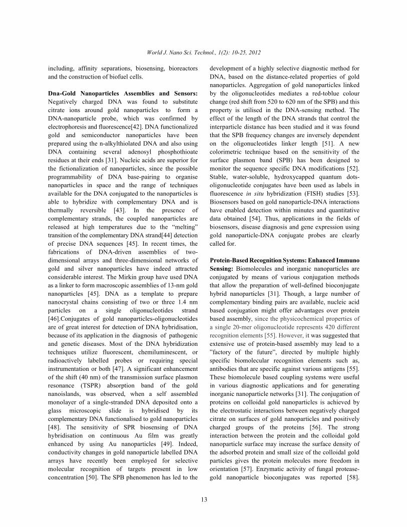
Photothermal Cancer Cell Therapy in NearinfraredRegion Using Anti-Egfr Antibody Conjugated GoldNanorods: Reducing a material’s size to the nanometerlength scale (which is the length scale of the electronicmotion that determines the material’s properties) makes itsensitive to further reduction in size or a change in shape.In semiconductor nanoparticles, the property changeresults from quantum confinement of the electronicmotion [89]. In metals the properties of the surfacebecome dominant and give nanoparticles new properties[90]. In noble metals the coherent collective oscillation ofelectrons in the conduction band induces large surfaceelectric fields which greatly enhance the radiativeproperties of gold and silver nanoparticles when they Fig. 2: NIR window interact with resonant electromagnetic radiation [91].This makes the absorption crossection of these enhancement of the absorption of nanorods is predictednanoparticles orders in magnitude stronger than the to be the strongest of all the different shapes of gold andstrongest absorbing molecules [92] and the light silver nanoparticles [109,110]. By changing the shape ofscattering crossecction orders in magnitude more intense gold nanoparticles to gold nanorods, not only canthan organic dyes [93]. Thus these particles act as one change the absorption and scatteringe nearexcellent sensors and novel contrast agents for optical infrared region. The absorption band of core-shelldetection due to their enhanced absorption and particles has been wavelength from visible to the NIRscattering, respectively. In addition, when it is realized region, but also increase their absorption and scatteringthat the strong absorbed radiation is converted efficiently crossections[107].into heat on a picoseconds time domain due to electron- In the present work, we demonstrate the potential usephonon and phonon-phonon processes [94], their of gold nanorods as a novel contrast reagent for selectivepotential use in photothermal therapy becomes obvious. photothermal therapy of cancer cells using a near infraredThe use of nanoparticles in medicine is one of the low energy cw laser. Solid gold nanorods have severalimportant directions that nanotechnology is taking at this advantages over other photothermal contrast agents.time. Their applications in drug delivery [95-97], cancer The synthesis of gold nanorods with various aspectcell diagnostics [97-100] and therapeutics [101] have been ratios which enables tunable absorption wavelength inactive fields of research. The scattering properties of gold the near infrared region [92,111,112] is quite simple andnanospheres have been used for cancer cell imaging well-established. The appropriate size of the nanorods isusing confocal microscopy [100,102] and simple dark field quite small and is potentially useful in applications suchmicroscopy [103]. Recently photothermal therapy usingthe absorption properties of antibody conjugated goldnanoshells [104]and solid gold nanospheres [105] havebeen demonstrated to selectively kill cancer cells leavingthe healthy cells unaffected. In order to use longwavelength laser irradiation that penetrates tissueoptimally (can be over 10 cm in penetration depthdepending on tissue types) for in vivo photothermaltreatment (650-900 nm) [106] the absorption band of thenanoparticles has to be in thtuned by adjusting the ratioof the thickness of the gold shell to the diameter of thesilica core (about 120 nm in diameter) and thus enablesphotothermal therapy in this region. Carbon nanotubesabsorb naturally in this region and have recently beenproposed as near-infrared therapy agents [108]. It isimportant to mention that surface plasmon field

World J. Nano Sci. Technol., 1(2): 10-25, 2012
17
as drug delivery and gene therapy. In addition, thebiosafety of metallic gold is well known and they havebeen used in vivo since the 1950’s [113] and recently thenoncytotoxicity of gold nanoparticles in human cells hasbeen studied in detail by Wyatt et al. [114].
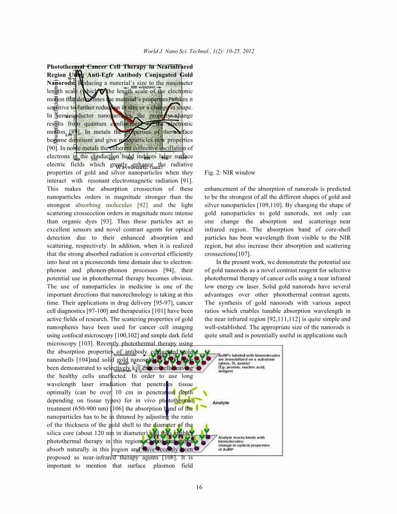
Gold Nanoparticles in Biosensor Applications: The basicprinciple involved in the design of a biosensor based ongold nanoparticles is that the AuNPs are functionalized orcapped with a thiolated biomolecule which uponidentifying the complementary biomolecule causeschange in the optical absorption of AuNPs [115]. Forexample, aptamer functionalized AuNPs specifically bindsto thrombin causing aggregation of AuNPs and redshifting the plasmon peak. The specific binding wastested by exposing aptamer functionalized AuNPs toother proteins (BSA or human IgG antibodies) where noAuNP aggregation was observed [116]. Similarly,immunoassays have been based on antigen-antibodyinteractions. AuNPs functionalized with antigen(antibody) aggregate when matching antibody (antigen)binds causing shift in the plasmon absorption [117,118].In addition to the above mentioned principle for designingbiosensors, Surface Enhanced Raman Scattering (SERS)has emerged as a powerful spectroscopic tool that [119]can be employed in detecting trace amounts of moleculesadsorbed on or present near metallic nanostructures alongwith structural and molecular information of themolecules.SERS now provides a great potential for label-free detection of biomolecules [120,121]. Significantefforts have focused on the binding of theoligonucleosides to metal surfaces and colloids for avariety of applications, including multiplexed DNAdetection technology [122], rapid sequencers based on
Fig. 3: Gold Nanoparticles in Biosensor

World J. Nano Sci. Technol., 1(2): 10-25, 2012
18
Fig. 4: Design strategy for bioconjugate /hybrid gold nanoparticles
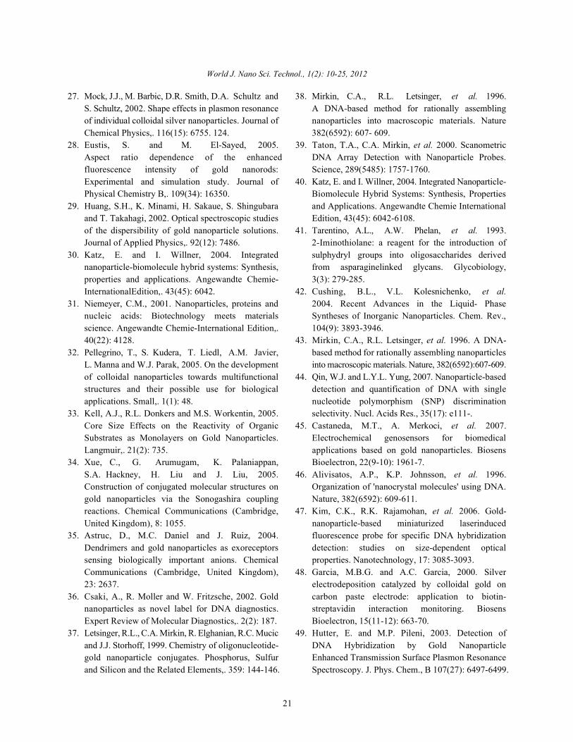
SERS from single DNA bases [120] and real-time DNA believed to exhibit metal chelating properties [137],detection methodology. For all the above applications contains disulfide and carboxylic acid groups tonew strategies for the synthesis, DNA-nanoparticle conjugate to peptide. S-S group acts as a chelating moietyassembly methods and development of nanoparticle- to hold the AuNP and 5 carbon atoms act as a spacebased SERS-active substrates are needed. In the present between S-S and the biomolecule. The thioctic acidproject we have developed a new SERS substrate based modified bombesin is used for AuNP bioconjugation.on gold nanoparticles in agarose matrix that providesbetter enhancement in Raman signal of DNA nucleosides Application of Gold Nanoparticles for Immunosensors:than that with commercially available gold nanoparticles. Immunosensors are important analytical tools based onBefore moving on to the project outline the following the detection of the binding event between antibody andsection discusses Raman scattering and SERS. antigen. The recent development of immunoassay
Bioconjugation of AuNPs: Hybrid gold nanoparticles are times, improving assay sensitivity, simplification andproduced by the interaction of highly reactive nascent automation of the assay procedures, low-volume analysis.AuNPs with chemical functionalities present on specific Among types of immunosensors, electrochemicalmolecules of biological interest (including peptides and immunosensors are attractive tools and have receivedproteins). The conjugation protocols that are applied for considerable attention because they are easy andproduction of radiolabel led bioconjugates, traditionally economical to mass production, they are robust and theyused for cancer diagnosis and therapy [123-126], can be achieve excellent detection limits with small analyteextended for the labeling nanoparticles of gold and other volumes. Furthermore, the availability of a variety of newmetals with tumor specific peptides. A hybrid gold materials with unique properties at nanoscale dimension,nanoparticle has 4 components: (i) AuNP, (ii) Chelating such as AuNPs, has attracted widespread attention inmoiety, (iii) Linker/Spacer and (iv) Cancer seeking peptide their utilization for the bioassay, especially forIn the present project, starch stabilized AuNPs are electrochemical detection. Recently, several novelutilized. The nature of bonding between starch and strategies have been proposed to develop electrochemicalAuNPs is a weak coordination bond between the hydroxyl immunosensors with high sensitivity using AuNPs. Agroups in starch and gold. However in presence of novel and sensitive electrochemical immunoassay forpowerful electron donor atoms such as S, this weak immunoglobulin G (IgG) has been developed by Limogescoordination bond is expected to break and starch and co-workers using a colloidal gold label via anodicmolecules detach from gold (Figure 1.9). The biomolecule stripping voltammetry technology. A low detection limitchosen for bioconjugation with AuNPs is the seven- (Concentration as low as 3×10 M) could be obtained,amino acid truncated bombesin analogue (BBN8-14) that which was competitive with colorimetric enzyme linkedis known to target gastrin releasing peptide (GRP) immuno-sorbent assay or with immunoassays based onreceptors that are over expressed on in a variety of fluorescent europium chelate labels. Furthermore, Shen’sneoplasma including small cell lung, prostate, breast, group reported a novel electrochemical immunoassaygastric, pancreatic, gastrointestinal carcinoid and colon based on the precipitation of silver on colloidal goldcancers[127-135]. To impart specificity hybrid AuNP, labels. After metal silver dissolution in an acidic solution,disulfide moiety is chosen as a chelating moiety. S-S the signal was indirectly determined by anodic strippinggroup undergo oxidative addition to AuNP and the voltammetry at a glassy carbon electrode.A detectionreaction is very selective, even in the presence of thiol limit as lowas 1 ng mL human IgG was achieved. Thegroups. Thioctic acid, a biological antioxidant [136] and enhancement in sensitivity for an electrochemical
techniques focused in most cases on decreasing analysis
-12
1

World J. Nano Sci. Technol., 1(2): 10-25, 2012
19
Fig. 5: Gold nanoparticles for immunosensors
immunoassay by the autocatalytic deposition of Au3+ electrochemical immunoassay electrode for detection ofonto AuNPs has been studied by Huang’s group. By carcinoembryonic antigen (CEA) has been developed bycoupling the autocatalytic deposition with square-wave Yao’s group. CEA antibody (CEAAb) was covalentlystripping voltammetry, the rabbit immunoglobulin G attached on glutathione (GSH) monolayer-modifiedanalyte could be determined quantitatively. A very low AuNPs and the resulting CEAAb-AuNPsdetection limit, 0.25 pgmL was obtained, which is three bioconjugates were immobilized on Au electrode by1
orders of magnitude lower than that obtained by a electrocopolymerization with o-aminophenol (OAP).conventional immunoassay using the same AuNPs labels. Electrochemical impedance spectroscopy studiesNovel enzyme-labeled electrochemical immunosensors demonstrated that the formation CEA antibody–antigenwere well developed by several groups. For instance, complexes increased the electron-transfer resistance. TheJu’s group reported that a highly hydrophilic and immunosensor could detect the CEA with a detection limitconductive colloidal AuNPs/titania sol–gel composite of 0.1 ng mL-1 and a linear range of 0.5–20 ng mL .membrane could be employed as electrochemicalsensing interface for horseradish peroxidase-labeled CONCLUSIONelectrochemical immunosensor. Later, a novelelectrochemical immunosensor for human chorionic Gold nanoparticles are currently being utilized ingonadotrophin (hCG) was developed by the same group several technological applications and are gainingvia the immobilization of hCG on AuNPs doped three- popularity as a form of counter measures against manydimensional (3D) sol–gel matrix. The 3D organized odds beared through conventional means. As a naturalcomposite structure was prepared by assembling AuNPs material, gold is known to be safe to man and produceinto a hydrolyzed (3-mercaptopropyl)-trimethoxysilane little to no allergic reactions when tested for curingsol–gel matrix, which showed good biocompatibility. After various diseases. Its wide and beneficial applicationsthe interfacial competitive immunoreaction, the formed are becoming more and more demanding. This is sure aHRPlabeled immunoconjugate showed good enzymatic very promising element to make our living long lastedactivity for the oxidation of ophenylenediamine by H O . gold. Development of environment friendly green2 2
The immunosensor showed good precision, high methodologies have been fabricated to producesensitivity, acceptable stability and reproducibility. biologically benign gold nanoparticles labeledLabel-free electrochemical immunosensors using AuNPs withbiologically relevant molecules. Furthermore, the goldas enhancing sensing component have been the focus of nanoparticles were evaluated fortheir in vitro stability andintense research due to their simplicity, speedy analysis in vivo biodistribution. Additionally, gold nanoparticlesand high sensitivity. The technique is mainly based on were conjugated with cancer seeking peptides to impartthe detectionof a change in physical properties as a result target specificity in hybrid gold nanoparticles for theirof antibody–antigen complex formation. The direct potential applications in cancer imaging and therapy. Golddetermination of immunospecies by detecting the nanoparticles trapped in an agarose matrix were evaluatedchange of impedance caused by immunoreactions has for their SERS properties with DNA nucleosides forbeen demonstrated. A simple and sensitive labelfree possible biosensor applications.
1

World J. Nano Sci. Technol., 1(2): 10-25, 2012
20
REFERENCE 17. Zharov, V.P., V. Galitovsky, et al. 2003.
1. Graham, T., 1861. Liquid Diffusion Applied toAnalysis. Philosophical Transactions of the RoyalSociety of London, 151: 183-224.
2. Kunckels, J., 1676. Nuetliche Observationes oderAnmerkungen von Auro und Argento Potabili.Hamburg.
3. Zsigmondy, R.A., 1926. Properties of colloids.4. Savage, G., 1973. Glass and Glassware. London,,
Octopus / Mayflower.5. Kahn, R.L., 1928. Serum Diagnosis for Syphilis.
In Colliodal Chemistry, Vol. II. J. Alexander,Ed. New York, The Chemical Catalog Co. II: 757.
6. Hauser, E.A., 1952. Aurum Potabile. Journal ofChemical Education, 29: 456-458.
7. Brown, D.H. and W.E. Smith, 1980. The Chemistry ofthe Gold Drugs Used in the Treatment of RheumatoidArthritis. Chemical Society Reviews, 9: 217-240.
8. Daniel, M.C. and D. Astruc, 2004. Gold Nanoparticles:Assembly, Supramolecular Chemistry, Quantum-Size-Related Properties and Applications toward Biology,Catalysis and Nanotechnology. Chem. Rev.,104(1): 293-346.
9. Francisci, A., 1618. Panacea Aurea-Auro Potabile.Hamburg, Bibliopolio Frobeniano.
10. Fulhame, M., 1794. An Essay on Combustion with aView to a New Art of Dying and Painting. London, J.Cooper.
11. Ostwald, W., 1915. Die Welt der VernachlässigtenDimensionen. Dresden,, Steinkopf.
12. Feynman, R.P., 1959. There's Plenty of Room at theBottom. A. P. Society. California.
13. Cushing, B.L., V.L. Kolesnichenko, et al. 2004.Recent Advances in the Liquid- PhaseSyntheses of Inorganic Nanoparticles. Chem. Rev.,104(9): 3893-3946.
14. Daniel, M.C. and D. Astruc, 2004. Gold Nanoparticles:Assembly, Supramolecular Chemistry, Quantum-Size-Related Properties and Applications toward Biology,Catalysis and Nanotechnology. Chem. Rev.,104(1): 293-346.
15. Chen, F., G.Q. Xu, et al. 2003. Preparation andassembly of colloidal gold nanoparticles in CTAB-stabilized reverse microemulsion. Materials Letters,57(21): 3282-3286
16. Love, J.C., L.A. Estroff, et al. 2005. Self-AssembledMonolayers of Thiolates on Metals as a Form ofNanotechnology. Chem. Rev., 105(4): 1103-1170.
"Photothermal detection of local thermal effectsduring selective nanophotothermolysis." AppliedPhysics Letters, 83(24): 4897-4899.
18. Park, J.H., Y.T. Lim, O.O. Park, J.K. Kim,J.W. Yu and Y.C. Kim, 2004. Polymer/Goldnanoparticle nanocomposite light-emittingdiodes: Enhancement of electroluminescencestability and quantum efficiency of blue-light-emitting polymers. Chemistry of Materials,16(4): 688.
19. Narayanan, R. and M.A. El-Sayed, 2005. Catalysiswith transition metal nanoparticles in colloidalsolution: Nanoparticle shape dependence andstability. Journal of Physical Chemistry B,.109(26): . 12663.
20. Aslan, K., Z. Jian, J.R. Lakowicz and C.D. Geddes,2004. Saccharide sensing using gold and silvernanoparticles - a review. Journal of Fluorescence,14(4): 391.
21. Riviere, C., F.P. Boudghene, F. Gazeau, J. Roger,J.N. Pons, et al., 2005. Iron oxide nanoparticle-labeledrat smooth muscle cells: Cardiac MR imaging forcell graft monitoring and quantitation. Radiology,235(3): 959.
22. Eustis, S. and M.A. El-Sayed, 2006. Why goldnanoparticles are more precious than pretty gold:Noble metal surface plasmon resonance and itsenhancement of the radiative and nonradiativeproperties of nanocrystals of different shapes.Chemical Society Reviews,. 35(3): 209.
23. Daniel, M.C. and D. Astruc, 2004. GoldNanoparticles: Assembly, Supramolecular Chemistry,Quantum-Size-Related Properties and Applicationstoward Biology, Catalysis and Nanotechnology.Chemical Reviews (Washington, DC, United States),.104(1): 293.
24. Berciaud, S., L. Cognet, P. Tamarat and B. Lounis,2005. Observation of intrinsic sizeeffects in theoptical response of individual gold nanoparticles.Nano Letters,. 5(3): 515.
25. Neeleshwar, S., C.L. Chen, C.B. Tsai, Y.Y. Chen,C.C. Chen, S.G. Shyu and M.S. Seehra, 2005. Size-dependent properties of CdSe quantum dots.Physical Review B,. 71(20).
26. Masumoto, Y. and K. Sonobe, 1997. Size-dependentenergy levels of CdTe quantum dots. PhysicalReview B,. 56(15): 9734.
World J. Nano Sci. Technol., 1(2): 10-25, 2012
21
27. Mock, J.J., M. Barbic, D.R. Smith, D.A. Schultz and 38. Mirkin, C.A., R.L. Letsinger, et al. 1996.S. Schultz, 2002. Shape effects in plasmon resonance A DNA-based method for rationally assemblingof individual colloidal silver nanoparticles. Journal of nanoparticles into macroscopic materials. NatureChemical Physics,. 116(15): 6755. 124. 382(6592): 607- 609.
28. Eustis, S. and M. El-Sayed, 2005. 39. Taton, T.A., C.A. Mirkin, et al. 2000. ScanometricAspect ratio dependence of the enhanced DNA Array Detection with Nanoparticle Probes.fluorescence intensity of gold nanorods: Science, 289(5485): 1757-1760.Experimental and simulation study. Journal of 40. Katz, E. and I. Willner, 2004. Integrated Nanoparticle-Physical Chemistry B,. 109(34): 16350. Biomolecule Hybrid Systems: Synthesis, Properties
29. Huang, S.H., K. Minami, H. Sakaue, S. Shingubara and Applications. Angewandte Chemie Internationaland T. Takahagi, 2002. Optical spectroscopic studies Edition, 43(45): 6042-6108.of the dispersibility of gold nanoparticle solutions. 41. Tarentino, A.L., A.W. Phelan, et al. 1993.Journal of Applied Physics,. 92(12): 7486. 2-Iminothiolane: a reagent for the introduction of
30. Katz, E. and I. Willner, 2004. Integrated sulphydryl groups into oligosaccharides derivednanoparticle-biomolecule hybrid systems: Synthesis, from asparaginelinked glycans. Glycobiology,properties and applications. Angewandte Chemie- 3(3): 279-285.InternationalEdition,. 43(45): 6042. 42. Cushing, B.L., V.L. Kolesnichenko, et al.
31. Niemeyer, C.M., 2001. Nanoparticles, proteins and 2004. Recent Advances in the Liquid- Phasenucleic acids: Biotechnology meets materials Syntheses of Inorganic Nanoparticles. Chem. Rev.,science. Angewandte Chemie-International Edition,. 104(9): 3893-3946.40(22): 4128. 43. Mirkin, C.A., R.L. Letsinger, et al. 1996. A DNA-
32. Pellegrino, T., S. Kudera, T. Liedl, A.M. Javier, based method for rationally assembling nanoparticlesL. Manna and W.J. Parak, 2005. On the development into macroscopic materials. Nature, 382(6592):607-609.of colloidal nanoparticles towards multifunctional 44. Qin, W.J. and L.Y.L. Yung, 2007. Nanoparticle-basedstructures and their possible use for biological detection and quantification of DNA with singleapplications. Small,. 1(1): 48. nucleotide polymorphism (SNP) discrimination
33. Kell, A.J., R.L. Donkers and M.S. Workentin, 2005. selectivity. Nucl. Acids Res., 35(17): e111-.Core Size Effects on the Reactivity of Organic 45. Castaneda, M.T., A. Merkoci, et al. 2007.Substrates as Monolayers on Gold Nanoparticles. Electrochemical genosensors for biomedicalLangmuir,. 21(2): 735. applications based on gold nanoparticles. Biosens
34. Xue, C., G. Arumugam, K. Palaniappan, Bioelectron, 22(9-10): 1961-7.S.A. Hackney, H. Liu and J. Liu, 2005. 46. Alivisatos, A.P., K.P. Johnsson, et al. 1996.Construction of conjugated molecular structures on Organization of 'nanocrystal molecules' using DNA.gold nanoparticles via the Sonogashira coupling Nature, 382(6592): 609-611.reactions. Chemical Communications (Cambridge, 47. Kim, C.K., R.K. Rajamohan, et al. 2006. Gold-United Kingdom), 8: 1055. nanoparticle-based miniaturized laserinduced
35. Astruc, D., M.C. Daniel and J. Ruiz, 2004. fluorescence probe for specific DNA hybridizationDendrimers and gold nanoparticles as exoreceptors detection: studies on size-dependent opticalsensing biologically important anions. Chemical properties. Nanotechnology, 17: 3085-3093.Communications (Cambridge, United Kingdom), 48. Garcia, M.B.G. and A.C. Garcia, 2000. Silver23: 2637. electrodeposition catalyzed by colloidal gold on
36. Csaki, A., R. Moller and W. Fritzsche, 2002. Gold carbon paste electrode: application to biotin-nanoparticles as novel label for DNA diagnostics. streptavidin interaction monitoring. BiosensExpert Review of Molecular Diagnostics,. 2(2): 187. Bioelectron, 15(11-12): 663-70.
37. Letsinger, R.L., C.A. Mirkin, R. Elghanian, R.C. Mucic 49. Hutter, E. and M.P. Pileni, 2003. Detection ofand J.J. Storhoff, 1999. Chemistry of oligonucleotide- DNA Hybridization by Gold Nanoparticlegold nanoparticle conjugates. Phosphorus, Sulfur Enhanced Transmission Surface Plasmon Resonanceand Silicon and the Related Elements,. 359: 144-146. Spectroscopy. J. Phys. Chem., B 107(27): 6497-6499.

World J. Nano Sci. Technol., 1(2): 10-25, 2012
22
50. Tirelli, N., 2006. (Bio)Responsive nanoparticles. 63. Connolly, S. and D. Fitzmaurice, 1999. ProgrammedCurrent Opinion in Colloid & Interface Science,11(4): 210-216.
51. Fischler, M. and U. Simon, 2007. DNA-BasedAssembly of Metal Nanoparticles: Structure andFunctionality. Charge Migration in DNA: 263-282.
52. Li, J., X. Chu, et al. 2005. A colorimetric method forpoint mutation detection using high-fidelity DNAligase. Nucl. Acids Res., 33(19): e168-.
53. Wu, S.M., X. Zhao, et al. 2006. Quantum-dot-labeledDNA probes for fluorescence in situ hybridization(FISH) in the microorganism Escherichia coli.Chemphyschem, 7(5): 1062-7.
54. Yáñez-Sedeño, P. and J.M. Pingarrón,2005. Gold nanoparticle-based electrochemicalbiosensors. Analytical and Bioanalytical Chemistry382(4): 884-886.
55. Mann, S., W. Shenton, et al. 2000. BiologicallyProgrammed Nanoparticle Assembly. AdvancedMaterials, 12(2): 147-150.
56. Xiao, Y., H.X. Ju, et al. 1999. Hydrogen peroxidesensor based on horseradish peroxidase-labeled Aucolloids immobilized on gold electrode surface bycysteamine monolayer. Analytica Chimica Acta,391(1): 73-82.
57. Liu, S., D. o. n. Leech, et al. 2003. Applicationof Colloidal Gold in Protein Immobilization,Electron Transfer and Biosensing. Analytical Letters,36(1): 1-19.
58. Phadtare, S., V.P.V. Kausik, et al. 2004."Immobilization and biocatalytic activity of fungalprotease on gold nanoparticle-loaded zeolitemicrospheres." Biotechnology and Bioengineering,85(6): 629-637.
59. Lin, F.Y.H., M. Sabri, et al. 2005. Development of aNanoparticle-Labeled Microfluidic Immunoassay forDetection of Pathogenic Microorganisms. Clin.Diagn. Lab. Immunol., 12(3): 418-425.
60. Thanh, N.T.K. and Z. Rosenzweig, 2002.Development of an Aggregation-BasedImmunoassay for Anti-Protein A Using GoldNanoparticles. Anal. Chem., 74(7): 1624-1628.
61. Jianrong, C., M. Yuqing, et al. 2004. Nanotechnologyand biosensors. Biotechnol Adv., 22(7): 505-18.
62. Garci,M.B.G., C.F. Sanchez, et al. 2000. Colloidal goldas an electrochemical label of streptavidin–biotininteraction. Biosensors and Bioelectronics,15(5-6): 315- 321.
Assembly of Gold Nanocrystals in AqueousSolution. Advanced Materials, 11(14): 1202-1205.
64. Shenton, W., S.A. Davis, et al. 1999. Directed Self-Assembly of Nanoparticles into MacroscopicMaterials Using Antibody-Antigen Recognition.Advanced Materials, 11(6): 449-452.
65. Velev, O.D. and E.W. Kaler, 1999. In Situ Assembly ofColloidal Particles into Miniaturized Biosensors.Langmuir, 15(11): 3693-3698.
66. Xiao, Y., H.X. Ju, et al. 1999. Hydrogen peroxidesensor based on horseradish peroxidase-labeled Aucolloids immobilized on gold electrode surface bycysteamine monolayer. Analytica Chimica Acta,391(1): 73-82.
67. Chan, W.C.W. and S. Nie, 1998. Quantum DotBioconjugates for Ultrasensitive NonisotopicDetection. Science, 281(5385): 2016-2018.
68. Han, M., X. Gao, et al. 2001. Quantum-dot-taggedmicrobeads for multiplexed optical coding ofbiomolecules. Nat Biotechnol., 19(7): 631-5.
69. Daniel, M.C. and D. Astruc, 2004. Gold Nanoparticles:Assembly, Supramolecular Chemistry, Quantum-Size-Related Properties and Applications toward Biology,Catalysis and Nanotechnology. Chem. Rev.,104(1): 293-346.
70. Lyon, L.A., M.D. Musick, et al. 1998.Colloidal Au-Enhanced Surface Plasmon ResonanceImmunosensing. Anal. Chem., 70(24): 5177-5183.
71. Dai, X., Y. Tan, et al. 2002. Formation of GoldNanoparticles in the Presence of o- Anisidine and theDependence of the Structure of Poly(o-anisidine) onSynthetic Conditions. Langmuir, 18(23): 9010-9016.
72. Shi Kam, N.W., M. O'Connell, et al. 2005. Carbonnanotubes as multifunctional biological transportersand near-infrared agents for selective cancer celldestruction. Proceedings of the National Academy ofSciences, 102(33): 11600-11605.
73. Bianco, A., K. Kostarelos, et al. 2005. Applications ofcarbon nanotubes in drug delivery. Curr Opin ChemBiol., 9(6): 674-9.
74. Jiang, W., B.Y. Kim, et al. 2007. Advances andchallenges of nanotechnology-based drug deliverysystems. Expert Opin Drug Deliv., 4(6): 621-633.
75. Hone, D.C., P.I. Walker, et al. 2002. Generation ofCytotoxic Singlet Oxygen via Phthalocyanine-Stabilized Gold Nanoparticles: A Potential DeliveryVehicle for Photodynamic Therapy. Langmuir,18(8): 2985-2987.

World J. Nano Sci. Technol., 1(2): 10-25, 2012
23
76. Hainfeld, J.F., 1992. Site-specific cluster labels. 91. El-Sayed, M.A. Acc., 2001. Preparation and GrowthUltramicroscopy, 46(1-4): 135-144. Mechanism of Gold Nanorods (NRs) Using, Chem.
77. Takizawa, T., K. Suzuki, et al. 1998. Correlative Res., 34(4): 257.Microscopy Using FluoroNanogold on Ultrathin 92. Link, S. and M.A.J. El-Sayed, 1999. GoldCryosections: Proof of Principle. J. Histochem. nanoparticles: interesting optical properties, Phys.Cytochem., 46(10): 1097-1102. Chem. B., 103: 8410.
78. Hainfeld, J.F. and F.R. Furuya, 1992. A 1.4-nm 93. Yguerabide, J., E. Yguerabide and E. Anal, 1998.gold cluster covalently attached to antibodies Light-scattering submicroscopic particles as highlyimproves immunolabeling. J. Histochem. Cytochem., fluorescent. Biochem., 262: 137.40(2): 177-184. 94. Link, S. and M.A. El-Sayed, 2000. Relative
79. Garcia, M.B.G. and A.C. Garcia, 2000. Silver Enhancement of Ultrafast Emission in Goldelectrodeposition catalyzed by colloidal gold on Nanorods. Int. Rev. Phys. Chem., 19: 409.carbon paste electrode: application to biotin- 95. West, J.L., N. Halas and J. Annu, 2003. Goldstreptavidin interaction monitoring. Biosens Nanocages: Bioconjugation and Their Potential UseBioelectron, 15(11-12): 663-70. as Optical, Rev. Biomed. Eng., 5: 285.
80. Csaki, A., R. Moller, et al. 2002. Gold nanoparticles as 96. Paciotti, G.F., L. Myer, D. Weinreich, D. Goia,novel label for DNA diagnostics. Expert Review of N. Pavel, R.E. McLaughlin and L. Tamarkin, 2004.Molecular Diagnostics, 2(2): 187-193. Drug Delivery, 11(3): 169.
81. Manimaran, M. and N.R. Jana, 2007. Detection of 97. Jain, K.K., 2005. New Aspects of the use ofprotein molecules by surfaceenhanced Raman Hyperbaric Oxygenation for Rehabilitation ofspectroscopy-based immunoassay using 2-5 nm gold Stroke Patients. Geriatrics and... Drug Discoverynanoparticle lables. Journal of Raman Spectroscopy Today”Technol. Cancer Res. and Treat., 4(4): 407.38(10): 1326-1331. 98. Wu, X., H. Liu, J. Liu, K.N. Haley, J.A. Treadway, J.P.
82. Lyon, L.A., M.D. Musick, et al. 1998. Colloidal Larson, N. Ge, F. Peale and M.P. Bruchez, 2003. Nat.Au-Enhanced Surface Plasmon Resonance Biotechnol., 21: 41.Immunosensing. Anal. Chem., 70(24): 5177-5183. 99. Chan, W.C.W., D.J. Maxwell, X. Gao, R.E. Bailey, M.
83. Lin, C.C., Y.C. Yeh, et al. 2002. Selective Binding of Han, S. Nie, Curr. Opin., 2002. Biotechnol., 13: 40.Mannose-Encapsulated Gold Nanoparticles to 100. Alivisatos, A.P. Nat., 2004. Protease-Modulated Type 1 Pili in Escherichia coli. J. Am. Chem. Soc., Cellular Uptake of Quantum Dots, Biotechnol.,124(14): 3508-3509 22(1): 47.
84. Jin, L. and R.V. Lloyd, 1997. In situ hybridization: 101. Sokolov, K., J. Aaron, B. Hsu, D. Nida,Methods and applications. Journal of Clinical A. Gillanwater, M. Follen, C. Macaulay, K. Adler-Laboratory Analysis, 11(1): 2-9. Storthz, B. Korgel, M. Discour, R. Pasqualini,
85. Thompson, R.H. and L.W. Swanson, 1998. W. Arap, W. Lam and R. Richartz-Kortum, 2003.Organization of inputs to the dorsomedial nucleus of Technol. Cancer Res. and Treat., 2(6): 491.the hypothalamus: a reexamination with Fluorogold 102. Hirsch, L.R., R.J. Stafford, J.A. Bankson,and PHAL in the rat. Brain Res Brain Res Rev., S.R. Sershen, B. Rivera, R.E. Rrice, J.D. Hazle,27(2): 89-118. N.J. Halas and J.L. West, Proc. 2003. Natl. Acad. Sci.
86. Yu, C.J., C.L. Su, et al. 2006. Separation of Acidic and USA, 100: 13549.Basic Proteins by Nanoparticle-Filled Capillary 103. Sokolov, K., M. Follen, J. Aaron, I. Pavlova,Electrophoresis. Anal. Chem., 78(23): 8004-8010. A. Malpica, R. Lotan and R. Richartz- Kortum, 2003.
87. Neiman, B., E. Grushka, et al. 2001. Use of Gold Cancer Res., 63: 1999.Nanoparticles To Enhance Capillary Electrophoresis. 104. El-Sayed, I.H.; X. Huang and M.A. El-Sayed, 2005.Anal. Chem., 73(21): 5220-5227. “Cancer Cells Assemble and Align Gold Nanorods
88. Pumera, M., J. Wang, et al. 2001. Gold Nanoparticle- Conjugated”Nano Letters, 5(5): 829.Enhanced Microchip Capillary Electrophoresis. Anal. 105. Loo, C., A. Lowery, N. Halas, J. West and R. Drezek,Chem., 73(22): 5625-5628. 2005. Immunotargeted Nanoshells for Integrated
89. Alivisatos, A.P., 1996. Science, pp: 271-933. Cancer Imaging, Nano letters, 5(4): 709.90. Kreibig, U. and M. Vollmer, 1995. Optical Properties 106. El-Sayed, I.H., X. Huang and M.A. El-Sayed, 2005.
of Metal Clusters; New York: Springer, Cancer Letters, in press.

World J. Nano Sci. Technol., 1(2): 10-25, 2012
24
107. Weissleder, R., 2001. Nat. Biotechnol., 19: 316. 121. Liu, G.L. and L.P. Lee, 2005. Nanowell surface108. Shi Kam, N.W., M. O’Connell, J.A. Wisdom enhanced Raman scattering arrays fabricated by
and H. Dai, Proc., 2005. Natl. Acad. Sci. USA, soft-lithography for label-free biomolecular102, 11600. detections in integrated microfluidics. Applied
109. Hao, E. and G.C.J. Schatz, 2004. Cancer cell imaging Physics Letters,. 87(7).and photothermal therapy in the near-infrared, 122. Cao, Y.C., J. Rongichao and C.A. Mirkin, 2002.Chem. Phys., 120: 357. Nanoparticles with Raman spectroscopic
110. Hao, E., G.C. Schatz and J.T.J. Hupp, 2004. Cancer fingerprints for DNA and RNA detection. Science,.Cell Imaging and Photothermal Therapy in the Near- 297(5586): 1536.Infrared, Fluorescence, 14: 331. 123. Hoffman, T.J., G.L. Sieckman, W.A. Volkert and
111. Murphy, C.J., T.K. Sau, A.M. Gole, C.J. Orendorff, J. H.S. Truman, 1996. Iodinated bombesin analogues:Gao, L. Gou, S.E. Hunyadi and T.J. Li, 2005. Phys. Effect of N-terminal vs side chain iodine attachmentChem. B., 109(29): 13857. on BBN/GRP receptor binding. Journal of Nuclear
112. Lance Kelly, K., Eduardo Coronado, Lin Lin Zhao Medicine,. 37(5): 850.and George C. Schatz, J., 2003. Phys. Chem. B., 124. Hoffman, T.J., T.P. Quinn and W.A. Volkert, 2001.107 (3), 668. Radiometallated receptor-avid peptide conjugates
113. Sherman, A.I., M. Ter-Pogossian, 1953. Lymph-node for specific in vivo targeting of cancer cells. Nuclconcentration of radioactive colloidal gold, Cancer, Med Biol,. 28(5): 527.6: 1238. 125. Karra, S.R., R. Schibli, H. Gali, K.V. Katti,
114. Connor, E.E., J. Mwamuka, A. Gole, C.J. Murphy and T.J. Hoffman, C. Higginbotham, G.L. Sieckman andM.D. Wyatt, 2005. Small, 1: 325. W.A. Volkert, 1999. 99mTc-labeling and in vivo
115. West, J.L. and N.J. Halas, 2003. Engineered studies of a bombesin analogue with a novel water-nanomaterials for biophotonics applications: soluble dithiadiphosphine-based bifunctionalImproving sensing, imaging and therapeutics. chelating agent. Bioconjug Chem,. 10(2): 254.Annual Review of Biomedical Engineering,. 5: 285. 126. Hu, F., C.S. Cutler, T. Hoffman, G. Sieckman,
116. Pavlov, V., Y. Xiao, B. Shlyahovsky and I. Willner, , W.A. Volkert and S.S. Jurisson, Pm-149 DOTA2004. Aptamer-functionalized Au nanoparticles for (2002)bombesin analogs for potential radiotherapy -the amplified optical detection of thrombin. Journal In vivo comparison with Sm-153 and Lu-177 labeledof the American Chemical Society,. 126(38): 11768. DO3A-amide-beta Ala-BBN(7-14)NH2. Nuclear
117. Raschke, G., T. Franzl, S. Kowarik, C. Soennichsen, Medicine and Biology,. 29(4): 423.T.A. Klar, J. Feldmann, A. Nichtl and K. Kuerzinger, 127. Saurin, J.C., J.P. Rouault, J. Abello, F. Berger,2004. Biomolecular sensor based on optical L. Remy and J.A. Chayvialle, 1999. High gastrinspectroscopy of single gold nanoparticles. Trends releasing peptide receptor mRNA level is related toin Optics and Photonics, 96(Conference on Lasers tumour dedifferentiation and lymphatic vesseland Electro-Optics, 2004): pp: CThI2/1. invasion in human colon cancer. Eur. J. Cancer,.
118. Nath, N. and A. Chilkoti, 2002. Immobilized gold 35(1): 125.nanoparticle sensor for label-free optical detection of 128. Tang, C., I. Biemond, G.J. Offerhaus, W. Verspagetbiomolecular interactions. Proceedings of SPIE-The and C.B. Lamers, 1997. Expression of receptors forInternational Society for Optical Engineering,. gut peptides in human pancreatic adenocarcinoma4626(Biomedical Nanotechnology Architectures and and tumour-free pancreas. Br. J. Cancer,.Applications): pp: 441. 75(10): 1467.
119. Bailey, R.C., J.M. Nam, C.A. Mirkin and J.T. Hupp, 129. Scott, N., E. Millward, E.J. Cartwright, S.R. Preston2003. Real-time multicolor DNA detection and P.L. Coletta, 2004. Gastrin releasing peptide andwith chemoresponsive diffraction gratings and gastrin releasing peptide receptor expression innanoparticle probes. Journal of the American gastrointestinal carcinoid tumours. J. Clin. Pathol,.Chemical Society,. 125(44): 13541. 57(2): 189.
120. Kneipp, K., H. Kneipp, I. Itzkan, R.R. Dasari and 130. Toi-Scott, M., C.L. Jones and M.A. Kane, 1996.M.S. Feld, 2002. Surface-enhanced Raman scattering Clinical correlates of bombesin-like peptide receptorand biophysics. Journal of Physics-Condensed subtype expression in human lung cancer cells.Matter,. 14(18): R597. Lung Cancer,. 15(3): 341.

World J. Nano Sci. Technol., 1(2): 10-25, 2012
25
131. Reubi, J.C., S. Wenger, J. Schmuckli-Maurer, 134. Chave, H.S., A.C. Gough, K. Palmer, S.R. Preston andJ.C. Schaer and M. Gugger, 2002. Bombesin J.N. Primrose, 2000. Bombesin family receptor andreceptor subtypes in human cancers: detection ligand gene expression in human colorectal cancerwith the universal radioligand (125)I-[D-TYR(6), and normal mucosa. Br. J. Cancer, 82(1): 124.beta-ALA(11), PHE(13), NLE(14)] bombesin(6- Clin 135. Patel, O., A. Shulkes and G.S. Baldwin, 2006. Gastrin-Cancer Res,. 8(4): 1139. releasing peptide and cancer.
132. Preston, S.R., L.F. Woodhouse, S. Jones-Blackett, 136. Biochim Biophys Acta, Packer, L., E.H. Witt and H.J.J.I. Wyatt and J.N. Primrose, 1993. High affinity 1995. Tritschler, alpha-Lipoic acid as a biologicalbinding sites for gastrin releasing peptide on human antioxidant. Free Radic Biol. Med., 19(2): 227.gastric cancer and Menetrier's mucosa. Cancer Res,. 137. Ou, P., H.J. Tritschler and S.P. Wolff, 1995.53(21): 5090. Thioctic (lipoic) acid: a therapeutic metalchelating
133. Markwalder, R. and J.C. Reubi, 1999. Gastrin- antioxidant? Biochem. Pharmacol., 50(1): 123.releasing peptide receptors in the human prostate:relation to neoplastic transformation. Cancer Res,.59(5): 1152.