Preparation of PLLA/PEG Nanofibers by Electrospinning and Potential Applications
Upload
khangminh22Category
view
2download
0
IOP PUBLISHING JOURNAL OF PHYSICS D: APPLIED PHYSICS
J. Phys. D: Appl. Phys. 41 (2008) 195301 (8pp) doi:10.1088/0022-3727/41/19/195301
Optimizing the size and surface propertiesof polyethylene glycol (PEG)–goldnanoparticles by intense x-ray irradiationChang-Hai Wang1, Chi-Jen Liu1, Cheng-Liang Wang1, Tzu-En Hua1,Judy M Obliosca1, K H Lee1, Y Hwu1,2,3,4,10, Chung-Shi Yang5,Ru-Shi Liu6, Hong-Ming Lin7, Jung-Ho Je8 and G Margaritondo9
1 Institute of Physics, Academia Sinica, Nankang, Taipei 115, Taiwan2 Department of Engineering and System Science, National Tsing Hua University, Hsinchu 300, Taiwan3 National Synchrotron Radiation Research Center, Hsinchu 300, Taiwan4 Institute of Opto-Electronics Sciences, National Taiwan Ocean University, Keelung 202, Taiwan5 Nanomedicine Research Center, National Health Research Institute, Hsinchu 350, Taiwan6 Department of Chemistry, National Taiwan University, Taipei 106, Taiwan7 Department of Materials Engineering, Tatung University, Taipei 104, Taiwan8 X-ray Imaging Center, Pohang University of Science and Technology, Pohang 790-784, Korea9 Ecole Polytechnique Federale de Lausanne (EPFL), CH-1015 Lausanne, Switzerland
E-mail: [email protected]
Received 14 May 2008, in final form 6 August 2008Published 5 September 2008Online at stacks.iop.org/JPhysD/41/195301
AbstractThe polyethylene glycol (PEG) modified gold nanoparticle complex was synthesized by aone-solution synchrotron x-ray irradiation method. The impact on the structure andmorphology of the gold nanoparticles of process parameters such as the PEG molecularweight, the PEG/gold molar ratio and the x-ray dosage were investigated. The size of PEGmodified gold particles was found to decrease with increasing PEG addition and x-ray dosage.With the capability to monitor the absorption spectra in situ during the fast synthesis process,this opens the way to accurate control of the size and distribution. PEG chains with anintermediate length (MW6000) were found optimal for size control and colloidal stability inbiologically relevant media. Our x-ray synthesized PEG–gold nanoparticles could findinteresting applications in nanoparticle-enhanced x-ray tumour imaging and therapy.
(Some figures in this article are in colour only in the electronic version)
1. Introduction
Nanotechnology is playing an increasingly important role inthe fight against cancer. The prospects of nanoparticle-basedcancer imaging and therapies have stimulated many studiesin this emerging field [1–4]. In particular, gold nanoparticlesare promising candidates as carriers for targeted drug delivery[5, 6], contrast agents [7, 8] or radiotherapy enhancers [9].
The expanding multi-functionalities of gold nanoparticlescreate challenges for material scientists since additionalfeatures such as higher concentration, smaller size, sufficientdispersion and colloidal stability are required for biomedical
10 Author to whom any correspondence should be addressed.
applications. This is especially true for in vivo biomedicalapplications in which, without surface modifications, theadministrated nanoparticles are eliminated from circulationwithin seconds to minutes through the reticulo-endothelialsystem (RES) [10]. One effective solution to preventopsonization and enable nanoparticles to circulate for a longtime is to construct a ‘stealth-shielding’ layer composed ofwater-soluble polymers on the surfaces of particulate vectors[11]. Four well-documented synthetic routes are used toprepare polymer stabilized gold nanoparticles [12]: covalent‘grafting from’ (relying on polymerization), covalent ‘graftingto’ (using sulfur-containing polymers), physisorption and post-modification of pre-formed Au NPs. These methods aresummarized in table 1 [5, 6, 8, 12, 13–29].
0022-3727/08/195301+08$30.00 1 © 2008 IOP Publishing Ltd Printed in the UK

J. Phys. D: Appl. Phys. 41 (2008) 195301 C-H Wang et al
Table 1. A summary of preparation approaches for polymer-protected gold nanoparticles.
Synthetic route Advantages Disadvantages Reference
Covalent graft from • Control polymer brush thickness; • Multi-steps [13–16]• Higher surface polymer graft density • Purification needed
Covalent graft to • One pot synthesis • Broad size distribution; [17–20]• Mono-dispersed Au NPs can be obtained • Long tethered polymer chain unfavourable
in aqueous and nonaqueous solutions for condensation and fractionation• Sulfur-containing polymer needed
Post-modification • Easy achievement of size selection; • Two-step procedures; [5, 6, 8, 12, 21–25]• Versatile surface property; • Low gold concentration to optimize
covalent bonding;• Sulfur-containing polymer needed
Physisorption • One pot synthesis • Broad size distribution; [26–29]• Unmodified polymer can be used • Less stable than covalent bonding;
• A high concentration of protectiveagent is required
These approaches, however, are affected by limitationsthat endanger their suitability for specific biomedicalapplication. To achieve maximum effectiveness in enhancingradiotherapy, Au nanoparticles must have optimal size andconcentration. Two polymer systems were used: polyethyleneglycol (PEG) and poly N -vinyl-2-pyrrolidone (PVP). Thesepolymers were successfully exploited to modify nanovectorsfor gene/drug delivery [30, 31], tumour targeting [5, 6, 32, 33]and x-ray imaging [8, 34]. For the in vivo applications usingPEG–gold nanoparticles, all the preparations were based onconventional chemical methods (i.e. sodium citrate or sodiumborohydrate reduction) and thiolated PEG [5, 6, 8, 30, 31, 35].However, the size of gold particles is normally larger than30 nm. In addition, due to the very low (∼0.25 mM of goldprecursor) concentration of colloidal gold, the nanosols mustbe condensed to reach higher concentration by high speedcentrifugation (e.g. 16 800 g for 30 nm PEG–thiol modifiedgold nanoparticles [8]). Furthermore, the high cost of thiolatedPEG could limit commercial applications.
We report here a one-solution synthesis approachusing x-ray irradiation to prepare polymer modified goldnanoparticles that is not affected by the above limitations. Themethod consists of bombarding a precursor solution containinggold ions and the polymeric stabilizers with intense x-raysproduced by a synchrotron source. The irradiation was foundto stimulate the formation of gold nanoparticles protected bythe polymer chains. Since there is no prior modification ofthe PEG, the polymeric chains are likely to be physicallyadsorbed on the particle surfaces. Compared with otherphysisorption methods, this approach has several advantages:(1) cleanliness, since the system is free of pre-added reducingagents and surfactant; (2) high reproducibility and capabilityto scale up for mass production; (3) unmodified PEG with lowconcentration are used; (4) easy increase in concentration withexcellent dispersion; (5) high colloidal stability in vivo andin vitro.
The intensity of the x-ray irradiation not only makesit possible to scale up the production, but can be used toaccurately control the nanoparticle size.
We investigated in detail the process parameters(molecular weight, polymer concentration and dosage) and
their effects on the structure and production rate of PEGmodified gold nanoparticles. The mechanism of PEG–goldnanoparticle formation is also discussed. The colloidalstability test confirmed that our high concentration PEG–goldnanosols are stable in typical in vivo and in vitro environmentsand therefore suitable for possible biomedical applications.
2. Materials and methods
2.1. X-ray synthesis
Gold nanoparticles were synthesized from a mixed aqueoussolution of hydrogen tetrachloroaurate trihydrate (HAuCl4 ·3H2O, Aldrich), NaOH (0.1 M, Showa Inc., Japan) andPEG (Showa Inc., Japan) solutions by bombardment withsynchrotron x-ray. The experiments were performed at theBL01A beam line of the NSRRC (National SynchrotronRadiation Research Center) storage rings [36]. Detaileddescriptions of the experimental system were previouslyreported [37–40]. The photon energy distribution was centredbetween 10 and 15 keV and the dose rate was 5.1±0.9 kGy s−1
as determined by a Fricke dosimeter with an estimated G valueof 13. PEG with different molecular weights (1500, 6000and 20 000) were investigated. The molar ratio PEG/gold ionranged from 0.004 to 3. The pH value of the 1 mM precursorsolution was set to 10.3 by adjusting the NaOH amount andmeasured with an Orion 720A pH-meter.
The initial PEG–Au colloidal solutions were condensedby combining centrifugation and solvent evaporation underreduced pressure. The centrifugation was performed with anEppendorf® 5810R, Germany, centrifuge and an Amicon®
Ultra-15, Millipore, US centrifugal filter at 4 ◦C. Thecentrifugation took place at 3200 g for 30 min. The goldsols were further condensed by a simple vacuum evaporationsystem. The final concentration of the PEG–Au colloidswas ∼75 mg ml−1, as determined by an induced coupledplasma optical emission spectrometer (ICP-OES, Perkin ElmerOptima 3000 DV, Perkin Elmer Co. Ltd, US).
2

J. Phys. D: Appl. Phys. 41 (2008) 195301 C-H Wang et al
Figure 1. TEM micrograph (a), histogram (b), XRD (c) and FTIR (d) spectra of synchrotron x-ray synthesized PEG–Au nanoparticles in asolution containing 2 × 10⁻³ M HAuCl4 and 3.3 × 10⁻⁵ M PEG (MW6000). The exposure time was 5 min. The HRTEM inset in (a) revealsthe Au (1 1 1) plane of individual nanoparticles.
2.2. Characterizations
UV and visible light absorption spectra were taken with aShimadzu UV-160 spectrometer with a 1 cm quartz cuvette.Fourier transform infrared spectra of solid PEG–gold sampleson a KBr plate were recorded in the spectral range4000–500 cm−1 using a JASCO 240 FTIR spectrometer. Thesurface charge of gold nanoparticle dispersions was measuredby a Malvern Zetasizer 3000 HAS (Malvern InstrumentsLtd, Malvern, Worcestershire, UK). The dynamic size of thenanoparticles in solutions was measured by dynamic lightscattering (DLS) size analyzer (Horiba LB-500, Horiba Co.Ltd, Japan). The particle morphology, structure and sizewere measured with a JEOL JEM 2010 F field emission guntransmission electron microscope (FEG-TEM) operating at200 kV. The samples for TEM measurements were preparedby placing droplets of nanoparticle-containing solution oncarbon-coated Cu grids and allowing them to dry.
2.3. Colloidal stability test
To test the re-dispersion capability, highly concentratedcolloid gold was re-dispersed into de-ionized (DI) waterto reach an optical density (OD) above 2. The size andoptical absorption of the re-dispersed colloidal gold werethen examined by monitoring the ultraviolet–visible (UV–VIS)absorption spectral changes. Gold nanoparticles were alsoevaluated for their colloidal stability in typical in vitro andin vivo environments. Aliquots of 0.5 ml of re-dispersedcondensed gold nanoparticles (OD > 2) were co-culturedwith 0.5 ml of different dispersion media including: (1) serum-free culture medium (DMEM); (2) culture medium with 5 wt%serum; (3) culture medium with 10 wt% serum; (4) NaCl (3 M);
(5) BSA (1 mM); (6) HBSS. Samples of the same PEG–Aunanoparticles in DI H2O were used as controls. The colloidalstability was examined by UV–VIS measurement for up to150 h. Two parameters directly correlated to colloidal stabilitywere used as ‘flocculation indicators’: (1) the peak maximumand (2) the full width at half maximum (FWHM) of the goldsurface plasmon resonance (SPR).
3. Results and discussion
3.1. Characterization of pegylated gold nanoparticles
TEM micrographs like figure 1(a), taken on our solutionsafter they dried up, showed that the PEG–Au nanoparticleswere spherical with 6.1 ± 1.9 nm diameter and reasonablesize distribution (figure 1(b)). The inset high-resolutionimage in figure 1(a) shows the Au (1 1 1) plane with a planarspacing of 2.32 Å confirming the crystal structure of thenanoparticles (note that HRTEM cannot reveal the adsorbedPEG because of its low electron density). The crystal natureof PEG–Au particles was also confirmed by x-ray diffraction(XRD) measurements (figure 1(c)). The estimated averagenanoparticle size from the broadening of the reflection peak(1 1 1) was ∼7.6 nm. The hydrodynamic size measured byDLS for re-dispersed PEG–Au in DI water was 27.9±8.1 nm.This implies that the thickness of the PEG coating was∼25 nm. A Fourier transform infrared (FTIR) spectrum ofPEG–Au is shown in figure 1(d). The broad band #1 in the3200–3600 cm−1 region is attributed to the OH group andsuggests that hydroxyl plays a role in linking the nanoparticlesto the PEG chains. Vibrational bands due to PEG were alsodetected, including the CH2 group (band #2 at 2884 cm−1) and
3
J. Phys. D: Appl. Phys. 41 (2008) 195301 C-H Wang et al
the C–O–C stretching band #3 at 1114 cm−1—confirming thatPEG chains were immobilized at the nanoparticle surfaces.
Our previous work [37–40] had developed a newapproach to prepare highly concentrated colloidal goldsolutions free of reducing agents and stabilizers by room-temperature synchrotron x-ray irradiation. This, aswe already mentioned, offers several advantages—quiterelevant to potential applications in the biomedical domain.However, biomedical application requires sufficient stabilityof the particles in vivo—not guaranteed by ‘naked’ goldnanoparticles. We report here on a development ofour approach that solves this crucial problem: by usingbiocompatible PEG. We can accurately define the size and
Table 2. Influences of PEG molecular weight and PEG/Au ratio onparticle size and distributiona.
PEG chain PEG/Au Particle Number oflength molar size particles
Sample (Dalton) ratio (mean ± SD nm) counted
1 0 0 20.4 ± 6.3 1862 1 500 0.657 8.9 ± 2.9 2443 1 500 3.285 9.2 ± 3.7 1634 6 000 0.004 10.6 ± 4.1 2535 6 000 0.165 6.1 ± 1.9 2676 6 000 0.325 5.4 ± 1.6 2757 6 000 1.625 4.9 ± 2 2598 6 000 2.929 5.6 ± 1.6 2879 20 000 0.0125 8.3 ± 1.9 219
10 20 000 0.05 8.3 ± 2.0 263
a Reaction condition: 1 mM of HAuCl4; pH = 10.3; 5 minexposure.
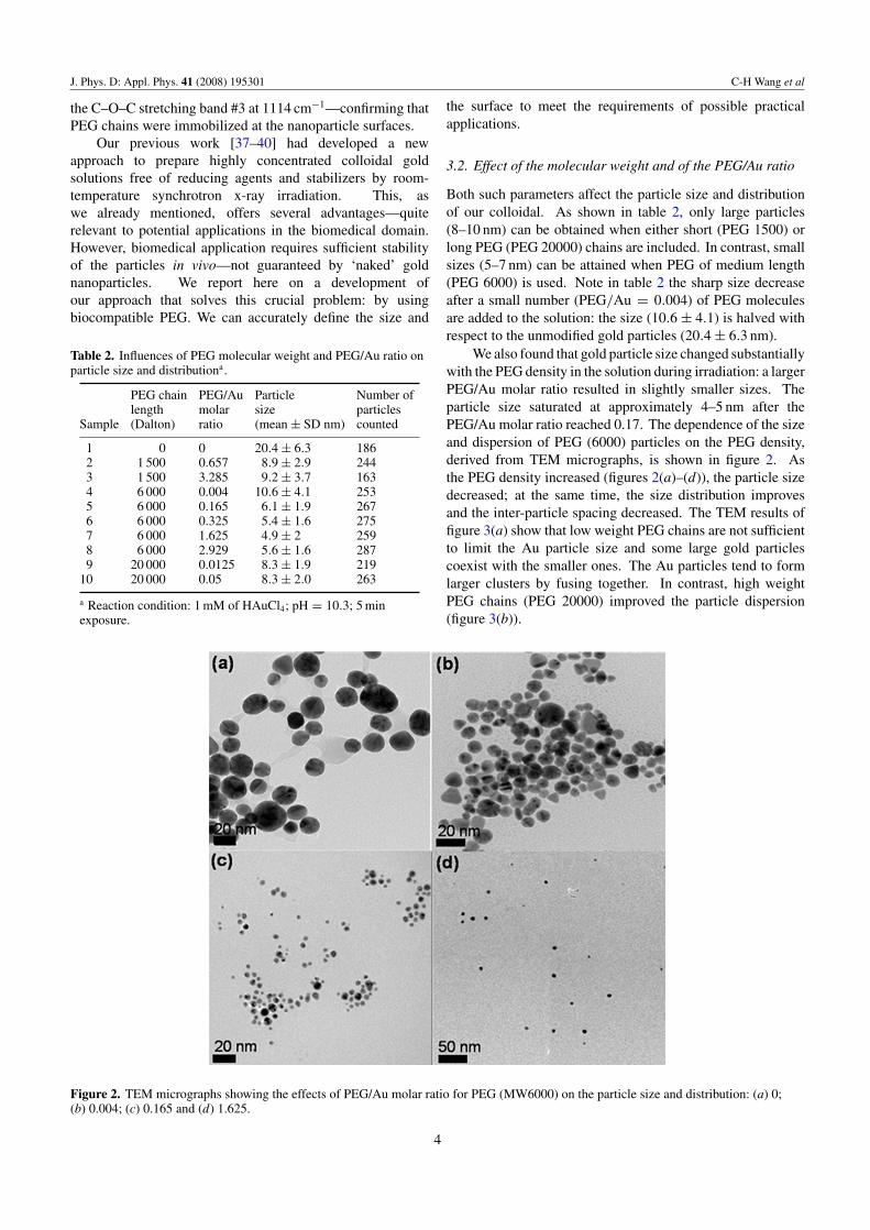
Figure 2. TEM micrographs showing the effects of PEG/Au molar ratio for PEG (MW6000) on the particle size and distribution: (a) 0;(b) 0.004; (c) 0.165 and (d) 1.625.
the surface to meet the requirements of possible practicalapplications.
3.2. Effect of the molecular weight and of the PEG/Au ratio
Both such parameters affect the particle size and distributionof our colloidal. As shown in table 2, only large particles(8–10 nm) can be obtained when either short (PEG 1500) orlong PEG (PEG 20000) chains are included. In contrast, smallsizes (5–7 nm) can be attained when PEG of medium length(PEG 6000) is used. Note in table 2 the sharp size decreaseafter a small number (PEG/Au = 0.004) of PEG moleculesare added to the solution: the size (10.6 ± 4.1) is halved withrespect to the unmodified gold particles (20.4 ± 6.3 nm).
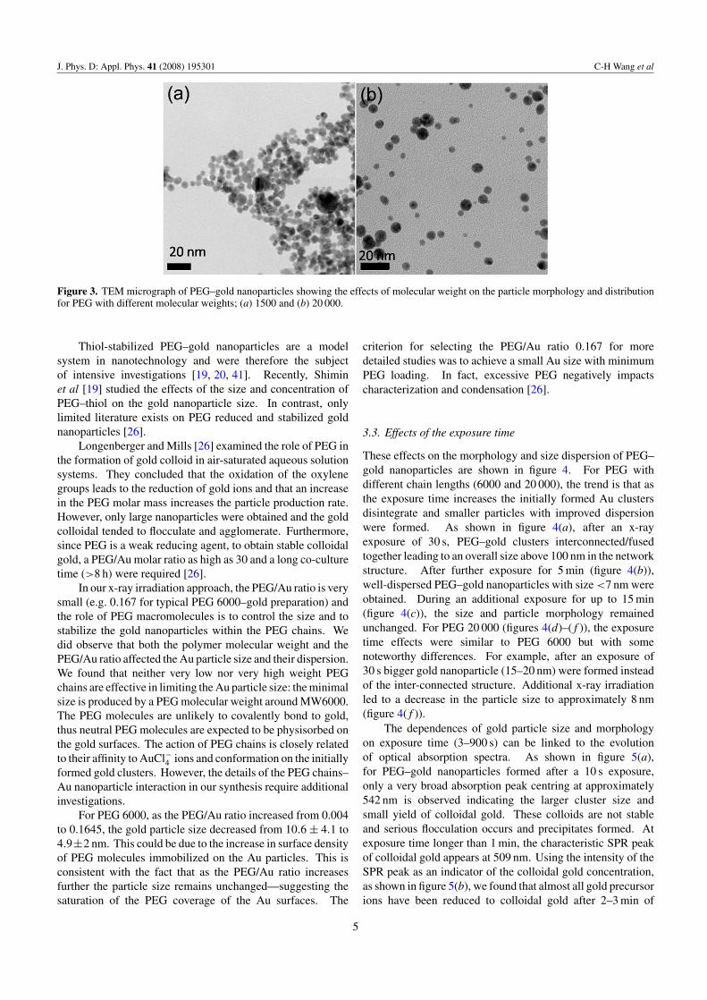
We also found that gold particle size changed substantiallywith the PEG density in the solution during irradiation: a largerPEG/Au molar ratio resulted in slightly smaller sizes. Theparticle size saturated at approximately 4–5 nm after thePEG/Au molar ratio reached 0.17. The dependence of the sizeand dispersion of PEG (6000) particles on the PEG density,derived from TEM micrographs, is shown in figure 2. Asthe PEG density increased (figures 2(a)–(d)), the particle sizedecreased; at the same time, the size distribution improvesand the inter-particle spacing decreased. The TEM results offigure 3(a) show that low weight PEG chains are not sufficientto limit the Au particle size and some large gold particlescoexist with the smaller ones. The Au particles tend to formlarger clusters by fusing together. In contrast, high weightPEG chains (PEG 20000) improved the particle dispersion(figure 3(b)).
4
J. Phys. D: Appl. Phys. 41 (2008) 195301 C-H Wang et al
Figure 3. TEM micrograph of PEG–gold nanoparticles showing the effects of molecular weight on the particle morphology and distributionfor PEG with different molecular weights; (a) 1500 and (b) 20 000.
Thiol-stabilized PEG–gold nanoparticles are a modelsystem in nanotechnology and were therefore the subjectof intensive investigations [19, 20, 41]. Recently, Shiminet al [19] studied the effects of the size and concentration ofPEG–thiol on the gold nanoparticle size. In contrast, onlylimited literature exists on PEG reduced and stabilized goldnanoparticles [26].
Longenberger and Mills [26] examined the role of PEG inthe formation of gold colloid in air-saturated aqueous solutionsystems. They concluded that the oxidation of the oxylenegroups leads to the reduction of gold ions and that an increasein the PEG molar mass increases the particle production rate.However, only large nanoparticles were obtained and the goldcolloidal tended to flocculate and agglomerate. Furthermore,since PEG is a weak reducing agent, to obtain stable colloidalgold, a PEG/Au molar ratio as high as 30 and a long co-culturetime (>8 h) were required [26].
In our x-ray irradiation approach, the PEG/Au ratio is verysmall (e.g. 0.167 for typical PEG 6000–gold preparation) andthe role of PEG macromolecules is to control the size and tostabilize the gold nanoparticles within the PEG chains. Wedid observe that both the polymer molecular weight and thePEG/Au ratio affected the Au particle size and their dispersion.We found that neither very low nor very high weight PEGchains are effective in limiting the Au particle size: the minimalsize is produced by a PEG molecular weight around MW6000.The PEG molecules are unlikely to covalently bond to gold,thus neutral PEG molecules are expected to be physisorbed onthe gold surfaces. The action of PEG chains is closely relatedto their affinity to AuCl−4 ions and conformation on the initiallyformed gold clusters. However, the details of the PEG chains–Au nanoparticle interaction in our synthesis require additionalinvestigations.
For PEG 6000, as the PEG/Au ratio increased from 0.004to 0.1645, the gold particle size decreased from 10.6 ± 4.1 to4.9±2 nm. This could be due to the increase in surface densityof PEG molecules immobilized on the Au particles. This isconsistent with the fact that as the PEG/Au ratio increasesfurther the particle size remains unchanged—suggesting thesaturation of the PEG coverage of the Au surfaces. The
criterion for selecting the PEG/Au ratio 0.167 for moredetailed studies was to achieve a small Au size with minimumPEG loading. In fact, excessive PEG negatively impactscharacterization and condensation [26].
3.3. Effects of the exposure time
These effects on the morphology and size dispersion of PEG–gold nanoparticles are shown in figure 4. For PEG withdifferent chain lengths (6000 and 20 000), the trend is that asthe exposure time increases the initially formed Au clustersdisintegrate and smaller particles with improved dispersionwere formed. As shown in figure 4(a), after an x-rayexposure of 30 s, PEG–gold clusters interconnected/fusedtogether leading to an overall size above 100 nm in the networkstructure. After further exposure for 5 min (figure 4(b)),well-dispersed PEG–gold nanoparticles with size <7 nm wereobtained. During an additional exposure for up to 15 min(figure 4(c)), the size and particle morphology remainedunchanged. For PEG 20 000 (figures 4(d)–( f )), the exposuretime effects were similar to PEG 6000 but with somenoteworthy differences. For example, after an exposure of30 s bigger gold nanoparticle (15–20 nm) were formed insteadof the inter-connected structure. Additional x-ray irradiationled to a decrease in the particle size to approximately 8 nm(figure 4( f )).
The dependences of gold particle size and morphologyon exposure time (3–900 s) can be linked to the evolutionof optical absorption spectra. As shown in figure 5(a),for PEG–gold nanoparticles formed after a 10 s exposure,only a very broad absorption peak centring at approximately542 nm is observed indicating the larger cluster size andsmall yield of colloidal gold. These colloids are not stableand serious flocculation occurs and precipitates formed. Atexposure time longer than 1 min, the characteristic SPR peakof colloidal gold appears at 509 nm. Using the intensity of theSPR peak as an indicator of the colloidal gold concentration,as shown in figure 5(b), we found that almost all gold precursorions have been reduced to colloidal gold after 2–3 min of
5

J. Phys. D: Appl. Phys. 41 (2008) 195301 C-H Wang et al
Figure 4. TEM micrographs showing the effects of x-ray dosage on gold particle size, distribution and morphology. PEG 6000: (a) 30 s,(b) 5 min and (c) 15 min; PEG 20000: (d) 30 s; (e) 1.5 min and ( f ) 5 min.
Figure 5. Visible optical absorption of PEG–Au nanosols prepared with various exposure times: (a) absorption spectra and (b) the variationof colloidal yield and gold SPR peak position.
exposure. This fast reducing speed and the ∼100% reductionefficiency adds to the advantage of our x-ray irradiationapproach.
It was also observed that the SPR peak position dependssensitively on the exposure time (figure 5(b)). The SPR peakfirstly blue shifts up and then reversely shifts to longerwavelength with x-ray exposure time longer than 1 min.Normally the SPR peak red shifts with increasing particlesize. The blue shift at exposure time shorter than 1 min isconsistent with this argument. However, the following red shiftis inconsistent with the TEM observation that the particle sizedecreases upon longer exposure. The temperature effect (thesolution temperature increases approximately 13 ◦C after 5 minexposure) also predicts a blue shift [42]. We can rule out thepossible influence of variation in inter-particle displacementsince mono-dispersed gold nanosols are obtained (figure 1(a)).Further investigations are required to explain the observed redshift of the SPR peak.
3.4. Colloidal stability
Stability tests were performed on re-dispersed nanoparticlesfor up to 150 h using UV–VIS absorption spectra, as shownin figure 6. Specifically, good colloidal stability wasdemonstrated by the stability of the SPR peak. This conclusionis valid for all dispersion media we explored except DMEM.In the latter case (figure 6(a)), after 100 h an additionalabsorption feature appeared at 585 nm that could be due toflocculation. The absorption spectra in figures 6(b)–(d) revealinstead excellent stability for the dispersion media containingHBSS, BSA and NaCl (3 M).
Quantitatively speaking, the stability can be characterizedin terms of the time variations of the so-called ‘flocculationindicators’: the SPR peak FWHM and peak position. Theresults, summarized in table 3, confirm the qualitative messageof figure 6, i.e. high stability for BSA (1 mM) and HBSS media,good stability for the NaCl medium and somewhat lower
6

J. Phys. D: Appl. Phys. 41 (2008) 195301 C-H Wang et al
Figure 6. UV–VIS spectra of re-dispersed PEG–Au colloid in different dispersion media: (a) DMEM containing 10% serum; (b) BSA;(c) HBSS and (d) NaCl (3 M).
Table 3. Time-dependent colloidal stability of re-dispersed PEG–Au colloid in different media.
HBSS BSA NaCl DMEM/F12Dispersion mediumtime point (h) Amax
a FWHMb Amax FWHM Amax FWHM Amax FWHM
0 82.3 521.4 523.4 85.1 521.4 83.5 521.4 84.71 81 521.4 523.4 81 521.4 83.5 521.4 88.33.5 81.5 519.4 523.4 86.6 519.4 83.5 521.4 87
25.5 83.5 519.4 523.4 89.1 521.4 86.1 521.4 99.5107 81.8 521.4 523.4 91.4 527.4 100 517.4 130.1152 83.5 521.4 523.4 94.9 529.4 98.7 513.4 139
a Absorption maximum at Au SPR.b Full width at half maximum.
stability for the DMEM medium. The colloidal stability ofPEG–gold nanoparticles was also examined with zeta potentialmeasurements. The zeta potential for the unmodified goldcolloidal was −57.8 ± 5 mV. For the PEG–gold colloidal, thezeta potential decreased to −20.1 ± 7.5 mV at a pH value of7.23: this can be attributed to the shielding effect of the neutralPEG chains immobilized on the particle surfaces.
Pegylation is well known to improve the colloidal stabilityof nanoparticles both in vitro and in vivo. Synchrotron x-rayderived PEG–gold nanoparticles are highly stable even atlow polymer concentrations and show no sign of flocculationor aggregation even after one year of storage at ambienttemperature. We found that such nanosols are stable undertypical in vitro and in vivo conditions for at least 150 h. Thestability in different dispersion media decreased according tothe sequence: HBSS > BSA (1 mM) > NaCl (3 M) > cellculture medium (DMEM).
The colloidal stability of PEG–gold nanoparticles could beexplained by the steric repulsion of the nanoparticles due to thepresence of PEG. We also found that PEG–Au is influencedmuch more by the adsorbed serum component than by the
ionic strength of the dispersion media, demonstrating stericstabilization dominates over electrostatic stabilization. Undertypical in vivo conditions with high ionic strength, PEG–gold nanosols exhibited superior colloidal stability whereasunmodified gold nanoparticles rapidly flocculated and formedsediments. The critical coagulation concentration (CCC)of PEG–gold determined by UV–VIS spectra was >2.5 M(whereas for unmodified gold it was >0.025 M). The stericrepulsion due to hydrophilic PEG molecules limited the sizeof the nanoparticles and resulted in good dispersion.
As shown in figure 6, the stability of PEG–gold nanosolsin the DMEM medium gradually decreased as the incubationtime increased. Serum adsorption on the surfaces of thePEG–Au particles is likely to contribute to this phenomenon.Serum protein adsorption on PEG–Au particles was revealedby directly examining the hydrodynamic size of the particlesloaded into the cell medium. The DLS measurement detecteda larger size, typically >100 nm, for PEG–Au nanoparticlesin DMEM medium compared with the control system (around29.8 nm). The stabilizing role of PEG depends on differentfactors such as the chain length, the inter-chain separation
7

J. Phys. D: Appl. Phys. 41 (2008) 195301 C-H Wang et al
and the PEG surface density [19, 43]. Therefore, the time-dependent colloidal stability of PEG–Au is probably due tothe incomplete coverage by immobilized PEG chains on thegold surfaces.
4. Conclusion
We developed a one-solution x-ray irradiation approachto prepare PEG modified gold nanoparticles using pristinePEG molecules. A low concentration of unmodified PEGmacromolecules was very effective in controlling particlesize and in stabilizing the gold nanoparticles. Both thePEG chain length and the PEG/Au ratio affected the size,morphology and colloidal stability of the particles. The PEG–gold nanosols could be easily concentrated and re-dispersedand demonstrated high stability under realistic biomedicalconditions.
Acknowledgments
This work was supported by the National Science Council, theAcademia Sinica (Taiwan), the Creative Research Initiatives(Functional X-ray Imaging) of MOST/KOSEF (Korea), theCenter for Biomedical Imaging (CIBM) in Lausanne, partiallyfunded by the Leenaards and Jeantet foundations and by theSwiss Fonds National de la Recherche Scientifique and bythe EPFL.
References
[1] Zharov V P, Galitovskaya E N, Johnson C and Kelly T 2005Lasers Surgery Medicine 37 219
[2] Ferrari M 2005 Nature Rev. Cancer 5 161[3] El-Sayed I H, Huang X H and El-Sayed M A 2006
Cancer Lett. 239 129[4] Caruthers S D, Wickline S A and Lanza G M 2007 Curr. Opin.
Biotechnol. 18 26[5] Paciotti G F, Myer L, Weinreich D, Goia D, Pavel N,
Mclaughlin R and Tamarkin L 2004 Drug Deliv. 11 169[6] Paciotti G F, Kingston D G I and Tamarkin L 2006 Drug
Develop. Res. 67 47[7] Hainfeld J F, Slatkin D N, Focella T M and Smilowitz H M
2006 Br. J. Radiol. 79 248[8] Kim D K, Park S J, Lee J H, Jeong Y Y and Jon S Y 2007
J. Am. Chem. Soc. 129 7661[9] Hainfeld J F, Slatkin D N and Smilowitz H M 2004 Phys. Med.
Biol. 49 N309[10] Owens D E III and Peppas N A 2006 Int. J. Pharm. 307 93[11] Vlerken L E V, Vyas T K and Amiji M M 2007 Pharm. Res.
24 1405[12] Shan J and Tenhu H 2007 Chem. Commun. 4580[13] Kotal A, Mandal T K and Walt D R 2005 J. Polymer Sci. A
43 3631
[14] Zhao H, Kang X and Liu L 2005 Macromolecules 38 10619[15] Raula J, Shan J, Nuopponen M, Niskanen A, Jiang H,
Kauppinen E and Tenhu H 2003 Langmuir 19 3499[16] Zhao W, Gao Y, Kandadai S A, Brook M A and Li Y 2006
Angew. Chem. Int. Edn 45 2409[17] Corbierre M K, Cameron N S and Lennox R B 2004 Langmuir
20 2867[18] Hussain I, Graham S, Wang Z, Tan B, Sherrington D C,
Rannard S P, Cooper A I and Brust M 2005 J. Am. Chem.Soc. 127 16398
[19] Shimin R G, Schoch A B and Braun P V 2004 Langmuir20 5613
[20] Sakura T, Takahashi T, Kataoka K and Nagasaki Y 2005Colloid Polymer Sci. 284 97
[21] Kang Y and Taton T A 2005 Macromolecules 38 6115[22] Becker C F W, Marsac Y, Hazarika P, Moser J, Goody R S and
Niemeyer C M 2007 ChemBioChem 8 32[23] Zubarev E R, Xu J, Sayyad A and Gibson J D 2006 J. Am.
Chem. Soc. 128 4958[24] Ujihara M, Mitamura K, Torikai N and Imae T 2006 Langmuir
22 3656[25] Shenoy D, Fu W, Li J, Crasto C, Jones G, Dimarzio C,
Sridhar S and Amiji M 2006 Int. J Nanomedicine 1 51[26] Longenberger L and Mills G 1995 J. Phys. Chem. 99 475[27] Ah C S, Yun Y J, Park H J, Kim W J, Ha D H and Yun W S
2005 Chem. Mater. 17 5558[28] Hoppe C E, Lazzari M, Pardinas-Blanco I and Lopez-Quintela
M A 2006 Langmuir 22 7027[29] Zhou M, Chen S and Zhao S 2006 J. Phys. Chem. B 110 4510[30] Kawano T, Yamagata M, Takahashi H, Niidome Y, Yamada S,
Katayama Y and Niidome T 2006 J. Control. Release111 382
[31] Bergen J M, Recum H A V, Goodman T T, Massey A P andPun S H 2006 Macromol. Biosci. 6 506
[32] Oyewumi M O, Yokel R A, Jay M, Coakley T and Mumper R J2004 J. Control. Release 95 613
[33] Liu Z, Cai W B, He L, Nakayama N, Chen K, Sun X M,Chen X Y and Dai H J 2007 Nature Nanotechnol. 2 47
[34] Rabin O, Perez J M, Grimm J, Wojtkiewicz G andWeissleder R 2006 Nature Mater. 5 118
[35] Kawano T, Katayama Y and Niidome Y 2006 J. Control.Release 114 343
[36] Margaritondo G, Hwu Y and Je J H 2004 Riv. Nuovo Cimento27 7
[37] Kim C C, Wang C H, Yang Y C, Hwu Y, Seol S K, Kwon Y B,Chen C H, Liou H W, Lin H M, Margaritondo G and Je J H2006 Mater. Chem. Phys. 100 292
[38] Yang Y C, Wang C H, Hwu Y and Je J H 2006 Mater. Chem.Phys. 100 72
[39] Wang C H et al 2007 Mater. Chem. Phys. 106 323[40] Wang C H, Chien C H, Yu Y L, Liu C J, Lee C F, Chen C H,
Hwu Y, Yang C H, Je J H and Margaritondo G 2007J. Synchrotron Radiat. 14 477
[41] Liu Y L, Shipton M K, Ryan J, Kaufman E D and Franzen S2007 Anal. Chem. 79 2221
[42] Stephan L and Mostafa A E S 1999 J. Phys. Chem. B 103 4212[43] Gref R, Luck M, Quellec P, Marchand M, Dellacherie E,
Harnisch S, Blunk T and Mulle R H 2000 Colloids Surf. B18 301
8
Copyright © 2022 FDOKUMEN