Organizational blind spots: splitting, blame and idealization in ...
Upload
khangminh22Category
view
0download
0
MAKIKO SHIRAKAWA, TOSHIYUKI OZAWA, CHIHARU TATEISHI et al. Intense Pulsed Light Therapy for Aberrant Mongolian Spots. Osaka City Medical Journal. 2012, 58, 59-65
Intense Pulsed Light Therapy for
Aberrant Mongolian Spots
MAKIKO SHIRAKAWA, TOSHIYUKI OZAWA, CHIHARU TATEISHI,
NAHO FUJII, DAISUKE SAKAHARA, and MASAMITSU ISHII
Citation Osaka City Medical Journal. Issue Date 2012-12
Type Journal Article Textversion Publisher
Right © Osaka City Medical Association. https://osakashi-igakukai.com/.
Placed on: Osaka City University Repository

Intense Pulsed Light Therapy for Aberrant Mongolian Spots
MAKIKO SHIRAKAWA1,2), TOSHIYUKI OZAWA2), CHIHARU TATEISHI1), NAHO FUJII2),
DAISUKE SAKAHARA2), and MASAMITSU ISHII1,2)
Departments of Dermatology1) and Plastic and Reconstructive Surgery2), Osaka City University, Graduate School of Medicine
- 59 -
Osaka City Med. J. Vol. 58, 59-65, 2012
Received January 31, 2012; accepted October 16, 2012.Correspondence to: Makiko Shirakawa, MD.
Department of Plastic and Reconstructive Surgery, Osaka City University, Graduate School ofMedicine, 1-4-3 Asahimachi, Abeno-ku, Osaka 545-8585, JapanTel: +81-6-6645-3892; Fax: +81-6-6646-6059E-mail: [email protected]
AbstractBackground
Aberrant Mongolian spots (AMS) distal to the lumbosacral region are thought to be more
likely to persist than typical sacral Mongolian spots. So far, Q-switched ruby laser (QSRL) has
been the treatment of choice for AMS.
Intense pulsed light (IPL) is obtained from flashlamp devices that emit wavelengths between
515 and 1200 nm. IPL has documented efficacy for the treatment of irregular pigmentation,
telangiectasia, rough skin texture, rhytids, hair removal, and vascular lesions, with several
filters being available that can be used to block shorter wavelengths from the skin.
As far as we could determine, there have been no clinical and histological studies on the
treatment of AMS with IPL. Accordingly, the aim of this study was to assess the clinical and
histological efficacy of IPL for AMS.
Methods
Seven patients (4 males and 3 females) presenting from September 2008 to July 2009 were
assessed. Their mean age was 2.0 years, ranging from 0 to 7 years. The IPL device used in this
study was a Natulight (Lumenis Ltd., Tokyo, Japan). Photographs were taken of all patients
with a high-resolution digital camera at baseline and 6 months after treatment. Skin biopsy
specimens were taken from 1 patient (case 4) before, immediately after, and 6 months after
treatment.
Results
According to the 7 family members of the patients, the outcome of IPL was graded as follows:
excellent improvement in 1 (14%), good improvement in 4 (57%), and slight improvement in 2
(29%). All families would have liked to continue IPL treatment. Evaluation of the effect of
treatment by a physician was less favorable, with excellent improvement in 1 (14%), good
improvement in 2 (29%), and slight improvement in 4 (57%). Histopathologic examination of the
pigmented region revealed the typical features of a Mongolian spot in the hematoxylin-eosin
stained section. Immediately after IPL, there were no changes in the dermis. At 6 months after
treatment, however, the number of melanocytes in the middle and upper dermis was obviously
decreased.
Conclusions
IPL is an effective method for the treatment of AMS.
Key Words: Intense pulsed light; Aberrant Mongolian spot; Mongolian spot
IntroductionMongolian spots are benign dermal melanocytoses that classically present as bluish-green
patches on the sacrococcygeal region at birth. These spots are very common in children of Asian
and African descent. The pigmentation gradually fades by 6 years of age in most cases1). Such
lesions are rarely seen after the age of 10 years, although persistent aberrant Mongolian spots
such as those on the middle or upper part of the back have been reported2-4).
Mongolian spots distal to the sacral region are more likely to persist than the typical sacral
spots2,5-7), and are called aberrant Mongolian spots (AMS). These spots have been treated with
the Q-switched ruby laser (QSRL)7). QSRL is well absorbed by melanin and can directly destroy
melanosomes, so effectively improves clinical conditions8,9). On the other hand, QSRL treatment
is invasive, so post-treatment care and prolonged downtime are inevitable, and sometimes causes
hyperpigmentation9,10). Another problem is that the irradiation spot of QSRL is small (most
QSRL spot sizes are about 5 mm or 6.5 mm), and sometimes we experience AMS covered wide
areas, so in such cases, general anesthesia is needed.
Intense pulsed light (IPL) is obtained from flashlamp device and is non-laser light with a
wavelength between 515 and 1200 nm. IPL has documented efficacy for the treatment of
irregular pigmentation, telangiectasia, rough skin texture, rhytids, hair removal, and vascular
lesions, with several filters being used to block shorter wavelengths11). IPL is noninvasive and it
is mainly employed to treat aging skin.
As far as we could determine, there have been no clinical or histological studies with regard to
treatment of AMS by IPL. Accordingly, this study was performed to determine the clinical and
histological efficacy of IPL for the treatment of AMS.
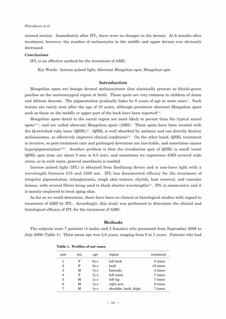
MethodsThe subjects were 7 patients (4 males and 3 females) who presented from September 2008 to
July 2009 (Table 1). Their mean age was 2.0 years, ranging from 0 to 7 years. Patients who had
- 60 -
Shirakawa et al
case
1
2
3
4
5
6
7
age
0y.o
3y.o
7y.o
1y.o
1y.o
1y.o
1y.o
sex
F
F
M
F
M
M
M
region
left back
back
buttocks
left waist
left leg
right arm
shoulder, back, thigh
treatment
5 times
13 times
4 times
7 times
7 times
9 times
7 times
Table 1. Profiles of our cases

a history of epilepsy or cutaneous photosensitivity were excluded.
Laser Parameters and Treatment Protocol
The IPL device used in this study was the Natulight (Lumenis Ltd., Tokyo, Japan). This
device incorporates optimal pulse technology that allows the user to select the wavelength range
required by employing specific cut-off filters. We used a 640 nm cut-off filter (which removed
shorter wavelengths from the broad-spectrum flash lamp output) and triple pulses. The 640 nm
cut-off filter was selected because light between 400 and 600 nm is strongly absorbed by collagen
fibers, so IPL with this cut-off filter causes less severe damage to dermal collagen fibers and the
light is more effectively absorbed by structures containing melanin than if another cut-off filter
was used 560 nm, etc.
Treatment was done at between 22 and 25 J/cm2 with a pulse duration of 4.8 ms, pulse delay
of 40 ms, and spot size of 8×35 mm. No local or general anesthesia was used. During IPL,
cooled colorless gel was applied to keep the filter in contact with the skin. Most procedures were
completed within 5 minutes. IPL was performed at 4-week intervals. Postoperatively, antibiotic
ointment was applied to the treated area. Bleaching agents such as hydroquinone were not used.
- 61 -
Intense Pulsed Light Therapy for Aberrant Mongolian Spots
A, A 7-year-old boy with AMS on thebuttocks before treatment (case 3). B, At six monthsafter 4 treatment sessions, there is an “excellent”cosmetic result.
Figure 1. A, A 1-year-old girl with AMS on the leftwaist before treatment (case 4). B, At six months after 7treatment sessions. The right side (dark part) isuntreated region. There is a remarkable changebetween treated and untreated region. The evaluationis “good”.
Figure 2.
A
B
A
B

Evaluation
Photographs were taken of all patients with a high-resolution digital camera at baseline and 6
months after treatment. The therapeutic effect of IPL was evaluated on the basis of color
improvement on the photographs by an experienced plastic surgeon who had not treated the
subjects. The satisfaction of each patient’s family was also determined on a scale containing 5
categories: worse than before treatment; no improvement; slight improvement; good
improvement; and excellent improvement.
- 62 -
Shirakawa et al
A, Before intense pulsed light (IPL)treatment. Numerous, diffusely scattered melanocytesin the dermis (H&E, ×100) (case 4). B, Immediatelyafter IPL treatment. There were no specific changes ofmelanocytes in the dermis (H&E, ×100). C, At 6 monthsafter treatment, the number of melanocytes in themiddle and upper layers of the dermis was obviouslydecreased (H&E, ×100).
Figure 3.
BA
C
case
1
2
3
4
5
6
7
Clinical improvement
Mild improvement
Good improvement
Excellent improvement
Good improvement
Mild improvement
Mild improvement
Mild improvement
Patient’s satisfaction rate
Good improvement
Good improvement
Excellent improvement
Good improvement
Good improvement
Mild improvement
Mild improvement
Table 2. Treatment results

Skin biopsy specimens were taken from 1 patient (case 4) before, immediately after, and 6
months after treatment.
ResultsIn the 7 patients, the ratings given by the families at the end of IPL were excellent
improvement in 1 (14%), good improvement in 4 (57%), and slight improvement in 2 (29%) (Table
2). All families would have liked to continue IPL treatment. Evaluation of IPL by the physician
was less favorable than by the families: excellent improvement in 1 (14%) (Fig. 1), good
improvement in 2 (29%) (Fig. 2) and slight improvement in 4 (57%) (Table 2).
Histopathologic examination of the pigmented region revealed typical findings of a Mongolian
spot (numerous scattered melanocytes in the dermis) in a hematoxylin-eosin stained section (Fig.
3A). Immediately after IPL, there were no specific changes of melanocytes in the dermis (Fig.
3B). At 6 months after treatment, however, the number of melanocytes in the middle and upper
layers of the dermis was obviously decreased (Fig. 3C).
DiscussionOur result showed both clinical and histological improvement of AMS at 6 months after
treatment with IPL. There was an obvious difference of the histological changes immediately
after treatment between QSRL and IPL. Because QSRL destroys melanosomes, immediately
after QSRL treatment, histological examination reveals damaged melanocytes in the dermis9). In
this study, immediately after IPL treatment, histological evaluation revealed no specific changes
of melanocytes in the dermis. One of the reasons for this difference is the different pulse
durations of IPL and QSRL. IPL has a longer pulse duration than that of QSRL. Considering
the thermal relaxation time of the target, photothermolysis of melanosomes requires a short
pulse duration of nanoseconds that can be obtained with QSRL12). IPL has a longer pulse
duration that results in diffusion of heat from the melanosomes to their surroundings, so IPL
cannot be used to destroy melanosomes without causing nonspecific dermal injury.
However, pathological examination at 6 months after IPL revealed that the number of
melanocytes was decreased in the middle and upper layers of the dermis of one of our patients.
Therefore, there may be another mechanism by which IPL affects melanocytes that differs from
direct thermolysis.
We hypothesized that IPL promotes the apoptosis of melonocytes in pigmented lesions. As
with the various lasers used to treat skin diseases, the mechanism of IPL can be explained by the
concept of selective photothermolysis13-15). Light energy is converted into heat that damages the
stroma of the dermis slightly and stimulates various cells to release cytokines and enzymes to
repair the damage. Fibroblasts are activated16,17) and synthesize more collagen, elastin, and
matrix proteins, which may result in smoother skin. According to Wong, part of the molecular
and signaling mechanism underlying this effect is up-regulation of the expression of typeⅢcollagen and transforming growth factor-β1 (TGF-β1)18).
Recent studies have revealed that TGF-β1 induces rapid apoptosis of unstimulated
melanocytes at picomolar concentrations in three-dimensional collagen culture19). Basal
keratinocytes express growth factors like fibroblast growth factor 2 (FGF-2) and nerve growth
factor, both of which are strong positive regulators of melanocyte growth and survival20,21), so such
- 63 -
Intense Pulsed Light Therapy for Aberrant Mongolian Spots

factors could presumably protect melanocytes from TGF-β-induced apoptosis19). However, the
melanocytes of AMS exist in the dermis where such factors cannot protect melanocytes from
TGF-β, so melanocytes in the AMS lesions are more susceptible to apoptosis than normal
melaonocytes.
In this study, the average treatment time of IPL for AMS was 7.4 times. But in our previous
study, the treatment time of QSRL was almost within 4 times7). So the treatment time of IPL
was more than that of QSRL. This result means IPL cannot directly destroy melanocytes
without causing dermal injury, but the mild, noninvasive treatment is easy to perform and
acceptable to patient’s family.
The advantages of IPL are that it is easy to perform with minimal preparation, limited post-
treatment care, little to no appreciable downtime, and minimal side effects. IPL devices also
have a large spot size, which is larger than that of most laser devices, so we can treat children
(particularly babies and infants) without local or general anesthesia. On the other hand, lasers
are more expensive and time-consuming when a large area is being treated with a small laser
tip, so sometimes local or general anesthesia is needed. These characteristics make IPL a good
choice for the treatment of AMS.
In summary, our study revealed that IPL is an effective method for treating AMS. On the
other hand, we are still far from throughly understanding the mechanism by which IPL improves
AMS, and further research is needed to elucidate this point.
References1. Leung AK. Mongolian spots in Chinese children. Int J Dermatol 1988;27:106-108.2. Leung AK, Kao CP, Leung AA. Persistent Mongolian spots in Chinese adults. Int J Dermatol 2005;44:43-
45.3. Hidano A. Persistent Mongolian spot in the adult. Arch Dermatol 1971;103:680-681.4. Inamadar AC, Palit A. Persistent, aberrant Mongolian spots in Sjögren-Larsson syndrome. Pediatr
Dermatol 2007;24:98-99.5. Cordova A. The Mongolian spot: a study of ethnic differences and a literature review. Clin Pediatr (Phila)
1981;20:714-719.6. Cole HN Jr, Hubler WR, Lund HZ. Persistent, aberrant mongolian spots. Arch Derm Syphilol 1950;61:244-
260.7. Shirakawa M, Ozawa T, Ohashi N, Ishii M, Harada T. Comparison of regional efficacy and complications in
the treatment of aberrant Mongolian spots with the Q-switched ruby laser. J cosmet Laser Ther 2010;12:138-142.
8. Kono T, Nozaki M, Chan HH, Mikashima Y. A retrospective study looking at the long-term complications ofQ-switched ruby laser in the treatment of nevus of Ota. Lasers Surg Med 2001;29:156-159.
9. Watanabe S, Takahashi H. Treatment of nevus of Ota with the Q-switched ruby laser. N Engl J Med 1994;331:1745-1750.
10. Kono T, Manstein D, Chan HH, Nozaki M, Anderson RR. Q-switched ruby versus long-pulsed dye laserdelivered with compression for treatment of facial lentigines in Asians. Lasers Surg Med 2006;38:94-97.
11. Fodor L, Carmi N, Fodor A, Ramon Y, Ullmann Y. Intense pulsed light for skin rejuvenation, hair removal,and vascular lesions: a patient satisfaction study and review of the literature. Ann Plast Surg. 2009;62:345-349.
12. Anderson RR, Parrish JA. Selective photothermolysis: precise microsurgery by selective absorption ofpulsed radiation. Science 1983;220:524-527.
13. Anderson RR, Parrish JA. The optics of human skin. J Invest Drematol 1981;77:13-19.14. Negishi K, Wakamatsu S, Kushikata N, Tezuka Y, Kotani Y, Shiba K. Full-face photorejuvenation of
photodamaged skin by intense pulsed light with integrated contact cooling: initial experiences in Asianpatients. Lasers Surg Med 2002;30:298-305.
15. Bitter PH. Noninvasive rejuvenation of photodamaged skin using serial, full-face intense pulsed light
- 64 -
Shirakawa et al

treatments. Dermatol Surg 2000;26:835-843.16. Labbe RF, Skogerboe KJ, Davis HA, Rettmer RL. Laser photobioactivation mechanism: in vitro studies
ascorbic acid uptake and hydroxyproline formation as biochemical markers of irradiation response. LasersSurg Med 1999;10:201-207.
17. Skinner SM, Gage JP, Wilce PA, Shaw RM. A preliminary study of the effects of laser radiation on collagenmetabolism in cell culture. Aust Dent J 1996;41:188-192.
18. Wong WR, Shyu WL, Tsai JW, Hsu KH, Pang JH. Intense pulsed light effects on the expression ofextracellular matrix protein and transforming growth factor beta-1 in skin dermal fibroblasts culturedwithin contracted collagen lattices. Dermatol Surg 2009;35:816-825.
19. Alanko T, Saksela O. Transforming growth factor beta1 induces apoptosis in normal melanocytes but not innevus cells grown in type 1 collagen gel. J Invest Dermatol 2000;115:286-291.
20. Halaban R, Langdon R, Birchall N, Cuono C, Baird A, Scott G, et al. Paracrine stimulation of melanocytesby keratinocytes through basic fibroblast growth factor. Ann N Y Acad Sci 1988;548:180-190.
21. Pincelli C, Yaar M. Nerve growth factor: its significance in cutaneous biology. J Investig Drematol SympProc 1997;2:31-36.
- 65 -
Intense Pulsed Light Therapy for Aberrant Mongolian Spots
Copyright © 2022 FDOKUMEN