Aberrant sphingomyelin 31P-NMR signatures in giant cell tumour of ...
35
Draft Aberrant sphingomyelin 31 P-NMR signatures in giant cell tumour of bone Journal: Biochemistry and Cell Biology Manuscript ID bcb-2020-0599.R1 Manuscript Type: Article Date Submitted by the Author: 20-May-2021 Complete List of Authors: Quiroz-Acosta, Tayde; Escuela Nacional de Medicina y Homeopatía, Instituto Politécnico Nacional, Sección de Estudios de Posgrado e Investigación Flores-Martinez, Yazmin; Escuela Nacional de Medicina y Homeopatía, Instituto Politécnico Nacional, Sección de Estudios de Posgrado e Investigación Becerra-Martínez, Elvia; Centro de Nanociencias y Micro y Nanotecnologías, Instituto Politécnico Nacional Pérez-Hernández, Elizabeth; UMAE de Traumatología, Ortopedia y Rehabilitación “Dr. Victorio de la Fuente Narváez" Pérez-Hernández, Nury; Escuela Nacional de Medicina y Homeopatía, Instituto Politécnico Nacional, Sección de Estudios de Posgrado e Investigación Bañuelos-Hernández, Angel Ernesto; Centro de Investigacion y de Estudios Avanzados del Instituto Politecnico Nacional, Departamento de Farmacologia Keyword: Giant cell tumour of bone, sphingomyelin synthase, sphingomyelinase, <sup>31</sup>P-NMR, glycerophospholipids Is the invited manuscript for consideration in a Special Issue? : Not applicable (regular submission) © The Author(s) or their Institution(s) Biochemistry and Cell Biology
-
Upload
khangminh22 -
Category
Documents
-
view
0 -
download
0
Transcript of Aberrant sphingomyelin 31P-NMR signatures in giant cell tumour of ...
Draft
Aberrant sphingomyelin 31P-NMR signatures in giant cell tumour of bone
Journal: Biochemistry and Cell Biology
Manuscript ID bcb-2020-0599.R1
Manuscript Type: Article
Date Submitted by the Author: 20-May-2021
Complete List of Authors: Quiroz-Acosta, Tayde; Escuela Nacional de Medicina y Homeopatía, Instituto Politécnico Nacional, Sección de Estudios de Posgrado e InvestigaciónFlores-Martinez, Yazmin; Escuela Nacional de Medicina y Homeopatía, Instituto Politécnico Nacional, Sección de Estudios de Posgrado e InvestigaciónBecerra-Martínez, Elvia; Centro de Nanociencias y Micro y Nanotecnologías, Instituto Politécnico NacionalPérez-Hernández, Elizabeth; UMAE de Traumatología, Ortopedia y Rehabilitación “Dr. Victorio de la Fuente Narváez"Pérez-Hernández, Nury; Escuela Nacional de Medicina y Homeopatía, Instituto Politécnico Nacional, Sección de Estudios de Posgrado e InvestigaciónBañuelos-Hernández, Angel Ernesto; Centro de Investigacion y de Estudios Avanzados del Instituto Politecnico Nacional, Departamento de Farmacologia
Keyword: Giant cell tumour of bone, sphingomyelin synthase, sphingomyelinase, <sup>31</sup>P-NMR, glycerophospholipids
Is the invited manuscript for consideration in a Special
Issue? :Not applicable (regular submission)
© The Author(s) or their Institution(s)
Biochemistry and Cell Biology
Draft
1
1 Aberrant sphingomyelin 31P-NMR signatures in giant cell tumour of bone
2 Tayde Quiroz-Acosta1, Yazmin Montserrat Flores-Martinez1, Elvia Becerra-Martínez2,
3 Elizabeth Pérez-Hernández3, Nury Pérez-Hernández1*, and Angel Ernesto Bañuelos-
4 Hernández4*
5
6 1 Escuela Nacional de Medicina y Homeopatía, Instituto Politécnico Nacional, Ciudad de
7 México, 07320, México.
8 2 Centro de Nanociencias y Micro y Nanotecnologías, Instituto Politécnico Nacional,
9 Ciudad de México, 07320, México.
10 3 UMAE de Traumatología, Ortopedia y Rehabilitación “Dr. Victorio de la Fuente
11 Narváez”, Instituto Mexicano del Seguro Social, Ciudad de México, 07760, México.
12 4 Programa de Posgrado en Farmacología, Centro de Investigación y de Estudios
13 Avanzados del Instituto Politécnico Nacional, A.P. 14-740, Ciudad de México, 07000,
14 México.
15
16 *Corresponding author: Angel Ernesto Bañuelos-Hernández, Programa de Posgrado en
17 Farmacología, Centro de Investigación y de Estudios Avanzados del Instituto
18 Politécnico Nacional, A.P. 14-740, Ciudad de México, 07000, México. E-mail:
20 Nury Pérez-Hernández, Escuela Nacional de Medicina y Homeopatía, Instituto
21 Politécnico Nacional, Ciudad de México, 07320, México. E-mail: [email protected]
Page 1 of 34
© The Author(s) or their Institution(s)
Biochemistry and Cell Biology
Draft
2
22 Abstract
23 An understanding of the biochemistry of the giant cell tumour of bone (GCTB) provides
24 an opportunity for the development of prognostic markers and identification of therapeutic
25 targets. Based on metabolomic analysis, we proposed glycerophospholipid metabolism
26 as the altered pathway in GCTB and the objective of this study was to identify these
27 altered metabolites. Using phosphorus-31 nuclear magnetic resonance spectroscopy
28 (31P-NMR), sphingomyelin was determined as the most dysregulated phospholipid in
29 tissue samples from six patients with GCTB; subsequently, enzymes related to its
30 biosynthesis and hydrolysis were examined using immunodetection techniques. High
31 expression of sphingomyelin synthases 1 and 2, but low expression of neutral
32 sphingomyelinase 2 (nSMase2), was found in GCTB tissues compared to non-neoplastic
33 bone tissues. Sphingomyelin/ ceramide biosynthesis is dysregulated in GCTB due to
34 alterations in the expression of SMS1, SMS2, and nSMase2.
35 Key Words: Giant cell tumour of bone, sphingomyelin, ceramide, sphingomyelin
36 synthase, sphingomyelinase, neutral sphingomyelinase 2, bone cancer, giant cell-rich
37 bone tumours, 31P-NMR, glycerophospholipids.
38
Page 2 of 34
© The Author(s) or their Institution(s)
Biochemistry and Cell Biology
Draft
3
39 Introduction
40 Giant cell tumour of bone (GCTB) is one of the most prevalent epiphyseal and meta-
41 epiphyseal neoplasms, which is locally aggressive, recurrent, and has high metastatic
42 potential to the lung (Montgomery et al., 2019; Muheremu and Niu, 2014; Santini-Araujo
43 E., 2015). This neoplasm accounts for 6% of the primary bone cancer and 20% of the
44 benign tumour cases reported in patients between 20 and 50 years of age in the long
45 bones with knee involvement, and it exhibits a slight predominance in women (Lin et al.,
46 2019; Montgomery et al., 2019). The histological composition of this tumour includes the
47 following three cell populations: a) multi-nucleated osteoclast-like giant cells (GCs), b)
48 mononuclear monocyte-macrophage cells (MCs), and c) stromal cells (SCs), which are
49 the neoplastic elements (Noh and Park, 2018; Schaefer and Hornick, 2018). The main
50 pathway involved in the pathogenesis of GCTB is linked to high levels of receptor activator
51 of nuclear factor-kappa B ligand (RANKL) in SCs, which stimulates formation of GCs from
52 monocytes and the differentiation of osteoclastic precursors, thereby activating osteolysis
53 (Wu et al., 2015).
54 The specific mutation of the H3F3A gene on chromosome 1 is useful for differentiating
55 this tumour from chondroblastoma, and this may be a potential biomarker for GCTB
56 (Yamamoto et al., 2020). The mutant histone H3G34W, encoded by the H3F3A gene, is
57 characterized by the exchange of the amino acid glycine (G) by tryptophan (W) at position
58 34 (p.Gly34Trp) (Yamamoto et al., 2020). An antibody directed against the H3G34W
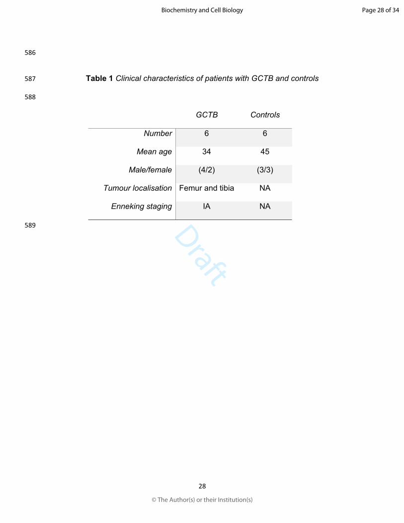
59 mutation, with high sensitivity and specificity for GCTB, exhibits diffuse nuclear staining
60 and is positive in 91% of the GCTB samples, but not in chondroblastoma and other types
61 of giant cell-rich bone tumours (Amary et al., 2017; Behjati et al., 2013).
Page 3 of 34
© The Author(s) or their Institution(s)
Biochemistry and Cell Biology

Draft
4
62 Currently, exploration of the cancer metabolome is of significance in the discovery of
63 diagnostic and predictive markers, since the phenotypic changes can be directly related
64 to their underlying biochemical changes (Armitage and Barbas, 2014). Recently, our
65 research group described the metabolomic profile of GCTB, which indicated
66 glycerophospholipid metabolism as one of the main deregulated pathways (Martínez-
67 López et al., 2017). Glycerophospholipids and sphingolipids are structurally complex
68 saponifiable lipids that form integral parts of cellular membranes and plasma lipoproteins.
69 They play an important role as intracellular signalling molecules during responses to
70 growth factors, differentiation, migration, and cell death (D'Angelo et al., 2018; Hannun
71 and Obeid, 2008; Iqbal et al., 2017; Lordan et al., 2017). Specifically, sphingomyelin (SM)
72 is a bioactive lipid and is the most abundant component of the plasma membrane. SM is
73 capable of forming lipid rafts that regulate the signalling by membrane receptors (Bienias
74 et al., 2016), owing to its ubiquity and abundance. Therefore, defects in SM biosynthesis
75 can directly or indirectly influence the normal physiology of the cell and lead to a
76 neoplastic transformation.
77 SM levels are crucial for cellular function; therefore, their biosynthesis and cellular
78 concentrations are strictly regulated. SM hydrolysis by sphingomyelinases (SMases)
79 increases the concentration of ceramide (Cer), another bioactive lipid, involved in cellular
80 proliferation, growth, and apoptosis (Bienias et al., 2016; Kolesnick, 2002). The levels of
81 SM and Cer are regulated by the concerted action of two enzyme families, SMases, which
82 convert SM into Cer, and sphingomyelin synthases, which convert Cer into SM. SM
83 biosynthesis is altered in certain types of epithelial tumours (Kozar et al., 2018; Li et al.,
84 2017; Nagahashi et al., 2016; Separovic et al., 2017; Sun and Leung, 1974), with an
Page 4 of 34
© The Author(s) or their Institution(s)
Biochemistry and Cell Biology
Draft
5
85 increase in SM and a decrease in Cer levels in hepatocarcinoma and ovarian cancer
86 (Kozar et al., 2018; Li et al., 2017), although the specific changes depend on the type of
87 tumour (D'Angelo et al., 2018). In the case of mesenchymal tumours, their lipid profile
88 and bioactive lipids are not well understood, because the focus has been on the sensitivity
89 to antineoplastic therapies, as observed in reports of haematopoietic malignancies
90 (Biswal et al., 2000; Ekiz and Baran, 2010; Ricci et al., 2006). The role of sphingolipids in
91 cancer is explored; however, until now, their contribution in most mesenchymal tumours
92 remains unclear. Therefore, the aim of this study was to analyse SM/Cer biosynthesis in
93 GCTB, a tumour of mesenchymal origin.
94 Materials and methods
95 Study population Six non-recurrent GCTB samples were obtained from surgical
96 intralesional excision from patients treated in the UMAE “Dr. Victorio de la Fuente
97 Narváez”, Instituto Mexicano del Seguro Social (IMSS) in Mexico City. The diagnosis was
98 based on clinical, radiological, and histopathological criteria. According to the Enneking
99 staging, all GCTB patients were categorised as IA, which corresponds to low-grade, intra-
100 compartmental lesions, without metastasis or pulmonary dissemination (Enneking et al.,
101 2003). For the control, six non-neoplastic cancerous bone tissue samples obtained from
102 the iliac crest were used. The tissues were dissected by a pathologist with expertise in
103 musculoskeletal pathology and then stored at -80°C. The medical histories of the patients
104 were examined and the samples of patients with metabolic, infectious, or concomitant
105 systemic pathologies and that of those, who received local or systemic adjuvant therapies
106 were discarded. All patients signed a consent authorisation according to the standardised
107 protocols of treatment and surgery, and the clinical information is summarised in Table 1.
Page 5 of 34
© The Author(s) or their Institution(s)
Biochemistry and Cell Biology
Draft
6
108 The Bioethics Committee of the Escuela Nacional de Medicina y Homeopatía del Instituto
109 Politécnico Nacional approved the procedures of this study. This study was conducted in
110 accordance with the Declaration of Helsinki.
111 Phospholipid extraction and phosphorus-31 nuclear magnetic resonance (31P-
112 NMR) spectroscopy Each frozen tissue sample was weighed (300 mg per sample) and
113 placed in a microtube. Then, 1 mL of a mixture of CDCl3:MeOD (2:1 v/v) was added (Folch
114 et al., 1957). The tissues were homogenised using zirconium beads in the Bead Bug
115 Homogenizer (Benchmark Scientific; Sayreville, NJ, USA) for 3 min at 180 Hz. The
116 samples were centrifuged at 13,500 × g for 10 min at 4°C, and the organic fraction (600
117 µL per sample) was collected in an NMR tube. Triphenylphosphine was added to achieve
118 a final concentration of 0.1 mM. 31P-NMR hydrogen-decoupled spectra were obtained
119 using the Bruker Avance 400 NMR spectrometer (Bruker Biospin, Karlsruhe, Germany).
120 The analyses were performed at 25°C with the probe PABBO BB-1H/D Z-GRD for 31P at
121 161.98 MHz using the pulse sequence zgpg30 and 30o pulse, sweep width of 12931.0
122 Hz, repetition time of 0.63 s, 20000 scans, and acquisition time of 0.6335 s. Spectra were
123 processed using the Bruker’s TopSpin software v.3.1 (Bruker Biospin, Karlsruhe,
124 Germany), by applying 1.0 Hz of line broadening. The identification of phospholipids was
125 conducted according to the methods described in earlier reports (Pierce and Komoroski,
126 1993).
127 Data analysis NMR profile The 31P-NMR spectra were aligned and the dimensionality
128 reduction was performed by lowering the original data points (32 k points) in bins of 0.04
129 ppm width (Anderson et al., 2011). To compensate for variations arising due to difference
130 in source cells and extraction, all the bins were normalised using the weight of the
Page 6 of 34
© The Author(s) or their Institution(s)
Biochemistry and Cell Biology
Draft
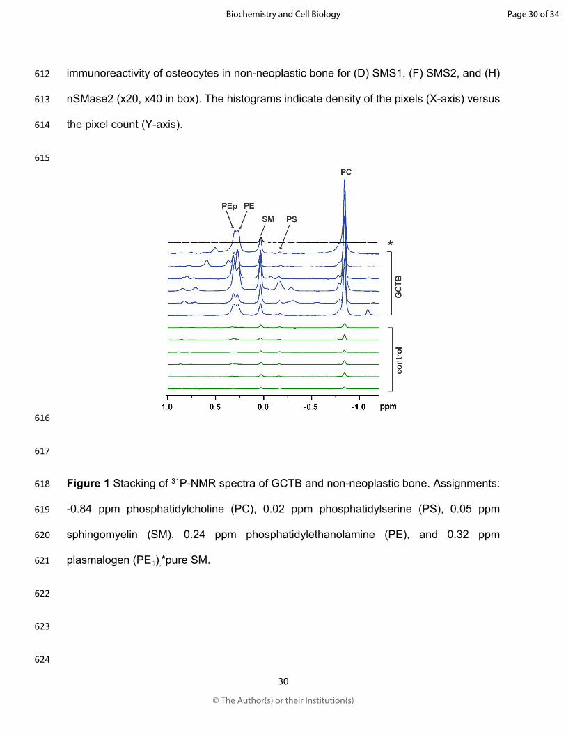
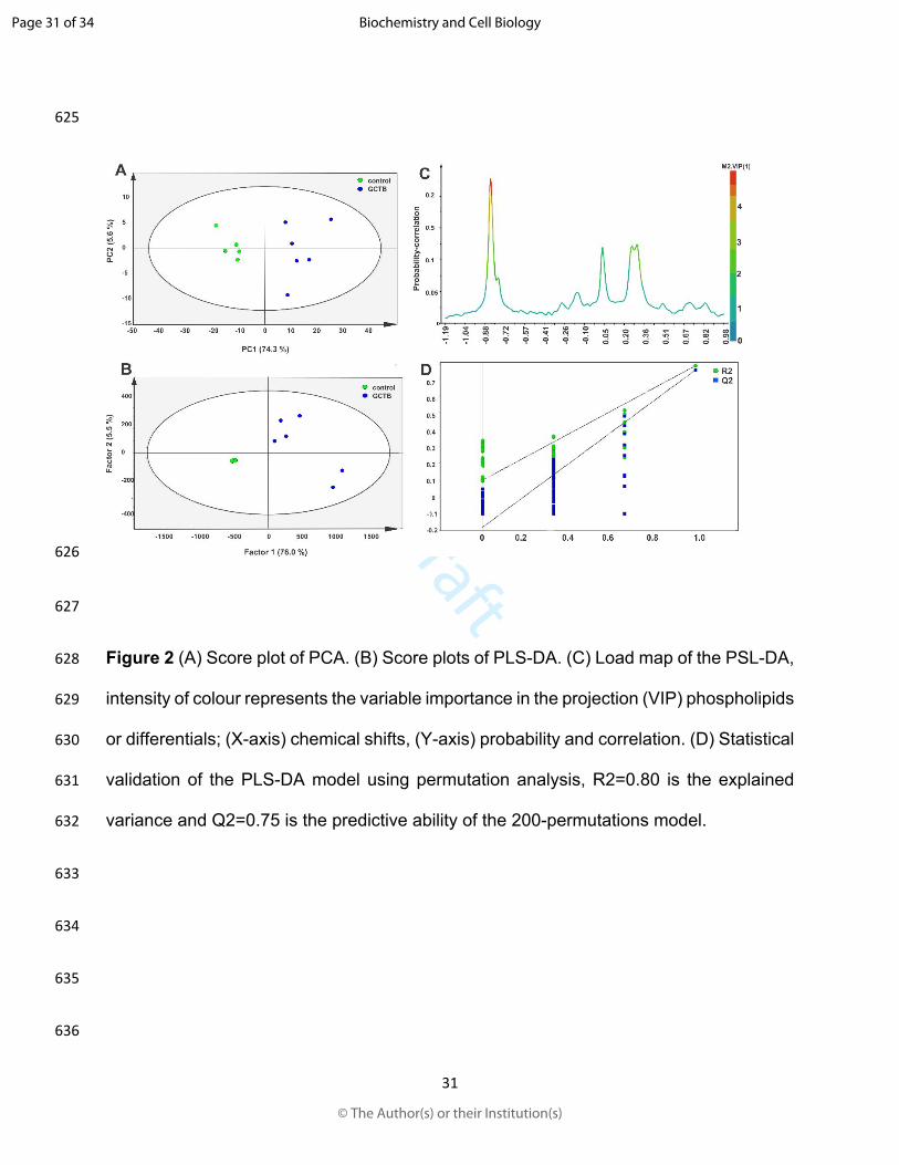
7
131 respective sample extract. The alignment, ‘binning’, and normalisation were performed
132 using the MNova software v.10.1 (Mestrelab Research, Santiago de Compostela, Spain).
133 Since the assigned signals were normalised with sample weights and internal standard,
134 the spectral intensities were compared to obtain a ratio between phospholipids.
135 Protein extraction and immunoblotting Proteins were extracted from tissues using
136 urea/thiourea lysis buffer containing 7 M urea, 2 M thiourea, and 4% (w/v) CHAPS
137 detergent in 30 mM Tris–HCl pH 8.5, and Halt Protease Inhibitor Cocktail (Thermo
138 Scientific, Waltham, MA USA). The tissues were homogenised using zirconium beads
139 and processed in the Bead Bug Microtube Homogenizer (Benchmark Scientific;
140 Sayreville, NJ, USA) for 3 min at 180 Hz. Extracts were centrifuged at 13,000 × g for 10
141 min at 4°C and precipitated with cold acetone (3:1 ratio) at -20°C overnight. Total protein
142 was quantified using the BCA method. Then, 10 µg of protein per sample was subjected
143 to SDS-PAGE on 10% acrylamide/bisacrylamide (30:1) gels at 120 V. The resolved
144 proteins were transferred onto a nitrocellulose membrane, 0.45 µm (Bio-Rad; Hercules,
145 CA, USA) for 40 min at 24 V using the Towbin transfer buffer, 25 mM Tris, 0.192 mM
146 glycine pH 8.6, and 10% methanol, using a trans-blot SD semi-dry electrophoretic transfer
147 cell (Bio-Rad; Hercules, CA, USA). The membranes were blocked using phosphate-
148 buffered saline-tween 0.25% (PBS-T) with 1% casein for 1 h. Membranes were then
149 incubated with the mouse monoclonal anti-β-tubulin (1:6000; Santa Cruz Biotechnology,
150 Dallas, TX, USA), rabbit polyclonal anti-sphingomyelin synthase 1 (anti-SMS1) (1:6000;
151 Thermo Scientific, Waltham, MA USA), rabbit polyclonal anti-sphingomyelin synthase 2
152 (anti-SMS2) (1 : 6000; Invitrogen, Carlsbad, CA, USA), and anti-neutral
153 sphingomyelinase 2 (anti-nSMase2) (1 : 6000; GeneTex, Irvine, CA, USA) for 1 h at room
Page 7 of 34
© The Author(s) or their Institution(s)
Biochemistry and Cell Biology
Draft
8
154 temperature (25°C). After rinsing with PBS-T for 30 min, membranes were incubated with
155 horseradish peroxidase (HRP)-conjugated anti-mouse and anti-rabbit IgG (1: 6000;
156 Sigma-Aldrich, St. Louis, MO, USA) antibodies for 1 h at room temperature. The blots
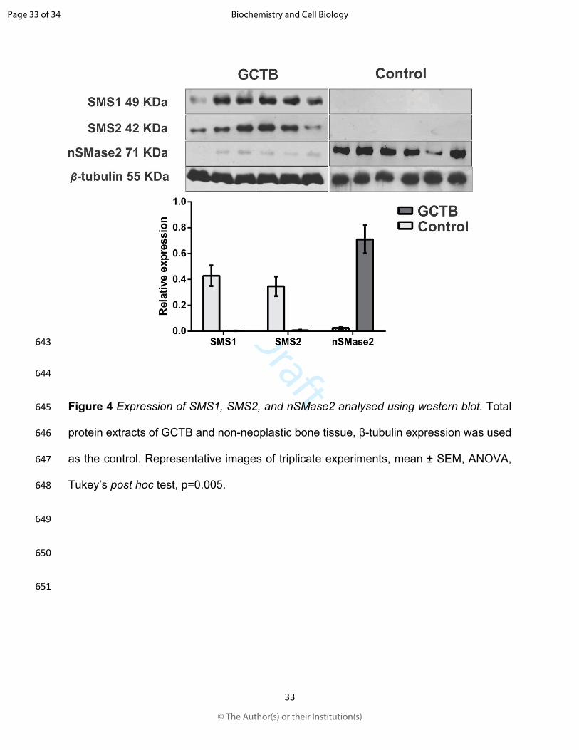
157 were developed using the Clarity Western ECL substrate (Bio-Rad, Hercules, CA, USA)
158 for autoradiography at 1-min exposure. The autoradiographs were scanned with the
159 EpiChemi Darkroom photo-documenting device (UVP; Upland, California, USA).
160 Densitometric analysis was performed using the Image Studio Lite software v.5.2, LI-COR
161 (Biosciences, Lincoln, Nebraska, USA).
162 Immunohistochemistry Tissue samples were fixed with 10% formalin-PBS, embedded
163 in paraffin, and sliced in 5 μm-thick sections for further analysis. The tissue sections were
164 de-paraffinised by heating the slides at 55°C for 30 min, followed by three washes using
165 xylol and gradual hydration with 100%, 95%, 85%, and 75% aqueous-ethanol. The
166 tissues were incubated with mouse monoclonal anti-β-tubulin (1:200; Santa Cruz
167 Biotechnology, Dallas, TX, USA), anti- rabbit polyclonal anti-sphingomyelin synthase 1
168 (anti-SMS1) (1:200; Thermo Scientific, Waltham, MA USA), rabbit polyclonal anti-
169 sphingomyelin synthase 2 (anti-SMS2) (1:200; Invitrogen, Carlsbad, CA, USA), and anti-
170 sphingomyelin phosphodiesterase 2 (anti-nSMase2) (1:200; GeneTex, Irvine, CA, USA).
171 Immuno-labelling was detected using the Histostain-Plus IHC Kit (Thermo Fisher,
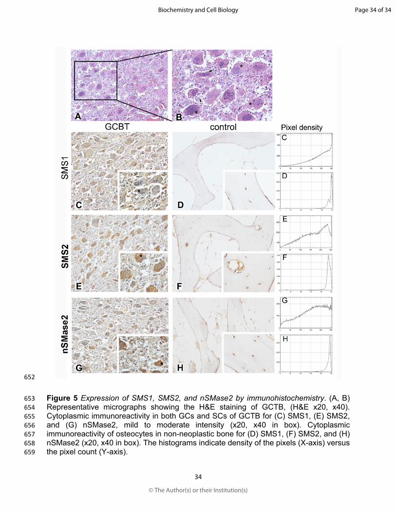
172 Waltham, MA, USA), according to the manufacturer’s instructions. Staining intensity was
173 determined using the IHC Profiler integrated in the ImageJ software v.1.46r (National
174 Institutes of Health, Bethesda, USA) (Varghese et al., 2014).
175 Statistical analysis The data matrix of NMR was analysed using multivariate statistics
176 of the SIMCA software v13.0.3 (Umetrics, Umea, Sweden). The data were normalised
Page 8 of 34
© The Author(s) or their Institution(s)
Biochemistry and Cell Biology
Draft
9
177 using logarithmic transformation and Pareto scaling. Principal Component Analysis (PCA)
178 was used for the general description of the system and the supervised method of Partial
179 Least Squares Discriminant Analysis (PLS-DA) was used for modelling using the same
180 parameters of centring and scaling. For immunoblot densitometric analysis, the results
181 were expressed as mean ± standard error (SEM) values from at least three independent
182 experiments (n=3). The differences among groups were analysed using two-way ANOVA
183 and the Tukey’s post hoc test. The GraphPad Prism software v.6.0 (GraphPad, La Jolla,
184 CA, USA) was used for statistical analysis. The significance was set at P<0.05.
185 Results
186 Evaluation of phospholipids (PLs) using 31P-NMR spectroscopy The 31P-NMR
187 spectra exhibited adequate resolution that allowed us to distinguish five metabolites (PLs)
188 in GCTB, which were expressed in significantly higher concentrations compared to those
189 in the non-neoplastic bone (Figure 1). The most noteworthy PL signals were identified at
190 approximately -1.0 and 0.5 ppm, which corresponded to phosphatidylcholine (PC) at -
191 0.84 ppm, phosphatidylserine (PS) at -0.02 ppm, sphingomyelin (SM) at 0.05 ppm,
192 phosphatidylethanolamine (PE) at 0.24 ppm, and plasmalogen (PEp) at 0.32 ppm. A
193 chemometric analysis was used to accurately assess the difference in the levels of PLs
194 between the GCTB and non-neoplastic bone tissue samples. The 31P-NMR spectra were
195 binned, and each bin was considered as a variable for construction of a data matrix with
196 the respective sample (Figure S1). A remarkable difference was observed between the
197 non-neoplastic tissue and the GCTB samples in the PCA (Figure 2A). The first principal
198 component (PC1) separated the samples and accounted for 74.3% of the variance within
199 the data (represented as circles in blue colour, Figure 2A), while the second principal
Page 9 of 34
© The Author(s) or their Institution(s)
Biochemistry and Cell Biology
Draft
10
200 component (PC2) accounted for 5.6% of the variance (green circles). Modelling with PLS-
201 DA (Figure 2B) confirmed the differences between the GCTB and control groups. In the
202 scoring graphs, the signals of some PLs determined the separation of two sample groups.
203 According to the loading plots, the signal intensity of PC, PE, PEp, and SM was
204 remarkably high in the GCTB samples compared to that in the non-neoplastic bone
205 samples. A pseudospectrum (Figure 2C) was constructed with the loading coefficients.
206 The high and red signals represent the variable importance in the projection (VIP) of the
207 chemical shift in the PLS-DA model. Finally, the GCTB model parameters were validated
208 using a permutation test (200 permutations) with a correlation of R2 = 0.80 and the
209 predictive capability of Q2 = 0.75 (Figure 2D). This result supports the hypothesis of an
210 altered SM metabolism. The control group exhibited a lower variance in the VIP for these
211 metabolites. In contrast, the GCTB group exhibited a higher variance for SM, which is
212 related to signalling and apoptosis, thereby indicating variations in this tumour, SM is one
213 of the main substrates of the Cer synthesis. An additional non multivariate analysis of the
214 VIP spectral intensities regarding the ratios of SM, PS, PE and PEp compared to main
215 phospholipid PC was performed (Figure 3), The PS/PC and SM/PC ratios were
216 significantly higher in control samples while PE/PS ratio were lower in controls and Pp/PC
217 was not different in both groups (Table S1).
218 SMS1, SMS2, and nSMase2 protein expression The metabolic conversion of Cer to
219 SM is catalysed mainly by SMS1 in the Golgi and by SMS2 in the Golgi and the cell
220 membrane. We analysed the expression of both enzymes (Figure 4A) and observed that
221 the relative expression of SMS1 (20.526 fold; p = 0.0030) and SMS2 (29.338 fold;
222 p=0.0066) were significantly higher in GCTB compared to those in non-neoplastic bone
Page 10 of 34
© The Author(s) or their Institution(s)
Biochemistry and Cell Biology
Draft
11
223 tissue (control group) (Figure 4B). The conversion of SM to Cer is catalysed by three
224 different types of SMases that are activated according to the pH of the microenvironment.
225 However, in this study, neutral sphingomyelinase 2 (nSMase2) was selected, since it is
226 the best-characterized SMase of the three enzymes (Tani and Hannun, 2007), and it
227 participates in bone mineralisation and cartilage development (Aubin et al., 2005; Stoffel
228 et al., 2019; Stoffel et al., 2007). Interestingly, a significant decrease in the relative
229 expression of nSMase2 was observed in GCTB compared to that in the non-neoplastic
230 bone tissue samples (0.0403 fold; p = 0.0014) (Figure 4B). These results suggest a
231 possible selective up-regulation of SMS1 and SMS2, and down-regulation of nSMase2 in
232 GCTB.
233 SMS1, SMS2, and nSMase2 immunohistochemical analysis The relative expression
234 of SMS1, SMS2, and nSMase2 was analysed using immunohistochemistry (Figure 5).
235 Previously, the tumour tissue was stained with hematoxylin and eosin (H&E) to identify
236 the cell types (Figure 5A and 5B). Numerous multinucleated GCs of the osteoclast type
237 (asterisks) and mononuclear SCs (arrows) are present, in addition to the abundant
238 vascular component, which are all characteristic elements of GCTB. Immunostaining for
239 SMS1 revealed mild cytoplasmic immunoreactivity in GCs, while the staining in SC was
240 scarce (Figure 5C), compared to that in non-neoplastic bone, where it was localised in
241 osteocytes (Figure 5D). We observed that the highest immunoreactivity for SMS2 was in
242 the GC cytoplasm (Figure 5E), and the intensity was moderate compared to that in the
243 control tissue (Figure 5F), where the intensity demonstrated a decreased presence.
244 nSMase2 was expressed in the cytoplasm of both GCs and SCs, at about 50% for both
245 cellular elements (Figure 5G). The expression was evident in the control tissue as well
Page 11 of 34
© The Author(s) or their Institution(s)
Biochemistry and Cell Biology
Draft
12
246 (Figure 5H). Image analysis and the semi-quantitative comparisons of the optical density
247 scores were performed using the ImageJ software and the IHC Profiler complement
248 (Varghese et al., 2014). The results of this analysis are shown as histograms of pixel
249 density. The upper histogram in each panel corresponds to the GCTB, while the lower
250 histogram corresponds to the non-neoplastic tissue (Figure 5). The semi-quantitative
251 analysis of the images showed a low IHC optical density score for SMS1 and SMS2 of
252 the GCTB compared to the non-neoplastic bone tissue; and a low score for nSMase2 in
253 both samples.
254 Discussion
255 Has been shown that the phospholipid content increase with cell transformation and
256 carcinoma progression (Cheng et al., 2016). For example, an increased of
257 phosphocholine, glycerophosphocholine, and free choline, has been detected by 1H MRS
258 (proton magnetic resonance spectroscopy (1H-MRS) in breast, prostate, ovarian,
259 endometrial, cervical, and brain cancers compared to non-cancerous healthy tissues
260 (Glunde et al., 2011). In relation to enzymes involved in phospholipid metabolism can be
261 highlighted the overexpression and elevated activity of choline kinase as factors that
262 contribute to increased phosphocholine and choline in various tumor tissues including
263 breast, ovarian, colorectal, prostate, lung, endometrial, and pancreatic cancer (Cheng et
264 al., 2016).
265 Concerning mesenchymal tumours such as bone sarcomas, the phospholipid metabolism
266 is scarcer. Until now it is known that in osteosarcoma, the expression of phospholipase
267 A2 which hydrolyses the ester bond of the sn-2 position membrane phospholipids
268 preferably PC releasing free fatty acids and lysophospholipid, is higher at mRNA and
Page 12 of 34
© The Author(s) or their Institution(s)
Biochemistry and Cell Biology
Draft
13
269 protein levels in patients with metastasis. (Liang et al., 2015). Consistently, in MG-63
270 osteosarcoma cell line have been shown the cytidine 5´-diphosphocholine (CDP-choline)
271 pathway is the only route for PC biosynthesis, while silencing Choline kinase-α (CKα) in
272 these cells reduce the cell proliferation and mineralization as well as slight decrease the
273 phosphocholine concentration (Li et al., 2014).
274 Unfortunately, there is limited knowledge on the metabolic changes in tumours of
275 mesenchymal origin, such as GCTB. In this study, we focussed on the evaluation of SM
276 biosynthesis, since it was the most important variable, based on the multivariate analysis
277 and the extraction of the VIP from the data obtained using 31P-NMR spectroscopy.
278 These results are concordant with the earlier reports. It is presently accepted that
279 enzymatic dysregulation in the SM metabolism in signalling plays a vital role in the
280 tumours of epithelial and haematological origin (Ogretmen, 2018). This explains the
281 reason for increased research interest in SM biosynthesis, because there are no reports
282 on its metabolic state in GCTB.
283 The enzymes which are directly responsible for the synthesis of SM include the ceramide
284 choline phosphotransferases or sphingomyelin synthases (SMSs). The reaction begins
285 with the transfer of a phosphocholine residue from phosphatidylcholine (PC) to the
286 primary hydroxyl group of the ceramide, resulting in SM and diacylglycerol (DAG)
287 (Holthuis and Luberto, 2010). Sphingomyelin phosphodiesterases or sphingomyelinases
288 (SMases) hydrolyse the phosphodiester bond of SM, thereby generating Cer and
289 phosphocholine (Bienias et al., 2016). Owing to the concerted action of these two enzyme
290 families, SM concentrations remain balanced in the intracellular compartments. The Golgi
291 apparatus and the plasma membrane are the major sites of SM synthesis (D'Angelo et
Page 13 of 34
© The Author(s) or their Institution(s)
Biochemistry and Cell Biology
Draft
14
292 al., 2018). Therefore, it is inevitable for these enzymes (SMS1 and SMS2) to be
293 distributed in the cytoplasm (see Figure 4). Similarly, the hydrolytic enzyme is also
294 commonly observed to be distributed in the cytoplasm, as SM and Cer, being two
295 bioactive lipids, are strictly regulated with respect to both the time of synthesis and
296 distribution. SM is an essential modulator of the structural properties of the plasma
297 membrane, which influences the accumulation of proteins involved in cell proliferation.
298 Therefore, it is not surprising that the dysregulation of its biosynthetic and hydrolytic
299 enzymes promotes tumourigenesis.
300 Our results indicated that the relative overexpression of SMS1 and SMS2 in GCTB
301 explained the increase in the intensity of the 31P-NMR signal of SM, however the lower
302 SM/PC ratio in GCTB indicates a possible increase in SM utilization in other processes
303 not dependent on ceramide synthesis. The low SM/PC ratio has not yet been described
304 in GCTB, high levels of SM are usually described as markers of high replicative rate and
305 rapid invasion of other neoplastic tissues, GCTB is considered benign although locally
306 aggressive. This finding is consistent with the increased expression of SMS2 in patients
307 with breast cancer, who exhibit metastases to the lymph node (Zheng et al., 2019) as well
308 as in patients with chemo-resistant leukaemia (Lafont et al., 2011). Leukemic cells and
309 GCs share a haematopoietic origin; thus, the highest contribution of SM in GCTB is
310 attributed to GCs, as suggested by our immunohistochemical results (Figure 4).
311 Regarding the sphingomyelinases which hydrolyse SM to ceramide and diacylglycerol
312 these enzymes have been implicated in various cancer pathologies (Shanbhogue and
313 Hannun, 2020). aSMase (acid sphingomyelinase) has been involved in metastasis
314 activation of melanome cells and in the mechanism of various anti-cancer drugs as
Page 14 of 34
© The Author(s) or their Institution(s)
Biochemistry and Cell Biology
Draft
15
315 gemcitabine or sunitib (Adada et al., 2016). Alk-SMase (alkaline sphingomyelinase)
316 shown decreased activity in biopsies of colorectal carcinoma, while alk-SMase knock-out
317 mice enhanced colonic tumourogenesis (Adada et al., 2016). Among nSMases family
318 consisting of nSMase1, nSMase2 and NSMase3, the second one is the most studied in
319 cancer pathophysiology (Clarke, 2018). Since nSMase-2 which participates in bone
320 mineralisation and cartilage development (Aubin et al., 2005; Stoffel et al., 2019; Stoffel
321 et al., 2007) we observed its down-regulation in GCTB. This result is in line with the results
322 of previous reports on decreased nSMase2 expression in breast and prostate cancer
323 owing to the loss of heterozygosity in chromosome 16q22.1, where nSMase2 gene is
324 localised (Filippova et al., 1998). A homozygous loss of nSMase2 was identified in a
325 mouse osteosarcoma cell line and when homozygosity was restored, the cell growth was
326 suppressed (Kim et al., 2008). Curiously, our results also revealed low basal levels of
327 SMS1 and SMS2 in the healthy tissue samples, which were almost undetectable under
328 the same conditions as the GCTB tissue samples. This is in line with the results from
329 reports on various cell types, which showed similar results, though with slight variations
330 among the different cell types (Holthuis and Luberto, 2010; Shakor et al., 2011).
331 In summary, our results indicated an overexpression of SMS1 and SMS2 and
332 downregulation of nSMase2 in GCTB, which favoured a disturbance of synthesis of SM
333 and a consequent imbalance in their cellular levels. This dysregulation could be related
334 to mechanisms already described for bone cells. For example, the knockdown of SMS2
335 inhibits the expression of RANKL mRNA in primary osteoblasts, which results in the
336 suppression of its differentiation to osteoclasts (Yoshikawa et al., 2019). Taken together,
337 this may suggest that in GCTB, the elevation of SMS2 increases the expression of RANKL
Page 15 of 34
© The Author(s) or their Institution(s)
Biochemistry and Cell Biology
Draft
16
338 and thus results in a greater proliferation of GCs during osteoclastogenesis. Another
339 remarkable cellular process linked to SMase activity is apoptosis (Hannun and Obeid,
340 2008; Kolesnick, 2002; van Blitterswijk et al., 2003). The synthesis of Cer by SMase
341 activity facilitates death-receptor clustering at the membrane surface (Goni and Alonso,
342 2009; Gulbins et al., 2004), especially nSMase2 activity, which is responsible for
343 synthesis of Cer in the internal leaflet of the plasma membrane (Takeda et al., 1999; van
344 Blitterswijk et al., 2003) This results in rapid increase in the Cer levels, resulting in the
345 formation of blebbing in the plasma membrane and subsequently, the apoptotic bodies
346 (van Blitterswijk et al., 2003). These lead to the Cer-dependent induction of apoptosis and
347 suppression of cell proliferation (Ekiz and Baran, 2010; Maguer-Satta, 1998). Based on
348 our results, it can be inferred that in GCTB, the expression of nSMase2 is down-regulated;
349 hence, it is plausible to observe a low concentration of Cer in these tissue samples.
350 Recently, the relevance of the nSMase2 in the exosome pathway was elucidated. An
351 exosome is a vesicle secreted by the cells that transfers cargo like proteins, glycoproteins,
352 lipids, and nucleic acids into target cells, thereby modulating cell communication (Pegtel
353 and Gould, 2019). In cancer, exosomes are related to tumour progression because of
354 their influence on mechanisms such as migration, invasion, evasion of immune cells, and
355 formation of a metastatic niche (Osaki and Okada, 2019). Exosomal transfer depends on
356 nSMase2 activity, and when this enzyme is suppressed, the metastatic capacity of cancer
357 cells decreases due to inhibition of angiogenesis (Kosaka et al., 2013). Although, the
358 exosome liberation from GCTB is not well understood, its importance in
359 osteoclastogenesis and osteogenesis as well the association between osteosarcoma
360 cells and the microenvironment cells are established (Kosaka et al., 2013; Perut et al.,
Page 16 of 34
© The Author(s) or their Institution(s)
Biochemistry and Cell Biology

Draft
17
361 2019). It is assumed that the imbalance in nSMase2 may also be related to exosome
362 release in GCTB; therefore, future studies are necessary to validate these hypotheses.
363 An optimal SM/Cer ratio is essential for determination of cell fate, whether survival or
364 death, and changes in the metabolism can interrupt this balance and promote
365 tumourigenesis, as probably is the case in GCTB. Recently, has been revised that specific
366 levels of SM depend on the type of cancers (Codini et al., 2021). Increased levels have
367 been found in hepatocellular carcinoma while lower were found in prostate and
368 oesophageal cancer, interestingly, sphingomyelin levels appear to be inversely correlated
369 with the aggressiveness of breast and colorectal cancer cell lines. Finally, the lower ratio
370 of SM/PC indicates a dysregulation of the levels of SM and overexpression of SMS-1, 2
371 and downregulation of nSMase founded in this study will be useful in understanding the
372 behaviour and progression of mesenchymal tumours, specifically GCTB.
373 Acknowledgements
374 We acknowledge BEIFI scholarship for the student Tayde Quiroz-Acosta and CONACYT
375 support register 2019-000006-01NACV-00402 for Yazmin Montserrat Flores-Martinez.
376 Competing interest statement
377 The authors declare that there is no interest conflict.
378 Authors contribution
379 Conceived and designed the experiments: Nury Pérez-Hernández, Elizabeth Pérez-
380 Hernández, Angel Ernesto Bañuelos-Hernández;
381 Performed the experiments: Tayde Quiroz-Acosta, Elvia Becerra-Martínez:
Page 17 of 34
© The Author(s) or their Institution(s)
Biochemistry and Cell Biology

Draft
18
382 Statistical analysis: Angel Ernesto Bañuelos-Hernández;
383 Wrote the paper: Yazmin M. Flores-Martinez, Nury Pérez-Hernández;
384 All authors read and approved the final manuscript.
385 Funding statement
386 We acknowledge financial support for this work from the SIP‐IPN 20210180.
387 Data availability statement
388 The data used to support the findings of this study are available from the corresponding
389 author upon request.
Page 18 of 34
© The Author(s) or their Institution(s)
Biochemistry and Cell Biology
Draft
19
390 References
391 Adada, M., Luberto, C., Canals, D. 2016. Inhibitors of the sphingomyelin cycle:
392 Sphingomyelin synthases and sphingomyelinases. Chem Phys Lipids. 197:45-59.
393 doi: 10.1016/j.chemphyslip.2015.07.008.
394 Amary, F., Berisha, F., Ye, H., Gupta, M., Gutteridge, A., Baumhoer, D., Gibbons, R.,
395 Tirabosco, R., O'Donnell, P., and Flanagan, A. M. 2017. H3F3A (Histone 3.3)
396 G34W Immunohistochemistry: A Reliable Marker Defining Benign and Malignant
397 Giant Cell Tumor of Bone. Am. J. Surg. Pathol. 41: 1059-1068.
398 doi:10.1097/PAS.0000000000000859
399 Anderson, P., Mahle, D., Doom, T., Reo, N., Delraso, N., and Raymer, M. 2011. Dynamic
400 adaptive binning: An improved quantification technique for NMR spectroscopic
401 data. Metabolomics. 7: 179-190. doi:10.1007/s11306-010-0242-7
402 Armitage, E. G., and Barbas, C. 2014. Metabolomics in cancer biomarker discovery:
403 current trends and future perspectives. J. Pharm. Biomed. Anal. 87: 1-11.
404 doi:10.1016/j.jpba.2013.08.041
405 Aubin, I., Adams, C. P., Opsahl, S., Septier, D., Bishop, C. E., Auge, N., Salvayre, R.,
406 Negre-Salvayre, A., Goldberg, M., Guenet, J. L., and Poirier, C. 2005. A deletion
407 in the gene encoding sphingomyelin phosphodiesterase 3 (Smpd3) results in
408 osteogenesis and dentinogenesis imperfecta in the mouse. Nat. Genet. 37: 803-
409 805. doi:10.1038/ng1603
410 Behjati, S., Tarpey, P. S., Presneau, N., Scheipl, S., Pillay, N., Van Loo, P., Wedge, D.
411 C., Cooke, S. L., Gundem, G., Davies, H., Nik-Zainal, S., Martin, S., McLaren, S.,
412 Goodie, V., Robinson, B., Butler, A., Teague, J. W., Halai, D., Khatri, B., Myklebost,
Page 19 of 34
© The Author(s) or their Institution(s)
Biochemistry and Cell Biology

Draft
20
413 O., Baumhoer, D., Jundt, G., Hamoudi, R., Tirabosco, R., Amary, M. F., Futreal,
414 P. A., Stratton, M. R., Campbell, P. J., and Flanagan, A. M. 2013. Distinct H3F3A
415 and H3F3B driver mutations define chondroblastoma and giant cell tumor of bone.
416 Nat. Genet. 45: 1479-1482. doi:10.1038/ng.2814
417 Bienias, K., Fiedorowicz, A., Sadowska, A., Prokopiuk, S., and Car, H. 2016. Regulation
418 of sphingomyelin metabolism. Pharmacol. Rep. 68: 570-581.
419 doi:10.1016/j.pharep.2015.12.008
420 Biswal, S. S., Datta, K., Acquaah-Mensah, G. K., and Kehrer, J. P. 2000. Changes in
421 ceramide and sphingomyelin following fludarabine treatment of human chronic B-
422 cell leukemia cells. Toxicology. 154: 45-53. doi:10.1016/s0300-483x(00)00296-1
423 Cheng, M., Bhujwalla, Z. M., Glunde, K. 2016. Targeting Phospholipid Metabolism in
424 Cancer. Front Oncol. 6:266. doi: 10.3389/fonc.2016.00266.
425 Codini, M., Garcia-Gil, M., Albi, E. 2021. Cholesterol and Sphingolipid Enriched Lipid
426 Rafts as Therapeutic Targets in Cancer. Int J Mol Sci. 13:726-743. doi:
427 10.3390/ijms220207266523.
428 Clarke, C. J. 2018. Neutral Sphingomyelinases in Cancer: Friend or Foe? Adv Cancer
429 Res. 140:97-119. doi: 10.1016/bs.acr.2018.04.010.
430 D'Angelo, G., Moorthi, S., and Luberto, C. 2018. Role and Function of Sphingomyelin
431 Biosynthesis in the Development of Cancer. Adv. Cancer. Res. 140: 61-96.
432 doi:10.1016/bs.acr.2018.04.009
433 Ekiz, H. A., and Baran, Y. 2010. Therapeutic applications of bioactive sphingolipids in
434 hematological malignancies. Int J Cancer. 127: 1497-1506. doi:10.1002/ijc.25478
Page 20 of 34
© The Author(s) or their Institution(s)
Biochemistry and Cell Biology
Draft
21
435 Enneking, W. F., Spanier, S. S., and Goodman, M. A. 2003. A system for the surgical
436 staging of musculoskeletal sarcoma. 1980. Clin. Orthop. Relat. Res. 153: 4-18.
437 doi:10.1097/01.blo.0000093891.12372.0f
438 Filippova, G. N., Lindblom, A., Meincke, L. J., Klenova, E. M., Neiman, P. E., Collins, S.
439 J., Doggett, N. A., and Lobanenkov, V. V. 1998. A widely expressed transcription
440 factor with multiple DNA sequence specificity, CTCF, is localized at chromosome
441 segment 16q22.1 within one of the smallest regions of overlap for common
442 deletions in breast and prostate cancers. Genes Chromosomes Cancer. 22: 26-
443 36. Retrieved from https://www.ncbi.nlm.nih.gov/pubmed/9591631
444 Folch, J., Lees, M., and Sloane Stanley, G. H. 1957. A simple method for the isolation
445 and purification of total lipides from animal tissues. J. Biol. Chem. 226: 497-509.
446 Retrieved from https://www.ncbi.nlm.nih.gov/pubmed/13428781
447 Glunde, K., Bhujwalla, Z. M., Ronen, S. M. 2011. Choline metabolism in malignant
448 transformation. Nat. Rev. Cancer 11:835–48. doi:10.1038/nrc3162
449 Goni, F. M., and Alonso, A. 2009. Effects of ceramide and other simple sphingolipids on
450 membrane lateral structure. Biochim. Biophys. Acta. 1788: 169-177.
451 doi:10.1016/j.bbamem.2008.09.002
452 Gulbins, E., Dreschers, S., Wilker, B., and Grassme, H. 2004. Ceramide, membrane rafts
453 and infections. J: Mo:l Med: (Berl). 82: 357-363. doi:10.1007/s00109-004-0539-y
454 Hannun, Y. A., and Obeid, L. M. 2008. Principles of bioactive lipid signalling: lessons from
455 sphingolipids. Nat. Rev. Mol. Cell Biol. 9: 139-150. doi:10.1038/nrm2329
Page 21 of 34
© The Author(s) or their Institution(s)
Biochemistry and Cell Biology
Draft
22
456 Holthuis, J. C., and Luberto, C. 2010. Tales and mysteries of the enigmatic sphingomyelin
457 synthase family. Adv. Exp. Med. Biol. 688: 72-85. doi:10.1007/978-1-4419-6741-
458 1_5
459 Iqbal, J., Walsh, M. T., Hammad, S. M., and Hussain, M. M. 2017. Sphingolipids and
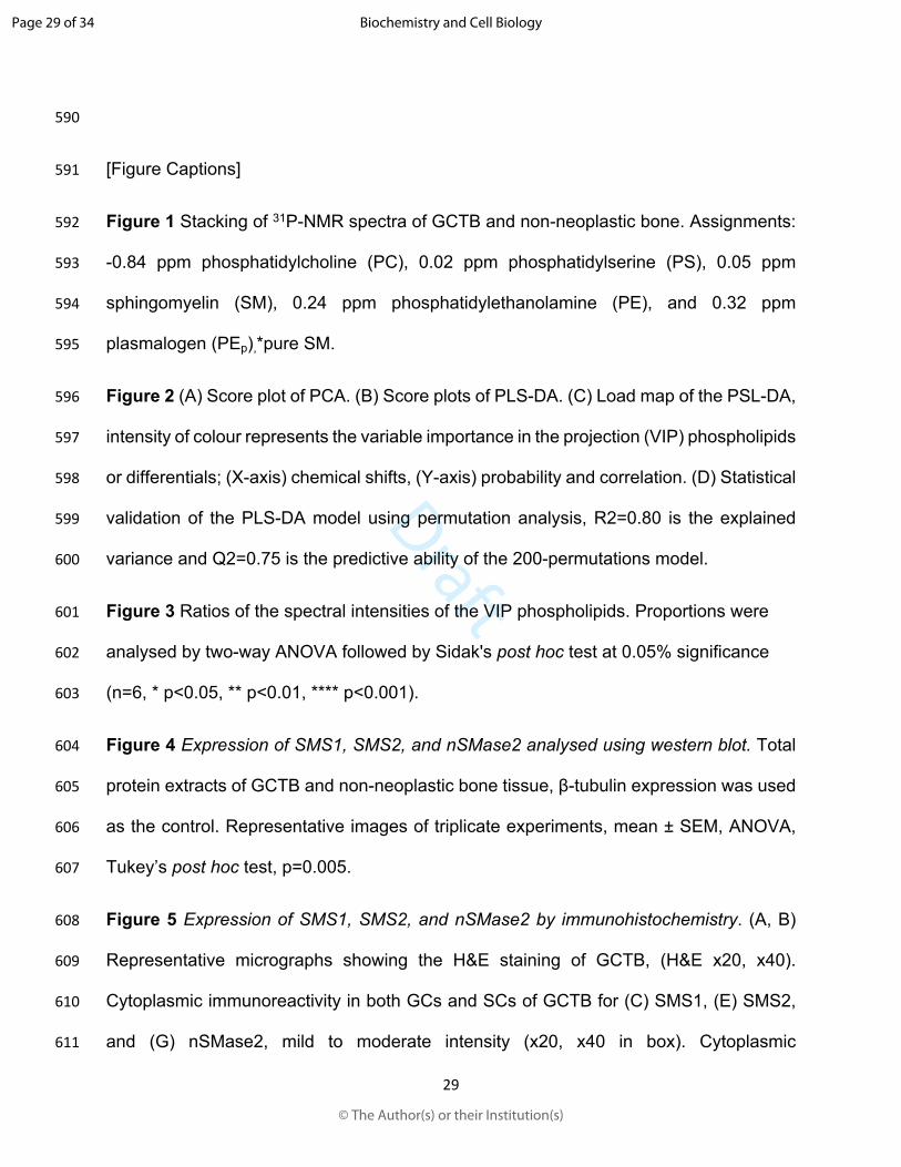
460 Lipoproteins in Health and Metabolic Disorders. Trends Endocrinol. Metab. 28:
461 506-518. doi:10.1016/j.tem.2017.03.005
462 Kim, W. J., Okimoto, R. A., Purton, L. E., Goodwin, M., Haserlat, S. M., Dayyani, F.,
463 Sweetser, D. A., McClatchey, A. I., Bernard, O. A., Look, A. T., Bell, D. W.,
464 Scadden, D. T., and Haber, D. A. 2008. Mutations in the neutral sphingomyelinase
465 gene SMPD3 implicate the ceramide pathway in human leukemias. Blood. 111:
466 4716-4722. doi:10.1182/blood-2007-10-113068
467 Kolesnick, R. 2002. The therapeutic potential of modulating the ceramide/sphingomyelin
468 pathway. J. Clin. Invest. 110: 3-8. doi:10.1172/JCI16127
469 Kosaka, N., Iguchi, H., Hagiwara, K., Yoshioka, Y., Takeshita, F., and Ochiya, T. 2013.
470 Neutral sphingomyelinase 2 (nSMase2)-dependent exosomal transfer of
471 angiogenic microRNAs regulate cancer cell metastasis. J. Biol. Chem. 288: 10849-
472 10859. doi:10.1074/jbc.M112.446831
473 Kozar, N., Kruusmaa, K., Bitenc, M., Argamasilla, R., Adsuar, A., Goswami, N., Arko, D.,
474 and Takac, I. 2018. Metabolomic profiling suggests long chain ceramides and
475 sphingomyelins as a possible diagnostic biomarker of epithelial ovarian cancer.
476 Clin. Chim. Acta. 481: 108-114. doi:10.1016/j.cca.2018.02.029
477 Lafont, E., Kitatani, K., Okazaki, T., and Segui, B. 2011. Regulation of death and growth
478 signals at the plasma membrane by sphingomyelin synthesis: implications for
Page 22 of 34
© The Author(s) or their Institution(s)
Biochemistry and Cell Biology
Draft
23
479 hematological malignancies. Recent Pat. Anticancer. Drug Discov. 6: 324-333.
480 doi:10.2174/157489211796957801
481 Li, Z., Guan, M., Lin, Y., Cui, X., Zhang, Y., Zhao, Z., and Zhu, J. 2017. Aberrant Lipid
482 Metabolism in Hepatocellular Carcinoma Revealed by Liver Lipidomics. Int. J. Mol.
483 Sci. 18. doi:10.3390/ijms18122550
484 Li, Z., Wu, G., van der Veen J. N., Hermansson, M., Vance, D. E. 2014.
485 Phosphatidylcholine metabolism and choline kinase in human osteoblasts.
486 Biochim. Biophys. Acta. 1841: 859-67. doi: 10.1016/j.bbalip.2014.02.004
487 Liang, S., Ren, Z., Han, X., Yang, J., Shan, L., Li L., Wang, B., Zhang, Q., Mu, T., Chen,
488 K., Xiong, S., Wang, G. 2015. PLA2G16 Expression in Human Osteosarcoma Is
489 Associated with Pulmonary Metastasis and Poor Prognosis. PLoS One.
490 18:e0127236. doi: 10.1371/journal.pone.0127236.
491 Lin, J. L., Wu, Y. H., Shi, Y. F., Lin, H., Nisar, M., Meftah, Z., Xu, C., Chen, J. X., and
492 Wang, X. Y. 2019. Survival and prognosis in malignant giant cell tumor of bone: A
493 population-based analysis from 1984 to 2013. J. Bone Oncol. 19: 100260.
494 doi:10.1016/j.jbo.2019.100260
495 Lordan, R., Tsoupras, A., and Zabetakis, I. 2017. Phospholipids of Animal and Marine
496 Origin: Structure, Function, and Anti-Inflammatory Properties. Molecules. 22.
497 doi:10.3390/molecules22111964
498 Maguer-Satta, V. 1998. CML and apoptosis: the ceramide pathway. Hematol. Cell. Ther.
499 40: 233-236. Retrieved from https://www.ncbi.nlm.nih.gov/pubmed/9844818
500 Martínez-López, F. J., Bañuelos-Hernández, A. E., Becerra-Martínez, E., Santini-Araujo,
501 E., Amaya-Zepeda, R. A., Pérez-Hernández, E., and Pérez-Hernández, N. 2017.
Page 23 of 34
© The Author(s) or their Institution(s)
Biochemistry and Cell Biology
Draft
24
502 1H NMR metabolomic signatures related to giant cell tumor of the bone. RSC
503 Advances. 7: 45385-45392. doi:10.1039/C7RA07138H
504 Montgomery, C., Couch, C., Emory, C. L., and Nicholas, R. 2019. Giant Cell Tumor of
505 Bone: Review of Current Literature, Evaluation, and Treatment Options. J. Knee
506 Surg. 32: 331-336. doi:10.1055/s-0038-1675815
507 Muheremu, A., and Niu, X. 2014. Pulmonary metastasis of giant cell tumor of bones.
508 World J. Surg. Oncol. 12: 261. doi:10.1186/1477-7819-12-261
509 Nagahashi, M., Tsuchida, J., Moro, K., Hasegawa, M., Tatsuda, K., Woelfel, I. A., Takabe,
510 K., and Wakai, T. 2016. High levels of sphingolipids in human breast cancer. J.
511 Surg. Res. 204: 435-444. doi:10.1016/j.jss.2016.05.022
512 Noh, B. J., and Park, Y. K. 2018. Giant cell tumor of bone: updated molecular
513 pathogenesis and tumor biology. Hum. Pathol. 81: 1-8.
514 doi:10.1016/j.humpath.2018.06.017
515 Ogretmen, B. 2018. Sphingolipid metabolism in cancer signalling and therapy. Nat. Rev.
516 Cancer. 18: 33-50. doi:10.1038/nrc.2017.96
517 Osaki, M., and Okada, F. 2019. Exosomes and Their Role in Cancer Progression. Yonago
518 Acta. Med. 62: 182-190. doi:10.33160/yam.2019.06.002
519 Pegtel, D. M., and Gould, S. J. 2019. Exosomes. Annu. Rev. Biochem. 88: 487-514.
520 doi:10.1146/annurev-biochem-013118-111902
521 Perut, F., Roncuzzi, L., and Baldini, N. 2019. The Emerging Roles of Extracellular
522 Vesicles in Osteosarcoma. Front. Oncol. 9: 1342. doi:10.3389/fonc.2019.01342
523 Pierce, J.M. and Komoriski R.A. 1993. Resolution of Phospholipid Molecular Species by
524 31P NMR. Magn. Res. Med. 29: 724-731. doi:10.1002/mrm.1910290603
Page 24 of 34
© The Author(s) or their Institution(s)
Biochemistry and Cell Biology
Draft
25
525 Ricci, C., Onida, F., and Ghidoni, R. 2006. Sphingolipid players in the leukemia arena.
526 Biochim. Biophys. Acta. 1758: 2121-2132. doi:10.1016/j.bbamem.2006.06.016
527 Santini-Araujo E., K. K. R., Bertoni F., Yong-Koo P. 2015. Tumors and Tumor-Like
528 Lesions of Bone. For Surgical Pathologists, Orthopedic Surgeons and Radiologists
529 (Springer Ed.). London.
530 Schaefer, I. M., and Hornick, J. L. 2018. Diagnostic Immunohistochemistry for Soft Tissue
531 and Bone Tumors: An Update. Adv. Anat. Pathol. 25: 400-412.
532 doi:10.1097/PAP.0000000000000204
533 Separovic, D., Shields, A. F., Philip, P. A., Bielawski, J., Bielawska, A., Pierce, J. S., and
534 Tarca, A. L. 2017. Altered Levels of Serum Ceramide, Sphingosine and
535 Sphingomyelin Are Associated with Colorectal Cancer: A Retrospective Pilot
536 Study. Anticancer Res. 37: 1213-1218. doi:10.21873/anticanres.11436
537 Shakor, A. B., Taniguchi, M., Kitatani, K., Hashimoto, M., Asano, S., Hayashi, A., Nomura,
538 K., Bielawski, J., Bielawska, A., Watanabe, K., Kobayashi, T., Igarashi, Y.,
539 Umehara, H., Takeya, H., and Okazaki, T. 2011. Sphingomyelin synthase 1-
540 generated sphingomyelin plays an important role in transferrin trafficking and cell
541 proliferation. J. Biol. Chem. 286: 36053-36062. doi:10.1074/jbc.M111.228593
542 Shanbhogue, P, Hannun, Y.A., 2020 Exploring the Therapeutic Landscape of
543 Sphingomyelinases. Handb Exp Pharmacol. 259:19-47. doi:
544 10.1007/164_2018_179
545 Stoffel, W., Hammels, I., Jenke, B., Schmidt-Soltau, I., and Niehoff, A. 2019. Neutral
546 Sphingomyelinase 2 (SMPD3) Deficiency in Mice Causes Chondrodysplasia with
Page 25 of 34
© The Author(s) or their Institution(s)
Biochemistry and Cell Biology
Draft
26
547 Unimpaired Skeletal Mineralization. Am. J. Pathol. 189: 1831-1845.
548 doi:10.1016/j.ajpath.2019.05.008
549 Stoffel, W., Jenke, B., Holz, B., Binczek, E., Gunter, R. H., Knifka, J., Koebke, J., and
550 Niehoff, A. 2007. Neutral sphingomyelinase (SMPD3) deficiency causes a novel
551 form of chondrodysplasia and dwarfism that is rescued by Col2A1-driven smpd3
552 transgene expression. Am. J. Pathol. 171: 153-161.
553 doi:10.2353/ajpath.2007.061285
554 Sun, G. Y., and Leung, B. S. 1974. Phospholipids and acyl groups of subcellular
555 membrane fractions from human intracranial tumors. J. Lipid Res. 15: 423-431.
556 Retrieved from https://www.ncbi.nlm.nih.gov/pubmed/4369007
557 Takeda, Y., Tashima, M., Takahashi, A., Uchiyama, T., and Okazaki, T. 1999. Ceramide
558 generation in nitric oxide-induced apoptosis. Activation of magnesium-dependent
559 neutral sphingomyelinase via caspase-3. J. Biol. Chem. 274: 10654-10660.
560 doi:10.1074/jbc.274.15.10654
561 Tani, M., and Hannun, Y. A. 2007. Neutral sphingomyelinase 2 is palmitoylated on
562 multiple cysteine residues. Role of palmitoylation in subcellular localization. J. Biol.
563 Chem. 282: 10047-10056. doi:10.1074/jbc.M611249200
564 van Blitterswijk, W. J., van der Luit, A. H., Veldman, R. J., Verheij, M., and Borst, J. 2003.
565 Ceramide: second messenger or modulator of membrane structure and dynamics?
566 Biochem. J. 369: 199-211. doi:10.1042/BJ20021528
567 Varghese, F., Bukhari, A. B., Malhotra, R., and De, A. 2014. IHC Profiler: an open source
568 plugin for the quantitative evaluation and automated scoring of
Page 26 of 34
© The Author(s) or their Institution(s)
Biochemistry and Cell Biology
Draft
27
569 immunohistochemistry images of human tissue samples. PLoS One. 9: e96801.
570 doi:10.1371/journal.pone.0096801
571 Wu, P. F., Tang, J. Y., and Li, K. H. 2015. RANK pathway in giant cell tumor of bone:
572 pathogenesis and therapeutic aspects. Tumour Biol. 36: 495-501.
573 doi:10.1007/s13277-015-3094-y
574 Yamamoto, H., Ishihara, S., Toda, Y., and Oda, Y. 2020. Histone H3.3 mutation in giant
575 cell tumor of bone: an update in pathology. Med. Mol. Morphol. 53: 1-6.
576 doi:10.1007/s00795-019-00238-1
577 Yoshikawa, Y., Yoshizawa, T., Domae, E., Hirai, Y., Kamada, A., Okazaki, T., and Ikeo,
578 T. 2019. Knockdown of sphingomyelin synthase 2 inhibits osteoclastogenesis by
579 decreasing RANKL expression in mouse primary osteoblasts. Biomed. Res. 40:
580 189-196. doi:10.2220/biomedres.40.189
581 Zheng, K., Chen, Z., Feng, H., Chen, Y., Zhang, C., Yu, J., Luo, Y., Zhao, L., Jiang, X.,
582 and Shi, F. 2019. Sphingomyelin synthase 2 promotes an aggressive breast
583 cancer phenotype by disrupting the homoeostasis of ceramide and sphingomyelin.
584 Cell Death Dis. 10: 157. doi:10.1038/s41419-019-1303-0
585
Page 27 of 34
© The Author(s) or their Institution(s)
Biochemistry and Cell Biology
Draft
28
586
587 Table 1 Clinical characteristics of patients with GCTB and controls
588
GCTB Controls
Number 6 6
Mean age 34 45
Male/female (4/2) (3/3)
Tumour localisation Femur and tibia NA
Enneking staging IA NA
589
Page 28 of 34
© The Author(s) or their Institution(s)
Biochemistry and Cell Biology
Draft
29
590
591 [Figure Captions]
592 Figure 1 Stacking of 31P-NMR spectra of GCTB and non-neoplastic bone. Assignments:
593 -0.84 ppm phosphatidylcholine (PC), 0.02 ppm phosphatidylserine (PS), 0.05 ppm
594 sphingomyelin (SM), 0.24 ppm phosphatidylethanolamine (PE), and 0.32 ppm
595 plasmalogen (PEp),*pure SM.
596 Figure 2 (A) Score plot of PCA. (B) Score plots of PLS-DA. (C) Load map of the PSL-DA,
597 intensity of colour represents the variable importance in the projection (VIP) phospholipids
598 or differentials; (X-axis) chemical shifts, (Y-axis) probability and correlation. (D) Statistical
599 validation of the PLS-DA model using permutation analysis, R2=0.80 is the explained
600 variance and Q2=0.75 is the predictive ability of the 200-permutations model.
601 Figure 3 Ratios of the spectral intensities of the VIP phospholipids. Proportions were
602 analysed by two-way ANOVA followed by Sidak's post hoc test at 0.05% significance
603 (n=6, * p<0.05, ** p<0.01, **** p<0.001).
604 Figure 4 Expression of SMS1, SMS2, and nSMase2 analysed using western blot. Total
605 protein extracts of GCTB and non-neoplastic bone tissue, β-tubulin expression was used
606 as the control. Representative images of triplicate experiments, mean ± SEM, ANOVA,
607 Tukey’s post hoc test, p=0.005.
608 Figure 5 Expression of SMS1, SMS2, and nSMase2 by immunohistochemistry. (A, B)
609 Representative micrographs showing the H&E staining of GCTB, (H&E x20, x40).
610 Cytoplasmic immunoreactivity in both GCs and SCs of GCTB for (C) SMS1, (E) SMS2,
611 and (G) nSMase2, mild to moderate intensity (x20, x40 in box). Cytoplasmic
Page 29 of 34
© The Author(s) or their Institution(s)
Biochemistry and Cell Biology
Draft
30
612 immunoreactivity of osteocytes in non-neoplastic bone for (D) SMS1, (F) SMS2, and (H)
613 nSMase2 (x20, x40 in box). The histograms indicate density of the pixels (X-axis) versus
614 the pixel count (Y-axis).
615
616
617
618 Figure 1 Stacking of 31P-NMR spectra of GCTB and non-neoplastic bone. Assignments:
619 -0.84 ppm phosphatidylcholine (PC), 0.02 ppm phosphatidylserine (PS), 0.05 ppm
620 sphingomyelin (SM), 0.24 ppm phosphatidylethanolamine (PE), and 0.32 ppm
621 plasmalogen (PEp),*pure SM.
622
623
624
Page 30 of 34
© The Author(s) or their Institution(s)
Biochemistry and Cell Biology
Draft
31
625
626
627
628 Figure 2 (A) Score plot of PCA. (B) Score plots of PLS-DA. (C) Load map of the PSL-DA,
629 intensity of colour represents the variable importance in the projection (VIP) phospholipids
630 or differentials; (X-axis) chemical shifts, (Y-axis) probability and correlation. (D) Statistical
631 validation of the PLS-DA model using permutation analysis, R2=0.80 is the explained
632 variance and Q2=0.75 is the predictive ability of the 200-permutations model.
633
634
635
636
Page 31 of 34
© The Author(s) or their Institution(s)
Biochemistry and Cell Biology

Draft
32
637
638
Rat
io
PS/PC
SM/PCPE/PC
PEp/PC0.0
0.2
0.4
0.6
0.8
ControlGCTB
**
****
* NS
639 Figure 3 Ratios of the spectral intensities of the VIP phospholipids. Proportions were
640 analysed by two-way ANOVA followed by Sidak's post hoc test at 0.05% significance
641 (n=6, * p<0.05, ** p<0.01, **** p<0.001).
642
Page 32 of 34
© The Author(s) or their Institution(s)
Biochemistry and Cell Biology
Draft
33
643
644
645 Figure 4 Expression of SMS1, SMS2, and nSMase2 analysed using western blot. Total
646 protein extracts of GCTB and non-neoplastic bone tissue, β-tubulin expression was used
647 as the control. Representative images of triplicate experiments, mean ± SEM, ANOVA,
648 Tukey’s post hoc test, p=0.005.
649
650
651
Page 33 of 34
© The Author(s) or their Institution(s)
Biochemistry and Cell Biology
Draft
34
652
653 Figure 5 Expression of SMS1, SMS2, and nSMase2 by immunohistochemistry. (A, B) 654 Representative micrographs showing the H&E staining of GCTB, (H&E x20, x40). 655 Cytoplasmic immunoreactivity in both GCs and SCs of GCTB for (C) SMS1, (E) SMS2, 656 and (G) nSMase2, mild to moderate intensity (x20, x40 in box). Cytoplasmic 657 immunoreactivity of osteocytes in non-neoplastic bone for (D) SMS1, (F) SMS2, and (H) 658 nSMase2 (x20, x40 in box). The histograms indicate density of the pixels (X-axis) versus 659 the pixel count (Y-axis).
Page 34 of 34
© The Author(s) or their Institution(s)
Biochemistry and Cell Biology