TSLP directly impairs pulmonary Treg function: association with aberrant tolerogenic immunity in...
11
RESEARCH Open Access TSLP directly impairs pulmonary Treg function: association with aberrant tolerogenic immunity in asthmatic airway Khoa D Nguyen, Christopher Vanichsarn, Kari C Nadeau * Abstract Background: Even though thymic stromal lymphopoietin (TSLP) has been implicated in the development of allergic inflammation, its influence on immune tolerance mediated by regulatory T cells (Treg) have not been explored. We aimed to dissect the influence of TSLP on immunosuppressive activities of Treg and its potential consequences in human allergic asthma. Methods: In vitro culture system was utilized to study the effects of TSLP on human Treg. The functional competency of pulmonary Treg from a cohort of 15 allergic asthmatic, 15 healthy control, and 15 non-allergic asthmatic subjects was also evaluated by suppression assays and flow cytometric analysis. Results: Activated pulmonary Treg expressed TSLP-R and responded to TSLP-mediated activation of STAT5. TSLP directly and selectively impaired IL-10 production of Treg and inhibited their suppressive activity. In human allergic asthma, pulmonary Treg exhibited a significant decrease in suppressive activity and IL-10 production compared to healthy control and non-allergic asthmatic counterparts. These functional alterations were associated with elevated TSLP expression in bronchoaveolar lavage fluid (BAL) of allergic asthmatic subjects. Furthermore, allergic asthmatic BAL could suppress IL-10 production by healthy control pulmonary Treg in a TSLP-dependent manner. Conclusions: These results provide the first evidences for a direct role of TSLP in the regulation of suppressive activities of Treg. TSLP mediated inhibition of Treg function might present a novel pathologic mechanism to dampen tolerogenic immune responses in inflamed asthmatic airway. Background Thymic stromal lymphopoietin (TSLP) was initially identified as being involved in lymphocyte development [1,2]. Subsequently, it was implicated in the induction of the pro-allergic phenotype in CD4+ effector T cells (Teff) [3]. Even though most studies to date have focused on the indirect mediation of allergic responses of TSLP via dendritic cells [4], it has been suggested that TSLP could directly expand CD4+ and CD8+ Teff [5, 6[. Recent studies revealed that TSLP could directly drive allergic responses of CD4+ Teff [7]. Studies of experimental models of asthma also indicated that TSLP-R-deficient animals failed to develop airway inflammation [4,8]. Conversely, over-expression of TSLP appeared to aggravate asthma symptoms [9]. Altogether, these evidences strongly suggested TSLP as a positive modulator of Th-2-biased inflammation. Regulatory T cells (Treg) have emerged as a key regu- lator of inflammatory responses in allergic disorders [10,11]. Treg are CD4+CD25hiCD127lo/-Foxp3+ cells that possess suppressive activities against cytokine pro- duction and proliferation of Teff [12]. In studies of human allergic asthma (AA), decreased frequency and diminished suppressive activity of pulmonary Treg have been documented [13]. Furthermore, murine data sug- gested that Treg-mediated suppression reversed airway hyper-responsiveness, inflammation, and remodeling [14]. Immuno-suppressive cytokines such as IL-10 and TGF-b have been implicated in immune regulation by Treg during airway inflammation. For instance, co- expression of IL-10 and TGF- b by Treg allowed complete inhibition of airway hyper-reactivity [15]. In addition, suppression of Der-p1 and Mycobacterium * Correspondence: [email protected] Department of Pediatrics, Stanford University, Stanford, CA, USA Nguyen et al. Allergy, Asthma & Clinical Immunology 2010, 6:4 http://www.aacijournal.com/content/6/1/4 ALLERGY, ASTHMA & CLINICAL IMMUNOLOGY © 2010 Nguyen et al; licensee BioMed Central Ltd. This is an Open Access article distributed under the terms of the Creative Commons Attribution License (http://creativecommons.org/licenses/by/2.0), which permits unrestricted use, distribution, and reproduction in any medium, provided the original work is properly cited.
Transcript of TSLP directly impairs pulmonary Treg function: association with aberrant tolerogenic immunity in...
RESEARCH Open Access
TSLP directly impairs pulmonary Treg function:association with aberrant tolerogenic immunity inasthmatic airwayKhoa D Nguyen, Christopher Vanichsarn, Kari C Nadeau*
Abstract
Background: Even though thymic stromal lymphopoietin (TSLP) has been implicated in the development ofallergic inflammation, its influence on immune tolerance mediated by regulatory T cells (Treg) have not beenexplored. We aimed to dissect the influence of TSLP on immunosuppressive activities of Treg and its potentialconsequences in human allergic asthma.
Methods: In vitro culture system was utilized to study the effects of TSLP on human Treg. The functionalcompetency of pulmonary Treg from a cohort of 15 allergic asthmatic, 15 healthy control, and 15 non-allergicasthmatic subjects was also evaluated by suppression assays and flow cytometric analysis.
Results: Activated pulmonary Treg expressed TSLP-R and responded to TSLP-mediated activation of STAT5. TSLPdirectly and selectively impaired IL-10 production of Treg and inhibited their suppressive activity. In human allergicasthma, pulmonary Treg exhibited a significant decrease in suppressive activity and IL-10 production compared tohealthy control and non-allergic asthmatic counterparts. These functional alterations were associated with elevatedTSLP expression in bronchoaveolar lavage fluid (BAL) of allergic asthmatic subjects. Furthermore, allergic asthmaticBAL could suppress IL-10 production by healthy control pulmonary Treg in a TSLP-dependent manner.
Conclusions: These results provide the first evidences for a direct role of TSLP in the regulation of suppressiveactivities of Treg. TSLP mediated inhibition of Treg function might present a novel pathologic mechanism todampen tolerogenic immune responses in inflamed asthmatic airway.
BackgroundThymic stromal lymphopoietin (TSLP) was initiallyidentified as being involved in lymphocyte development[1,2]. Subsequently, it was implicated in the induction ofthe pro-allergic phenotype in CD4+ effector T cells(Teff) [3]. Even though most studies to date havefocused on the indirect mediation of allergic responsesof TSLP via dendritic cells [4], it has been suggestedthat TSLP could directly expand CD4+ and CD8+ Teff[5, 6[. Recent studies revealed that TSLP could directlydrive allergic responses of CD4+ Teff [7]. Studies ofexperimental models of asthma also indicated thatTSLP-R-deficient animals failed to develop airwayinflammation [4,8]. Conversely, over-expression of TSLPappeared to aggravate asthma symptoms [9]. Altogether,
these evidences strongly suggested TSLP as a positivemodulator of Th-2-biased inflammation.Regulatory T cells (Treg) have emerged as a key regu-
lator of inflammatory responses in allergic disorders[10,11]. Treg are CD4+CD25hiCD127lo/-Foxp3+ cellsthat possess suppressive activities against cytokine pro-duction and proliferation of Teff [12]. In studies ofhuman allergic asthma (AA), decreased frequency anddiminished suppressive activity of pulmonary Treg havebeen documented [13]. Furthermore, murine data sug-gested that Treg-mediated suppression reversed airwayhyper-responsiveness, inflammation, and remodeling[14]. Immuno-suppressive cytokines such as IL-10 andTGF-b have been implicated in immune regulation byTreg during airway inflammation. For instance, co-expression of IL-10 and TGF-b by Treg allowedcomplete inhibition of airway hyper-reactivity [15]. Inaddition, suppression of Der-p1 and Mycobacterium
* Correspondence: [email protected] of Pediatrics, Stanford University, Stanford, CA, USA
Nguyen et al. Allergy, Asthma & Clinical Immunology 2010, 6:4http://www.aacijournal.com/content/6/1/4 ALLERGY, ASTHMA & CLINICAL
IMMUNOLOGY
© 2010 Nguyen et al; licensee BioMed Central Ltd. This is an Open Access article distributed under the terms of the Creative CommonsAttribution License (http://creativecommons.org/licenses/by/2.0), which permits unrestricted use, distribution, and reproduction inany medium, provided the original work is properly cited.
vaccae-induced airway inflammation was dependent onIL-10 and/or TGF-b production by Foxp3+ Treg[16,17]. Therefore, modulation of the expression ofthese effector molecules by pulmonary Treg might playa critical role in regulating airway immune responses.TSLP has been implicated in the development of Treg
[18]. Disruption of TSLP signaling by TLSP receptordeletion impaired intra-thymic generation of Treg butdid not affect their peripheral repertoire [19]. Consistentwith these results, blocking TSLP-R led to a delayedfunctional maturation of thymic Treg [20]. Since TSLPand Treg have been suggested to play opposite modula-tory roles in allergic inflammation, we aimed to explorethe potential regulatory interaction between TSLP andTreg. Here we provided data that link TSLP signaling tothe inhibition of Treg function as well as its implicationsin the context of peripheral tolerance in AA.
MethodsHuman subjectsThe study was approved by the Stanford AdministrativePanel on Human Subjects in Medical Research. Studypopulation included 15 AA subjects, 15 HC subjects, and15 NA subjects. All subjects signed informed consentforms before participating in the study. Asthma diagnosiswas established by evidences of episodic and partiallyreversible airflow obstruction or airway hyper-respon-siveness, and exclusion of alternative diagnoses (NHLBIExpert Panel Report-3 2007). All patients had been fol-lowed for at least 6 months by a board certified Asthma,Allergy, and Immunology specialist at Stanford to ensurethe diagnosis was correct. AA subjects were distinguishedfrom NA subjects based on history of allergic symptoms,elevated blood IgE levels (above 50 IU/ml), and positiveskin tests to allergens. Spirometry was performed by afully qualified respiratory therapist and study coordina-tor, both of whom have over 15 years of experiences inasthma studies. Patients with FEV1 below 60% were con-sidered severe. Those in the range of 60%-80% were con-sidered moderate and those with FEV1 above 80% wereconsidered mild. Comprehensive clinical data were col-lected at each patient visit including history, diseaseseverity, medication status, common allergens, IgE level,and FEV1 (Additional file 1, Table S1). HC were definedas non-smoking subjects greater than 17 years of agewith a total serum IgE of less than 25 IU/ml, negativeskin testing (as compared to positive histamine control),and no evidence of lung disease or allergic symptoms. Inaddition, there was no evidence of obstructive or restric-tive lung disease for HC on spirometry testing.
Cell isolationBAL samples were collected with a standardized proto-col for clinical research at Lucile Packard’s Children
Hospital. After being collected, BAL samples (approxi-mately 3 ml for each subject) were spun down at 1800rpm for 15 minutes. Undiluted BAL supernatants werecollected and filtered with 45 μm filters (BD Bios-ciences) and stored at -80°C until analysis. Cell pelletswere subjected to downstream isolation. UntouchedCD3+ T cells from BAL were first isolated by depletingB cells, monocytes/dendritic cells, NK cells, and granu-locytes with pan T cell selection kit II (Miltenyi). CD4+T cells were then isolated from these CD3+ T cells withCD4-microbeads (Miltenyi). Circulating CD4+ T cellswere isolated from peripheral blood by CD4+ T cellRosette kit (Stemcell) to deplete other cell lineagesincluding B cells, monocytes/dendritic cells, NK cells,granulocytes, and non CD4+ T cells. Purified CD4+ Tcell fraction, which contained virtually no DR+ antigenpresenting cells (data not shown), was stained withCD25 (BD Biosciences) and CD127 (Biolegend) antibo-dies and sorted for CD4+CD25hiCD127lo/- Treg andTeff. Cells were rested in RPMI + 10% FBS + 1% L-Glu-tamine (complete media) after isolation. All procedureswere performed with manufacturers’ standard protocols.
Cell stimulationFor cytokine priming experiments, cells were cultured at1*105 cells per ml for 18 hours at 37°C in completemedia in the presence or absence of 50 pg/ml recombi-nant IL-2, IL-7, and TSLP (Peprotech). For BAL incuba-tion, pulmonary Treg from the same HC subject werecultured at 1*105 cells per 900 μl for 18 hours at 37°Cin complete media in the presence of BAL from differ-ent AA and NA subjects. 100 μL of BAL supernatantswere added to 900 μl of cell suspension. To determinethe role of IL-10 in Treg-mediated suppression, recom-binant IL-10 (Peprotech) or IL-10-blocking antibody(R&D) was added to suppression assays at differentdoses. Optimal volumes of BAL and cytokine/blockingantibody concentrations were experimentally deter-mined. For neutralization experiments, blocking TSLP-Rand isotype control antibody (R&D) were introduced tocell cultures at 10 μg/ml for 0.5 hours at 37°C beforeBAL supernatants were added. Optimal concentrationsof blocking reagents were experimentally determined orused at doses recommended by the manufacturers.
Immune phenotyping via FACS and ELISAPhenotypes of immune cells were detected with antibo-dies against CD3, CD4, CD25, CD127, Foxp3, CTLA-4,LAG-3, OX40, and CD40L (Biolegend). For cytokine sti-mulation, Treg and Teff were cultured at 1*105 cells perml for 2 hours after isolation. After 2 hours, 1 ml of cellsuspension was stimulated with 50 ng/ml PMA (Sigma)and 1 μg/ml Ionomycin (Sigma) for 5 hours in the pre-sence of Brefeldin A (diluted 1× solution was added in
Nguyen et al. Allergy, Asthma & Clinical Immunology 2010, 6:4http://www.aacijournal.com/content/6/1/4
Page 2 of 11
the last 2.5 hours, Biolegend) for intracellular cytokinestaining or in the absence of Brefeldin A for ELISA.Staining for membrane-bound TGF-b was performedwith standard surface staining protocols. For intracellu-lar cytokine detection, stimulated cells were fixed andpermeablized in 200 μL of Cytofix/Cytoperm solution(BD Biosciences) and then stained with antibodiesagainst IL-4, IL-10, and TNF-a (Biolegend) in Permwashsolution (BD Biosciences) in a final volume of 100 μL(BD Biosciences). Data acquisition threshold was set onforward scatter channel to exclude dead cells and debriswith very low size. Compensation of flow cytometricdata was performed electronically with Flow Jo (Trees-tar) for standardization. Quantitation of secreted cyto-kines was performed with ELISA kits for IL-4, IL-10,TGF-b (R&D), and TSLP (eBioscience). Total proteinamount in BAL supernatants was determined by Brad-ford assays. TSLP level in BAL samples was normalizedto total protein amount. All procedures were performedwith manufacturers’ standard protocols.
Quantitation of TSLP-R mRNARNA was isolated using RNeasy kits (Qiagen) accordingto the manufacturer’s protocol. Similar cell numbers(1*105 for peripheral blood cells and 5*104 for BALcells) were used for each subject. For cDNA synthesis,500 ng total RNA was transcribed with cDNA transcrip-tion reagents (Applied Biosystems) using random hex-amers, according to the manufacturer’s protocol. Geneexpression was measured in real-time using primers andother reagents purchased from Applied Biosystems andSuperarray. All PCR assays were performed in tripli-cates. Data was presented as relative fold expression ofTSLP-R to the expression of the housekeeping geneb2-microglobulin.
Detection of phosphorylated signal transducer andactivator of transcription 5 (pSTAT5)T cells were cultured at 1*105 cells per ml in completemedia at 37°C for 18 hours after isolation. After 18hours, 1 μl recombinant IL-2, IL-7, and TSLP (Pepro-tech) were added to V-bottom 96-well plates and 100 μlof cells were added to these wells with cytokines (finalcytokine concentrations were 50 pg/ml). Optimal con-centration and stimulation duration were experimentallydetermined. For ELISA, cells were lysed after being sti-mulated for 15 minutes. Lysates were analyzed forpSTAT5 by phospho-ELISA kits (R&D). For phospho-flow cytometry, cells were stimulated for 15 minutes at37°C before being fixed with 10 μL of 10% paraformal-dehyde at 37°C. Fixed cells were washed with PBS andpermeablized with ice-cold methanol for 10 minutes.Permeablized cells were washed again with PBS andstained with pSTAT5 antibody (BD Biosciences). All
procedures were performed with manufacturers’ stan-dard protocols. JAK3 inhibitor (WHI-P131, Calbiochem)was used at optimal concentration (78 μM) recom-mended by the manufacturer.
Suppression assaysStandard thymidine-based suppression assays were per-formed to analyze Treg function. Treg and Teff werecultured at 3,750 cells per well in complete media withallogeneic irradiated CD3-depleted peripheral bloodmononuclear cells (antigen presenting cells or APC), at37,500 cells per well (1:1:10 ratio). Assays with 1:4:10ratio of Treg:Teff: APC were also performed. Anti-CD3antibodies (clone UCHT1, BD) were pre-coated onU-bottom 96 well plates at 5 μg/ml overnight at 37°Cbefore suppression assays were performed. Additionalmedia was added so the final volume in each well was200 μl. On day 6, cells were pulsed with 1 μCi thymi-dine (25 μl) per well and harvested on day 7 with aTomtec cell harvester. Thymidine incorporation wasdetermined using a 1450 microbeta Wallac Trilux liquidscintillation counter. Stimulation assays were set upsimilarly with allogeneic irradiated APC and only onetype of T cells. All assays were performed in triplicates.
Statistical analysisAll statistical procedures were performed with Prismsoftware (GraphPad). Non-parametric statistical testswere used for analysis of cohorts with small samplesizes (15 or less). Differences with p < 0.05 were consid-ered statistically significant.
Results and DiscussionActivated Treg express TSLP-R and directly respond toTSLP-mediated activation of STAT5TSLP-R expression was first examined on purified CD3/CD28-activated pulmonary T cell subsets from healthycontrol (HC) subjects as described previously [5]. mRNAexpression of TSLP-R was significantly higher in pulmon-ary Treg compared to pulmonary Teff (Figure 1A). Flowcytometry (FACS) analysis showed that, compared to pul-monary Teff, a significantly higher percentage of pulmon-ary Treg, express TSLP-R (Figure 1A). Consistent withthese results, expression of TSLP-R positively correlatedwith CD25 expression and negative correlated with CD127expression by tri-color FACS staining (Additional file 1,Figure S1A). TSLP signaling requires two receptor compo-nents, IL-7Ra and TSLP-R 21, the former of which wasexpressed at low level on Treg. Thus, to determine whetherthis pattern of high TSLP-R and low IL-7Ra expressionwas sufficient for TSLP signaling in Treg, we used phos-pho-ELISA, which allows measurement of protein expres-sion in rare cell subsets, to quantify the expressionof phosphorylated STAT5 (pSTAT5) by purified
Nguyen et al. Allergy, Asthma & Clinical Immunology 2010, 6:4http://www.aacijournal.com/content/6/1/4
Page 3 of 11

CD3/CD28-activated pulmonary Treg in response torecombinant TSLP. Our analysis showed that level ofpSTAT5 in TSLP-stimulated pulmonary Treg was signifi-cantly elevated compared to that of un-stimulated cells(Figure 1B). The responsiveness of pulmonary Treg toTSLP was confirmed with phospho-flow cytometry (Figure1B). While TSLP and IL-7 both signal via IL-7Ra, JAK3phosphorylation was observed only in response to IL-7[21-23]. Thus, signaling events triggered by binding of IL7to IL-7Ra (the only signaling component in IL-7 receptorcomplex), but not binding of TSLP to TSLP-R (a part ofTSLP receptor complex, which consists of two signalingcomponents IL-7Ra and TSLP-R), resulted in JAK3 activa-tion and subsequent induction of phosphorylated STAT5.To determine which receptor component (IL-7Ra vs.TSLP-R) in TSLP receptor complex was responsible foractivation of STAT5 in response to TSLP, we introduced a
JAK3 inhibitor into the stimulation assays. Phospho ELISAshowed that in the presence of the JAK3 inhibitor, STAT5activation was abrogated in response to IL-7 but not TSLP(Additional file 1, Figure S1B). These results suggested thatsignaling via TLSP-R, but not IL-7Ra, in response to TSLPwas likely to be required for the induction of phosphory-lated STAT5 in Treg. Consistent with previous findings 5,activated Teff could response to TSLP (Figure 1C). Similarresults were also observed in circulating Treg and Teff(Additional file 1, Figure S2). Collectively, our resultsdemonstrated that Treg expressed functional TSLP-R anddirectly responded to TSLP-mediated activation of STAT5.
TSLP-primed Treg exhibit suboptimal suppressiveactivitiesSubsequently, in vitro stimulation assays were utilized todetermine the effects of TSLP signaling in Treg. Purified
Figure 1 Pulmonary Treg express functional TSLP-R. A. (Top) TSLP-R expression at protein (% of positive cells) and mRNA levels in HCpulmonary Treg and Teff (n = 14). (Bottom) Representative FACS plots of TSLP-R expression by HC pulmonary Treg and Teff. B. (Left)Phosphorylated STAT5 (pSTAT5) expression, measured by ELISA, in HC pulmonary Treg in response to IL-2, IL-4, IL-7, and TSLP (n = 7). (Right)Representative FACS plots of pSTAT5 expression in HC pulmonary Treg in response to different stimuli. C. pSTAT5 expression, measured by ELISA,in HC pulmonary Teff in response to IL-2, IL-4, IL-7, and TSLP (n = 7). Wilcoxon tests were used for statistical analysis. Horizontal bars representedmedian values as indicated throughout the figure.
Nguyen et al. Allergy, Asthma & Clinical Immunology 2010, 6:4http://www.aacijournal.com/content/6/1/4
Page 4 of 11
CD3/CD28-activated HC pulmonary Treg were incu-bated with 50 pg/ml TSLP for 18 hours before beingsubjected to downstream assays. TSLP-primed pulmon-ary Treg did not exhibit increased proliferation (Addi-tional file 1, Figure S3A). Expression of surface andintracellular molecules associated with suppressive func-tion of pulmonary Treg such as LAG-3, CTLA-4, andFoxp3 was not significantly altered after exposure toTSLP (Additional file 1, Figure S4). CD40L and OX40,molecules that have been associated with TSLP-mediated induction of pro-inflammatory cytokine pro-duction and inhibition of IL-10 production by T cells[24,25] were expressed at similar levels between TSLP-primed and un-stimulated pulmonary Treg (Additionalfile 1, Figure S4). To further characterize the effects ofTSLP on Treg function, we performed in vitro suppres-sion assays to assess the ability of Treg to inhibit Teffproliferation after they have been exposed to TSLP.CD3/CD28-activated Treg were pre-incubated withTSLP for 18 hours and underwent to 3 washes toremove TSLP in the pre-incubation cultures before sub-jected to suppression assays. We found decreased sup-pressive activities of TSLP-primed pulmonary Tregcompared to cultures with un-stimulated pulmonaryTreg (Figure 2A). A similar effect of TSLP was observedin circulating T cells (Additional file 1, Figure S3B, C).Since Treg exert their suppressive activity via cell con-tact as well as release of suppressive molecules [26-29],we tested whether the inhibitory effects of TSLPoccurred via the former mechanism with transwellassays. Treg were able to suppress Teff proliferation inthe presence of transwell inserts even though their sup-pression potency decreased (Additional file 1, FigureS3D). Interestingly, decrease in suppressive activities ofTSLP-primed Treg was also observed in the presence oftranswell inserts (Additional file 1, Figure S3D). Thus,suppressive activity of pulmonary Treg was significantlyreduced by exposure to TSLP. This effect did not occurvia cell-contact dependent suppressive mechanisms andwas likely to be mediated via TSLP-mediated inhibitionof soluble factors produced by Treg. These results wereconsistent with our previous findings which showed thatTSLP did not alter the expression of LAG-3 and CTLA-4, molecules that are involved in cell-contact dependentsuppressive activities of Treg [30].
TSLP suppresses IL-10 production by TregWe next explored the influence of TSLP on Treg-derived soluble mediators and their potential associationwith the TSLP-induced impairment of Treg function.Purified CD3/CD28-activated pulmonary Treg wereincubated with 50 pg/ml TSLP for 18 hours before cyto-kine detection. Intracellular staining showed that TSLP-primed pulmonary Treg exhibited a significant reduction
in IL-10 expression compared to those that were notexposed to TSLP (Figure 2B). A similar reduction in IL-10 expression by TSLP-primed pulmonary Treg wasconfirmed via ELISA (Figure 2B). Surprisingly, this effectwas only present in the pulmonary Treg subset as simi-lar TSLP-priming experiments of pulmonary Teffshowed no significant changes in IL-10 expression bythese cells (Figure 2B). Furthermore, no inhibitoryeffects of TSLP on the production of immunosuppres-sive cytokines TGF-b expression by pulmonary Tregwas not observed (Additional file 1, Figure S5A). TSLPalso did not enhance the production of pro-inflamma-tory cytokines IL-4 and TNF-a in pulmonary Treg andTeff (Additional file 1, Figure S5A, B). We also exam-ined the priming effects of TSLP on circulating T cellsubsets and found that TLSP was also able to suppresstheir IL-10 production (Additional file 1, Figure S6).Consistent with our findings on pulmonary cells, thisinhibitory effect of TSLP on IL-10 production was speci-fic to circulating Treg but not Teff (Additional file 1,Figure S6). Altogether, these findings suggested thatTSLP directly inhibited IL-10 production by humanTreg.To determine whether IL-10 inhibition was involved
in TSLP-induced impairment of Treg function, weattempted to rescue the reduced suppression of Teffproliferation mediated by TSLP-primed Treg with exo-genous IL-10. Addition of recombinant IL-10 to sup-pression assays with TSLP-primed Treg significantlyreduced thymidine uptakes in these cultures (Figure 2C,Additional file 1, Figure S7A). Conversely, blocking IL-10 by neutralizing antibodies in suppression assays withTreg that were not exposed to TSLP significantlyincreased cell proliferation (Figure 2C, Additional file 1,Figure S7B). Altogether, these results demonstrated thatTSLP-mediated inhibition of Treg function might becontributed by their suppressive effects on IL-10 pro-duction of Treg.
Reduced function of allergic asthmatic pulmonary Tregwas associated with elevated airway TSLPTo explore the potentially pathological role of TSLP-mediated inhibition of Treg function, we next examinedthe interaction between TSLP and Treg in allergicasthma. Elevated TSLP expression has been observed inbronchoaveolar lavage fluid (BAL) of allergic asthmatic(AA) subjects [31,32]. Thus, it is likely that AA pulmon-ary Treg function might be influenced by elevatedexpression of BAL TSLP. Pulmonary Treg from HC,AA, and non-allergic asthmatic (NA) subjects were puri-fied and subjected to in vitro functional assays (Addi-tional file 1, Figure S8A). AA pulmonary Treg activatedwith PMA and Ionomycin showed a significant decreasein IL-10 expression compared to HC and NA
Nguyen et al. Allergy, Asthma & Clinical Immunology 2010, 6:4http://www.aacijournal.com/content/6/1/4
Page 5 of 11
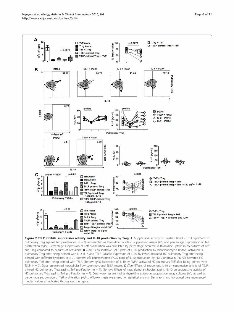
Figure 2 TSLP inhibits suppressive activity and IL-10 production by Treg. A. Suppressive activity of un-stimulated vs. TSLP-primed HCpulmonary Treg against Teff proliferation (n = 8) represented as thymidine counts in suppression assays (left) and percentage suppression of Teffproliferation (right). Percentage suppression of Teff proliferation was calculated by percentage decrease in thymidine uptake in co-cultures of Teffand Treg compared to cultures of Teff alone. B. (Top) Representative FACS plots of IL-10 production by PMA/Ionomycin (PMA/I) activated HCpulmonary Treg after being primed with IL-2, IL-7, and TSLP. (Middle) Expression of IL-10 by PMA/I activated HC pulmonary Treg after beingprimed with different cytokines (n = 7). (Bottom left) Representative FACS plots of IL-10 production by PMA/Ionomycin (PMA/I) activated HCpulmonary Teff after being primed with TSLP. (Bottom right) Expression of IL-10 by PMA/I activated HC pulmonary Teff after being primed withTSLP (n = 7). Data represented intracellular flow cytometric and ELISA results. C. (Top) Effects of exogenous IL-10 on suppressive activity of TSLP-primed HC pulmonary Treg against Teff proliferation (n = 7). (Bottom) Effects of neutralizing antibodies against IL-10 on suppressive activity ofHC pulmonary Treg against Teff proliferation (n = 7). Data were represented as thymidine uptake in suppression assay cultures (left) as well aspercentage suppression of Teff proliferation (right). Wilcoxon tests were used for statistical analysis. Bar graphs and horizontal bars representedmedian values as indicated throughout the figure.
Nguyen et al. Allergy, Asthma & Clinical Immunology 2010, 6:4http://www.aacijournal.com/content/6/1/4
Page 6 of 11

counterparts (Figure 3A). No significant changes in IL-10, TNF-a production by pulmonary Teff; and IL-4,TNF-a, and TGF-b expression by pulmonary Treg wereobserved among 3 subject groups (Additional file 1, Fig-ure S8B). We also observed an increase in cell prolifera-tion in suppression assays with AA pulmonary Treg andautologous Teff compared to assays with HC and NAcells (Figure 3B). Furthermore, in allogeneic suppressionassays, AA pulmonary Treg showed a significantdecrease in suppressive activity against HC pulmonaryTeff compared to HC pulmonary Treg (Figure 3C). Onthe other hand, HC pulmonary Treg suppressed theproliferation of AA and HC pulmonary Teff equivalently(Figure 3C). Collectively, these results suggested the pre-sence of defective IL-10 production and suppressivefunction by AA pulmonary Treg.
Elevated expression of TSLP in AA BAL is necessary for itssuppressive effects on Treg functionConsistent with previous findings, we found elevatedexpression of TSLP in AA BAL (Figure 4A). ElevatedBAL TSLP expression was significantly correlated withreduced IL-10 expression and suppressive function ofpulmonary AA Treg (Figure 4B). These results thus sug-gested that the selective dysfunction in IL-10 productionby pulmonary Treg in AA might be associated with invivo priming of these cells by TSLP. We next examinedwhether BAL from AA subjects with high concentra-tions of TSLP could induce functional changes in HCpulmonary Treg as previously seen with recombinantTSLP. BAL samples from AA subjects with highestlevels of TSLP were selected for this experiment. BALsupernatants were introduced to CD3/CD28 activatedHC pulmonary Treg cultures at a 1:10 volume ratio for18 hours and Treg were subsequently analyzed for IL-10production. Surprisingly, we found that HC pulmonaryTreg incubated with AA BAL showed significantlydecreased expression of IL-10 (Figure 4C). In contrast,in similar priming experiments, BAL samples from NAsubjects failed to inhibit IL-10 production by HC pul-monary Treg (Figure 4D). Furthermore, this reductionin IL-10 expression by HC pulmonary Treg mediated byAA BAL was reversed by neutralizing TSLP with 10 μg/ml of a blocking antibody against TSLP-R (Figure 4C).On the other hand, parallel cultures with low-endotoxinisotype control antibodies at a similar concentrationfailed to reverse the inhibition of IL-10 production ofTreg by AA BAL (Figure 4C). Altogether, these resultsshowed that IL-10 production by pulmonary HC Tregcould be inhibited by exposure to AA-airway-derived-TSLP.It has been well established that TSLP is a master reg-
ulator of airway inflammation because of its abundantexpression in airway epithelial cells as well as its ability
to instruct antigen presenting cells to prime the devel-opment of pathogenic Th-2 helper T cells. Here weshowed that functional TSLP-R was expressed on Tregfrom both blood and pulmonary compartments.Furthermore, TSLP directly activated the signal-transdu-cing molecule STAT5 by Treg and suppressed theirsuppressive activities and production of the immunosup-pressive cytokine IL-10. Our results thus pointed to apotentially novel mechanism by which TSLP mightdirectly dampen tolerogenic immune responses of Treg,and subsequently prolong the course of inflammation.TSLP signaling pathway resembles that of a cytokine
family, including IL-2, IL-7, and IL-15, which all acti-vates STAT5. Surprisingly, unlike TSLP, cytokines suchas IL-2 and IL-15 have been reported to enhance IL-10production by Treg [33,34]. Thus, distinctions in signal-ing pathways downstream of TSLP-R-TSLP ligation withrespect to the activation of intracellular signaling mole-cules other than STAT5 might be required for the inhi-bition of Treg function by TSLP. In our study, theinhibitory effect of TSLP on IL-10 production was speci-fic to Treg but not Teff. This result might be explainedby potential differences in signaling circuitries betweenTreg and Teff, which have been observed in AKT/mTOR cascade [35]. Proteomic analysis of intracellularsignaling molecules in Treg and Teff is underway in ourlaboratory to provide further insights to thisphenomenon.Contrary to a previous report which showed that
TSLP could enhance IL-4 production by Teff isolatedfrom allergen-sensitized mice [7], we did not find amodulatory role of TSLP on IL-4 production by Teff. Itis worth noting that antigen presenting cells, whichinclude monocytes and dendritic cells, were depleted inour culture system while present in theirs. Therefore, itwas possible that the ability of TSLP to stimulate IL-4production by Teff required cross-talk between dendriticcells and TSLP-primed Teff even in the absence ofdirect TSLP-dendritic cell interaction. In addition, Jianget al showed that blocking TSLP reduced thymic Foxp3expression [36]. However, we did not find a modulatoryrole of TSLP on the expression of Foxp3 by peripheralTreg. Thjs discrepancy might be due to changes inmicroenvironments in the thymus vs. peripheral tissueswith respect to the presence of different cellular subsetsand cytokines, which may have an impact on Treghomeostasis. Alternatively, modulation of Foxp3 expres-sion in Treg by TSLP might be temporally regulated:naïve thymic derived Treg, but not circulating/tissueresident Treg, might be susceptible to TSLP-mediatedup-regulation of Foxp3.Our study also showed an association between TSLP-
Treg interaction and defective function of pulmonaryTreg in human AA. Pulmonary Treg showed decreased
Nguyen et al. Allergy, Asthma & Clinical Immunology 2010, 6:4http://www.aacijournal.com/content/6/1/4
Page 7 of 11
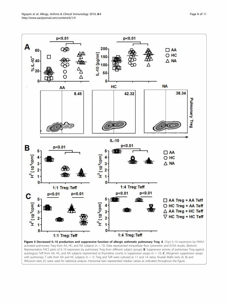
Figure 3 Decreased IL-10 production and suppressive function of allergic asthmatic pulmonary Treg. A. (Top) IL-10 expression by PMA/Iactivated pulmonary Treg from AA, HC, and NA subjects (n = 13). Data represented intracellular flow cytometric and ELISA results. (Bottom)Representative FACS plots of IL-10 expression by pulmonary Treg from different subject groups. B. Suppressive activity of pulmonary Treg againstautologous Teff from AA, HC, and NA subjects represented as thymidine counts in suppression assays (n = 13). C. Allogeneic suppression assayswith pulmonary T cells from AA and HC subjects (n = 7). Treg and Teff were cultured at 1:1 and 1:4 ratios. Kruskal Wallis tests (A, B) andWilcoxon tests (C) were used for statistical analysis. Horizontal bars represented median values as indicated throughout the figure.
Nguyen et al. Allergy, Asthma & Clinical Immunology 2010, 6:4http://www.aacijournal.com/content/6/1/4
Page 8 of 11

IL-10 production, which was correlated with theincreased expression of TSLP in BAL of these subjects.Consistent with the potential role of TSLP in the sup-pression of IL-10 by pulmonary AA Treg, we showedthat TSLP derived from BAL of AA subjects was neces-sary for the direct suppression of IL-10 production byHC Treg in vitro. Alternatively, decreased IL-10 produc-tion might result from in vivo exposure of AA Treg todifferent stimuli in AA airway and interaction amongTSLP, pulmonary dendritic cells, and pulmonary Treg,respectively. A previous study showed that defective IL-10 production was observed on steroid-resistant asth-matic subjects which could be pharmacologicallyreversed by calcitriol [37]. In this study, the populationof regulatory T cells being examined was IL-10 produ-cing CD4+ T cells (Tr1). Thus, it is not known whetherIL-10 production by natural Foxp3-expressing Treg is
also defective. Our preliminary studies showed that ster-oid-resistant asthmatics also had elevated TSLP levels intheir BAL (unpublished observations), suggesting thatthis increased expression of TSLP might also have aninhibitory effect on expression of IL-10 by natural Treg.Studies to elaborate on these findings as well as to char-acterize the role of TSLP on IL-10 producing CD4+T cells (Tr1) and the ability of calcitriol to reverse theseeffects of TSLP on Treg production of IL-10 are under-way in our laboratory.Defective IL-10 production by pulmonary Treg was
found only in AA but not NA subjects. These resultswere consistent with previous observations of immuno-logical differences in these two sub-types of asthma [38].Nevertheless, our data did not rule out the possibilitythat pulmonary Treg exhibit distinct suboptimal regula-tory functions in other allergic and pulmonary diseases.
Figure 4 Impaired allergic asthmatic pulmonary Treg function is associated with elevated airway TSLP. A. TSLP expression in BAL of AA,HC, and NA subjects (n = 15). B. Correlation of TLSP levels in BAL of AA subjects with IL-10 production and suppression activity of AApulmonary Treg (n = 13). Straight lines represented best-fitted lines. Dotted lines represented 95% confident intervals. C. (Top) Effects of blockingTLSP-R antibodies on AA BAL-mediated inhibition of IL-10 production by HC pulmonary Treg (n = 5). (Bottom) Representative FACS plots of IL-10production by HC pulmonary Treg in different stimulatory conditions. D. Effects of NA BAL on IL-10 production by HC pulmonary Treg (n = 5).Data represented intracellular flow cytometric and ELISA results. Kruskal Wallis tests (A), Spearman tests (B) and Wilcoxon tests (C) were used forstatistical analysis. Horizontal bars represented median values as indicated throughout the figure.
Nguyen et al. Allergy, Asthma & Clinical Immunology 2010, 6:4http://www.aacijournal.com/content/6/1/4
Page 9 of 11
A potential explanation is that each disease possesses asignature inflammatory environment. Thus, the effectsof inflammatory cytokine milieu of each disease on pul-monary Treg function might very well be different.Modulation of TSLP signaling has been shown to be
influential to airway inflammation in experimental mod-els of asthma [39]. Blockade of TSLP-R suppressed aller-gic inflammation by altering dendritic cell function.Deletion of an intracellular regulator of TSLP produc-tion, SOCS7, also led to increased allergic phenotype[40]. Along with these results, our data suggest that,besides preventing airway inflammation, TSLP-targetedtherapies might also be beneficial in modulatingimmune tolerance by Treg in allergic diseases.
ConclusionsOur study presents a potentially novel antigen-present-ing-cell-independent mechanism by which TSLP mightcontribute to the exacerbation of airway inflammationby directly inhibiting tolerogenic immune responses ofpulmonary Treg.
Additional file 1: Supplementary Figures and Table. The file includesFigures S1-8 and Table S1.
AbbreviationsHC: healthy controls; AA: allergic asthmatics; NA: non-allergic asthmatics;Treg: CD4+CD25hiCD127lo/-regulatory T cells; Teff: CD4+CD25-T cells; TSLP:thymic stromal lymphopoietin; pSTAT5: phosphorylated signal transducerand activator of transcription 5; BAL: bronchoaveolar lavage fluid; PBMC:peripheral blood mononuclear cells; FEV1: forced expiratory volume in 1second; FACS: fluorescent activated cell sorting; MFI: mean fluorescenceintensity.
AcknowledgementsThe study was supported by grants from the Mary Hewitt LovelessFoundation, the Parker B. Francis Foundation, and the American Academy ofAllergy, Asthma, and Immunology. None of the authors have conflictingfinancial interests. We are grateful to the asthmatic and healthy subjectswho provided samples for the study.
Authors’ contributionsKDN designed the study and wrote the article. KCN oversaw the project andrecruited study subjects. KDN, KCN, and CV performed experiments andanalyzed the data. All authors read and approved the final manuscript.
Competing interestsThe authors declare that they have no competing interests.
Received: 10 January 2010 Accepted: 15 March 2010Published: 15 March 2010
References1. Vosshenrich CA, Cumano A, Müller W, Di Santo JP, Vieira P: Thymic
stromal-derived lymphopoietin distinguishes fetal from adult B celldevelopment. Nat Immunol 2003, 4:773-779.
2. Al-Shami A, et al: A role for thymic stromal lymphopoietin in CD4(+) Tcell development. J Exp Med 2004, 200:159-168.
3. Soumelis V, et al: Human epithelial cells trigger dendritic cell mediatedallergic inflammation by producing TSLP. Nat Immunol 2002, 3:673-680.
4. Zhou B, et al: Thymic stromal lymphopoietin as a key initiator of allergicairway inflammation in mice. Nat Immunol 2005, 6:1047-1053.
5. Rochman I, Watanabe N, Arima K, Liu YJ, Leonard WJ: Cutting edge: directaction of thymic stromal lymphopoietin on activated human CD4+ Tcells. J Immunol 2007, 178:6720-6724.
6. Akamatsu T, et al: Human TSLP directly enhances expansion of CD8+ Tcells. Clin Exp Immunol 2008, 154:98-106.
7. He R, et al: TSLP acts on infiltrating effector T cells to drive allergic skininflammation. Proc Natl Acad Sci USA 2008, 105:11875-11880.
8. Al-Shami A, Spolski R, Kelly J, Keane-Myers A, Leonard WJ: A role for TSLPin the development of inflammation in an asthma model. J Exp Med2005, 202:829-839.
9. Zhang Z, et al: Thymic stromal lymphopoietin overproduced bykeratinocytes in mouse skin aggravates experimental asthma. Proc NatlAcad Sci USA 2009, 106:1536-1541.
10. Akdis CA, Akdis M: Mechanisms and treatment of allergic disease in thebig picture of regulatory T cells. J Allergy Clin Immunol 2009, 123:735-746.
11. Herrick CA, Bottomly K: To respond or not to respond: T cells in allergicasthma. Nat Rev Immunol 2003, 3:405-412.
12. Miyara M, Sakaguchi S: Natural regulatory T cells: mechanisms ofsuppression. Trends Mol Med 2007, 13:108-116.
13. Hartl D, et al: Quantitative and functional impairment of pulmonary CD4+CD25hi regulatory T cells in pediatric asthma. J Allergy Clin Immunol2007, 119:1258-1266.
14. Kearley J, Robinson DS, Lloyd CM: CD4+CD25+ regulatory T cells reverseestablished allergic airway inflammation and prevent airway remodeling.J Allergy Clin Immunol 2008, 122:617-624.
15. Presser K, et al: Coexpression of TGF-beta1 and IL-10 enables regulatoryT cells to completely suppress airway hyperreactivity. J Immunol 2008,181:7751-7758.
16. Zuany-Amorim C, et al: Suppression of airway eosinophilia by killedMycobacterium vaccae-induced allergen-specific regulatory T-cells. NatMed 2002, 8:625-629.
17. Hammad H, et al: Activation of the D prostanoid 1 receptor suppressesasthma by modulation of lung dendritic cell function and induction ofregulatory T cells. J Exp Med 2007, 204:357-367.
18. Watanabe N, et al: Hassall’s corpuscles instruct dendritic cells to induceCD4+CD25+ regulatory T cells in human thymus. Nature 2005,436:1181-1185.
19. Jiang Q, Coffield VM, Kondo M, Su L: TSLP is involved in expansion ofearly thymocyte progenitors. BMC Immunol 2007, 8:11.
20. Mazzucchelli R, et al: Development of regulatory T cells requires IL-7Ralpha stimulation by IL-7 or TSLP. Blood 2008, 112:3283-3292.
21. Pandey A, et al: Cloning of a receptor subunit required for signaling bythymic stromal lymphopoietin. Nat Immunol 2000, 1:59-64.
22. Park LS, et al: Cloning of the murine thymic stromal lymphopoietin(TSLP) receptor: formation of a functional heteromeric complex requiresinterleukin 7 receptor. J Exp Med 2000, 192:659-670.
23. Quentmeier H, et al: Cloning of human thymic stromal lymphopoietin(TSLP) and signaling mechanisms leading to proliferation. Leukemia 2001,8:1286-1292.
24. Ito T, et al: TSLP-activated dendritic cells induce an inflammatory Thelper type 2 cell response through OX40 ligand. J Exp Med 2005,202:1213-1223.
25. Gilliet M, et al: Human dendritic cells activated by TSLP and CD40Linduce proallergic cytotoxic T cells. J Exp Med 2003, 197:1059-1063.
26. Thornton AM, Shevach EM: CD4+CD25+ immunoregulatory T cellssuppress polyclonal T cell activation in vitro by inhibiting interleukin 2production. J Exp Med 1998, 188:287.
27. Shevach EM: Mechanisms of foxp3+ T regulatory cell-mediatedsuppression. Immunity 2009, 30:636-645.
28. Vignali DA, Collison LW, Workman CJ: How regulatory T cells work. NatRev Immunol 2008, 8:523-532.
29. Tang Q, Bluestone J: The Foxp3+ regulatory T cell: a jack of all trades,master of regulation. Nat Immunol 2008, 9:239-244.
30. Wing K, Sakaguchi S: Regulatory T cells exert checks and balances on selftolerance and autoimmunity. Nat Immunol 2010, 11:7-13.
31. Ying S, et al: Thymic stromal lymphopoietin expression is increased inasthmatic airways and correlates with expression of Th2-attractingchemokines and disease severity. J Immunol 2005, 174:8183-8190.
Nguyen et al. Allergy, Asthma & Clinical Immunology 2010, 6:4http://www.aacijournal.com/content/6/1/4
Page 10 of 11

32. Ying S, et al: Expression and cellular provenance of thymic stromallymphopoietin and chemokines in patients with severe asthma andchronic obstructive pulmonary disease. J Immunol 2008, 181:2790-2798.
33. Wilson MS, et al: Suppression of murine allergic airway disease by IL-2:anti-IL-2 monoclonal antibody-induced regulatory T cells. J Immunol2008, 181:6942-6954.
34. Lee CC, Lin SJ, Cheng PJ, Kuo ML: The regulatory function of umbilicalcord blood CD4 CD25 T cells stimulated with anti-CD3/anti-CD28 andexogenous interleukin (IL)-2 or IL-15. Pediatr Allergy Immunol 2009,20(7):624-632(9).
35. Zeiser R, et al: Differential impact of mammalian target of rapamycininhibition on CD4+CD25+Foxp3+ regulatory T cells compared withconventional CD4+ T cells. Blood 2008, 111:453-462.
36. Jiang Q, Su H, Knudsen G, Helms W, Su L: Delayed functional maturationof natural regulatory T cells in the medulla of postnatal thymus: role ofTSLP. BMC Immunol 2006, 7:6.
37. Xystrakis E, et al: Reversing the defective induction of IL-10-secretingregulatory T cells in glucocorticoid-resistant asthma patients. J Clin Invest2006, 116:146-155.
38. Nguyen KD, Vanichsarn C, Fohner A, Nadeau KC: Selective deregulation inchemokine signaling pathways of CD4+CD25(hi)CD127(lo)/(-) regulatoryT cells in human allergic asthma. J Allergy Clin Immunol 2009, 123:933-939.
39. Shi L, et al: Local blockade of TSLP receptor alleviated allergic disease byregulating airway dendritic cells. Clin Immunol 2008, 129:202-210.
40. Knisz J, et al: Loss of SOCS7 in mice results in severe cutaneous diseaseand increased mast cell activation. Clin Immunol 2009, 132:277-284.
doi:10.1186/1710-1492-6-4Cite this article as: Nguyen et al.: TSLP directly impairs pulmonary Tregfunction: association with aberrant tolerogenic immunity in asthmaticairway. Allergy, Asthma & Clinical Immunology 2010 6:4.
Submit your next manuscript to BioMed Centraland take full advantage of:
• Convenient online submission
• Thorough peer review
• No space constraints or color figure charges
• Immediate publication on acceptance
• Inclusion in PubMed, CAS, Scopus and Google Scholar
• Research which is freely available for redistribution
Submit your manuscript at www.biomedcentral.com/submit
Nguyen et al. Allergy, Asthma & Clinical Immunology 2010, 6:4http://www.aacijournal.com/content/6/1/4
Page 11 of 11