The L¹dek-Œnie¿nik Metamorphic Unit - Recent State of Knowledge
Upload
independentCategory
view
0download
0
BioMed CentralBMC Immunology
ss
Open AcceResearch articleSerum from mice immunized in the context of Treg inhibition identifies DEK as a neuroblastoma tumor antigenJin Zheng1, M Eric Kohler1, Qingrong Chen2, James Weber1, Javed Khan2, Bryon D Johnson1 and Rimas J Orentas*1Address: 1Department of Pediatrics, Medical College of Wisconsin and the Children's Research Institute, Children's Hospital of Wisconsin. 8701 Watertown Plank Rd. Milwaukee, WI 53226, USA and 2National Cancer Institute, Pediatric Oncology Branch, Oncogenomics Section, U.S. National Institutes of Health, Advanced Technology Center, 8717 Grovemont Circle. Gaithersburg, MD 20877, USA
Email: Jin Zheng - [email protected]; M Eric Kohler - [email protected]; Qingrong Chen - [email protected]; James Weber - [email protected]; Javed Khan - [email protected]; Bryon D Johnson - [email protected]; Rimas J Orentas* - [email protected]
* Corresponding author
AbstractBackground: We have developed a cell-based vaccine that features the expression of both CD80and CD86 on the surface of a murine neuroblastoma cell line. The cellular immunity induced bythis vaccine is enhanced by treatment with antibody that interferes with T-regulatory cell (Treg)function and we report here that immunization combined with interfering with Treg function alsoproduces a profound serological effect. Serum from mice immunized with our cell-based vaccine inthe context of Treg blockade was used to screen a cDNA expression library constructed from theparental neuroblastoma tumor cell line, AGN2a.
Results: Serum from mice vaccinated in the context of Treg blockade identified a number ofpotentially oncogenic transcripts that may serve as important immune targets in a tumor-derivedcDNA library screen. This novel approach identified far more candidates than could be seen withserum derived from vaccine-treated only, Treg-depleted only, or tumor-bearing mice. The mostcommonly identified tumor-associated antigen, using serum from immunized and Treg-depletedmice, was the DEK oncogene. Altered expression of the DEK oncogene has been implicated in anumber of human cancers. Importantly, we were able to demonstrate that the DEK oncogene alsoinduces a T cell response.
Conclusion: The use of post-vaccine immune serum in this report differs from previousapproaches where serum collected at the time of cancer onset or diagnosis and was used for tumorantigen identification. We hypothesize that the use of diagnostic serum samples may be inadequatefor the clinical translation of this approach, and that identification of protective immunogenic tumorantigens may require the use of serum from post-treatment or vaccinated subjects. Theidentification of DEK as a tumor-associated antigen capable of eliciting a T cell response validatesour experimental approach and argues for the antigens we have identified here to be evaluated astargets of effector immunity and as vaccine candidates.
Published: 30 March 2007
BMC Immunology 2007, 8:4 doi:10.1186/1471-2172-8-4
Received: 24 October 2006Accepted: 30 March 2007
This article is available from: http://www.biomedcentral.com/1471-2172/8/4
© 2007 Zheng et al; licensee BioMed Central Ltd. This is an Open Access article distributed under the terms of the Creative Commons Attribution License (http://creativecommons.org/licenses/by/2.0), which permits unrestricted use, distribution, and reproduction in any medium, provided the original work is properly cited.
Page 1 of 15(page number not for citation purposes)
BMC Immunology 2007, 8:4 http://www.biomedcentral.com/1471-2172/8/4
BackgroundAdvanced neuroblastoma poses a grave clinical challengeand still awaits effective therapy. Early clinical observa-tions, combined with a slight but demonstrable positiveimpact of bone marrow transplantation on outcome hasmotivated the development of immune approaches totherapy [1-4]. In murine models of human neuroblast-oma, anti-tumor immunity can be generated using cell-based vaccines where tumor cells have been geneticallymodified to express soluble cytokines or cell-surfaceimmunostimulatory molecules [5-7]. Our own work hasdemonstrated that cancer cell-based vaccines expressingmultiple immune co-stimulatory molecules in the murineneuroblastoma cell line AGN2a can transform this tumorcell line in to a vaccine that induces strong cell-basedimmunity to the unmodified parental cell line [8,9].Based on the ability to induce an immune response withcancer cell-based vaccines, human trials with neuroblast-oma patients have been carried out [10]. Although thesecell-based cancer vaccines did not prove immediatelyeffective, they were demonstrated to be safe and are ripefor further optimization [11].
In experimental systems, immunity to neuroblastoma canbe amplified by the blockade of T-regulatory cell (Treg)function with anti-CD25 antibody (B.D. Johnson, et al.,2007, J. Immunother., in press). Treg are known to sup-press the immune response to self-antigens, includingtumor-self antigens, and thwarting this tolerogenic role bytheir depletion has become a major focus in the develop-ment of new immunotherapeutic strategies to treathuman malignancy [12,13]. Golgher et al. have demon-strated that CD25+ T cell depletion uncovers immuneresponses to the tumor cell type used as a vaccine, andimportantly that this response broadens to include othersyngeneic tumor cell types [14]. Given the ability toinduce immune recognition of what are normally consid-ered "self" antigens upon Treg blockade, we reasoned thattreatment of experimental animals with cell-based cancervaccines in the context of anti-CD25 antibody treatmentwould induce a strong anti-neuroblastoma immuneresponse. The proposed use of serology to uncover T cellantigens is supported by the recent description of anti-body as well as T cell responses to the DBY minor histo-compatibility antigen in allogeneic stem celltransplantation [15,16]. The breaking of tolerance to self-antigens with Treg depletion may be functionally analo-gous to the anti-tumor effect seen in allogeneic bone mar-row transplantation, whose primary side-effect, graft-versus-host disease, is evidence that tolerance to normalself antigens has been modified.
The serological analysis of recombinant cDNA expressionlibraries (SEREX) constructed from patient tumor wasestablished by Sahin and Tureci who demonstrated that
this process identifies T-cell antigens as well as B-cell anti-gens [17,18]. SEREX continues to be employed in patientstudies and has even proven to identify intracellular anti-gens targeted by the immune system [19]. The identifica-tion of the NY-ESO-1 antigen in patients by SEREXdemonstrated that both MHC class II restricted epitopesand MHC class I-restricted (HLA-A2) epitopes, targets ofcytotoxic T cell responses, could be identified with thistechnique [19]. We present a new means to identifyimmunogenic tumor antigens. In this report we employserum from experimental animals that have been vacci-nated in the context of anti-CD25 antibody treatment, asopposed to using sera from tumor-bearing animals, whichwould be the equivalent of using serum from newly diag-nosed patients. The use of immune serum-SEREX hasallowed us to identify new tumor-associated antigens inour neuroblastoma model. Notably, we demonstrate thatone of the antigens identified by our immune-SEREXapproach, the DEK oncoprotein, induces a T cell responseas well as an antibody response. Translation of this con-cept in to clinical studies would require the used of post-treatment or even post-vaccination serum, as opposed tothe initial samples commonly harvested at the time ofdiagnosis.
ResultsThe modification of syngeneic tumor cell lines withimmune co-stimulatory molecules can transform lethaltumor cell lines into effective vaccines. The immunizationof A/J mice with a cell-based vaccine, AGN2a-CD80/CD86, has been demonstrated to induce a strong cellularimmune response [8]. This immune response is enhancedwhen the vaccine is given in the context of anti-CD25 anti-body (clone PC61) as a means to block and/or deplete T-regulatory cells. We sought to explore the specificity of theimmunoglobulin response to this cell based vaccine, as ameans to potentially uncover new tumor-associated anti-gens. To do so, we constructed a cDNA library usingmRNA purified from the AGN2a cell line and expressedproteins encoded by tumor- derived mRNA in E. coliusing the λZAPII phage system. To identify clones express-ing antigenic epitopes recognized by IgG present inimmune sera, a total of 2 × 10⁵ clones were screened. Fifty-eight unique plaques were identified as expressing pro-teins that could be recognized by IgG antibody whenserum from AGN2a-CD80/CD86+PC61 treated mice wasused at a dilution of 1:250. When pooled serum (n = 5 foreach group) from naïve mice, tumor-bearing mice, micetreated with PC61 alone, or mice treated with two weeklyinjections of the vaccine cell-line AGN2a-CD80/CD86alone was used to screen the cDNA library 0, 0, 0, or 5unique clones, respectively, were identified. Antibody-reactive plaques identified by serum from the AGN2a-CD80/CD86+PC61-treated mice were picked, used to re-infect E. coli, and once more serologically screened. A
Page 2 of 15(page number not for citation purposes)
BMC Immunology 2007, 8:4 http://www.biomedcentral.com/1471-2172/8/4
third round of plaque purification was not required, as italways resulted in uniform recognition of phage-expressed proteins. In vivo excision was performed withhelper phage and phagemid DNA isolated. Eachphagemid insert was DNA sequenced using both T7 andT3 primers, and this sequence was used to search on-linedatabases. The 58 clones identified by immune serum-SEREX screening encoded 25 individual proteins, as listedin Table 1. Most notable was the frequency with whichDEK was detected. While all antigens listed in Table 1were detected once from a single positive clone, the DEKantigen was detected 34 separate times. Careful inspectionof the cDNA library clones demonstrates that most tran-scripts seem to have some role in neuronal differentiation,cell cycle control, or have previously been identified astranscripts that are over-expressed in other malignancies.The final 4 transcripts listed in Table 1 have no publishedor annotated functional analysis associated with them,beyond their naming and characterization using bioinfor-matic techniques. Primers were generated for each cloneidentified, and expression of each verified by PCR analysisof AGN2a-derived cDNA (not shown).
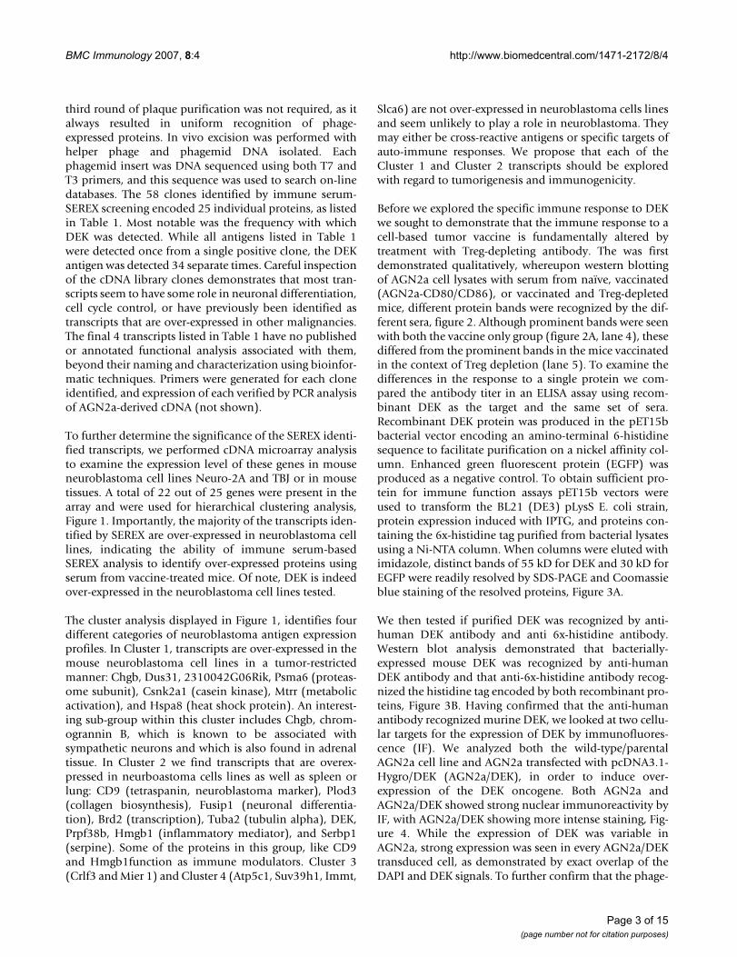
To further determine the significance of the SEREX identi-fied transcripts, we performed cDNA microarray analysisto examine the expression level of these genes in mouseneuroblastoma cell lines Neuro-2A and TBJ or in mousetissues. A total of 22 out of 25 genes were present in thearray and were used for hierarchical clustering analysis,Figure 1. Importantly, the majority of the transcripts iden-tified by SEREX are over-expressed in neuroblastoma celllines, indicating the ability of immune serum-basedSEREX analysis to identify over-expressed proteins usingserum from vaccine-treated mice. Of note, DEK is indeedover-expressed in the neuroblastoma cell lines tested.
The cluster analysis displayed in Figure 1, identifies fourdifferent categories of neuroblastoma antigen expressionprofiles. In Cluster 1, transcripts are over-expressed in themouse neuroblastoma cell lines in a tumor-restrictedmanner: Chgb, Dus31, 2310042G06Rik, Psma6 (proteas-ome subunit), Csnk2a1 (casein kinase), Mtrr (metabolicactivation), and Hspa8 (heat shock protein). An interest-ing sub-group within this cluster includes Chgb, chrom-ogrannin B, which is known to be associated withsympathetic neurons and which is also found in adrenaltissue. In Cluster 2 we find transcripts that are overex-pressed in neurboastoma cells lines as well as spleen orlung: CD9 (tetraspanin, neuroblastoma marker), Plod3(collagen biosynthesis), Fusip1 (neuronal differentia-tion), Brd2 (transcription), Tuba2 (tubulin alpha), DEK,Prpf38b, Hmgb1 (inflammatory mediator), and Serbp1(serpine). Some of the proteins in this group, like CD9and Hmgb1function as immune modulators. Cluster 3(Crlf3 and Mier 1) and Cluster 4 (Atp5c1, Suv39h1, Immt,
Slca6) are not over-expressed in neuroblastoma cells linesand seem unlikely to play a role in neuroblastoma. Theymay either be cross-reactive antigens or specific targets ofauto-immune responses. We propose that each of theCluster 1 and Cluster 2 transcripts should be exploredwith regard to tumorigenesis and immunogenicity.
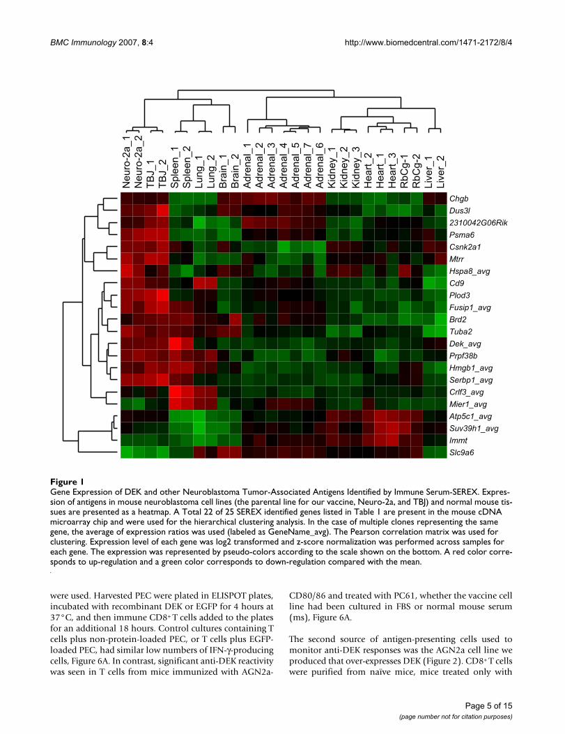
Before we explored the specific immune response to DEKwe sought to demonstrate that the immune response to acell-based tumor vaccine is fundamentally altered bytreatment with Treg-depleting antibody. The was firstdemonstrated qualitatively, whereupon western blottingof AGN2a cell lysates with serum from naïve, vaccinated(AGN2a-CD80/CD86), or vaccinated and Treg-depletedmice, different protein bands were recognized by the dif-ferent sera, figure 2. Although prominent bands were seenwith both the vaccine only group (figure 2A, lane 4), thesediffered from the prominent bands in the mice vaccinatedin the context of Treg depletion (lane 5). To examine thedifferences in the response to a single protein we com-pared the antibody titer in an ELISA assay using recom-binant DEK as the target and the same set of sera.Recombinant DEK protein was produced in the pET15bbacterial vector encoding an amino-terminal 6-histidinesequence to facilitate purification on a nickel affinity col-umn. Enhanced green fluorescent protein (EGFP) wasproduced as a negative control. To obtain sufficient pro-tein for immune function assays pET15b vectors wereused to transform the BL21 (DE3) pLysS E. coli strain,protein expression induced with IPTG, and proteins con-taining the 6x-histidine tag purified from bacterial lysatesusing a Ni-NTA column. When columns were eluted withimidazole, distinct bands of 55 kD for DEK and 30 kD forEGFP were readily resolved by SDS-PAGE and Coomassieblue staining of the resolved proteins, Figure 3A.
We then tested if purified DEK was recognized by anti-human DEK antibody and anti 6x-histidine antibody.Western blot analysis demonstrated that bacterially-expressed mouse DEK was recognized by anti-humanDEK antibody and that anti-6x-histidine antibody recog-nized the histidine tag encoded by both recombinant pro-teins, Figure 3B. Having confirmed that the anti-humanantibody recognized murine DEK, we looked at two cellu-lar targets for the expression of DEK by immunofluores-cence (IF). We analyzed both the wild-type/parentalAGN2a cell line and AGN2a transfected with pcDNA3.1-Hygro/DEK (AGN2a/DEK), in order to induce over-expression of the DEK oncogene. Both AGN2a andAGN2a/DEK showed strong nuclear immunoreactivity byIF, with AGN2a/DEK showing more intense staining, Fig-ure 4. While the expression of DEK was variable inAGN2a, strong expression was seen in every AGN2a/DEKtransduced cell, as demonstrated by exact overlap of theDAPI and DEK signals. To further confirm that the phage-
Page 3 of 15(page number not for citation purposes)

BMC Immunology 2007, 8:4 http://www.biomedcentral.com/1471-2172/8/4
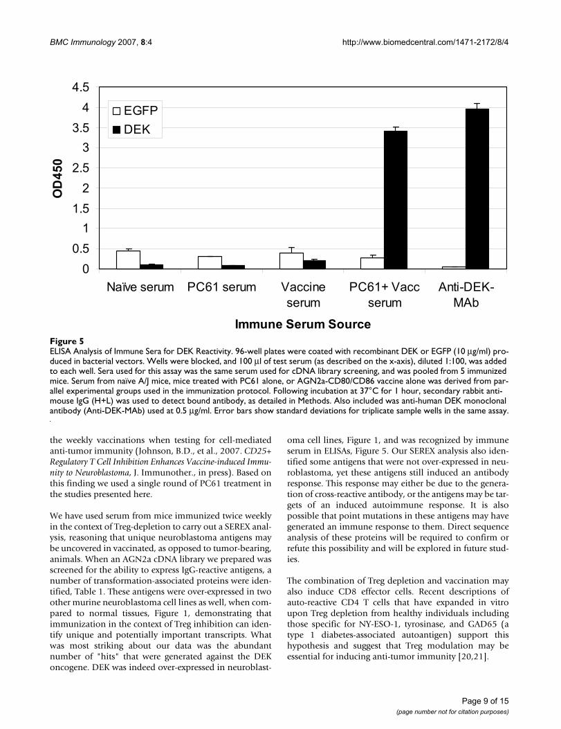
expressed DEK cDNA was a genuine immune target wetested serum from tumor-vaccinated mice for the presenceof DEK-reactive immunoglobulin by ELISA. Using poly-styrene plates coated with recombinant DEK, DEK-reac-tive IgG could be readily detected in serum from miceimmunized with AGN2a-CD80/86 in the context of PC61treatment, Figure 5. Anti-human DEK antibody was usedas a positive control antibody in the ELISA. This assay alsosupports our hypothesis that vaccination in the context ofTreg depletion alters the nature of the immune responseto cell-based tumor vaccines. High titer DEK antibodyproduction required the depletion of Treg, while vaccina-tion with the cell based vaccine, AGN2a-CD80/CD86, didnot produce anti-DEK IgG.
To investigate the CD8+ T cell response against DEK inmice that had received a cell-based cancer vaccine, A/J
mice were given two weekly subcutaneous (s.c.) injectionsof 2 × 10⁶ irradiated AGN2a-CD80/CD86 cells cultured inDMEM supplemented with 2% mouse serum or 10% FBS,and Treg activity inhibited by the i.p. injection of clonePC61 anti-CD25 antibody three days prior to the first vac-cination. Mouse serum-cultured vaccine cell lines wereused in order to control for potential FBS reactivity, whichhas been reported in the analysis of CD4 T cell responses.AGN2a-CD80/86 was cultured in normal mouse serum atleast two weeks prior to immunization studies. Spleno-cytes were collected 5 days after the second vaccination,depleted of RBCs, and CD8+ T cells purified by immu-nomagnetic selection. T cell responses to DEK weredetected in IFN-γ ELISPOT assays using two differentsources of antigen-presenting cells. In the first assay, peri-toneal exudate cells (PEC, which are primarily composedof macrophages) loaded with recombinant DEK protein
Table 1: Neuroblastoma antigens identified by immune serum and SEREX.
Identity of cDNA SEREX hit Accession Characteristics Ref
DEK BC055451 p53 interaction, blocks apoptosisDNA binding/chromosomal organization,Proofreading of splicingFused to can in leukemia
Wise-Draper '06Waldman '04Soares '06Garcon '05
Hspa8, heat shock protein 8 (Hsc70) NM_031165 Promotes cell-cycle progression (w/cyclin D1)Binds/regulates dbl proto-oncogene
Diehl '03Kauppinen '05
Fusip1, FUS interacting protein (Nssr, TASR) BC0832082 Neuronal Differentiation, modulatesneuronal mRNA splicingOver-expressed in liposarcoma and leukemia
Liu '04Fushimi '05Clinton '02
Hmgb1, high mobility group box-1 BC008565 Inflammatory mediatorDNA binding, architecture regulation
Andreason '02Stott '06
Atp5c1, ATP synthase (mitochondrial F1 complex, gamma polypeptide 1) BC010700 ATP biosynthesis, upregulated in STI571-resistant leukemia
Hofmann '02
Sfrs5, splicing factor, arginine/serine-rich (SRp40, HRS) BC085267 Splicing factor, Akt substrateApoptosis resistance
Patel '05Tidd '03
Prpf38b, pre-mRNA processing factor (PRP38) BC050900 snRNP, required for spliceosome activity Xie '98Tuba1, tubulin, alpha 1 BC085256 Upregulated in carcinoma Qi '05Chn1, chimerin 1 BC010825 Neurotrophin target Mizuno '04Csnk2a1, casein kinase II, alpha 1 polypeptide BC060742 Phosphorylates/activates NFkappaB Chantome '04Mkks, McKusick-Kaufman syndrome protein NM_021527 Chaperonin, required for cytokenesis Kim '05Mier1, mesoderm induction early response 1 (Mi-er1, er1) AK129405 Fibroblast growth factor induced,
HDAC1 interaction, transcription factorDing '03Post '05
MTTR, 5-methyltetrahydrofolate-homocysteine methyltransferase reductase BC025942 Mutations seen in neural tube defectsInfluences ALL susceptibility
Wilson '99Gemmati '04
Psma6, proteasome subunit, alpha type 6 NM_011968 Upregulated in hepatocellular ca. Cui '06CD9 NM_007657 Cell surface tetraspannin, neuronal
differentiation and neuroblastoma markerIshibashi '04Komada '00
Chromogranin B, Chgb (secretogranin I) NM_007694 Present in neuroblastoma secretory vesicles
Goodall '97
Brd2, bromodomain containing 2 (NAT, Rnf3, Ring3, Fsrg1) NM_001025387 Binds to KSHV, LANA-1 proteinLymphomagenesis
Viejo-Borbolla '05Greenwald '04
Crlf3, cytokine receptor-like factor 3 AL591113 Upregulated in skin cancer Dang '06Plod3, procollagen-lysine, 2-oxoglutarate 5-dioxygenase 3 (LH3) NM_011962 Collagen biosynthesis
Upregulated in chemo resistant ca.Rautavuoma '04Cleator '05
Immt, inner membrane protein mitochondrial (HMP; P87; P89; P87/89) AK038129 Energy metabolism John '05Suv39h2, suppressor of variegation 3–9 homolog 2 AF149205 Epigenetic regulation of telomere length Garcia-Cao '04Serbp1, serpine1 mRNA binding protein BC006030 inferred function (by Gene Ontology)Dus3l, dihydrouridine synthase 3-like AY040840 inferred function (by Gene Ontology)Slc9a6, Solute carrier family 9 (Na+/H+ exchanger) AK028350 inferred function (by Gene Ontology)2310042G06Rik BC026968 Riken cDNA, predicted protein
The table lists the name (identity) of the recombinant plaques in the AGN2a cDNA library that expressed IgG-reactive protein using serum from mice immunized with AGN2a-CD80/86 in the context of PC61 treatment three days prior to first vaccine. Each transcript was identified a single time, with the exception of DEK, as detailed in Results. The table also lists the accession number, relevant characteristics of the identified protein, and the references that initially reported these functions. All information is derived from publicly available databases including Entrez Gene, National Center for Biotechnology Information, National Library of Medicine, U.S. National Institutes of Health.
Page 4 of 15(page number not for citation purposes)
BMC Immunology 2007, 8:4 http://www.biomedcentral.com/1471-2172/8/4
were used. Harvested PEC were plated in ELISPOT plates,incubated with recombinant DEK or EGFP for 4 hours at37°C, and then immune CD8+ T cells added to the platesfor an additional 18 hours. Control cultures containing Tcells plus non-protein-loaded PEC, or T cells plus EGFP-loaded PEC, had similar low numbers of IFN-γ-producingcells, Figure 6A. In contrast, significant anti-DEK reactivitywas seen in T cells from mice immunized with AGN2a-
CD80/86 and treated with PC61, whether the vaccine cellline had been cultured in FBS or normal mouse serum(ms), Figure 6A.
The second source of antigen-presenting cells used tomonitor anti-DEK responses was the AGN2a cell line weproduced that over-expresses DEK (Figure 2). CD8+ T cellswere purified from naïve mice, mice treated only with
Gene Expression of DEK and other Neuroblastoma Tumor-Associated Antigens Identified by Immune Serum-SEREXFigure 1Gene Expression of DEK and other Neuroblastoma Tumor-Associated Antigens Identified by Immune Serum-SEREX. Expres-sion of antigens in mouse neuroblastoma cell lines (the parental line for our vaccine, Neuro-2a, and TBJ) and normal mouse tis-sues are presented as a heatmap. A Total 22 of 25 SEREX identified genes listed in Table 1 are present in the mouse cDNA microarray chip and were used for the hierarchical clustering analysis. In the case of multiple clones representing the same gene, the average of expression ratios was used (labeled as GeneName_avg). The Pearson correlation matrix was used for clustering. Expression level of each gene was log2 transformed and z-score normalization was performed across samples for each gene. The expression was represented by pseudo-colors according to the scale shown on the bottom. A red color corre-sponds to up-regulation and a green color corresponds to down-regulation compared with the mean.
Page 5 of 15(page number not for citation purposes)
BMC Immunology 2007, 8:4 http://www.biomedcentral.com/1471-2172/8/4
Page 6 of 15(page number not for citation purposes)
Western Blot Analysis of Whole Tumor Cell LysatesFigure 2Western Blot Analysis of Whole Tumor Cell Lysates. (A) AGN2a cell lysate was prepared (as in Methods) and the indicated immune serum used at a dilution of 1:100. Lane 1, molecular weight marker (mw); Lane 2, naïve serum; Lane 3, serum from PC61-treated mice; Lane 4, serum from AGN2a-CD80/86 immunized mice; Lane 5, serum from AGN2a-CD80/86+PC61 (Treg-depleted)treated mice. (B) SDS-PAGE analysis showing, Lane 1, mw marker (mw) and, Lane 2, total AGN2a lysate (5 × 10⁴ cell equivalents/well).
A 1 2 3 4 5
B 1 2
97
64
51
39
28
19
97
64
51
39
28
19
191
KD
KD

BMC Immunology 2007, 8:4 http://www.biomedcentral.com/1471-2172/8/4
Page 7 of 15(page number not for citation purposes)
SDS-PAGE and Western Blot Analysis of Recombinant DEKFigure 3SDS-PAGE and Western Blot Analysis of Recombinant DEK. (A) Bacterial lysates and Ni-NTA column purified protein were diluted in reducing sample loading buffer, resolved by SDS-PAGE and stained with Coomassie blue. Lanes were loaded with 1) molecular weight (mw) marker, 2) control bacterial lysate, 10 μl, 3) pET-15b/DEK lysate, 10 μl, 4) 1.5 μg Ni-NTA column-puri-fied DEK, 5) pET-15b/EGFP lysate, 10 μl, 6) 1.5 μg Ni-NTA column-purified EGFP. (B) Proteins were resolved by SDS-PAGE and then transferred to PVDF membrane using a Bis-Tris electrophoresis buffer system for western blot analysis. In blot (a) lanes contained: 1) mw marker, 2) control bacterial lysate, 3) pET-15b/DEK lysate, 10 μl, 4) 0.15 μg Ni-NTA column-purified DEK, 5) pET-15b/EGFP lysate, 10 μl, 6) 0.15 μg Ni-NTA column-purified EGFP. Blot (a) was probed with anti-6x-His antibody. For blot (b) a separate gel containing the same samples loaded in blot (a) was probed with anti-human DEK monoclonal anti-body. Bound antibody was detected in both blots with alkaline phosphatase (AP)-conjugated rabbit anti-mouse IgG (H+L) sec-ondary antibody, as described in Methods. Data is representative of more than three separate experiments.
1 2 3 4 5 6
64
39
51
97kD
28
19
64 kD
51
39
28
(a) 1 2 3 4 5 6 (b) 1 2 3 4 5 6
A
B
Anti-DEK Anti-6x-His

BMC Immunology 2007, 8:4 http://www.biomedcentral.com/1471-2172/8/4
PC61 (anti-CD25 antibody), or from mice that receivedboth the AGN2a-CD80/86 vaccine and PC61. While naïveor PC61-treated mice did not show any IFN-γ ELISPOTactivity in response to AGN2a, vaccinated mice showedrelatively strong ELISPOT reactivity against both unmodi-fied AGN2a tumor cells and heightened responses againstthe DEK-over-expressing cell line, Figure 6B. Takentogether these assays demonstrate that anti-DEK CD8 Tcell responses are induced by our vaccine+PC61 immuni-zation protocol. This also validates that our serologiccDNA screening assay, based on the use of immune serumrather than tumor-onset serum, can identify important Tcell epitopes.
DiscussionSEREX analysis has been used to identify tumor-associ-ated antigens in a number of malignancies. Primarily,patient serum has been used to screen tumor-derivedcDNA libraries. The immunological rationale for theSEREX approach is that unique tumor antigens shouldinduce an antibody response. However, the induction of
tolerance to tumor antigens is now recognized to be a for-midable obstacle to inducing anti-tumor immunity. Itmay well be that the antibody specificities present intumor-bearing patients or animals represent antigens towhich a tolerized, or non-tumoricidal immune responsehas been generated.
During our use of a pre-clinical model to test novel cell-based vaccines for neuroblastoma, we found that transfec-tion of the AGN2a cell line with immune co-stimulatorymolecules transforms the immunologically silent tumorinto a powerful locus of immune activation. Moreover,immunization with a genetically modified tumor cell line,AGN2a-CD80/86, is even more effective when adminis-tered in the context of Treg blockade/depletion with ananti-CD25 mAb, PC61. In using PC61 it is also possible todeplete activated effectors, as all T cells express CD25upon stimulation. Although we have not directly exploredeffects on antibody production, a single round of PC61treatment prior to two weekly injections with a cell-basedvaccine is superior to depletion three days prior to each of
Immunofluorescence (IF) Analysis of DEK expressionFigure 4Immunofluorescence (IF) Analysis of DEK expression. AGN2a (column 2) or AGN2a/DEK (columns 1 and 3) were plated on glass chamber slides, fixed with 4% paraformaldehyde and visualized using a Nuance multispectral imaging system (Nuance 1P46, Cambridge Research and Instrumentation, Inc. Woburn, MA) mounted on a Zeiss Axio Imager Z1 microscope. Image files (200×) were captured and merged using the Axiovision 4.5 package (open program). For all cells, nuclei were stained blue with DAPI. Cells were stained with anti-human DEK antibody (1:100, columns 1 and 2) or similarly diluted isotype control anti-body (column 3). Bound antibody was detected with goat anti-mouse IgG (H+L) conjugated to Alexa Fluor 555. Row (A) shows the merged DAPI and Alexa Fluor 555 image, and row (B) shows the staining of the Alexa Fluor 555 image alone. Data is representative of more than three separate experiments.
AGN2a/DEK AGN2a AGN2a/DEK (control)
A
B
Page 8 of 15(page number not for citation purposes)
BMC Immunology 2007, 8:4 http://www.biomedcentral.com/1471-2172/8/4
the weekly vaccinations when testing for cell-mediatedanti-tumor immunity (Johnson, B.D., et al., 2007. CD25+Regulatory T Cell Inhibition Enhances Vaccine-induced Immu-nity to Neuroblastoma, J. Immunother., in press). Based onthis finding we used a single round of PC61 treatment inthe studies presented here.
We have used serum from mice immunized twice weeklyin the context of Treg-depletion to carry out a SEREX anal-ysis, reasoning that unique neuroblastoma antigens maybe uncovered in vaccinated, as opposed to tumor-bearing,animals. When an AGN2a cDNA library we prepared wasscreened for the ability to express IgG-reactive antigens, anumber of transformation-associated proteins were iden-tified, Table 1. These antigens were over-expressed in twoother murine neuroblastoma cell lines as well, when com-pared to normal tissues, Figure 1, demonstrating thatimmunization in the context of Treg inhibition can iden-tify unique and potentially important transcripts. Whatwas most striking about our data was the abundantnumber of "hits" that were generated against the DEKoncogene. DEK was indeed over-expressed in neuroblast-
oma cell lines, Figure 1, and was recognized by immuneserum in ELISAs, Figure 5. Our SEREX analysis also iden-tified some antigens that were not over-expressed in neu-roblastoma, yet these antigens still induced an antibodyresponse. This response may either be due to the genera-tion of cross-reactive antibody, or the antigens may be tar-gets of an induced autoimmune response. It is alsopossible that point mutations in these antigens may havegenerated an immune response to them. Direct sequenceanalysis of these proteins will be required to confirm orrefute this possibility and will be explored in future stud-ies.
The combination of Treg depletion and vaccination mayalso induce CD8 effector cells. Recent descriptions ofauto-reactive CD4 T cells that have expanded in vitroupon Treg depletion from healthy individuals includingthose specific for NY-ESO-1, tyrosinase, and GAD65 (atype 1 diabetes-associated autoantigen) support thishypothesis and suggest that Treg modulation may beessential for inducing anti-tumor immunity [20,21].
ELISA Analysis of Immune Sera for DEK ReactivityFigure 5ELISA Analysis of Immune Sera for DEK Reactivity. 96-well plates were coated with recombinant DEK or EGFP (10 μg/ml) pro-duced in bacterial vectors. Wells were blocked, and 100 μl of test serum (as described on the x-axis), diluted 1:100, was added to each well. Sera used for this assay was the same serum used for cDNA library screening, and was pooled from 5 immunized mice. Serum from naïve A/J mice, mice treated with PC61 alone, or AGN2a-CD80/CD86 vaccine alone was derived from par-allel experimental groups used in the immunization protocol. Following incubation at 37°C for 1 hour, secondary rabbit anti-mouse IgG (H+L) was used to detect bound antibody, as detailed in Methods. Also included was anti-human DEK monoclonal antibody (Anti-DEK-MAb) used at 0.5 μg/ml. Error bars show standard deviations for triplicate sample wells in the same assay.
0
0.5
1
1.5
2
2.5
3
3.5
4
4.5
Naïve serum PC61 serum Vaccine
serum
PC61+ Vacc
serum
Anti-DEK-
MAb
Immune Serum Source
OD
45
0
EGFP
DEK
Page 9 of 15(page number not for citation purposes)

BMC Immunology 2007, 8:4 http://www.biomedcentral.com/1471-2172/8/4
Page 10 of 15(page number not for citation purposes)
IFN-γ ELISPOT Analysis of CD8 T cells from Immunized MiceFigure 6IFN-γ ELISPOT Analysis of CD8 T cells from Immunized Mice. CD8 T cells from immunized and control mice were purified by immunomagnetic sorting and co-incubated with the indicated APCs (protein-loaded PEC or tumor cells) for18 hours. A) 1 × 10⁵ syngeneic PEC loaded with buffer (no protein), recombinant EGFP, or recombinant DEK were co-cultured with 1 × 10⁵
CD8+ T cells from the following groups of mice: naïve (stripes), PC61 treated (gray), PC61 treated plus AGN2a-CD80/86 vac-cinated (cultured in normal mouse serum (ms)), (white), and PC61 treated plus AGN2a-CD80/86 vaccinated (cultured in FBS), (black). B) 5 × 10⁴ CD8 T cells purified from naïve mice, PC61-treated mice, or AGN2a-CD80/86+PC61 treated mice were co-cultured with 1 × 10⁴ tumor cells: AGN2a (white), AGN2a/pcDNA3.1 (gray, vector only control), AGN2a/DEK (stripes), or cultured alone (black, T cells only control). Data is representative of three separate experiments. Each experiment consisted of T cells purified and pooled from 5 mice. Error bars represent the standard deviation calculated from triplicate ELISPOT wells.
A
B

BMC Immunology 2007, 8:4 http://www.biomedcentral.com/1471-2172/8/4
The identification of DEK as a tumor-associated antigen inmurine neuroblastoma cell lines is fascinating due to theexpression of DEK in an expanding number of humancancers. Both DEK and E2F3 have been identified as over-expressed transcripts due to chromosome 6p gains inretinoblastoma, a common pediatric malignancy [22].Genomic gains of oncogenes like DEK are likely to beselected for because they confer a growth advantage to themalignancy, and 6p gains have been described in anumber of malignancies including bladder and gastro-esophageal cancer, osteosarcoma, and melanoma [23-26].DEK was first identified as part of a fusion protein with theCAN/NUP 214 nucleoprotein in an acute myeloid leuke-mia, AML, sub-type through an "in-frame" translocationof chromosome 6 and 9 [27]. Biochemically, DEK isknown to regulate transcription and contains a DNAbinding domain, several phosphorylation sites, and aSAF-Box (scaffold attachment factor). DNA binding isdependent on phosphorylation by casein kinase 2 (CK2)which appears to regulate its transcriptional regulatoryfunction [28-30]. Of note, we also identified CK2 byimmune serum-SEREX, Table 1. Most recently DEK wasdemonstrated to reside in the spliceosome, and that uponphosphorylation, DEK associates with U2AF, enforcing 3'splice site discrimination, preventing U2AF from bindingto pyridine tracts not followed by AG sequence [31]. Theassociation with DEK over-expression or inactivation withhuman disease may be related to alterations in splice-siterecognition and intron removal [32]. Evidence that DEKregulates key genomic responses to DNA damage wasdemonstrated by the ability of a partial fragment of DEK,isolated by a cDNA library screen, to complement anataxia-telangiectasia phenotype in vitro [33]. In ourimmune-SEREX screen we identified partial transcripts aswell, generated from an internal ATG sequence, that wererecognized by immune sera (not shown). The associationof DEK with cellular transformation has left little doubt ofits oncogenic potential. DEK has been shown to inhibitsenescence in cells infected with high-risk papillomavirusand to associate with the latency protein LANA-1expressed by KSHV (Kaposi's sarcoma-associated herpes-virus) [34,35]. In the AML subset containing thet(6;9)(p23;q34) chromosomal abnormality, monitoringof the DEK-CAN transcript by RT-PCR shows an exact cor-relation to therapeutic outcome [36].
DEK is a nuclear protein, and antibody responses to DEKhave been proposed to be indicators of autoimmune dis-ease. However, in a report by Dong et al., it was proposedthat anti-DEK antibodies are not a marker for any specificdisease, but a marker for a subset of autoimmunity associ-ated with IFN-γ production [37]. This is fascinating, as thebreaking of tolerance to a self/tumor-associated antigen,as demonstrated by an IFN-γ mediated immune response,is a key characteristic of generating Th1 immunity. Further
evidence for the ability to generate an immune responsespecific for DEK was seen when a human CD4 T cell clonespecific to the DEK-CAN fusion protein produced IFN-γupon co-culture with dendritic cells loaded with eitherapoptotic or necrotic t(6;9) leukemia cells [38]. Anotherpotential mechanism for the induction of an immuneresponse to self-proteins is the production of a truncatedtranscript by the tumor itself. Immune responses to atruncated HER-2/neu protein are far greater than to nativeprotein and have become a new focus for vaccine develop-ment [39]. Although full-length recombinant DEK wasused throughout our studies, the isolation of a partialtranscript from the AGN2a cDNA library leaves this a pos-sibility in our in vivo vaccine studies.
SEREX-based analysis of human cancers is entering a newphase of development. Studies from the laboratory of Dr.L. Old are beginning to describe how in different clinicalsituations, it is the SEREX-identified antibody targets thatdefine discrete sets of over-expressed protein antigens thatpredict tumor pathophysiology. Notably, their currenthypothesis is that SEREX specifically identifies tumor/selfantigens that are recognized by CD4+CD25+ regulatory Tcells, and that without induction of an inflammatoryimmune response that includes CTL activity, immuniza-tion with SEREX-identified antigens alone may actuallyenhance tumor progression [40,41]. Our approach avoidsthis concern, in that tumor antigens we identify in thecontext of cell-based vaccination and Treg inhibitionoccur in a physiological environment where protectivecytolytic T cell responses are being induced by the tumorcell-based vaccine [8]. We anticipate testing our hypothe-sis in human subjects immediately after the initial courseof chemotherapy, where tumor antigen has been newlyloaded in to the antigen-presenting cells, or following spe-cific immunotherapeutic trials.
ConclusionWe have presented a new rationale for tumor antigenidentification using serum from mice vaccinated in thecontext of altered tolerance, by blocking the function ofTreg cells. This procedure, which was developed for induc-ing strong T cell responses, also generated a strong sero-logical response. Few if any antigens were identified intumor-bearing, or Treg-blocked only animals. However,vaccination in the context of Treg depletion producedserum from which we were able to generate a candidatetumor antigen list, and we demonstrate that for one ofthese antigens, the DEK oncogene, a strong T cell responseis also induced. A number of these antigens are associatedwith tumorigenicity and should be explored in their ownright. We conclude that tolerance mechanisms are opera-tive in tumor-bearing animals, and that these may blockeffective tumor antigen identification by serologicalscreening of cDNA libraries (SEREX). To extrapolate these
Page 11 of 15(page number not for citation purposes)

BMC Immunology 2007, 8:4 http://www.biomedcentral.com/1471-2172/8/4
findings to human disease, serology-based antigen discov-ery should be carried out not with onset or diagnosticserum (which is most commonly banked), but withserum derived from treated patients in which antigenloading in to the immune system is optimal and in whichTreg effects may be minimized.
MethodsMice and tumor cell linesA/J mice, 6–8 weeks of age, were purchased from JacksonLaboratories (Bar Harbor, ME). Mice were housed in theMedical College of Wisconsin Biomedical ResourceCenter (AALAC accredited) and all protocols wereapproved by the MCW Institutional Animal Care and UseCommittee. AGN2a, an aggressive clone of Neuro2a, wasderived from successive in vivo passage, and AGN2a trans-fectants that permanently express CD80 and CD86(AGN2a-CD80/86) have been previously described [8].
Generation of immune serumA/J mice were given two weekly subcutaneous (s.c.) injec-tions of 2 × 10⁶ irradiated AGN2a-CD80/86 cells. Forblockade/depletion of T-regulatory cells, mice received500 μg of bioreactor generated (Integra CL 1000, Chur,Switzerland) anti-CD25 monoclonal antibody (mAb),clone PC61, by intraperitoneal (i.p.) injection 3 days priorto the first vaccination. Blood was collected 5 days afterthe second vaccination, incubated at 37°C for 30 min,centrifuged at 800 × g for 10 min, and then stored at -80°C.
Construction of cDNA expression librariesTotal RNA was isolated from AGN2a using Trizol (Invitro-gen) according to the manufacturer's protocol and mRNApurified using the Oligotex mRNA Kit (Qiagen). 5 μg ofpurified poly-A mRNA was used to construct a cDNAlibrary using the ZAP Express cDNA Synthesis Kit and ZAPExpress cDNA Gigapack III Gold Cloning Kit (Stratagene,Inc., La Jolla, CA). Three libraries based on size fractiona-tion of packaged cDNA inserts were created. cDNA frag-ments were cloned into the λZAPII Express Vector(Stratagene), packaged into phage particles, and used totransfect E. coli, resulting in at least 1.25 × 10⁵ primaryrecombinants per library. We screened the library with thehighest titer (2 × 10⁷ pfu/ml) and the most representativeinsert size species for mammalian mRNA (600–2500 bpas determined by PAGE).
Immunoscreening of the AGN2a cDNA libraryProteins encoded by the cDNA expression library wereprobed with sera from AGN2a-CD80/86+PC61 vacci-nated mice (pooled from 5 mice). Recombinant phage ata concentration of 5,000 pfu per plate (150 mm2) wereamplified for 4 hr at 42°C until plaques were visible andthen transferred to nitrocellulose membranes pre-wetted
with 10 mM IPTG (Invitrogen) for an additional 3.5 hr at37°C. Membranes were then washed 3 times with TBST(20 mM Tris-Cl, 150 mM NaCl, 0.05% Tween 20, pH7.5),blocked with 1% bovine serum albumin (BSA, SigmaA3803) in TBS, and then incubated with a 1:250 dilutionof immune serum, which had been pre-adsorbed with E.coli phage lysate following the manufacturer's protocol(Stratagene). Bound antibody was detected by incubationwith alkaline phosphatase-conjugated rabbit anti-mouseIgG (H+L) (Abcam) and visualized by staining with 4-nitro blue tetrazolium chloride/5-bromo-4-chloro-3-indoyl-phosphate (NBT/BCIP, picoBLUE Immunoscreen-ing Kit, Stratagene). Positive clones were subcloned andre-screened as above. In vivo excision was carried out withplaques that proved positive upon secondary screening inorder to generate the pBK-CMV phagemid containing thecloned insert. Phagemid was isolated with QIAprep col-umns (QIAGEN) and the size of the cDNA insert analyzedby XhoI/EcoRI restriction digest. Inserts were sequencedusing T7 and T3 primers by automated DNA sequencing(ABI 3100, MCW Protein and Nucleic Acid Facility). Toverify that phagemid-encoded cDNA was expressed byAGN2a, newly generated cDNA (with Olido-dT andSuperScript III, as above) was screened by PCR (34 cyclesat an annealing temperature of 55°C) using primers spe-cific for each gene. Primers were designed on-line usingPrimer3 [42].
Expression of DEK proteinFull-length DEK (GeneBank, BC055451) was cloned fromAGN2a cDNA using the following primers: (fwd) 5'-GGAATTCCATATGCCGGGTCCCAGGGAAGAG, (rev) 5'-CGGGATCCTCAAGAAATTAGCTCTTTTACAG. A smallportion of non-translated 5' sequence was included toproduce an in-frame product for protein expression andto overcome the repetitive GC-rich region just prior to thestart codon. Primers used to amplify and clone EGFPwere: (fwd) 5'-CATATGGTGAGCAAGGGCGAG, (rev) 5'-GGATCCGCTTTACTTGTACAGCT. NdeI and BamHIrestricted PCR fragments were ligated into pET-15b(Novagen, Madison, WI) and recombinant plasmid insertsequence (pET-15/DEK and pET-15/EGFP) was verified byDNA sequencing. Plasmids were transformed into E. colistrain BL21 (DE3) and gene expression induced with 0.8mM isopropyl-β-D-thiogalactopyranoside (IPTG, Invitro-gen). Prokaryotically-expressed proteins were purifiedusing a Ni-NTA Purification System (Invitrogen) and ana-lyzed by SDS-PAGE and western blotting. Bacterial lysateswere lysed in reducing loading buffer (NuPAGE system,Invitrogen), proteins resolved by SDS-PAGE, and the pro-teins transferred to PVDF membranes (Invitrolon, 0.45μm, Invitrogen) using a NuPAGE Bis-Tris electrophoresissystem (Invitrogen). Blots were probed with anti-humanDEK (BD Biosciences) and anti-His antibody (Serotec,Raleigh, MC) at a 1:1000 dilution, followed by alkaline
Page 12 of 15(page number not for citation purposes)

BMC Immunology 2007, 8:4 http://www.biomedcentral.com/1471-2172/8/4
phosphatase conjugated rabbit anti-mouse IgG (H+L)(Abcam) at a 1:2500 dilution. NBT/BCIP was used for APdetection (picoBLUE Immunoscreening Kit, Stratagene).
To produce stable transfected cell lines, AGN2a was trans-fected by electroporation with the pcDNA3.1-Hygro vec-tor (Invitrogen) encoding DEK. Transfected cells wereselected by culture in 400 μg/ml hygromycin (Invitrogen),cloned by limiting dilution, and subclones selected foruniform DEK expression by immunofluorescent staining,as follows: 1 × 10⁵ cells were plated overnight in glasschamber slides (Nalge, Nunc International), washed 2×with PBS, and fixed with 4% paraformaldehyde (Sigma)for 15 minutes at room temperature. The slides wererinsed in PBS, blocked with 10% normal goat serum, andthen incubated overnight at 4°C with mouse anti-humanDEK (1:100, BD Biosciences) or isotype control (IgG1,1:100, BD Biosciences). Slides were then rinsed in PBSand then incubated for 1 hour with Alexa Fluor 555-con-jugated goat anti-mouse IgG (H+L), (1:1000, Invitrogen).After rinsing in PBS, the cells were incubated for 5 minuteswith 0.3 mM DAPI (Invitrogen) at room temperature.Slides were mounted with Vectashield (Vector Laborato-ries) and microscopically inspected.
Western blot analysisAGN2a cells were washed twice with PBS, resuspended inPBS at 5 × 10⁶/ml, and diluted in three volumes 4× samplebuffer (NuPAGE LDS Sample Buffer, Invitrogen, Inc.). (A)Lane 1, molecular weight (mw) marker and lanes 2through 5, boiled AGN2a cell lysate (5 × 10⁴ cell/well).After resolution of proteins by SDS-PAGE (12%, NuPAGEgel system, Invitrogen, Inc.), proteins were transferred toPVDF membrane using a Bis-Tris electrophoresis buffersystem and transferred to Invitrolon PVDF membrane (asabove), then cut into strips. Lanes were blocked by incu-bation for 1 hr at room temperature in 5% non-fat drymilk and 1% BSA in Tris-buffered saline, pH 7.5, rinsedand then incubated in: Lane 2, serum from naïve mice;Lane 3, serum from PC61 treated mice; Lane 4, serumfrom AGN2a-CD80/86 immunized mice; and Lane 5,serum from AGN2a-CD80/86 immunized +PC61 treatedmice, diluted 1:100 in blocking buffer. Bound antibodywas detected with biotin-conjugated goat anti-mouse IgG(Biotin-SP-conjugated AffiniPure Goat Anti-Mouse IgG,Fcγ Fragment Specific, Jackson ImmunoResearch Labora-tories, Inc., West Grove, PA) diluted 1:1000 in blockingbuffer, washed, then incubated with AP labeled-ExtraAvi-din (Extravidin-Alkaline Phosphatase, Sigma) anddetected with picoBLUE as above. Data is representative ofmore than three separate experiments. (B) Lane 1, molec-ular weight marker; Lane 2, Coomassie blue stain of pro-teins resolved by SDS-PAGE of the AGN2a cell lysate.
Vaccination and immune assaysA/J mice were immunized by subcutaneous (s.c.) injec-tion of 2 × 10⁶ irradiated (5000 rad) AGN2a-CD80/86cells cultured in DMEM supplemented with 2% mouseserum (Equitech-Bio) or 10% FBS (Gemini Bio-Products)in the context of PC61 treatment (as above). Five days fol-lowing the second of two weekly vaccinations, spleno-cytes were collected, depleted of red cells, and CD8+ T cellspurified using the CD8a (Ly-2) Microbead kit (MiltenyiBiotech) on an AutoMACS device (Miltenyi Biotech).ELISPOT analysis to enumerate CD8+ IFN-γ-producingcells was carried out using the BD ELISPOT mouse IFN-γSet and 96-well PVDF membrane plates (Millipore, Bed-ford, MA) according to the manufacturer's protocols. Insome assays, peritoneal exudate cells (PEC) were used asantigen-presenting cells. 1 × 10⁵ PEC from naïve A/J micewere placed in ELISPOT wells and loaded with protein byincubating in 100 μl media containing recombinant DEKor EGFP at 25 μg/ml for 4 hours at 37°C. 1 × 10⁵ CD8+ Tcells (in 100 μl) were added to each well for 18 hr to testfor antigen recognition. For direct recognition of tumorcells, 1 × 10⁴ neuroblastoma cells (AN2a or AGN2a/DEK)were incubated with 5 × 10⁴ CD8+ T cells. Spots werecounted using an automated reader (Immunospot 3,C.T.L., Ltd., Cleveland, OH).
DEK-specific IgG was detected by coating 96-well plates(EIA/RIA, Costar, Corning, NY) with bacterially-expressedDEK or EGFP (1 μg per well) in carbonate buffer (45.3mM NaHCO3, 18.2 mM Na2CO3, pH 9.6). Diluted serawas added to blocked wells and detected with rabbit anti-mouse IgG (H+L) labeled with alkaline phosphatase(Abcam) and developed with NNBT/BCIP (Stratagene).
Microarray analysisThe normal mouse tissues (brain, heart, lung, rib cage,spleen, liver and kidney) and tumors established usingTBJ and Neuro-2a murine neuroblastoma cell lines wereused for microarray analysis. For each tissue or tumor, atleast two samples were used. Total RNA was purified usinga combination of Trizol extraction followed by Qiagencolumn purification [43]. We utilized mouse cDNAmicroarray chips consisting of 19940 probes representing13,958 non-redundant genes in a cDNA microarray exper-iment carried out precisely as previously described [43].NIH3T3 RNA was used as reference in all hybridizations.To investigate the expression of SEREX identified tran-scripts in different mouse tissues and neuroblastoma celllines, we used UGRepAcc to match the SEREX identifiedgenes and the genes existing in cDNA array, 22 of 25SEREX-identified genes having matches. In the case ofmultiple clones representing the same gene, the average ofexpression ratio was used. The data were log2 transformedand z-score normalization was performed across samplesfor each gene. Hierarchical clustering analysis was per-
Page 13 of 15(page number not for citation purposes)

BMC Immunology 2007, 8:4 http://www.biomedcentral.com/1471-2172/8/4
formed using the Pearson distance as the distance meas-ure.
Authors' contributionsJZ was responsible for experimental procedures and car-ried out cloning, plaque screening, T cell assays. MEK car-ried out experiments and developed protein purificationprotocols. QC analyzed and complied gene expressionprofiling data. JW carried out experiments and assisted inanalyzing data. JK established normal tissue and neurob-lastoma expression profiling databases. BJ designed andanalyzed all immune assays in animals. RO was responsi-ble for the overall design and execution of the experimen-tal program. All authors have read and approved the finalmanuscript.
AcknowledgementsThis work was supported by the Midwest Athletes Against Childhood Can-cer (MACC Fund, Inc., Milwaukee, WI).
References1. Evans AE, Hummeler K: The significance of primitive cells in
marrow aspirates of children with neuroblastoma. Cancer1973, 32:906-912.
2. Bill AH: The implications of immune reactions to neuroblast-oma. Surgery 1969, 66(2):415-418.
3. Hellstrom I, Hellstrom KE, Pierce GE, Bill AH: Demonstration ofcell-bound and humoral immunity against neuroblastomacells. ProcNatlAcadSciUSA 1968, 60:1231-1238.
4. Matthay KK, Villablanca JG, Seeger RC, Stram DO, Harris RE, RamsayNK, Swift P, Shimada H, Black CT, Brodeur GM, Gerbing RB, Rey-nolds CP: Treatment of high-risk neuroblastoma with inten-sive chemotherapy, radiotherapy, autologous bone marrowtransplantation, and 13-cis-retinoic acid. New England Journal ofMedicine 1999, 341(16):1165-1173.
5. Katsanis E, Orchard PJ, Bausero MA, Gorden KB, McIvor RS, BlazarBR: Interleukin-2 gene transfer into murine neuroblastomadecreases tumorigenicity and enhances systemic immunitycausing regression of preestablished retroperitonealtumors. Journal of Immunotherapy 1994, 15:81-90.
6. Hock RA, Reynolds BD, Tucker-McClung CL, Kwok WW: Humanclass II major histocompatibility complex gene transfer intomurine neuroblastoma leads to loss of tumorigenicity,immunity against subsequent tumor challenge, and elimina-tion of microscopic preestablished tumors. Journal of Immuno-therapy 1995, 17:12-18.
7. Katsanis E, Bausero MA, Xu H, Orchard PJ, Xu Z, McIvor RS, BrianAA, Blazar BR: Transfection of the mouse ICAM-1 gene intomurine neuroblastoma enhances susceptibility to lysis,reduces in vivo tumorigenicity and decreases ICAM-2-dependent killing. Cancer Immunol Immunother 1994, 38:135-141.
8. Johnson BD, Yan X, Schauer DW, Orentas RJ: Dual expression ofCD80 and CD86 produces a tumor vaccine superior to singleexpression of either molecule. Cellular Immunology 2003,222:15-26.
9. Johnson BD, Gershan JA, Natalia N, Zujewski H, Weber JJ, Yan X,Orentas RJ: Neuroblastoma cells transiently transfected tosimultaneously express the co-stimulatory molecules CD54,CD80, CD86, and CD137L generate antitumor immunity inmice. Journal of Immunotherapy 2005, 28:449-460.
10. Rousseau RF, Haight AE, Hirschmann-Jax C, Yvon ES, Rill DR, Mei Z,Smith SC, Inman S, Cooper K, Alcoser P, Grilley B, Gee A, Popek E,Davidoff A, Bowman LC, Brenner MK, Strother D: Local and sys-temic effects of an allogeneic tumor cell vaccine combininghuman lymphotactin with interleukin-2 in patients withadvanced or refractory neuroblastoma. Blood 2003,101(5):1718-1726.
11. Rousseau RF, Brenner MK: Vaccine therapies for pediatricmalignacies. The Cancer Journal 2005, 11(4):331-339.
12. Dudley ME, Wunderlich J, Robbins PF, Yang JC, Hwu P, Schwartsen-truber DJ, Topalian S, Sherry R, Restifo NP, Hubicki AM, RobinsonMR, Raffeld M, Duray P, Seipp CA, Rogers-Freezer L, Morton KE,Mavroukakis SA, White DE, Rosenberg SA: Cancer regression andautoimmunity in patients after clonal repopulation with anti-tumor lymphocytes. Science 2002, 298:850-854.
13. Sakaguchi S, Sakaguchi N, Shimizu J, Yamazaki S, Sakihama T, Itoh M,Huniyasu Y, Nomura T, Toda M, Takahashi T: Immunologic toler-ance maintained by CD25+CD4+ regualtory T cells: theircommon role in controlling autoimmunity, tumor immu-nity, and transplantation tolerance. Immunological Reviews 2001,182:18-23.
14. Golgher D, Jones E, Powrie F, Elliot T, Gallimore A: Depletion ofCD25+ regulatory cells uncovers immune responses toshared murine tumor rejection antigens. EurJImmunol 2002,32:3267-3275.
15. Miklos DB, Kim HT, Zorn E, Hochberg EP, Guo L, Mattes-Ritz A,Viatte S, Soiffer RJ, Antin JH, Ritz J: Antibody response to DBYminor histocompatibility antigen is induced after allogeneicstem cell transplantation and in healthy female donors. Blood2004, 103:353-359.
16. Zorn E, Miklos DB, Floyd BH, Mattes-Ritz A, Gua L, Soiffer RJ, AntinJH, Ritz J: Minor histocompatibility antigen DBY elicits a coor-dinated B and T cell response after allogeneic stem celltransplantation. JExpMed 2004, 199(8):1133-1142.
17. Sahin U, Tureci O, Schmitt H, Cochlovius B, Johannes T, Schmits R,Stenner F, Luo G, Schobert I, Pfreundshuh M: Human neoplasmselicit multiple specific immune responses in the autologoushost. ProcNatlAcadSciUSA 1995, 92:11810-11813.
18. Sahin U, Tureci O, Pfreundshuh M: Serological identification ofhuman tumor antigens. Current Opinion in Immunology 1997,9:709-716.
19. Chen YT, Scanlan MJ, Obata Y, Old LJ, Rosenberg SA: Identificationof Human Tumor Antigens by Serological Expression Clon-ing. In Prinicples and Practice of Biological Therapy of Cnacer VolumeThird. Philadelphia , Lippincott Williams & Williams; 2000:557.
20. Nishikawa H, Jäger.E., Ritter G, Old LJ, Gnjatic S: CD4+ CD25+ reg-ulatoy T cells control the induction of antigen-specific CD4+helper T cell responses in cancer patients. Blood 2005,106:1008-1011.
21. Danke NA, Koelle DM, Yee C, Beharay S, Kwok WW: Autoreac-tive T cells in healthy individuals. Journal of Immunology 2004,172:5967-5972.
22. Grasemann K, Gratias S, Stephan H, Schuler A, Schramm A, Klein-Hit-pass L, Rieder H, Schneider S, Kappes F: Gains and overexpressionidentify DEK and E2F3 as targets of chromosome 6p gains inretinoblastoma. Oncogene 2005, 24:6441-6449.
23. Hoglund M, Gisselsson D, Hansen GB, White VA, Sall T, Mitelman F,Horsman D: Dissectingg karyotypic paterns in malignantmelanomas: temporal clustering of losses and gains inmelanoma karyotypic evolution. International Journal of Cancer2004, 108:57-65.
24. Koon N, Zaika A, Moskaluk CA, Frierson HF, Knuutila S, Powell SA,El-Rifai W: Clustering of molecular alterations in gastro-esophageal carcinomas. Neoplasia 2004, 6(2):143-149.
25. Lau CC, Harris CP, Lu XY, Perlaky L, Gogineli S, Murali C, Hicks J,Johnson ME, Davino NA, Huvos AG, Meyers PA, Healy JH, Gorlick R,Rao PH: Frequent amplification and rearrangement of chro-mosomal bands 6p12-021 and 17p11.12 in osteosarcoma.Genes, Chromosomes and Cancer 2003, 39(1):11-21.
26. Wu Q, Hoffmann MJ, Hartmann FH, Schulz WA: Amplification andoverexpression of the ID4 gene at 6p22.3 in bladder cancer.Molecular Cancer 2005, 4(1):16.
27. von Lindern M, Fornerod M, van Baal S, Jaegle M, de Witt T, Buijis A,Grosveld G: The translocation (6;9) assocaited with a specificsubtype of acute myeloid leukemia, results in the fusion oftwo genes DEK and CAN, and the expression of a chimeric,leukemia-specific dek-can mRNA. Molecular and Cellular Biology1992, 12(4):1687-1697.
28. Fu GK, Grosveld G, Markovitz DM: DEK, an autoantigen invo-loved in a chromosomal translocation in acute myelogenousleukemia, binds to the HIV-2 enhancer. ProcNatlAcadSciUSA1997, 94:1811-1815.
29. Kappes F, Damoc C, Knippers R, Przbylski M, Pinna LA, Gruss C:Phosphorylation by protein kinase CK2 changes the DNA
Page 14 of 15(page number not for citation purposes)

BMC Immunology 2007, 8:4 http://www.biomedcentral.com/1471-2172/8/4
Publish with BioMed Central and every scientist can read your work free of charge
"BioMed Central will be the most significant development for disseminating the results of biomedical research in our lifetime."
Sir Paul Nurse, Cancer Research UK
Your research papers will be:
available free of charge to the entire biomedical community
peer reviewed and published immediately upon acceptance
cited in PubMed and archived on PubMed Central
yours — you keep the copyright
Submit your manuscript here:http://www.biomedcentral.com/info/publishing_adv.asp
BioMedcentral
binding properties of the human chromatin protein DEK.Molecular and Cellular Biology 2004, 24(13):6011-6020.
30. Kappes F, Scholten I, Richter N, Gruss N, Waldmann T: Functionaldomains of the ubiquitous chromatin protein DEK. Molecularand Cellular Biology 2004, 24(13):6000-6010.
31. Soares LMM, Zanier K, Mackereth C, Sattler M, Valcarcel J: Intronremoval requires proofreading of U2AF/3' splice site recog-nition by DEK. Science 2006, 312:1961-1965.
32. Waldmann T, Scholten I, Kappes F, Hu HG, Knippers R: The DEKprotein-an abundant and ubiquitoous constituent of mam-malian chromatin. Gene 2004, 343:1-9.
33. Meyn MS, Lu-Kuo JM, Herxzing LBK: Expression cloning of multi-ple human cDNAs that complement the phenotypic defectsof ataxia-telangiectasia group D fibroblasts. Am J Hum Genet1993, 53:1206-1216.
34. Krithivas A, Fujimuro M, Weidner M, Young DB, Hayward SD: Pro-tein interactions targeting the latency-associaited nuclearantigen of Kaposi's sarcoma-associated herpesvirus to cellchromosomes. Jounral of Virology 2002, 76(22):11596-11604.
35. Wise-Drapper TM, Allen HV, Thobe MN, Jones EE, Habash KB,Munger K, Wells SI: The human DEK proto-oncogene is asenescence inhibitor and an upregulated target of high-riskhuman papillomavirus E7. Journal of Virology 2005,79(22):14309-14317.
36. Garcon L, Libura M, Delabesse E, Valensi F, Asnafi V, Berger C, Sch-mitt C, Leblanc T, Buzyn A, Macintyre E: DEK-CAN molecularmonitoring of myeloid malignancies could aid therapeuticstratification. Leukemia 2005, 19:1338-1344.
37. Dong X, Wang J, Kabir FN, Shaw M, Reed AM, Stein L, Andrade LEC,Trevisani VFM, Miller ML, Fuji T, Akizuki M, Pachman LM, Satoh M,Reeves WH: Autoantibodies to DEK oncoprotein in humaninflammatory disease. Arthritis and Rheumatism 2000,43(1):85-93.
38. Makita M, Azuma T, Hamaguchi H, Niiya H, Kojima K, Fujuta S, Tani-moto M, Harada M, Yasukawa M: Leukemia-associated fusionproteins, dek-can and bcr-abl, represetn immunogenic HLA-DR-restricted epitopes recognized by fusion peptide-specificCD4+ T lymphocytes. Leukemia 2002, 16:2400-2407.
39. Kitano S, Kageyama S, Nagata Y, Miyahara Y, Hiasa A, Naota H, Oku-mura S, Imai H, Shiraishi T, Masuya M, Nishikawa M, Sunamoto J, Aki-yoshi K, Kanematsu T, Scott AM, Murphy R, Hoffman EW, Old LJ,Shiku H: HER2-specific T-cell immune responses in patientsvaccinated with truncated HER2 protein complexes withnanogels of cholesteryl pullulan. Clin Cancer Res 2006,12(24):7937-7405.
40. Nishikawa H, Kato T, Tawara I, Takemitsu T, Saito K, Wang L, Ikara-shi Y, Wakasugi H, Nakayama T, Taniguchi M, Kuribayashi K, Old LJ,Shiku H: Accelerated chemically induced tumor developmentmediated by CD4+CD25+ regulatory T cells in wild-typehosts. ProcNatlAcadSciUSA 2005, 102(26):9253-9257.
41. Nishikawa H, Kato T, Hiasa A, Tawara I, Ikeda H, Ikarashi Y, WakasugiH, Kronenberg M, Nakayama T, Tanigichi M, Kuribayashi K, Old LJ,Shiku H: CD4+ CD25+ T cells responding to serologicallydefined autoantigens suppress antitumor immuneresponses. ProcNatlAcadSciUSA 2003, 100(19):10902-10906.
42. Rozen S, Skaletsky H: Primer3 on the WWW for general usersand for biologist programmers. In Bioinformatics Methods and Pro-tocols:Methods in Molecular Biology Edited by: Krawetz S, Misener S.Totowa, NJ , Humana Press; 2000:365-386.
43. Wei JS, Greer BT, Westerman F, Steinberg SM, Son CG, Chen QR,Whiteford CC, Bilke S, Krasnoselsky AL, Cenacchi N, Catchpoole D,Berthold F, Schwab M, Khan J: Prediction of Cinical OutcomeUsing Gene Expression Profiling and Artifical Neural Net-works for Patients with Neuroblastoma. Cancer Research 2004,64:6883-3891.
Page 15 of 15(page number not for citation purposes)
Copyright © 2022 FDOKUMEN