Characterization of a Xenopus laevis mitochondrial protein with a high affinity for supercoiled DNA
Upload
independentCategory
view
0download
0
Che
VSa
b
a
ARR1AA
KHCDR
1
amic[trnt[Hf
t
0d
Vaccine 27 (2009) 1032–1039
Contents lists available at ScienceDirect
Vaccine
journa l homepage: www.e lsev ier .com/ locate /vacc ine
hallenge studies in Rhesus monkeys immunized with candidateepatitis E vaccines: DNA, DNA-prime-protein-boost and DNA-proteinncapsulated in liposomes
idya A. Arankallea,∗, Kavita S. Lolea, Tejaswini M. Deshmukha,hubham Srivastavaa, Umesh S. Shaligramb
Hepatitis Division, National Institute of Virology, Microbial Containment Complex, Sus Road, Pashan, Pune 411021, IndiaSerum Institute of India, 212/2 Hadapsar, Pune 411028, India
r t i c l e i n f o
rticle history:eceived 2 September 2008eceived in revised form9 November 2008ccepted 28 November 2008vailable online 16 December 2008
eywords:
a b s t r a c t
Complete ORF2 gene (1983 bp) of hepatitis E virus (HEV) and the 450 bp region within ORF2 contain-ing neutralizing epitope (NE) cloned in pVAX1 and corresponding proteins expressed in baculovirus andprokaryotic systems respectively were evaluated as vaccine candidates. Two doses of liposome encapsu-lated DNA plus corresponding protein with both ORF2 and NE regions (Lipo-ORF2-DP and Lipo-NE-DP)showed 100% seroconversion and comparable anti-HEV titres in Swiss albino mice. These vaccine can-didates were further evaluated as DNA, DNA-prime-protein-boost (DPPB) and liposome formulations inRhesus monkeys. Monkeys receiving ORF2/NE DNA seroconverted after fourth dose while those immu-
epatitis Eandidate vaccine efficacyNA-protein-liposomeshesus monkeys
nized employing ORF2-DPPB format seroconverted at 7 weeks post third dose. In view of the delayed weakantibody response, these monkeys were not challenged. Though Lipo-ORF2-DP was immunogenic, 2 ofthe 4 monkeys developed HEV infection following homologous virus challenge of 100 Monkey InfectiousDose50. Both monkeys immunized with Lipo-NE-DP and 1 of the 2 monkeys immunized with NE-DPPBshowed complete protection, the second monkey being protected from hepatitis with limited viral repli-cation. Irrespective of the type of immunogen, all challenged monkeys were protected from hepatitis. The
E-DP
results document Lipo-N. Introduction
Hepatitis E, one of the major causes of acute hepatitis in sporadicnd epidemic forms in the developing countries, is primarily trans-itted by the faecal–oral route. Hepatitis E is usually a self-limiting
nfection with low mortality. However, in pregnant women, espe-ially in the third trimester, the mortality rate may be as high as 25%1,2]. In sporadic setting, men and non-pregnant women succumbo fulminant hepatitis E [3]. The disease was earlier thought to beestricted to developing countries. However, hepatitis cases amongon-travellers are being increasingly reported in developed coun-ries. Zoonoses is emerging as an important transmission mode4–8]. The causative agent, hepatitis E virus (HEV) belongs to family
epeviridae and genus hepevirus. The virus has special predilectionor young adults [9].HEV is a non-enveloped virus with a single-stranded, posi-
ive sense RNA genome of approximately 7.2 kb in length. The
∗ Corresponding author. Tel.: +91 20 25871194; fax: +91 20 25871895.E-mail address: [email protected] (V.A. Arankalle).
264-410X/$ – see front matter © 2008 Elsevier Ltd. All rights reserved.oi:10.1016/j.vaccine.2008.11.097
to be a promising vaccine candidate needing further evaluation.© 2008 Elsevier Ltd. All rights reserved.
genome contains a short 5′ non-coding region (5′ NCR), 3 openreading frames (ORFs), and a short 3′ NCR terminated by a poly-Atract. Non-structural and structural proteins are encoded by ORF1(approximately 5 kb) and ORF2 (approximately 2 kb) respectively;ORF3 (342 nt) overlaps ORF2 and encodes a small phosphoprotein.
In the absence of a suitable cell culture system or convenientanimal model conventional methods cannot be attempted for thevaccine development. The capsid protein (ORF2) is mainly tar-geted for possible use as candidate vaccine. Recombinant proteinsexpressed in baculovirus system [10] or bacteria [11] and DNA[12,13] vaccines have been evaluated in primate models. A recombi-nant protein-based vaccine has undergone successful clinical trialin humans in Nepal [14].
We tried DNA alone and DNA-prime-protein-boost (DPPB)approaches in mice employing either complete ORF2 or the smallerregion containing the neutralizing epitope (NE) [15]. Both humoraland cellular immune responses were observed in mice immu-
nized with different immunogens. Subsequently, another approachof encapsulating DNA and corresponding recombinant protein inliposome [16] was tried in mice. Both NE and ORF2 regions wereevaluated. These formulations elicited excellent humoral responsein terms of early seroconversion and high anti-HEV titres. The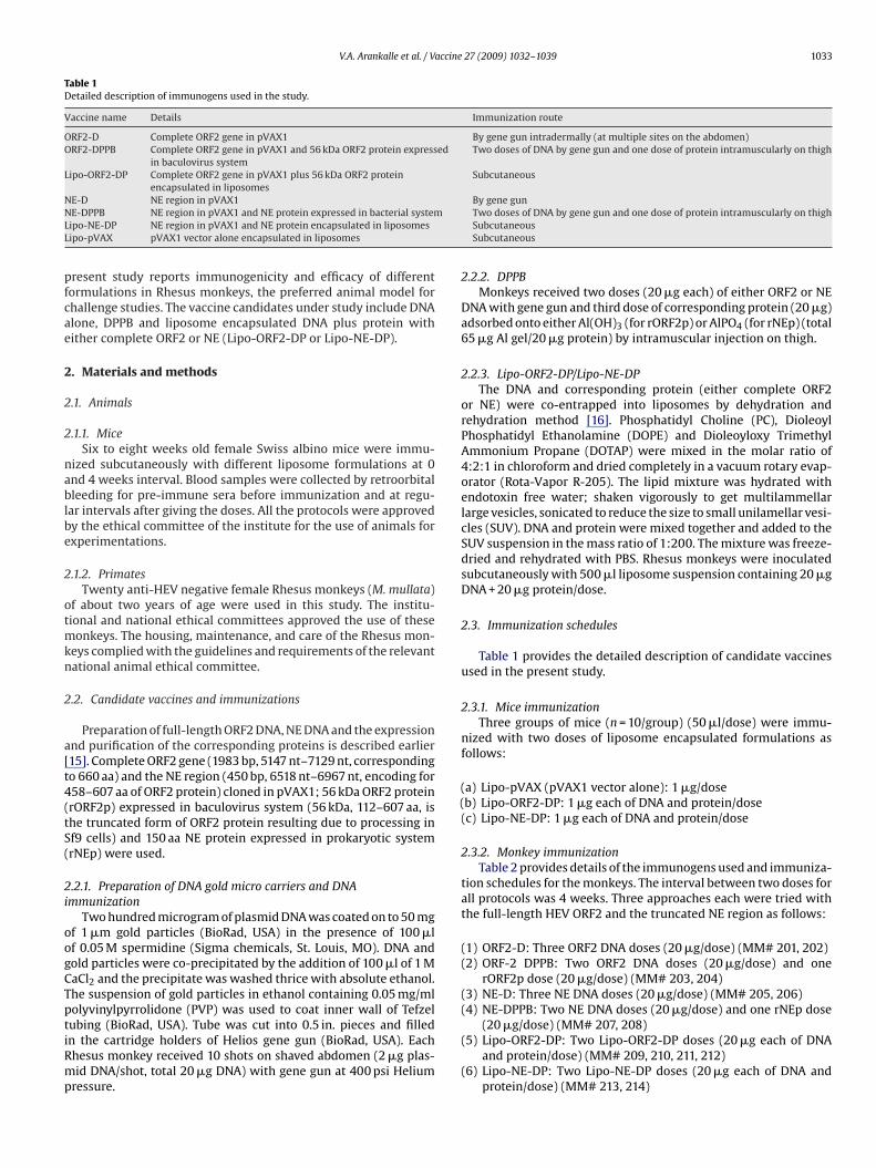
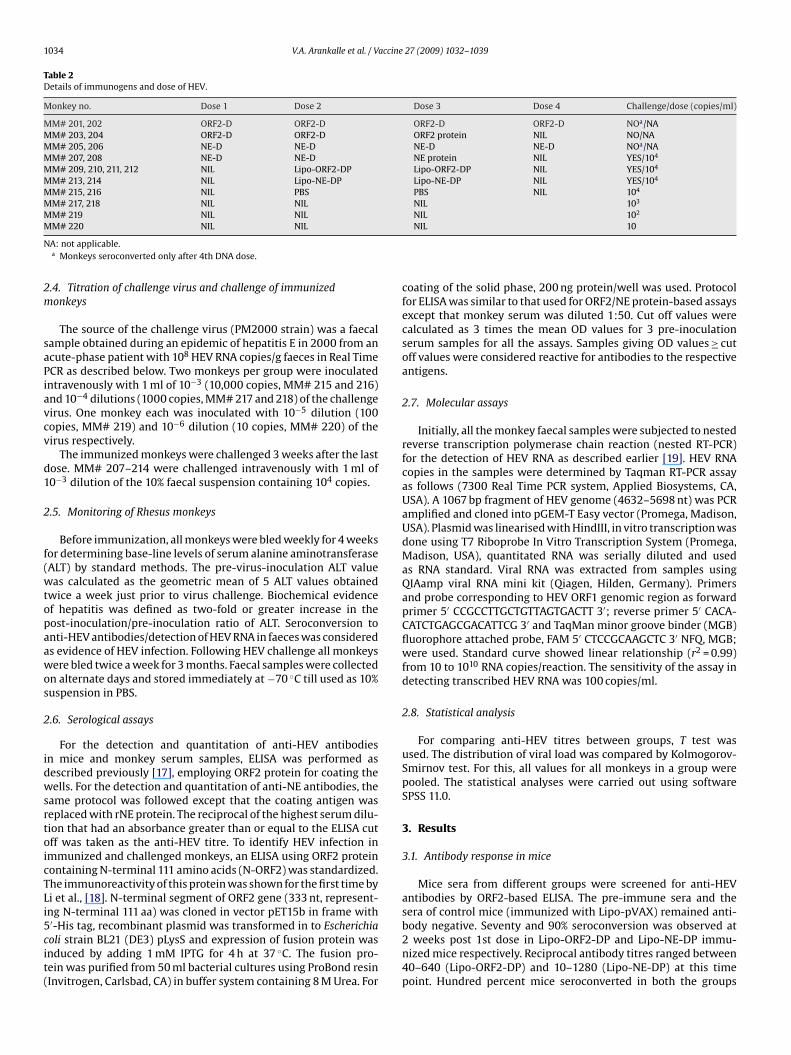
V.A. Arankalle et al. / Vaccine 27 (2009) 1032–1039 1033
Table 1Detailed description of immunogens used in the study.
Vaccine name Details Immunization route
ORF2-D Complete ORF2 gene in pVAX1 By gene gun intradermally (at multiple sites on the abdomen)ORF2-DPPB Complete ORF2 gene in pVAX1 and 56 kDa ORF2 protein expressed
in baculovirus systemTwo doses of DNA by gene gun and one dose of protein intramuscularly on thigh
Lipo-ORF2-DP Complete ORF2 gene in pVAX1 plus 56 kDa ORF2 proteinencapsulated in liposomes
Subcutaneous
NNLL
pfcae
2
2
2
nablbe
2
otmkn
2
a[t4(tS(
2i
oogCTptiRmp
((
(4) NE-DPPB: Two NE DNA doses (20 �g/dose) and one rNEp dose
E-D NE region in pVAX1E-DPPB NE region in pVAX1 and NE protein expressed in bacterial systemipo-NE-DP NE region in pVAX1 and NE protein encapsulated in liposomesipo-pVAX pVAX1 vector alone encapsulated in liposomes
resent study reports immunogenicity and efficacy of differentormulations in Rhesus monkeys, the preferred animal model forhallenge studies. The vaccine candidates under study include DNAlone, DPPB and liposome encapsulated DNA plus protein withither complete ORF2 or NE (Lipo-ORF2-DP or Lipo-NE-DP).
. Materials and methods
.1. Animals
.1.1. MiceSix to eight weeks old female Swiss albino mice were immu-
ized subcutaneously with different liposome formulations at 0nd 4 weeks interval. Blood samples were collected by retroorbitalleeding for pre-immune sera before immunization and at regu-
ar intervals after giving the doses. All the protocols were approvedy the ethical committee of the institute for the use of animals forxperimentations.
.1.2. PrimatesTwenty anti-HEV negative female Rhesus monkeys (M. mullata)
f about two years of age were used in this study. The institu-ional and national ethical committees approved the use of these
onkeys. The housing, maintenance, and care of the Rhesus mon-eys complied with the guidelines and requirements of the relevantational animal ethical committee.
.2. Candidate vaccines and immunizations
Preparation of full-length ORF2 DNA, NE DNA and the expressionnd purification of the corresponding proteins is described earlier15]. Complete ORF2 gene (1983 bp, 5147 nt–7129 nt, correspondingo 660 aa) and the NE region (450 bp, 6518 nt–6967 nt, encoding for58–607 aa of ORF2 protein) cloned in pVAX1; 56 kDa ORF2 proteinrORF2p) expressed in baculovirus system (56 kDa, 112–607 aa, ishe truncated form of ORF2 protein resulting due to processing inf9 cells) and 150 aa NE protein expressed in prokaryotic systemrNEp) were used.
.2.1. Preparation of DNA gold micro carriers and DNAmmunization
Two hundred microgram of plasmid DNA was coated on to 50 mgf 1 �m gold particles (BioRad, USA) in the presence of 100 �lf 0.05 M spermidine (Sigma chemicals, St. Louis, MO). DNA andold particles were co-precipitated by the addition of 100 �l of 1 MaCl2 and the precipitate was washed thrice with absolute ethanol.he suspension of gold particles in ethanol containing 0.05 mg/mlolyvinylpyrrolidone (PVP) was used to coat inner wall of Tefzel
ubing (BioRad, USA). Tube was cut into 0.5 in. pieces and filledn the cartridge holders of Helios gene gun (BioRad, USA). Eachhesus monkey received 10 shots on shaved abdomen (2 �g plas-id DNA/shot, total 20 �g DNA) with gene gun at 400 psi Heliumressure.
By gene gunTwo doses of DNA by gene gun and one dose of protein intramuscularly on thighSubcutaneousSubcutaneous
2.2.2. DPPBMonkeys received two doses (20 �g each) of either ORF2 or NE
DNA with gene gun and third dose of corresponding protein (20 �g)adsorbed onto either Al(OH)3 (for rORF2p) or AlPO4 (for rNEp) (total65 �g Al gel/20 �g protein) by intramuscular injection on thigh.
2.2.3. Lipo-ORF2-DP/Lipo-NE-DPThe DNA and corresponding protein (either complete ORF2
or NE) were co-entrapped into liposomes by dehydration andrehydration method [16]. Phosphatidyl Choline (PC), DioleoylPhosphatidyl Ethanolamine (DOPE) and Dioleoyloxy TrimethylAmmonium Propane (DOTAP) were mixed in the molar ratio of4:2:1 in chloroform and dried completely in a vacuum rotary evap-orator (Rota-Vapor R-205). The lipid mixture was hydrated withendotoxin free water; shaken vigorously to get multilammellarlarge vesicles, sonicated to reduce the size to small unilamellar vesi-cles (SUV). DNA and protein were mixed together and added to theSUV suspension in the mass ratio of 1:200. The mixture was freeze-dried and rehydrated with PBS. Rhesus monkeys were inoculatedsubcutaneously with 500 �l liposome suspension containing 20 �gDNA + 20 �g protein/dose.
2.3. Immunization schedules
Table 1 provides the detailed description of candidate vaccinesused in the present study.
2.3.1. Mice immunizationThree groups of mice (n = 10/group) (50 �l/dose) were immu-
nized with two doses of liposome encapsulated formulations asfollows:
a) Lipo-pVAX (pVAX1 vector alone): 1 �g/doseb) Lipo-ORF2-DP: 1 �g each of DNA and protein/dose
(c) Lipo-NE-DP: 1 �g each of DNA and protein/dose
2.3.2. Monkey immunizationTable 2 provides details of the immunogens used and immuniza-
tion schedules for the monkeys. The interval between two doses forall protocols was 4 weeks. Three approaches each were tried withthe full-length HEV ORF2 and the truncated NE region as follows:
(1) ORF2-D: Three ORF2 DNA doses (20 �g/dose) (MM# 201, 202)(2) ORF-2 DPPB: Two ORF2 DNA doses (20 �g/dose) and one
rORF2p dose (20 �g/dose) (MM# 203, 204)(3) NE-D: Three NE DNA doses (20 �g/dose) (MM# 205, 206)
(20 �g/dose) (MM# 207, 208)(5) Lipo-ORF2-DP: Two Lipo-ORF2-DP doses (20 �g each of DNA
and protein/dose) (MM# 209, 210, 211, 212)(6) Lipo-NE-DP: Two Lipo-NE-DP doses (20 �g each of DNA and
protein/dose) (MM# 213, 214)
1034 V.A. Arankalle et al. / Vaccine 27 (2009) 1032–1039
Table 2Details of immunogens and dose of HEV.
Monkey no. Dose 1 Dose 2 Dose 3 Dose 4 Challenge/dose (copies/ml)
MM# 201, 202 ORF2-D ORF2-D ORF2-D ORF2-D NOa/NAMM# 203, 204 ORF2-D ORF2-D ORF2 protein NIL NO/NAMM# 205, 206 NE-D NE-D NE-D NE-D NOa/NAMM# 207, 208 NE-D NE-D NE protein NIL YES/104
MM# 209, 210, 211, 212 NIL Lipo-ORF2-DP Lipo-ORF2-DP NIL YES/104
MM# 213, 214 NIL Lipo-NE-DP Lipo-NE-DP NIL YES/104
MM# 215, 216 NIL PBS PBS NIL 104
MM# 217, 218 NIL NIL NIL 103
MM# 219 NIL NIL NIL 102
M
N
2m
saPiavcv
d1
2
f(wtopaawos
2
idwsrtoicTLi5cit(
M# 220 NIL NIL
A: not applicable.a Monkeys seroconverted only after 4th DNA dose.
.4. Titration of challenge virus and challenge of immunizedonkeys
The source of the challenge virus (PM2000 strain) was a faecalample obtained during an epidemic of hepatitis E in 2000 from ancute-phase patient with 108 HEV RNA copies/g faeces in Real TimeCR as described below. Two monkeys per group were inoculatedntravenously with 1 ml of 10−3 (10,000 copies, MM# 215 and 216)nd 10−4 dilutions (1000 copies, MM# 217 and 218) of the challengeirus. One monkey each was inoculated with 10−5 dilution (100opies, MM# 219) and 10−6 dilution (10 copies, MM# 220) of theirus respectively.
The immunized monkeys were challenged 3 weeks after the lastose. MM# 207–214 were challenged intravenously with 1 ml of0−3 dilution of the 10% faecal suspension containing 104 copies.
.5. Monitoring of Rhesus monkeys
Before immunization, all monkeys were bled weekly for 4 weeksor determining base-line levels of serum alanine aminotransferaseALT) by standard methods. The pre-virus-inoculation ALT valueas calculated as the geometric mean of 5 ALT values obtained
wice a week just prior to virus challenge. Biochemical evidencef hepatitis was defined as two-fold or greater increase in theost-inoculation/pre-inoculation ratio of ALT. Seroconversion tonti-HEV antibodies/detection of HEV RNA in faeces was considereds evidence of HEV infection. Following HEV challenge all monkeysere bled twice a week for 3 months. Faecal samples were collected
n alternate days and stored immediately at −70 ◦C till used as 10%uspension in PBS.
.6. Serological assays
For the detection and quantitation of anti-HEV antibodiesn mice and monkey serum samples, ELISA was performed asescribed previously [17], employing ORF2 protein for coating theells. For the detection and quantitation of anti-NE antibodies, the
ame protocol was followed except that the coating antigen waseplaced with rNE protein. The reciprocal of the highest serum dilu-ion that had an absorbance greater than or equal to the ELISA cutff was taken as the anti-HEV titre. To identify HEV infection inmmunized and challenged monkeys, an ELISA using ORF2 proteinontaining N-terminal 111 amino acids (N-ORF2) was standardized.he immunoreactivity of this protein was shown for the first time byi et al., [18]. N-terminal segment of ORF2 gene (333 nt, represent-ng N-terminal 111 aa) was cloned in vector pET15b in frame with
′-His tag, recombinant plasmid was transformed in to Escherichiaoli strain BL21 (DE3) pLysS and expression of fusion protein wasnduced by adding 1 mM IPTG for 4 h at 37 ◦C. The fusion pro-ein was purified from 50 ml bacterial cultures using ProBond resinInvitrogen, Carlsbad, CA) in buffer system containing 8 M Urea. ForNIL 10
coating of the solid phase, 200 ng protein/well was used. Protocolfor ELISA was similar to that used for ORF2/NE protein-based assaysexcept that monkey serum was diluted 1:50. Cut off values werecalculated as 3 times the mean OD values for 3 pre-inoculationserum samples for all the assays. Samples giving OD values ≥ cutoff values were considered reactive for antibodies to the respectiveantigens.
2.7. Molecular assays
Initially, all the monkey faecal samples were subjected to nestedreverse transcription polymerase chain reaction (nested RT-PCR)for the detection of HEV RNA as described earlier [19]. HEV RNAcopies in the samples were determined by Taqman RT-PCR assayas follows (7300 Real Time PCR system, Applied Biosystems, CA,USA). A 1067 bp fragment of HEV genome (4632–5698 nt) was PCRamplified and cloned into pGEM-T Easy vector (Promega, Madison,USA). Plasmid was linearised with HindIII, in vitro transcription wasdone using T7 Riboprobe In Vitro Transcription System (Promega,Madison, USA), quantitated RNA was serially diluted and usedas RNA standard. Viral RNA was extracted from samples usingQIAamp viral RNA mini kit (Qiagen, Hilden, Germany). Primersand probe corresponding to HEV ORF1 genomic region as forwardprimer 5′ CCGCCTTGCTGTTAGTGACTT 3′; reverse primer 5′ CACA-CATCTGAGCGACATTCG 3′ and TaqMan minor groove binder (MGB)fluorophore attached probe, FAM 5′ CTCCGCAAGCTC 3′ NFQ, MGB;were used. Standard curve showed linear relationship (r2 = 0.99)from 10 to 1010 RNA copies/reaction. The sensitivity of the assay indetecting transcribed HEV RNA was 100 copies/ml.
2.8. Statistical analysis
For comparing anti-HEV titres between groups, T test wasused. The distribution of viral load was compared by Kolmogorov-Smirnov test. For this, all values for all monkeys in a group werepooled. The statistical analyses were carried out using softwareSPSS 11.0.
3. Results
3.1. Antibody response in mice
Mice sera from different groups were screened for anti-HEVantibodies by ORF2-based ELISA. The pre-immune sera and thesera of control mice (immunized with Lipo-pVAX) remained anti-
body negative. Seventy and 90% seroconversion was observed at2 weeks post 1st dose in Lipo-ORF2-DP and Lipo-NE-DP immu-nized mice respectively. Reciprocal antibody titres ranged between40–640 (Lipo-ORF2-DP) and 10–1280 (Lipo-NE-DP) at this timepoint. Hundred percent mice seroconverted in both the groups
V.A. Arankalle et al. / Vaccine
Fig. 1. (a) Percent seroconversion in Swiss albino mice at 2 weeks (2 weeks post dose1) and 6 weeks (2 weeks post dose 2). Mice groups (n = 10/group) were immunizedat 0 and 4 weeks with either Lipo-ORF2-DP or Lipo-NE-DP. (b) Serum HEV-specificanti-rORF2p IgG antibody mean log (10) titres detected in ELISA in Swiss albinomLt
aaAg
3
kTssttdakiepa(ipc
non-significant (p > 0.4).
ice. Mice groups (n = 10/group) were immunized at 0 and 4 weeks with eitheripo-ORF2-DP or Lipo-NE-DP. Error bars represent standard error of the mean logitres.
t 2 weeks post 2nd dose (6 weeks) with further increase in thentibody titres to 800–6400 and 800–12,800 respectively (Fig. 1).t both time points anti-HEV titres were comparable in both theroups.
.2. Titration of the challenge inoculum
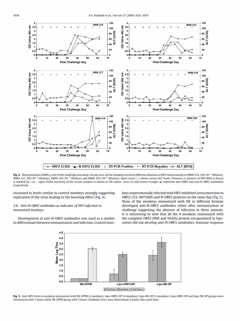
As the challenge inoculum was not pre-titrated in Rhesus mon-eys, number of HEV RNA copies was determined employing Realime PCR and was estimated to be 107 copies/ml of the 10% stooluspension. Considering Real Time PCR to be ∼10-fold more sen-itive than infectivity in monkeys, the challenge dose was decidedo be 10−3, i.e. 104 copies/ml. Fig. 2 depicts dynamics of HEV infec-ion in control, unimmunized monkeys inoculated with differentoses of the virus. Both monkeys inoculated with 104 (MM# 215nd 216) and 103 (MM# 217 and 218) copies each and the only mon-ey infected with 102 copies (MM# 219) showed evidence of HEVnfection as indicated by seroconversion to anti-HEV antibodies andxcretion of the virus in faeces. Rise in serum ALT (≥two-times there-virus-inoculation levels) was recorded for MM# 215, 216, 218nd 219. The monkey (MM# 220) inoculated with 10−6 dilution
10 copies/ml) remained IgG-anti-HEV negative. Thus the infectiv-ty titre of the 10% stool suspension was estimated to be 106 fiftyercent monkey infectious dose (MID50) per gram of faeces and thehallenge inoculum contained 100 MID50 HEV.27 (2009) 1032–1039 1035
3.3. Anti-HEV response in immunized monkeys
Tables 1 and 2 provide details of the immunogens and differentvaccine approaches evaluated. All the pre-immune sera taken priorto the first immunization were negative for IgG-anti-HEV.
3.3.1. Vaccine approaches not yielding encouraging resultsNone of the monkeys receiving ORF2-DNA, NE-DNA or ORF2-
DNA followed by ORF2 protein boost showed the presence ofanti-HEV antibodies. As the protocol demanded challenge after thethird dose and these monkeys did not seroconvert after the thirddose, we took the option of use of fourth DNA dose at 12 weeksfor checking seroconversion rather than challenging the monkeysin the absence of anti-HEV antibodies. These monkeys showedweak (anti-HEV titre 1:100) seroconversion at 2 weeks post fourthdose. Thus of the 6 approaches tested in Rhesus monkeys, threeapproaches including ORF2 and NE DNA alone and ORF2 DNA fol-lowed by protein boost were observed to be less effective.
3.3.2. Vaccine approaches evaluated by virus challengeThe monkeys immunized employing NE-DPPB approach did not
develop anti-HEV antibodies after two DNA doses. Seroconversionwas noted 4 weeks after the protein boost (titres 1:1600 and 800).Of the four monkeys receiving Lipo-ORF2-DP, one seroconverted3 weeks after the first dose (anti-HEV titre 1:100), all four beinganti-HEV positive 1 week after the second dose (anti-HEV titres:1:100–800). Both the monkeys receiving Lipo-NE-DP seroconverted3 weeks after the first dose, anti-HEV titres being 800 and 100respectively. One week after the second dose, the anti-HEV titresrose to 6400 and 800. Thus, for each group, the pattern of serumantibody was similar in all the monkeys, although antibody levelwas different. As compared to ORF2, anti-HEV titres produced byNE were higher, though statistically insignificant (p = 0.053) (Fig. 3).
3.4. Dynamics of HEV infection in control monkeys
Both the monkeys (MM# 215 and 216) infected with 100 MID50challenge virus exhibited moderate rise in serum ALT levels,maximum values being 77 and 42 IU/litre on 38 and 45 days post-inoculation respectively (Fig. 2). Virus excretion as measured byReal Time PCR was evident for 6 weeks, the maximum viral loadbeing 1.5 × 10⁷ and 6.0 × 10⁶ copies/g stool. Seroconversion fol-lowed by high titres of anti-HEV antibodies were recorded.
3.5. Assessment of HEV infection in challenged monkeys
Monkeys from NE-DPPB, Lipo-NE-DP and Lipo-ORF2-DP groupswere evaluated for protection following challenge. Irrespective oftype of immunogen, none of the challenged monkeys exhibitedraised ALT levels and were protected from hepatitis (Table 3). Com-plete protection from infection was offered by Lipo-NE-DP, bothanimals not excreting the virus. Similarly, one of the monkeysimmunized with NE-DPPB (MM# 207) also did not show any evi-dence of virus replication, HEV RNA being absent in all the faecalsamples screened. The other animal (MM# 208) showed reducedexcretion for a shorter time. Lipo-ORF2-DP was least effective, allthe 4 monkeys excreting the virus for extended period of time.As compared to the control monkeys, overall viral load in fae-ces was significantly less in monkeys immunized with NE-DPPB(p < 0.001); no difference was noted in Lipo-ORF2-DP immunizedmonkeys (p > 0.4). Among NE-immunized groups the difference was
Comparison of IgG-anti-HEV titres in the challenged monkeysshowed that except two monkeys (MM# 209 and 211) immunizedwith Lipo-ORF2-DP, all other monkeys exhibited either same ordeclining antibody titres. Anti-HEV titres in MM# 209 and 211
1036 V.A. Arankalle et al. / Vaccine 27 (2009) 1032–1039
F onkeyM on). Oi alues;r
ir
3i
t
Fi
ig. 2. Determination of MID50 titre of the challenge inoculum. On day zero, all the mM# 217, 218 (10−4 dilution), MM# 219 (10−5 dilution) and MM# 220 (10−6 diluti
s marked by + or − signs. ELISA reactivity of the serum samples is shown as OD vespectively.
ncreased to levels similar to control monkeys strongly suggestingeplication of the virus leading to the boosting effect (Fig. 4).
.6. Anti-N-ORF2 antibodies as indicator of HEV infection inmmunized monkeys
Development of anti-N-ORF2 antibodies was used as a markero differentiate between immunization and infection. Control mon-
ig. 3. Anti-HEV titres in monkeys immunized with NE-DPPB (2 monkeys), Lipo-ORF2-DPmmunized with 2 doses while NE-DPPB group with 3 doses. Antibody titres were determ
s received different dilutions of HEV intravenously as MM# 215, 216 (10−3 dilution),pen circle (©) shows serum ALT levels. Presence or absence of HEV RNA in faecescross (x) and closed triangle (�) represent anti-ORF2 and anti-N-ORF2 antibodies
keys experimentally infected with HEV exhibited seroconversion toORF2 (112–607/660) and N-ORF2 proteins on the same day (Fig. 2).None of the monkeys immunized with NE in different formats
developed anti-N-ORF2 antibodies either after immunization orchallenge suggesting the absence of infection in these animals.It is interesting to note that all the 4 monkeys immunized withthe complete ORF2 DNA and 56 kDa protein encapsulated in lipo-somes did not develop anti-N-ORF2 antibodies. Immune response(4 monkeys), Lipo-NE-DP (2 monkeys). Lipo-ORF2-DP and Lipo-NE-DP groups wereined 4 weeks after each dose.

V.A. Arankalle et al. / Vaccine 27 (2009) 1032–1039 1037
Fig. 4. HEV challenge of monkeys immunized with different preparations. Dose-1, 2 and 3 show schedule of immunizations. Arrow indicates viral challenge of 102 MID50.Open circle (©) shows serum ALT levels. Presence or absence of HEV RNA in faeces is marked by + or − signs. ELISA reactivity of the serum samples is shown as OD values;cross (x) and closed triangle (�) represents anti-ORF2 and anti-N-ORF2 antibodies respectively. MM# 207, 208 were immunized with NE DNA-prime-protein-boost, MM#209–212 were immunized with ORF2-liposome and MM# 213, 214 received NE-liposome. The dynamics of HEV infection in control monkeys (MM# 215 and 216) inoculatedwith the same dose of the virus (102 MID50) is depicted in Fig. 2.
Table 3Summary of challenge experiment.
Vaccine type Monkey no. Reciprocal anti-HEVtitre at challenge
Peak/pre-challenge ratio of ALTvalues (weeks elevated)
HEV RNA copies in faeces, peak titre/ml in10% stool suspension (duration in weeks)
NE DPPB MM# 207 1600 1.0 (0) Not detectedMM# 208 800 1.7 (0) 2.2 × 10⁴ (3)
Lipo-ORF2-DP MM# 209 200 1.1 (0) 2.9 × 10⁶ (6)MM# 210 1600 1.09 (0) 1.0 × 10⁴ (3)MM# 211 200 1.09 (0) 2.6 × 10⁶ (5)MM# 212 200 1.35 (0) 1.0 × 10⁴ (6)
Lipo-NE-DP MM# 213 12,800 1.06 (0) Not detectedMM# 214 1600 1.06 (0) Not detected
Placebo MM# 215 <10 3.6 (2) 1.5 × 10⁷ (6)MM# 216 <10 2.1 (1) 6.0 × 10⁶ (6)

1 accine
wtOpaAkekdOt
3f
biTtaipiwoiawB
4
bi(sstotHLm
acNoeotnpakagit
(l
038 V.A. Arankalle et al. / V
as mainly targeted against the 112+ protein component. However,wo monkeys (MM# 209 and 211) showing >10-fold rise in anti-RF2 antibodies after challenge (evidence of infection) showed theresence of anti-N-ORF2 antibodies (Fig. 4). Rise in anti-HEV titresnd the detection of anti-N-ORF2 antibodies were simultaneous.nti-N-ORF2 antibodies were not detected in the other two mon-eys (MM# 210 and 212) immunized with Lipo-ORF2-DP and notxhibiting rise in anti-ORF2 titres after challenge and all the 4 mon-eys immunized with NE in different formats. These results clearlyemonstrated that two of the four monkeys immunized with Lipo-RF2-DP developed HEV infection following challenge whereas all
he four monkeys immunized with NE were protected.
.7. Comparison of anti-HEV titres employing rORF2p and rNEpor ELISA
As anti-NE antibodies were proposed to be the neutralizing anti-odies, we compared antibody titres following immunization and
nfection employing rORF2p and rNEp as coating antigens in ELISA.he titres were similar using both antigens. The anti-NE titres athe time of challenge in NE-immunized monkeys and protectedgainst hepatitis E were 800, 1600, 1600 and 12,800. In ORF2-mmunized monkeys, two with anti-NE titres of 1600 and 200 wererotected while the other two with the titres of 200 each were
nfected, though protected from disease. Overall, all the monkeysith anti-NE titres of ≥200 were protected from hepatitis. Two
f the three monkeys with titres of 200 each showed evidence ofnfection and extensive virus replication. Of the five monkeys withnti-NE titres >200 (≥800) three exhibited sterilizing immunityhile two showed evidence of infection with reduced viral load.oth the control monkeys developed high anti-NE titres (>12,800).
. Discussion
Open reading frame 2 encoding a protein of 660 amino acids haseen the target for vaccine development for hepatitis E employ-
ng different approaches, the most successful being the 56 kDa112–607 aa) recombinant protein expressed in baculovirus expres-ion system [10,14]. Present study documents the utility of themaller (458–607, 150 aa) NE region containing the putative neu-ralization epitope [20] as a vaccine candidate. In continuationf mice experiments [15] evaluation of a different concept led tohe striking observation of early seroconversion and high anti-EV titres in mice immunized with two doses of Lipo-ORF2-DP oripo-NE-DP (Fig. 1) and prompted us to evaluate this approach inacaques.We tried both ORF2 and NE regions in three formats, i.e., DNA
lone, DNA-prime-protein-boost and encapsulation of DNA and theorresponding protein in liposomes. As DNA vaccine, both ORF2 andE were not able to mount antibody response when used in 3 dosesf 20 �g each with gene gun. An additional dose was required tolicit the antibody response in these monkeys questioning utilityf HEV DNA vaccine. As the protocol demanded challenge after thehird dose and the monkeys remained anti-HEV negative, we didot challenge them. Therefore, role of cell-mediated immunity inrotection against HEV infection could not be evaluated. Kamili etl. [13] have described complete protection of cynomolgus mon-eys immunized with 4 doses of full-length ORF2 DNA with theid of gene gun after challenge with 10,000 MID50 of the heterolo-ous challenge. We did observe superiority of gene gun in inducing
mmune response to the candidate DNA vaccines in mice, thoughhe results were not reproduced in Rhesus monkeys [15].In the prime-boost approach, ORF2 showed late seroconversion7 weeks post third dose) and therefore this group was not chal-enged. For NE, as against 3 DNA doses, DPPB approach was found to
27 (2009) 1032–1039
induce high titres of anti-HEV antibodies and protection indicatingimportant role of the NE protein in mounting immune response. Wedid not evaluate NE protein alone as an immunogen as the responsewas poor in mice [15].
Both ORF2 and NE were immunogenic when administered asLipo-DP formulations. NE appeared to be a better immunogen, bothimmunized animals seroconverting after one dose as compared to1 of the 4 animals immunized with Lipo-ORF2-DP. Though bothORF2 and NE produced comparable anti-HEV titres, their abilityto protect following HEV challenge was different. Excellent protec-tion was offered by Lipo-NE-DP as both the animals were protectedfrom the disease as well as infection. Absence of excretion of thevirus and anti-N-ORF2 antibodies strongly suggests development ofsterilizing immunity in these animals. Though Lipo-ORF2-DP wasimmunogenic and protected all the animals from hepatitis, 2 of the4 challenged monkeys showed evidence of HEV infection as indi-cated by rise in anti-ORF2 antibodies, extensive replication of thevirus and development of anti-N-ORF2 antibodies. This study alsoconfirms the findings of Zhou et al. [21] that anti-N-ORF2 antibodiesrepresent a useful serological marker for diagnosis of HEV infec-tion in individuals as well as animals immunized with ORF2-basedvaccines and warrants evaluation in an endemic setting.
Though anti-N-ORF2 antibodies were detected during infectionwith the virus, these antibodies were not detected in monkeysimmunized with complete ORF2 DNA in Lipo-ORF2-DP. Whetherthis reflects modulation of the immune response by the 112–607protein component of the vaccine remains to be seen.
We would like to point out here that NE was also shown to be agood immunogen in the DPPB format. MM# 207 developed steril-izing immunity whereas MM# 208 showed significantly low viralload, both monkeys being negative for anti-N-ORF2 antibodies.
We used homologous virus (genotype 1) for challenge. How-ever, based on the cross-genotype protection reported so far [22]we tend to believe that the candidate vaccines will offer protec-tion against all the HEV genotypes. Low virus dose (100 MID50)used for challenge reflecting natural exposure led to moderate (77)and marginally high (42) rise in ALT levels in the control animals.Demonstration of protection against severe hepatitis followinghigher challenge virus dose is essential in subsequent experiments.Our results clearly demonstrate utility of NE in protecting animalsagainst infection, the most important finding of the study.
This study confirms the observations by Zhou et al. [20] that theanti-HEV profiles in experimentally infected monkeys were almostidentical with either 112–607 aa or 458–607 aa ORF2 proteins ascoating antigens in ELISA. A striking finding was, despite substan-tial replication of the virus, no ALT rise was observed in two of thechallenged monkeys (MM# 209 and 211). We agree with the possi-bility proposed by Zhang et al. [23] that the extent of liver damagein hepatitis E may not be a direct reflection of virus titre.
In our earlier experiments in mice, ORF2 DNA when admin-istered with gene gun was found to be better immunogen thanNE DNA whereas in the DPPB format, boosting effect was promi-nent with NE protein [15]. Absence of anti-HEV antibodies after 3ORF2/NE DNA doses, late serocoversion with ORF2 DPPB approachand better boosting effect of NE protein demonstrate partial agree-ment between results obtained in mice and monkeys.
Zhou et al. [21] have proposed the monitoring of anti-NEantibodies in assessing neutralizing antibody response followingimmunization. From our study it appears that anti-NE titre as lowas 200 may indicate protection against disease. However, theseantibodies do not seem to reflect protection from infection. Consid-
ering the fact that NE offers complete protection of monkeys, thepossibility of non-anti-NE antibody mediated immune response inprotection needs to be evaluated.Though 56 kDa protein vaccine has undergone a successful clin-ical trial, attempts for the development of better vaccines must

accine
ceoplotlLoNutmo
Di4mie
A
otRWPco
R
[
[
[
[
[
[
[
[
[
[
[
[
[23] Zhang M, Emerson S, Nguyen H, Engle R, Govindarajan S, Blackwelder W, et al.Recombinant vaccine against hepatitis E: duration of protective immunity in
V.A. Arankalle et al. / V
ontinue. Our experiments provide the possibility of use of a small,asy to purify protein employing bacterial expression system withbvious advantages over the baculovirus expression with multipleurification steps [24]. In the DNA/protein liposome encapsu-
ation format, NE was distinctly superior to ORF2. Comparisonf liposome-NE protein with liposome-NE-protein-DNA (showno be the best formulation in this study using 2 animals) in aarger number of Rhesus monkeys would be the next logical step.iposome-based candidate vaccines are already in different phasesf clinical trial (Clinical Trial Identifier No. NCT00001042 ando. NCT00197301, http://www.ClinicalTrials.gov). The technologysed in encapsulation does not need sophisticated instrumen-ation/protocols and could be easily produced by a vaccine
anufacturing company of international standards from a devel-ping country where the disease is endemic.
In conclusion, our results document that both ORF2 and NENA were poorly immunogenic in macaques. NE region was highly
mmunogenic in DPPB as well as DP-liposome formats, 3 of theimmunized monkeys developing sterilizing immunity. The for-ulation generated by the encapsulation of NE DNA and protein
n liposome offered best protection and merits further in-depthvaluation.
cknowledgements
The authors thank Dr. Mishra, A.C., Director, National Institutef Virology and Dr. Kapre, S.V., Executive Director, Serum Insti-ute of India, Pune for all the encouragement. Timely help by Dr.aut, C.S. is gratefully acknowledged. Sincere thanks are due to Mr.alkoli, V.M., Jawalkar, P.B., Tilekar, B.N., Ranawade, S.S. and Sarje,
. for excellent technical assistance and Walimbe, A.M. for statisti-al analysis. Financial support was provided by the Indian Councilf Medical Research, India.
eferences
[1] Khuroo MS, Teli RT, Skidmore S, Sofi MA, Khuroo HI. Incidence and severity ofviral hepatitis in pregnancy. Am J Med 1981;70:252–5.
[2] Boccia D, Guthmann J, Klovstad H, Hamid N, Tatay M, Ciglenecki I, et al. Highmortality associated with an outbreak of Hepatitis E among displaced personsin Darfur, Sudan. Clin Infect Dis 2006;42:1679–84.
[3] Arankalle VA, Jha J, Favorov MO, Chaudhari A, Fields AH, Banerjee K. Contribu-tion of HEV and HCV in causing fulminant non-A, non-B hepatitis in WesternIndia. J Viral Hepat 1995;2:189–93.
[4] Drobeniuc J, Favorov MO, Shapiro CN, Bell BP, Mast EE, Dadu A, et al. HepatitisE virus antibody prevalence among persons who work with swine. J Infect Dis2001;184:1594–7.
[5] Meng XJ, Halbur PG, Shapiro MS, Govindarajan S, Bruna JD, Mushahwar IK,et al. Genetic and experimental evidence for cross-species infection by swinehepatitis E virus. J Virol 1998;72:9714–21.
[
27 (2009) 1032–1039 1039
[6] Tamada Y, Yano K, Yatsuhashi H, Inoue O, Mawatari F, Ishibashi H. Consumptionof wild boar linked to cases of hepatitis E. J Hepatol 2004;40:869–70.
[7] Tei S, Kitajima N, Ohara S, Inoue Y, Miki M, Yamatani T, et al. Consumption ofuncooked deer meat as a risk factor for hepatitis E virus infection: an age andsex-matched case-control study. J Med Virol 2004;74:67–70.
[8] Yazaki Y, Mizuo H, Takahashi M, Nishizawa T, Sasaki N, Gotanda Y, et al. Spo-radic acute or fulminant hepatitis E in Hokkaido, Japan, may be food-borne, assuggested by the presence of hepatitis E virus in pig liver as food. J Gen Virol2003;84:2351–7.
[9] Viswanathan R, Naidu S. Infectious hepatitis in Delhi 1955–1956: a criticalstudy; observations in pregnant women. Indian J Med Res 1957;45(Suppl.1):1–30.
10] Tsarev SA, Tsareva TS, Emerson SU, Govindarajan S, Shapiro M, Gerin JL, et al.Recombinant vaccine against hepatitis E: dose response and protection againstheterologous challenge. Vaccine 1997;15:1834–8.
[11] Li SW, Zhang J, Li YM, Ou SH, Huang GY, He ZQ, et al. A bacterially expressedparticulate hepatitis E vaccine: antigenicity, immunogenicity and protectivityon primates. Vaccine 2005;23:2893–901.
12] Kamili S, Spelbring J, Krawczynski K. DNA vaccination against hepatitis E virusinfection in cynomolgus macaques. J Gastroenterol Hepatol 2002;17(Suppl.3):S365–9.
13] Kamili S, Spelbring J, Carson D, Krawczynski K. Protective efficacy of hepatitisE virus DNA vaccine administered by gene gun in the cynomolgus macaquemodel of infection. J Infect Dis 2004;189:258–64.
14] Shrestha MP, Scott RM, Shrestha SK, Myint KSA, Mammen MP, Joshi DM,et al. Safety and efficacy of a hepatitis E virus vaccine. N Engl J Med2007;356:895–903.
15] Deshmukh TM, Lole KS, Tripathy AS, Arankalle VA. Immunogenicity of candi-date hepatitis E virus DNA vaccine expressing complete and truncated ORF2 inmice. Vaccine 2007;25:4350–60.
16] Gregoriadis G, McCormack B, Obrenovic M, Saffie R, Zadi B, Perrie Y. Vaccineentrapment in liposomes. Methods 1999;19:156–62.
17] Arankalle VA, Lole KS, Deshmukh TM, Chobe LP, Gandhe SS. Evaluation of human(genotype 1) and swine (genotype 4)-ORF-2 based ELISAs for anti-HEV IgMand IgG detection in an endemic country and search for type 4 human HEVinfections. J Viral Hepat 2007;14:435–45.
18] Li F, Torresi J, Locarnini SA, Zhuang H, Zhu W, Guo X, et al. Amino-terminalepitopes are exposed when full-length open reading frame 2 of hepatitis E virusis expressed in Escherichia coli, but carboxy-terminal epitopes are masked. J MedVirol 1997;52:289–300.
19] Arankalle VA, Chobe LP, Joshi MV, Chadha MS, Kundu B, Walimbe AM. Humanand swine hepatitis E viruses from Western India belong to different genotypes.J Hepatol 2002;36:417–25.
20] Zhou YH, Purcell RH, Emerson SU. An ELISA for putative neutralizing antibod-ies to hepatitis E virus detects antibodies to genotypes 1, 2, 3, and 4. Vaccine2004;22:2578–85.
21] Zhou YH, Purcell RH, Emerson SU. A truncated ORF2 protein contains the mostimmunogenic site on ORF2: antibody responses to non-vaccine sequences fol-lowing challenge of vaccinated and non-vaccinated macaques with hepatitis Evirus. Vaccine 2005;2(23):3157–65.
22] Purcell RH, Nguyen H, Shapiro M, Engle RE, Govindarajan S, Blackwelder WC,et al. Pre-clinical immunogenicity and efficacy trial of a recombinant hepatitisE vaccine. Vaccine 2003;21:2607–15.
Rhesus macaques. Vaccine 2002;20:3285–91.24] Robinson RA, Burgess WH, Emerson SU. Structural characterization of recombi-
nant hepatitis E virus ORF2 proteins in baculovirus-infected insect cells. ProteinExpr Purif 1998;12:75–84.
Copyright © 2022 FDOKUMEN