Biochemical filtering of a protein-protein docking simulation ...
Upload
independentCategory
view
0download
0
The Xanthomonas citri effector protein PthA interacts with citrusproteins involved in nuclear transport, protein folding andubiquitination associated with DNA repair
MARIANE NORONHA DOMINGUES1, T IAGO ANTONIO DE SOUZA1, RAÚL ANDRÉS CERNADAS1,† ,MARIA LUIZA PEIXOTO DE OLIVEIRA1, CÁSSIA DOCENA2,‡ , CHUCK SHAKER FARAH2 ANDCELSO EDUARDO BENEDETTI1,*1Laboratório Nacional de Biociências, Centro Nacional de Pesquisa em Energia e Materiais, R. Giuseppe Máximo Scolfaro 10000, Campinas, SP, CP6192, Brazil2Departamento de Bioquímica, Instituto de Química, Universidade de São Paulo, Av. Prof Lineu Prestes 748, CEP 05508-000, São Paulo, SP, Brazil
SUMMARY
Xanthomonas axonopodis pv. citri utilizes the type III effectorprotein PthA to modulate host transcription to promote citruscanker. PthA proteins belong to the AvrBs3/PthA family and carrya domain comprising tandem repeats of 34 amino acids thatmediates protein–protein and protein–DNA interactions. Weshow here that variants of PthAs from a single bacterial strainlocalize to the nucleus of plant cells and form homo- and het-erodimers through the association of their repeat regions. Wehypothesize that the PthA variants might also interact withdistinct host targets. Here, in addition to the interaction witha-importin, known to mediate the nuclear import of AvrBs3, wedescribe new interactions of PthAs with citrus proteins involvedin protein folding and K63-linked ubiquitination. PthAs 2 and 3preferentially interact with a citrus cyclophilin (Cyp) and withTDX, a tetratricopeptide domain-containing thioredoxin. In addi-tion, PthAs 2 and 3, but not 1 and 4, interact with the ubiquitin-conjugating enzyme complex formed by Ubc13 and ubiquitin-conjugating enzyme variant (Uev), required for K63-linkedubiquitination and DNA repair. We show that Cyp, TDX and Uevinteract with each other, and that Cyp and Uev localize to thenucleus of plant cells. Furthermore, the citrus Ubc13 and Uevproteins complement the DNA repair phenotype of the yeastDubc13 and Dmms2/uev1a mutants, strongly indicating thatthey are also involved in K63-linked ubiquitination and DNArepair. Notably, PthA 2 affects the growth of yeast cells in thepresence of a DNA damage agent, suggesting that it inhibitsK63-linked ubiquitination required for DNA repair.
INTRODUCTION
Citrus canker, caused by the bacterial pathogen Xanthomonasaxonopodis pv. citri, is characterized by water-soaked eruptionsand pustule formation on the surface of the host plant (Bruningsand Gabriel, 2003). Although it is known that canker lesionsresult from the intense division and expansion of the host cells atthe site of infection, the molecular events leading to cell hyper-trophy and hyperplasia are not fully comprehended. Previousstudies have shown that X. axonopodis pv. citri infectionchanges the transcription of a large set of host genes associatedwith cell division and expansion, including genes involved inribosome biogenesis, cell wall remodelling, vesicle trafficking,and the synthesis and mobilization of auxin and gibberellin(Cernadas et al., 2008). The expression of the cell wall remodel-ling genes, as well as those involved in the synthesis and mobi-lization of auxin and gibberellin, was shown to be similarlyregulated by auxin and gibberellin, indicating that X. axonopodispv. citri changes the active contents of both hormones topromote cell division and enlargement (Cernadas and Benedetti,2009). Indeed, it was found that both auxin and gibberellin arerequired for canker development and that polar transport ofauxin and vesicle trafficking mediate this process (Cernadas andBenedetti, 2009; Cernadas et al., 2008).
The transcriptional changes associated with the growth andexpansion of citrus cells in response to X. axonopodis pv. citriinfection are thought to be triggered by the Type III effectorprotein PthA. PthA is recognized as the major determinant of X.axonopodis pv. citri pathogenicity (Al-Saadi et al., 2007; Brun-ings and Gabriel, 2003). This protein is required for pathogengrowth and canker elicitation, and is sufficient to promote hyper-trophy and hyperplasia in citrus (Duan et al., 1999; Swarup et al.,1991, 1992). PthA belongs to the AvrBs3/PthA protein family.Members of this family, also known as transcription activator-like(TAL) effectors, are translocated by the Type III secretion system
*Correspondence: Email: [email protected]†Present address: Department of Plant Pathology, Iowa State University, Ames, IA, USA.‡Present address: Fundação Oswaldo Cruz, 50670-420, Recife, PE, Brazil.
MOLECULAR PLANT PATHOLOGY DOI: 10.1111/J .1364-3703.2010.00636.X
© 2010 THE AUTHORSJOURNAL COMPILATION © 2010 BLACKWELL PUBLISHING LTD 1

and function as transcriptional activators in the host (Kayet al., 2007; Römer et al., 2007; Sugio et al., 2007; Yang et al.,2006).
One of the most surprising features of AvrBs3/PthA proteins isthat they have an internal region comprising variable, nearlyidentical tandem repeats of 34 amino acids that determine bothpathogenicity and avirulence (Kay and Bonas, 2009; Swarupet al., 1992; Yang and Gabriel, 1995). For instance, AvrBs3, themost well-characterized TAL effector, dimerizes through itsrepeat region and interacts with an a-importin (a-Imp) thatmediates its transfer to the nucleus of the host cell (Gürlebecket al., 2005; Szurek et al., 2001). In the nucleus, AvrBs3 recog-nizes plant promoters and activates the transcription of a helix–loop–helix factor required for cell enlargement in susceptibleplants, whereas, in resistant plants, it transactivates its cognateBs3 resistance gene, triggering a hypersensitive reaction (Kayet al., 2007; Römer et al., 2007). Most remarkably, it is the repeatregion that mediates DNA recognition, and the amino acid poly-morphism found within the repeat units determines the DNAsequence specificity of TAL effectors (Boch et al., 2009; Moscouand Bogdanove, 2009).
Many Xanthomonas pathogens contain multiple variants ofAvrBs3/PthA proteins.The existence of multiple copies of avrBs3/pthA genes in a single strain is thought to facilitate recombina-tion and rapid adaptation to new hosts (Yang and Gabriel,1995). However, in the light of the findings of Boch et al. (2009)and Moscou and Bogdanove (2009), it is expected that multiplevariants of TAL effectors would also directly affect the transcrip-tion of target genes in the host and, consequently, the outcomeof the disease process. Accordingly, it has been shown thatmultiple TAL effectors contribute additively to symptom devel-opment (Yang and White, 2004; Yang et al., 1996). The X. axo-nopodis pv. citri strain 306 (da Silva et al., 2002) contains fourrelated PthA proteins (PthA 1–4) that differ from each otheressentially by the number of their 34-amino-acid repeats and bythe polymorphic residues found within the repeat units(Fig. S1A,B, see Supporting information). These PthA variants arethe equivalents of the four functionally characterized PthAs ofstrain 3213 (Al-Saadi et al., 2007). Interestingly, it was foundthat all X. axonopodis pv. citri strains carry one PthA variant with17.5 repeats that determines the pathogenicity on citrus, andthat the other variants of different repeat sizes do not comple-ment a knockout mutation of the 17.5-repeat PthA (Al-Saadiet al., 2007). Thus, how PthA variants with 15.5 (PthAs 2 and 3)and 16.5 (PthA 1) repeats contribute to symptom development isnot yet clear. Microarray analyses of sweet orange epicotylstransformed with individual PthAs have shown that PthA 4 (17.5repeats) up-regulates a larger set of citrus genes associated withcell division and expansion relative to PthA 2. Nevertheless, anumber of genes implicated in canker development areup-regulated by both PthAs, and some are specifically induced by
PthA 2 (A. Pereira and C. Benedetti, unpublished data), indicat-ing that they contribute to symptom development in an additiveor synergistic manner.
Although the ultimate target of Type III TAL effectors appearsto be the plant nuclear DNA (Kay et al., 2007; Römer et al., 2007;Sugio et al., 2007; Yang et al., 2006), it is reasonable to expectthat these proteins might interact with other host factors todirect the transcriptional activation of their target genes. Thusfar, a-Imp is the only TAL effector interactor known (Szureket al., 2001). Therefore, considering the sequence variabilityamong the four PthA internal repeats (Fig. S1, see SupportingInformation), and given the extraordinary phenotypes elicited byTAL effectors in the host, we hypothesized that the PthA variantsmight differentially interact with distinct host proteins in addi-tion to a-Imp.
To gain insights into the functional dynamics of TAL effectors,we combined two-hybrid assays with in vitro binding andcomplementation studies to identify and characterize the inter-actions of each of the X. axonopodis pv. citri PthAs with sweetorange proteins. In addition to the identification of a citrusa-Imp similar to that responsible for the nuclear import ofAvrBs3 (Szurek et al., 2001), we describe new interactions ofPthA proteins with components of a citrus protein compleximplicated in protein folding and DNA repair.
RESULTS
PthA proteins localize to the nucleus and formhomo- and heterodimers
Figure 1A shows that all PthA variants localize to the nucleus ofNicotiana benthamiana cells and, in some nuclei, the proteinsappear to concentrate into multiple nuclear dots, indicating thatthey might be associated with sites of increased transcriptionalactivity.
As AvrBs3 dimerizes in the cytosol of the host cell prior to itsnuclear import (Gürlebeck et al., 2005), we hypothesized thatthe four PthA variants would possibly form homo- and/or het-erodimers in the host cell. To test this assumption, we usedtwo-hybrid assays to verify all possible interactions among thePthA proteins.We observed that all PthAs are capable of forminghomodimers, but only PthAs 2 and 3 heterodimerize with eachother and with PthAs 1 and 4 (Fig. 1B). Surprisingly, PthAs 1 and4 do not form heterodimers even under less stringent conditionsof interaction, i.e. in the presence of adenine (Fig. 1C). As shownfor AvrBs3, the repeat domain of PthA was sufficient for homo-and heterodimerization (Fig. 1D). In addition, a truncated versionof PthA2 (5.5rep + CT2), carrying the last 5.5 repeat units plusthe C-terminus (Fig. S1C, see Supporting Information), alsoformed homo- and heterodimers with PthAs 2 and 3, respectively(Fig. 1D).
2 M. N. DOMINGUES et al .
© 2010 THE AUTHORSJOURNAL COMPILATION © 2010 BLACKWELL PUBLISHING LTDMOLECULAR PLANT PATHOLOGY

Identification of sweet orange interactors of PthA
To identify interactors of PthAs, we performed two-hybrid screen-ing of a citrus cDNA library using PthAs 2,3 and 4 as bait (Fig. S1C,see Supporting Information). The majority of the identified pro-teins can be classified into four major functional categories,including protein folding,nuclear transport, transcriptional controland DNA repair. The screenings made with PthA 4 retrievedprimarily proteins associated with transcriptional control, RNAstabilization and DNA repair, carrying either DNA- or RNA-bindingmotifs. These interactions will, however, be reported elsewhere.Below, we describe the interactions of the four PthAs with thecitrus proteins implicated in protein folding, K63-linked ubiquiti-nation and nuclear transport, identified with PthAs 2 and 3.
Of particular relevance was the identification of interactionswith a-Imp, which is 87% identical to the Capsicum annuuma-Imp shown to interact with AvrBs3 and to mediate its nuclearimport (Szurek et al., 2001), and with citrus cyclophilin (Cyp),thioredoxin (TDX) and ubiquitin-conjugating enzyme variant(Uev). The citrus Cyp has a single peptidyl-prolyl cis–transisomerase (PPiase) domain and is 84% identical to ROC1, anArabidopsis thaliana Cyp required for the activation of AvrRpt2inside the host cell (Coaker et al., 2005). TDX (tetratricopeptidedomain-containing thioredoxin) is a two-domain protein withdisulphide reductase activity found only in plants (Vignols et al.,2003). Its N-terminus contains a tetratricopeptide repeat (TPR)found in the co-chaperone Hsp70-interacting protein HIP,whereas its C-terminus carries a thioredoxin (TRX) domain(Fig. 2B). The citrus Uev is closely related to the yeast methyl-methanesulphonate 2 (MMS2) and its human orthologue Uev1a,which act in conjunction with Ubc13 to promote K63-linkedubiquitination of components of the DNA repair machinery(Hofmann and Pickart, 1999; Petroski et al., 2007; Windheimet al., 2008).
To confirm the interactions, the full-length Cyp, TDX and Uevproteins were used as prey in two-hybrid assays against all fourPthA baits. We also used an a-Imp construct in which the first80 residues carrying the N-terminal auto-inhibitory region (Har-reman et al., 2003) were deleted. Figure 2A shows that bothPthAs 2 and 3 interact more strongly than PthAs 1 and 4 with thecitrus proteins. Indeed, with the exception of a-Imp, PthA 4interacts weakly with Cyp, TDX and Uev, and the interactions ofPthA 1 with the citrus prey are even weaker, as observed insynthetic complete medium lacking tryptophan, leucine andhistidine but containing adenine (SC - Leu - Trp - His + Ade)(Fig. 2A).
As TDX was the only prey protein displaying two functionaldomains (Fig. 2B), we tested the requirement of each of itsdomains for interaction with PthAs. Figure 2C shows that boththe TPR and TRX domains of TDX strongly and preferentiallyinteract with PthAs 2 and 3, whereas only the TRX domain
Fig. 1 PthA proteins localize to the nucleus and form homo- andheterodimers. (A) Fluorescence of PthA–green fluorescent protein (GFP)fusions in the nucleus of transiently transfected Nicotiana benthamiana cells72 h after Agrobacterium tumefaciens infiltration. The top panels show thePthA4–GFP and 4′,6-diamidino-2-phenylindole (DAPI) fluorescence of thesame cell at ¥400 magnification. The bottom panels show the localizationof PthA1– and PthA2–GFP fusions in nuclear dots at ¥1000 magnification.(B) Yeast two-hybrid assays showing that PthAs 2 and 3 form homodimersbut also heterodimerize with each other and with PthAs 1 and 4. Yeastcells co-transformed with the bait and prey constructs are shown at the topof the plate, whereas cells carrying the bait construct plus the empty pOADor the prey construct plus the empty pOBD are shown at the sides, asindicated. (C) Under less stringent conditions of interaction (syntheticcomplete medium lacking histidine but containing adenine, SC - His +Ade), PthAs 1 and 4 homodimerize but do not self-interact. (D) The repeatdomain (RD2) as well as the 5.5rep + CT2 of PthA2 were sufficient forhomo- and heterodimerization with PthAs 2 and 3, respectively. In (B–D),bait and prey are indicated horizontally and vertically, respectively.
PthA interaction with citrus proteins 3
© 2010 THE AUTHORSJOURNAL COMPILATION © 2010 BLACKWELL PUBLISHING LTD MOLECULAR PLANT PATHOLOGY

interacts with PthAs 1 and 4, and only under less stringentconditions (Fig. 2C).
To confirm the interactions observed in yeast, glutathionetransferase (GST) pulldown assays were carried out using PthAs2 and 4 as representative bait. This choice was based on thedifferential interaction shown by the PthA variants with the preyproteins, i.e. PthA 1 interacts weakly with the prey, PthAs 2 and
3 show similar interaction patterns with all prey, and both havethe same number of repeat units, and PthA4, carrying 17.5repeat units, is the form required to elicit canker on citrus (Al-Saadi et al., 2007; Duan et al., 1999). Consistent with the two-hybrid data, pulldown assays revealed that PthA 2 binds to allprey proteins, including the TPR and TRX domains of TDX. Stron-ger interactions were observed with a-Imp and TDX, as observed
Fig. 2 Differential interactions between PthA proteins with the citrus a-importin (a-Imp), cyclophilin (Cyp), thioredoxin (TDX) and ubiquitin-conjugating enzymevariant (Uev). (A) Yeast two-hybrid assays showing that PthAs 2 and 3 interact most strongly with the citrus proteins, whereas PthAs 1 and 4 interact with theprey under less stringent conditions (synthetic complete medium lacking histidine but containing adenine, SC - His + Ade). The disposition of the yeastco-transformants in the plates is the same as shown in Fig. 1. (B) Schematic representation of the tetratricopeptide repeat (TPR) and thioredoxin (TRX) domainsof TDX with the numbers representing the positions of the amino acid residues. The TPR (residues 1–178) and TRX (residues 179–328) domains were subclonedinto the pOAD vector for two-hybrid assays. (C) Differential interactions between the four PthA proteins with the TDX domains, showing the preferentialinteraction of PthAs 2 and 3 with both TPR and TRX domains, relative to PthAs 1 and 4. PthA 4 and the TRX domain of TDX self-interacted in the presence ofadenine. (D, E) Glutathione transferase (GST) pulldown assays with PthAs and the citrus proteins. 6 ¥ His–PthAs 2 and 4 were expressed in Escherichia coli andpurified by affinity chromatography. The purified proteins were subjected to GST pulldown assays, as described in Experimental procedures, and the resultingsamples were separated by electrophoresis and probed with the anti-PthA and anti-GST sera. (D) PthA2 binds to all citrus proteins (GST fusions), but morestrongly to a-Imp and TDX. (E) Pronounced interaction of PthA4 with a-Imp. No interactions were detected between PthA 4 and Cyp or the TPR domain ofTDX. Purified PthA proteins used as controls are shown on the left. The GST fusions are indicated by asterisks and the molecular sizes of the proteins are shownon the left. The corresponding sodium dodecylsulphate (SDS) gels are shown in Fig. S2 (see Supporting Information).
4 M. N. DOMINGUES et al .
© 2010 THE AUTHORSJOURNAL COMPILATION © 2010 BLACKWELL PUBLISHING LTDMOLECULAR PLANT PATHOLOGY

in yeast (Fig. 2D). Similarly, we observed a good correspondencebetween the two-hybrid and in vitro protein interaction assaysperformed with PthA 4 (Fig. 2E). PthA 4 binds more tightly toa-Imp than to Cyp and Uev. In addition, only the TDX domain ofTDX interacts with PthA 4 in vitro (Fig. 2E).
Cyp, TDX and Uev interact with each other
As described in the Discussion, Cyp, TDX and Uev have beenassociated with protein folding, activation and quality control.We thus tested whether they could interact with each other.Figure 3A shows that TDX interacts with Cyp and Uev, and thatboth TPR and TRX domains contribute to these interactions.Although the binding of TDX to Uev is relatively weak in the invitro pulldown assays relative to the binding with Cyp, it isnotable that significantly larger amounts of Uev were pulleddown with TDX in the presence of Cyp (Fig. 3B). Similarly, largeramounts of Cyp were detected when Uev was added in thepulldown with TDX (Fig. 3B). Considering that the interactionbetween Cyp and Uev is relatively weak (Fig. 3A,B), this syner-gism of interaction is best interpreted as evidence of the forma-tion of a protein complex involving TDX, Cyp and Uev.
The citrus Ubc13 interacts with PthA2 and forms aheterodimer with Uev
The role of the Uev–Ubc13 heterodimer in the K-63 ubiquitina-tion of components of the DNA damage repair machinery hasbeen well documented in yeast, animals and plants (Broomfieldet al., 1998; Hofmann and Pickart, 1999; Motegi et al., 2008;Panier and Durocher, 2009; Wen et al., 2006, 2008). Given thehigh degree of identity shared by Ubc13 proteins, we were ableto identify the citrus Ubc13 orthologue. We therefore investi-gated whether the citrus Ubc13 would interact with Uev, as hasbeen observed in yeast and vertebrates. Figure 4A shows thatUbc13 strongly interacts with Uev, but not with TDX or Cyp. Inaddition, consistent with the results shown in Fig. 2, Ubc13interacts with PthAs 2 and 3 only (Fig. 4A). These interactionswere also confirmed by in vitro pulldown assays, where wedetected strong binding of Ubc13 to Uev and PthA2, but nobinding to PthA4 (Fig. 4B).
The PthA repeat domain and leucine-rich repeat(LRR) are important for protein–protein interactions
Yeast two-hybrid assays showed that the repeat domain ofPthA2 (RD2), as well as the 5.5rep + CT2 fragment (Fig. S1C,see Supporting Information), interacts with Cyp, TDX, Uev,Ubc13 and a-Imp, except that the interaction of RD2 witha-Imp is less pronounced (Fig. 5A). Interestingly, LRR, which islocated adjacent to the repeat region and is invariable among
Fig. 3 The citrus proteins cyclophilin (Cyp), thioredoxin (TDX) andubiquitin-conjugating enzyme variant (Uev) interact with each other. (A)Yeast two-hybrid assays showing interactions between TDX, Cyp and Uev,as well as between Cyp and Uev. The tetratricopeptide repeat (TPR) andthioredoxin (TRX) domains of TDX also interact with Cyp and Uev. Thedisposition of the yeast co-transformants in the plates is the same asshown in Fig. 1. (B) Western blot of glutathione transferase (GST) pulldownassays using GST–TDX or GST–Cyp as bait and 6 ¥ His–Cyp and 6 ¥His–Uev as prey. Consistent with the in vivo assay shown in (A), strongerinteractions between TDX and Cyp are observed in the presence of Uev,suggesting the existence of a protein complex formed by TDX, Cyp and Uev.The corresponding sodium dodecylsulphate (SDS) gel is shown in Fig. S2(see Supporting Information).
PthA interaction with citrus proteins 5
© 2010 THE AUTHORSJOURNAL COMPILATION © 2010 BLACKWELL PUBLISHING LTD MOLECULAR PLANT PATHOLOGY

PthAs (Fig. S1, see Supporting Information), interacts only witha-Imp (Fig. 5A). These results were also confirmed by GST pull-down assays, which showed that both RD2 and 5.5rep + CT2bind to the citrus proteins (Fig. 5B). The C-terminus of PthAstransactivated the yeast reporter genes and so could not beused in the in vivo assays. In this case, we demonstrated byGST pulldown that the C-terminus of PthA2 (CT2), carrying LRRand the nuclear localization signal (NLS) (Fig. S1, see Support-ing Information), binds to a-Imp only (Fig. 5B). Taken together,the results indicate that the repeat region, which is sufficientfor homo- and heterodimerization of PthAs (Fig. 1C), also con-tributes to the interactions with Cyp, TDX, Uev and Ubc13,whereas LRR and NLS appear to be required for interactionwith a-Imp only.
Citrus Uev and Ubc13 complement the Dubc13 andDmms2/uev1a yeast mutants
The yeast mutants Dubc13 and Dmms2/uev1a are defective inK63-linked ubiquitination and, as a result, are unable to grow in
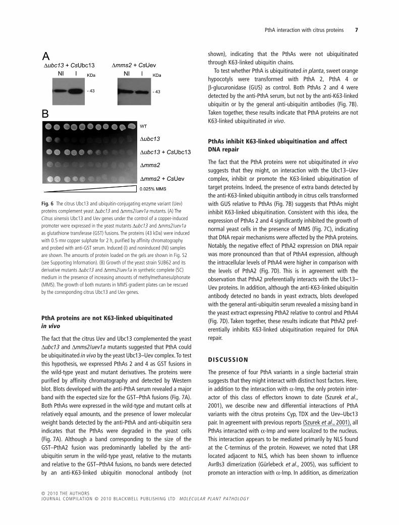
the presence of MMS as they are defective in DNA damagerepair (Broomfield et al., 1998; Hofmann and Pickart, 1999). Totest whether the corresponding citrus proteins are also involvedin DNA repair, we expressed the citrus Ubc13 and Uev genes inthe corresponding yeast mutants (Fig. 6A). We observed that thegrowth of both mutants in MMS gradient plates can be rescuedby the citrus proteins, strongly indicating that the citrus Ubc13and Uev are functional homologues of the yeast proteins(Fig. 6B).
Fig. 4 The citrus Ubc13 interacts with ubiquitin-conjugating enzymevariant (Uev) and PthA proteins. (A) Sweet orange Ubc13 interacts with Uevand with PthAs 2 and 3. The disposition of the yeast co-transformants inthe plates is the same as shown in Fig. 1. (B) Western blots of glutathionetransferase (GST) pulldown assays using GST–Ubc13 as bait and 6 ¥His–Uev and 6 ¥ His–PthAs 2 and 4 as prey. According to the assay shownin (A), Ubc13 binds to Uev and PthA2, but not to PthA 4. Purified PthAproteins used as controls and the molecular sizes of the proteins are shownon the left. The corresponding sodium dodecylsulphate (SDS) gels areshown in Fig. S2 (see Supporting Information).
Fig. 5 The repeat domain and leucine-rich repeat (LRR) are important forprotein–protein interactions. (A) Yeast two-hybrid assays showing theinteractions of the internal repeat domain of PthA2 (RD2) and the 5.5rep +CT2 construct with all citrus proteins. LRR interacted with a-importin(a-Imp) only. (B) Western blot of glutathione transferase (GST) pulldownassays showing that both RD2 and 5.5rep + CT2 constructs bind to citrusproteins (GST fusions). The C-terminus of PthA2 (CT2), carrying LRR and thenuclear localization signal (NLS), binds to a-Imp only. Purified PthA proteinsused as controls and the molecular sizes of the proteins are shown on theleft. The corresponding sodium dodecylsulphate (SDS) gels are shown inFig. S2 (see Supporting Information).
6 M. N. DOMINGUES et al .
© 2010 THE AUTHORSJOURNAL COMPILATION © 2010 BLACKWELL PUBLISHING LTDMOLECULAR PLANT PATHOLOGY
PthA proteins are not K63-linked ubiquitinatedin vivo
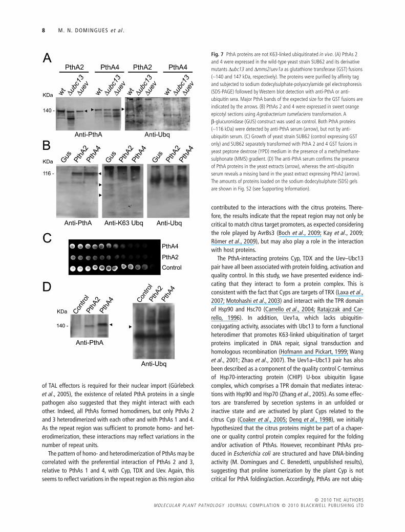
The fact that the citrus Uev and Ubc13 complemented the yeastDubc13 and Dmms2/uev1a mutants suggested that PthA couldbe ubiquitinated in vivo by the yeast Ubc13–Uev complex.To testthis hypothesis, we expressed PthAs 2 and 4 as GST fusions inthe wild-type yeast and mutant derivatives. The proteins werepurified by affinity chromatography and detected by Westernblot. Blots developed with the anti-PthA serum revealed a majorband with the expected size for the GST–PthA fusions (Fig. 7A).Both PthAs were expressed in the wild-type and mutant cells atrelatively equal amounts, and the presence of lower molecularweight bands detected by the anti-PthA and anti-ubiquitin seraindicates that the PthAs were degraded in the yeast cells(Fig. 7A). Although a band corresponding to the size of theGST–PthA2 fusion was predominantly labelled by the anti-ubiquitin serum in the wild-type yeast, relative to the mutantsand relative to the GST–PthA4 fusions, no bands were detectedby an anti-K63-linked ubiquitin monoclonal antibody (not
shown), indicating that the PthAs were not ubiquitinatedthrough K63-linked ubiquitin chains.
To test whether PthA is ubiquitinated in planta, sweet orangehypocotyls were transformed with PthA 2, PthA 4 orb-glucuronidase (GUS) as control. Both PthAs 2 and 4 weredetected by the anti-PthA serum, but not by the anti-K63-linkedubiquitin or by the general anti-ubiquitin antibodies (Fig. 7B).Taken together, these results indicate that PthA proteins are notK63-linked ubiquitinated in vivo.
PthAs inhibit K63-linked ubiquitination and affectDNA repair
The fact that the PthA proteins were not ubiquitinated in vivosuggests that they might, on interaction with the Ubc13–Uevcomplex, inhibit or promote the K63-linked ubiquitination oftarget proteins. Indeed, the presence of extra bands detected bythe anti-K63-linked ubiquitin antibody in citrus cells transformedwith GUS relative to PthAs (Fig. 7B) suggests that PthAs mightinhibit K63-linked ubiquitination. Consistent with this idea, theexpression of PthAs 2 and 4 significantly inhibited the growth ofnormal yeast cells in the presence of MMS (Fig. 7C), indicatingthat DNA repair mechanisms were affected by the PthA proteins.Notably, the negative effect of PthA2 expression on DNA repairwas more pronounced than that of PthA4 expression, althoughthe intracellular levels of PthA4 were higher in comparison withthe levels of PthA2 (Fig. 7D). This is in agreement with theobservation that PthA2 preferentially interacts with the Ubc13–Uev proteins. In addition, although the anti-K63-linked ubiquitinantibody detected no bands in yeast extracts, blots developedwith the general anti-ubiquitin serum revealed a missing band inthe yeast extract expressing PthA2 relative to control and PthA4(Fig. 7D). Taken together, these results indicate that PthA2 pref-erentially inhibits K63-linked ubiquitination required for DNArepair.
DISCUSSION
The presence of four PthA variants in a single bacterial strainsuggests that they might interact with distinct host factors. Here,in addition to the interaction with a-Imp, the only protein inter-actor of this class of effectors known to date (Szurek et al.,2001), we describe new and differential interactions of PthAvariants with the citrus proteins Cyp, TDX and the Uev–Ubc13pair. In agreement with previous reports (Szurek et al., 2001), allPthAs interacted with a-Imp and were localized to the nucleus.This interaction appears to be mediated primarily by NLS foundat the C-terminus of the protein. However, we noted that LRRlocated adjacent to NLS, which has been shown to influenceAvrBs3 dimerization (Gürlebeck et al., 2005), was sufficient topromote an interaction with a-Imp. In addition, as dimerization
Fig. 6 The citrus Ubc13 and ubiquitin-conjugating enzyme variant (Uev)proteins complement yeast Dubc13 and Dmms2/uev1a mutants. (A) TheCitrus sinensis Ubc13 and Uev genes under the control of a copper-inducedpromoter were expressed in the yeast mutants Dubc13 and Dmms2/uev1aas glutathione transferase (GST) fusions. The proteins (43 kDa) were inducedwith 0.5 mM copper sulphate for 2 h, purified by affinity chromatographyand probed with anti-GST serum. Induced (I) and noninduced (NI) samplesare shown. The amounts of protein loaded on the gels are shown in Fig. S2(see Supporting Information). (B) Growth of the yeast strain SUB62 and itsderivative mutants Dubc13 and Dmms2/uev1a in synthetic complete (SC)medium in the presence of increasing amounts of methylmethanesulphonate(MMS). The growth of both mutants in MMS gradient plates can be rescuedby the corresponding citrus Ubc13 and Uev genes.
PthA interaction with citrus proteins 7
© 2010 THE AUTHORSJOURNAL COMPILATION © 2010 BLACKWELL PUBLISHING LTD MOLECULAR PLANT PATHOLOGY
of TAL effectors is required for their nuclear import (Gürlebecket al., 2005), the existence of related PthA proteins in a singlepathogen also suggested that they might interact with eachother. Indeed, all PthAs formed homodimers, but only PthAs 2and 3 heterodimerized with each other and with PthAs 1 and 4.As the repeat region was sufficient to promote homo- and het-erodimerization, these interactions may reflect variations in thenumber of repeat units.
The pattern of homo- and heterodimerization of PthAs may becorrelated with the preferential interaction of PthAs 2 and 3,relative to PthAs 1 and 4, with Cyp, TDX and Uev. Again, thisseems to reflect variations in the repeat region as this region also
contributed to the interactions with the citrus proteins. There-fore, the results indicate that the repeat region may not only becritical to match citrus target promoters, as expected consideringthe role played by AvrBs3 (Boch et al., 2009; Kay et al., 2009;Römer et al., 2009), but may also play a role in the interactionwith host proteins.
The PthA-interacting proteins Cyp, TDX and the Uev–Ubc13pair have all been associated with protein folding, activation andquality control. In this study, we have presented evidence indi-cating that they interact to form a protein complex. This isconsistent with the fact that Cyps are targets of TRX (Laxa et al.,2007; Motohashi et al., 2003) and interact with the TPR domainof Hsp90 and Hsc70 (Carrello et al., 2004; Ratajczak and Car-rello, 1996). In addition, Uev1a, which lacks ubiquitin-conjugating activity, associates with Ubc13 to form a functionalheterodimer that promotes K63-linked ubiquitination of targetproteins implicated in DNA repair, signal transduction andhomologous recombination (Hofmann and Pickart, 1999; Wanget al., 2001; Zhao et al., 2007). The Uev1a–Ubc13 pair has alsobeen described as a component of the quality control C-terminusof Hsp70-interacting protein (CHIP) U-box ubiquitin ligasecomplex, which comprises a TPR domain that mediates interac-tions with Hsp90 and Hsp70 (Zhang et al., 2005). As some effec-tors are transferred by secretion systems in an unfolded orinactive state and are activated by plant Cyps related to thecitrus Cyp (Coaker et al., 2005; Deng et al., 1998), we initiallyhypothesized that the citrus proteins might be part of a chaper-one or quality control protein complex required for the foldingand/or activation of PthAs. However, recombinant PthAs pro-duced in Escherichia coli are structured and have DNA-bindingactivity (M. Domingues and C. Benedetti, unpublished results),suggesting that proline isomerization by the plant Cyp is notcritical for PthA folding/action. Accordingly, PthAs are not ubiq-
Fig. 7 PthA proteins are not K63-linked ubiquitinated in vivo. (A) PthAs 2and 4 were expressed in the wild-type yeast strain SUB62 and its derivativemutants Dubc13 and Dmms2/uev1a as glutathione transferase (GST) fusions(~140 and 147 kDa, respectively). The proteins were purified by affinity tagand subjected to sodium dodecylsulphate-polyacrylamide gel electrophoresis(SDS-PAGE) followed by Western blot detection with anti-PthA or anti-ubiquitin sera. Major PthA bands of the expected size for the GST fusions areindicated by the arrows. (B) PthAs 2 and 4 were expressed in sweet orangeepicotyl sections using Agrobacterium tumefaciens transformation. Ab-glucuronidase (GUS) construct was used as control. Both PthA proteins(~116 kDa) were detected by anti-PthA serum (arrow), but not by anti-ubiquitin serum. (C) Growth of yeast strain SUB62 (control expressing GSTonly) and SUB62 separately transformed with PthA 2 and 4 GST fusions inyeast peptone dextrose (YPD) medium in the presence of a methylmethane-sulphonate (MMS) gradient. (D) The anti-PthA serum confirms the presenceof PthA proteins in the yeast extracts (arrow), whereas the anti-ubiquitinserum reveals a missing band in the yeast extract expressing PthA2 (arrow).The amounts of proteins loaded on the sodium dodecylsulphate (SDS) gelsare shown in Fig. S2 (see Supporting Information).
8 M. N. DOMINGUES et al .
© 2010 THE AUTHORSJOURNAL COMPILATION © 2010 BLACKWELL PUBLISHING LTDMOLECULAR PLANT PATHOLOGY
uitinated in vivo, but apparently inhibit K63-linked ubiquitina-tion associated with DNA repair. Thus, our data strongly point toa model in which PthAs would inhibit the activity of a citrusprotein complex associated with DNA repair. Interestingly, othercitrus proteins identified as PthA4 interactors, including a highmobility group (HMG) protein, a homologue of the mammaliantranslin-associated factor (TRAX) and an SMC (structural main-tenance of chromosomes), are associated with DNA repair andchromatin stability (Erdemir et al., 2002; Hirano, 2002; Prasadet al., 2007).
Although the role of Uev–Ubc13 in response to DNA damagehas been well established in yeast, animals and plants (Broom-field et al., 1998; Hofmann and Pickart, 1999; Motegi et al., 2008;Panier and Durocher, 2009;Wen et al., 2006, 2008), recent studieshave shown that K63-linked ubiquitination also serves as a tar-geting signal for the 26S proteasome (Saeki et al., 2009).This is inline with recent findings showing that CHIP targets the degrada-tion of mammalian SRC-3, a transcriptional co-activator associ-ated with tumour progression (Kajiro et al., 2008). It is thusattractive to speculate that, if a CHIP-related complex operates incitrus, its inhibition by PthAs might favour canker development.
Alternatively, it is notable that citrus Cyp is 64% identical toyeast Cyp A, a protein that decreases gene silencing (Arévalo-Rodríguez and Heitman, 2005; Arévalo-Rodríguez et al., 2000)and interacts with Ess1, a prolyl-isomerase that affects chroma-tin remodelling and the process of transcription through isomer-ization of prolines of the C-terminal domain of the RNApolymerase II (Hani et al., 1999; Krishnamurthy et al., 2009; Wuet al., 2000). Interestingly, Ess1 activity is affected by a ubiquitinligase that promotes K63 ubiquitination and the degradation ofRNA polymerase II (Somesh et al., 2007; Wu et al., 2001). As theubiquitination and degradation of RNA polymerase II occur inresponse to DNA damage and during transcriptional elongationarrest, as a mechanism to avoid the transcription of damagedchromatin and prolonged transcriptional arrest of active genes(Daulny and Tansey, 2009; Somesh et al., 2005, 2007), wepropose that PthAs might increase the rates of transcriptionthrough the modulation of the activity of a protein complexassociated with chromatin remodelling and transcriptionalcontrol. This idea is in agreement with the expected role of PthAsas transcriptional activators, and is supported by the fact thatboth Cyp and Uev identified here localize to the nucleus (Fig. S3,see Supporting Information). A possible role of the citrus Cyp inPthA-dependent transcription is under investigation.
EXPERIMENTAL PROCEDURES
Plant material and bacterial infiltration
Six-month-old plants of sweet orange (Citrus sinensis) wereobtained from certified nurseries and kept in a growth room at
25–28 °C under 14 h/day fluorescent light. Leaves were infil-trated with suspensions of X. axonopodis pv. citri, as describedpreviously (Cernadas et al., 2008). Leaf sectors were infiltratedwith approximately 0.3 mL of an aqueous suspension of bacte-rial cells [optical density at 600 nm (OD600 nm) = 0.5].
Leaves of N. benthamiana were infiltrated with Agrobacte-rium tumefaciens suspensions (OD600 nm = 0.5) carrying theconstructs pBI-35S::PthA(1-4)-GFP, pBI-35S::GFP, pBI-35S::GUS,pBI-35S::DsRed, pBI-35S::Cyp-DsRed or pBI-35S::Uev-DsRed. Thesubcellular localization of the fusion proteins was visualized byfluorescence microscopy equipped with filters for green fluores-cent protein (GFP), red fluorescent protein (DsRed) and 4′,6-diamidino-2-phenylindole (DAPI) fluorescence.
Citrus cDNA library construction and yeasttwo-hybrid assays
Messenger RNA was extracted from sweet orange leaves 48 hafter X. axonopodis pv. citri infiltration using Trizol and FastTrack(Invitrogen). RNA samples from noninfiltrated and infiltrated leafsectors were reverse-transcribed using an oligo-dT with a NotIrestriction site. cDNAs were ligated to SalI adaptors, digestedwith SalI/NotI and ligated into a modified pOAD vector (Uetzet al., 2000) carrying a NotI site between SalI and PstI. Thegenerated cDNA library containing approximately 0.8 ¥ 106 inde-pendent clones was moved into the Saccharomyces cerevisiaestrain PJ694a (MATa trp1-901 leu2-3 112 ura3-52 his3-200gal4D gal80D LYS2::GAL1-HIS3 GAL2-ADE2 met2::GAL7-lacZ)(James et al., 1996) by high-efficiency lithium acetate transfor-mation (Gietz and Woods, 2002).
The genes corresponding to PthAs 1–4 were amplified from X.axonopodis pv. citri plasmids and cloned into the NdeI/EcoRIsites of pET28 (Novagen). The genes were subsequently digestedwith NotI and subcloned into pOBD/pOAD vectors (Uetz et al.,2000). This created an N-terminal truncation of the proteinslacking the first 128 amino acid residues (Fig. S1C, see Support-ing Information). This construct was chosen to avoid the trans-activation of the yeast HIS3 reporter gene by the N-terminus (notshown). The constructs were verified by DNA sequencing andused as bait/prey in two-hybrid assays.
The initial two-hybrid screening was performed on syntheticcomplete medium lacking tryptophan, leucine, histidine andadenine (SC - Trp - Leu - His - Ade), supplemented with 5 mM
3-aminotriazole (3AT), using PthAs 2 and 3 as bait. pOAD plas-mids recovered from positive clones were sequenced and, whenrequired, the full-length citrus cDNAs, including Ubc13, wereobtained by reverse transcription-polymerase chain reaction (RT-PCR) and subcloned downstream of and in frame with the fusionproteins Gal4 (pOAD), GST (pGEX-4T1) and 6 ¥ His (pET28). Bait(pOBD) and prey (pOAD) constructs, including controls (emptypOBD + pOAD prey and pOBD bait + empty pOAD), were used to
PthA interaction with citrus proteins 9
© 2010 THE AUTHORSJOURNAL COMPILATION © 2010 BLACKWELL PUBLISHING LTD MOLECULAR PLANT PATHOLOGY

co-transform PJ694a cells. The cells were grown on SC - Trp -Leu - His (�Ade) containing 0, 3 or 5 mM 3AT for 5 days at30 °C. The cDNA sequences of the citrus Cyp, TDX, Uev, Ubc13and a-Imp were deposited in the GENBANK database asGQ853548, GQ853549, GQ455410, GQ862814 and GQ475487,respectively.
Protein purification and GST pulldown
The 6 ¥ His-tagged proteins PthA2, PthA4, 5.5rep + CT2, CT2 andUev were expressed in E. coli BL21(DE3) cells and purified bymetal affinity chromatography. Briefly, cultures were grown at25 °C in Luria–Bertani (LB) medium containing kanamycin(50 mg/mL) to OD600 nm = 0.6, followed by induction with 0.4 mM
isopropyl-thio-b-D-galactopyranoside (IPTG) for 3 h. Cells har-vested by centrifugation were suspended in binding buffer[20 mM Tris-HCl, pH 8.0, 300 mM NaCl, 5 mM imidazole, 20%glycerol, 1 mM phenylmethylsulphonylfluoride (PMSF), 0.5 mM
dithiothreitol (DTT)] and incubated on ice with lysozyme (1 mg/mL) for 30 min. Bacterial cells were sonicated and the clarifiedsupernatants were incubated with nickel or cobalt resins for 4 hat 4 °C under slow agitation.The beads were washed three timeswith two column volumes of washing buffer (20 mM Tris-HCl, pH8.0, 300 mM NaCl, 15 mM imidazole, 20% glycerol, 1 mM PMSF,0.5 mM DTT) and the retained proteins were eluted with washingbuffer containing up to 200 mM imidazole.
Prey proteins, subcloned into the SalI/NotI sites of pGEX-4T,were expressed in BL21(DE3) cells on induction with IPTG for 2 hat 30–37 °C. Cell pellets were suspended in phosphate-bufferedsaline (PBS) containing 1 mM DTT and lysozyme. After sonicationand centrifugation, soluble fractions of GST fusions were immo-bilized on glutathione resin and nonbound proteins wereremoved with three PBS washes. Thirty micrograms of the puri-fied 6 ¥ His fusion proteins were incubated with the resinscontaining GST or the GST fusion proteins for 2 h at 4 °C. Thebeads were washed four times with PBS and the resin-boundproteins were resolved on 10–13% sodium dodecylsulphate-polyacrylamide electrophoresis (SDS-PAGE) gels. Proteins weretransferred onto nylon membranes, probed with anti-PthA(1 : 5000), anti-6 ¥ His (1 : 3000) or anti-GST (1 : 1000) sera anddeveloped with the ECL kit (GE Healthcare).
Functional complementation of yeast mutants
The yeast strain SUB62 (MATa lys2-801leu2-3, 112 ura3-52 his3-D200 trp1-1[am]) and its single mutant derivatives Dubc13 andDmms2 (uev1a) (Hofmann and Pickart, 1999) were transformedwith the citrus Ubc13 or Uev genes cloned into pYEX-4T (Clon-tech). Transformants were grown on SC - Leu - Ura plates at30 °C for 4 days. For qualitative analysis of MMS sensitivity,liquid cultures were grown overnight to exponential phase in
yeast peptone dextrose (YPD) medium and plated in YPD supple-mented with 0.025% MMS gradient (Pastushok et al., 2005). Theplates were incubated at 30 °C for 24 h.
In vivo ubiquitination
PthA ubiquitination in yeast was evaluated using the wild-typeyeast strain SUB62 and the corresponding mutant derivativesubc13D and UevD.The strains were transformed with PthA 2 and4 GST fusions cloned into pYEX4T-1 and grown in SC - Leu - Urafor 4 days at 30 °C. Liquid cultures were grown to OD600nm = 0.8and protein expression was induced with 0.5 mM CuSO4 for 2 h.Cells were collected by centrifugation (6000 g for 15 min at4 °C), resuspended in PBS containing 1 mM PMSF and lysed withglass beads and freeze–thaw cycles. Recombinant proteins werepurified using glutathione sepharose beads. Purified proteinswere analysed by 7% SDS-PAGE and probed with the anti-PthAand anti-ubiquitin sera.
To evaluate possible PthA ubiquitination in planta, PthAs 2and 4 were expressed in etiolated epicotyls of sweet orange‘Hamlin’ according to de Oliveira et al. (2009). Surface-sterilizedseeds were sown in half-strength Murashige and Skoog (MS)basal medium supplemented with 50 mg/L myo-inositol and25 g/L sucrose. Seeds were incubated at 27 � 2 °C under darkconditions for 6 weeks. Epicotyl sections of etiolated seedlingswere cut transversally into 1-cm segments and incubated for15 min with Agrobacterium tumefaciens EHA105 (OD600nm = 1.0)carrying the pBI121-PthA2, pBI121-PthA4 or pBI121-GUS con-struct in MS basal medium containing 100 mM acetosyringone.Excess of Agrobacterium suspension was removed by blotting,and co-cultivation was carried out in the dark at 26 � 2 °C for2 days. Epicotyls were macerated in liquid nitrogen and resus-pended in SDS-PAGE sample buffer. Soluble proteins were recov-ered by centrifugation (16 000 g for 30 min at 4 °C), resolved by10% SDS-PAGE and detected by the anti-PthA, anti-ubiquitin(Sigma) and anti-polyubiquitin K63-linkage-specific cloneHWA4C4 (Biomol International) antibodies.
Inhibition of K63-linked ubiquitination-mediated DNArepair in yeast
PthAs 2 and 4 were expressed in the SUB62 wild-type strain.Transformants were selected on SC - Leu - Ura plates at 30 °Cfor 4 days. Liquid cultures were grown overnight to exponentialphase in YPD medium and plated in YPD supplemented with a0.025% MMS gradient. The plates were incubated at 30 °C for24 h and the expression of PthAs 2 and 4 was detected byWestern blot with anti-PthA serum.
ACKNOWLEDGEMENTS
We thank Dr Robert Cohen and Dr Candice Carlile for providingthe yeast strain SUB62 and the Dubc13 and Dmms2 mutants.
10 M. N. DOMINGUES et al .
© 2010 THE AUTHORSJOURNAL COMPILATION © 2010 BLACKWELL PUBLISHING LTDMOLECULAR PLANT PATHOLOGY

This work was supported by Fundação de Amparo à Pesquisado Estado de São Paulo (FAPESP) (grants 98/14138-2;00/10266-8; 03/08316-5; 07/06686-0). MND, TAS, RAC, MLPO,CD and CEB received fellowships from FAPESP and the Con-selho Nacional de Desenvolvimento Científico e Tecnológico(CNPq).
REFERENCES
Al-Saadi, A., Reddy, J.D., Duan, Y.P., Brunings, A.M., Yuan, Q. andGabriel, D.W. (2007) All five host-range variants of Xanthomonas citricarry one pthA homolog with 17.5 repeats that determines pathogenic-ity on citrus, but none determine host-range variation. Mol. Plant–Microbe Interact. 20, 934–943.
Arévalo-Rodríguez, M. and Heitman, J. (2005) Cyclophilin A is localizedto the nucleus and controls meiosis in Saccharomyces cerevisiae.Eukaryot. Cell, 4, 17–29.
Arévalo-Rodríguez, M., Cardenas, M.E., Wu, X., Hanes, S.D. andHeitman, J. (2000) Cyclophilin A and Ess1 interact with and regulatesilencing by the Sin3-Rpd3 histone deacetylase. EMBO J. 19, 3739–3749.
Boch, J., Scholze, H., Schornack, S., Landgraf, A., Hahn, S., Kay, S.,Lahaye, T., Nickstadt, A. and Bonas, U. (2009) Breaking the code ofDNA-binding specificity of TAL-Type III effectors. Science, 326, 1509–1512.
Broomfield, S., Chow, B.L. and Xiao, W. (1998) MMS2, encoding aubiquitin-conjugating-enzyme-like protein, is a member of the yeasterror-free postreplication repair pathway. Proc. Natl. Acad. Sci. USA, 95,5678–5683.
Brunings, A.M. and Gabriel, D.W. (2003) Xanthomonas citri: breakingthe surface. Mol. Plant Pathol. 4, 141–157.
Carrello, A., Allan, R.K., Morgan, S.L., Owen, B.A., Mok, D., Ward,B.K., Minchin, R.F., Toft, D.O. and Ratajczak, T. (2004) Interaction ofthe Hsp90 cochaperone cyclophilin 40 with Hsc70. Cell Stress Chaper-ones, 9, 167–181.
Cernadas, R.A. and Benedetti, C.E. (2009) Role of auxin and gibberellinin citrus canker development and in the transcriptional control of cell-wall remodeling genes modulated by Xanthomonas axonopodis pv citri.Plant Sci. 177, 190–195.
Cernadas, R.A., Camillo, L.R. and Benedetti, C.E. (2008) Transcriptionalanalysis of the sweet orange interaction with the citrus canker patho-gens Xanthomonas axonopodis pv. citri and Xanthomonas axonopodispv. aurantifolii. Mol. Plant Pathol. 9, 609–631.
Coaker, G., Falick, A. and Staskawicz, B. (2005) Activation of a phyto-pathogenic bacterial effector protein by a eukaryotic cyclophilin.Science, 308, 548–550.
Daulny, A. and Tansey, W.P. (2009) Damage control: DNA repair,transcription, and the ubiquitin-proteasome system. DNA Repair, 8,444–448.
Deng, W., Chen, L., Wood, D.W., Metcalfe, T., Liang, X., Gordon, M.P.,Comai, L. and Nester, E.W. (1998) Agrobacterium VirD2 proteininteracts with plant host cyclophilins. Proc. Natl. Acad. Sci. USA, 95,7040–7045.
Duan, Y.P., Castañeda, G.Z., Erdos, G. and Gabriel, D.W. (1999) Expres-sion of a single, host-specific, bacterial pathogenicity gene in plant cellselicits division, enlargement, and cell death. Mol. Plant–MicrobeInteract. 12, 556–560.
Erdemir, T., Bilican, B., Oncel, D., Goding, C.R. and Yavuzer, U. (2002)DNA damage-dependent interaction of the nuclear matrix protein C1Dwith translin-associated factor X (TRAX). J. Cell Sci. 115, 207–216.
Gietz, R.D. and Woods, R.A. (2002) Transformation of yeast by lithiumacetate/single-stranded carrier DNA/polyethylene glycol method.Methods Enzymol. 350, 87–96.
Gürlebeck, D., Szurek, B. and Bonas, U. (2005) Dimerization of thebacterial effector protein AvrBs3 in the plant cell cytoplasm prior tonuclear import. Plant J. 42, 175–187.
Hani, J., Schelbert, B., Bernhardt, A., Domdey, H., Fischer, G., Wie-bauer, K. and Rahfeld, J.U. (1999) Mutations in a peptidylprolyl-cis/trans-isomerase gene lead to a defect in 3′-end formation of a pre-mRNA in Saccharomyces cerevisiae. J. Biol. Chem. 274, 108–116.
Harreman, M.T., Hodel, M.R., Fanara, P., Hodel, A.E. and Corbett, A.H.(2003) The auto-inhibitory function of importin alpha is essential in vivo.J. Biol. Chem. 278, 5854–5863.
Hirano, T. (2002) The ABCs of SMC proteins: two-armed ATPases forchromosome condensation, cohesion, and repair. Genes Dev. 16, 399–414.
Hofmann, R.M. and Pickart, C.M. (1999) Noncanonical MMS2-encodedubiquitin-conjugating enzyme functions in assembly of novel polyubiq-uitin chains for DNA repair. Cell, 96, 645–653.
James, P., Halladay, J. and Craig, E.A. (1996) Genomic libraries and ahost strain designed for highly efficient two-hybrid selection in yeast.Genetics, 144, 1425–1436.
Kajiro, M., Hirota, R., Nakajima, Y., Kawanowa, K., So-ma, K., Ito, I.,Yamaguchi, Y., Ohie, S.H., Kobayashi, Y., Seino, Y., Kawano, M.,Kawabe, Y., Takei, H., Hayashi, S., Kurosumi, M., Murayama, A.,Kimura, K. and Yanagisawa, J. (2008) The ubiquitin ligase CHIP acts asan upstream regulator of oncogenic pathways. Nat. Cell Biol. 11, 312–319.
Kay, S. and Bonas, U. (2009) How Xanthomonas type III effectors manipu-late the host plant. Curr. Opin. Microbiol. 12, 37–43.
Kay, S., Hahn, S., Marois, E., Hause, G. and Bonas, U. (2007) A bacterialeffector acts as a plant transcription factor and induces a cell sizeregulator. Science, 318, 648–651.
Kay, S., Hahn, S., Marois, E., Wieduwild, R. and Bonas, U. (2009)Detailed analysis of the DNA recognition motifs of the Xanthomonastype III effectors AvrBs3 and AvrBs3Deltarep16. Plant J. 59, 859–871.
Krishnamurthy, S., Ghazy, M.A., Moore, C. and Hampsey, M. (2009)Functional interaction of the Ess1 prolyl isomerase with components ofthe RNA polymerase II initiation and termination machineries. Mol. Cell.Biol. 29, 2925–2934.
Laxa, M., König, J., Dietz, K.J. and Kandlbinder, A. (2007) Role of thecysteine residues in Arabidopsis thaliana cyclophilin CYP20-3 inpeptidyl-prolyl cis–trans isomerase and redox-related functions.Biochem. J. 401, 287–297.
Moscou, M.J. and Bogdanove, A.J. (2009) A simple cipher governs DNArecognition by TAL effectors. Science, 326, 1501.
Motegi, A., Liaw, H.J., Lee, K.Y., Roest, H.P., Maas, A., Wu, X.,Moinova, H., Markowitz, S.D., Ding, H., Hoeijmakers, J.H. andMyung, K. (2008) Polyubiquitination of proliferating cell nuclearantigen by HLTF and SHPRH prevents genomic instability from stalledreplication forks. Proc. Natl. Acad. Sci. USA, 105, 12 411–12 416.
Motohashi, K., Koyama, F., Nakanishi, Y., Ueoka-Nakanishi, H. andHisabori, T. (2003) Chloroplast cyclophilin is a target protein of thiore-
PthA interaction with citrus proteins 11
© 2010 THE AUTHORSJOURNAL COMPILATION © 2010 BLACKWELL PUBLISHING LTD MOLECULAR PLANT PATHOLOGY

doxin. Thiol modulation of the peptidyl-prolyl cis–trans isomerase activ-ity. J. Biol. Chem. 278, 31 848–31 852.
de Oliveira, M.L.P., Febres, V.J., Costa, M.G.C., Moore, G.A. andOtoni, W.C. (2009) High-efficiency Agrobacterium-mediated transfor-mation of citrus via sonication and vacuum infiltration. Plant Cell Rep.28, 387–395.
Panier, S. and Durocher, D. (2009) Regulatory ubiquitylation in responseto DNA-double strand breaks. DNA Repair, 8, 436–443.
Pastushok, L., Moraes, T.F., Ellison, M.J. and Xiao, W. (2005) A singleMms2 ‘key’ residue insertion into a Ubc13 pocket determines the inter-face specificity of a human Lys63 ubiquitin conjugation complex. J. Biol.Chem. 280, 17 891–17 900.
Petroski, M.D., Zhou, X., Dong, G., Daniel-Issakani, S., Payan, D.G.and Huang, J. (2007) Substrate modification with lysine 63-linkedubiquitin chains through the UBC13-UEV1A ubiquitin-conjugatingenzyme. J. Biol. Chem. 282, 29936–29945.
Prasad, R., Liu, Y., Deterding, L.J., Poltoratsky, V.P., Kedar, P.S.,Horton, J.K., Kanno, S., Asagoshi, K., Hou, E.W., Khodyreva, S.N.,Lavrik, O.I., Tomer, K.B., Yasui, A. and Wilson, S.H. (2007) HMGB1 isa cofactor in mammalian base excision repair. Mol. Cell, 27, 829–841.
Ratajczak, T. and Carrello, A. (1996) Cyclophilin 40 (CyP-40), mapping ofits hsp90 binding domain and evidence that FKBP52 competes withCyP-40 for hsp90 binding. J. Biol. Chem. 271, 2961–2965.
Römer, P., Hahn, S., Jordan, T., Strauss, T., Bonas, U. and Lahaye, T.(2007) Plant pathogen recognition mediated by promoter activation ofthe pepper Bs3 resistance gene. Science, 318, 645–648.
Römer, P., Strauss, T., Hahn, S., Scholze, H., Morbitzer, R., Grau, J.,Bonas, U. and Lahaye, T. (2009) Recognition of AvrBs3-like proteins ismediated by specific binding to promoters of matching pepper Bs3alleles. Plant Physiol. 150, 1697–1712.
Saeki, Y., Kudo, T., Sone, T., Kikuchi, Y., Yokosawa, H., Toh-e, A. andTanaka, K. (2009) Lysine 63-linked polyubiquitin chain may serve as atargeting signal for the 26S proteasome. EMBO J. 28, 359–371.
da Silva, A.C., Ferro, J.A., Reinach, F.C., Farah, C.S., Furlan, L.R.,Quaggio, R.B., Monteiro-Vitorello, C.B., Van Sluys, M.A., Almeida,N.F., Alves, L.M., do Amaral, A.M., Bertolini, M.C., Camargo, L.E.,Camarotte, G., Cannavan, F., Cardozo, J., Chambergo, F., Ciapina,L.P., Cicarelli, R.M., Coutinho, L.L., Cursino-Santos, J.R., El-Dorry,H., Faria, J.B., Ferreira, A.J., Ferreira, R.C., Ferro, M.I., Formighieri,E.F., Franco, M.C., Greggio, C.C., Gruber, A., Katsuyama, A.M.,Kishi, L.T., Leite, R.P., Lemos, E.G., Lemos, M.V., Locali, E.C.,Machado, M.A., Madeira, A.M., Martinez-Rossi, N.M., Martins,E.C., Meidanis, J., Menck, C.F., Miyaki, C.Y., Moon, D.H., Moreira,L.M., Novo, M.T., Okura, V.K., Oliveira, M.C., Oliveira, V.R., Pereira,H.A., Rossi, A., Sena, J.A., Silva, C., de Souza, R.F., Spinola, L.A.,Takita, M.A., Tamura, R.E., Teixeira, E.C., Tezza, R.I., Trindade dosSantos, M., Truffi, D., Tsai, S.M., White, F.F., Setubal, J.C. andKitajima, J.P. (2002) Comparison of the genomes of two Xanthomonaspathogens with differing host specificities. Nature, 417, 459–463.
Somesh, B.P., Reid, J., Liu, W.F., Søgaard, T.M., Erdjument-Bromage,H., Tempst, P. and Svejstrup, J.Q. (2005) Multiple mechanisms con-fining RNA polymerase II ubiquitylation to polymerases undergoingtranscriptional arrest. Cell, 121, 913–923.
Somesh, B.P., Sigurdsson, S., Saeki, H., Erdjument-Bromage, H.,Tempst, P. and Svejstrup, J.Q. (2007) Communication between distantsites in RNA polymerase II through ubiquitylation factors and the poly-merase CTD. Cell, 129, 57–68.
Sugio, A., Yang, B., Zhu, T. and White, F.F. (2007) Two type III effectorgenes of Xanthomonas oryzae pv. oryzae control the induction of thehost genes OsTFIIAg1 and OsTFX1 during bacterial blight of rice. Proc.Natl. Acad. Sci. USA, 104, 10 720–10 725.
Swarup, S., De Feyter, R., Brlansky, R.H. and Gabriel, D.W. (1991) Apathogenicity locus from Xanthomonas citri enables strains from severalpathovars of X. campestris to elicit canker like lesions on citrus. Phyto-pathology, 81, 803–809.
Swarup, S., Yang, Y., Kingsley, M.T. and Gabriel, D.W. (1992) AnXanthomonas citri pathogenicity gene, pthA, pleiotropically encodesgratuitous avirulence on nonhosts. Mol. Plant–Microbe Interact. 5, 204–213.
Szurek, B., Marois, E., Bonas, U. and Ackerveken, G. (2001) Eukaryoticfeatures of the Xanthomonas type III effector AvrBs3: protein domainsinvolved in transcriptional activation and the interaction with nuclearimport receptors from pepper. Plant J. 26, 523–534.
Uetz, P., Giot, L., Cagney, G., Mansfield, T.A., Judson, R.S., Knight,J.R., Lockshon, D., Narayan, V., Srinivasan, M., Pochart, P., Qureshi-Emili, A., Li, Y., Godwin, B., Conover, D., Kalbfleisch, T., Vijayada-modar, G., Yang, M., Johnston, M., Fields, S. and Rothberg, J.M.(2000) A comprehensive analysis of protein–protein interactions in Sac-charomyces cerevisiae. Nature, 403, 623–627.
Vignols, F., Mouaheb, N., Thomas, D. and Meyer, Y. (2003) Redoxcontrol of Hsp70-Co-chaperone interaction revealed by expression of athioredoxin-like Arabidopsis protein. J. Biol. Chem. 278, 4516–4523.
Wang, C., Deng, L., Hong, M., Akkaraju, G.R., Inoue, J. and Chen, Z.J.(2001) TAK1 is a ubiquitin-dependent kinase of MKK and IKK. Nature,412, 346–351.
Wen, R., Newton, L., Li, G., Wang, H. and Xiao, W. (2006) Arabidopsisthaliana UBC13: implication of error-free DNA damage toleranceand Lys63-linked polyubiquitylation in plants. Plant Mol. Biol. 61, 241–253.
Wen, R., Torres-Acosta, J.A., Pastushok, L., Lai, X., Pelzer, L., Wang, H.and Xiao, W. (2008) Arabidopsis UEV1D promotes Lysine-63-linkedpolyubiquitination and is involved in DNA damage response. Plant Cell,20, 213–227.
Windheim, M., Peggie, M. and Cohen, P. (2008) Two different classes ofE2 ubiquitin-conjugating enzymes are required for the mono-ubiquitination of proteins and elongation by polyubiquitin chains with aspecific topology. Biochem. J. 409, 723–729.
Wu, X., Wilcox, C.B., Devasahayam, G., Hackett, R.L., Arévalo-Rodríguez, M., Cardenas, M.E., Heitman, J. and Hanes, S.D. (2000)The Ess1 prolyl isomerase is linked to chromatin remodeling complexesand the general transcription machinery. EMBO J. 19, 3727–3738.
Wu, X., Chang, A., Sudol, M. and Hanes, S.D. (2001) Genetic interac-tions between the ESS1 prolyl-isomerase and the RSP5 ubiquitin ligasereveal opposing effects on RNA polymerase II function. Curr. Genet. 40,234–242.
Yang, Y. and Gabriel, D.W. (1995) Intragenic recombination of a singleplant pathogen gene provides a mechanism for the evolution of newhost specificities. J. Bacteriol. 177, 4963–4968.
Yang, B. and White, F.F. (2004) Diverse members of the AvrBs3/PthAfamily of type III effectors are major virulence determinants in bacterialblight disease of rice. Mol. Plant–Microbe Interact. 17, 1192–1200.
Yang, Y., Yuan, Q. and Gabriel, D.W. (1996) Watersoaking function(s) ofXcmH1005 are redundantly encoded by members of the Xanthomonasavr/pth gene family. Mol. Plant–Microbe Interact. 9, 105–113.
12 M. N. DOMINGUES et al .
© 2010 THE AUTHORSJOURNAL COMPILATION © 2010 BLACKWELL PUBLISHING LTDMOLECULAR PLANT PATHOLOGY
Yang, B., Sugio, A. and White, F.F. (2006) Os8N3 is a host disease-susceptibility gene for bacterial blight of rice. Proc. Natl. Acad. Sci. USA,103, 10 503–10 508.
Zhang, M., Windheim, M., Roe, S.M., Peggie, M., Cohen, P., Prodro-mou, C. and Pearl, L.H. (2005) Chaperoned ubiquitylation-crystalstructures of the CHIP U box E3 ubiquitin ligase and a CHIP-Ubc13–Uev1a complex. Mol. Cell, 20, 525–538.
Zhao, G.Y., Sonoda, E., Barber, L.J., Oka, H., Murakawa, Y., Yamada,K., Ikura, T., Wang, X., Kobayashi, M., Yamamoto, K., Boulton, S.J.and Takeda, S. (2007) A critical role for the ubiquitin-conjugatingenzyme Ubc13 in initiating homologous recombination. Mol. Cell, 25,663–675.
SUPPORTING INFORMATION
Additional Supporting Information may be found in the onlineversion of this article:
Fig. S1 PthA proteins and constructs used in two-hybrid andpulldown assays. (A) Schematic representation of the four PthAsfrom Xanthomonas axonopodis pv. citri strain 306. Dark-greybars indicate the 34-amino-acid repeats depicted in (B). Light-grey boxes indicate the C-terminal leucine-rich repeat (LRR) andthe acidic domain (AD) flanking three nuclear localizationsignals (NLSs). The numbers represent the positions of the aminoacid residues and the letters indicate residue variation outsidethe repetitive domain. The proteins were expressed in Escheri-chia coli as 6 ¥ His fusions and used in glutathione transferase(GST) pulldown assays. (B) Sequence alignment of the 34-amino-acid repeats of the four PthA proteins, depicting the regions ofincreased amino acid variation (*). Comma indicates ‘and’,whereas hyphen indicates ‘to’. (C) Schematic representation ofthe PthA constructs in pOBD/pOAD used as bait/prey in the
two-hybrid assays. The internal repeat domain of PthA2 (RD2),the invariable LRR and the C-terminal (CT2) and C-terminal withthe last 5.5 repeats of PthA2 (5.5rep + CT2) were also expressedas 6 ¥ His fusions for pulldown assays.Fig. S2 Coomassie blue-stained sodium dodecylsulphate-polyacrylamide gel electrophoresis (SDS-PAGE) gels correspond-ing to the Western blots shown in Figs 2D, E, 3B, 4B, 5B, 6A, 7A, Band D, showing the relative amounts of proteins loaded in eachlane. The molecular size markers are indicated on the left. Theglutathione transferase (GST) fusion proteins used in the pull-down assays of Figs 2D, E, 3B, 4B and 5B are indicated by theasterisks. The gels corresponding to Fig. 6A show the copper-induced expression (I) of citrus ubiquitin-conjugating enzymevariant (Uev) and Ubc13 in the yeast mutants, relative to nonin-duced samples (NI). Arrows indicate the induced bands. The gelscorresponding to Fig. 7A show the GST–PthA samples purifiedfrom wild-type and mutant yeast cells. NI (noninduced), I(induced), S (soluble), FT (flow-through),W (wash) and E (elution).The ‘E’ samples were loaded side by side in the blot shown inFig. 7A. The gels corresponding to Fig. 7B and D show theamounts of proteins loaded from plant and yeast cells expressingPthAs 2 and 4. Arrows indicate the positions of PthA bands.Fig. S3 Nuclear localization of cyclophilin (Cyp) and ubiquitin-conjugating enzyme variant (Uev) in Nicotiana benthamianacells. Fluorescence of Uev–red fluorescent protein (DsRed) andCyp–DsRed fusions in the nucleus of transiently transfected N.benthamiana cells 72 h after Agrobacterium tumefaciens infil-tration. 4′,6-Diamidino-2-phenylindole (DAPI) fluorescence ofthe same cells is shown alongside.
Please note: Wiley-Blackwell are not responsible for the contentor functionality of any supporting materials supplied by theauthors. Any queries (other than missing material) should bedirected to the corresponding author for the article.
PthA interaction with citrus proteins 13
© 2010 THE AUTHORSJOURNAL COMPILATION © 2010 BLACKWELL PUBLISHING LTD MOLECULAR PLANT PATHOLOGY
Copyright © 2022 FDOKUMEN


![[Current Topics in Microbiology and Immunology] || Modulation of the Ubiquitination Machinery by Legionella](https://static.fdokumen.com/doc/165x107/63328412f0080405510482a9/current-topics-in-microbiology-and-immunology-modulation-of-the-ubiquitination.jpg)