Biophysics of protein evolution and evolutionary protein biophysics
36
, 20140419, published 27 August 2014 11 2014 J. R. Soc. Interface Tobias Sikosek and Hue Sun Chan biophysics Biophysics of protein evolution and evolutionary protein References http://rsif.royalsocietypublishing.org/content/11/100/20140419.full.html#ref-list-1 This article cites 435 articles, 131 of which can be accessed free Subject collections (313 articles) computational biology (354 articles) biophysics (49 articles) bioinformatics Articles on similar topics can be found in the following collections Email alerting service here right-hand corner of the article or click Receive free email alerts when new articles cite this article - sign up in the box at the top http://rsif.royalsocietypublishing.org/subscriptions go to: J. R. Soc. Interface To subscribe to on August 27, 2014 rsif.royalsocietypublishing.org Downloaded from on August 27, 2014 rsif.royalsocietypublishing.org Downloaded from
Transcript of Biophysics of protein evolution and evolutionary protein biophysics
, 20140419, published 27 August 201411 2014 J. R. Soc. Interface Tobias Sikosek and Hue Sun Chan biophysicsBiophysics of protein evolution and evolutionary protein
Referenceshttp://rsif.royalsocietypublishing.org/content/11/100/20140419.full.html#ref-list-1
This article cites 435 articles, 131 of which can be accessed free
Subject collections
(313 articles)computational biology � (354 articles)biophysics �
(49 articles)bioinformatics � Articles on similar topics can be found in the following collections
Email alerting service hereright-hand corner of the article or click Receive free email alerts when new articles cite this article - sign up in the box at the top
http://rsif.royalsocietypublishing.org/subscriptions go to: J. R. Soc. InterfaceTo subscribe to
on August 27, 2014rsif.royalsocietypublishing.orgDownloaded from on August 27, 2014rsif.royalsocietypublishing.orgDownloaded from

on August 27, 2014rsif.royalsocietypublishing.orgDownloaded from
rsif.royalsocietypublishing.org
Headline reviewCite this article: Sikosek T, Chan HS. 2014
Biophysics of protein evolution and evolution-
ary protein biophysics. J. R. Soc. Interface 11:
20140419.
http://dx.doi.org/10.1098/rsif.2014.0419
Received: 22 April 2014
Accepted: 28 July 2014
Subject Areas:biophysics, bioinformatics,
computational biology
Keywords:adaptation, promiscuous functions,
conformational dynamics, hidden states,
protein folding, protein – protein interactions
Authors for correspondence:Tobias Sikosek
e-mail: [email protected]
Hue Sun Chan
e-mail: [email protected]
& 2014 The Author(s) Published by the Royal Society. All rights reserved.
Biophysics of protein evolution andevolutionary protein biophysics
Tobias Sikosek1,2,3 and Hue Sun Chan1,2,3
1Department of Biochemistry, 2Department of Molecular Genetics, and 3Department of Physics,University of Toronto, Toronto, Ontario, Canada M5S 1A8
TS, 0000-0001-9929-3525
The study of molecular evolution at the level of protein-coding genes often
entails comparing large datasets of sequences to infer their evolutionary relation-
ships. Despite the importance of a protein’s structure and conformational
dynamics to its function and thus its fitness, common phylogenetic methods
embody minimal biophysical knowledge of proteins. To underscore the
biophysical constraints on natural selection, we survey effects of protein
mutations, highlighting the physical basis for marginal stability of natural glob-
ular proteins and how requirement for kinetic stability and avoidance of
misfolding and misinteractions might have affected protein evolution. The
biophysical underpinnings of these effects have been addressed by models
with an explicit coarse-grained spatial representation of the polypeptide chain.
Sequence–structure mappings based on such models are powerful conceptual
tools that rationalize mutational robustness, evolvability, epistasis, promiscuous
function performed by ‘hidden’ conformational states, resolution of adaptive
conflicts and conformational switches in the evolution from one protein fold
to another. Recently, protein biophysics has been applied to derive more accu-
rate evolutionary accounts of sequence data. Methods have also been
developed to exploit sequence-based evolutionary information to predict bio-
physical behaviours of proteins. The success of these approaches demonstrates
a deep synergy between the fields of protein biophysics and protein evolution.
1. IntroductionBiological evolution uses mutations as its basic working material. Mutations
occur in DNA molecules through various mechanisms. Some mutations are rela-
tively ‘silent’ in that their effects are less appreciable, whereas others have a more
prominent impact on the biological function. The most immediate effect of a
mutation is the alteration of the DNA molecule itself and thus, possibly, its affi-
nities to bind certain proteins or RNA. Given the vastness of many genomes, it
was once believed that many mutations in DNA fall in regions that have no bio-
logical function. However, with increasing knowledge of the functional roles of
non-coding DNA sequences, the proportion of genomes that is considered
non-functional has decreased significantly [1]. Regions of the genome that do
encode for a functional RNA or protein can undergo several different kinds of
mutations, such as insertions, deletions and duplications of entire segments of
DNA. The present review focuses primarily on the effect of point mutations
(change of a single nucleotide) and will consider only proteins but not RNA,
although many general principles of evolution are applicable to both classes of
biomolecules. We refer to other authors for the evolution of protein structures
via sequence re-arrangements such as domain-wise evolution [2–4], the fusion
of small peptide fragments [5] or the ‘chimeric’ recombination of fragments
that is also exploited in protein engineering [6–9].
Current study of molecular evolution can benefit from a huge amount of
sequence data, but only a relatively small body of structural data. Conse-
quently, many approaches in evolutionary studies are predominantly
sequence-based. A prime example is phylogenetic inference methods based
upon multiple sequence alignments. Mostly, the biophysical foundation of
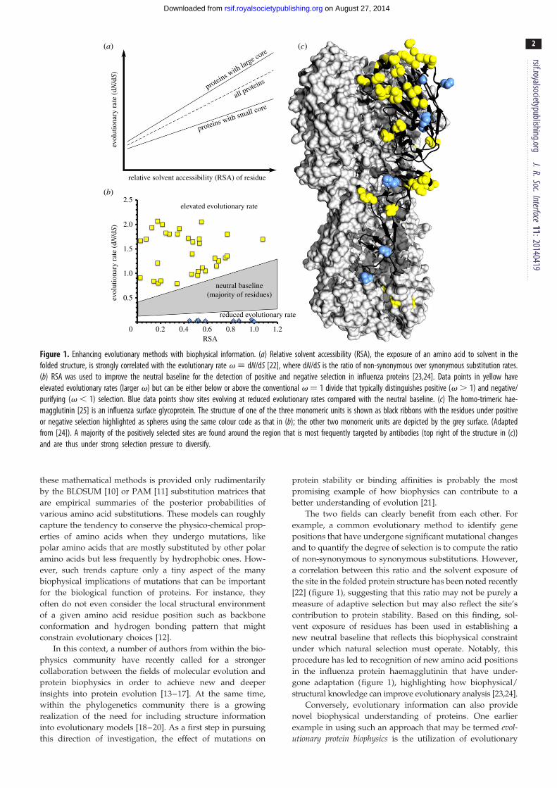
relative solvent accessibility (RSA) of residue
elevated evolutionary rate
reduced evolutionary rate
neutral baseline(majority of residues)
RSA0 0.2 0.4 0.6 0.8 1.0 1.2
2.5
2.0
1.5
1.0
0.5
(a)
(b)
(c)
evol
utio
nary
rat
e (d
N/d
S)ev
olut
iona
ry r
ate
(dN
/dS)
proteins with small core
proteins with large core
all proteins
Figure 1. Enhancing evolutionary methods with biophysical information. (a) Relative solvent accessibility (RSA), the exposure of an amino acid to solvent in thefolded structure, is strongly correlated with the evolutionary rate v ; dN/dS [22], where dN/dS is the ratio of non-synonymous over synonymous substitution rates.(b) RSA was used to improve the neutral baseline for the detection of positive and negative selection in influenza proteins [23,24]. Data points in yellow haveelevated evolutionary rates (larger v) but can be either below or above the conventional v ¼ 1 divide that typically distinguishes positive (v . 1) and negative/purifying (v , 1) selection. Blue data points show sites evolving at reduced evolutionary rates compared with the neutral baseline. (c) The homo-trimeric hae-magglutinin [25] is an influenza surface glycoprotein. The structure of one of the three monomeric units is shown as black ribbons with the residues under positiveor negative selection highlighted as spheres using the same colour code as that in (b); the other two monomeric units are depicted by the grey surface. (Adaptedfrom [24]). A majority of the positively selected sites are found around the region that is most frequently targeted by antibodies (top right of the structure in (c))and are thus under strong selection pressure to diversify.
rsif.royalsocietypublishing.orgJ.R.Soc.Interface
11:20140419
2
on August 27, 2014rsif.royalsocietypublishing.orgDownloaded from
these mathematical methods is provided only rudimentarily
by the BLOSUM [10] or PAM [11] substitution matrices that
are empirical summaries of the posterior probabilities of
various amino acid substitutions. These models can roughly
capture the tendency to conserve the physico-chemical prop-
erties of amino acids when they undergo mutations, like
polar amino acids that are mostly substituted by other polar
amino acids but less frequently by hydrophobic ones. How-
ever, such trends capture only a tiny aspect of the many
biophysical implications of mutations that can be important
for the biological function of proteins. For instance, they
often do not even consider the local structural environment
of a given amino acid residue position such as backbone
conformation and hydrogen bonding pattern that might
constrain evolutionary choices [12].
In this context, a number of authors from within the bio-
physics community have recently called for a stronger
collaboration between the fields of molecular evolution and
protein biophysics in order to achieve new and deeper
insights into protein evolution [13–17]. At the same time,
within the phylogenetics community there is a growing
realization of the need for including structure information
into evolutionary models [18–20]. As a first step in pursuing
this direction of investigation, the effect of mutations on
protein stability or binding affinities is probably the most
promising example of how biophysics can contribute to a
better understanding of evolution [21].
The two fields can clearly benefit from each other. For
example, a common evolutionary method to identify gene
positions that have undergone significant mutational changes
and to quantify the degree of selection is to compute the ratio
of non-synonymous to synonymous substitutions. However,
a correlation between this ratio and the solvent exposure of
the site in the folded protein structure has been noted recently
[22] (figure 1), suggesting that this ratio may not be purely a
measure of adaptive selection but may also reflect the site’s
contribution to protein stability. Based on this finding, sol-
vent exposure of residues has been used in establishing a
new neutral baseline that reflects this biophysical constraint
under which natural selection must operate. Notably, this
procedure has led to recognition of new amino acid positions
in the influenza protein haemagglutinin that have under-
gone adaptation (figure 1), highlighting how biophysical/
structural knowledge can improve evolutionary analysis [23,24].
Conversely, evolutionary information can also provide
novel biophysical understanding of proteins. One earlier
example in using such an approach that may be termed evol-utionary protein biophysics is the utilization of evolutionary

rsif.royalsocietypublishing.orgJ.R.Soc.Interface
11:20140419
3
on August 27, 2014rsif.royalsocietypublishing.orgDownloaded from
data on the PDZ domain family to predict energetically
coupled positions on the protein, some of which are spatially
far apart [26]. Another example is the inference of structural
information from protein sectors, which are co-evolving clus-
ters of spatially proximate and physically interacting amino
acids within a protein structure. A protein such as rat trypsin
[27], for example, can have several such clusters that have dis-
tinct functions and evolve independently (figure 2). The
existence of protein sectors raises fundamental concerns
over phylogenetic methods that assume no such biophysical
interactions, because those methods led to inconsistent phylo-
genetic trees depending on whether they are deduced from
all mutations of the protein or from considering only
mutations within a sector. However, with appropriate analy-
sis, biophysical studies of proteins can use this type of
evolutionary information to predict the correct fold of a
protein, deduce interactions between protein monomeric
units in a multiple-chain protein complex and identify
hitherto unknown functional conformations [28–32].
In the following, we first discuss the basic constraints of
biophysics on evolution by surveying salient biophysical
consequences of protein mutations. We then outline recent
advances in using biophysical concepts to shed light on
experimentally observed evolutionary behaviours.
Figure 2. Co-evolving residues in rat trypsin (PDB code 3TGI), a serine pro-tease. Protein sectors are networks of co-evolving residues with independentfunctions [27]. Here, the three sectors of serine proteases are shown in red(substrate specificity), blue (thermal stability) and green (catalysis). Knownfunctional residues are shown as sticks. The existence of protein sectorshas important consequences for phylogenetic analyses, since each sectorevolves independently. Protein sectors were identified using the statisticalcoupling analysis (SCA) approach, whereas a different approach, direct coup-ling analysis (DCA), yielded a partially different set of co-evolving residues(dashed lines). Residue pairs from DCA have successfully been used incombination with structure-based models to predict native structure,protein – protein interactions and conformational changes [28,29]. Theseexamples illustrate how the fields of biophysics and molecular evolutioncan benefit from each other. (Adapted from [27,29].)
2. Biophysical consequences of proteinmutations
2.1. Mutational effects on the thermodynamic stabilityof protein folded states
For proteins that have a globular folded native structure, the
thermodynamic stability of the folded structure relative to
the ensemble of unfolded conformations is determined
by the balance between the interactions that favour the
folded state and the conformational entropy that favours the
unfolded state. The more stable a protein, the more difficult
it is to unfold (denature) under high temperatures or high con-
centrations of denaturing chemicals. To illustrate the energetic
balance governing protein stability and its kinetic implications,
the conformational diversity of the unfolded state and the
essentially unique native structure of a globular protein is
often depicted by a funnel-like representation of the free
energy landscape of the protein conformations. The folded
state is situated at the bottom of the funnel whereas the
unfolded state populates the top of the funnel [33–36] (see
for example the top-left drawing in figure 3).
In protein evolution studies, stability is often used as a
proxy for the fidelity of a protein function, because a suffi-
cient stability of the native state is often required for
function [21]. Although a protein’s function is not equivalent
to its stability, experimental support exists for a positive cor-
relation between protein functionality and native stability
(e.g. [39–41]). This relationship can be seen very clearly in
a recent experiment demonstrating how the evolutionary tra-
jectory of influenza nucleoprotein is probably constrained to
avoid low-stability sequences [42] (see further discussion in
§3.8). In general, a mutation that decreases the stability of a
protein is more probable than a mutation that does not
decrease the protein’s stability to lead to the formation of
other non-functional structures that would be detrimental
to the protein’s original (wild-type) biological function, and
in the worst case can cause serious harm to the organism.
The qualitative impact of a mutation on the folded state of
a protein can often be anticipated. In globular proteins, surface
residues are mostly polar and charged, while core residues
have a higher tendency to be hydrophobic [43,44]. Mutations
that conserve these properties are less likely to result in a
large change in stability. In addition, the statistical propensities
for certain amino acids to occur in a particular type of second-
ary structure have also been compiled and can be used to
predict probable mutational effects on secondary structure
(e.g. [45]). A recent comprehensive review of numerous stu-
dies of mutants occurring in natural protein families and
superfamilies shows clearly that amino acid substitutions are
constrained differently—i.e. their viabilities vary—in different
local environments as defined by the main-chain secondary
structure, solvent accessibility and hydrogen bonding [12].
Using stability as proxy for function, quantitative stability
prediction is widely used to address the effect of mutations on
protein function. Many tools exist to calculate an estimated
DDG, or change in free energy, after one or more mutations

unfolded protein chain
folding
misfolding
surfacechange
no effect(neutral)
functional,foldedprotein
other proteins
no interactionsfunctionalinteraction
interactionpartner
mutatedproteins
failed interaction
misinteractionnew interaction
mutation
aggregation
Figure 3. Schematics of some of the possible effects of mutations on protein folding and interaction. The top-left cartoon of the folding landscape of a globularprotein shows the correctly folded structure as the global free energy minimum, whereas a shallower minimum corresponds to a misfolded structure. Interactions ofthe original protein are indicated by black arrows; those of the mutant are indicated in red. Mutations can lead to misfolding and/or aggregation and/or mis-interactions. Mutations can also lead to no apparent changes (neutral mutation). Some non-neutral mutations, however, can lead to new functionalinteractions that can then be subject to evolutionary selection. Note that the depiction of interactions between folded proteins as a ‘lock and key’ fit betweenspecific shapes is adopted here merely to simplify the schematic representation. The perspective conveyed by the present figure does not preclude more dynamicbinding mechanisms such as induced fit [37] and conformational selection [38].
rsif.royalsocietypublishing.orgJ.R.Soc.Interface
11:20140419
4
on August 27, 2014rsif.royalsocietypublishing.orgDownloaded from
[46–52]. Most of these methods focus on a static reference
structure for which an energy or a score is calculated according
to an empirical forcefield. To implement the mutation,
the structure is computationally modified; energy is then re-
calculated and compared against the pre-mutation wild-type
value. DDG prediction is widely used to screen large numbers
of mutations, often in combination with laboratory exper-
iments [53–57]. The approach has also served as fitness
estimators in simulation studies of protein evolution [58,59].
One obvious limitation of these DDG prediction methods is
that, with few exceptions [60–62], they consider only a single
‘native’ protein conformation. In essence, these methods disre-
gard mutational effects on the unfolded state and often ignore
the possibility of structural adjustment of the folded state in
response to the mutation. The accuracy of these methods is
limited because in reality the mutational effects on protein
stability are determined by the balance between the impact
of the mutation on the folded and the unfolded states. More-
over, these methods do not address possible change from
one folded structure to another, nor the possibility of misfold-
ing; but conformational transition is crucial for exploring new
protein functions during evolution, with polar-to-hydrophobic
substitutions having a higher potential to lead to alternative
folded structures [63–65]. In fact, sometimes a mutation may
seem harmless in the native structure but can have dramatic
effects during the folding process so that the native state
might not even be formed (see §2.2).
In principle, with improved algorithms and appropriate ato-
mistic forcefields, extensive molecular dynamics simulations that
sample both the folded and unfolded conformations may pro-
vide more accurate stability predictions [66], even predictions
of conformation transition [67–69]. But currently the compu-
tational cost for such simulations is very high; thus molecular
dynamics cannot yet be used for large-scale mutation screenings.
rsif.royalsocietypublishing.orgJ.R.Soc.Interface
11:20140419
5
on August 27, 2014rsif.royalsocietypublishing.orgDownloaded from
2.2. Effects of mutation on folding kinetics andintermediate statesThe impact of mutations on a globular protein is not limited
to its folded structure. The folding process itself is altered
by mutations, even when the end-point of the folding kinetics
of the mutant is essentially the same folded structure as
that of the original sequence. Kinetics of folding is often
two-state-like for small, single-domain proteins [70] but tran-
siently populated intermediate states are observed in many
other proteins [71]. Mutations can affect folding speeds
of both two-state-like and non-two-state proteins by modulat-
ing the interactions that favour the native state [72–75] or
through strengthening certain non-native interactions not
present in the folded structure [76,77].
Folding kinetics can be subject to natural selection. A recent
estimate pointed to an overall increase in folding speed during
evolution. Specifically, the folding speeds of a-proteins (folded
structures consisting mostly of a-helices) have increased
throughout evolution whereas those of b-proteins (folded
structures consisting mostly of b-sheets) appear to have been
decreasing in the last 1.5 billion years [78]. In an earlier study
of conserved amino acid positions across protein families, it
was concluded that conserved sites are important for function
or stability, and that there has been ‘evolutionary pressure
towards fast (not necessarily the fastest) folding of several pro-
teins’ [79]. By contrast, a subsequent investigation of 48 natural
mutants with single-site substitutions in the hydrophobic core
of the SH3 domain (a b-protein; not considered in [79]) indi-
cated that conservation correlates well with unfolding rates
but not the folding rates of the mutants. In other words,
mutants with slower unfolding rates occur more frequently
than mutants with faster unfolding rates, but a positive or
negative correlation between folding rate with occurrence
frequency was not observed. This finding suggests that evol-
ution selects more strongly for a slower unfolding rate than
faster folding rate, at least for the SH3 family [80].
In this regard, a recent survey argued that protein kinetic
stability, i.e. a slow unfolding rate, is often more strongly
selected by evolution than thermodynamic stability, most
probably because kinetic instability (a faster unfolding rate)
facilitates irreversible alteration processes such as amyloid
formation and other forms of detrimental protein aggregation
even if overall thermodynamic stability is maintained by a
higher folding rate [81]. Echoing the aforementioned study
of SH3 domains, an investigation of 27 single-substitution
variants of thioredoxin—the fold of which is apparently
extremely ancient in evolutionary history [82]—indicates
that viable mutants can at most be 2 kcal mol21 less stable
than the wild-type, but a significant correlation exists
between slower unfolding rate and the occurrence frequency
of a given residue in sequence alignments, again suggesting a
significant natural selection for slower unfolding rates [83].
For proteins that undergo folding with significantly
populated transient intermediates, a mutation may stabilize
or destabilize the intermediate conformations, or even abro-
gate the intermediates encountered in the folding of the
original sequence, or create new intermediates. In fact, in
some experiments, mutations were intentionally introduced
to stabilize various folding intermediates to facilitate their
characterization [84,85]. In one case, swapping certain hydro-
phobic core residues between two related proteins could also
swap the associated folding intermediates [86]. In more
extreme cases, a mutation could lead to the formation of
different folding intermediates or even different folded struc-
tures with potentially severe implications for protein function
and aggregation [87]. In particular, highly abundant proteins
with relatively low solubilities are prone to aggregate [88]. An
increasing number of neurodegenerative and other varieties
of prion and amyloid diseases are now known to be caused
by misfolded structures (different ‘native’ structures) or by
aggregation/oligomerization of intermediate conformational
states, with propensity for misfolding increased by certain
mutations [87,89] (figure 3). Cataracts in the human eye are
also found to be caused by accumulation of misfolded
proteins [90] and associated with mutations that led to abnor-
mal folding behaviour [91,92]. As exemplified by the mouse
prion protein and consistent with the general observation
of evolutionary selection for kinetic stability [81], the folding
and maintenance of the non-disease folded form of some of
the pertinent proteins (the misfolded forms of which are
implicated in diseases) is under kinetic rather than thermo-
dynamic control [93]. Consistent with these observations,
the experimentally observed distribution of protein evolution
rates may be rationalized by an evolutionary process that
selects against misfolding [94].
In the cellular environment, mutations can affect not only
the folding kinetics of a protein in isolation but also how it
interacts with the complex cellular machinery while it is fold-
ing. Inasmuch as folding kinetics is concerned, the in vivotranslational rate can affect co-translational folding [95,96]
because, for example, fast-translating codons can be useful
for avoiding misfolding. In this regard, even synonymous
mutations that do not change the amino acid sequence of a
protein can lead to altered folding pathways in the cell [97].
2.3. Interactions and misinteractionsThe biological functions of most proteins require them to
interact with other proteins and/or other biomolecules [98].
Mutations affect these interactions and can lead to misinterac-
tions [99]. A classic example is the glutamic acid to valine
mutation in haemoglobin [100] that causes aggregation of hae-
moglobin and consequently sickle-cell anaemia [101]. More
recent examples include mutations implicated in prion, amy-
loid and other misfolding diseases mentioned above [102] as
well as disease-causing mutations that disrupt or weaken the
proper binding between two proteins [103,104].
The cellular environment is crowded [105,106]. This crowd-
edness is probably dictated by biophysical constraints imposed
by a living cell’s need for efficient rates of biochemical reactions
[107]. Within the cellular confine, a given protein can potentially
come into contact with a large number of other proteins
[108,109]. Although the possibility of non-specific binding prob-
ably constitutes a biophysical constraint that might have
restricted the number of proteins in a cell [110], natural proteins
can function by being remarkably specific binders. This inter-
action specificity entails not only favourable binding with a
protein’s target molecule(s) but also extremely unfavourable—
essentially absence of—binding with many other molecules.
This requirement is conceptually similar to the well-known prin-
ciple for protein design, i.e. that an optimized sequence has to
‘design in’ the target structure as well as ‘design out’ alternative
structures [111]. Many natural proteins have evolved not only to
fold to the functional native state but also to strongly destabilize
non-native intermediate states [112] by increasing the energetic

rsif.royalsocietypublishing.orgJ.R.Soc.Interface
11:20140419
6
on August 27, 2014rsif.royalsocietypublishing.orgDownloaded from
separation between the folded and unfolded states [113,114]
such that the folding–unfolding transition is switch-like
[36,115]. Therefore, in line with both the folding and interaction
requirements, functional proteins have to disfavour non-
native intra-protein interactions as well as discriminate against
detrimental inter-protein misinteractions (figure 3).
There is a biophysical limit to evolutionary optimization
of protein binding specificity, however. Because proteins are
made up of a finite alphabet of amino acid residues [116],
the heterogeneity, or designability, of their interactions
are constrained by the physico-chemical properties of the
alphabet. It is not physically possible to eliminate all favour-
able interactions between a protein and all other proteins
except its presumed functional partner(s). In other words,
misinteractions cannot be eliminated completely by optimiz-
ation. In the living cell, there can be more misinteractions
because some evolving proteins have not had time to mini-
mize them [117]. In fact, even the folded form of a globular
protein is probably a metastable state, whereas amyloid
[118] or prion-like [119] aggregates are expected to be
thermodynamically more stable configurations at longer
timescales. Therefore, binding should not be understood as
an all-or-none proposition; instead it is a question of binding
affinities that can vary over a wide range. Although proteins
bind their evolved interaction partners particularly strongly,
they probably also interact transiently with many other pro-
teins, albeit with low affinities. Currently it is not feasible
to identify the effects of a given mutation on the many poss-
ible interactions a protein can engage in, especially when the
mutation has no detectable effect on the main function.
Nonetheless, computational prediction methods are being
developed to perform efficient tests for potential binding
between large numbers of proteins [120].
Any mutation on a protein can potentially increase the
binding strength with some molecular partners. If this
change alters the cellular biochemistry, the mutation may
be subject to either positive or negative natural selection
(figure 3). A misinteraction is created by mutation if an orig-
inally negligible protein–protein interaction is strengthened
to an appreciable level. If the misinteraction is beneficial, it
can underpin a new oligomeric state or promiscuous function
of the protein which can then be positively selected [121,122]
(see further discussion in §3.5). In those cases, computational
modelling suggests that positive selection of an interacting
region can also facilitate evolution of globally well-packed
globular structures in the interacting proteins [123,124].
Protein–protein interactions require geometric coupling
of the protein interfaces. Mutations within the interfaces
naturally have a direct impact on binding; mutations outside
the interface can affect binding allosterically as well
[125] (see further discussion in §2.7.1). Biophysically, new
protein–protein interactions are not unlikely to emerge.
A recent survey of heterodimers found that functional bind-
ing interfaces bury a surface area between 380 and 3400 A2
[126]. Another recent study indicated that only two amino
acid substitutions are needed to shift the average amino
acid composition of a 1000 A2, approximately 28-residue
non-interacting protein surface to that of a protein–protein
interface [127]. In this light, transient binding may be possible
with even smaller interfaces. One can imagine a ‘grey area’ of
interface sizes where a single surface mutation may signifi-
cantly increase the binding affinity to a new substrate.
There are also overlapping binding interfaces that bind
different substrates [128,129], which can be created easily
via mutations from an original interface that binds only one
substrate. This perspective is consistent with a recent directed
evolution study on the bacterial immunity protein Im9. The
wild-type Im9 primarily inhibits deoxyribonuclease ColE9
but also inhibits ColE7 promiscuously, i.e. to a much lesser
extent. The experiment shows that it can evolve readily into
a primary ColE7-inhibitor with an approximately 105-fold
increase in affinity and 108-fold increase in selectivity via a
‘generalist’ intermediate that allows for rapid evolutionary
divergence [130].
2.4. Marginal native stabilitySince native stability is required for globular proteins to per-
form their biological functions (§2.1) and to avoid misfolding
and aggregation (§2.2), it might seem that a higher native
stability should always be desirable and therefore favoured
by evolution. However, natural globular proteins are not
extremely stable. An early survey of the thermal stability of
12 proteins at 258C showed considerable variation of native
stability among them, with average stabilizing free energies
of 0.05–0.12 kcal per mole of amino acid residues [131].
This and other experimental data indicate an approximate
native stability of 5–15 kcal mol21 for a natural globular
protein with about 100 amino acids. These findings have
since been rationalized theoretically by considering the
strength of intra-protein interactions and conformational
entropy [44,132]. This experimental level of stability of natu-
ral globular proteins is often characterized as ‘marginally
stable’. ‘Marginal’ here points to the relatively small free ener-
gies of folding. Sometimes the term also refers to the fact that
the net balance of 5–15 kcal mol21 for native stability is the
result of a partial cancellation of two much larger free energies
on the order of 100–200 kcal mol21 contributed by favourable
intra-protein interactions on one hand and conformational
entropy on the other [44].
If evolutionary selection for stability is expected, why are
natural proteins only marginally stable? One possible reason
is that native stability is not the only requirement on a func-
tional globular protein. Conformational flexibility is crucial
for certain biological functions. Therefore, adaptation
towards increased conformational flexibility might have
acted as a check against proteins evolving to become extre-
mely stable [21,133,134], suggesting that marginal stability
can be an adaptive trait.
2.4.1. Marginal stability may not be an adaptive propertyIs a strong selection pressure for marginal stability necessary
to account for the experimentally observed marginal stability
of natural proteins? Biophysics-based models have suggested
otherwise by showing that marginal stability could be a non-
adaptive property [135,136]. The number of sequences
encoding for a given structure generally decreases with
native stability. Hence, even in the absence of any evolution-
ary selection, there are more sequences encoding for a given
native structure with low stabilities than sequences encoding
for the same structure with high stabilities. This phenomenon
is a basic property of protein sequence space and is consistent
with the ‘superfunnel’ perspective [137] (§3.2.3). Therefore, as
long as a certain minimum stability requirement for folding
and function is met, random mutational drift will lead an
evolving population to a region of sequence space that

rsif.royalsocietypublishing.orgJ.R.Soc.Interface
11:20140419
7
on August 27, 2014rsif.royalsocietypublishing.orgDownloaded from
encodes with marginal stabilities (close to the minimum
required stability) simply because there are more sequences
with that property [135]. In a more recent model, an evolved
population is seen to prefer marginal stability even when the
model fitness function increases exponentially with native
stability [136]. In this view, if marginal stability of a protein
is functionally beneficial, it may represent a ‘spandrel’
[135], i.e. a tendency occurring originally for non-adaptive
reasons that is exploited subsequently by biology [138].
This population consideration argues convincingly that
there might not have been extensive positive evolutionary
selection to decrease the stabilities of globular proteins. A fun-
damental issue that remains to be addressed, however, is the
extent of evolutionary selection to increase stability. This ques-
tion asks whether the stabilities of natural proteins are close
to their biophysical maximum, as envisioned in the super-
funnel picture (§3.2.3) or are far from a biophysically
possible maximum that was not selected evolutionarily.
Notably, both of the models discussed above [135,136] posit
that there are amino acid sequences that can fold to a given
structure uniquely with native stabilities far exceeding the
experimentally observed stabilities of natural proteins.
Results of the random mutation model of Taverna &
Goldstein [135] show a significant population of sequences
encoding with higher native stabilities than the sequences
around the peak of the steady-state population. Therefore, if
the sequences near the peak of the population distribution
are taken as models for natural proteins, their results suggest
that a significant fraction of mutations of natural proteins
would lead to higher native stabilities (although that fraction
is smaller than the fraction of mutations leading to lower
native stabilities). In a more recent model of Goldstein
[136], it is stated specifically that the 300-residue protein
used in the study can potentially reach an extremely high
stability of 118 kcal mol21 but the evolved population has a
stability of only about 9 kcal mol21.
2.4.2. How stable can real proteins be?Is it physically possible for some amino acid sequences to
fold with exceedingly high stability? The perspective from
experiments is different from that suggested by Goldstein
[136]. Among 290 single-residue substitutions of staphylococ-
cal nuclease created artificially by Shortle and co-workers
[139–141], 257 are destabilizing, five lead to stabilities
essentially the same as that of the wild-type (approx.
5.5 kcal mol21), only 28 are stabilizing. Moreover, each destabi-
lizing artificial mutation destabilizes by more than 2.08 kcal
mol21 on average (maximum¼ 7.5 kcal mol21), whereas each
stabilizing artificial mutation stabilizes by only 0.36 kcal mol21
on average (maximum ¼ 1.0 kcal mol21). A similar trend is
exhibited by the 98 artificial mutants of chymotrypsin inhibitor
2 studied by Fersht and co-workers [142] (77 with a single sub-
stitution, 17 with two substitutions, and four with three
substitutions): 90 artificial mutants are less stable than the
wild-type (7.6 kcal mol21), only eight artificial mutants are
more stable than the wild-type. On average, a destabiliz-
ing mutation destabilizes by 1.67 kcal mol21 (maximum¼
4.93 kcal mol21 among single-substitution mutants), whereas
a stabilizing mutation stabilizes by only 0.18 kcal mol21
(maximum ¼ 0.42 kcal mol21). These data suggest that the
stabilities of natural proteins are close to, albeit not exactly at,
the maximum achievable by sequences in the immediate
sequence-space neighbourhood of the wild-type sequence.
However, when larger numbers of amino acid substitutions
are applied to a wild-type, an increase in thermodynamic
and/or kinetic stability of 3–4 kcal mol21 has been observed
in several proteins (e.g. [143,144]).
There is no experimental evidence to date indicating the
existence of polypeptides that encode for an essentially
unique folded structure with native stability as high as approxi-
mately 0.4 kcal per mole of amino acid residues as posited by
Goldstein [136]. A case in point is the 93-residue designed
protein Top7, which is already characterized as extremely
stable. Its stability is approximately 13 kcal mol21 at 258C[145]. Although this level of native stability is significantly
higher than several single-domain proteins [146] including the
97-residue S6 with similar secondary structure (native
stability¼ 8.5 kcal mol21) [147], the stability of the artificially
designed Top7 is still within the 5–15 kcal mol21 native stab-
ility range long recognized for natural proteins [44,131]. The
highest stability achieved by more recent attempts to design
stable proteins is 14.9 kcal mol21, or 0.14 kcal per mole of
amino acid residues for a 110-residue construct [148]. In this
light, the 118 kcal mol21 stability estimated in [136] is physically
unrealistic. This exceedingly high estimate is probably an arte-
fact of the non-explicit-chain approach used in the study (for
a discussion of explicit- versus non-explicit-chain protein
models, see [36]), which tends to underestimate mutational
effects on the unfolded states. From a protein biophysics stand-
point, however, any given mutation not only impacts the free
energy of the native state but can also have a significant effect
on the denatured (unfolded) state, and the effects on the two
states often partially cancel, such that extremely high native
stability is physically not possible [149].
2.4.3. Reconciling evolutionary selection for stability withmarginal stability
Taken together, the above discussion indicates that funda-
mentally, natural globular proteins without disulfide and
other cross-links are marginally stable because of the physical
constraints on native stability itself. Exceedingly high native
stability is physically impossible. Because there are more
sequences encoding for lower stabilities than higher stabilities
[135,137], extensive evolutionary selection to decrease native
stability is not necessary, though selection for local flexibility
may sometimes result in functional globular proteins that are
not the most stable possible for the given folds [21,133,134].
Experimental evidence abounds, however, for evolutionary
selection for higher native stability [40,83] (see §2.1), though
not necessarily the highest once a certain threshold for
function is achieved [150], as illustrated by the data on the
artificial mutants of staphylococcal nuclease and chymo-
trypsin inhibitor 2 discussed in §2.4.2. Therefore, natural
globular proteins are marginally stable (because of biophysi-
cal constraints) but they are nonetheless nearly maximally
stable (by evolution) for the structures they fold to.
This conclusion is supported by theory: neutral net topo-
logy in protein sequence space tends to concentrate large
evolving populations toward sequences that are mutatio-
nally most robust [137,151]. These sequences are often also
thermodynamically most stable [137]. But random mutations
alone—in the absence of a fitness drive towards higher native
stability—are not sufficient to produce a highly concentrated
population at the most stable ‘prototype’ sequence at the
rsif.royalsocietypublishing.orgJ.R.Soc.Interface
11:20140419
8
on August 27, 2014rsif.royalsocietypublishing.orgDownloaded from
bottom of the sequence-space superfunnel because of the
large number of sequences that are less stable [40,137,152,
153]. Therefore, the experimental observation that natural
proteins are often a nearly most stable sequence that behaves
like a prototype sequence suggests strongly that they are
results of positive selection for higher native stability (see
further discussion in §3.2.3).
2.5. Geometric/topological constraints imposed by thenative structure
The Top7 example mentioned in §2.4.2 also offers insights
into other aspects of the interplay between biophysical
constraints and evolution. It shows that the tendency to mis-
fold does not necessarily diminish with increasing native
stability: despite the high native stability of Top7, its folding
kinetics is complex, probably involving multiple kinetic traps
[154,155]. Theoretical considerations indicate that the lack of
two-state-like behaviour of Top7 is probably caused more
fundamentally by its peculiar native structure, more so than
the fact that it is an artificially designed protein that did
not undergo natural selection [156]. Thus, native geometry
or topology (the pattern of residue–residue contacts in the
native structure) probably impose a physical constraint on
the level of stability and folding cooperativity that natural
or artificial selection can achieve [156,157]. In this connection,
it has been shown using simple lattice protein models that not
all protein structures are equally encodable [158] or design-
able [159–161]. Some structures may not be encodable
at all [158,162]. This represents another set of biophysical
constraints under which protein evolution must operate.
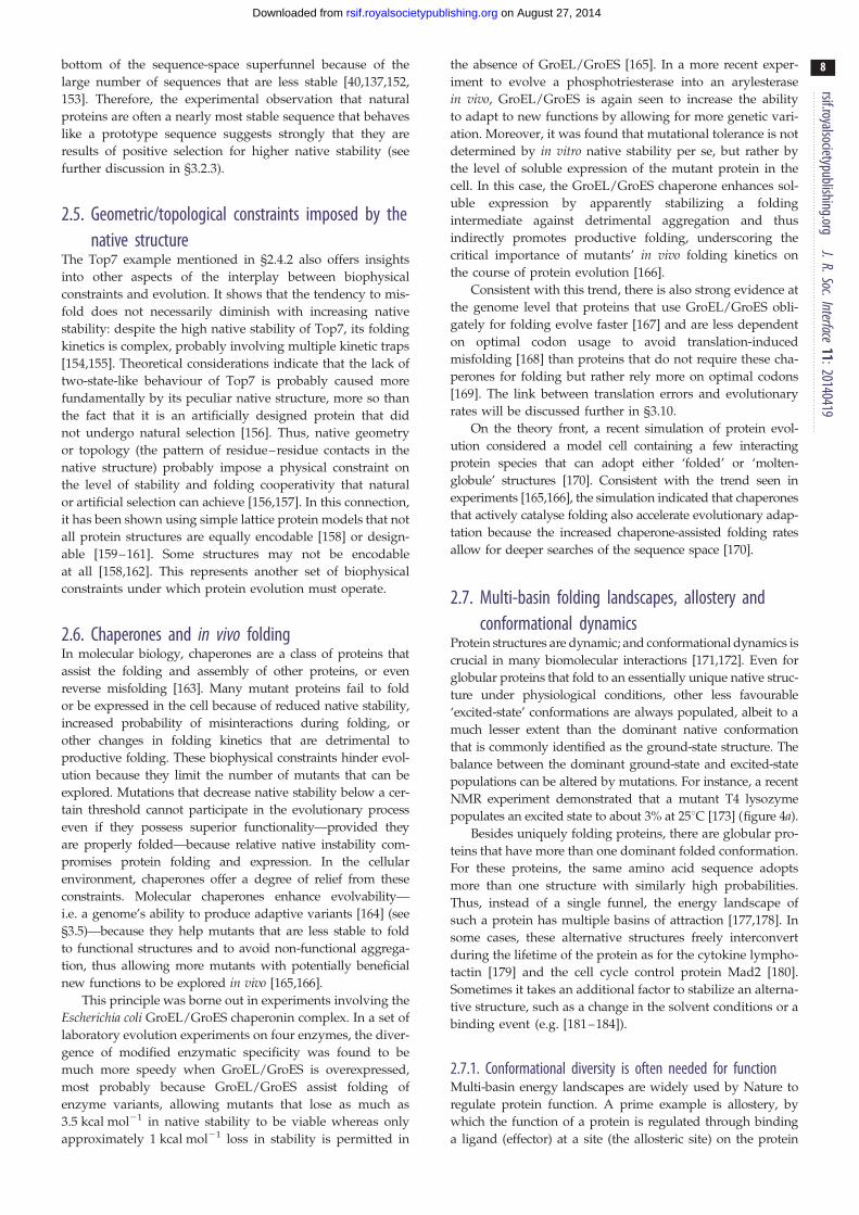
2.6. Chaperones and in vivo foldingIn molecular biology, chaperones are a class of proteins that
assist the folding and assembly of other proteins, or even
reverse misfolding [163]. Many mutant proteins fail to fold
or be expressed in the cell because of reduced native stability,
increased probability of misinteractions during folding, or
other changes in folding kinetics that are detrimental to
productive folding. These biophysical constraints hinder evol-
ution because they limit the number of mutants that can be
explored. Mutations that decrease native stability below a cer-
tain threshold cannot participate in the evolutionary process
even if they possess superior functionality—provided they
are properly folded—because relative native instability com-
promises protein folding and expression. In the cellular
environment, chaperones offer a degree of relief from these
constraints. Molecular chaperones enhance evolvability—
i.e. a genome’s ability to produce adaptive variants [164] (see
§3.5)—because they help mutants that are less stable to fold
to functional structures and to avoid non-functional aggrega-
tion, thus allowing more mutants with potentially beneficial
new functions to be explored in vivo [165,166].
This principle was borne out in experiments involving the
Escherichia coli GroEL/GroES chaperonin complex. In a set of
laboratory evolution experiments on four enzymes, the diver-
gence of modified enzymatic specificity was found to be
much more speedy when GroEL/GroES is overexpressed,
most probably because GroEL/GroES assist folding of
enzyme variants, allowing mutants that lose as much as
3.5 kcal mol21 in native stability to be viable whereas only
approximately 1 kcal mol21 loss in stability is permitted in
the absence of GroEL/GroES [165]. In a more recent exper-
iment to evolve a phosphotriesterase into an arylesterase
in vivo, GroEL/GroES is again seen to increase the ability
to adapt to new functions by allowing for more genetic vari-
ation. Moreover, it was found that mutational tolerance is not
determined by in vitro native stability per se, but rather by
the level of soluble expression of the mutant protein in the
cell. In this case, the GroEL/GroES chaperone enhances sol-
uble expression by apparently stabilizing a folding
intermediate against detrimental aggregation and thus
indirectly promotes productive folding, underscoring the
critical importance of mutants’ in vivo folding kinetics on
the course of protein evolution [166].
Consistent with this trend, there is also strong evidence at
the genome level that proteins that use GroEL/GroES obli-
gately for folding evolve faster [167] and are less dependent
on optimal codon usage to avoid translation-induced
misfolding [168] than proteins that do not require these cha-
perones for folding but rather rely more on optimal codons
[169]. The link between translation errors and evolutionary
rates will be discussed further in §3.10.
On the theory front, a recent simulation of protein evol-
ution considered a model cell containing a few interacting
protein species that can adopt either ‘folded’ or ‘molten-
globule’ structures [170]. Consistent with the trend seen in
experiments [165,166], the simulation indicated that chaperones
that actively catalyse folding also accelerate evolutionary adap-
tation because the increased chaperone-assisted folding rates
allow for deeper searches of the sequence space [170].
2.7. Multi-basin folding landscapes, allostery andconformational dynamics
Protein structures are dynamic; and conformational dynamics is
crucial in many biomolecular interactions [171,172]. Even for
globular proteins that fold to an essentially unique native struc-
ture under physiological conditions, other less favourable
‘excited-state’ conformations are always populated, albeit to a
much lesser extent than the dominant native conformation
that is commonly identified as the ground-state structure. The
balance between the dominant ground-state and excited-state
populations can be altered by mutations. For instance, a recent
NMR experiment demonstrated that a mutant T4 lysozyme
populates an excited state to about 3% at 258C [173] (figure 4a).
Besides uniquely folding proteins, there are globular pro-
teins that have more than one dominant folded conformation.
For these proteins, the same amino acid sequence adopts
more than one structure with similarly high probabilities.
Thus, instead of a single funnel, the energy landscape of
such a protein has multiple basins of attraction [177,178]. In
some cases, these alternative structures freely interconvert
during the lifetime of the protein as for the cytokine lympho-
tactin [179] and the cell cycle control protein Mad2 [180].
Sometimes it takes an additional factor to stabilize an alterna-
tive structure, such as a change in the solvent conditions or a
binding event (e.g. [181–184]).
2.7.1. Conformational diversity is often needed for functionMulti-basin energy landscapes are widely used by Nature to
regulate protein function. A prime example is allostery, by
which the function of a protein is regulated through binding
a ligand (effector) at a site (the allosteric site) on the protein

bi-stable
bi-stable
bi-stable
(a)
(b)
(c)
(d )
(e)
L99A
G113A
N11L
K21P G11V
L12N
R119P
T4 lysozyme
Arc repressor
cystein-rich domain NW1
GA
P22 Cro l Cro
GBGA98
chameleon
GB9827 mut.
9 mut. 14 mut.
21 mut.L45Y
double mutant
double mutant
double mutant
Figure 4. (Caption opposite.)
Figure 4. (Opposite.) Examples of experimentally designed bi-stable proteinsand mutation-induced structural switches. (a) Wild-type T4 lysozyme was mutated(L99A) to create an internal cavity that allows for the population of an excited-stateconformation with an altered helical segment (blue; left). This T4 lysozyme variantcould be further transformed via a single G113A substitution into a bi-stableprotein that also populates a new folded structure in which the local structureof the helical segment is modified (red; right). An additional R119P substitutionon this L99A, G113A variant then leads to a protein that adopts the conformationon the right as its essentially unique native structure [173]. (b) Wild-type Arcrepressor is a homo-dimeric protein. Each monomeric unit contributes ab-strand to form a two-stranded antiparallel b-sheet (blue; left). This shared con-figuration becomes bi-stable with the introduction of a single N11L substitution toeach of the monomeric units. The mutated sequence now populates the originalstructure as well as a new structure with the b-strands changed into two shorthelices (red; right). An additional L12N substitution on each of the monomericunits results in a sequence that adopts the new configuration on the right asits essentially unique native structure [65]. (c) The cysteine-rich domain NW1forms a stable structural element (blue; left) with three disulfide bonds (yellowsticks) between the residue pairs (8,20), (12,25) and (16,24). A single K21P sub-stitution results in a bi-stable mutant that also populates a structure with adifferent overall conformation (red, right) and an alternate disulfide-bonding pat-tern, now between residue pairs (8,24), (12,20) and (16,25). Introduction of asingle G11V substitution on this bi-stable mutant results in a sequence thatadopts the conformation on the right as its essentially unique native structure[174]. (d ) Two domains of streptococcal Protein B, named GA and GB, with a3a and a 4b þ a-fold, respectively, and no significant sequence similarity,were transformed into each other by a series of point mutations that resultedin a structure pair GA98 and GB98 that allows the switch between the two struc-tures with just a single L45Y mutation. GA98 exhibits a small 4b þ a populationand thus may also be regarded as bi-stable [175]. (e) The viral P22 Cro and l Croare DNA-binding proteins. Encoded by different sequences, they have structurallyvery similar helical N-terminal domains (represented by the yellow and greenribbon, respectively) but have structurally distinct C-terminal domains. P22 Crohas a helical C-terminal, whereas the C-terminal of the homo-dimeric l Croforms a b-sheet. A 24-residue chameleon sequence created largely by mixing resi-dues from the helical and sheet-forming C-termini adopts different secondarystructure depending on whether it is inserted in the P22 or l context [176].Sequence and structural information presented in this figure was taken fromthe cited original references.
rsif.royalsocietypublishing.orgJ.R.Soc.Interface
11:20140419
9
on August 27, 2014rsif.royalsocietypublishing.orgDownloaded from
that changes the structure and/or dynamics of the protein’s
active, functional site positioned at a distance from the allo-
steric site [185]. Allostery is important for biological function
and its malfunction is implicated in disease processes
[186,187]. Mutations affect allostery. Mutational effects on
allostery can be subtle because allosteric communication
between the allosteric and active sites can be underpinned
by multiple mechanisms [188,189]. Nonetheless, mutational
effects on allostery can be rationalized by computational
approaches in some instances [190].
Conformational flexibility, dynamics of protein folded
states and allosteric transitions often can be deduced to a
reasonable degree from the structure(s) of the protein in ques-
tion using elastic network models for folded-state dynamics
[191–194] or native-centric Go-like potentials [195] with mul-
tiple folding basins ([196]; reviewed in [177]). Similar to the
aforementioned case for the probable existence of geometric/
topological constraints on the evolution of folding stability
and cooperativity (§2.5), the success of structure-based
native-centric modelling in rationalizing conformational
dynamics and allosteric transitions suggests that there are sig-
nificiant structural constraints on the evolution of functional
folded-state dynamics. The computational efficiency of elastic
network models also allows enzymes that are dissimilar in
sequence and structure yet probably perform similar functions
to be detected by their similar dynamic properties [194,197],
making it possible for relationships between evolutionary con-
servation and conformational dynamics to be explored [198].
Allostery is envisioned to have evolved by oligomerization,
gene fusion and/or recruitment of unused/flexible parts of a
pre-existing protein structure (reviewed in [199]). The latter
evolutionary route may proceed by positive selection of oppor-
tunistic binding of excited-state conformations. The mechanism
of such binding may lie anywhere between the ‘conformational
selection’ and ‘induced fit’ scenarios [177,200]. Evolution has
apparently exploited latent allosteric potentials entailed by con-
formational dynamics in this manner, as in the case of Ste5
activators that target MAP kinases in yeast [201].
Opportunistic binding of excited-state conformations can
also facilitate evolution of new functions that are not necess-
arily allosteric [122,202,203]. During such an evolutionary
process, a sequence with a multi-basin energy landscape
can serve as an evolutionary bridge. In particular, the evol-
utionary intermediate of two sequences each encoding for a

rsif.r
10
on August 27, 2014rsif.royalsocietypublishing.orgDownloaded from
different dominant structure can be a bi-stable sequence that
folds to both structures with equal or similar probabilities
[161,178,204].
oyalsocietypublishing.orgJ.R.Soc.Interface
11:20140419
2.7.2. Bi-stable proteins and conformational switchesExperiments in several laboratories have found cases where a
single mutation was able to either create a bi-stable protein
from a uniquely folding protein or completely switch one
uniquely folding protein to another with a new native structure
[65,173–175,205,206]. Although these cases of mutation-
induced structure switches were artificially engineered, they
demonstrated that it is generally possible for bi-stable proteins
to arise through mutations during natural evolution.
An early example of mutation-induced structure switch-
ing was the Arc repressor, which is a homodimer with a
two-stranded inter-unit b-sheet. Experiments by Cordes
et al. [205] showed that the b-sheet in the wild-type protein
can be changed to a pair of 310-helices by two amino acid sub-
stitutions that swap the neighbouring sequence positions of
an asparagine and a leucine. A subsequent experiment indi-
cated that a mutant with a single asparagine-to-leucine
substitution has approximately equal populations of the
b-sheet and helical forms, and thus may be regarded as an
evolutionary bridge [65] (figure 4b). A recent study showed
further that if two more polar or charged to hydrophobic sub-
stitutions are introduced, the resulting triple mutant adopts
an octamer configuration with approximately half the helical
content of wild-type Arc, indicating that new protein–protein
interactions and novel oligomeric states can readily result
from a small number of mutations [207].
Experimental mutagenesis has uncovered a similar be-
haviour in the cysteine-rich domains (CRD) of cnidarian
nematocyst proteins. Different CRDs fold to either one of
two structures with different disulfide-bonding arrangements
despite high sequence similarity and identical sequence pat-
terns for their cysteines. Meier et al. [174] found that a CRD
sequence that folds to one disulfide arrangement can be con-
verted to another disulfide arrangement by only two amino
acid substitutions, one from lysine to proline and the other
from glycine to valine, whereas the single-substitution
mutant with only the lysine-to-proline mutation behaves as
an evolutionary bridge that populates both disulfide arrange-
ments (figure 4c). This finding again underscores that large
structural changes can be effected by minimal changes in
the amino acid sequence.
The study by Alexander et al. [175] of the GA/GB system
showed that a single leucine-to-tyrosine substitution can con-
vert a sequence encoding for an albumin-binding 3a (GA)
structure to a sequence encoding for an immunoglobulin-
binding 4b þ a (GB) structure (figure 4d ). A subsequent
experiment on two other mutants identified two additional
3a$ 4b þ a structure switches induced by a single amino
acid substitution [206]. Interestingly, a mutant with a confor-
mational ensemble that is 95% 3a and only 5% 4b þ a when
measured in isolation nevertheless binds immunoglobulin
but not albumin [206], providing an excellent example of
how protein–protein interactions can dramatically shift the
conformational distributions of the binding partners [200].
Another recent example of an artificial ‘evolutionary
intermediate’ is a 24-residue sequence that can adopt either
the a-helical or b-sheet C-terminal conformations, respect-
ively, of transcription factors P22 Cro and l Cro, depending
on whether the designed sequence is fused with the N-term-
inal domain of P22 Cro or l Cro [176] (figure 4e). In this case,
the naturally occurring wild-type 24-residue C-terminal
sequences of P22 Cro and l Cro have only five identical
amino acid positions, whereas the amino acid residues of
the designed sequence at all but four positions are either
identical to that in the wild-type P22 Cro or in the wild-
type l Cro. This finding underscores the critical role of ter-
tiary context in determining secondary structure in proteins
[208]. Although the designed sequence is nine and 14 substi-
tutions away from the corresponding sequences in wild-type
P22 Cro and l Cro, respectively, the successful design of a
structurally ambivalent ‘chameleon’ sequence in this exper-
iment suggests that a smooth evolution transition from one
Cro fold to another is possible [176].
Computation-assisted design of conformational switches
has seen notable success [209,210]; but it is still a challenge
to apply our current biophysical knowledge to provide a fun-
damental physical rationalization for experimentally observed
conformational switching. For the GA/GB system, a mutation-
induced gradual stabilization of one structure over another
was demonstrated using a common software for DDG predic-
tion (§2.1) [178]. However, the mutation-induced GA/GB
conformational switching was not reproduced in atomistic
molecular dynamics simulations [67], even though a part of
the simulated energetics is consistent with experiment [68].
The structural plasticity in bi-stable and multi-stable pro-
teins probably plays an important role in protein evolution
[122,211,212]. Conformational switches and bridge sequences
facilitate evolution by allowing continuous or near-
continuous transition from one folded structure to another.
The experiments in figure 4 suggest that, under certain cir-
cumstances, multi-functional proteins can be created by
only a few mutations that stabilize certain hidden or excited
states. A situation where it is advantageous to take such a
route is the coevolution of pathogens and their hosts, a
highly competitive evolutionary process that demands fre-
quent change of protein shapes and functions. It is thus
unsurprising that bi-stability and multi-specificity are exhib-
ited in antibodies [213], antimicrobial peptides in natural
plant defence [214] and antiviral proteins [215].
2.8. Intrinsic disorderWhen structural plasticity is extreme, one might expect a
multi-stable sequence to morph into one without a discrete
set of clearly discernible favoured conformations. This in
itself is not surprising because an overwhelming majority of
polypeptides with random amino acid sequences do not
fold to a unique structure [116]. What is remarkable, in the
context of our decades-long near-exclusive focus on proteins
with well-ordered structures, is the existence of many func-
tional proteins with such extreme conformational diversity.
Although our main concern here is evolution of globular pro-
teins, it is important to recognize that intrinsically disordered
proteins (IDPs) or intrinsically disordered regions (IDRs) play
key roles in cellular processes [216–222].
2.8.1. Any protein conformational state can potentially havebiological function
With the discovery of functional IDPs/IDRs, it has become
abundantly clear that biology can exploit any protein confor-
mational state that it finds useful. In this respect, an
rsif.royalsocietypublishing.orgJ.R.Soc.Interface
11:20140419
11
on August 27, 2014rsif.royalsocietypublishing.orgDownloaded from
intriguing recent suggestion is that although avoidance of
amyloid-like aggregation has apparently been a driving
force of protein evolution [223] (§2.2), it is possible that
modern protein folds have an amyloid origin in evolution
[224]. For IDPs/IDRs, current understanding of the evolution
of the triplet genetic code [225] suggests that the amino acid
composition of primordial polypeptides was conducive to
more disordered conformations before the modern genetic
code for a 20-letter amino acid alphabet was completed
[222]. However, surveys of modern proteomes indicate that
IDPs/IDRs are more common in eukaryotes than in prokar-
yotes: more than 32% of amino acid residues in eukaryotic
proteins are in IDPs/IDRs whereas the corresponding per-
centage is less than 27% for prokaryotic proteins. This
pattern suggests that the proteins in the last universal ances-
tor were probably well structured and emergence of the
IDPs/IDRs observed today was relatively late [222], perhaps
coinciding with an evolutionary trend that has witnessed a
general decrease in protein hydrophobicity [226].
According to one estimate, more than 30% of eukaryotic
proteins have IDRs of more than 50 consecutive residues
[216], consisting of more proline, glycine and charged resi-
dues but fewer hydrophobic residues [227,228]. IDPs/IDRs
are involved in fundamental processes such as transcription,
translation and cell cycle regulation that, when they malfunc-
tion, can lead to cancer. The essential role of IDPs/IDRs in
mediating biological regulation suggests that, in some situ-
ations, they have certain advantages over folded proteins in
recognition and binding [229]. For instance, their ability to
flexibly bind to many different partners has allowed them
to occupy hub-like roles in protein–protein interaction net-
works [230,231]. They can also encode relatively larger
intermolecular interfaces to economize genome and cell
sizes [218]. Protein–protein interactions for some IDPs/
IDRs entail significant folding upon binding [219], while
others undergo only restricted local ordering at the binding
site with other parts of the protein remaining disordered,
thus forming a dynamic ‘fuzzy’ complex [220,232–236].
2.8.2. Biophysical constraints on evolution of intrinsicallydisordered proteins and regions
What can be expected of the biophysical constraints on the
evolution of IDPs/IDRs? IDPs/IDRs do not fold to a unique
structure. Therefore, in contrast to many globular proteins,
the energy landscapes of IDPs/IDRs are not funnel-like
[222]. As far as near-neutral mutations [237,238] are concerned,
one might expect less biophysical constraints on IDP/IDR
evolution than on globular protein evolution because for
IDPs/IDRs there is no need to maintain an essentially
unique folded structure. However, it can also be argued that
evolution of certain IDPs/IDRs may be subject to even more
restrictive constraints because of their requirement to bind to
multiple partners. As a result, these IDP/IDRs may suffer
from low mutational robustness similar to that of bi-stable
globular proteins that play the role of an evolutionary bridge
between two folded structures [178,239]. Nevertheless, even
in such cases, IDPs/IDRs in a neutral net might only need
to conserve certain functional residues that are compatible
with multiple binding partners while imposing few constraints
on mutations at amino acid sites in the rest of the protein.
These expectations are largely consistent with database
studies and experiments. Phylogenetic analyses indicate
that IDRs generally evolved faster than ordered regions of
proteins, but some IDRs such as DNA-binding regions
evolved slower [240,241]. For proteins that have both ordered
and disordered regions, mutations in IDRs lead to smaller
stability changes than in ordered regions. Thus, IDPs/IDRs
may enhance protein evolvability and the development of
new functions [242], as evolutionary changes in protein
sequence and structure are often correlated with local
flexibility and disorder [243].
The biophysical constraints on IDP/IDR evolution [244]
are quite different from those on folded protein evolution
[12]. In fact, the accepted amino acid substitutions in IDPs/
IDRs resemble those in solvent-exposed loops and turns of
globular proteins [244]. Chemical composition defined as the
fraction of positive, negative, polar, hydrophobic and special
(proline and glycine) residues is often maintained across IDR
orthologues that otherwise exhibit little conservation [245].
This observation is in line with the finding that whether an
IDP is elastomeric or amyloidic depends largely on the relative
compositions of proline and glycine [228], and is consistent
with the central role of aromatic composition in a set of IDP
interactions that are presumably underpinned by cation–p
attraction [236]. Relative to the substitution matrices for globular
proteins, substitution matrices for IDPs/IDRs entail a generally
higher probability of evolutionary changes, but some residues
such as tryptophan and tyrosine tend to be highly conserved
in IDPs/IDRs, perhaps because of their critical role in
protein–protein interfaces [244,246].
It should be recognized that IDP/IDR conformations are
far from random. Biological functions of proteins are always
underpinned by conformational structures. In this respect,
the difference between IDPs/IDRs and ordered proteins is
that the IDP/IDR function is conferred by a much more
diverse conformational ensemble than for globular proteins.
The transient, ‘fuzzy’ tertiary contacts in IDP/IDR con-
formations are often important for the function; hence
mutations that disrupt such contacts can be extremely detri-
mental to function. An example of how a single mutation
can disrupt IDP function is the threonine-to-arginine mutation
at position 45 of the cyclin-dependent kinase inhibitor Sic1
[234,247]. This amino acid substitution leads to a dramatic
increase in its hydrodynamic radius [234] and, at the same
time, a serious disruption of its biological function in regulat-
ing the cell cycle [247]. Current biophysical understanding of
this and other mutational effects on IDP/IDR conformational
distribution is limited. Much remains to be discovered about
the evolution of these proteins.
2.9. Protein dynamics and phenotypic plasticity: what isa molecular phenotype?In the study of molecular evolution, the term genotype is
used for the inheritable part of genetic information; whereas
phenotype refers to the biomolecules of interest that are pro-
duced based on the genotypic information. In theoretical
studies of protein evolution, as a modelling simplification, the
genotype may be identified with the amino acid sequence
because as far as in vitro protein folding is concerned, it con-
tains essentially the same information as the nucleic acid
sequence that encodes it. This is a simplified approach that
neglects in vivo complexities such as the fact that synonymous
mutations can lead to altered cellular folding pathways (§2.2).
In principle, the molecular phenotype should encompass all

rsif.royalsocietypublishing.orgJ.R.Soc.Interface
11:20140419
12
on August 27, 2014rsif.royalsocietypublishing.orgDownloaded from
properties—including but not limited to biological
functions—of the protein encoded by the genotype. In prac-
tice, molecular phenotypes in theoretical and experimental
investigations are defined, and thus are restricted, by the
question being addressed. However, an oversimplified view
of molecular phenotypes that is too restrictive can hinder
understanding of important principles of protein evolution.
For globular proteins that have an essentially unique
folded structure, a practical and seemingly natural definition
of molecular phenotype of a given amino acid sequence is its
structure as deposited in the Protein Data Bank (PDB). This
practice is useful for constructing a neutral net of sequences
that encode uniquely for the same protein structure and the
evolution from one such phenotype to another [137,161].
However, this simplistic view of molecular phenotype
neglects the dynamic nature of proteins. Recent advances in
experimental techniques, especially those using NMR, have
enabled detailed characterizations of the dynamic properties
of proteins [173,248–250] and, in conjunction with compu-
tation, allowed for the construction of ensembles of diverse
conformations of disordered proteins based on NMR and
other experimental measurements [251,252]. As a result of
these experimental advances and the theoretical energy land-
scape perspective [34,253,254], our view of how protein
molecules function has undergone a drastic change in the
past two decades, with increasing recognition of the bio-
physical, biological and evolutionary significance of protein
dynamics [255,256].
Because of the role of dynamics in protein function
(§2.7 and 2.8), identifying a protein’s molecular phenotype
only with its native folded structure is often too restric-
tive. Ideally, the molecular phenotype of an amino acid
sequence should correspond to the totality of its biologically
relevant properties. Although it may not be practical to enu-
merate many properties of a protein, for many applications
the molecular genotype should at least be understood as an
ensemble of conformations with a sequence-specific and
environment-dependent distribution. Within this ensemble,
certain phenotypic properties, such as the presence of a
secondary structure in the protein conformation, are not
necessarily fixed but can undergo thermal fluctuations or
environment-induced changes. This phenomenon is referred
to as single-genotype phenotypic fluctuation or phenotypic
plasticity, which can underpin important evolutionary
responses to environmental changes [257].
Phenotypic plasticity tends to enhance evolvability. This
trend can be seen clearly in an experimental evolution
study of E. coli cells that express mutants of green fluor-
escence protein. In this experiment, mutants leading to a
larger fluctuation in fluorescence among cells containing
the same green fluorescence protein gene were found to exhi-
bit a higher rate of evolution [258]. A positive correlation
between single-genotype phenotypic fluctuation and evolva-
bility has also been rationalized recently by computational
models of proteins [259] and RNA [260]. As mentioned in
§2.7.2 above, plasticity of molecular phenotype (i.e. confor-
mational diversity) is generally conducive to higher
evolution rates that can be beneficial to organisms in rapidly
changing environments [261,262] through selection of ‘moon-
lighting’ [263] promiscuous functions [264]. We will elaborate
below on how the relatively new view of protein dynamics
and conformational distribution [202,203,265] has enriched
our understanding of evolution.
3. Applications of biophysics-based models tounderstand protein evolution
To gain insights into how proteins evolve, various models of
the mapping from protein sequence (genotype) space to
structure (phenotype) space have been constructed. Here,
we focus largely on models with an explicit representation
of the protein chain and biophysics-based interactions
because these models provide a better delineation of what
is physically plausible for real proteins than other models
of molecular evolution that postulate such a mapping in the
absence of or with little biophysical considerations.
3.1. Protein sequence and structure spaces: evolutionmeets biophysics
Recent genomic and proteomic initiatives have greatly
advanced our knowledge of the global sequence–structure
relationship and the evolution of the primary, secondary,
supersecondary, domain, tertiary and quaternary/complex
structures of proteins [266–269]. An earlier study suggested
that a/b folds appeared later in evolution than other struc-
tural classes [266]. However, more recent investigations
indicate that the globular protein architectures observed
today had emerged during evolution roughly in the follow-
ing order: a/b (e.g. TIM-barrel), a þ b, all-a, all-b, then
multi-domain proteins [268]. Consistent with this perspec-
tive, a core of functional diversity corresponding mostly to
the more ancient a/b folds in the protein structure space
has been identified [270]. Interestingly, a computer modelling
study of the dynamic properties of protein fold space
suggests that a/b folds are also more stable than other fold
classes [271].
In the space of all possible amino acid sequences, an over-
whelming majority of sequences do not have a biological
function. It has been estimated that the probability of finding
a functional protein among random amino acid sequences is
approximately 10211 [272]. Evolutionarily, natural protein
sequences are still diverging from one another today, albeit
at a slow rate because biophysical and functional constraints
allow only about 2% of amino acid sites to be mutated [273].
This ongoing divergence means that the coverage of the space
of all possible sequences by biologically viable sequences has
been and still is increasing; i.e., there has been a continuing
expansion of the ‘protein universe’ since the beginning of
life on the Earth [273].
There is consensus among researchers that the repertoire
of globular protein folds is probably finite. The SCOP classi-
fication [274] currently identifies about 1200 different folds in
the PDB. Estimates for the total number of possible folds
range from about 1000 [275], 2000 [276] to 10 000 [277].
Fold classifications are inherently difficult and estimation of
the total number of possible folds is sensitive to the definition
of a fold (see [278] and references therein). Nonetheless, in
most cases, structures of recently sequenced proteins are
related to known folds [279], suggesting that the existing
PDB structures are probably a near-complete representation
of all biologically viable globular protein folds.
A broader issue is whether the observed natural globular
protein folds constitute a relatively small subset selected by
evolution from a much larger collection of all physically poss-
ible compact conformations. Biophysics and polymer physics

rsif.royalsocietypublishing.orgJ.R.Soc.Interface
11:20140419
13
on August 27, 2014rsif.royalsocietypublishing.orgDownloaded from
have shed light on this question. A hallmark of most globular
proteins is their helical and/or sheet-like organization. These
secondary structures facilitate backbone–backbone hydrogen
bonding in the folded protein core (reviewed in [12]). Second-
ary structures are conducive to tight tertiary packing as
well. It has been shown that secondary-structure-like chain
organization is enhanced by conformational compactness
[280–282], but in the absence of hydrogen bonding such
structures exhibit deviations from sharply defined a-helices
and b-sheets [283–285]. These findings suggest that biophysi-
cal constraints of conformational compactness in conjunction
with hydrogen bonding can go a long way in accounting for
the basic architecture of globular protein folds.
A more recent study using computational sampling of
homopolypeptide conformations suggested further that the
current repertoire of globular protein folds is nearly complete
in its coverage of all physically possible compact folds [286].
However, studies by three other groups have found instead
that the current fold repertoire represents only a small
fraction of all possible folds [287–289]. In particular, an
investigation of the compact conformations of 60-residue
polyvaline concluded that known protein folds constitute
only approximately 5% of all physically possible folds, and
that on average the natural folds have more local intrachain
contacts (i.e. lower contact orders [146]) than the set of all
possible folds, suggesting an evolutionary preference for
structures with lower contact orders [288]. In response, a
recent study argued that inasmuch as an appropriate cri-
terion for matching simulated compact conformations
and natural folds is applied, the existing library of single-
domain PDB structures is probably complete in covering all
physically possible folds [290]. A separate study by the
same group indicated that computer-generated compact con-
formations tend to contain cavities resembling binding
pockets in natural proteins as well, even in the absence of
selection, suggesting that ‘many features of biochemical func-
tion arise from the physical properties of proteins that
evolution likely fine-tunes to achieve specificity’ [291].
While the degree to which evolution has shaped the space
of known protein folds remains to be further elucidated, the
investigative effort described above is an excellent illustration
of how explicit-chain biophysical models can be harnessed to
address fundamental questions in protein evolution.
3.2. Simple exact models and other explicit-chaincoarse-grained models of protein evolution
Sequence and structure spaces of proteins are vast.
Coarse-grained explicit-chain models are valuable tools
in the study of protein evolution. Currently, modelling
the sequence–structure mapping by energetically and
structurally high-resolution representations is often not prac-
tical, especially for addressing large-scale evolutionary
changes involving many different protein folds. In this
regard, lattice models—wherein conformations of model pro-
teins are configured on two- or three-dimensional (2D or 3D)
lattices—are particularly useful because of their compu-
tational tractability [292–294]. In view of their historical
and current utility for investigating fundamental evolution-
ary issues (see, e.g., recent applications of 2D lattice models
to study the basis of homology modelling [295], adaptive
conflict [239] (§3.6) and long-term survivability [296]), a
detailed assessment of the biophysical foundation of these
models is in order.
3.2.1. Conformational enumerationAmong lattice models of protein evolution, simple exact
models allow for exhaustive enumeration of all possible
sequences and structures in the model [292]. These models
include the two-letter 2D hydrophobic-polar (HP) model
that uses a reduced alphabet consisting of only two types
of residues, hydrophobic (H) and polar (P), to capture the
prominent effects of hydrophobic interactions in protein ener-
getics [137,161,162,297–299] (figure 5), two-letter variants of
the 2D HP model (e.g. [293,301,302]), and a four-letter 2D
model that also includes residues behaving somewhat
like positive and negative charges [303]. Some 20-letter
2D models may also be considered as exact, because
they consider all possible mutations in the immediate
sequence-space neighbourhood of a given sequence [40,153].
Other types of lattice models have been used to study
protein evolution as well. These models either restrict chain
conformations to be maximally compact so as to allow
model proteins with longer chain lengths to be studied
[152,160,304], or rely to various degrees on sequence-space
sampling instead of considering all possible sequences
because of their usage of a 20-letter amino acid alphabet
that entails many more sequences (e.g. [40,153,305]) or both
(e.g. [306,307]). Restricting model structure space to maxi-
mally compact conformations in 2D [306] or 3D [307,308]
reduces computation drastically because such conformations
constitute only a tiny fraction of all possible conformations
[280,309–312]. For instance, whereas the total number of all
distinguishable conformations (not related by rotations and
reflections) for a chain with 25 residues configured on
the 2D square lattice is 5 768 299 665 [313], the number of
maximally compact 25-residue conformations restricted to a
5 � 5 square, as considered in the study of Taverna &
Goldstein [306], is only 1081 [309]. In 3D, the total number
of all possible conformations (not related by rotations and
inversions) on the simple cubic lattice for a chain with 27 resi-
dues is 11 447 808 041 780 409 [313,314], but the number of
maximally compact 27-residue conformations restricted to a
3 � 3 � 3 cube, as considered by Deeds & Shakhnovich
[307], is only 103 346 [280].
However, from a biophysical standpoint, it is important
to keep in mind that real protein chains are not restricted
to be maximally compact. Although behavioural trends
predicted by models that use only maximally compact con-
formations may sometimes correlate with models that
consider the full conformational ensemble, significant distor-
tions of protein folding energetics are introduced by this
approach [158,292]. In particular, for a given model sequence
with a physically plausible interaction, the lowest-energy
structure among maximally compact conformations may
not be the true ground state structure, which is often less
than maximally compact [299,315,316].
3.2.2. Model interactions and their biophysical basisWe next consider the physicality of the model interaction
potential. Several examples presented below to illustrate
recent biophysical insights into basic principles of protein
evolution are based on the 2D HP model (figure 5). We
choose the 2D HP model for this purpose because owing to
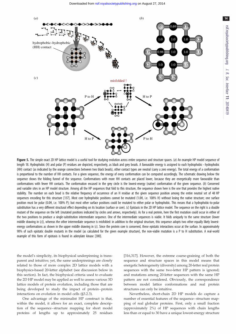
hydrophobic–hydrophobic(HH) contact
0 0.69 0.19
1.0 1.0 1.0 0.25
0.980.19 1.0 1.0
1.00.21 1.0
0.31
H to P
H to P
P to H
P to H
misfolded !0.29
0.17
0.13
(a)
(c)
(b)
Figure 5. The simple exact 2D HP lattice model is a useful tool for studying evolution across entire sequence and structure spaces. (a) An example HP model sequence oflength 18. Hydrophobic (H) and polar (P) residues are depicted, respectively, as black and grey beads. A favourable energy is assigned to each hydrophobic – hydrophobic(HH) contact (as indicated by the orange connections between two black beads), other contact types are neutral (carry a zero energy). The total energy of a conformationis proportional to the number of HH contacts. For a given sequence, the energy of every conformation can be computed accordingly. The schematic drawing below thesequence shows the folding funnel of the sequence. Conformations with more HH contacts are placed lower, because they are energetically more favourable thanconformations with fewer HH contacts. The conformation encased in the grey circle is the lowest-energy (native) conformation of the given sequence. (b) Conservedand variable sites in an HP model structure. Among all the HP sequences that fold to this structure, the sequence shown here is the one that provides the highest nativestability. The number on each bead is the relative frequency of occurrence of an H residue at the given sequence position among the entire neutral set of 48 HPsequences encoding for this structure [137]. Most core hydrophobic positions cannot be mutated (1.0H, i.e. 100% H) without losing the native structure; one surfaceposition must be polar (0.0H, i.e. 100% P); but most other surface positions could be mutated to either polar or hydrophobic. This means that a hydrophobic-to-polarsubstitution has a very different structural effect depending on its location (surface or core). (c) Epistasis in the 2D HP lattice model. The sequence on the right is a doublemutant of the sequence on the left (mutated positions indicated by circles and arrows, respectively). As for a real protein, here the first mutation could occur in either ofthe two positions to produce a single-substitution intermediate sequence. One of the intermediate sequences is viable. It folds uniquely to the same structure (lowermiddle drawing in (c)), whereas the other intermediate sequence is misfolded: in addition to the original structure, this sequence adopts two other equally likely lowest-energy conformations as shown in the upper middle drawing in (c). Since the protein core is conserved, these epistatic interactions occur at the surface. In approximately90% of such epistatic double mutants in the model (as calculated for the given example structure), the non-viable mutation is a P to H substitution. A real-worldexample of this form of epistasis is found in adenylate kinase [300].
rsif.royalsocietypublishing.orgJ.R.Soc.Interface
11:20140419
14
on August 27, 2014rsif.royalsocietypublishing.orgDownloaded from
the model’s simplicity, its biophysical underpinning is trans-
parent and intuitive; yet, the same underpinnings are closely
related to those of more complex 2D lattice models with a
biophysics-based 20-letter alphabet (see discussion below in
this section). In fact, the biophysical criteria used to evaluate
the 2D HP model may be applied as well to assess various 3D
lattice models of protein evolution, including those that are
being developed to study the impact of protein–protein
interactions on evolution in model cells (§3.2.3).
One advantage of the minimalist HP construct is that,
within the model, it allows for an exact, complete descrip-
tion of the sequence–structure mapping for short model
proteins of lengths up to approximately 25 residues
[316,317]. However, the extreme coarse-graining of both the
sequence and structure spaces in this model means that
energetic heterogeneity (diversity) among 20-letter real protein
sequences with the same two-letter HP pattern is ignored;
and mutations among 20-letter sequences with the same HP
pattern are not considered. Obviously, the correspondence
between model lattice conformations and real protein
structures can only be intuitive.
Nevertheless, short-chain 2D HP models do capture a
number of essential features of the sequence–structure map-
ping of real globular proteins. First, only a small fraction
(approximately 2%) of HP sequences with chain lengths
less than or equal to 30 have a unique lowest-energy structure

rsif.royalsocietypublishing.orgJ.R.Soc.Interface
11:20140419
15
on August 27, 2014rsif.royalsocietypublishing.orgDownloaded from
[158,162,299], consistent with experimental observations that
only a tiny percentage of random amino acid sequences
(much lower than 2%) can fold and/or have a biological func-
tion [116,272]. Experimentally, it is very rare for folded and/
or functional sequences to arise in random sequence search,
but binary HP patterning can help with artificial design of
such sequences [318,319]. Second, the small fraction of
model HP sequences that have a unique lowest-energy 2D
structure exhibit statistics of HP patterns similar to that of
real proteins [320,321]. Third, many of the lowest-energy
2D HP structures are highly compact but not maximally com-
pact, as for real globular proteins [158,299], and a significant
fraction of compact structures are not encodable by any HP
sequence as its unique native structure [158,162]. The latter
observation may bear on the question of whether the cur-
rently known set of globular protein folds is nearly
complete in its coverage of all physically possible compact
folds, as discussed in §3.1 [286–290], but one has to also
keep in mind that the HP model interaction potential is less
heterogeneous, and thus entails fewer encodable structures,
than model potentials that contain repulsive interactions or
otherwise more heterogeneous interactions [158,322,323].
Fourth, the 2D HP lattice model provides sequences that act
like evolutionary bridges [161,178,204,239,299] (see §3.5),
encode for autonomous folding units [292,316,324], and exhi-
bit homology-like behaviours [295], all similar to properties
observed in real proteins.
Two likely physical reasons underlie the similarity
between the sequence–structure mapping of the 2D HP
model and that of real globular proteins. First, the HP
model potential captures the prominent effect of hydrophobic
interactions in protein folding [44]. Similar to the hydro-
phobic effects operating in real proteins, the model
potential leads to folded structures with a hydrophobic core
and mostly polar residues on the surface (see examples in
figure 5). Because the surface-core ratio of folded structures
on the 2D square lattice with chain lengths approximately
16 is similar to the surface-core ratio of 3D folded structures
with chain length approximately 150 [317], the energetics of
short-chain 2D HP models should bear resemblance to that
of real proteins with approximately 100 amino acid residues.
Second, as has been argued [324], although the simple poten-
tials of the HP model and its two-letter variants, and for that
matter even 20-letter lattice potentials, are not sufficient to
capture more detailed thermodynamic [325] and/or kinetic
[326] properties of protein folding, the HP potential may
still provide a useful caricature of the mapping between
sequence and folded structure of real globular proteins
because of the ‘consistency principle’ [327] or ‘principle of
minimal frustration’ [328]. These principles stipulate consist-
ency or near-consistency among various energy terms that
contribute to the stability of natural proteins. Therefore, in
this perspective, the folded state of a protein—at least for a
natural, evolved protein—is expected to be a lowest-energy
structure for a hydrophobic interaction potential similar
to the one prescribed by the HP model, although other
interaction types may provide it with additional stabilization.
Besides the HP and HP-like models, many of the 20-letter
models adopt interaction energies derived from the PDB-
based Miyazawa–Jernigan (MJ) statistical potential [329].
The original MJ potential (Table V of [329]), as was used in
[40,153,296,330], embodies the hydrophobic effect, thus it
tends to place non-polar residues in the folded protein core
as in real proteins (reviewed in [116,204]). However, certain
modified, ‘shifted’ forms of MJ potentials that are similar to
table VI of [329] with prominent repulsive energies [331]
do not embody this biophysical property (reviewed in
[158,204]), making it problematic to interpret some of the pre-
dictions from such models. For instance, in some cases, a
shifted-MJ potential may lead to nominally charged residues
instead of hydrophobic residues occupying the core of a
model protein [114,204,331].
These limitations notwithstanding, all aforementioned lat-
tice models provided useful evolutionary insights. Different
approaches are often complementary because they tackle
different aspects of protein evolution. However, caution
should always be used to take these models’ limitations
into account so as not to over-interpret model predictions.
Major earlier progress of lattice models of protein evolution
can be found in several reviews [292,332–334]. We now
recall briefly some of the significant early efforts before
highlighting recent advances.
3.2.3. Predictions and rationalizationsIn several 2D [137,161,293,299,303,304,323] and 3D [305,335]
lattice models as well as an off-lattice model [304], protein
sequence space was found to be organized as multiple neu-
tral nets. A neutral net is a network of sequences that are
connected by single-point substitutions and encoding for
the same folded structure. In a few studies that addressed
the global connectivity of sequence space, it has been
observed further that neutral nets for different folded struc-
tures are interconnected to form a dominant ‘supernet’
covering most of the sequence space [298,303,324] in a
manner similar to that envisioned by Maynard Smith [336].
Consistent with experiment [139–142] (§2.4.2), mutations
on encoding sequences often result in sequences encoding
for the same structure [137,297,305], especially if the mutation
site is on the surface of the folded protein [297]. In other
words, many sequences are stable against mutation, a
property referred to as mutational robustness [306].
Several models indicate that the topology of a protein
neutral net has a superfunnel organization [137], wherein
sequences encoding for the same structure tend to centre
around a prototype sequence with maximum mutational
robustness as well as maximum thermodynamic stability
for the encoded native structure [137,293,304,323,335]. Muta-
tional stability is generally correlated with thermodynamic
stability [114,137], such that sequences at the edge of the neu-
tral net have lower native stability [137,299,337]. Consistent
with this trend, a 20-letter 2D lattice model predicts that,
with increasing number of amino acid substitutions, the
probability for a protein to retain its original structure
declines exponentially [40].
Evolution of protein function has been explored using 2D
square-lattice [296,301,302,330] and 3D diamond-lattice [335]
model sequences that encode folded structures with a bind-
ing site. In the 2D square-lattice model studies of Bloom
et al. [330,338], folded-state stability was found to promote
evolvability of new binding functions, consistent with exper-
imental observations. These simulated evolution processes
allow for extensive exploration of sequence space. However,
no structural change to the fold of the evolving lattice protein
was seen during the simulation. Thus, the scope of protein
evolution in these models [330,338] is largely limited to the

rsif.royalsocietypublishing.orgJ.R.Soc.Interface
11:20140419
16
on August 27, 2014rsif.royalsocietypublishing.orgDownloaded from
development of new binding abilities through modifications
of surface residues, while the evolved protein retains its orig-
inal structural scaffold. For larger structural changes, one
expects that it would be harder for a more stable protein to
evolve to a specific new fold, because a more stable sequence
is farther away from the edge of the neutral net where it can
switch to another fold (§2.7.2).
More recently, the binding between 2D square-lattice
model proteins and lattice ligands was used to investigate
short-term versus long-term evolutionary success. Interest-
ingly, this study by Feldman and co-workers indicate that
although the model evolution process is stochastic, long-
term evolutionary success—as determined by stability of
the evolving protein and its binding with a given ligand—
is ‘surprisingly predictable’ from the founding sequence
of a lineage. In this lattice protein model, long-term survivabil-
ity is only partially determined by short-term fitness, i.e. short-
term adaptive success does not guarantee long-term survival
of a lineage [296].
2D lattice models have been applied to compare the evol-
utionary effects of point mutations and recombination
[152,324,335]. An initial study showed that crossover of two
encoding sequences is an effective way of producing a new
encoding sequence, suggesting that local sequence patterns
are important for determining whether the full protein
sequence can fold to a unique structure [324]. This theoreti-
cal prediction is consistent with subsequent experiments on
b-lactamase indicating that for a given number of amino acid
substitutions, recombined variants are much more likely to
retain function than variants generated by random point
mutations [339]. In another simulation study, evolutionary
dynamics that admits both point mutations and recombination
leads to a much higher concentration of population in the pro-
totype sequence than if evolution proceeds via point mutations
alone, suggesting a significant role of recombination in the
prototype-like behaviours of natural proteins [152].
Lattice models have also been used to investigate how
mutagenesis data may be exploited to improve forcefields
for protein structure prediction [340,341]. In addition, they
have provided insights into the relationship between the
native contact pattern of a target structure and its designabi-
lity [342], the degree to which evolutionary selection at the
molecular and/or organism level has led to the observed
scale-free distribution of protein structure similarities
[307,334], and the biophysical basis [343,344] of methods
that use evolutionary/mutational information as a probe to
reveal long-range energetic coupling in proteins [26].
An exciting recent development of 3D maximally com-
pact lattice protein models is their application to the study
of fitness and population dynamics of model cells containing
multiple proteins. One model assumes that all proteins in a
model cell are essential and that the fitness of the cell
depends on the stability of the least stable proteins it contains.
Real-life-like properties such as preferred folds and protein
families readily emerge from these simple assumptions
[308]. Introduction of various protein–protein interactions
to this modelling set-up by Shakhnovich and co-workers
[345] has provided further rationalization for the emergence
of species-like collections of model cells with very similar
sequence make-ups, an increased rate of mutation in stress
response [346], as well as a trade-off between strengthening
functional interactions and avoidance of misinteractions as
observed in experimental proteomic data [347]. More
recently, Zhang and co-workers [99] developed a related lat-
tice approach to model evolution of protein–protein
interactions that offers an explanation of slow evolution of
highly expressed proteins in terms of stronger constraints
on these proteins to avoid misinteractions.
3.3. Energy, entropy, fitness, protein neutral nets andfitness/mortality landscapes
Protein evolution can be formulated in terms of population
dynamics on a fitness or adaptive landscape in which a fit-
ness function assigns a fitness value to each protein
sequence, with evolving populations migrating to areas of
higher fitness over time [348–350]. Certain parallels may be
drawn between fitness landscapes defined on sequence
spaces and energy landscapes defined on protein confor-
mational spaces. Mathematically, both protein sequence and
conformational spaces are high-dimensional. As for protein
energy landscapes, fitness landscapes are intrinsically high-
dimensional constructs, even though fitness and energy land-
scapes are often depicted as 1D profiles or 2D surfaces for
metaphorical, conceptual visualization [34,254,317,348,351].
With this in mind, it is important to focus on the quantita-
tive fitness function itself and not to over-interpret the
picturesque 1D and 2D landscape representations [351].
The biological fitness concept has been compared to the
physical quantity of negative energy or negative free energy
[352]. There is an obvious analogy between the sequence-
space distribution of steady-state evolutionary population
and the conformational-space distribution of equilibrium
population. Just as lower-energy states and higher-entropy
macrostates are more favoured in statistical mechanical sys-
tems, higher-fitness sequences and phenotypes encoded by
more sequences are expected to be more populated during
evolution. Under certain limiting conditions, a direct quanti-
tative correspondence can be made between fitness and
energy, as well as between population size and inverse temp-
erature [352]. In general, however, there is an important
difference between the analogous roles of energy and fitness:
insofar as a statistical mechanical system is ergodic [353] and
total population is conserved, the equilibrium population of a
given state is determined by its energy in accordance with the
Boltzmann distribution. By contrast, for evolution, total
population can be increased by reproduction and decreased
by lethal mutations. As a result, the steady-state population
of a given sequence in a large evolving population is deter-
mined not only by reproductive fitness of the sequence but
also by its connectivities to neighbouring viable sequences.
This issue will be addressed in the discussion on mutational
robustness in §3.4.
Studies of molecular evolution indicate that evolutionary
pathways on fitness landscapes are subjected to various con-
straints [351,354]. The topographies of fitness landscapes of
real proteins are far different, on average, from those postu-
lated theoretically by random assignments of fitness [351].
This observation underscores the importance of biophysical
considerations in constructing model fitness landscapes. As
an illustration, figure 6 shows the sequence space of a 2D
HP model (figure 6a), part of its neutral net organization
(figure 6b), and a model fitness landscape that identifies
fitness with native stability (figure 6c). As discussed
above, the biophysics embodied by the HP model imparts
it with several essential protein-like properties. For instance,

(a)
(c)
foldable sequence space
sequence spacepromiscuity and evolvability
thermodynamic stability and mutational robustness
stab
ility
(b) neutral networks
Figure 6. The space of foldable HP sequences gives a glimpse of what real protein sequence space might look like. (a) A representation of the adjacency network ofapproximately 25 000 HP sequences of length 18, each with either a unique ground-state structure or up to six different but equally populated ground-statestructures. Only a small minority of sequences is not connected to the dominant ‘supernet’. (b) Local sequence neighbourhoods form clusters of sequencesthat fold to the same ground-state structure. These clusters are called neutral nets or neutral networks, each of which is shown in a different colour here.Some of the neutral nets overlap. (c) Thermodynamic stabilities of the folded ground states of the sequences (stability increases in the downward direction)are plotted using a schematic planar of sequence space as in (b). A distinct funnel-like organization for each neutral net is apparent. These sequence-space funnelsare referred to as superfunnels [137]. Typically, there is a single prototype sequence per neutral net that is thermodynamically maximally stable and also tolerates thehighest number of neutral mutations (i.e. those resulting in a sequence within the neutral net). The ground-state structure in the HP model is shown for eachneutral net. Sequences with high stability (at the bottom of the landscape) have a clear tendency to also have more neutral mutations and thus higher mutationalrobustness. In contrast, promiscuity and multi-functionality is likely encoded in less stable sequences, including sequences with two or more ground-state structures.These sequences are located further away from the prototype sequence but closer to other neutral nets; hence they have a higher evolvability—meaning a higherprobability to acquire new functions through further mutations (see also figure 8). The network layouts here and in subsequent figures were constructed in such away that the lengths of the edges roughly reflect the Hamming distances between the sequences connected by the edges [239]. Because of the large number ofsequences depicted, the network drawings in the present figure convey only a heuristic impression of the sequence connections. More detailed descriptions of someof the neutral nets shown in figures 5 – 9 are provided in [137,239].
rsif.royalsocietypublishing.orgJ.R.Soc.Interface
11:20140419
17
on August 27, 2014rsif.royalsocietypublishing.orgDownloaded from
consideration of all model HP sequences that fold uniquely to
the native structure in figure 5a [137,204] shows that certain
sites are conserved, and that the probability that a mutation
is viable is site-dependent (figure 5b), echoing the context-
dependent substitution rates of amino acids in real proteins
(§1). It is also noteworthy that because the main biophysical
driving forces in protein folding are different from that in
RNA secondary structure formation, the global organization
of neutral nets of globular proteins is probably very different
[161,355] from that of RNA [356–359].
It is conventional to associate increasing fitness with
upward movement on fitness landscapes [348]. For the
model fitness landscape in figure 6c, however, we have
chosen to represent increasing fitness with downward move-
ment. As discussed elsewhere, such landscapes may be
referred to as ‘inverse-fitness’ or ‘mortality’ landscapes
[324]. We prefer such depictions because our inescapable
experience with gravity on Earth makes it easier for us to
appreciate natural driving forces that point downward than
ones that point upward. In this respect, mortality landscapes
may offer a better metaphor for the drive by natural selection
towards lower mortality and thus higher fitness. In analogy
with the most favoured protein conformation having the
lowest free energy in the conformational energy landscape,
the fittest sequence is seen as situated at the bottom of an
attractive basin with lowest mortality in the sequence-space
mortality landscape [324].
3.4. Mutational robustness, sequence-space topologyand population evolution
Experiments showed that many natural globular proteins are
robust to mutations [40,53,55,202,360]. The observation that
proteins with diverse primary sequences can be grouped
into families with very similar structure and function gives
further illustration of this robustness [361]. A folded structure
that has a larger neutral net is more designable [160,322].
Compared to a less designable structure, it is expected to be
more robust against mutations and thus more likely to be
populated by evolution. From a biophysical standpoint,
robustness may be viewed as a form of entropy in sequence
space [306]. As discussed above, an idea of sequence-space
‘entropy’ (§3.3) is useful for analysing possible origins of
marginal stability of natural globular proteins [135] (§2.4).
There are at least two biophysical reasons for the
observed mutational robustness of natural globular proteins.
First, a significant part of the stabilization of specific second-
ary structures in protein comes from backbone hydrogen

(a) no selection for stability
(b) selection for stability
Figure 7. Interplay of network-topology and fitness effects on evolutionarypopulation. In this figure, the 132 HP model sequences in an extended neu-tral net are depicted as nodes on a planar representation of sequence space(light blue surface), with each edge denoting a single-point mutation. Thesequences either fold to the native structure in figure 5b uniquely or foldto that structure as well as at most five other ground-state structures[137]. Mutations leading to a sequence outside the net are deemed lethalin the present consideration. Here, steady-state distribution of evolutionarypopulations (i.e. at mutation – selection balance) of an infinite, asexuallyreproducing sequence population within the neutral net is computed without(a) or with (b) positive selection for native stability. The distributions ofsteady-state populations are shown by the histograms in logarithmic scaleswith the insets showing the same distributions in linear scales. Fractionalpopulations of sequences that fold uniquely and sequences that fold to mul-tiple ground-state structures are shown by vertical bars in lighter and darkercolours, respectively. To better highlight the overall trend, different verticalscales are used in (a,b); and only fractional populations more than 0.1%are plotted. (a) With no selection for native stability, all sequences in thenet have equal fitness. In this case, the uneven distribution in steady-statepopulations originates entirely from network topology. Among all 132sequences, the prototype sequence has the highest steady-state populationthat encompasses, however, only 3.31% of total population. (b) When fitnessis proportional to native stability, the majority of the steady-state population(78.3%) is concentrated at the prototype sequence. The fitness functions usedfor (a) and (b) here correspond to the single-structure fitness function definedin [239] with u ¼ 0; t ¼ 1 and u ¼ t ¼ 1, respectively.
rsif.royalsocietypublishing.orgJ.R.Soc.Interface
11:20140419
18
on August 27, 2014rsif.royalsocietypublishing.orgDownloaded from
bonding. With proline as the only exception, amino acid sub-
stitutions do not change the ability of backbone atoms to
form hydrogen bonds. While the nature of the amino acid
sidechain has an effect on the preferred backbone dihedral
angles and interactions with surrounding atoms, an a-helix
or a b-sheet can be formed by many different combinations
of amino acids along the primary sequence [362,363].
Second, the biophysics of hydrophobicity results in a rough
grouping of a globular protein’s amino acids into non-polar
residues in a largely conserved hydrophobic core and polar
surface residues. Surface residues with higher solvent accessi-
bility are more tolerant to mutations [22] and thus contribute
to mutational robustness because, on an individual basis,
they are less crucial for stability (see lattice model example
in figure 5b).
Mutational robustness is not determined solely by the
number of sequences encoding for a certain structure or
property. Another important factor that contributes to muta-
tional robustness of any given sequence is its connectivity
with other viable sequences. The pattern of mutational con-
nections among sequences has also been referred to as
sequence-space topology [137,151]. Among sequences with
the same fitness, analytical and simulation studies have
shown that evolving populations tend to prefer sequences
that have more viable mutants. Fundamentally, this relative
concentration of steady-state evolutionary population at
sequences that are mutationally more stable (i.e. robust)
arises from the fact that they lose less population to lethal
mutations than sequences that are mutationally less robust
[137,151,292]. As long as the evolving population N is suffi-
ciently large and satisfies mN � 1 , where m is the mutation
rate [151], the phenomenon is independent of m and N[137,151]. A lattice-model example that illustrates how
sequence-space topology affects evolutionary population
under the mN � 1 condition is provided in figure 7. It
should be noted, however, that when the mN � 1 condition
is not satisfied, the significance of sequence-space topology
on evolutionary population diminishes [151,153]. In the limit
of mN � 1, evolutionary population dynamics on a neutral
net becomes a random walk without regard to the relative
abundance of connectivities of different sequences [151].
Figure 7a demonstrates that the role of fitness in steady-
state evolutionary population distribution does not corre-
spond directly to that of energy in equilibrium population
distribution. Microstates having the same energy have equal
equilibrium populations in a canonical ensemble [353]. In
contrast, although all sequences are assumed to be equally
fit in figure 7a; their steady-state evolutionary populations
are different when mN � 1 because of sequence-space top-
ology, with the prototype sequence that has maximum
native stability and is also mutationally most robust achiev-
ing the highest population among sequences belonging to
this sequence-space superfunnel [137]. However, this relative
concentration of population at the prototype sequence is
weak [137,152,153,335]. Thus, sequence-space topology
alone is probably insufficient to account for the experimen-
tally observed dominance of prototype-like sequences in
natural proteins [139–142]. This recognition led to the argu-
ment in §2.4.2 that the distribution of evolutionary
population in natural proteins has been driven significantly
by selection for native thermodynamic and/or kinetic stab-
ility. Here, using the same explicit-chain lattice modelling
set-up, we show in figure 7b how a selection for stability
may work in concert with sequence-space topology to beget
a dominant population at the prototype sequence. This
example also illustrates how mutational robustness of natural
globular proteins may have arisen by selection for native
stability without a direct selection on robustness itself [364].
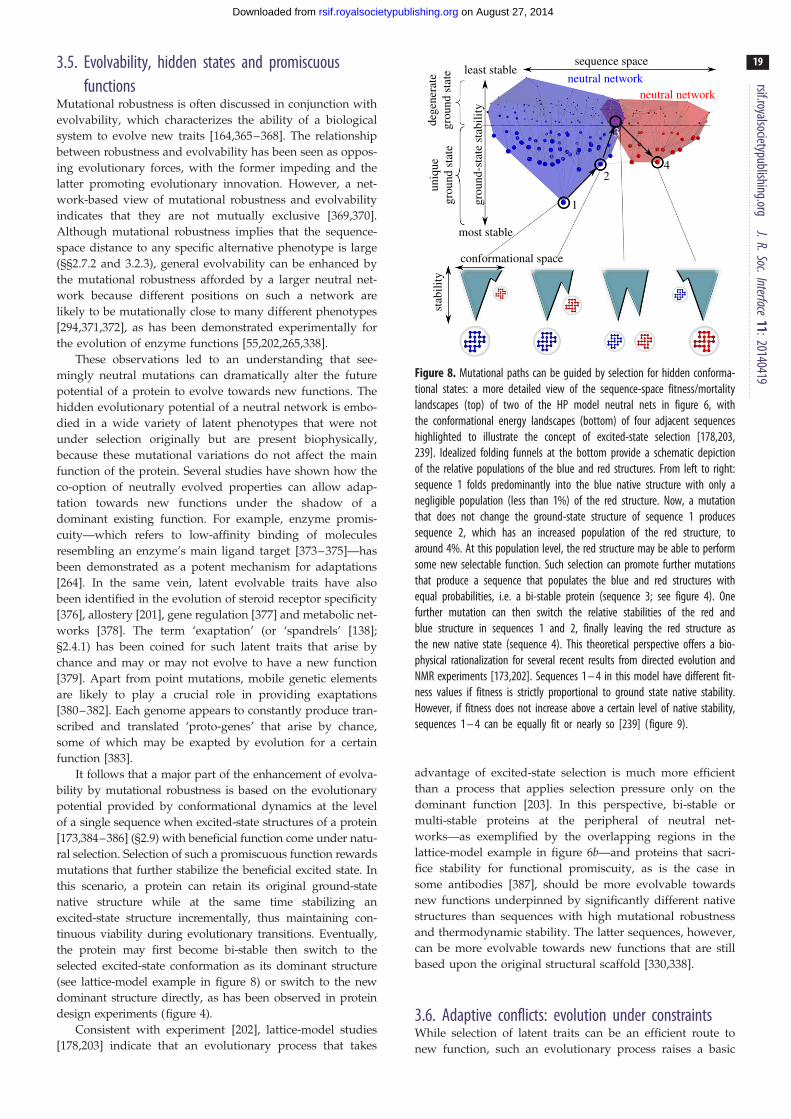
most stable
sequence space
1
24
3
neutral network
grou
nd-s
tate
sta
bilit
y
dege
nera
tegr
ound
sta
teun
ique
grou
nd s
tate
least stableneutral network
conformational space
stab
ility
Figure 8. Mutational paths can be guided by selection for hidden conforma-tional states: a more detailed view of the sequence-space fitness/mortalitylandscapes (top) of two of the HP model neutral nets in figure 6, withthe conformational energy landscapes (bottom) of four adjacent sequenceshighlighted to illustrate the concept of excited-state selection [178,203,239]. Idealized folding funnels at the bottom provide a schematic depictionof the relative populations of the blue and red structures. From left to right:sequence 1 folds predominantly into the blue native structure with only anegligible population (less than 1%) of the red structure. Now, a mutationthat does not change the ground-state structure of sequence 1 producessequence 2, which has an increased population of the red structure, toaround 4%. At this population level, the red structure may be able to performsome new selectable function. Such selection can promote further mutationsthat produce a sequence that populates the blue and red structures withequal probabilities, i.e. a bi-stable protein (sequence 3; see figure 4). Onefurther mutation can then switch the relative stabilities of the red andblue structure in sequences 1 and 2, finally leaving the red structure asthe new native state (sequence 4). This theoretical perspective offers a bio-physical rationalization for several recent results from directed evolution andNMR experiments [173,202]. Sequences 1 – 4 in this model have different fit-ness values if fitness is strictly proportional to ground state native stability.However, if fitness does not increase above a certain level of native stability,sequences 1 – 4 can be equally fit or nearly so [239] (figure 9).
rsif.royalsocietypublishing.orgJ.R.Soc.Interface
11:20140419
19
on August 27, 2014rsif.royalsocietypublishing.orgDownloaded from
3.5. Evolvability, hidden states and promiscuousfunctions
Mutational robustness is often discussed in conjunction with
evolvability, which characterizes the ability of a biological
system to evolve new traits [164,365–368]. The relationship
between robustness and evolvability has been seen as oppos-
ing evolutionary forces, with the former impeding and the
latter promoting evolutionary innovation. However, a net-
work-based view of mutational robustness and evolvability
indicates that they are not mutually exclusive [369,370].
Although mutational robustness implies that the sequence-
space distance to any specific alternative phenotype is large
(§§2.7.2 and 3.2.3), general evolvability can be enhanced by
the mutational robustness afforded by a larger neutral net-
work because different positions on such a network are
likely to be mutationally close to many different phenotypes
[294,371,372], as has been demonstrated experimentally for
the evolution of enzyme functions [55,202,265,338].
These observations led to an understanding that see-
mingly neutral mutations can dramatically alter the future
potential of a protein to evolve towards new functions. The
hidden evolutionary potential of a neutral network is embo-
died in a wide variety of latent phenotypes that were not
under selection originally but are present biophysically,
because these mutational variations do not affect the main
function of the protein. Several studies have shown how the
co-option of neutrally evolved properties can allow adap-
tation towards new functions under the shadow of a
dominant existing function. For example, enzyme promis-
cuity—which refers to low-affinity binding of molecules
resembling an enzyme’s main ligand target [373–375]—has
been demonstrated as a potent mechanism for adaptations
[264]. In the same vein, latent evolvable traits have also
been identified in the evolution of steroid receptor specificity
[376], allostery [201], gene regulation [377] and metabolic net-
works [378]. The term ‘exaptation’ (or ‘spandrels’ [138];
§2.4.1) has been coined for such latent traits that arise by
chance and may or may not evolve to have a new function
[379]. Apart from point mutations, mobile genetic elements
are likely to play a crucial role in providing exaptations
[380–382]. Each genome appears to constantly produce tran-
scribed and translated ‘proto-genes’ that arise by chance,
some of which may be exapted by evolution for a certain
function [383].
It follows that a major part of the enhancement of evolva-
bility by mutational robustness is based on the evolutionary
potential provided by conformational dynamics at the level
of a single sequence when excited-state structures of a protein
[173,384–386] (§2.9) with beneficial function come under natu-
ral selection. Selection of such a promiscuous function rewards
mutations that further stabilize the beneficial excited state. In
this scenario, a protein can retain its original ground-state
native structure while at the same time stabilizing an
excited-state structure incrementally, thus maintaining con-
tinuous viability during evolutionary transitions. Eventually,
the protein may first become bi-stable then switch to the
selected excited-state conformation as its dominant structure
(see lattice-model example in figure 8) or switch to the new
dominant structure directly, as has been observed in protein
design experiments (figure 4).
Consistent with experiment [202], lattice-model studies
[178,203] indicate that an evolutionary process that takes
advantage of excited-state selection is much more efficient
than a process that applies selection pressure only on the
dominant function [203]. In this perspective, bi-stable or
multi-stable proteins at the peripheral of neutral net-
works—as exemplified by the overlapping regions in the
lattice-model example in figure 6b—and proteins that sacri-
fice stability for functional promiscuity, as is the case in
some antibodies [387], should be more evolvable towards
new functions underpinned by significantly different native
structures than sequences with high mutational robustness
and thermodynamic stability. The latter sequences, however,
can be more evolvable towards new functions that are still
based upon the original structural scaffold [330,338].
3.6. Adaptive conflicts: evolution under constraintsWhile selection of latent traits can be an efficient route to
new function, such an evolutionary process raises a basic

commencement of selectionfor promiscuous function
high population ofbi-stable proteins
steady state:subfunctionalization
gene duplications fixed due todosage effect
DW^ = 0.398
DW^ = 0.09
DW^ = 0.008
Figure 9. The simulated evolution, with gene duplication, of an essentially infi-nite HP sequence population under an adaptive conflict of two selection pressures.Here, four stages of the evolutionary dynamics are shown by representativechanges in the distribution of evolutionary population and average population fit-nessDW from one stage to the next. The two adjacent neutral nets (blue and red)are the same as those in figure 8. Distributions of population are plotted by log-arithmic scales in the same style as figure 7. Initially, before the native structure ofthe red network is selected, nearly the entire population occupies the most stableHP sequence of the blue network. After selection pressure is imposed simul-taneously for both the blue and red structures (figure 8), the red structurebecomes a selectable promiscuous function in the model. After a number of gen-erations, bi-stable proteins (magenta) appear as high-fitness evolutionaryintermediates that are prone to undergo gene duplication. Duplicated bi-stablesequences that have maximum fitness in the model (because of an assumed ben-eficial dosage increase) then slowly give way to equally fit subfunctionalized(functionally diverged) gene pairs occupying those regions of the neutral networkswith the highest mutational robustness. In other words, this model shows thatsome subfunctionalization processes can be non-adaptive in that subfunctionaliza-tion can be driven by sequence-space topology even when the total fitness of aduplicated pair of bi-stable proteins is the same as that of a pair of prototypesequences each folding uniquely to one or the other selected structures, as inthis model. Consistent with this perspective, and as indicated by the last DWvalues in this figure, the final-stage optimization of population distributionaround the two prototype sequences provides only a very small increase in fitnessvalue. Details of this model can be found in [239].
rsif.royalsocietypublishing.orgJ.R.Soc.Interface
11:20140419
20
on August 27, 2014rsif.royalsocietypublishing.orgDownloaded from
question regarding biophysical limits on the degree of multi-
functionality or promiscuity that can be carried within one
protein molecule. Multi-functionality also bears the danger
of creating an adaptive conflict. Such a conflict can emerge
whenever adaptation on the same gene is driven by two or
more different or even mutually exclusive functional require-
ments. In the extreme case of viruses with severely
constrained genome sizes, adaptive conflict can arise from
overlapping open reading frames encoding for different
proteins within the same DNA sequence [388]. For a multi-
functional protein, adaptive conflict arises when enhancing
one subfunction impedes another subfunction.
As far as adaptive conflicts in a protein is concerned, if
multi-functionality is realized by the presence of different
binding interfaces on the protein surface, a small number of
such interfaces may coexist, limited by such factors as the
protein size and/or surface area and the number of surface
hydrophobic residues that can be tolerated without causing
misfolding. Binding interfaces can also overlap, using
some but not all of the same residues for different ligands
[128,129]. In that case, an adaptive conflict may be antici-
pated since increasing the binding affinity for one interface
through mutations may interfere with the binding affinity
of the second interface, and vice versa. If multi-functionality
is underpinned by bi-stability or multi-stability, i.e. the coex-
istence of and dynamic inter-conversion between alternative
functional conformations (§2.7.2), it is expected biophysically
that only a narrow capacity for accommodating several dis-
tinct conformations can exist in the lifetime of a globular
protein and perhaps a somewhat higher capacity for doing
so in disordered proteins. During evolution, a general mech-
anism for resolving adaptive conflict is offered by gene
duplication and subfunctionalization [389], which we will
discuss briefly in §3.7.
3.7. Protein divergence driven by gene duplication andmutational robustness
Evolution of bi-stable and more generally multi-stable proteins
is an efficient means to meet new functional needs; but multi-
functional proteins often serve as evolutionary intermediates
rather than long-term solutions. Explicit-chain biophysical
models suggest that multi-stable globular proteins are muta-
tionally much less robust than globular proteins with an
essentially unique native structure [161,178,239]. This trend is
readily seen in the 2D HP lattice model example in figure 9,
which shows that the sequence-space ‘entropy’ of bi-stable
sequences (magenta area) is much smaller than that of
sequences folding uniquely for either one of the two structures
encoded by the bi-stable sequences (blue and red areas). This is
a prediction that should be testable experimentally, for
example, by using the recently designed bi-stable proteins
[65,174,206] (figure 4). In this picture, the short-term advan-
tage of bi-stability/bi-functionality is expected to give way
eventually to an alternative sequence-space arrangement that
is mutationally more robust, provided that the gene encoding
for the bi-stable protein is duplicated at some point in the
evolutionary process.
Subfunctionalization, or functional divergence after gene
duplication, is a ubiquitous phenomenon in evolution
[389,390]. For example, an experimental study of the recon-
structed common ancestor of the fluorescent proteins in
corals that emit either red or green light was found to emit

rsif.royalsocietypublishing.orgJ.R.Soc.Interface
11:20140419
21
on August 27, 2014rsif.royalsocietypublishing.orgDownloaded from
both red and green light [391]. Subfunctionalization of a
duplicated multi-functional gene is probably a more efficient
evolutionary route than neofunctionalization, which necessi-
tates evolution of new function in a duplicated gene from
scratch. However, in the subfunctionalization route, this pro-
cess can be facilitated by selection on latent traits (§3.6) before
gene duplication [239]. In the lexicon of fitness landscape,
gene duplication amounts to doubling the number of dimen-
sions of sequence space and thus may be viewed as an
‘extradimensional bypass mechanism’ for resolving adaptive
conflicts [392].
Functional or structural divergence can be driven by an
increase in fitness when a pair of identical bi-stable sequences
is transformed into two subfunctionalized sequences. Gener-
ally speaking, such an increase in fitness is biophysically
plausible because each of the subfunctionalized sequences
may afford a higher kinetic stability [81] to one or the other
functional structure than that provided by a bi-stable sequence
[239]. However, divergence does not always have to be adap-
tive. Even if the fitness of a subfunctionalized pair is
identical to the fitness of a pair of bi-stable sequences, the
inherent tendency of protein evolution towards higher
mutational robustness can still drive subfunctionalization.
Biophysically, an ensemble of protein structures subjected to
more overlapping functional constraints is likely to be more
restricted in sequence space, resulting in low mutational
robustness, as is exemplified in figure 9. When the constraints
are lifted through gene duplication or changes in the environ-
mental selection pressures, evolution will naturally favour
mutations that result in higher mutational robustness even if
there is no gain in functional fitness for the proteins in the pro-
cess. This scenario is illustrated by the model evolutionary
dynamics in figure 9. Simulation results summarized in this
figure indicate that after an adaptive pressure to simul-
taneously select two structures is imposed, the evolving
proteins first attempt a short-term resolution of the adap-
tive conflict using bi-stable sequences with low mutational
robustness. Subsequently, upon gene duplication, a process
of divergence that is essentially neutral ensues, with each
copy of the originally bi-stable protein evolving towards
the central, high-robustness region of one of the two
neighbouring neutral networks [239] (figure 9).
3.8. Epistasis and co-evolution of interacting amino acidresidues
The phenomenon of epistasis, referring originally to non-addi-
tivity of genetic effects caused by gene interactions (e.g. [393]),
can also manifest within a protein molecule. Mutational effects
on stability or function at different sites of a protein can be
non-additive when the sites are energetically coupled [394].
A consequence of this biophysical property is that the overall
evolutionary effect of multiple mutations can depend on the
order in which the mutations are made. For the same given set
of mutations, it may be that one temporal order of mutations
is evolutionarily favoured because it entails a monotonic
increase in fitness, whereas another order of mutations is disfa-
voured because it involves an intermediate step that decreases
fitness. Several studies have demonstrated this type of epista-
tic behaviour in proteins and its constraints on evolutionary
pathways [42,53,300,395–398]. For instance, experiments on
adenylate kinase indicate that a double mutant with higher stab-
ility can only be obtained via one mutation path [300]. When the
order of the two mutations was changed the protein could not
fold. This type of behaviour is readily observed in simple bio-
physical models of protein evolution, as is illustrated by the
HP model example in figure 5c.
An implication of epistasis is that the propensity for a viable
mutation at a certain site in a protein structure may not be fixed.
Rather it should depend on the preceding mutations at other
sites. This phenomenon is illustrated by a recent simulation
study of the evolution of purple acid phosphatase by generating
random mutations in the structure [58]. Based on stability calcu-
lations in the model, the propensities for all amino acids at
selected solvent-exposed and buried sites were determined.
The results indicated considerable variations of these propensi-
ties because stability effects of amino acid substitutions at a
given site change with time as the evolutionary process pro-
gresses. The simulation showed that an enforced destabilizing
mutation (which could arise in real proteins owing to functional
constraints such as the need to preserve an active site) can be
compensated by subsequent mutations, thus increasing the
future viability of the already-mutated residue at that same
site and rendering the reverse mutation detrimental and
therefore less probable [58]. Although this model probably over-
estimated native stability as well as stability effects of mutations
and underestimated the probability for misfolding because of
its simplified treatment of the unfolded state (see discussion
above on marginal stability; §2.4.2), it offers an excellent eluci-
dation of the ‘holistic’ nature of intra-protein interactions and
the biophysical forces that govern the mutational effect on stab-
ility and how it may depend on the temporal order in which the
mutations occur [58].
Epistatic effects are common but not universal. Strong epis-
tasis arising from significant evolutionary shifts in the stability
effects of mutations as envisioned in [58] may even be rare [57].
In the example of the influenza nucleoprotein, an experimental
analysis of mutations in a set of homologues showed that stab-
ility effects of mutations with no clear functional benefit are
largely conserved across homologues, mostly additive and
exhibit no aforementioned [58] strong dependence on temporal
order [57]. It has been argued that mutations in viral proteins
in general—which probably have evolved to buffer deleterious
mutations—are not likely to exhibit strong epistasis [399].
Whenever the functional benefits outweigh the cost of destabi-
lization of a mutation, strong epistatic effects are more likely to
follow [21]. Nonetheless, a weaker form of epistasis can occur
even if stability effects of mutations are conserved because
different temporal orders of stability changes can result in
drastically different survivabilities. For instance, a recent exper-
imental study of the 39 mutations on the nucleoprotein of the
influenza virus between years 1968 and 2007 identified several
mutations that decrease the stability of the protein significantly
when introduced individually to the starting 1968 protein, thus
suggesting strongly that these mutations were preceded by
‘enabling’ mutations that increase native stability. An inferred
evolutionary trajectory was constructed based on the stability
constraints [42]. Epistasis has also been revealed by studying
disease-causing single mutations in humans and comparing
them with compensated mutations that do not cause disease
in other species [400–402]. One estimate indicates that 80%
of pathological mutations result in protein stability changes
[403]. When the compensated pathological mutations are
compared against uncompensated pathological mutations,
compensated mutations are mostly found at solvent-exposed
positions and the amino acid substitutions are ‘milder’,

rsif.royalsocietypublishing.orgJ.R.Soc.Interface
11:20140419
22
on August 27, 2014rsif.royalsocietypublishing.orgDownloaded from
entailing, for example, less changes in hydrophobicity
[400–402]. These experimental trends are consistent with the
biophysical principles of protein structure and stability
expounded here. A more detailed discussion of the biophysi-
cal/structural basis of epistasis and compensatory evolution
is available in a recent review [404].
3.9. Fitness landscapes for multiple phenotypicproperties
Natural protein evolution takes place in a highly wired, inter-
acting molecular system. Ultimately, therefore, studies of
protein evolution have to take into account a complex mol-
ecular context [392] (see §3.11). The expanded concept of
molecular phenotype discussed in §2.9 is an attempt towards
a better account of this biophysical reality. In this regard,
highly simplified yet promising explicit-chain biophysical
models have recently been developed to study protein evol-
ution in the context of protein–protein interactions [99,347]
(§3.2.3) We have also summarized explicit-chain biophysical
models that take into consideration the ensemble nature
of a protein’s conformational phenotype, and how these
models can provide insights into selection of promiscuous
function, bi-stability and structural divergence (§3.2).
With the understanding that the fitness of a protein
sequence depends not only on its ground-state native structure,
but also on its entire conformational distribution as well as
potential functional interactions and detrimental misinterac-
tions with other molecules, a critical issue in modelling is
how to assess contributions from different phenotypic proper-
ties to the overall fitness. For example, in the bi-stable fitness
landscape in figures 8 and 9, the combined fitness is taken
as the sum of two fitness values, one for each structure
[178,239]. Although this modelling scheme is useful for
illustrating general principles, it would be too simplistic
when applied to real-life situations. Different forms of multi-
functionality may require different rules for combining fitness
contributions. Ideally, a fitness function should include not
only positive contributions from selected biophysical proper-
ties, but also account for the negative effects on folding rates
and misfolding, as well as aggregation and misinteraction.
Thus, protein evolution in general entails a multi-factorial
optimization problem where only something like a Pareto
optimality can be achieved, i.e. a satisfaction of multiple
optimality criteria just above a minimum threshold of optimal-
ity for each criterion [405]. One simple example would be the
trade-off between thermodynamic stability of a folded protein
versus the need for conformational dynamics in biochemi-
cal functions and degradation. Both these criteria probably
cannot be fully satisfied, but a Pareto optimality may be
achieved such that the protein is stable enough to maintain
the same fold yet flexible enough to allow binding.
Constructing more realistic fitness functions will be a
challenging task. Genomic information is abundant; but
pinpointing mutational impact on cellular function by
experiment is often daunting. Theoretical/computational
investigations can assist greatly in this endeavour by devel-
oping more comprehensive models that account for various
biophysical constraints on protein evolution. Two recent
examples will be discussed in §3.10 and 3.11 to illustrate
how incorporating information about protein–protein inter-
actions into biophysical models can advance understanding
of experimentally observed evolutionary patterns.
3.10. Biophysical links between protein expression leveland evolutionary rateA fundamental evolutionary question is why different pro-
teins evolve at different rates. What makes some proteins
less likely to accept new mutations than others? Can the
different evolutionary rates be explained in terms of the
biophysical constraints on mutations as outlined in figure 3?
One hypothesis is that proteins that are functionally more
important are more conserved, because the cell cannot risk
their function to be compromised in any way, even slightly.
Some authors have linked the evolutionary rate to the position
of a protein in the protein–protein interaction network, finding
that ‘hub’ proteins involved in many interactions evolve more
slowly [406]. Empirically, evolutionary rate was found to be
most strongly anticorrelated with the expression level [407].
This trend is not inconsistent with the functional argument.
A protein is essential to an organism if the organism fails to
survive when the gene encoding for the protein is deleted
from its genome [408,409]. Many essential proteins are
highly expressed, as the cell needs a constant supply for its
most basic and vital functions. However, is the slower evol-
utionary rate of highly expressed proteins a result of the
importance of their functions or a more direct consequence
of their high concentrations in the cell? Several biophysical
mechanisms have been proposed for the latter scenario. Here
we summarize briefly two mechanisms that are based, respect-
ively, on protein misfolding and protein misinteraction, noting
however that an explanation in terms of mRNA folding rate
has also been put forth recently [410]. Multiple mechanisms
can be at play because the proposed mechanisms are not
mutually exclusive.
All proteins have to avoid misfolding. Taking the popu-
lation of a protein sequence as a whole, a protein that is
abundantly populated provides more opportunity for the for-
mation of detrimental misfolded structures than a protein
that is sparsely populated; thus the constraint imposed by
misfolding avoidance is stronger on protein sequences with
higher populations. A similar consideration applies to the
misinteractions, which will be discussed further below.
Accordingly, the need to prevent or at least minimize mis-
folding caused by translation error has been proposed as a
major constraint on the evolution of highly expressed pro-
teins [411–414], leading to slower evolution. Consistent
with this picture, highly expressed proteins are selected to
be more robust against translation errors by using synon-
ymous codons with the smallest chance of producing non-
synonymous changes as a result of translation errors. Apart
from translation errors, the need to avoid misfolding of the
properly translated protein also constitutes a strong evol-
utionary constraint on highly expressed proteins, resulting
in preferential usage of amino acid residues that minimize
misfolding [413]. These restrictive requirements lead to pro-
teins that are both slowly evolving and thermodynamically
more stable [414,415].
Another probable biophysical constraint behind the anti-
correlation between protein expression level and evolutionary
rate is the need for a protein to avoid misinteraction with
other proteins [99]. This selection pressure affects primarily sur-
face residues that can potentially participate in interactions
between the protein and other molecules. Thus, its effects are
to some degree complementary to that arising from the need
for folding stability, kinetic accessibility of the folded structure

rsif.royalsocietypublishing.orgJ.R.Soc.Interface
11:20140419
23
on August 27, 2014rsif.royalsocietypublishing.orgDownloaded from
and avoidance of misfolding. The latter selection pressures
affect primarily buried residues but can also affect surface resi-
dues. Misinteractions may be caused by the same exposed
hydrophobic surface residues that are part of the functional
protein–protein interactions, leading to an adaptive conflict
between increasing the strength of functional interactions and
avoiding misinteractions [347]. This conflict can result in
further constraints that limit the viability of mutants of
highly expressed proteins. As a consequence of these biophysi-
cal constraints, evolution might have increased the proportion
of functional monomeric proteins with hydrophilic surfaces,
reduced the abundance of functional multi-chain complexes,
weakened the strengths of functional interactions, or increased
the degree of disordered protein interactions to minimize
exposed hydrophobics while still allowing many interaction
partners [230,347]. In other words, such strategies might have
contributed to the evolution of interaction network topologies
that can better alleviate the conflict between functional
interactions and misinteractions [110].
3.11. Protein evolution in the context of functionalnetworks
As emphasized above, proteins do not act in isolation in
living organisms; hence a full understanding of the func-
tion and evolution of a protein should take into account
its interactions with other biomolecules and metabolites
[392,416]. It would be daunting to account for these inter-
actions in all their complexity at the molecular level.
Using simple explicit-chain protein models, conceptual
advances were made in elucidating how the biophysical
constraint of misinteraction avoidance might impact protein
evolution [99,347] (§3.10). However, investigators have to
rely upon abstract descriptions of protein interactions,
using model parameters extracted from experimental data
on binding and on the effects of enzymatic activities on bio-
chemical reaction rates. With an increasing repertoire of
genomic data, this approach has produced significant pro-
gress. For instance, the recent mapping of a realistic
network of DNA sequences bound by the same transcrip-
tion factor [417] has afforded new evidence in support of
the idea that a large genotype network enhances both
mutational robustness and evolvability (§3.5).
Important advances have also been made by taking an
abstract approach in the study of metabolic networks [418].
Notably, a reductive evolution algorithm was applied to
determine minimal viable genomes for E. coli [419]. In prin-
ciple, the effect of a mutation on metabolism is difficult to
predict, because it affects not only the activity of the mutated
protein but also many downstream events. Yet metabolic net-
works are often found to be robust against perturbations such
as gene deletions and loss-of-function mutations because of
‘distributed robustness’, i.e. an ability of the network to com-
pensate for the local perturbation by systemic adjustments
[420]. In silico metabolic networks have also shed light on
the evolution of specialist versus generalist enzymes. By ana-
lysing a model network of E. coli metabolism, it was found
that specialists are very efficient at catalysing single metabolic
reaction steps, responsible for a high metabolic throughput,
and often essential to the cell. These functional roles necessi-
tate more regulation of its activity. Consequently, specialists
require a much higher degree of maintenance than generalists
that are promiscuous and multi-functional. This model study
thus offered an explanation for why specialists have not
replaced all the generalists in real organisms [421].
A more recent computational study used flux balance
analysis [422] and random re-wiring of a realistic model
metabolic network [423] to study evolution of a model cell
under a selection pressure to survive on a given carbon
source [378]. The simulation showed that selection for one
carbon source also allows the model cell to survive on a
number of other carbon sources that were not selected for.
This finding demonstrated that metabolic systems embody
latent evolutionary potentials, and that beneficial traits can
arise non-adaptively through exaptations [379] in the absence
of selection at the level of metabolic network [378], as is the
case we have seen at the molecular level (§3.5).
Although it is currently not feasible to apply explicit-
chain models of proteins in the simulation of a realistic cellu-
lar metabolic network, a recent evolutionary population
dynamics study was able to incorporate energetic infor-
mation of explicit-chain continuum (off-lattice) models for
10 proteins from the folate biosynthetic pathway [59]. The
study considers a population of 1000 model cells. The fitness
of each model cell is taken to be the total metabolic output of
the model biosynthetic pathway minus the number of mis-
folded proteins, with both of these quantities dependent
upon the thermodynamic folded-state stabilities of the
proteins. Protein stabilities are in turn computed using a bio-
physical potential function. Simulation results from this
model provide a protein-based molecular biophysical ration-
alization for the distribution of stabilizing and destabilizing
mutations and other experimentally observed patterns of
polymorphisms [59].
4. Outlook: enriching the biophysics of proteinevolution
As this review emphasizes, evolution is ultimately a physico-
chemical process that cannot be fully comprehended without
biophysics. Likewise, because evolution happened under
biophysical constraints, evolutionary information can help
decipher aspects of protein biophysics that are still too com-
plex or too costly to be tackled by first-principles physical
or chemical methods. In closing, we provide a few further
examples to showcase the productive research directions
in which future progress will probably be made by this
synergistic approach.
4.1. Synergy between biophysics and the study ofprotein evolution
Perhaps the most direct way to access the change in bio-
physical properties during the long evolutionary history of
natural proteins is to perform experiments on ‘resurrected’
ancestral proteins. Recently, several putative ancestral
protein sequences have been constructed computationally
using common phylogenetic methods and then synthesized
in the laboratory [82,391,424–428]. We have mentioned
thioredoxin as one of the proteins that were studied in
this manner in the discussion on kinetic stability (§2.2).
Another interesting case is the reconstruction of the evol-
ution of steroid receptors [376,429], revealing that ancient
steroid receptors were already able to bind to the hormone
aldosterone. But aldosterone only became available to
rsif.royalsocietypublishing.orgJ.R.Soc.Interface
11:20140419
24
on August 27, 2014rsif.royalsocietypublishing.orgDownloaded from
mammalian cells much later during evolution, indicating
once again that latent functions, or exaptations (§3.5), can
play important roles in protein evolution [376]. The study
of these putative ancestral proteins indicates further that
epistatic interactions within the structure of hormone recep-
tors have led to surprisingly irreversible evolutionary
pathways [430].
The utility of an ancestral reconstruction, however, is
only as good as the accuracy of the phylogenetic relation-
ships it assumes. As mentioned earlier, mutation rates
depend not only on amino acid residue type, but also on
the conformational context of the site being mutated; but
many common phylogenetic methods do not account for
this dependence (§1). One of the structural properties that
exhibit significant correlation with mutation rate is confor-
mational diversity. The conformational diversity exhibited
by a single protein sequence is also reflected in the confor-
mational diversity among the sequences in the family to
which the protein belongs [431]. Local packing density of
an amino acid position, which correlates negatively with
local backbone conformational diversity (flexibility), was
also found to correlate negatively with evolution rate. In
other words, amino acid positions that have a lower local
packing density and are more flexible locally tend to
evolve faster. The correlation of evolutionary rate with
local packing density is even stronger than that with solvent
accessibility [432]. More recently, it has been suggested
further that this strong correlation is a reflection of a funda-
mental relationship between evolutionary rate and the
energetic stress caused by random mutations since average
mutational stress is proportional to local packing density
in an idealized elastic network model of protein structure
[433]. This general trend is also consistent with the obser-
vation that IDRs typically evolve faster than globular
proteins except when the IDR site is involved in the binding
of multiple partners (§2.8.2). Taken together, the findings
summarized above contradict another group’s earlier find-
ing that evolutionary rate is negatively correlated with
conformational diversity [434,435]. The correlation coeffi-
cients computed from the limited dataset considered in
the earlier study were, however, all very small [434].
As mentioned in §1, a recent method makes use of the
information about solvent exposure of residues in a known
protein structure to identify sites in a protein-coding gene
that have undergone positive or negative evolutionary selec-
tion [23,24]. Although synonymous mutations are not
necessarily neutral [95–97,436], the ratio of non-synonymous
over synonymous codon replacement rates, v ¼ dN/dS, is
commonly used to detect adaptation in a given phylogenetic
tree of related gene sequences. In such studies, most
mutations do not show any signs of adaptation, with v � 1,
and are thus considered ‘neutral’; v . 1 is usually taken to
indicate positive selection but negative selection is difficult
to decipher from v values. The predictive power of this
method hinges on an accurate discrimination between neu-
tral, adaptive and conservative v signatures. Recent studies
have shown that by considering solvent-exposed areas of
mutation sites, this discrimination can be improved in a
protein-specific manner. In this new approach, instead of
using the same underlying assumptions for every protein
family, as has been commonly practiced, biophysical con-
sideration of solvent exposure is used to customize and
improve the accuracy of the model for the specific protein
under investigation (figure 1). This enriched methodology
provides more accurate evolutionary inference than
approaches that do not consider the conformational context
of the mutation sites, but it requires the 3D structure of the
protein in question. Hopefully, with better protein structure
prediction techniques [437,438], it may be possible to apply
structure-based phylogenetic reconstruction methods
routinely by starting with sequence information alone.
4.2. Evolutionary protein biophysics: evolutionaryinformation benefiting biophysical studies ofproteins
The advances summarized above exemplify how biophysics
can assist in the study of evolution. In the following, we
describe briefly three examples in which evolutionary data
have assisted in biophysical studies of proteins. The first
example concerns prediction of mutational change in protein
stability, DDG. As described in §2.1, a number of biophysical
methods for DDG prediction exist but are limited in various
respects. In this context, it has been shown recently that a
Bayesian method for inferring DDG values of individual
mutations from the evolutionary information embodied in
homologous sequences can achieve accuracy exceeding
pure biophysical methods and sequence-based consensus
approaches. The method was applied to predict stabilizing
mutations for influenza haemagglutinin. Subsequent exper-
iment demonstrated that some of the mutations do allow a
temperature-sensitive virus to grow at a higher temperature,
attesting to the utility of this evolution-based method in
improving biophysical understanding [439].
Another example that we have mentioned briefly is the
detection of protein sectors from coevolution data (§1). Evo-
lutionarily, protein sectors are largely independent of one
another even though they are parts of the same protein.
Amino acid residues within a sector are physically connected
in the folded structure and are correlated evolutionarily
[27] (figure 2). Sectors constitute sparse networks of co-evol-
ving amino acid residues comprising only a minority of the
residues in a protein. A recent high-throughput saturation
point mutagenesis study of a PDZ domain (1577 mutations
were tested) showed that sector positions are functionally
less tolerant to mutation than non-sector positions [440].
These observations suggest that coevolution data can be
used in general to gain insight into the biophysics of
functional binding.
Coevolution data have also been applied to predict bio-
physical interactions in proteins, as mentioned briefly in §1
[28–32]. A computational algorithm has recently been devel-
oped to use pure sequence information to predict contacts
within a protein structural domain. The approach is useful
for predicting the native structure when sequence data are
abundant but a structure has not been determined experimen-
tally for a protein [31]. Even more interestingly, and going
beyond earlier seminal findings [26], it was found that
sequence information can reveal residue interactions that are
not present in the PDB structure, including interactions
between structural domains [31] as well as interactions
involved in alternative conformational states with evolutio-
narily conserved functional significance [29]. Most recently,
coevolutionary information of several protein families
has been applied to determine a theoretical sequence-space
rsif.royalsocietypublishing.orgJ.R.Soc.Interface
11:20140419
25
on August 27, 2014rsif.royalsocietypublishing.orgDownloaded from
‘selection temperature’ Tsel that can be related to the Tf/Tg
ratio between the folding and glass-transition temperatures
in protein folding [441]. Because Tf/Tg is a biophysical
measure of folding cooperativity [36,254], this latest
analysis [441] demonstrates remarkably how fundamental
biophysical principles can be revealed by evolutionary
data. All in all, the examples in this section illustrate a gen-
eral productive research approach of evolutionary protein
biophysics, in which the current deluge of sequence-based
evolutionary data is harnessed to extract important bio-
physical/structural information to improve understanding
of protein function.
4.3. Role of theory and computationTheoretical/computational methods are an integral part
of the biophysical study of protein evolution (§3.2–3.4).
Quantitative biophysical modelling is indispensable in the
formulation of concepts, rationalization of existing exper-
imental data, discovery of novel hypotheses and provision
of predictions for subsequent experimental testing. Recent
models not only addressed evolution of individual proteins
but have also begun to take into account the interaction
and metabolic networks in model organisms (§3.11). More
complex models tend to be richer in that they have the
capacity to provide non-trivial predictions that are not
immediately obvious from the modelling set-up. Nonethe-
less, even simple explicit-chain models that serve largely
to confirm expected trends are useful for conceptualizing
how known evolutionary behaviours might have arisen
from the physical forces that govern protein properties.
Despite improved model sophistication and the tremen-
dous recent increase in computational power, models of
protein evolution have to rely on representations that are
coarse-grained and thus the models are inevitably highly
simplified caricatures of the complex real situation they
seek to mimic. In general, model predictions are sensitive
to the assumed parameter values; but a precise corres-
pondence between these values and physical reality often
cannot be readily established. With these limitations in
mind, theoreticians should strive to perform more explora-
tion, as controls, of multiple parameter sets and alternative
modelling set-ups that are biophysically plausible. Enhanced
efforts in scenario classification are needed in general to better
delineate the logical relationship between the assumptions
and predictions of any given model.
4.4. Evolution within and across protein families andsuperfamilies
As outlined above, much progress has been made in exper-
imental and theoretical studies of evolution within a protein
family or superfamily. Compared to mutational changes that
convert one protein fold to another, mutational changes
that maintain essentially the same folded structure are
computationally less costly to simulate; their study is more
amenable to experimental techniques such as directed evol-
ution, and can also benefit from the availability of abundant
genomic data. It is more challenging to study fold-altering
protein evolution. Nonetheless, notable recent experimental
advances have been made in the design and structural
characterization of bi-stable proteins and conformatio-
nal switches (§2.7; figure 4). Understanding structural
innovation entails biophysical accounting of conformational
diversity—which underpins functional promiscuity—as well
as the plasticity, or ensemble nature of molecular phenotype
(§2.9). Here, we have placed considerable emphasis on this
broader perspective of protein evolution, although theoreti-
cal investigation in this area is only in its infancy (§§2.7 and
3.5–3.7; figures 4 and 8). We hope to witness more advances
in this direction. It is exciting to better understand not only
how proteins evolved within a family or a superfamily but
also, more fundamentally, how the structural families
originated in the first place.
5. Concluding remarksThe aim of this review is to provide a broad sketch of the fun-
damental biophysical forces that both enable and constrain
protein evolution. Starting with the effects of mutations on
protein stability, folding kinetics, interactions, functional
dynamics, promiscuous functions, conformational switch
and conformational disorder, these findings are then linked
to broader evolutionary themes including the global and
local organization of protein sequence and structure space,
simple models of the protein sequence–structure mapping,
fitness/mortality landscapes, sequence-space topology
and mutational robustness, adaptive conflict and its possible
resolution by selection of promiscuous function and subfunc-
tionalization driven by mutational robustness, evolvability,
epistasis and intra-cellular networks. We have highlighted
advances made through computational models, especially
simple exact and other explicit-chain models of protein evol-
ution, because many insightful discoveries in biophysics of
protein evolution were pioneered through simple, coarse-
grained modelling of biological or biophysical processes
that are too complex to be studied in atomistic details. As
far as simplified models are concerned, explicit-chain
models with biophysics-based interactions enjoy a clear phys-
ical advantage over theories that contain little or no
biophysical consideration of protein structure and dynamics.
We have also summarized several recent experimental
advances that bear on the biophysics of evolution, as many
questions that have arisen from theory and simulation can
only be answered definitely by further experiments. Even
so, this review touches upon only a small fraction of all the
exciting discoveries that have been made lately. Looking
into the future, we expect to witness increasing collaboration
between the fields of biophysics and evolution as well as
between theory/computation and experiment to decipher
many aspects of the evolutionary forces that have been
shaping the biological roles of proteins.
Acknowledgements. We thank Jesse Bloom, Xavier de la Cruz, JulieForman-Kay, Alessandro Laio, Austin Meyer, Marc Ostermeier, JoseSanchez-Ruiz, Andreas Wagner and Claus Wilke for helpful discus-sions. H.S.C. wishes to take this opportunity to thank ErichBornberg-Bauer specially for a fruitful and pleasurable collaborationon evolutionary studies over many years. Part of this work was pre-sented at the 2014 Meeting of the Society for Molecular Biology andEvolution (San Juan, Puerto Rico) by T.S., who gratefully acknowl-edges a travel award he received from the Canadian Institutesof Health Research (CIHR) Training Program in ‘Protein Foldingand Interaction Dynamics: Principles and Diseases’ at the Universityof Toronto.
Funding statement. This work was supported by a CIHR grant to H.S.C.and the computational resource provided by SciNet of Compute Canada.
26
on August 27, 2014rsif.royalsocietypublishing.orgDownloaded from
References
rsif.royalsocietypublishing.orgJ.R.Soc.Interface
11:20140419
1. Dunham I et al. 2012 An integrated encyclopedia ofDNA elements in the human genome. Nature 489,57 – 74. (doi:10.1038/nature11247)
2. Vogel C, Bashton M, Kerrison ND, Chothia C,Teichmann SA. 2004 Structure, function and evolutionof multidomain proteins. Curr. Opin. Struct. Biol. 14,208 – 216. (doi:10.1016/j.sbi.2004.03.011)
3. Moore AD, Bjorklund AK, Ekman D, Bornberg-BauerE, Elofsson A. 2008 Arrangements in the modularevolution of proteins. Trends Biochem. Sci. 33,444 – 451. (doi:10.1016/j.tibs.2008.05.008)
4. Bornberg-Bauer E, Alba MM. 2013 Dynamics andadaptive benefits of modular protein evolution.Curr. Opin. Struct. Biol. 23, 459 – 466. (doi:10.1016/j.sbi.2013.02.012)
5. Soding J, Lupas AN. 2003 More than the sum oftheir parts: on the evolution of proteins frompeptides. BioEssays 25, 837 – 846. (doi:10.1002/bies.10321)
6. Hocker B, Claren J, Sterner R. 2004 Mimicking enzymeevolution by generating new (ba)8-barrels from(ba)4-half-barrels. Proc. Natl Acad. Sci. USA 101,16 448 – 16 453. (doi:10.1073/pnas.0405832101)
7. Carbone MN, Arnold FH. 2007 Engineering byhomologous recombination: exploring sequence andfunction within a conserved fold. Curr. Opin. Struct.Biol. 17, 454 – 459. (doi:10.1016/j.sbi.2007.08.005)
8. Hocker B. 2013 Engineering chimaeric proteins fromfold fragments: ‘hopeful monsters’ in proteindesign. Biochem. Soc. Trans. 41, 1137 – 1140.(doi:10.1042/BST20130099)
9. Smith MA, Romero PA, Wu T, Brustad EM, ArnoldFH. 2013 Chimeragenesis of distantly-relatedproteins by noncontiguous recombination. ProteinSci. 22, 231 – 238. (doi:10.1002/pro.2202)
10. Henikoff S, Henikoff JG. 1992 Amino acidsubstitution matrices from protein blocks. Proc. NatlAcad. Sci. USA 89, 10 915 – 10 919. (doi:10.1073/pnas.89.22.10915)
11. Dayhoff M, Schwartz R, Orcutt B. 1978 A model ofevolutionary change in proteins. In Atlas of proteinsequence and structure (ed MO Dayhoff ), pp. 345 –352. Washington, DC: National Biomedical ResearchFoundation.
12. Worth CL, Gong S, Blundell TL. 2009 Structural andfunctional constraints in the evolution of proteinfamilies. Nat. Rev. Mol. Cell Biol. 10, 709 – 720.(doi:10.1038/nrm2762)
13. Wilke CO. 2012 Bringing molecules back intomolecular evolution. PLoS Comput. Biol. 8,e1002572. (doi:10.1371/journal.pcbi.1002572)
14. Liberles DA et al. 2012 The interface of proteinstructure, protein biophysics, and molecularevolution. Protein Sci. 21, 769 – 785. (doi:10.1002/pro.2071)
15. Studer RA, Dessailly BH, Orengo CA. 2013 Residuemutations and their impact on protein structure andfunction: detecting beneficial and pathogenicchanges. Biochem. J. 449, 581 – 594. (doi:10.1042/BJ20121221)
16. Harms MJ, Thornton JW. 2013 Evolutionarybiochemistry: revealing the historical and physicalcauses of protein properties. Nat. Rev. Genet. 14,559 – 571. (doi:10.1038/nrg3540)
17. Bordner AJ, Mittelmann HD. 2014 A newformulation of protein evolutionary models thataccount for structural constraints. Mol. Biol. Evol. 31,736 – 749. (doi:10.1093/molbev/mst240)
18. Rodrigue N, Philippe H. 2010 Mechanistic revisionsof phenomenological modeling strategies inmolecular evolution. Trends Genet. 26, 248 – 252.(doi:10.1016/j.tig.2010.04.001)
19. Kleinman CL, Rodrigue N, Lartillot N, Philippe H.2010 Statistical potentials for improved structurallyconstrained evolutionary models. Mol. Biol. Evol. 27,1546 – 1560. (doi:10.1093/molbev/msq047)
20. Le SQ, Gascuel O. 2010 Accounting for solventaccessibility and secondary structure in proteinphylogenetics is clearly beneficial. Syst. Biol. 59,277 – 287. (doi:10.1093/sysbio/syq002)
21. DePristo MA, Weinreich DM, Hartl DL. 2005Missense meanderings in sequence space: abiophysical view of protein evolution. Nat. Rev.Genet. 6, 678 – 687. (doi:10.1038/nrg1672)
22. Franzosa EA, Xia Y. 2009 Structural determinants ofprotein evolution are context-sensitive at theresidue level. Mol. Biol. Evol. 26, 2387 – 2395.(doi:10.1093/molbev/msp146)
23. Scherrer MP, Meyer AG, Wilke CO. 2012 Modelingcoding-sequence evolution within the context ofresidue solvent accessibility. BMC Evol. Biol. 12, 179.(doi:10.1186/1471-2148-12-179)
24. Meyer AG, Wilke CO. 2013 Integrating sequencevariation and protein structure to identify sitesunder selection. Mol. Biol. Evol. 30, 36 – 44. (doi:10.1093/molbev/mss217)
25. Stevens J, Corper AL, Basler CF, Taubenberger JK,Palese P, Wilson IA. 2004 Structure of the uncleavedhuman H1 hemagglutinin from the extinct 1918influenza virus. Science 303, 1866 – 1870. (doi:10.1126/science.1093373)
26. Lockless SW, Ranganathan R. 1999 Evolutionarilyconserved pathways of energetic connectivity inprotein families. Science 286, 295 – 299. (doi:10.1126/science.286.5438.295)
27. Halabi N, Rivoire O, Leibler S, Ranganathan R. 2009Protein sectors: evolutionary units of three-dimensional structure. Cell 138, 774 – 786. (doi:10.1016/j.cell.2009.07.038)
28. Morcos F et al. 2010 Modeling conformationalensembles of slow functional motions in Pin1-WW.PLoS Comput. Biol. 6, e1001015. (doi:10.1371/journal.pcbi.1001015)
29. Morcos F, Jana B, Hwa T, Onuchic JN. 2013Coevolutionary signals across protein lineages helpcapture multiple protein conformations. Proc. NatlAcad. Sci. USA 110, 205 33 – 205 38. (doi:10.1073/pnas.1315625110)
30. Schug A, Weigt M, Onuchic JN, Hwa T, Szurmant H.2009 High-resolution protein complexes from
integrating genomic information with molecularsimulation. Proc. Natl Acad. Sci. USA 106, 22 124 –22 129. (doi:10.1073/pnas.0912100106)
31. Morcos F et al. 2011 Direct-coupling analysis ofresidue coevolution captures native contactsacross many protein families. Proc. Natl Acad.Sci. USA 108, E1293 – E1301. (doi:10.1073/pnas.1111471108)
32. Dago AE, Schug A, Procaccini A, Hoch JA, Weigt M,Szurmant H. 2012 Structural basis of histidinekinase autophosphorylation deduced by integratinggenomics, molecular dynamics, and mutagenesis.Proc. Natl Acad. Sci. USA 109, E1733 – E1742.(doi:10.1073/pnas.1201301109)
33. Leopold PE, Montal M, Onuchic JN. 1992 Proteinfolding funnels: a kinetic approach to the sequence-structure relationship. Proc. Natl Acad. Sci. USA 89,8721 – 8725. (doi:10.1073/pnas.89.18.8721)
34. Dill KA, Chan HS. 1997 From Levinthal to pathwaysto funnels. Nat. Struct. Biol. 4, 10 – 19. (doi:10.1038/nsb0197-10)
35. Onuchic JN, Wolynes PG. 2004 Theory of proteinfolding. Curr. Opin. Struct. Biol. 14, 70 – 75. (doi:10.1016/j.sbi.2004.01.009)
36. Chan HS, Zhang Z, Wallin S, Liu Z. 2011Cooperativity, local-nonlocal coupling, andnonnative interactions: principles of protein foldingfrom coarse-grained models. Annu. Rev. Phys. Chem.62, 301 – 326. (doi:10.1146/annurev-physchem-032210-103405)
37. Koshland DE. 1958 Application of a theory of enzymespecificity to protein synthesis. Proc. Natl Acad. Sci. USA44, 98 – 104. (doi:10.1073/pnas.44.2.98)
38. Csermely P, Palotai R, Nussinov R. 2010 Induced fit,conformational selection and independent dynamicsegments: an extended view of binding events.Trends Biochem. Sci. 35, 539 – 546. (doi:10.1016/j.tibs.2010.04.009)
39. Bellotti V, Stoppini M, Mangione PP, Fornasieri A, MinL, Merlini G, Ferri G. 1996 Structural and functionalcharacterization of three human immunoglobulinkappa light chains with different pathologicalimplications. Biochim. Biophys. Acta 1317, 161 – 167.(doi:10.1016/S0925-4439(96)00049-X)
40. Bloom JD, Silberg JJ, Wilke CO, Drummond DA,Adami C, Arnold FH. 2005 Thermodynamicprediction of protein neutrality. Proc. Natl Acad.Sci. USA 102, 606 – 611. (doi:10.1073/pnas.0406744102)
41. Mayer S, Rudiger S, Ang HC, Joerger AC, Fersht AR.2007 Correlation of levels of folded recombinantp53 in Escherichia coli with thermodynamic stabilityin vitro. J. Mol. Biol. 372, 268 – 276. (doi:10.1016/j.jmb.2007.06.044)
42. Gong LI, Suchard MA, Bloom JD. 2013 Stability-mediated epistasis constrains the evolution of aninfluenza protein. eLife 2, e00631. (doi:10.7554/eLife.00631)
43. Kauzmann W. 1959 Some factors in theinterpretation of protein denaturation. Adv. Protein

rsif.royalsocietypublishing.orgJ.R.Soc.Interface
11:20140419
27
on August 27, 2014rsif.royalsocietypublishing.orgDownloaded from
Chem. 14, 1 – 63. (doi:10.1016/S0065-3233(08)60608-7)
44. Dill KA. 1990 Dominant forces in protein folding.Biochemistry 29, 7133 – 7155. (doi:10.1021/bi00483a001)
45. Rost B. 2001 Review: protein secondary structureprediction continues to rise. J. Struct. Biol. 134,204 – 218. (doi:10.1006/jsbi.2001.4336)
46. Guerois R, Nielsen JE, Serrano L. 2002 Predictingchanges in the stability of proteins and proteincomplexes: a study of more than 1000 mutations.J. Mol. Biol. 320, 369 – 387. (doi:10.1016/S0022-2836(02)00442-4)
47. Capriotti E, Fariselli P, Casadio R. 2005 I-Mutant2.0:predicting stability changes upon mutation from theprotein sequence or structure. Nucleic Acids Res. 33,W306 – W310. (doi:10.1093/nar/gki375)
48. Parthiban V, Gromiha MM, Schomburg D. 2006CUPSAT: prediction of protein stability upon pointmutations. Nucleic Acids Res. 34, W239 – W242.(doi:10.1093/nar/gkl190)
49. Yin S, Ding F, Dokholyan NV. 2007 Eris: anautomated estimator of protein stability. Nat.Methods 4, 466 – 467. (doi:10.1038/nmeth0607-466)
50. Wang Q, Canutescu AA, Dunbrack RL. 2008 SCWRLand MolIDE: computer programs for side-chainconformation prediction and homology modeling.Nat. Protoc. 3, 1832 – 1847. (doi:10.1038/nprot.2008.184)
51. Dehouck Y, Grosfils A, Folch B, Gilis D, Bogaerts P,Rooman M. 2009 Fast and accurate predictions ofprotein stability changes upon mutations usingstatistical potentials and neural networks:poPMuSiC-2.0. Bioinformatics 25, 2537 – 2543.(doi:10.1093/bioinformatics/btp445)
52. Kellogg EH, Leaver-Fay A, Baker D. 2011 Role ofconformational sampling in computing mutation-induced changes in protein structure and stability.Proteins 79, 830 – 838. (doi:10.1002/prot.22921)
53. Bershtein S, Segal M, Bekerman R, Tokuriki N,Tawfik DS. 2006 Robustness-epistasis link shapesthe fitness landscape of a randomly drifting protein.Nature 444, 929 – 932. (doi:10.1038/nature05385)
54. Tokuriki N, Stricher F, Schymkowitz J, Serrano L,Tawfik DS. 2007 The stability effects of proteinmutations appear to be universally distributed.J. Mol. Biol. 369, 1318 – 1332. (doi:10.1016/j.jmb.2007.03.069)
55. Bershtein S, Goldin K, Tawfik DS. 2008 Intenseneutral drifts yield robust and evolvable consensusproteins. J. Mol. Biol. 379, 1029 – 1044. (doi:10.1016/j.jmb.2008.04.024)
56. Bloom JD, Nayak JS, Baltimore D. 2011A computational-experimental approachidentifies mutations that enhance surface expressionof an oseltamivir-resistant influenza neuraminidase.PLoS ONE 6, e22201. (doi:10.1371/journal.pone.0022201)
57. Ashenberg O, Gong LI, Bloom JD. 2013 Mutationaleffects on stability are largely conserved duringprotein evolution. Proc. Natl Acad. Sci. USA 110,21 071 – 21 076. (doi:10.1073/pnas.1314781111)
58. Pollock DD, Thiltgen G, Goldstein RA. 2012 Aminoacid coevolution induces an evolutionary Stokesshift. Proc. Natl Acad. Sci. USA 109, E1352 – E1359.(doi:10.1073/pnas.1120084109)
59. Serohijos AWR, Shakhnovich EI. 2014 Contribution ofselection for protein folding stability in shaping thepatterns of polymorphisms in coding regions. Mol.Biol. Evol. 31, 165 – 176. (doi:10.1093/molbev/mst189)
60. Benedix A, Becker CM, de Groot BL, Caflisch A,Bockmann RA. 2009 Predicting free energy changesusing structural ensembles. Nat. Methods 6, 3 – 4.(doi:10.1038/nmeth0109-3)
61. Willis JR, Briney BS, DeLuca SL, Crowe JE, Meiler J.2013 Human germline antibody gene segmentsencode polyspecific antibodies. PLoS Comput. Biol.9, e1003045. (doi:10.1371/journal.pcbi.1003045)
62. Howell SC, Inampudi KK, Bean DP, Wilson CJ. 2014Understanding thermal adaptation of enzymesthrough the multistate rational design and stabilityprediction of 100 adenylate kinases. Structure 22,218 – 229. (doi:10.1016/j.str.2013.10.019)
63. Cordes MHJ, Sauer RT. 1999 Tolerance of a proteinto multiple polar-to-hydrophobic surfacesubstitutions. Protein Sci. 8, 318 – 325. (doi:10.1110/ps.8.2.318)
64. Gu H, Doshi N, Kim DE, Simons KT, Santiago JV,Nauli S, Baker D. 1999 Robustness of protein foldingkinetics to surface hydrophobic substitutions. ProteinSci. 8, 2734 – 2741. (doi:10.1110/ps.8.12.2734)
65. Cordes MHJ, Burton RE, Walsh NP, McKnight CJ,Sauer RT. 2000 An evolutionary bridge to a newprotein fold. Nat. Struct. Biol. 7, 1129 – 1132.(doi:10.1038/81985)
66. Seeliger D, de Groot BL. 2010 Proteinthermostability calculations using alchemical freeenergy simulations. Biophys. J. 98, 2309 – 2316.(doi:10.1016/j.bpj.2010.01.051)
67. Allison JR, Bergeler M, Hansen N, van Gunsteren WF.2011 Current computer modeling cannot explain whytwo highly similar sequences fold into differentstructures. Biochemistry 50, 10 965 – 10 973. (doi:10.1021/bi2015663)
68. Hansen N, Allison JR, Hodel FH, van Gunsteren WF.2013 Relative free enthalpies for point mutations intwo proteins with highly similar sequences butdifferent folds. Biochemistry 52, 4962 – 4970.(doi:10.1021/bi400272q)
69. Roy A, Perez A, Dill KA, Maccallum JL. 2014Computing the relative stabilities and the per-residue components in protein conformationalchanges. Structure 22, 168 – 175. (doi:10.1016/j.str.2013.10.015)
70. Baker D. 2000 A surprising simplicity to proteinfolding. Nature 405, 39 – 42. (doi:10.1038/35011000)
71. Brockwell DJ, Radford SE. 2007 Intermediates:ubiquitous species on folding energy landscapes?Curr. Opin. Struct. Biol. 17, 30 – 37. (doi:10.1016/j.sbi.2007.01.003)
72. Matthews CR, Hurle MR. 1987 Mutant sequences asprobes of protein folding mechanisms. BioEssays 6,254 – 257. (doi:10.1002/bies.950060603)
73. Shortle D. 1989 Probing the determinants of proteinfolding and stability with amino acid substitutions.J. Biol. Chem. 264, 5315 – 5318.
74. Jackson SE, ElMasry N, Fersht AR. 1993 Structure ofthe hydrophobic core in the transition state forfolding of chymotrypsin inhibitor 2: a critical test ofthe protein engineering method of analysis.Biochemistry 32, 11 270 – 11 278. (doi:10.1021/bi00093a002)
75. Lawrence C, Kuge J, Ahmad K, Plaxco KW. 2010Investigation of an anomalously acceleratingsubstitution in the folding of a prototypical two-state protein. J. Mol. Biol. 403, 446 – 458. (doi:10.1016/j.jmb.2010.08.049)
76. Viguera AR, Vega C, Serrano L. 2002 Unspecifichydrophobic stabilization of folding transition states.Proc. Natl Acad. Sci. USA 99, 5349 – 5354. (doi:10.1073/pnas.072387799)
77. Zarrine-Afsar A, Wallin S, Neculai AM, Neudecker P,Howell PL, Davidson AR, Chan HS. 2008 Theoreticaland experimental demonstration of the importanceof specific nonnative interactions in protein folding.Proc. Natl Acad. Sci. USA 105, 9999 – 10 004.(doi:10.1073/pnas.0801874105)
78. Debes C, Wang M, Caetano-Anolles G, Grater F.2013 Evolutionary optimization of protein folding.PLoS Comput. Biol. 9, e1002861. (doi:10.1371/journal.pcbi.1002861)
79. Mirny LA, Shakhnovich EI. 1999 Universallyconserved positions in protein folds: readingevolutionary signals about stability, folding kineticsand function. J. Mol. Biol. 291, 177 – 196. (doi:10.1006/jmbi.1999.2911)
80. Di Nardo AA, Larson SM, Davidson AR. 2003 Therelationship between conservation, thermodynamicstability, and function in the SH3 domainhydrophobic core. J. Mol. Biol. 333, 641 – 655.(doi:10.1016/j.jmb.2003.08.035)
81. Sanchez-Ruiz JM. 2010 Protein kinetic stability.Biophys. Chem. 148, 1 – 15. (doi:10.1016/j.bpc.2010.02.004)
82. Ingles-Prieto A, Ibarra-Molero B, Delgado-DelgadoA, Perez-Jimenez R, Fernandez JM, Gaucher EA,Sanchez-Ruiz JM, Gavira JA. 2013 Conservation ofprotein structure over four billion years. Structure21, 1690 – 1697. (doi:10.1016/j.str.2013.06.020)
83. Godoy-Ruiz R, Ariza F, Rodriguez-Larrea D, Perez-Jimenez R, Ibarra-Molero B, Sanchez-Ruiz JM. 2006Natural selection for kinetic stability is a likely originof correlations between mutational effects onprotein energetics and frequencies of amino acidoccurrences in sequence alignments. J. Mol. Biol.362, 966 – 978. (doi:10.1016/j.jmb.2006.07.065)
84. Gsponer J, Hopearuoho H, Whittaker SB-M, SpenceGR, Moore GR, Paci E, Radford SE, Vendruscolo MH.2006 Determination of an ensemble of structuresrepresenting the intermediate state of the bacterialimmunity protein Im7. Proc. Natl Acad. Sci. USA103, 99 – 104. (doi:10.1073/pnas.0508667102)
85. Kato H, Feng H, Bai Y. 2007 The folding pathway ofT4 lysozyme: the high-resolution structure andfolding of a hidden intermediate. J. Mol. Biol. 365,870 – 880. (doi:10.1016/j.jmb.2006.10.047)
rsif.royalsocietypublishing.orgJ.R.Soc.Interface
11:20140419
28
on August 27, 2014rsif.royalsocietypublishing.orgDownloaded from
86. Dalessio PM, Boyer JA, McGettigan JL, Ropson IJ.2005 Swapping core residues in homologousproteins swaps folding mechanism. Biochemistry 44,3082 – 3090. (doi:10.1021/bi048125u)
87. Valastyan JS, Lindquist S. 2014 Mechanisms ofprotein-folding diseases at a glance. Dis. ModelMech. 7, 9 – 14. (doi:10.1242/dmm.013474)
88. Ciryam P, Tartaglia GG, Morimoto RI, Dobson CM,Vendruscolo MH. 2013 Widespread aggregation andneurodegenerative diseases are associated withsupersaturated proteins. Cell Rep. 5, 1 – 10. (doi:10.1016/j.celrep.2013.09.043)
89. Jahn TR, Parker MJ, Homans SW, Radford SE. 2006Amyloid formation under physiological conditionsproceeds via a native-like folding intermediate. Nat.Struct. Mol. Biol. 13, 195 – 201. (doi:10.1038/nsmb1058)
90. Surguchev A, Surguchov A. 2010 Conformationaldiseases: looking into the eyes. Brain Res. Bull. 81,12 – 24. (doi:10.1016/j.brainresbull.2009.09.015)
91. Das P, King JA, Zhou R. 2011 Aggregation of g-crystallins associated with human cataracts viadomain swapping at the C-terminal b-strands. Proc.Natl Acad. Sci. USA 108, 10 514 – 10 519. (doi:10.1073/pnas.1019152108)
92. Ji F, Jung J, Koharudin LMI, Gronenborn AM. 2013The human W42R gD-crystallin mutant structureprovides a link between congenital and age-relatedcataracts. J. Biol. Chem. 288, 99 – 109. (doi:10.1074/jbc.M112.416354)
93. Baskakov IV, Legname G, Prusiner SB, Cohen FE.2001 Folding of prion protein to its native a-helicalconformation is under kinetic control. J. Biol. Chem.276, 19 687 – 19 690. (doi:10.1074/jbc.C100180200)
94. Lobkovsky AE, Wolf YI, Koonin EV. 2010 Universaldistribution of protein evolution rates as aconsequence of protein folding physics. Proc. NatlAcad. Sci. USA 107, 2983 – 2988. (doi:10.1073/pnas.0910445107)
95. Ciryam P, Morimoto RI, Vendruscolo MH, DobsonCM, O’Brien EP. 2013 In vivo translation rates cansubstantially delay the cotranslational folding of theEscherichia coli cytosolic proteome. Proc. Natl Acad.Sci. USA 110, E132 – E140. (doi:10.1073/pnas.1213624110)
96. Sander IM, Chaney JL, Clark PL. 2014 ExpandingAnfinsen’s principle: contributions of synonymouscodon selection to rational protein design. J. Am.Chem. Soc. 136, 858 – 861. (doi:10.1021/ja411302m)
97. Tsai C-J, Sauna ZE, Kimchi-Sarfaty C, Ambudkar SV,Gottesman MM, Nussinov R. 2008 Synonymousmutations and ribosome stalling can lead toaltered folding pathways and distinct minima.J. Mol. Biol. 383, 281 – 291. (doi:10.1016/j.jmb.2008.08.012)
98. Nooren IMA, Thornton JM. 2003 Diversity of protein-protein interactions. EMBO J. 22, 3486 – 3492.(doi:10.1093/emboj/cdg359)
99. Yang J-R, Liao B-Y, Zhuang S-M, Zhang J. 2012Protein misinteraction avoidance causes highlyexpressed proteins to evolve slowly. Proc. Natl Acad.Sci. USA 109, E831 – E840. (doi:10.1073/pnas.1117408109)
100. Ingram VM. 1957 Gene mutations in humanhaemoglobin: the chemical difference betweennormal and sickle cell haemoglobin. Nature 180,326 – 328. (doi:10.1038/180326a0)
101. Pauling L, Itano H, Singer S, Wells I. 1949 Sicklecell anemia, a molecular disease. Science 110,543 – 548. (doi:10.1126/science.110.2865.543)
102. Meyer V et al. 2014 Single mutations in taumodulate the populations of fibril conformersthrough seed selection. Angew. Chem. Int. Ed. Engl.53, 1590 – 1593. (doi:10.1002/anie.201308473)
103. Schuster-Bockler B, Bateman A. 2008 Proteininteractions in human genetic diseases. GenomeBiol. 9, R9. (doi:10.1186/gb-2008-9-1-r9)
104. Wang X, Wei X, Thijssen B, Das J, Lipkin SM, Yu H.2012 Three-dimensional reconstruction of proteinnetworks provides insight into human geneticdisease. Nat. Biotechnol. 30, 159 – 164. (doi:10.1038/nbt.2106)
105. Ellis RJ, Minton AP. 2006 Protein aggregation incrowded environments. Biol. Chem. 387, 485 – 497.(doi:10.1515/BC.2006.064)
106. Gershenson A, Gierasch LM. 2010 Protein folding inthe cell: challenges and progress. Curr. Opin. Struct.Biol. 21, 32 – 41. (doi:10.1016/j.sbi.2010.11.001)
107. Dill KA, Ghosh K, Schmit JD. 2011 Physical limits ofcells and proteomes. Proc. Natl Acad. Sci. USA 108,17 876 – 17 882. (doi:10.1073/pnas.1114477108)
108. Levy ED, De S, Teichmann SA. 2012 Cellularcrowding imposes global constraints on thechemistry and evolution of proteomes. Proc. NatlAcad. Sci. USA 109, 20 461 – 20 466. (doi:10.1073/pnas.1209312109)
109. Sarkar M, Smith AE, Pielak GJ. 2013 Impact ofreconstituted cytosol on protein stability. Proc. NatlAcad. Sci. USA 110, 19 342 – 19 347. (doi:10.1073/pnas.1312678110)
110. Johnson ME, Hummer G. 2011 Nonspecific bindinglimits the number of proteins in a cell and shapestheir interaction networks. Proc. Natl Acad. Sci. USA108, 603 – 608. (doi:10.1073/pnas.1010954108)
111. Yue K, Dill KA. 1992 Inverse protein foldingproblem: designing polymer sequences. Proc. NatlAcad. Sci. USA 89, 4163 – 4167. (doi:10.1073/pnas.89.9.4163)
112. Isogai Y. 2006 Native protein sequences aredesigned to destabilize folding intermediates.Biochemistry 45, 2488 – 2492. (doi:10.1021/bi0523714)
113. Sali A, Shakhnovich EI, Karplus M. 1994 Kinetics ofprotein folding. A lattice model study of therequirements for folding to the native state. J. Mol.Biol. 235, 1614 – 1636. (doi:10.1006/jmbi.1994.1110)
114. Broglia RA, Tiana G, Roman HH, Vigezzi E,Shakhnovich EI. 1999 Stability of designed proteinsagainst mutations. Phys. Rev. Lett. 82, 4727 – 4730.(doi:10.1103/PhysRevLett.82.4727)
115. Chan HS, Shimizu S, Kaya H. 2004 Cooperativityprinciples in protein folding. Methods Enzymol. 380,350 – 379. (doi:10.1016/S0076-6879(04)80016-8)
116. Chan HS. 1999 Folding alphabets. Nat. Struct. Biol.6, 994 – 996. (doi:10.1038/14876)
117. Tompa P, Tusnady GE, Cserzo M, Simon I. 2001Prion protein: evolution caught en route. Proc. NatlAcad. Sci. USA 98, 4431 – 4436. (doi:10.1073/pnas.071308398)
118. Baldwin AJ et al. 2011 Metastability of native proteinsand the phenomenon of amyloid formation. J. Am.Chem. Soc. 133, 14 160 – 14 163. (doi:10.1021/ja2017703)
119. Harrison PM, Chan HS, Prusiner SB, Cohen FE. 2001Conformational propagation with prion-likecharacteristics in a simple model of protein folding.Protein Sci. 10, 819 – 835. (doi:10.1110/ps.38701)
120. Lopes A, Sacquin-Mora S, Dimitrova V, Laine E,Ponty Y, Carbone A. 2013 Protein – proteininteractions in a crowded environment: an analysisvia cross-docking simulations and evolutionaryinformation. PLoS Comput. Biol. 9, e1003369.(doi:10.1371/journal.pcbi.1003369)
121. Andreeva A, Murzin AG. 2006 Evolution of proteinfold in the presence of functional constraints. Curr.Opin. Struct. Biol. 16, 399 – 408. (doi:10.1016/j.sbi.2006.04.003)
122. Tokuriki N, Tawfik DS. 2009 Protein dynamism andevolvability. Science 324, 203 – 207. (doi:10.1126/science.1169375)
123. Yomo T, Saito S, Sasai M. 1999 Gradualdevelopment of protein-like global structuresthrough functional selection. Nat. Struct. Biol. 6,743 – 746. (doi:10.1038/11512)
124. Nagao C, Terada TP, Yomo T, Sasai M. 2005Correlation between evolutionary structuraldevelopment and protein folding. Proc. Natl Acad.Sci. USA 102, 18 950 – 18 955. (doi:10.1073/pnas.0509163102)
125. Perica T, Chothia C, Teichmann SA. 2012 Evolutionof oligomeric state through geometric coupling ofprotein interfaces. Proc. Natl Acad. Sci. USA 109,8127 – 8132. (doi:10.1073/pnas.1120028109)
126. Chen J, Sawyer N, Regan L. 2013 Protein-proteininteractions: general trends in the relationshipbetween binding affinity and interfacial buriedsurface area. Protein Sci. 22, 510 – 515. (doi:10.1002/pro.2230)
127. Levy ED. 2010 A simple definition of structuralregions in proteins and its use in analyzing interfaceevolution. J. Mol. Biol. 403, 660 – 670. (doi:10.1016/j.jmb.2010.09.028)
128. Davis FP, Sali A. 2010 The overlap of small moleculeand protein binding sites within families of proteinstructures. PLoS Comput. Biol. 6, e1000668. (doi:10.1371/journal.pcbi.1000668)
129. Dasgupta B, Nakamura H, Kinjo AR. 2011 Distinctroles of overlapping and non-overlapping regions ofhub protein interfaces in recognition of multiplepartners. J. Mol. Biol. 411, 713 – 727. (doi:10.1016/j.jmb.2011.06.027)
130. Levin KB, Dym O, Albeck S, Magdassi S, Keeble AH,Kleanthous C, Tawfik DS. 2009 Followingevolutionary paths to protein – protein interactionswith high affinity and selectivity. Nat. Struct. Mol.Biol. 16, 1049 – 1055. (doi:10.1038/nsmb.1670)
131. Privalov PL, Gill SJ. 1988 Stability of proteinstructure and hydrophobic interaction. Adv. Protein

rsif.royalsocietypublishing.orgJ.R.Soc.Interface
11:20140419
29
on August 27, 2014rsif.royalsocietypublishing.orgDownloaded from
Chem. 39, 191 – 234. (doi:10.1016/S0065-3233(08)60377-0)
132. Pace CN. 2001 Polar group burial contributes moreto protein stability than nonpolar group burial.Biochemistry 40, 310 – 313. (doi:10.1021/bi001574j)
133. Zavodszky P, Kardos J, Svingor A, Petsko GA. 1998Adjustment of conformational flexibility is a keyevent in the thermal adaptation of proteins. Proc.Natl Acad. Sci. USA 95, 7406 – 7411. (doi:10.1073/pnas.95.13.7406)
134. Beadle BM, Shoichet BK. 2002 Structural bases ofstability – function tradeoffs in enzymes. J. Mol.Biol. 321, 285 – 296. (doi:10.1016/S0022-2836(02)00599-5)
135. Taverna DM, Goldstein RA. 2002 Why are proteinsmarginally stable? Proteins 46, 105 – 109. (doi:10.1002/prot.10016)
136. Goldstein RA. 2011 The evolution and evolutionaryconsequences of marginal thermostability inproteins. Proteins 79, 1396 – 1407. (doi:10.1002/prot.22964)
137. Bornberg-Bauer E, Chan HS. 1999 Modelingevolutionary landscapes: mutational stability,topology, and superfunnels in sequence space. Proc.Natl Acad. Sci. USA 96, 10 689 – 10 694. (doi:10.1073/pnas.96.19.10689)
138. Gould SJ, Lewontin RC. 1979 The spandrels of SanMarco and the panglossian paradigm: a critique ofthe adaptationist programme. Proc. R. Soc. Lond. B205, 581 – 598. (doi:10.1098/rspb.1979.0086)
139. Shortle D, Stites WE, Meeker AK. 1990 Contributionsof the large hydrophobic amino acids to thestability of staphylococcal nuclease. Biochemistry 29,8033 – 8041. (doi:10.1021/bi00487a007)
140. Green SM, Meeker AK, Shortle D. 1992 Contributions ofthe polar, uncharged amino acids to the stability ofstaphylococcal nuclease: evidence for mutational effectson the free energy of the denatured state. Biochemistry31, 5717 – 5728. (doi:10.1021/bi00140a005)
141. Meeker AK, Garcia-Moreno B, Shortle D. 1996Contributions of the ionizable amino acids to thestability of staphylococcal nuclease. Biochemistry 35,6443 – 6449. (doi:10.1021/bi960171+)
142. Itzhaki LS, Otzen DE, Fersht AR. 1995 The structureof the transition state for folding of chymotrypsininhibitor 2 analysed by protein engineeringmethods: evidence for a nucleation-condensationmechanism for protein folding. J. Mol. Biol. 254,260 – 288. (doi:10.1006/jmbi.1995.0616)
143. Serrano L, Day AG, Fersht AR. 1993 Step-wisemutation of barnase to binase. A procedure forengineering increased stability of proteins and anexperimental analysis of the evolution of proteinstability. J. Mol. Biol. 233, 305 – 312. (doi:10.1006/jmbi.1993.1508)
144. Zhao H, Arnold FH. 1999 Directed evolution convertssubtilisin E into a functional equivalent ofthermitase. Protein Eng. Des. Sel. 12, 47 – 53.(doi:10.1093/protein/12.1.47)
145. Kuhlman B, Dantas G, Ireton GC, Varani G, StoddardBL, Baker D. 2003 Design of a novel globularprotein fold with atomic-level accuracy. Science 302,1364 – 1368. (doi:10.1126/science.1089427)
146. Plaxco KW, Simons KT, Baker D. 1998 Contact order,transition state placement and the refolding rates ofsingle domain proteins. J. Mol. Biol. 277, 985 – 994.(doi:10.1006/jmbi.1998.1645)
147. Miller EJ, Fischer KF, Marqusee S. 2002Experimental evaluation of topological parametersdetermining protein-folding rates. Proc. Natl Acad.Sci. USA 99, 10 359 – 10 363. (doi:10.1073/pnas.162219099)
148. Koga N, Tatsumi-Koga R, Liu G, Xiao R, Acton TB,Montelione GT, Baker D. 2012 Principles fordesigning ideal protein structures. Nature 491,222 – 227. (doi:10.1038/nature11600)
149. Shortle D, Chan HS, Dill KA. 1992 Modeling theeffects of mutations on the denatured states ofproteins. Protein Sci. 1, 201 – 215. (doi:10.1002/pro.5560010202)
150. Arnold FH, Wintrode PL, Miyazaki K, Gershenson A.2001 How enzymes adapt: lessons from directedevolution. Trends Biochem. Sci. 26, 100 – 106.(doi:10.1016/S0968-0004(00)01755-2)
151. Van Nimwegen E, Crutchfield JP, Huynen M. 1999Neutral evolution of mutational robustness. Proc.Natl Acad. Sci. USA 96, 9716 – 9720. (doi:10.1073/pnas.96.17.9716)
152. Xia Y, Levitt M. 2002 Roles of mutation andrecombination in the evolution of proteinthermodynamics. Proc. Natl Acad. Sci. USA 99,10 382 – 10 387. (doi:10.1073/pnas.162097799)
153. Bloom JD, Raval A, Wilke CO. 2007 Thermodynamicsof neutral protein evolution. Genetics 175,255 – 266. (doi:10.1534/genetics.106.061754)
154. Watters AL, Deka P, Corrent C, Callender D, Varani G,Sosnick T, Baker D. 2007 The highly cooperativefolding of small naturally occurring proteins is likelythe result of natural selection. Cell. 128, 613 – 624.(doi:10.1016/j.cell.2006.12.042)
155. Zhang Z, Chan HS. 2010 Competition betweennative topology and nonnative interactions insimple and complex folding kinetics of natural anddesigned proteins. Proc. Natl Acad. Sci. USA 107,2920 – 2925. (doi:10.1073/pnas.0911844107)
156. Zhang Z, Chan HS. 2009 Native topology of thedesigned protein Top7 is not conducive tocooperative folding. Biophys. J. 96, L25 – L27.(doi:10.1016/j.bpj.2008.11.004)
157. Badasyan A, Liu Z, Chan HS. 2008 Probing possibledownhill folding: native contact topology likely places asignificant constraint on the folding cooperativity ofproteins with�40 residues. J. Mol. Biol. 384, 512 – 530.(doi:10.1016/j.jmb.2008.09.023)
158. Chan HS, Dill KA. 1996 Comparing folding codes forproteins and polymers. Proteins 24, 335 – 344.(doi:10.1002/(SICI)1097-0134(199603)24:3,335::AID-PROT6.3.0.CO;2-F)
159. Govindarajan S, Goldstein RA. 1995 Searching forfoldable protein structures using optimized energyfunctions. Biopolymers 36, 43 – 51. (doi:10.1002/bip.360360105)
160. Li H, Helling R, Tang C, Wingreen NS. 1996Emergence of preferred structures in a simple modelof protein folding. Science 273, 666 – 669. (doi:10.1126/science.273.5275.666)
161. Bornberg-Bauer E. 1997 How are model proteinstructures distributed in sequence space? Biophys. J.73, 2393 – 2403. (doi:10.1016/S0006-3495(97)78268-7)
162. Chan HS, Dill KA. 1991 ‘Sequence space soup’ ofproteins and copolymers. J. Chem. Phys. 95,3775 – 3787. (doi:10.1063/1.460828)
163. Kim YE, Hipp MS, Bracher A, Hayer-Hartl M, HartlFU. 2013 Molecular chaperone functions in proteinfolding and proteostasis. Annu. Rev. Biochem. 82,323 – 355. (doi:10.1146/annurev-biochem-060208-092442)
164. Wagner GP, Altenberg L. 1996 Perspective: complexadaptations and the evolution of evolvability.Evolution 50, 967 – 976. (doi:10.2307/2410639)
165. Tokuriki N, Tawfik DS. 2009 Chaperoninoverexpression promotes genetic variation andenzyme evolution. Nature 459, 668 – 673. (doi:10.1038/nature08009)
166. Wyganowski KT, Kaltenbach M, Tokuriki N. 2013GroEL/ES buffering and compensatory mutationspromote protein evolution by stabilizing foldingintermediates. J. Mol. Biol. 425, 3403 – 3414.(doi:10.1016/j.jmb.2013.06.028)
167. Bogumil D, Dagan T. 2010 Chaperonin-dependentaccelerated substitution rates in prokaryotes.Genome Biol. Evol. 2, 602 – 608. (doi:10.1093/gbe/evq044)
168. Warnecke T, Hurst LD. 2010 GroEL dependencyaffects codon usage–support for a critical role ofmisfolding in gene evolution. Mol. Syst. Biol. 6, 340.(doi:10.1038/msb.2009.94)
169. O’Brien EP, Vendruscolo M, Dobson CM. 2014 Kineticmodelling indicates that fast-translating codons cancoordinate cotranslational protein folding byavoiding misfolded intermediates. Nat. Commun. 5,2988. (doi:10.1038/ncomms3988)
170. Cetinbas M, Shakhnovich EI. 2013 Catalysis ofprotein folding by chaperones acceleratesevolutionary dynamics in adapting cell populations.PLoS Comput. Biol. 9, e1003269. (doi:10.1371/journal.pcbi.1003269)
171. Kim H, Abeysirigunawarden SC, Chen K, Mayerle M,Ragunathan K, Luthey-Schulten Z, Ha T, WoodsonSA. 2014 Protein-guided RNA dynamics during earlyribosome assembly. Nature 506, 334 – 338. (doi:10.1038/nature13039)
172. Seo M-H, Park J, Kim E, Hohng S, Kim H-S. 2014Protein conformational dynamics dictate thebinding affinity for a ligand. Nat. Commun. 5, 3724.(doi:10.1038/ncomms4724)
173. Bouvignies G et al. 2011 Solution structure of aminor and transiently formed state of a T4 lysozymemutant. Nature 477, 111 – 114. (doi:10.1038/nature10349)
174. Meier S, Jensen PR, David CN, Chapman J, HolsteinTW, Grzesiek S, Ozbek S. 2007 Continuous molecularevolution of protein-domain structures by singleamino acid changes. Curr. Biol. 17, 173 – 178.(doi:10.1016/j.cub.2006.10.063)
175. Alexander PA, He Y, Chen Y, Orban J, Bryan PN.2009 A minimal sequence code for switchingprotein structure and function. Proc. Natl Acad. Sci.
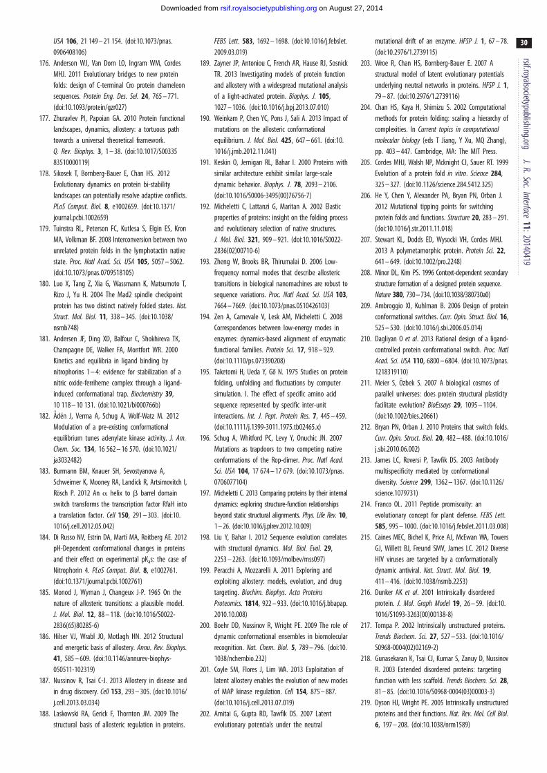
rsif.royalsocietypublishing.orgJ.R.Soc.Interface
11:20140419
30
on August 27, 2014rsif.royalsocietypublishing.orgDownloaded from
USA 106, 21 149 – 21 154. (doi:10.1073/pnas.0906408106)
176. Anderson WJ, Van Dorn LO, Ingram WM, CordesMHJ. 2011 Evolutionary bridges to new proteinfolds: design of C-terminal Cro protein chameleonsequences. Protein Eng. Des. Sel. 24, 765 – 771.(doi:10.1093/protein/gzr027)
177. Zhuravlev PI, Papoian GA. 2010 Protein functionallandscapes, dynamics, allostery: a tortuous pathtowards a universal theoretical framework.Q. Rev. Biophys. 3, 1 – 38. (doi:10.1017/S0033583510000119)
178. Sikosek T, Bornberg-Bauer E, Chan HS. 2012Evolutionary dynamics on protein bi-stabilitylandscapes can potentially resolve adaptive conflicts.PLoS Comput. Biol. 8, e1002659. (doi:10.1371/journal.pcbi.1002659)
179. Tuinstra RL, Peterson FC, Kutlesa S, Elgin ES, KronMA, Volkman BF. 2008 Interconversion between twounrelated protein folds in the lymphotactin nativestate. Proc. Natl Acad. Sci. USA 105, 5057 – 5062.(doi:10.1073/pnas.0709518105)
180. Luo X, Tang Z, Xia G, Wassmann K, Matsumoto T,Rizo J, Yu H. 2004 The Mad2 spindle checkpointprotein has two distinct natively folded states. Nat.Struct. Mol. Biol. 11, 338 – 345. (doi:10.1038/nsmb748)
181. Andersen JF, Ding XD, Balfour C, Shokhireva TK,Champagne DE, Walker FA, Montfort WR. 2000Kinetics and equilibria in ligand binding bynitrophorins 1 – 4: evidence for stabilization of anitric oxide-ferriheme complex through a ligand-induced conformational trap. Biochemistry 39,10 118 – 10 131. (doi:10.1021/bi000766b)
182. Aden J, Verma A, Schug A, Wolf-Watz M. 2012Modulation of a pre-existing conformationalequilibrium tunes adenylate kinase activity. J. Am.Chem. Soc. 134, 16 562 – 16 570. (doi:10.1021/ja3032482)
183. Burmann BM, Knauer SH, Sevostyanova A,Schweimer K, Mooney RA, Landick R, Artsimovitch I,Rosch P. 2012 An a helix to b barrel domainswitch transforms the transcription factor RfaH intoa translation factor. Cell 150, 291 – 303. (doi:10.1016/j.cell.2012.05.042)
184. Di Russo NV, Estrin DA, Martı MA, Roitberg AE. 2012pH-Dependent conformational changes in proteinsand their effect on experimental pKas: the case ofNitrophorin 4. PLoS Comput. Biol. 8, e1002761.(doi:10.1371/journal.pcbi.1002761)
185. Monod J, Wyman J, Changeux J-P. 1965 On thenature of allosteric transitions: a plausible model.J. Mol. Biol. 12, 88 – 118. (doi:10.1016/S0022-2836(65)80285-6)
186. Hilser VJ, Wrabl JO, Motlagh HN. 2012 Structuraland energetic basis of allostery. Annu. Rev. Biophys.41, 585 – 609. (doi:10.1146/annurev-biophys-050511-102319)
187. Nussinov R, Tsai C-J. 2013 Allostery in disease andin drug discovery. Cell 153, 293 – 305. (doi:10.1016/j.cell.2013.03.034)
188. Laskowski RA, Gerick F, Thornton JM. 2009 Thestructural basis of allosteric regulation in proteins.
FEBS Lett. 583, 1692 – 1698. (doi:10.1016/j.febslet.2009.03.019)
189. Zayner JP, Antoniou C, French AR, Hause RJ, SosnickTR. 2013 Investigating models of protein functionand allostery with a widespread mutational analysisof a light-activated protein. Biophys. J. 105,1027 – 1036. (doi:10.1016/j.bpj.2013.07.010)
190. Weinkam P, Chen YC, Pons J, Sali A. 2013 Impact ofmutations on the allosteric conformationalequilibrium. J. Mol. Biol. 425, 647 – 661. (doi:10.1016/j.jmb.2012.11.041)
191. Keskin O, Jernigan RL, Bahar I. 2000 Proteins withsimilar architecture exhibit similar large-scaledynamic behavior. Biophys. J. 78, 2093 – 2106.(doi:10.1016/S0006-3495(00)76756-7)
192. Micheletti C, Lattanzi G, Maritan A. 2002 Elasticproperties of proteins: insight on the folding processand evolutionary selection of native structures.J. Mol. Biol. 321, 909 – 921. (doi:10.1016/S0022-2836(02)00710-6)
193. Zheng W, Brooks BR, Thirumalai D. 2006 Low-frequency normal modes that describe allosterictransitions in biological nanomachines are robust tosequence variations. Proc. Natl Acad. Sci. USA 103,7664 – 7669. (doi:10.1073/pnas.0510426103)
194. Zen A, Carnevale V, Lesk AM, Micheletti C. 2008Correspondences between low-energy modes inenzymes: dynamics-based alignment of enzymaticfunctional families. Protein Sci. 17, 918 – 929.(doi:10.1110/ps.073390208)
195. Taketomi H, Ueda Y, Go N. 1975 Studies on proteinfolding, unfolding and fluctuations by computersimulation. I. The effect of specific amino acidsequence represented by specific inter-unitinteractions. Int. J. Pept. Protein Res. 7, 445 – 459.(doi:10.1111/j.1399-3011.1975.tb02465.x)
196. Schug A, Whitford PC, Levy Y, Onuchic JN. 2007Mutations as trapdoors to two competing nativeconformations of the Rop-dimer. Proc. Natl Acad.Sci. USA 104, 17 674 – 17 679. (doi:10.1073/pnas.0706077104)
197. Micheletti C. 2013 Comparing proteins by their internaldynamics: exploring structure-function relationshipsbeyond static structural alignments. Phys. Life Rev. 10,1 – 26. (doi:10.1016/j.plrev.2012.10.009)
198. Liu Y, Bahar I. 2012 Sequence evolution correlateswith structural dynamics. Mol. Biol. Evol. 29,2253 – 2263. (doi:10.1093/molbev/mss097)
199. Peracchi A, Mozzarelli A. 2011 Exploring andexploiting allostery: models, evolution, and drugtargeting. Biochim. Biophys. Acta ProteinsProteomics. 1814, 922 – 933. (doi:10.1016/j.bbapap.2010.10.008)
200. Boehr DD, Nussinov R, Wright PE. 2009 The role ofdynamic conformational ensembles in biomolecularrecognition. Nat. Chem. Biol. 5, 789 – 796. (doi:10.1038/nchembio.232)
201. Coyle SM, Flores J, Lim WA. 2013 Exploitation oflatent allostery enables the evolution of new modesof MAP kinase regulation. Cell 154, 875 – 887.(doi:10.1016/j.cell.2013.07.019)
202. Amitai G, Gupta RD, Tawfik DS. 2007 Latentevolutionary potentials under the neutral
mutational drift of an enzyme. HFSP J. 1, 67 – 78.(doi:10.2976/1.2739115)
203. Wroe R, Chan HS, Bornberg-Bauer E. 2007 Astructural model of latent evolutionary potentialsunderlying neutral networks in proteins. HFSP J. 1,79 – 87. (doi:10.2976/1.2739116)
204. Chan HS, Kaya H, Shimizu S. 2002 Computationalmethods for protein folding: scaling a hierarchy ofcomplexities. In Current topics in computationalmolecular biology (eds T Jiang, Y Xu, MQ Zhang),pp. 403 – 447. Cambridge, MA: The MIT Press.
205. Cordes MHJ, Walsh NP, Mcknight CJ, Sauer RT. 1999Evolution of a protein fold in vitro. Science 284,325 – 327. (doi:10.1126/science.284.5412.325)
206. He Y, Chen Y, Alexander PA, Bryan PN, Orban J.2012 Mutational tipping points for switchingprotein folds and functions. Structure 20, 283 – 291.(doi:10.1016/j.str.2011.11.018)
207. Stewart KL, Dodds ED, Wysocki VH, Cordes MHJ.2013 A polymetamorphic protein. Protein Sci. 22,641 – 649. (doi:10.1002/pro.2248)
208. Minor DL, Kim PS. 1996 Context-dependent secondarystructure formation of a designed protein sequence.Nature 380, 730 – 734. (doi:10.1038/380730a0)
209. Ambroggio XI, Kuhlman B. 2006 Design of proteinconformational switches. Curr. Opin. Struct. Biol. 16,525 – 530. (doi:10.1016/j.sbi.2006.05.014)
210. Dagliyan O et al. 2013 Rational design of a ligand-controlled protein conformational switch. Proc. NatlAcad. Sci. USA 110, 6800 – 6804. (doi:10.1073/pnas.1218319110)
211. Meier S, Ozbek S. 2007 A biological cosmos ofparallel universes: does protein structural plasticityfacilitate evolution? BioEssays 29, 1095 – 1104.(doi:10.1002/bies.20661)
212. Bryan PN, Orban J. 2010 Proteins that switch folds.Curr. Opin. Struct. Biol. 20, 482 – 488. (doi:10.1016/j.sbi.2010.06.002)
213. James LC, Roversi P, Tawfik DS. 2003 Antibodymultispecificity mediated by conformationaldiversity. Science 299, 1362 – 1367. (doi:10.1126/science.1079731)
214. Franco OL. 2011 Peptide promiscuity: anevolutionary concept for plant defense. FEBS Lett.585, 995 – 1000. (doi:10.1016/j.febslet.2011.03.008)
215. Caines MEC, Bichel K, Price AJ, McEwan WA, TowersGJ, Willett BJ, Freund SMV, James LC. 2012 DiverseHIV viruses are targeted by a conformationallydynamic antiviral. Nat. Struct. Mol. Biol. 19,411 – 416. (doi:10.1038/nsmb.2253)
216. Dunker AK et al. 2001 Intrinsically disorderedprotein. J. Mol. Graph Model 19, 26 – 59. (doi:10.1016/S1093-3263(00)00138-8)
217. Tompa P. 2002 Intrinsically unstructured proteins.Trends Biochem. Sci. 27, 527 – 533. (doi:10.1016/S0968-0004(02)02169-2)
218. Gunasekaran K, Tsai CJ, Kumar S, Zanuy D, NussinovR. 2003 Extended disordered proteins: targetingfunction with less scaffold. Trends Biochem. Sci. 28,81 – 85. (doi:10.1016/S0968-0004(03)00003-3)
219. Dyson HJ, Wright PE. 2005 Intrinsically unstructuredproteins and their functions. Nat. Rev. Mol. Cell Biol.6, 197 – 208. (doi:10.1038/nrm1589)
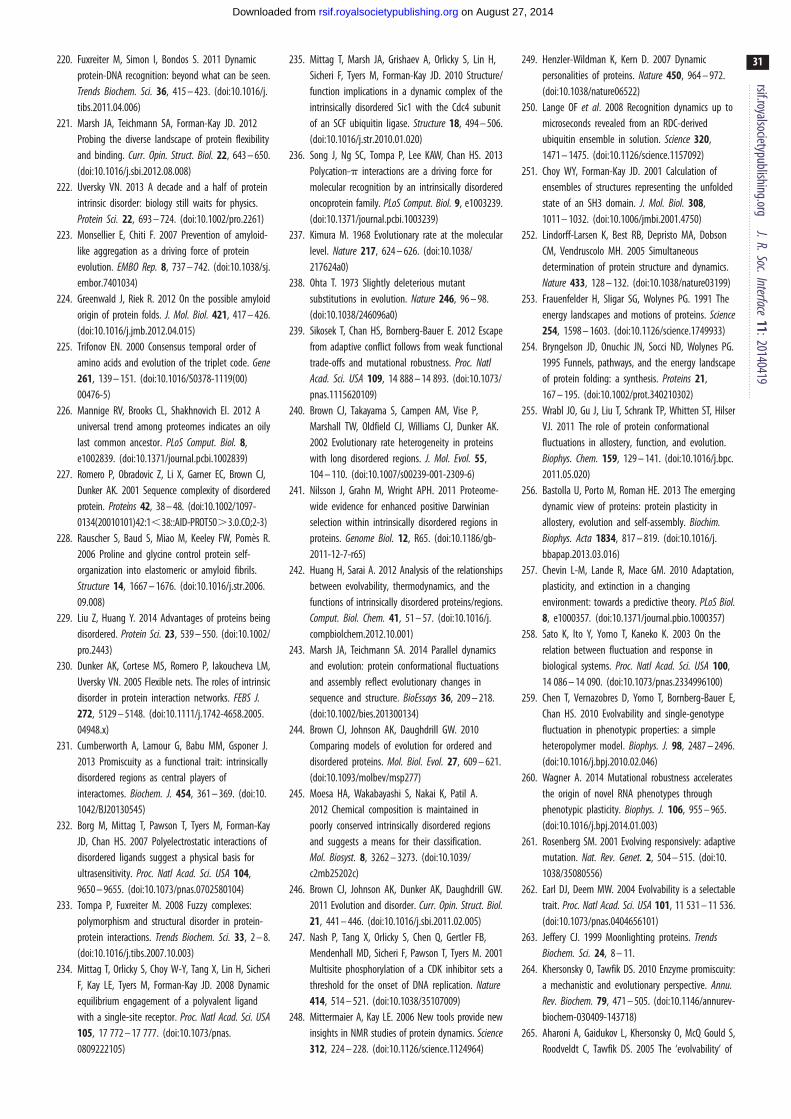
rsif.royalsocietypublishing.orgJ.R.Soc.Interface
11:20140419
31
on August 27, 2014rsif.royalsocietypublishing.orgDownloaded from
220. Fuxreiter M, Simon I, Bondos S. 2011 Dynamicprotein-DNA recognition: beyond what can be seen.Trends Biochem. Sci. 36, 415 – 423. (doi:10.1016/j.tibs.2011.04.006)
221. Marsh JA, Teichmann SA, Forman-Kay JD. 2012Probing the diverse landscape of protein flexibilityand binding. Curr. Opin. Struct. Biol. 22, 643 – 650.(doi:10.1016/j.sbi.2012.08.008)
222. Uversky VN. 2013 A decade and a half of proteinintrinsic disorder: biology still waits for physics.Protein Sci. 22, 693 – 724. (doi:10.1002/pro.2261)
223. Monsellier E, Chiti F. 2007 Prevention of amyloid-like aggregation as a driving force of proteinevolution. EMBO Rep. 8, 737 – 742. (doi:10.1038/sj.embor.7401034)
224. Greenwald J, Riek R. 2012 On the possible amyloidorigin of protein folds. J. Mol. Biol. 421, 417 – 426.(doi:10.1016/j.jmb.2012.04.015)
225. Trifonov EN. 2000 Consensus temporal order ofamino acids and evolution of the triplet code. Gene261, 139 – 151. (doi:10.1016/S0378-1119(00)00476-5)
226. Mannige RV, Brooks CL, Shakhnovich EI. 2012 Auniversal trend among proteomes indicates an oilylast common ancestor. PLoS Comput. Biol. 8,e1002839. (doi:10.1371/journal.pcbi.1002839)
227. Romero P, Obradovic Z, Li X, Garner EC, Brown CJ,Dunker AK. 2001 Sequence complexity of disorderedprotein. Proteins 42, 38 – 48. (doi:10.1002/1097-0134(20010101)42:1,38::AID-PROT50.3.0.CO;2-3)
228. Rauscher S, Baud S, Miao M, Keeley FW, Pomes R.2006 Proline and glycine control protein self-organization into elastomeric or amyloid fibrils.Structure 14, 1667 – 1676. (doi:10.1016/j.str.2006.09.008)
229. Liu Z, Huang Y. 2014 Advantages of proteins beingdisordered. Protein Sci. 23, 539 – 550. (doi:10.1002/pro.2443)
230. Dunker AK, Cortese MS, Romero P, Iakoucheva LM,Uversky VN. 2005 Flexible nets. The roles of intrinsicdisorder in protein interaction networks. FEBS J.272, 5129 – 5148. (doi:10.1111/j.1742-4658.2005.04948.x)
231. Cumberworth A, Lamour G, Babu MM, Gsponer J.2013 Promiscuity as a functional trait: intrinsicallydisordered regions as central players ofinteractomes. Biochem. J. 454, 361 – 369. (doi:10.1042/BJ20130545)
232. Borg M, Mittag T, Pawson T, Tyers M, Forman-KayJD, Chan HS. 2007 Polyelectrostatic interactions ofdisordered ligands suggest a physical basis forultrasensitivity. Proc. Natl Acad. Sci. USA 104,9650 – 9655. (doi:10.1073/pnas.0702580104)
233. Tompa P, Fuxreiter M. 2008 Fuzzy complexes:polymorphism and structural disorder in protein-protein interactions. Trends Biochem. Sci. 33, 2 – 8.(doi:10.1016/j.tibs.2007.10.003)
234. Mittag T, Orlicky S, Choy W-Y, Tang X, Lin H, SicheriF, Kay LE, Tyers M, Forman-Kay JD. 2008 Dynamicequilibrium engagement of a polyvalent ligandwith a single-site receptor. Proc. Natl Acad. Sci. USA105, 17 772 – 17 777. (doi:10.1073/pnas.0809222105)
235. Mittag T, Marsh JA, Grishaev A, Orlicky S, Lin H,Sicheri F, Tyers M, Forman-Kay JD. 2010 Structure/function implications in a dynamic complex of theintrinsically disordered Sic1 with the Cdc4 subunitof an SCF ubiquitin ligase. Structure 18, 494 – 506.(doi:10.1016/j.str.2010.01.020)
236. Song J, Ng SC, Tompa P, Lee KAW, Chan HS. 2013Polycation-p interactions are a driving force formolecular recognition by an intrinsically disorderedoncoprotein family. PLoS Comput. Biol. 9, e1003239.(doi:10.1371/journal.pcbi.1003239)
237. Kimura M. 1968 Evolutionary rate at the molecularlevel. Nature 217, 624 – 626. (doi:10.1038/217624a0)
238. Ohta T. 1973 Slightly deleterious mutantsubstitutions in evolution. Nature 246, 96 – 98.(doi:10.1038/246096a0)
239. Sikosek T, Chan HS, Bornberg-Bauer E. 2012 Escapefrom adaptive conflict follows from weak functionaltrade-offs and mutational robustness. Proc. NatlAcad. Sci. USA 109, 14 888 – 14 893. (doi:10.1073/pnas.1115620109)
240. Brown CJ, Takayama S, Campen AM, Vise P,Marshall TW, Oldfield CJ, Williams CJ, Dunker AK.2002 Evolutionary rate heterogeneity in proteinswith long disordered regions. J. Mol. Evol. 55,104 – 110. (doi:10.1007/s00239-001-2309-6)
241. Nilsson J, Grahn M, Wright APH. 2011 Proteome-wide evidence for enhanced positive Darwinianselection within intrinsically disordered regions inproteins. Genome Biol. 12, R65. (doi:10.1186/gb-2011-12-7-r65)
242. Huang H, Sarai A. 2012 Analysis of the relationshipsbetween evolvability, thermodynamics, and thefunctions of intrinsically disordered proteins/regions.Comput. Biol. Chem. 41, 51 – 57. (doi:10.1016/j.compbiolchem.2012.10.001)
243. Marsh JA, Teichmann SA. 2014 Parallel dynamicsand evolution: protein conformational fluctuationsand assembly reflect evolutionary changes insequence and structure. BioEssays 36, 209 – 218.(doi:10.1002/bies.201300134)
244. Brown CJ, Johnson AK, Daughdrill GW. 2010Comparing models of evolution for ordered anddisordered proteins. Mol. Biol. Evol. 27, 609 – 621.(doi:10.1093/molbev/msp277)
245. Moesa HA, Wakabayashi S, Nakai K, Patil A.2012 Chemical composition is maintained inpoorly conserved intrinsically disordered regionsand suggests a means for their classification.Mol. Biosyst. 8, 3262 – 3273. (doi:10.1039/c2mb25202c)
246. Brown CJ, Johnson AK, Dunker AK, Daughdrill GW.2011 Evolution and disorder. Curr. Opin. Struct. Biol.21, 441 – 446. (doi:10.1016/j.sbi.2011.02.005)
247. Nash P, Tang X, Orlicky S, Chen Q, Gertler FB,Mendenhall MD, Sicheri F, Pawson T, Tyers M. 2001Multisite phosphorylation of a CDK inhibitor sets athreshold for the onset of DNA replication. Nature414, 514 – 521. (doi:10.1038/35107009)
248. Mittermaier A, Kay LE. 2006 New tools provide newinsights in NMR studies of protein dynamics. Science312, 224 – 228. (doi:10.1126/science.1124964)
249. Henzler-Wildman K, Kern D. 2007 Dynamicpersonalities of proteins. Nature 450, 964 – 972.(doi:10.1038/nature06522)
250. Lange OF et al. 2008 Recognition dynamics up tomicroseconds revealed from an RDC-derivedubiquitin ensemble in solution. Science 320,1471 – 1475. (doi:10.1126/science.1157092)
251. Choy WY, Forman-Kay JD. 2001 Calculation ofensembles of structures representing the unfoldedstate of an SH3 domain. J. Mol. Biol. 308,1011 – 1032. (doi:10.1006/jmbi.2001.4750)
252. Lindorff-Larsen K, Best RB, Depristo MA, DobsonCM, Vendruscolo MH. 2005 Simultaneousdetermination of protein structure and dynamics.Nature 433, 128 – 132. (doi:10.1038/nature03199)
253. Frauenfelder H, Sligar SG, Wolynes PG. 1991 Theenergy landscapes and motions of proteins. Science254, 1598 – 1603. (doi:10.1126/science.1749933)
254. Bryngelson JD, Onuchic JN, Socci ND, Wolynes PG.1995 Funnels, pathways, and the energy landscapeof protein folding: a synthesis. Proteins 21,167 – 195. (doi:10.1002/prot.340210302)
255. Wrabl JO, Gu J, Liu T, Schrank TP, Whitten ST, HilserVJ. 2011 The role of protein conformationalfluctuations in allostery, function, and evolution.Biophys. Chem. 159, 129 – 141. (doi:10.1016/j.bpc.2011.05.020)
256. Bastolla U, Porto M, Roman HE. 2013 The emergingdynamic view of proteins: protein plasticity inallostery, evolution and self-assembly. Biochim.Biophys. Acta 1834, 817 – 819. (doi:10.1016/j.bbapap.2013.03.016)
257. Chevin L-M, Lande R, Mace GM. 2010 Adaptation,plasticity, and extinction in a changingenvironment: towards a predictive theory. PLoS Biol.8, e1000357. (doi:10.1371/journal.pbio.1000357)
258. Sato K, Ito Y, Yomo T, Kaneko K. 2003 On therelation between fluctuation and response inbiological systems. Proc. Natl Acad. Sci. USA 100,14 086 – 14 090. (doi:10.1073/pnas.2334996100)
259. Chen T, Vernazobres D, Yomo T, Bornberg-Bauer E,Chan HS. 2010 Evolvability and single-genotypefluctuation in phenotypic properties: a simpleheteropolymer model. Biophys. J. 98, 2487 – 2496.(doi:10.1016/j.bpj.2010.02.046)
260. Wagner A. 2014 Mutational robustness acceleratesthe origin of novel RNA phenotypes throughphenotypic plasticity. Biophys. J. 106, 955 – 965.(doi:10.1016/j.bpj.2014.01.003)
261. Rosenberg SM. 2001 Evolving responsively: adaptivemutation. Nat. Rev. Genet. 2, 504 – 515. (doi:10.1038/35080556)
262. Earl DJ, Deem MW. 2004 Evolvability is a selectabletrait. Proc. Natl Acad. Sci. USA 101, 11 531 – 11 536.(doi:10.1073/pnas.0404656101)
263. Jeffery CJ. 1999 Moonlighting proteins. TrendsBiochem. Sci. 24, 8 – 11.
264. Khersonsky O, Tawfik DS. 2010 Enzyme promiscuity:a mechanistic and evolutionary perspective. Annu.Rev. Biochem. 79, 471 – 505. (doi:10.1146/annurev-biochem-030409-143718)
265. Aharoni A, Gaidukov L, Khersonsky O, McQ Gould S,Roodveldt C, Tawfik DS. 2005 The ‘evolvability’ of

rsif.royalsocietypublishing.orgJ.R.Soc.Interface
11:20140419
32
on August 27, 2014rsif.royalsocietypublishing.orgDownloaded from
promiscuous protein functions. Nat. Genet. 37,73 – 76. (doi:10.1038/ng1482)
266. Hou J, Sims GE, Zhang C, Kim S-H. 2003 A globalrepresentation of the protein fold space. Proc. NatlAcad. Sci. USA 100, 2386 – 2390. (doi:10.1073/pnas.2628030100)
267. Sippl MJ. 2009 Fold space unlimited. Curr. Opin.Struct. Biol. 19, 312 – 320. (doi:10.1016/j.sbi.2009.03.010)
268. Caetano-Anolles G, Wang M, Caetano-Anolles D,Mittenthal JE. 2009 The origin, evolution andstructure of the protein world. Biochem. J. 417,621 – 637. (doi:10.1042/BJ20082063)
269. Caetano-Anolles G, Kim KM, Caetano-Anolles D.2012 The phylogenomic roots of modernbiochemistry: origins of proteins, cofactors andprotein biosynthesis. J. Mol. Evol. 74, 1 – 34.(doi:10.1007/s00239-011-9480-1)
270. Osadchy M, Kolodny R. 2011 Maps of proteinstructure space reveal a fundamental relationshipbetween protein structure and function. Proc. NatlAcad. Sci. USA 108, 12 301 – 12 306. (doi:10.1073/pnas.1102727108)
271. Minary P, Levitt M. 2008 Probing protein fold spacewith a simplified model. J. Mol. Biol. 375,920 – 933. (doi:10.1016/j.jmb.2007.10.087)
272. Keefe AD, Szostak JW. 2001 Functional proteinsfrom a random-sequence library. Nature 410,715 – 718. (doi:10.1038/35070613)
273. Povolotskaya IS, Kondrashov FA. 2010 Sequencespace and the ongoing expansion of the proteinuniverse. Nature 465, 922 – 926. (doi:10.1038/nature09105)
274. Murzin AG, Brenner SE, Hubbard T, Chothia C. 1995SCOP: a structural classification of proteins databasefor the investigation of sequences and structures.J. Mol. Biol. 247, 536 – 540. (doi:10.1006/jmbi.1995.0159)
275. Wolf YI, Grishin NV, Koonin EV. 2000 Estimating thenumber of protein folds and families from completegenome data. J. Mol. Biol. 299, 897 – 905. (doi:10.1006/jmbi.2000.3786)
276. Govindarajan S, Recabarren R, Goldstein RA. 1999Estimating the total number of protein folds. Proteins35, 408 – 414. (doi:10.1002/(SICI)1097-0134(19990601)35:4,408::AID-PROT4.3.0.CO;2-A)
277. Coulson AFW, Moult J. 2002 A unifold, mesofold,and superfold model of protein fold use. Proteins46, 61 – 71. (doi:10.1002/prot.10011)
278. Kolodny R, Pereyaslavets L, Samson AO, Levitt M.2013 On the universe of protein folds. Annu. Rev.Biophys. 42, 559 – 582. (doi:10.1146/annurev-biophys-083012-130432)
279. Godzik A. 2011 Metagenomics and the proteinuniverse. Curr. Opin. Struct. Biol. 21, 398 – 403.(doi:10.1016/j.sbi.2011.03.010)
280. Chan HS, Dill KA. 1990 The effects of internalconstraints on the configurations of chainmolecules. J. Chem. Phys. 92, 3118 – 3135. (doi:10.1063/1.458605)
281. Chan HS, Dill KA. 1990 Origins of structure inglobular proteins. Proc. Natl Acad. Sci. USA 87,6388 – 6392. (doi:10.1073/pnas.87.16.6388)
282. Maritan A, Micheletti C, Trovato A, Banavar JR. 2000Optimal shapes of compact strings. Nature 406,287 – 290. (doi:10.1038/35018538)
283. Gregoret LM, Cohen FE. 1991 Protein folding. Effectof packing density on chain conformation. J. Mol.Biol. 219, 109 – 122. (doi:10.1016/0022-2836(91)90861-Y)
284. Hunt NG, Gregoret LM, Cohen FE. 1994 The originsof protein secondary structure. Effects of packingdensity and hydrogen bonding studied by a fastconformational search. J. Mol. Biol. 241, 214 – 225.(doi:10.1006/jmbi.1994.1490)
285. Yee DP, Chan HS, Havel TF, Dill KA. 1994 Doescompactness induce secondary structure in proteins?A study of poly-alanine chains computed bydistance geometry. J. Mol. Biol. 241, 557 – 573.(doi:10.1006/jmbi.1994.1531)
286. Zhang Y, Hubner IA, Arakaki AK, Shakhnovich EI,Skolnick J. 2006 On the origin and highly likelycompleteness of single-domain protein structures.Proc. Natl Acad. Sci. USA 103, 2605 – 2610. (doi:10.1073/pnas.0509379103)
287. Taylor WR, Chelliah V, Hollup SM, MacDonald JT,Jonassen I. 2009 Probing the ‘dark matter’ ofprotein fold space. Structure 17, 1244 – 1252.(doi:10.1016/j.str.2009.07.012)
288. Cossio P, Trovato A, Pietrucci F, Seno F, Maritan A,Laio A. 2010 Exploring the universe of proteinstructures beyond the Protein Data Bank. PLoSComput. Biol. 6, e1000957. (doi:10.1371/journal.pcbi.1000957)
289. Dai L, Zhou Y. 2011 Characterizing the existing andpotential structural space of proteins by large-scalemultiple loop permutations. J. Mol. Biol. 408,585 – 595. (doi:10.1016/j.jmb.2011.02.056)
290. Skolnick J, Zhou H, Brylinski M. 2012 Furtherevidence for the likely completeness of thelibrary of solved single domain protein structures.J. Phys. Chem. B 116, 6654 – 6664. (doi:10.1021/jp211052j)
291. Skolnick J, Gao M. 2013 Interplay of physics andevolution in the likely origin of protein biochemicalfunction. Proc. Natl Acad. Sci. USA 110, 9344 – 9349.(doi:10.1073/pnas.1300011110)
292. Chan HS, Bornberg-Bauer E. 2002 Perspectives onprotein evolution from simple exact models. Appl.Bioinform. 1, 121 – 144.
293. Xia Y, Levitt M. 2004 Funnel-like organization insequence space determines the distributions ofprotein stability and folding rate preferred byevolution. Proteins 55, 107 – 114. (doi:10.1002/prot.10563)
294. Greenbury SF, Johnston IG, Louis AA, Ahnert SE.2014 A tractable genotype-phenotype mapmodelling the self-assembly of protein quaternarystructure. J. R. Soc. Interface 11, 20140249. (doi:10.1098/rsif.2014.0249)
295. Moreno-Hernandez S, Levitt M. 2012 Comparativemodeling and protein-like features of hydrophobic-polar models on a two-dimensional lattice. Proteins80, 1683 – 1693. (doi:10.1002/prot.24067)
296. Palmer ME, Moudgil A, Feldman MW. 2013 Long-term evolution is surprisingly predictable in lattice
proteins. J. R. Soc. Interface 10, 20130026. (doi:10.1098/rsif.2013.0026)
297. Lau KF, Dill KA. 1990 Theory for protein mutabilityand biogenesis. Proc. Natl Acad. Sci. USA 87,638 – 642. (doi:10.1073/pnas.87.2.638)
298. Lipman DJ, Wilbur WJ. 1991 Modelling neutral andselective evolution of protein folding. Proc. R. Soc.Lond. B 245, 7 – 11. (doi:10.1098/rspb.1991.0081)
299. Holzgrafe C, Irback A, Troein C. 2011 Mutation-induced fold switching among lattice proteins.J. Chem. Phys. 135, 195101. (doi:10.1063/1.3660691)
300. Miller C, Davlieva M, Wilson C, White KI, CounagoR, Wu G, Myers JC, Wittung-Stafshede P, Shamoo Y.2010 Experimental evolution of adenylate kinasereveals contrasting strategies toward proteinthermostability. Biophys. J. 99, 887 – 896. (doi:10.1016/j.bpj.2010.04.076)
301. Hirst JD. 1999 The evolutionary landscape offunctional model proteins. Protein Eng. Des. Sel. 12,721 – 726. (doi:10.1093/protein/12.9.721)
302. Blackburne BP, Hirst JD. 2001 Evolution offunctional model proteins. J. Chem. Phys. 115,1935. (doi:10.1063/1.1383051)
303. Burke S, Elber R. 2011 Super folds, networks, andbarriers. Proteins 80, 463 – 470. (doi:10.1002/prot.23212)
304. Noirel J, Simonson T. 2008 Neutral evolution ofproteins: the superfunnel in sequence space and itsrelation to mutational robustness. J. Chem. Phys.129, 185104. (doi:10.1063/1.2992853)
305. Bastolla U, Roman HE, Vendruscolo MH. 1999Neutral evolution of model proteins: diffusion insequence space and overdispersion. J. Theor. Biol.200, 49 – 64. (doi:10.1006/jtbi.1999.0975)
306. Taverna DM, Goldstein RA. 2002 Why are proteinsso robust to site mutations? J. Mol. Biol. 315,479 – 484. (doi:10.1006/jmbi.2001.5226)
307. Deeds EJ, Shakhnovich EI. 2005 The emergence ofscaling in sequence-based physical models ofprotein evolution. Biophys. J. 88, 3905 – 3911.(doi:10.1529/biophysj.104.051433)
308. Zeldovich KB, Chen P, Shakhnovich BE, ShakhnovichEI. 2007 A first-principles model of early evolution:emergence of gene families, species, and preferredprotein folds. PLoS Comput. Biol. 3, e139. (doi:10.1371/journal.pcbi.0030139)
309. Chan HS, Dill KA. 1989 Compact polymers.Macromolecules 22, 4559 – 4573. (doi:10.1021/ma00202a031)
310. Pande VS, Joerg C, Grosberg AY, Tanaka T. 1994Enumerations of the Hamiltonian walks on a cubicsublattice. J. Phys. A Math. Gen. 27, 6231 – 6236.(doi:10.1088/0305-4470/27/18/030)
311. Lee JH, Kim S-Y, Lee J. 2011 Parallel algorithm forcalculation of the exact partition function of alattice polymer. Comput. Phys. Commun. 182,1027 – 1033. (doi:10.1016/j.cpc.2011.01.004)
312. Schram RD, Schiessel H. 2013 Exact enumeration ofHamiltonian walks on the 4�4�4 cube andapplications to protein folding. J. Phys. A Math.Theor. 46, 485001. (doi:10.1088/1751-8113/46/48/485001)

rsif.royalsocietypublishing.orgJ.R.Soc.Interface
11:20140419
33
on August 27, 2014rsif.royalsocietypublishing.orgDownloaded from
313. Lee J. 2004 Exact partition function zeros of two-dimensional lattice polymers. J. Korean Phys. Soc.44, 617 – 620. (doi:10.3938/jkps.44.617)
314. Clisby N, Liang R, Slade G. 2007 Self-avoiding walkenumeration via the lace expansion. J. Phys. AMath. Theor. 40, 10 973 – 11 017. (doi:10.1088/1751-8113/40/36/003)
315. Yue K, Fiebig KM, Thomas PD, Chan HS,Shakhnovich EI, Dill KA. 1995 A test of latticeprotein folding algorithms. Proc. Natl Acad. Sci. USA92, 325 – 329. (doi:10.1073/pnas.92.1.325)
316. Irback A, Troein C. 2002 Enumerating designingsequences in the HP model. J. Biol. Phys. 28, 1 – 15.(doi:10.1023/A:1016225010659)
317. Dill KA, Bromberg S, Yue K, Fiebig KM, Yee DP,Thomas PD, Chan HS. 1995 Principles of proteinfolding—a perspective from simple exact models.Protein Sci. 4, 561 – 602. (doi:10.1002/pro.5560040401)
318. Kamtekar S, Schiffer J, Xiong H, Babik J, Hecht M.1993 Protein design by binary patterning of polarand nonpolar amino acids. Science 262,1680 – 1685. (doi:10.1126/science.8259512)
319. Urvoas A, Valerio-Lepiniec M, Minard P. 2012Artificial proteins from combinatorial approaches.Trends Biotechnol. 30, 512 – 520. (doi:10.1016/j.tibtech.2012.06.001)
320. Irback A, Sandelin E. 2000 On hydrophobicitycorrelations in protein chains. Biophys. J. 79,2252 – 2258. (doi:10.1016/S0006-3495(00)76472-1)
321. Irback A, Peterson C, Potthast F. 1996 Evidence fornonrandom hydrophobicity structures in proteinchains. Proc. Natl Acad. Sci. USA 93, 9533 – 9538.(doi:10.1073/pnas.93.18.9533)
322. Buchler NE, Goldstein RA. 1999 Effect of alphabetsize and foldability requirements on proteinstructure designability. Proteins 34, 113 – 124.(doi:10.1002/(SICI)1097-0134(19990101)34:1,113::AID-PROT9.3.0.CO;2-J)
323. Wroe R, Bornberg-Bauer E, Chan HS. 2005Comparing folding codes in simple heteropolymermodels of protein evolutionary landscape:robustness of the superfunnel paradigm. Biophys. J.88, 118 – 131. (doi:10.1529/biophysj.104.050369)
324. Cui Y, Wong WH, Bornberg-Bauer E, Chan HS. 2002Recombinatoric exploration of novel foldedstructures: a heteropolymer-based model of proteinevolutionary landscapes. Proc. Natl Acad. Sci. USA99, 809 – 814. (doi:10.1073/pnas.022240299)
325. Chan HS. 2000 Modeling protein density of states:additive hydrophobic effects are insufficient forcalorimetric two-state cooperativity. Proteins 40,543 – 571. (doi:10.1002/1097-0134(20000901)40:4,543::AID-PROT20.3.0.CO;2-O)
326. Chan HS. 1998 Protein folding. Matching speed andlocality. Nature 392, 761 – 763. (doi:10.1038/33808)
327. Go N. 1983 Theoretical studies of protein folding.Annu. Rev. Biophys. Bioeng. 12, 183 – 210. (doi:10.1146/annurev.bb.12.060183.001151)
328. Bryngelson JD, Wolynes PG. 1987 Spin glasses andthe statistical mechanics of protein folding. Proc.Natl Acad. Sci. USA 84, 7524 – 7528. (doi:10.1073/pnas.84.21.7524)
329. Miyazawa S, Jernigan RL. 1985 Estimation ofeffective interresidue contact energies from proteincrystal structures: quasi-chemical approximation.Macromolecules 18, 534 – 552. (doi:10.1021/ma00145a039)
330. Bloom JD, Wilke CO, Arnold FH, Adami C. 2004Stability and the evolvability of function in a modelprotein. Biophys. J. 86, 2758 – 2764. (doi:10.1016/S0006-3495(04)74329-5)
331. Abkevich VI, Gutin AM, Shakhnovich EI. 1994 Specificnucleus as the transition state for protein folding:evidence from the lattice model. Biochemistry 33,10 026 – 10 036. (doi:10.1021/bi00199a029)
332. Xia Y, Levitt M. 2004 Simulating protein evolutionin sequence and structure space. Curr. Opin. Struct.Biol. 14, 202 – 207. (doi:10.1016/j.sbi.2004.03.001)
333. Goldstein RA. 2008 The structure of protein evolutionand the evolution of protein structure. Curr. Opin. Struct.Biol. 18, 170 – 177. (doi:10.1016/j.sbi.2008.01.006)
334. Zeldovich KB, Shakhnovich EI. 2008 Understandingprotein evolution: from protein physics to Darwinianselection. Annu. Rev. Phys. Chem. 59, 105 – 127.(doi:10.1146/annurev.physchem.58.032806.104449)
335. Blackburne BP, Hirst JD. 2005 Population dynamicssimulations of functional model proteins. J. Chem.Phys. 123, 154907. (doi:10.1063/1.2056545)
336. Maynard Smith J. 1970 Natural selection and theconcept of a protein space. Nature 225, 563 – 564.(doi:10.1038/225563a0)
337. Bastolla U, Vendruscolo MH, Roman HE. 2000Structurally constrained protein evolution: resultsfrom a lattice simulation. Eur. Phys. J. B 15,385 – 397. (doi:10.1007/s100510051140)
338. Bloom JD, Labthavikul ST, Otey CR, Arnold FH. 2006Protein stability promotes evolvability. Proc. NatlAcad. Sci. USA 103, 5869 – 5874. (doi:10.1073/pnas.0510098103)
339. Drummond DA, Silberg JJ, Meyer MM, Wilke CO,Arnold FH. 2005 On the conservative nature ofintragenic recombination. Proc. Natl Acad. Sci. USA102, 5380 – 5385. (doi:10.1073/pnas.0500729102)
340. Cui Y, Wong W. 2000 Multiple-sequence informationprovides protection against mis-specified potentialenergy functions in the lattice model of proteins.Phys. Rev. Lett. 85, 5242 – 5245. (doi:10.1103/PhysRevLett.85.5242)
341. Nanda V, DeGrado WF. 2005 Automated use ofmutagenesis data in structure prediction. Proteins59, 454 – 466. (doi:10.1002/prot.20382)
342. England JL, Shakhnovich BE, Shakhnovich EI. 2003Natural selection of more designable folds: amechanism for thermophilic adaptation. Proc. NatlAcad. Sci. USA 100, 8727 – 8731. (doi:10.1073/pnas.1530713100)
343. Noivirt-Brik O, Unger R, Horovitz A. 2009 Analysingthe origin of long-range interactions in proteinsusing lattice models. BMC Struct. Biol. 9, 4. (doi:10.1186/1472-6807-9-4)
344. Liu Z, Chen J, Thirumalai D. 2009 On the accuracy ofinferring energetic coupling between distant sites inprotein families from evolutionary imprints:illustrations using lattice model. Proteins 77,823 – 831. (doi:10.1002/prot.22498)
345. Heo M, Kang L, Shakhnovich EI. 2009 Emergence ofspecies in evolutionary ‘simulated annealing’. Proc.Natl Acad. Sci. USA 106, 1869 – 1874. (doi:10.1073/pnas.0809852106)
346. Heo M, Shakhnovich EI. 2010 Interplay betweenpleiotropy and secondary selection determines rise andfall of mutators in stress response. PLoS Comput. Biol.6, e1000710. (doi:10.1371/journal.pcbi.1000710)
347. Heo M, Maslov S, Shakhnovich EI. 2011 Topology ofprotein interaction network shapes proteinabundances and strengths of their functional andnonspecific interactions. Proc. Natl Acad. Sci. USA108, 4258 – 4263. (doi:10.1073/pnas.1009392108)
348. Wright S. 1932 The roles of mutations, inbreeding,crossbreeding and selection in evolution. In Proc.6th Int. Congr. Genet, vol. 1, pp. 356 – 366.Menasha, WI: Brooklyn Botanical Garden.
349. Kauffman S, Levin S. 1987 Towards a general theoryof adaptive walks on rugged landscapes. J. Theor. Biol.128, 11 – 45. (doi:10.1016/S0022-5193(87) 80029-2)
350. Voigt CA, Kauffman S, Wang ZG. 2000 Rationalevolutionary design: the theory of in vitro proteinevolution. Adv. Protein Chem. 55, 79 – 160. (doi:10.S0065-3233(01)55003-2)
351. Carneiro M, Hartl DL. 2010 Adaptive landscapes andprotein evolution. Proc. Natl Acad. Sci. USA 107,1747 – 1751. (doi:10.1073/pnas.0906192106)
352. Sella G, Hirsh AE. 2005 The application of statisticalphysics to evolutionary biology. Proc. Natl Acad.Sci. USA 102, 9541 – 9546. (doi:10.1073/pnas.0501865102)
353. Pathria R. 1980 Statistical mechanics. Oxford, UK:Pergamon Press.
354. Lobkovsky AE, Wolf YI, Koonin EV. 2013 Quantifyingthe similarity of monotonic trajectories in rough andsmooth fitness landscapes. Mol. Biosyst. 9,1627 – 1631. (doi:10.1039/c3mb25553k)
355. Ferrada E, Wagner A. 2012 A comparison ofgenotype – phenotype maps for RNA and proteins.Biophys. J. 102, 1916 – 1925. (doi:10.1016/j.bpj.2012.01.047)
356. Fontana W, Schuster P. 1987 A computer model ofevolutionary optimization. Biophys. Chem. 26,123 – 147. (doi:10.1016/0301-4622(87)80017-0)
357. Fontana W, Stadler P, Bornberg-Bauer E,Griesmacher T, Hofacker I, Tacker M, Tarazona P,Weinberger E, Schuster P. 1993 RNA folding andcombinatory landscapes. Phys. Rev. E 47,2083 – 2099. (doi:10.1103/PhysRevE.47.2083)
358. Schuster P, Fontana W, Stadler PF, Hofacker IL. 1994From sequences to shapes and back: a case study inRNA secondary structures. Proc. R. Soc. Lond. B 255,279 – 284. (doi:10.1098/rspb.1994.0040)
359. Ancel LW, Fontana W. 2000 Plasticity,evolvability, and modularity in RNA. J. Exp. Zool.288, 242 – 283. (doi:10.1002/1097-010X(20001015)288:3,242::AID-JEZ5.3.0.CO;2-O)
360. Guo HH, Choe J, Loeb LA. 2004 Protein tolerance torandom amino acid change. Proc. Natl Acad. Sci. USA101, 9205 – 9210. (doi:10.1073/pnas.0403255101)
361. Punta M et al. 2012 The Pfam protein familiesdatabase. Nucleic Acids Res. 40, D290 – D301.(doi:10.1093/nar/gkr1065)
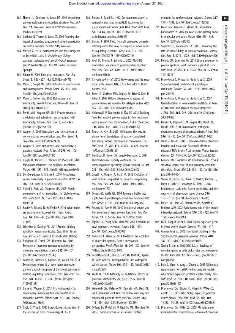
rsif.royalsocietypublishing.orgJ.R.Soc.Interface
11:20140419
34
on August 27, 2014rsif.royalsocietypublishing.orgDownloaded from
362. Thorne JL, Goldman N, Jones DT. 1996 Combiningprotein evolution and secondary structure. Mol. Biol.Evol. 13, 666 – 673. (doi:10.1093/oxfordjournals.molbev.a025627)
363. Goldman N, Thorne JL, Jones DT. 1998 Assessing theimpact of secondary structure and solvent accessibilityon protein evolution. Genetics 149, 445 – 458.
364. Massey SE. 2010 Pseudaptations and the emergenceof beneficial traits. In Evolutionary biology—concepts, molecular and morphological evolution(ed. P Pontarotti), pp. 81 – 98. Berlin, Germany:Springer.
365. Kitano H. 2004 Biological robustness. Nat. Rev.Genet. 5, 826 – 837. (doi:10.1038/nrg1471)
366. Masel J, Siegal ML. 2009 Robustness: mechanismsand consequences. Trends Genet. 25, 395 – 403.(doi:10.1016/j.tig.2009.07.005)
367. Masel J, Trotter MV. 2010 Robustness andevolvability. Trends Genet. 26, 406 – 414. (doi:10.1016/j.tig.2010.06.002)
368. Rorick MM, Wagner GP. 2011 Protein structuralmodularity and robustness are associated withevolvability. Genome Biol. Evol. 3, 456 – 475.(doi:10.1093/gbe/evr046)
369. Wagner A. 2008 Neutralism and selectionism: anetwork-based reconciliation. Nat. Rev. Genet. 9,965 – 974. (doi:10.1038/nrg2473)
370. Wagner A. 2008 Robustness and evolvability: aparadox resolved. Proc. R. Soc. B 275, 91 – 100.(doi:10.1098/rspb.2007.1137)
371. Draghi JA, Parsons TL, Wagner GP, Plotkin JB. 2010Mutational robustness can facilitate adaptation.Nature 463, 353 – 355. (doi:10.1038/nature08694)
372. Bornberg-Bauer E, Kramer L. 2010 Robustnessversus evolvability: a paradigm revisited. HFSP J. 4,105 – 108. (doi:10.2976/1.3404403)
373. Nobeli I, Favia AD, Thornton JM. 2009 Proteinpromiscuity and its implications for biotechnology.Nat. Biotechnol. 27, 157 – 167. (doi:10.1038/nbt1519)
374. Babtie A, Tokuriki N, Hollfelder F. 2010 What makesan enzyme promiscuous? Curr. Opin. Chem.Biol. 14, 200 – 207. (doi:10.1016/j.cbpa.2009.11.028)
375. Schreiber G, Keating AE. 2011 Protein bindingspecificity versus promiscuity. Curr. Opin. Struct.Biol. 21, 50 – 61. (doi:10.1016/j.sbi.2010.10.002)
376. Bridgham JT, Carroll SM, Thornton JW. 2006Evolution of hormone-receptor complexity bymolecular exploitation. Science 312, 97 – 101.(doi:10.1126/science.1123348)
377. Rebeiz M, Jikomes N, Kassner VA, Carroll SB. 2011Evolutionary origin of a novel gene expressionpattern through co-option of the latent activities ofexisting regulatory sequences. Proc. Natl Acad. Sci.USA 108, 10 036 – 10 043. (doi:10.1073/pnas.1105937108)
378. Barve A, Wagner A. 2013 A latent capacity forevolutionary innovation through exaptation inmetabolic systems. Nature 500, 203 – 206. (doi:10.1038/nature12301)
379. Gould S, Vrba E. 1982 Exaptation-a missing term inthe science of form. Paleobiology 8, 4 – 15.
380. Brosius J, Gould SJ. 1992 On ‘genomenclature’: acomprehensive (and respectful) taxonomy forpseudogenes and other ‘junk DNA’. Proc. Natl Acad.Sci. USA 89, 10 706 – 10 710. (doi:10.1093/oxfordjournals.molbev.a025627)
381. Brosius J. 1999 RNAs from all categories generateretrosequences that may be exapted as novel genesor regulatory elements. Gene 238, 115 – 134.(doi:10.1016/S0378-1119(99)00227-9)
382. Krull M, Brosius J, Schmitz J. 2005 Alu-SINEexonization: en route to protein-coding function.Mol. Biol. Evol. 22, 1702 – 1711. (doi:10.1093/molbev/msi164)
383. Carvunis A-R et al. 2012 Proto-genes and de novogene birth. Nature 487, 370 – 374. (doi:10.1038/nature11184)
384. Fraser JS, Clarkson MW, Degnan SC, Erion R, Kern D,Alber T. 2009 Hidden alternative structures ofproline isomerase essential for catalysis. Nature 462,669 – 673. (doi:10.1038/nature08615)
385. Vallurupalli P, Bouvignies G, Kay LE. 2012 Studying‘invisible’ excited protein states in slow exchangewith a major state conformation. J. Am. Chem. Soc.134, 8148 – 8161. (doi:10.1021/ja3001419)
386. Sekhar A, Kay LE. 2013 NMR paves the way foratomic level descriptions of sparsely populated,transiently formed biomolecular conformers. Proc.Natl Acad. Sci. USA 110, 12 867 – 12 874. (doi:10.1073/pnas.1305688110)
387. Dimitrov JD, Kaveri SV, Lacroix-Desmazes S. 2014Thermodynamic stability contributes toimmunoglobulin specificity. Trends Biochem. Sci. 39,221 – 226. (doi:10.1016/j.tibs.2014.02.010)
388. Sabath N, Wagner A, Karlin D. 2012 Evolution ofviral proteins originated de novo by overprinting.Mol. Biol. Evol. 29, 3767 – 3780. (doi:10.1093/molbev/mss179)
389. Conant GC, Wolfe KH. 2008 Turning a hobby intoa job: how duplicated genes find new functions. Nat.Rev. Genet. 9, 938 – 950. (doi:10.1038/nrg2482)
390. Soskine M, Tawfik DS. 2010 Mutational effects andthe evolution of new protein functions. Nat. Rev.Genet. 11, 572 – 582. (doi:10.1038/nrg2808)
391. Ugalde JA, Chang BSW, Matz MV. 2004 Evolution ofcoral pigments recreated. Science 305, 1433.(doi:10.1126/science.1099597)
392. Gutierrez J, Maere S. 2014 Modeling the evolutionof molecular systems from a mechanisticperspective. Trends Plant Sci. 19, 292 – 303. (doi:10.1016/j.tplants.2014.03.004)
393. Corbett-Detig RB, Zhou J, Clark AG, Hartl DL, AyrolesJF. 2013 Genetic incompatibilities are widespreadwithin species. Nature 504, 135 – 137. (doi:10.1038/nature12678)
394. Wells JA. 1990 Additivity of mutational effects inproteins. Biochemistry 29, 8509 – 8517. (doi:10.1021/bi00489a001)
395. Weinreich DM, Delaney NF, Depristo MA, Hartl DL.2006 Darwinian evolution can follow only very fewmutational paths to fitter proteins. Science 312,111 – 114. (doi:10.1126/science.1123539)
396. Ortlund EA, Bridgham JT, Redinbo MR, Thornton JW.2007 Crystal structure of an ancient protein:
evolution by conformational epistasis. Science 317,1544 – 1548. (doi:10.1126/science.1142819)
397. Breen MS, Kemena C, Vlasov PK, Notredame C,Kondrashov FA. 2012 Epistasis as the primary factorin molecular evolution. Nature 490, 535 – 538.(doi:10.1038/nature11510)
398. Soylemez O, Kondrashov FA. 2012 Estimating therate of irreversibility in protein evolution. GenomeBiol. Evol. 4, 1213 – 1222. (doi:10.1093/gbe/evs096)
399. Pollock DD, Goldstein RA. 2014 Strong evidence forprotein epistasis, weak evidence against it. Proc.Natl Acad. Sci. USA 111, E1450. (doi:10.1073/pnas.1401112111)
400. Ferrer-Costa C, Orozco M, de la Cruz X. 2004Sequence-based prediction of pathologicalmutations. Proteins 57, 811 – 819. (doi:10.1002/prot.20252)
401. Ferrer-Costa C, Orozco M, de la Cruz X. 2007Characterization of compensated mutations in termsof structural and physico-chemical properties.J. Mol. Biol. 365, 249 – 256. (doi:10.1016/j.jmb.2006.09.053)
402. Baresic A, Hopcroft LEM, Rogers HH, Hurst JM,Martin ACR. 2010 Compensated pathogenicdeviations: analysis of structural effects. J. Mol. Biol.396, 19 – 30. (doi:10.1016/j.jmb.2009.11.002)
403. Wang Z, Moult J. 2003 Three-dimensional structurallocation and molecular functional effects ofmissense SNPs in the T cell receptor Vbeta domain.Proteins 53, 748 – 757. (doi:10.1002/prot.10522)
404. Ivankov DN, Finkelstein AV, Kondrashov FA. 2014 Astructural perspective of compensatory evolution.Curr. Opin. Struct. Biol. 26, 104 – 112. (doi:10.1016/j.sbi.2014.05.004)
405. Shoval O, Sheftel H, Shinar G, Hart Y, Ramote O,Mayo A, Dekel E, Kavanagh K, Alon U. 2012Evolutionary trade-offs, Pareto optimality, and thegeometry of phenotype space. Science 336,1157 – 1160. (doi:10.1126/science.1217405)
406. Fraser HB, Hirsh AE, Steinmetz LM, Scharfe C,Feldman MW. 2002 Evolutionary rate in the proteininteraction network. Science 296, 750 – 752. (doi:10.1126/science.1068696)
407. Pal C, Papp B, Hurst L. 2001 Highly expressed genesin yeast evolve slowly. Genetics 71, 416 – 417.
408. Giaever G et al. 2002 Functional profiling of theSaccharomyces cerevisiae genome. Nature 418,387 – 391. (doi:10.1038/nature00935)
409. Zhang R, Lin Y. 2009 DEG 5.0, a database ofessential genes in both prokaryotes and eukaryotes.Nucleic Acids Res. 37, D455 – D458. (doi:10.1093/nar/gkn858)
410. Park C, Chen X, Yang J, Zhang J. 2013 Differentialrequirements for mRNA folding partially explainwhy highly expressed proteins evolve slowly. Proc.Natl Acad. Sci. USA 110, E678 – E686. (doi:10.1073/pnas.1218066110)
411. Drummond DA, Bloom JD, Adami C, Wilke CO,Arnold FH. 2005 Why highly expressed proteinsevolve slowly. Proc. Natl Acad. Sci. USA 102,14 338 – 14 343. (doi:10.1073/pnas.0504070102)
412. Drummond DA, Wilke CO. 2008 Mistranslation-induced protein misfolding as a dominant constraint
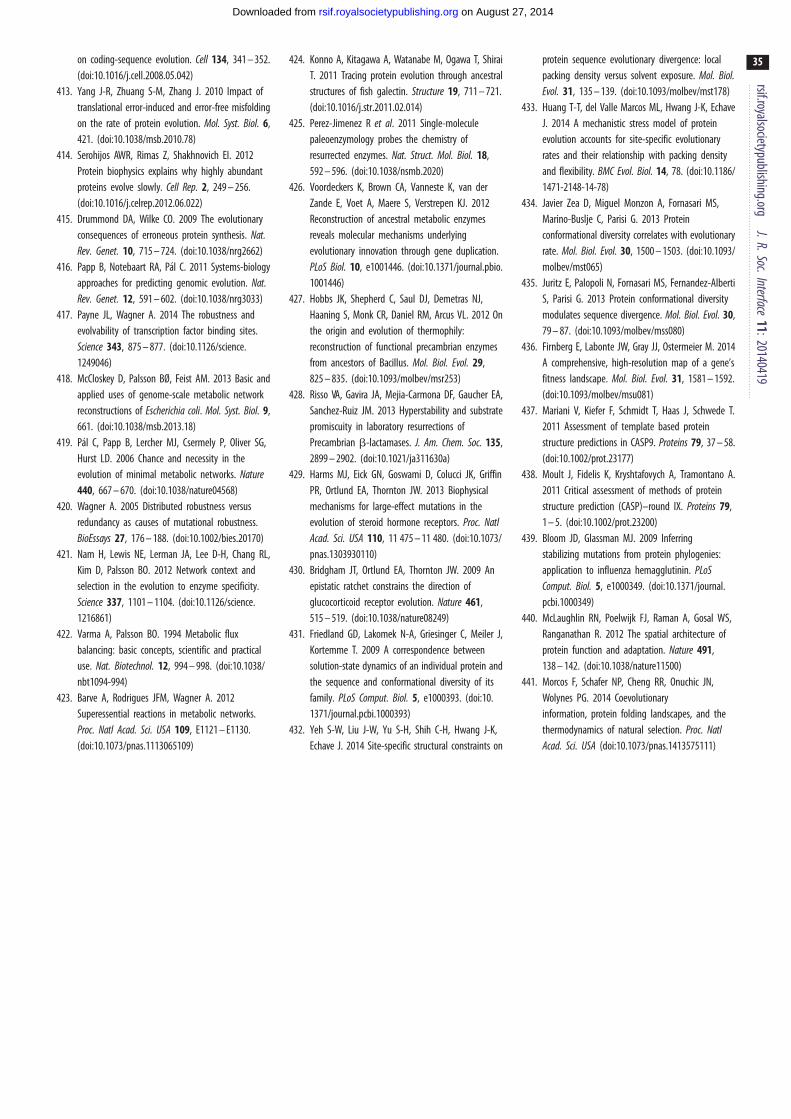
rsif.royalsocietypublishing.orgJ.R.Soc.Interface
11:20140419
35
on August 27, 2014rsif.royalsocietypublishing.orgDownloaded from
on coding-sequence evolution. Cell 134, 341 – 352.(doi:10.1016/j.cell.2008.05.042)
413. Yang J-R, Zhuang S-M, Zhang J. 2010 Impact oftranslational error-induced and error-free misfoldingon the rate of protein evolution. Mol. Syst. Biol. 6,421. (doi:10.1038/msb.2010.78)
414. Serohijos AWR, Rimas Z, Shakhnovich EI. 2012Protein biophysics explains why highly abundantproteins evolve slowly. Cell Rep. 2, 249 – 256.(doi:10.1016/j.celrep.2012.06.022)
415. Drummond DA, Wilke CO. 2009 The evolutionaryconsequences of erroneous protein synthesis. Nat.Rev. Genet. 10, 715 – 724. (doi:10.1038/nrg2662)
416. Papp B, Notebaart RA, Pal C. 2011 Systems-biologyapproaches for predicting genomic evolution. Nat.Rev. Genet. 12, 591 – 602. (doi:10.1038/nrg3033)
417. Payne JL, Wagner A. 2014 The robustness andevolvability of transcription factor binding sites.Science 343, 875 – 877. (doi:10.1126/science.1249046)
418. McCloskey D, Palsson BØ, Feist AM. 2013 Basic andapplied uses of genome-scale metabolic networkreconstructions of Escherichia coli. Mol. Syst. Biol. 9,661. (doi:10.1038/msb.2013.18)
419. Pal C, Papp B, Lercher MJ, Csermely P, Oliver SG,Hurst LD. 2006 Chance and necessity in theevolution of minimal metabolic networks. Nature440, 667 – 670. (doi:10.1038/nature04568)
420. Wagner A. 2005 Distributed robustness versusredundancy as causes of mutational robustness.BioEssays 27, 176 – 188. (doi:10.1002/bies.20170)
421. Nam H, Lewis NE, Lerman JA, Lee D-H, Chang RL,Kim D, Palsson BO. 2012 Network context andselection in the evolution to enzyme specificity.Science 337, 1101 – 1104. (doi:10.1126/science.1216861)
422. Varma A, Palsson BO. 1994 Metabolic fluxbalancing: basic concepts, scientific and practicaluse. Nat. Biotechnol. 12, 994 – 998. (doi:10.1038/nbt1094-994)
423. Barve A, Rodrigues JFM, Wagner A. 2012Superessential reactions in metabolic networks.Proc. Natl Acad. Sci. USA 109, E1121 – E1130.(doi:10.1073/pnas.1113065109)
424. Konno A, Kitagawa A, Watanabe M, Ogawa T, ShiraiT. 2011 Tracing protein evolution through ancestralstructures of fish galectin. Structure 19, 711 – 721.(doi:10.1016/j.str.2011.02.014)
425. Perez-Jimenez R et al. 2011 Single-moleculepaleoenzymology probes the chemistry ofresurrected enzymes. Nat. Struct. Mol. Biol. 18,592 – 596. (doi:10.1038/nsmb.2020)
426. Voordeckers K, Brown CA, Vanneste K, van derZande E, Voet A, Maere S, Verstrepen KJ. 2012Reconstruction of ancestral metabolic enzymesreveals molecular mechanisms underlyingevolutionary innovation through gene duplication.PLoS Biol. 10, e1001446. (doi:10.1371/journal.pbio.1001446)
427. Hobbs JK, Shepherd C, Saul DJ, Demetras NJ,Haaning S, Monk CR, Daniel RM, Arcus VL. 2012 Onthe origin and evolution of thermophily:reconstruction of functional precambrian enzymesfrom ancestors of Bacillus. Mol. Biol. Evol. 29,825 – 835. (doi:10.1093/molbev/msr253)
428. Risso VA, Gavira JA, Mejia-Carmona DF, Gaucher EA,Sanchez-Ruiz JM. 2013 Hyperstability and substratepromiscuity in laboratory resurrections ofPrecambrian b-lactamases. J. Am. Chem. Soc. 135,2899 – 2902. (doi:10.1021/ja311630a)
429. Harms MJ, Eick GN, Goswami D, Colucci JK, GriffinPR, Ortlund EA, Thornton JW. 2013 Biophysicalmechanisms for large-effect mutations in theevolution of steroid hormone receptors. Proc. NatlAcad. Sci. USA 110, 11 475 – 11 480. (doi:10.1073/pnas.1303930110)
430. Bridgham JT, Ortlund EA, Thornton JW. 2009 Anepistatic ratchet constrains the direction ofglucocorticoid receptor evolution. Nature 461,515 – 519. (doi:10.1038/nature08249)
431. Friedland GD, Lakomek N-A, Griesinger C, Meiler J,Kortemme T. 2009 A correspondence betweensolution-state dynamics of an individual protein andthe sequence and conformational diversity of itsfamily. PLoS Comput. Biol. 5, e1000393. (doi:10.1371/journal.pcbi.1000393)
432. Yeh S-W, Liu J-W, Yu S-H, Shih C-H, Hwang J-K,Echave J. 2014 Site-specific structural constraints on
protein sequence evolutionary divergence: localpacking density versus solvent exposure. Mol. Biol.Evol. 31, 135 – 139. (doi:10.1093/molbev/mst178)
433. Huang T-T, del Valle Marcos ML, Hwang J-K, EchaveJ. 2014 A mechanistic stress model of proteinevolution accounts for site-specific evolutionaryrates and their relationship with packing densityand flexibility. BMC Evol. Biol. 14, 78. (doi:10.1186/1471-2148-14-78)
434. Javier Zea D, Miguel Monzon A, Fornasari MS,Marino-Buslje C, Parisi G. 2013 Proteinconformational diversity correlates with evolutionaryrate. Mol. Biol. Evol. 30, 1500 – 1503. (doi:10.1093/molbev/mst065)
435. Juritz E, Palopoli N, Fornasari MS, Fernandez-AlbertiS, Parisi G. 2013 Protein conformational diversitymodulates sequence divergence. Mol. Biol. Evol. 30,79 – 87. (doi:10.1093/molbev/mss080)
436. Firnberg E, Labonte JW, Gray JJ, Ostermeier M. 2014A comprehensive, high-resolution map of a gene’sfitness landscape. Mol. Biol. Evol. 31, 1581 – 1592.(doi:10.1093/molbev/msu081)
437. Mariani V, Kiefer F, Schmidt T, Haas J, Schwede T.2011 Assessment of template based proteinstructure predictions in CASP9. Proteins 79, 37 – 58.(doi:10.1002/prot.23177)
438. Moult J, Fidelis K, Kryshtafovych A, Tramontano A.2011 Critical assessment of methods of proteinstructure prediction (CASP)–round IX. Proteins 79,1 – 5. (doi:10.1002/prot.23200)
439. Bloom JD, Glassman MJ. 2009 Inferringstabilizing mutations from protein phylogenies:application to influenza hemagglutinin. PLoSComput. Biol. 5, e1000349. (doi:10.1371/journal.pcbi.1000349)
440. McLaughlin RN, Poelwijk FJ, Raman A, Gosal WS,Ranganathan R. 2012 The spatial architecture ofprotein function and adaptation. Nature 491,138 – 142. (doi:10.1038/nature11500)
441. Morcos F, Schafer NP, Cheng RR, Onuchic JN,Wolynes PG. 2014 Coevolutionaryinformation, protein folding landscapes, and thethermodynamics of natural selection. Proc. NatlAcad. Sci. USA (doi:10.1073/pnas.1413575111)