A quantitative FRET approach to characterize protein-protein ...

Structural and mutational analyses of protein–proteininteractions between transthyretin and retinol-bindingproteinGiuseppe Zanotti1,2, Claudia Folli3, Laura Cendron1,2, Beatrice Alfieri3, Sonia K. Nishida4,Francesca Gliubich1,*, Nicola Pasquato1, Alessandro Negro5 and Rodolfo Berni3
1 Department of Chemical Sciences and Institute of Biomolecular Chemistry-CNR, University of Padua, Italy
2 Venetian Institute of Molecular Medicine, Padua, Italy
3 Department of Biochemistry and Molecular Biology, University of Parma, Italy
4 Department of Medicine, Federal University of Sao Paulo, Brazil
5 Department of Experimental Veterinary Sciences and CRIBI, University of Padua, Italy
Keywords
Fab; mutational analysis; protein–protein
interactions; retinol-binding protein;
transthyretin
Correspondence
G. Zanotti, Department of Chemical
Sciences, University of Padua, Via Marzolo
1, 35131 Padua, Italy
Fax: +39 049 8275239
Tel: +39 049 8275245
E-mail: [email protected]
R. Berni, Department of Biochemistry and
Molecular Biology, University of Parma, V.le
G.P. Usberti 23 ⁄ A, 43100 Parma, Italy
Fax: +39 0521 905151
Tel: +39 0521 905645
E-mail: [email protected]
*Present address
KCL Enterprises Ltd, London, UK
Database
Atomic coordinates and structure factors
have been deposited at the Protein Data
Bank (PDB) (http://www.rcsb.org) for imme-
diate release: PDB 3BSZ for the TTR–RBP–
Fab complex and PDB 3BT0 and 3CXF
for the V20S and Y114H TTR variants,
respectively
(Received 23 July 2008, revised
22 September 2008, accepted 25
September 2008)
doi:10.1111/j.1742-4658.2008.06705.x
Transthyretin is a tetrameric binding protein involved in the transport of
thyroid hormones and in the cotransport of retinol by forming a complex
in plasma with retinol-binding protein. In the present study, we report the
crystal structure of a macromolecular complex, in which human transthy-
retin, human holo-retinol-binding protein and a murine anti-retinol-binding
protein Fab are assembled according to a 1 : 2 : 2 stoichiometry. The main
interactions, both polar and apolar, between retinol-binding protein and
transthyretin involve the retinol hydroxyl group and a limited number of
solvent exposed residues. The relevance of transthyretin residues in com-
plex formation with retinol-binding protein has been examined by muta-
tional analysis, and the structural consequences of some transthyretin point
mutations affecting protein–protein recognition have been investigated.
Despite a few exceptions, in general, the substitution of a hydrophilic for a
hydrophobic side chain in contact regions results in a decrease or even a
loss of binding affinity, thus revealing the importance of interfacial hydro-
phobic interactions and a high degree of complementarity between retinol-
binding protein and transthyretin. The effect is particularly evident when
the mutation affects an interacting residue present in two distinct subunits
of transthyretin participating simultaneously in two interactions with a reti-
nol-binding protein molecule. This is the case of the amyloidogenic I84S
replacement, which abolishes the interaction with retinol-binding protein
and is associated with an altered retinol-binding protein plasma transport
in carriers of this mutation. Remarkably, some of the residues in mutated
human transthyretin that weaken or abolish the interaction with retinol-
binding protein are present in piscine transthyretin, consistent with the lack
of interaction between retinol-binding protein and transthyretin in fish.
Abbreviations
PDB, Protein Data Bank; RBP, retinol-binding protein; TTR, transthyretin.
FEBS Journal 275 (2008) 5841–5854 ª 2008 The Authors Journal compilation ª 2008 FEBS 5841

Transthyretin (TTR), a homotetramer of approxi-
mately 55 kDa, is a thyroid hormone-binding protein
present in the extracellular fluids of vertebrates,
where it participates, together with other binding
proteins, in the distribution of thyroid hormones
(thyroxine and triiodothyronine) [1]. It was generated
during early vertebrate evolution as a result of a
duplication event in the gene encoding 5-hydroxyiso-
urate hydrolase, an enzyme distributed in several
prokaryotes and in several eukaryotic lineages and
involved in purine metabolism [2–6]. The extracellu-
lar transport of retinol (vitamin A alcohol) is specifi-
cally mediated by retinol-binding protein (RBP, also
known as RBP 4), a monomeric protein of 21 kDa
that delivers the vitamin molecule to the target cells
[7,8], where a membrane RBP receptor represents a
major mediator of cellular vitamin A uptake [9,10].
TTR and RBP are synthesized primarily by the
hepatocytes and are secreted into the circulation,
where RBP is found bound to TTR. The association
of RBP with TTR increases the stability of the reti-
nol–RBP complex [11,12] and, according to various
lines of evidence [13–15], is believed to reduce the
glomerular filtration of the relatively small RBP mol-
ecule. In turn, the stability of the RBP–TTR com-
plex is strongly affected by the presence of retinol
bound to RBP within the complex, a feature that is
believed to be of physiological significance. The
affinity of holoRBP for TTR is significantly higher
than that of apoRBP [16,17], which is consistent
with holoRBP being retained in the circulation as
the protein–protein complex and with the uncom-
plexed apoRBP molecule, resulting from the delivery
of retinol, being selectively cleared from the circula-
tion by glomerular filtration. TTR has been associ-
ated with human diseases. It is one of several
proteins that can produce the extracellular accumula-
tion in tissues of protein aggregates, in the form of
fibrils, which are responsible for degenerative diseases
known as amyloidoses; to date, more than 100 point
mutations have been described for TTR, most of
which are involved in familial amyloidoses [18,19].
Moreover, a protective role of TTR in Alzheimer’s
disease has recently been proposed [20,21]. RBP is
an adipocyte-derived ‘signal’ that may contribute to
the pathogenesis of insulin resistance [22].
Well-refined crystal structures of TTR from differ-
ent vertebrate species, including mammals [23–25],
chicken [26] and fish (sea bream) [27,28], and of
RBP from mammals [29–33] and chicken [34], have
been described. The crystal structures of heterologous
(human TTR–chicken RBP) [35] and homologous
(human TTR–human RBP) [36] TTR–holoRBP com-
plexes have also been determined, both characterized
by a 1 : 2 TTR : RBP stoichiometry in which each
TTR-bound RBP molecule interacts simultaneously
with three TTR subunits [35,36]. TTR is a tetrameric
protein formed by the assembly of four identical sub-
units. Each monomer is composed of eight anti-par-
allel b-strands (A–H), arranged in a topology similar
to the Greek key b-barrel, with a short a-helixlocated at the end of b-strand E. In the tetramer,
the four monomers are organized as a dimer of
dimers. Specifically, two monomers are held together
to form a stable dimer through a net of H-bond
interactions involving the two edge b-strands H and
F. To form the tetramer, two dimers associate back
to back, mainly through hydrophobic contacts
between residues of the loops formed by b-strands A
and B and b-strands G and H. One of the two-fold
symmetry axes of the tetramer is coincident with a
long channel that transverses the entire molecule and
harbors two binding sites for thyroid hormones.
RBP is a single domain protein, made up of an
N-terminal coil, eight anti-parallel b-strands (A–H)
and a short a-helix close to the C-terminus. The
core of the protein is the internal cavity of an eight-
stranded up-and-down b-barrel. The vitamin mole-
cule is accommodated within the cavity of the barrel:
the b-ionone ring is innermost, the polyene chain is
fully extended and the hydroxyl end group is almost
solvent exposed, in the region of the loops that con-
nect b-strands A and B, C and D and E and F and
surround the entrance of the b-barrel at the open
end of the cavity. As a result of evolutionary
restraints imposed by the multiple interactions estab-
lished by both RBP and TTR, a high degree of
structural similarity appears to be preserved for these
two proteins from phylogenetically distant vertebrate
species. It should be noted, however, that piscine
TTR and RBP lack the ability to form a protein–
protein complex [27]. The molecular basis of the
evolution of the two piscine proteins into proteins
that possess the ability to interact with each other in
terrestrial vertebrates remains to be clarified.
In the present study, we report on the structure of
a complex formed by the association of human TTR,
human holoRBP and a murine anti-RBP Fab, and
on a mutational analysis of the RBP-binding determi-
nants present in the human TTR molecule. The data
provide insight into the molecular basis of the altered
plasma transport of RBP in carriers of a relevant
amyloidogenic TTR mutation (I84S) and of the
changes in the TTR molecule that have affected its
ability to interact with RBP during the course of
vertebrate evolution.
Transthyretin–retinol-binding protein interactions G. Zanotti et al.
5842 FEBS Journal 275 (2008) 5841–5854 ª 2008 The Authors Journal compilation ª 2008 FEBS

Results
Structure of a complex between human TTR,
human holoRBP and an anti-RBP Fab
The crystal structure of the human TTR–RBP com-
plex bound to an anti-RBP Fab could be determined
at a resolution of 3.36 A, similar to that obtained for
the crystal structures of TTR–RBP complexes [35,36].
The molecular model, obtained by molecular replace-
ment starting from the available high resolution crys-
tal structures of RBP and TTR, is of reasonable
quality. The anti-RBP Fab is bound to an RBP epi-
tope that is well separated from the TTR-binding
determinants present in the RBP molecule. It inter-
acts with RBP on the side opposite to that involved
in the binding to TTR (Fig. 1A), so that the interac-
tions between RBP and TTR are not affected by the
RBP-bound Fab. The entire complex, composed of
one TTR tetramer and two holoRBP molecules in
complex with Fab, is present in the asymmetric unit
(Fig. 1A). The two RBP–Fab sub-complexes are
arranged symmetrically around the two-fold axis of
TTR running through the central channel that trans-
verses the TTR molecule. RBP and TTR substan-
tially maintain the structure they have in the
uncomplexed state, with only minor changes in con-
tact regions.
The region of entrance of retinol into the b-barrelcavity of RBP (i.e. loops 32–36, 63–67 and 92–98),
and retinol itself, participate in the recognition of
TTR, involving residues that are essentially located in
loops connecting b-strands of the tetrameric protein,
in such a way that one RBP molecule interacts simul-
taneously with three TTR subunits (Table 1 and
Fig. 1A,B). The RBP–TTR interactions are both
polar or apolar (Table 1). The contact surface of
TTR is characterized by a prevalence of hydrophobic
residues in the case of the subunits B and C and of
hydrophilic residues in the case of the subunit A.
Val20, Trp79, Leu82, Ile84, Pro113 and Tyr114 from
both subunits B and C of TTR form a hydrophobic
patch, which is in contact in the protein–protein com-
plex with the hydrophobic patch formed by the resi-
dues Trp67, Leu63, Leu64, Val69, Phe96 and Leu97
of RBP. Instead, the interacting hydrophilic residues
from the subunit A of TTR are closer to the border
of the contact surface of TTR. Four H-bonds, mainly
between RBP and the subunit B of TTR, are also
present (Table 1). The area buried on the two pro-
teins upon complex formation is 1443 A2, out of a
total area of 9307 and 21502 A2 for the two sepa-
rated RBP and TTR molecules, respectively.
The Fab–RBP interaction involves the region pre-
ceding the a-helix and the C-terminal b-strand of RBP
and the hyper-variable regions of Fab: loops 53–56
and 100–103 and the short helix 28–32 of chain H and
loops 31–36 and 53–56 of chain L. The interactions,
which are mainly polar and comprise several H-bonds,
are summarized in Table 2. The number of residues
involved in the formation of the Fab–RBP complex is
smaller than that of interacting residues in the RBP–
TTR complex (Table 2) but the surface buried upon
complex formation is comparable to that for the RBP–
TTR complex (1588 A2). The contacts between inter-
acting surfaces of RBP and Fab are shown in Fig. 1C.
Mutational analysis of the RBP-binding
determinants of TTR
The amino acid sequences of RBP and TTR from dif-
ferent vertebrate species showing the residues of
human RBP and TTR that are mainly involved in
interactions, according to our structure of the TTR–
RBP–Fab complex, are shown in Fig. 2. The func-
tional and structural consequences of several TTR
point mutations on the RBP–TTR interactions have
been investigated. The values of the dissociation con-
stants for several complexes between human TTR vari-
ants and human RBP, as determined by means of
fluorescence anisotropy titrations (Fig. 3A), are
reported in Table 3. For some TTR mutations that
abolish or weaken significantly the RBP–TTR interac-
tions, the crystal structures of the variants have been
determined or already available structures have been
examined to provide details of the interference with
protein–protein recognition by mutations.
V20S TTR variant
Two Val20 residues present in two distinct TTR sub-
units (B and C, according to our designation of TTR
subunits) are located at the center of a large hydropho-
bic patch in the contact area between TTR and RBP,
so that their replacement by a hydrophilic residue is
expected to significantly impair protein–protein inter-
actions. Accordingly, the V20S mutation has been
found to almost abolish the binding affinity between
RBP and TTR (Table 3 and Fig. 3A). To investigate
the structural consequences of the V20S replacement
on the RBP–TTR recognition, we have determined the
1.6 A resolution crystal structure of the V20S TTR
variant. The overall structure is very similar to that of
the wild-type protein (PDB: 1F41) [25]: a superposition
of equivalent Ca atoms gives an rmsd of 0.40 and
0.68 A for subunits A and B, respectively. However,
G. Zanotti et al. Transthyretin–retinol-binding protein interactions
FEBS Journal 275 (2008) 5841–5854 ª 2008 The Authors Journal compilation ª 2008 FEBS 5843

A
B
C
Fig. 1. Structure of the TTR–RBP–Fab complex. (A) Representation of the overall structure of the TTR–RBP–Fab complex. TTR: chain A,
red; chain B, green; chain C, magenta; chain D, orange. RBP: chains E and F, yellow. Fab: heavy chains H and N, cyan; light chains L
and M, blue. (B) Detail of the contact between the TTR subunits A, B and C and the RBP molecule E [left side, color codes as in (A)].
Center and right drawings show the interacting surfaces of RBP (center) and of the TTR subunits A, B and C (right) rotated by approxi-
mately 90� counterclockwise and clockwise, respectively, compared to the previous view. It is possible to appreciate how a protuberance
formed by loops 63–67 and 92–98 on the RBP surface fits into a crevice formed by the arrangement of three TTR subunits. (C) Detail of
the contact between the RBP molecule F and the Fab chains H and L [left side, color codes as in (A)]. Center and right drawings show
the interacting surfaces of RBP (center) and Fab (right), rotated through a vertical axis by approximately 90� counterclockwise and clock-
wise, respectively, compared to the previous view. Residues 163–169 and the region preceding the a-helix contribute to the formation of
the RBP epitope.
Transthyretin–retinol-binding protein interactions G. Zanotti et al.
5844 FEBS Journal 275 (2008) 5841–5854 ª 2008 The Authors Journal compilation ª 2008 FEBS
significant differences are observed for loop region
98–103, which connects b-strands G to F and is flexi-
ble in most TTR structures, and for loop 19–22, which
connects b-strands A and B and hosts the mutation.
The movement of the latter loop is not large (the Caatom of Val20 is displaced by 1.6 A from its position
in the wild-type structure) but the entire loop 19–22 is
displaced by more than 1.5 A from its original position
(Fig. 3B), so that the change in the positions of Val20
and Arg21 of two distinct TTR subunits (Table 1) may
interfere simultaneously with two interactions estab-
lished with RBP.
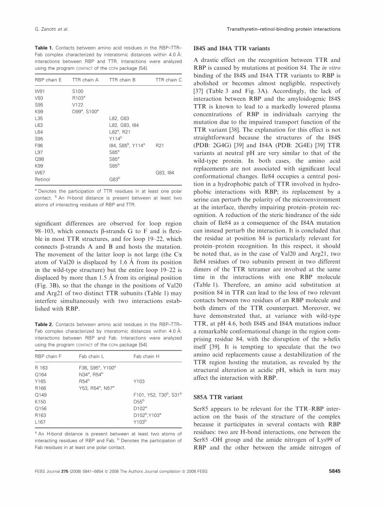
I84S and I84A TTR variants
A drastic effect on the recognition between TTR and
RBP is caused by mutations at position 84. The in vitro
binding of the I84S and I84A TTR variants to RBP is
abolished or becomes almost negligible, respectively
[37] (Table 3 and Fig. 3A). Accordingly, the lack of
interaction between RBP and the amyloidogenic I84S
TTR is known to lead to a markedly lowered plasma
concentrations of RBP in individuals carrying the
mutation due to the impaired transport function of the
TTR variant [38]. The explanation for this effect is not
straightforward because the structures of the I84S
(PDB: 2G4G) [39] and I84A (PDB: 2G4E) [39] TTR
variants at neutral pH are very similar to that of the
wild-type protein. In both cases, the amino acid
replacements are not associated with significant local
conformational changes. Ile84 occupies a central posi-
tion in a hydrophobic patch of TTR involved in hydro-
phobic interactions with RBP; its replacement by a
serine can perturb the polarity of the microenvironment
at the interface, thereby impairing protein–protein rec-
ognition. A reduction of the steric hindrance of the side
chain of Ile84 as a consequence of the I84A mutation
can instead perturb the interaction. It is concluded that
the residue at position 84 is particularly relevant for
protein–protein recognition. In this respect, it should
be noted that, as in the case of Val20 and Arg21, two
lle84 residues of two subunits present in two different
dimers of the TTR tetramer are involved at the same
time in the interactions with one RBP molecule
(Table 1). Therefore, an amino acid substitution at
position 84 in TTR can lead to the loss of two relevant
contacts between two residues of an RBP molecule and
both dimers of the TTR counterpart. Moreover, we
have demonstrated that, at variance with wild-type
TTR, at pH 4.6, both I84S and I84A mutations induce
a remarkable conformational change in the region com-
prising residue 84, with the disruption of the a-helixitself [39]. It is tempting to speculate that the two
amino acid replacements cause a destabilization of the
TTR region hosting the mutation, as revealed by the
structural alteration at acidic pH, which in turn may
affect the interaction with RBP.
S85A TTR variant
Ser85 appears to be relevant for the TTR–RBP inter-
action on the basis of the structure of the complex
because it participates in several contacts with RBP
residues: two are H-bond interactions, one between the
Ser85 -OH group and the amide nitrogen of Lys99 of
RBP and the other between the amide nitrogen of
Table 1. Contacts between amino acid residues in the RBP–TTR–
Fab complex characterized by interatomic distances within 4.0 A:
interactions between RBP and TTR. Interactions were analyzed
using the program CONTACT of the CCP4 package [54].
RBP chain E TTR chain A TTR chain B TTR chain C
W91 S100
V93 R103a
S95 V122
K99 D99a, S100a
L35 L82, G83
L63 L82, G83, I84
L64 L82a, R21
S95 Y114b
F96 I84, S85b, Y114a R21
L97 S85a
Q98 S85a
K99 S85b
W67 G83, I84
Retinol G83b
a Denotes the participation of TTR residues in at least one polar
contact. b An H-bond distance is present between at least two
atoms of interacting residues of RBP and TTR.
Table 2. Contacts between amino acid residues in the RBP–TTR–
Fab complex characterized by interatomic distances within 4.0 A:
interactions between RBP and Fab. Interactions were analyzed
using the program CONTACT of the CCP4 package [54].
RBP chain F Fab chain L Fab chain H
R 163 F36, S95a, Y100a
Q164 N34a, R54b
Y165 R54b Y103
R166 Y53, R54a, N57a
Q149 F101, Y52, T30b, S31b
K150 D55b
Q156 D102a
R163 D102b,Y103a
L167 Y103b
a An H-bond distance is present between at least two atoms of
interacting residues of RBP and Fab. b Denotes the participation of
Fab residues in at least one polar contact.
G. Zanotti et al. Transthyretin–retinol-binding protein interactions
FEBS Journal 275 (2008) 5841–5854 ª 2008 The Authors Journal compilation ª 2008 FEBS 5845

Ser85 and the carbonyl oxygen of Phe96 of RBP
(Table 1). Although the latter can be preserved in the
S85A variant, the former is lost, possibly contributing
to an approximately five-fold decrease in binding affin-
ity caused by the mutation (Table 3). It can be specu-
lated that the loss of interactions caused by the
A
B
Fig. 2. Multiple sequence alignments for RBP (A) and TTR (B) from different vertebrate species. The residues that are identical in all the
sequences considered for each alignment are shaded in red; the residues that are identical or chemically similar in at least five sequences
for each alignment are denoted by red characters (similarity groups are: HKR, DE, STNQ, AVLIM, FYW, PG, C). The amino acid residues in
the human TTR and RBP sequences more directly involved in RBP–TTR interactions (Table 1) are denoted by arrowheads. Numbering and
secondary structure elements are based on the structures of human RBP (PDB: 1JYD) and TTR (PDB: 1F41). GenBank or SwissProt acces-
sion numbers are: human TTR, PO2766; rat TTR, NP_036813; chicken TTR, CAA43000; zebrafish TTR, AAH81488; sea bream TTR,
AF059193; trout TTR, CB497711 (EST sequence); human RBP, P02753; rat RBP, NM_013162; chicken RBP, P41263; zebrafish RBP,
EF373650; sea bream RBP, AAF79021; trout RBP, P24774. Sequences containing the signal peptide have been reported when the N-termini
of the mature proteins are not known. Sequence alignments were constructed by CLUSTALW [58] and rendered with ESPRIPT [59].
Transthyretin–retinol-binding protein interactions G. Zanotti et al.
5846 FEBS Journal 275 (2008) 5841–5854 ª 2008 The Authors Journal compilation ª 2008 FEBS

mutation may be partially compensated by a novel
hydrophobic interaction in a hydrophobic patch
involving Ala85.
D99A and S100E TTR variants
These are important mutations because each of them
causes an approximately 20-fold decrease in binding
affinity of TTR for RBP (Table 3 and Fig. 3A). Both
replaced residues interact with Lys99 of RBP and,
moreover, Ser100 is close to Trp91 (Table 1). Conse-
quently, it is conceivable that the replacements of
Asp99 by a hydrophobic residue and of Ser100 by a
charged residue significantly perturb the interactions.
However, it is likely that the effects of the D99A and
S100E mutations are less drastic compared to the I84S
and V20S replacements because the interactions involv-
ing residues at positions 99 and 100 are present at the
periphery of a contact area between RBP and TTR.
Moreover, the new electrostatic situation for the TTR
variants could result in new interactions of the
mutated residues with the solvent.
Y114F and Y114H TTR variants
A special case is represented by Tyr114: its substitution
for a phenylalanine lowers the dissociation constant of
the protein–protein complex by an approximate factor
of two, whereas the dissociation constant is three-fold
higher when Tyr114 is replaced by a histidine (Table 3
and Fig. 3A). We have also determined the crystal
structure of the amyloidogenic Y114H variant: no sig-
nificant conformational changes have been observed,
and, in particular, the side chain of His114 maintains
the same position and orientation compared to
Tyr114. The same holds for the position and orienta-
tion of the Phe114 residue present in the structure of
both chicken TTR [26], which interacts well with RBP
[17,40], and piscine TTR [27]. The -OH group of
Tyr114 forms an H-bond with the -OH group of Ser95
of RBP. Moreover, Tyr114 is in a hydrophobic patch
of TTR, despite its proximity to the protein surface.
Its replacement by a phenylalanine is not drastic in
terms of modification of the surface potential but it
leads to the loss of an H-bond interaction. It must be
assumed that the loss of a H-bond interaction for the
Y114F variant is compensated by some conforma-
tional rearrangements that result in stronger hydro-
A
B
Fig. 3. Human TTR mutations affecting TTR–RBP interactions.
(A) Typical fluorescence anisotropy titrations of human holoRBP
(3 lM) with human TTR: wild-type, black; V20S TTR, gray; I84S
TTR, red; D99A TTR, green; Y114H TTR, blue; Y114F TTR, orange.
Fluorescence anisotropy values are plotted as a function of human
TTR molar concentration. Lines represent theoretical binding curves
(for details, see Experimental procedures) corresponding to dissoci-
ation constants of 0.34 lM for wild-type TTR, 5.99 lM for D99A
TTR, 1.04 lM for Y114H TTR and 0.17 lM for Y114F TTR. (B) Ste-
reo view showing the superposition of the Ca chain traces of wild-
type TTR (PDB: 1F41, red) and the V20S TTR variant (green) in the
area around residue 20. The side chains of residues Ser20 or Val20
and Arg21 are shown.
Table 3. Dissociation constants of complexes between human holoRBP and human TTR variants as determined by means of fluorescence
anisotropy titrations. Data represent the average of at least three independent measurements.
TTR Wild-type V20Sa V30Ma,b L55Pa,b L58Hb T60Aa,b I84Sa,b,c I84Aa S85A D99Ac S100Ec Y114Ha,b Y114Fc
KD (lM) 0.35 –d 0.22 0.66 0.31 0.43 –e –d 1.64 6.23 5.81 1.12 0.18
a The structure for the TTR variant is available: V20S and Y114H [present study]; V30M [55]; L55P, [56]; T60A, [57]; I84S and I84A, [39].b Amyloidogenic TTR variant. c Position affected by significant amino acid replacements in piscine TTR compared to human TTR (Fig. 2B).d Almost negligible interaction. e Lack of interaction.
G. Zanotti et al. Transthyretin–retinol-binding protein interactions
FEBS Journal 275 (2008) 5841–5854 ª 2008 The Authors Journal compilation ª 2008 FEBS 5847

phobic interactions, possibly explaining the higher
affinity between RBP and this TTR variant. The
replacement of Tyr114 by a potentially charged histi-
dine could have a marked effect on the surface poten-
tial; the finding that the Y114H mutation does not
drastically impair the RBP–TTR interaction suggests
that His114 is not protonated in the protein–protein
complex.
Modeling of the interactions of piscine TTR and
RBP with protein counterparts within the
TTR–RBP–Fab complex
The observation that human TTR and RBP are bound
in the TTR–RBP–Fab complex without undergoing sig-
nificant conformational changes compared to the un-
complexed proteins prompted us to assess the ability of
the two proteins from fish, which are unable to interact
with each other [27], to fit within the structure of the
TTR–RBP–Fab complex by replacing the correspond-
ing human proteins present in the complex. The struc-
ture of sea bream (Sparus aurata) TTR is known [27],
whereas no structure for a fish RBP is available to date.
Therefore, only a theoretical model for the structure of
sea bream RBP could be obtained with the Swiss-
Model server [41]. The structure of sea bream TTR was
superimposed on that of human TTR present in the
TTR–RBP–Fab complex, giving rise to a hypothetical
model of the mixed piscine TTR–human RBP complex
(Fig. 4). The most relevant difference can be observed
for loop 98–102 of all the subunits of TTR, especially
for subunits A and C, due to some remarkable muta-
tions affecting interacting residues (Table 3 and
Fig. 2B). The conformation of loop 80–85 of piscine
TTR also changes slightly (Fig. 4), possibly due to
relevant mutations in this area (Table 3 and Fig. 2B).
The same holds for a model in which the human RBP
structure is also replaced by a theoretical sea bream
RBP structure, giving rise to a hypothetical piscine
RBP–TTR complex (Fig. 4). Conformational differ-
ences for TTR along with point mutations that have no
effect on TTR structure (Fig. 2B) may account for the
experimentally determined lack of binding affinity
between piscine TTR and RBP, as well as between
piscine TTR and human RBP [27]. Only limited confor-
mational differences between piscine and human RBP
are found. Accordingly, the degree of conservation of
the putative interacting residues is remarkably higher in
piscine RBP than in piscine TTR; the only significant
amino acid difference in piscine RBP compared to
human RBP is K99T (Fig. 2A). These features may
explain the existence of an affinity, albeit weak,
between piscine RBP and human TTR [27,42].
Discussion
The human holoRBP–TTR complex is relatively weak,
being characterized by a dissociation constant of
approximately 0.35 lm (Table 3). It has been suggested
that this feature may be correlated with the need for
the presence in plasma of a small but significant
amount of uncomplexed holoRBP, which can thus
leave the circulation more easily to deliver the retinol
to the target tissues [7]. A limited number of residues
and retinol itself are mainly responsible for the rela-
tively weak RBP–TTR interaction. An important role
played by the retinol hydroxyl end group in the
interaction is consistent with the low binding affinity
of apoRBP compared to that of holoRBP for TTR
[16,17] and with the drastic interference with the
interaction between the two proteins by RBP-bound
fenretinide, a retinoid that bears a bulky end group in
place of the retinol hydroxyl group [43,44]. Moreover,
the conformational change affecting one of the loops
Fig. 4. Modeling of piscine TTR and RBP
within the TTR–RBP–Fab complex. Stereo
view showing interacting regions between
RBP (magenta) and TTR (orange) in the
human RBP–TTR complex bound to Fab,
with a superimposed model of the piscine
RBP–TTR complex (green) based on the
structure of sea bream TTR (PDB: 1OO2)
and on a hypothetical model of the piscine
RBP structure. The two regions of TTR that
differ significantly in the structures (98–102
and 80–85) are labeled.
Transthyretin–retinol-binding protein interactions G. Zanotti et al.
5848 FEBS Journal 275 (2008) 5841–5854 ª 2008 The Authors Journal compilation ª 2008 FEBS
surrounding the opening of the b-barrel (in particular,
residues Leu35 and Phe36) in apo-RBP compared to
holoRBP [30,31] is likely to contribute to the weaken-
ing of the interaction of apoRBP with TTR due to the
involvement of such a loop in RBP–TTR recognition.
Despite a few exceptions, the substitutions of hydro-
philic for hydrophobic side chains in TTR contact
regions generally have a rather pronounced dissociat-
ing effect on the RBP–TTR complex, consistent with
the important role played by interfacial apolar inter-
actions. In general, the changes in the TTR molecule
induced by amyloidogenic mutations do not interfere
with the interactions between RBP and TTR, unless
the mutations are located in contact areas. The amyloi-
dogenic mutations V30M, L55P, L58H and T60A have
a limited effect on the binding affinity between RBP
and TTR (Table 3), in accordance with the lack of the
replaced residues in contact areas; moreover, it can be
inferred that such amyloidogenic mutations do not
cause large conformational changes in the TTR mole-
cule that might affect indirectly the RBP–TTR recogni-
tion. Conversely, the amyloidogenic I84S mutation,
which affects a residue that is crucial for protein–pro-
tein interactions, causes the lack of recognition
between RBP and TTR and an altered plasma trans-
port of RBP by TTR [37,38]. It might be hypothesized
that the ability of RBP to interact well with relevant
amyloidogenic TTR variants, such as V30M, L55P,
L58H and T60A, can protect them from amyloid
aggregation. However, it should be noted that the
plasma concentration of TTR is significantly higher
than that of RBP [7], so that a protective effect of
RBP on TTR can only be limited.
Despite the high symmetry of TTR, which is a homo-
tetramer with virtually four identical binding sites for
RBP, a 1 : 1 TTR : RBP complex is believed to be
present in plasma due to the excess of TTR over RBP
[7]. Binding data obtained in solution [17,37,45] and
structural data [35,36] have shown that a maximum of
two RBP molecules can be bound by one TTR tetra-
mer. The binding of two RBP molecules to an uncom-
plexed TTR tetramer partially hinders the potential
binding of two nearby RBP molecules, thereby limiting
the possible interactions with tetrameric TTR to two
RBP molecules [35,36]. However, two distinct macro-
molecular organizations, both accounting for the 1 : 2
TTR : RBP stoichiometry, have been described for
the heterologous and the homologous RBP–TTR
complexes [35,36]. The crystal structure of the TTR
tetramer is characterized by 222 symmetry. One of the
three orthogonal two-fold axes runs through the
central channel harboring the two thyroid hormone
binding sites. In the heterologous chicken RBP–human
TTR complex, the two TTR-bound RBP molecules are
related by one of the two available two-fold axes that
are perpendicular to the central channel of TTR [35].
Instead, in the homologous human RBP–human TTR
complex, as well as in the case of our structure of the
TTR–RBP–Fab complex, the two TTR-bound RBP
molecules are related by the two-fold axis running
through the central channel of TTR [36] (Fig. 1A).
Because the two situations are chemically equivalent, a
possible explanation for the observed different assem-
bly is that, in solution, both modes of assembly can be
present and that the crystallization process selects one
of them according to the best packing.
By comparing the structure of the human TTR–
human RBP complex bound to Fab with those of the
heterologous human TTR–chicken RBP complex [35]
and of the homologous human TTR–human RBP
complex [36], a good correspondence between these
structures with regard to interacting surfaces of TTR
and RBP has been found. In the case of the human
TTR–human RBP complex, however, it should be
noted that one of the two TTR-bound RBP molecules
has been reported to participate in the interaction with
the last C-terminal amino acid residues (especially
Leu182 and Leu183), thereby generating an asymmetry
within the complex [36]. At variance with this observa-
tion, our TTR–RBP–Fab structure does not reveal the
presence of interactions between the carboxy terminus
of RBP and TTR. On the other hand, it should be
noted that chicken RBP, in which eight C-terminal res-
idues are missing compared to human RBP (Fig. 2A),
binds to human TTR with an affinity similar to that
exhibited by human RBP [40; C. Folli and R. Berni,
unpublished data], which suggests that the carboxy
terminus of human RBP is not so crucial for the inter-
action with TTR.
The RBP–TTR complex is normally isolated from
the serum of terrestrial vertebrates, such as mammals
and birds [7]. Moreover, purified human and chicken
RBP and TTR have been found to cross-interact [40].
By contrast, RBP could be isolated from the serum of
different fish species only as uncomplexed protein
[27,42,46], suggesting that, in fish, it is present in the
circulation as an uncomplexed protein without affinity
for TTR. In accordance with this observation, the lack
of binding affinity between purified piscine RBP and
TTR has been established [27]. The comparison of the
amino acid sequences of piscine RBPs and TTRs with
those of the same proteins from terrestrial vertebrates
reveals the presence of remarkable differences in
regions involved in protein–protein interactions for
TTR, whereas only limited differences are present in
the case of RBP (Fig. 2A,B). In particular, the amino
G. Zanotti et al. Transthyretin–retinol-binding protein interactions
FEBS Journal 275 (2008) 5841–5854 ª 2008 The Authors Journal compilation ª 2008 FEBS 5849

acid replacements at positions 82, 84, 99 and 100 in
piscine TTR compared to the human and chicken pro-
teins are drastic and are present at positions critical
for the interaction between RBP and TTR (Table 1
and Fig. 2B), and some of them (I84S, D99A and
S100E) are shown in the present study to impair or
abolish protein–protein recognition (Table 3 and
Fig. 3A). The results obtained are consistent with the
notion that evolutionary changes affecting a limited
number of surface-exposed residues led to the appear-
ance in terrestrial vertebrates of the TTR function of
cotransport of retinol through the interaction with
holoRBP in plasma, in addition to that of the distribu-
tion of thyroid hormones in the extracellular fluids.
Experimental procedures
Materials
HoloRBP was purified from human plasma as reported
previously [17]. Recombinant wild-type human TTR and
TTR variants I84S and I84A were prepared and quantified
as described previously [39]. All chemicals were of analytical
grade.
Site-directed mutagenesis, bacterial expression
and purification of human TTR variants
The recombinant human TTR variants V20S, L55P, L58H,
T60A, S85A, D99A, S100E, Y114F and Y114H were pre-
pared by PCR using the plasmid pET11b-human TTR [39]
as template, a high-fidelity thermostable DNA polymerase
(Pfu Ultra II Fusion HS DNA polymerase; Stratagene, La
Jolla, CA, USA) and mutagenic primers complementary to
opposite strands. For each mutation, the product of reac-
tion was treated with DpnI (New England Biolabs, Beverly,
MA, USA) to digest the parental DNA template. This pro-
cedure allowed us to select the newly synthesized and
potentially mutated plasmids. The products of each diges-
tion were used to transform Escherichia coli XL1 Blue cells.
Single clones were then sequenced to confirm the occur-
rence of the desired mutation. Finally, mutant plasmids
were electroporated into E. coli BL21 (DE3) cells. The
expression of TTR variants was induced by 1 mm isopropyl
thio-b-d-galactoside and, after incubation for 4 h at 30 �C,cells were disrupted by sonication. TTR variants were puri-
fied as described for wild-type TTR [39].
Determination of the amino acid sequences of
anti-RBP Fab variable domains
Total RNA obtained from the cell line producing the anti-
RBP murine monoclonal antibody A8P3 [47] was subjected
to retrotranscription into cDNA employing the Superscript
Preamplification System (Gibco, Gaithersburg, MD, USA).
The cDNA sequences encoding for the variable domains of
the H and L chains of the monoclonal antibody A8P3 were
PCR amplified using a mixture of 18 5¢ primer VKBACK
mix and a mixture of five 3¢ primer VKFOR for the VK
gene and 20 5¢ primer VHBACK mix and a mixture of five
3¢ primer VHFOR mix for the VH gene [48]. Each domain
was cloned using the TA cloning kit (Invitrogen, Carlsbad,
CA, USA) and sequenced using an automated model 377
sequenator (Applied Biosystems, Foster City, CA, USA).
The amino acid sequences of the variable domains of L and
H chains of the antibody A8P3 are provided in Fig. S1.
Binding assay for the interaction between
holoRBP and TTR variants
To study the in vitro interaction of holoRBP with TTR
variants, the highly fluorescent RBP-bound retinol provides
an intense signal which is suitable for fluorescence polariza-
tion measurements [40]. The intensities of the vertical (I ||)
and horizontal (I^) components of the fluorescence of
RBP-bound retinol (excitation at 330 nm and emission at
460 nm) were recorded at an angle of 90� to the vertically
polarized excitation beam. A correction factor, G, equal to
I ¢^ ⁄ I ¢|| (where the primes indicate excitation polarized in a
perpendicular direction) was used to correct for the unequal
transmission of differently polarized light. Fluorescence
anisotropy (A) was determined according to the equation:
A = (I || ) GI^) ⁄ (I ||+ 2GI^). Human holoRBP (0.7–
3.0 lm) in 0.05 m sodium phosphate (pH 7.2) and 0.15 m
NaCl, at 20 �C, was titrated by adding aliquots of concen-
trated solutions of human TTR (wild-type or mutant
forms) to the RBP-containing cuvette and the increase in
fluorescence anisotropy of the RBP-bound retinol upon
complex formation was monitored. The fraction of RBP
bound by TTR (a) was calculated for every point of the
titration curves using the equation: a = (A ) Ao) ⁄(Amax ) Ao), where A represents the fluorescence anisotropy
value of RBP-bound retinol for a certain molar concen-
tration of TTR, and Amax and Ao are the two limiting
anisotropy values (i.e. in the presence of an excess saturat-
ing TTR and in the absence of TTR, respectively). Binding
data were analyzed as described [12]. Fluorescence anisot-
ropy measurements were carried out with a LS-50B spectro-
fluorometer (Perkin-Elmer, Waltham, MA, USA).
Crystallization, data collection, structure
determination and refinement for the
TTR–RBP–Fab complex and the V20S and
Y114 TTR variants
Crystallization and preliminary X-ray data for the macro-
molecular complex formed by human transthyretin, human
holoRBP and a murine anti-RBP Fab have been reported
Transthyretin–retinol-binding protein interactions G. Zanotti et al.
5850 FEBS Journal 275 (2008) 5841–5854 ª 2008 The Authors Journal compilation ª 2008 FEBS
previously [49]. The structure has been solved by molecular
replacement, using the models of the single components of
the complex as templates (for RBP, PDB: 1RBP; for TTR,
PDB: 1F41; for Fab, a model was obtained with the server
EXPASY) [41]. The crystallographic refinement was carried
on using the software cns [50], imposing noncrystallo-
graphic symmetry restraints throughout all cycles. Three
hundred and thirty-four water molecules were added in the
last cycles of the refinement. The addition of these water
molecules caused 0.02 and 0.01 unit reductions, respec-
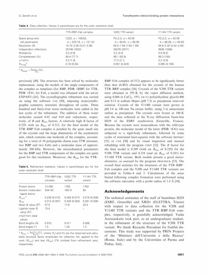
tively, of R and Rfree factors. A relatively high R factor of
0.239, with an Rfree of 0.312, for the final model of the
TTR–RBP–Fab complex is justified by the quite small size
of the crystals and the large dimensions of the asymmetric
unit, which contains one molecule of the complex, account-
ing for a total of 10 polypeptide chains (one TTR tetramer,
two RBP and two Fab) and a molecular mass of approxi-
mately 200 kDa. However, the stereochemical parameters
for the RBP and TTR components of the complex are quite
good for this resolution. Moreover, the Rfree for the TTR–
RBP–Fab complex (0.312) appears to be significantly better
than that (0.403) obtained for the crystals of the human
TTR–BRP complex [36]. Crystals of the V20S TTR variant
were obtained at 295 K by the vapor diffusion method,
using 0.066 m CaCl2, 19% (w ⁄ v) poly(ethyelene glycol)-400
and 0.13 m sodium Hepes (pH 7.5) as precipitant reservoir
solution. Crystals of the Y114H variant were grown at
pH 5.6 in 100 mm Na citrate buffer, using 2 m ammonium
sulfate as precipitant. The crystals were frozen at 100 K
and the data collected at the X-ray diffraction beam-line
ID29 of the ESRF synchrotron (Grenoble, France).
Because the crystals were isomorphous with the wild-type
protein, the molecular model of the latter (PDB: 1F41) was
subjected to a rigid-body refinement, followed by some
cycles of restrained least-squares with the software refmac
[51] or cns [50] and by visual inspection and manual
rebuilding with the program coot [52]. The R factor for
the final model is 0.209 (with an Rfree of 0.229) for the
V20S TTR variant and 0.218 (and an Rfree of 0.281) for
Y114H TTR variant. Both models present a good stereo-
chemistry, as assessed by the program procheck [53]. The
overall final statistics for the structures of the TTR–RBP–
Fab complex and the V20S and Y114H TTR variants are
provided in Tables 4 and 5. Calculations of the areas
buried following complex formation were performed using
the software areaimol with a probe radius of 1.4 A [54].
Acknowledgements
The technical assistance of the staff of beamlines ID29
(ESRF, Grenoble) and XRD1 (ELETTRA, Trieste)
with respect to data collection for the V20S and
Y114H TTR variants and the TTR–RBP–Fab com-
plex, respectively, is gratefully acknowledged. Vaida
Arcisauskaite took part, as an undergraduate student,
in the refinement of the structure of the V20S TTR
variant. We thank Riccardo Percudani for fruitful dis-
cussions. This study was supported by PRIN Projects
of the ‘Ministero dell’Universita e della Ricerca’
(Rome, Italy) and by the Universities of Parma and
Padua, Italy.
Table 4. Data collection. Values in parentheses are for the outer resolution shell.
TTR–RBP–Fab complex V20S TTR variant Y114H TTR variant
Space group and
cell parameters
C222, a = 159.62,
b = 223.18, c = 121.43
P21212, a = 42.00
b = 83.81, c = 65.58
P21212, a = 42.56
b = 86.28, c = 64.83
Resolution (A) 15.72–3.36 (3.51–3.36) 65.5–1.59 (1.63–1.59) 64.8–2.30 (2.42–2.30)
Independent reflections 25746 (1622) 30276 (2071) 9936 (1599)
Multiplicity 3.0 (2.6) 3.2 (2.0) 5.9 (6.2)
Completeness (%) 84.4 (77.7) 99.1 (92.9) 99.3 (100)
<I ⁄ r(I )> 3.3 (1.9) 17.3 (2.1) 5.2 (3.9)
Rmergea 0.16 (0.34) 0.051 (0.423) 0.095 (0.156)
a Rmerge ¼ Rhkl Ihkl� Ihklh ij jRIhkl
.
Table 5. Refinement statistics. Values in parentheses are for the
outer resolution shell.
TTR–RBP–Fab
complex
V20S TTR
variant
Y114H TTR
variant
Protein atoms 13 058 1765 1783
Solvent molecules ⁄ligand atoms
334 ⁄ 42 165 ⁄ 0 94
Rcryst.a 0.239 (0.313) 0.209 (0.277) 0.218 (0.258)
Rfree 0.312 (0.357) 0.229 (0.303) 0.281 (0.326)
Mean B value (A2) 37.5 17.8 23.3
Ligands mean B
value (A2)
13.4 – –
rmsd from ideal
values
Bond lengths (A) 0.010 0.011 0.006
Bond angles (�) 1.5 1.29 1.3
a Rcryst ¼ Rhkl Foj j�k Fcj jj jRhkl Foj j , where |Fo| and |Fc| are the observed and calcu-
lated structure factor amplitudes for reflection hkl, applied to the
work (Rcryst) and test (Rfree) (7% omitted from refinement) sets,
respectively.
G. Zanotti et al. Transthyretin–retinol-binding protein interactions
FEBS Journal 275 (2008) 5841–5854 ª 2008 The Authors Journal compilation ª 2008 FEBS 5851

References
1 Richardson SJ, Monk JA, Shepherdley CA, Ebbesson
LO, Sin F, Power DM, Frappell PB, Kohrle J & Renfree
MB (2005) Developmentally regulated thyroid hormone
distributor proteins in marsupials, a reptile, and fish. Am
J Physiol Regul Integr Comp Physiol 288, R1264–R1272.
2 Ramazzina I, Folli C, Secchi A, Berni R & Percudani R
(2006) Completing the uric acid degradation pathway
through phylogenetic comparison of whole genomes.
Nat Chem Biol 2, 144–148.
3 Hennebry SC, Law RH, Richardson SJ, Buckle AM &
Whisstock JC (2006) The crystal structure of the trans-
thyretin-like protein from Salmonella dublin, a prokary-
ote 5-hydroxyisourate hydrolase. J Mol Biol 359,
1389–1399.
4 Jung DK, Lee Y, Park SG, Park BC, Kim GH & Rhee
S (2006) Structural and functional analysis of PucM, a
hydrolase in the ureide pathway and a member of the
transthyretin-related protein family. Proc Natl Acad Sci
USA 103, 9790–9795.
5 Zanotti G, Cendron L, Ramazzina I, Folli C, Percudani
R & Berni R (2006) Structure of zebra fish HIUase:
Insights into evolution of an enzyme to a hormone
transporter. J Mol Biol 363, 1–9.
6 Lundberg E, Backstrom S, Sauer UH & Sauer-Eriksson
AE (2006) The transthyretin-related protein: structural
investigation of a novel protein family. J Struct Biol
155, 445–457.
7 Goodman DS. (1984) Plasma retinol-binding protein. In
The Retinoids (Sporn MB, Roberts AB & Goodman
DS, eds), pp. 41–88. Academic Press, New York, NY.
8 Zanotti G & Berni R (2004) Plasma retinol-binding
protein: structure and interactions with retinol,
retinoids, and transthyretin. Vitam Horm 69, 271–295.
9 Kawaguchi R, Yu J, Honda J, Hu J, Whitelegge J, Ping
P, Wiita P, Bok D & Sun H (2007) A membrane
receptor for retinol binding protein mediates cellular
uptake of vitamin A. Science 315, 820–825.
10 Redondo C, Vouropoulou M, Evans J & Findlay JB
(2008) Identification of the retinol-binding protein
(RBP) interaction site and functional state of RBPs for
the membrane receptor. FASEB J 22, 1043–1054.
11 Goodman DS & Raz A (1972) Extraction and recombi-
nation studies of the interaction of retinol with human
plasma retinol-binding protein. J Lipid Res 13, 338–347.
12 Folli C, Viglione S, Busconi M & Berni R (2005) Bio-
chemical basis for retinol deficiency induced by the
I41N and G75D mutations in human plasma retinol-
binding protein. Biochem Biophys Res Commun 336,
1017–1022.
13 Episkopou V, Maeda S, Nishiguchi S, Shimada K,
Gaitanaris GA, Gottesman ME & Robertson EJ (1993)
Disruption of the transthyretin gene results in mice with
depressed levels of plasma retinol and thyroid hormone.
Proc Natl Acad Sci USA 90, 2375–2379.
14 Quadro L, Blaner WS, Salchow DJ, Vogel S, Piantedosi
R, Gouras P, Freeman S, Cosma MP, Colantuoni V &
Gottesman ME (1999) Impaired retinal function and
vitamin A availability in mice lacking retinol-binding
protein. EMBO J 18, 4633–4644.
15 van Bennekum AM, Wei S, Gamble MV, Vogel S,
Piantedosi R, Gottesman M, Episkopou V & Blaner
WS (2001) Biochemical basis for depressed serum
retinol levels in transthyretin-deficient mice. J Biol
Chem 276, 1107–1113.
16 Fex G, Albertsson PA & Hansson B (1979) Interaction
between prealbumin and retinol-binding protein studied
by affinity chromatography, gel filtration and two-phase
partition. Eur J Biochem 99, 353–360.
17 Malpeli G, Folli C & Berni R (1996) Retinoid binding
to retinol-binding protein and the interference with the
interaction with transthyretin. Biochim Biophys Acta
1294, 48–54.
18 Damas AM & Saraiva MJ (2000) Review: TTR amyloi-
dosis-structural features leading to protein aggregation
and their implications on therapeutic strategies. J Struct
Biol 130, 290–299.
19 Benson MD & Kincaid JC (2007) The molecular bio-
logy and clinical features of amyloid neuropathy.
Muscle Nerve 36, 411–423.
20 Buxbaum JN, Ye Z, Reixach N, Friske L, Levy C, Das
P, Golde T, Masliah E, Roberts AR & Bartfai T (2008)
Transthyretin protects Alzheimer’s mice from the
behavioral and biochemical effects of Abeta toxicity.
Proc Natl Acad Sci USA 105, 2681–2686.
21 Costa R, Goncalves A, Saraiva MJ & Cardoso I (2008)
Transthyretin binding to A-Beta peptide – impact on
A-Beta fibrillogenesis and toxicity. FEBS Lett 582, 936–
942.
22 Yang Q, Graham TE, Mody N, Preitner F, Peroni OD,
Zabolotny JM, Kotani K, Quadro L & Kahn BB
(2005) Serum retinol binding protein 4 contributes to
insulin resistance in obesity and type 2 diabetes. Nature
436, 356–362.
23 Blake CC, Geisow MJ, Oatley SJ, Rerat B & Rerat C
(1978) Structure of prealbumin: secondary, tertiary and
quaternary interactions determined by Fourier refine-
ment at 1.8 A. J Mol Biol 121, 339–356.
24 Wojtczak A (1997) Crystal structure of rat transthyretin
at 2.5 A resolution: first report on a unique tetrameric
structure. Acta Biochim Pol 44, 505–517.
25 Hornberg A, Eneqvist T, Olofsson A, Lundgren E &
Sauer-Eriksson AE (2000) A comparative analysis of 23
structures of the amyloidogenic protein transthyretin.
J Mol Biol 302, 649–669.
26 Sunde M, Richardson SJ, Chang L, Pettersson TM,
Schreiber G & Blake CC (1996) The crystal structure of
Transthyretin–retinol-binding protein interactions G. Zanotti et al.
5852 FEBS Journal 275 (2008) 5841–5854 ª 2008 The Authors Journal compilation ª 2008 FEBS
transthyretin from chicken. Eur J Biochem 236, 491–
499.
27 Folli C, Pasquato N, Ramazzina I, Battistutta R, Zan-
otti G & Berni R (2003) Distinctive binding and struc-
tural properties of piscine transthyretin. FEBS Lett 555,
279–284.
28 Eneqvist T, Lundberg E, Karlsson A, Huang S, Santos
CR, Power DM & Sauer-Eriksson AE (2004) High res-
olution crystal structures of piscine transthyretin reveal
different binding modes for triiodothyronine and thy-
roxine. J Biol Chem 279, 26411–26416.
29 Cowan SW, Newcomer ME & Jones TA (1990) Crystal-
lographic refinement of human serum retinol binding
protein at 2A resolution. Proteins 8, 44–61.
30 Zanotti G, Ottonello S, Berni R & Monaco HL (1993)
Crystal-structure of the trigonal form of human plasma
retinol-binding protein at 2.5-angstrom resolution.
J Mol Biol 230, 613–624.
31 Zanotti G, Berni R & Monaco HL (1993)
Crystal-structure of liganded and unliganded forms of
bovine plasma retinol-binding protein. J Biol Chem
268, 10728–10738.
32 Zanotti G, Panzalorto M, Marcato A, Malpeli G, Folli
C & Berni R (1998) Structure of pig plasma retinol-
binding protein at 1.65 angstrom resolution. Acta Crys-
tallogr D Biol Crystallogr 54, 1049–1052.
33 Calderone V, Berni R & Zanotti G (2003) High-resolu-
tion structures of retinol-binding protein in complex
with retinol: pH-induced protein structural changes in
the crystal state. J Mol Biol 329, 841–850.
34 Zanotti G, Calderone V, Beda M, Malpeli G, Folli C &
Berni R (2001) Structure of chicken plasma retinol-
binding protein. Biochim Biophys Acta-Protein Struct
Molec Enzym 1550, 64–69.
35 Monaco HL, Rizzi M & Coda A (1995) Structure of a
complex of two plasma proteins: transthyretin and reti-
nol-binding protein. Science 268, 1039–1041.
36 Naylor HM & Newcomer ME (1999) The structure
of human retinol-binding protein (RBP) with its
carrier protein transthyretin reveals an interaction
with the carboxy terminus of RBP. Biochemistry 38,
2647–2653.
37 Berni R, Malpeli G, Folli C, Murrell JR, Liepnieks JJ
& Benson MD (1994) The Ile-84–>Ser amino acid sub-
stitution in transthyretin interferes with the interaction
with plasma retinol-binding protein. J Biol Chem 269,
23395–23398.
38 Waits RP, Yamada T, Uemichi T & Benson MD (1995)
Low plasma concentrations of retinol-binding protein in
individuals with mutations affecting position 84 of the
transthyretin molecule. Clin Chem 41, 1288–1291.
39 Pasquato N, Berni R, Folli C, Alfieri B, Cendron L &
Zanotti G (2007) Acidic pH-induced conformational
changes in amyloidogenic mutant transthyretin. J Mol
Biol 366, 711–719.
40 Kopelman M, Cogan U, Mokady S & Shinitzky M
(1976) The interaction between retinol-binding proteins
and prealbumins studied by fluorescence polarization.
Biochim Biophys Acta 439, 449–460.
41 Schwede T, Kopp J, Guex N & Peitsch MC (2003)
SWISS-MODEL: an automated protein homology-mod-
eling server. Nucleic Acids Res 31, 3381–3385.
42 Berni R, Stoppini M & Zapponi MC (1992) The piscine
plasma retinol-binding protein. Purification, partial
amino acid sequence and interaction with mammalian
transthyretin of rainbow trout (Oncorhynchus mykiss)
retinol-binding protein. Eur J Biochem 204, 99–106.
43 Berni R, Clerici M, Malpeli G, Cleris L & Formelli F
(1993) Retinoids: in vitro interaction with retinol-bind-
ing protein and influence on plasma retinol. FASEB J
7, 1179–1184.
44 Zanotti G, Marcello M, Malpeli G, Folli C, Sartori G
& Berni R (1994) Crystallographic studies on complexes
between retinoids and plasma retinol-binding protein.
J Biol Chem 269, 29613–29620.
45 Tragardh L, Anundi H, Rask L, Sege K & Peterson PA
(1980) On the stoichiometry of the interaction between
prealbumin and retinol-binding protein. J Biol Chem
255, 9243–9248.
46 Shidoji Y & Muto Y (1977) Vitamin A transport in
plasma of the non-mammalian vertebrates: isolation
and partial characterization of piscine retinol-binding
protein. J Lipid Res 18, 679–691.
47 Pereira AB, Nishida SK, Vieira JG, Lombardi MT,
Silva MS, Ajzen H & Ramos OL (1993) Monoclonal
antibody-based immunoenzymometric assays of retinol-
binding protein. Clin Chem 39, 472–476.
48 Orlandi R, Gussow DH, Jones PT & Winter G (1989)
Cloning immunoglobulin variable domains for expres-
sion by the polymerase chain reaction. Proc Natl Acad
Sci USA 86, 3833–3837.
49 Malpeli G, Zanotti G, Gliubich F, Rizzotto A, Nish-
ida SK, Folli C & Berni R (1999) Crystallization and
preliminary X-ray data for the human transthyretin-
retinol-binding protein (RBP) complex bound to an
anti-REP Fab. Acta Crystallogr D Biol Crystallogr 55,
276–278.
50 Brunger AT, Adams PD, Clore GM, DeLano WL,
Gros P, Grosse-Kunstleve RW, Jiang JS, Kuszewski J,
Nilges M, Pannu NS et al. (1998) Crystallography &
NMR system: a new software suite for macromolecular
structure determination. Acta Crystallogr D Biol Crys-
tallogr 54, 905–921.
51 Murshudov GN, Vagin AA & Dodson EJ (1997)
Refinement of macromolecular structures by the maxi-
mum-likelihood method. Acta Crystallogr D Biol Crys-
tallogr 53, 240–255.
52 Emsley P & Cowtan K (2004) Coot: model-building
tools for molecular graphics. Acta Crystallogr D Biol
Crystallogr 60, 2126–2132.
G. Zanotti et al. Transthyretin–retinol-binding protein interactions
FEBS Journal 275 (2008) 5841–5854 ª 2008 The Authors Journal compilation ª 2008 FEBS 5853

53 Laskowski RA, Macarthur MW, Moss DS & Thornton
JM (1993) Procheck – a program to check the stereo-
chemical quality of protein structures. J Appl Crystal-
logr 26, 283–291.
54 Collaborative Computational Project, Number 4.
(1994) The CCP4 suite: programs for protein crystal-
lography. Acta Crystallogr D Biol Crystallogr 50,
760–763.
55 Hamilton JA, Steinrauf LK, Braden BC, Liepnieks J,
Benson MD, Holmgren G, Sandgren O & Steen L
(1993) The x-ray crystal structure refinements of normal
human transthyretin and the amyloidogenic Val-30–
>Met variant to 1.7-A resolution. J Biol Chem 268,
2416–2424.
56 Sebastiao MP, Saraiva MJ & Damas AM (1998) The
crystal structure of amyloidogenic Leu55 –> Pro
transthyretin variant reveals a possible pathway for
transthyretin polymerization into amyloid fibrils. J Biol
Chem 273, 24715–24722.
57 Schormann N, Murrell JR & Benson MD (1998) Ter-
tiary structures of amyloidogenic and non-amyloido-
genic transthyretin variants: new model for amyloid
fibril formation. Amyloid 5, 175–187.
58 Thompson JD, Higgins DG & Gibson TJ (1994)
CLUSTAL W: improving the sensitivity of progressive
multiple sequence alignment through sequence weight-
ing, position-specific gap penalties and weight matrix
choice. Nucleic Acids Res 22, 4673–4680.
59 Gouet P, Courcelle E, Stuart DI & Metoz F (1999)
ESPript: analysis of multiple sequence alignments in
PostScript. Bioinformatics 15, 305–308.
Supporting information
The following supplementary material is available:
Fig. S1. Amino acid sequences of the variable domains
of the H and L chains of the anti-RBP monoclonal
antibody A8P3.
This supplementary material can be found in the
online version of this article.
Please note: Wiley-Blackwell is not responsible for the
content or functionality of any supplementary material
supplied by the authors. Any queries (other than miss-
ing material) should be directed to the corresponding
author for the article.
Transthyretin–retinol-binding protein interactions G. Zanotti et al.
5854 FEBS Journal 275 (2008) 5841–5854 ª 2008 The Authors Journal compilation ª 2008 FEBS
Copyright © 2022 FDOKUMEN