Genetic Variation Shapes Protein Networks Mainly through Non-transcriptional Mechanisms
Upload
independentCategory
view
0download
0
ARTICLE IN PRESS
Physica A 352 (2005) 1–27
0378-4371/$ -
doi:10.1016/j
�CorrespoE-mail ad
www.elsevier.com/locate/physa
Characterization and modeling ofprotein–protein interaction networks
Vittoria Colizzaa, Alessandro Flamminia, Amos Maritanb,Alessandro Vespignania,�
aSchool of Informatics and Biocomplexity Center, Indiana University, Bloomington, IN 47408, USAbINFM and Dipartment of Physics, Universita di Padova, Via Marzolo 8, 35131 Padova, Italy
Available online 13 January 2005
Abstract
The recent availability of high-throughput gene expression and proteomics techniques has
created an unprecedented opportunity for a comprehensive study of the structure and dynamics
of many biological networks. Global proteomic interaction data, in particular, are synthetically
represented as undirected networks exhibiting features far from the random paradigm which has
dominated past effort in network theory. This evidence, along with the advances in the theory of
complex networks, has triggered an intense research activity aimed at exploiting the
evolutionary and biological significance of the resulting network’s topology. Here we present
a review of the results obtained in the characterization and modeling of the yeast Saccharomyces
Cerevisiae protein interaction networks obtained with different experimental techniques. We
provide a comparative assessment of the topological properties and discuss possible biases in
interaction networks obtained with different techniques. We report on dynamical models based
on duplication mechanisms that cast the protein interaction networks in the family of
dynamically growing complex networks. Finally, we discuss various results and analysis
correlating the networks’ topology with the biological function of proteins.
r 2005 Published by Elsevier B.V.
PACS: 82.39.Rt; 87.14.Ee; 89.75.Hc
Keywords: Protein interaction networks; Complex networks; Evolution modeling
see front matter r 2005 Published by Elsevier B.V.
.physa.2004.12.030
nding author.
dress: [email protected] (A. Vespignani).

ARTICLE IN PRESS
V. Colizza et al. / Physica A 352 (2005) 1–272
1. Introduction
Complex biological functions in living organisms rarely depend on singlecomponents and the possibility of gathering data on the global genomic andproteomic scale has created an unprecedented opportunity to develop comprehensiveexplanations for biological phenomena. In particular, one of the most importantaspects of biological complexity is encapsulated in the structure and dynamics of themany networks emerging at different organizational levels, ranging from intracel-lular biochemical pathways to ecological interactions [1–6]. While the data setsavailable to us are often incomplete, yet they suffice for analysis, model developmentand prediction through model simulations. In addition, the last years have witnessedthe developing of a large body of work on the statistical characterization and theoryof evolving complex networks, activating an entire research field concerned with theanalysis of complex biological networks, in particular focusing on their structure andtopology [7–10].A prominent example in this area is provided by the protein interaction network
(PIN) of various organisms which can be mathematically represented as graphswhose nodes symbolize proteins and edges connect pairs of interacting proteins.Global PINs have been collected in particular for the Saccharomyces cerevisiae ayeast of the class Hemiascomycetes [11–14], but extensive data are being gathered onhigher organisms such as Drosophila melanogaster [15]. Noticeably, all interactiondata sets exhibit a non-trivial topological structure of the networks, showing a broadconnectivity distribution PðkÞ; i.e., the probability that any given protein interactswith k other proteins. This feature implies the statistical abundance of ‘‘hubs’’, thatis nodes with a large connectivity, and prompt to a complex architecture that hasfound further support in the non-trivial correlation and hierarchical featuresobserved in the networks topology [16–18]. Interestingly, these properties are sharedby many biological networks that appear to have recurrent architectural principlesthat might point to common organizational mechanisms [9,10,19]. The resultingnetworks topology is clearly interwoven with the biological significance of thenetwork’s topology and analysis in this direction have indeed pointed out correlationsignatures between gene knock-out lethality and the connectivity of the encodedprotein [20], negative correlation between the evolution rate of a protein and itsconnectivity [21,22], and functional constraints in protein complexes [23]. At thesame time, topological information is being exploited in predictive methods forprotein functional assignment and theoretical models are being developed for theformation of PINs. Despite a careful scrutiny of the possible biases in interactionnetworks obtained with different techniques is needed [24–26], the results obtainedso far on protein interaction networks might open new paths in our understanding ofthe biological complexity at the ‘‘omic’’ level.Our aim, here, is to provide an overview of the main results obtained in this area,
by privileging the perspective emerged with the use of statistical physics methods andthe theory of evolving networks. We start by reviewing the most importantexperimental techniques used to gather data on protein interactions and the pros andcons as well as the biases intrinsically present in each of them. In the following we

ARTICLE IN PRESS
V. Colizza et al. / Physica A 352 (2005) 1–27 3
focus on the analysis of the S. cerevisiae PIN, presenting a discussion of thetopological properties of graphs obtained from different data sets. We then report onworks concerning the development of biological evolutionary models that reproducethe structure we observe in PINs. This corresponds to the solution of an ‘‘inverse’’problem: i.e., given the topology of the network what is the dynamical evolution thatgives raise to the observed architectures? This is a fascinating issue since thedevelopment of successful models amounts to the understanding of the evolutionaryprocess that has generated the biological life. Finally, we will discuss the approachesdeveloped to facilitate the functional annotation of proteins for which we have fewor no functional information at all. In particular we will describe the basic strategy atthe basis of global optimization methods that takes into account the whole set ofinteractions of each uncharacterized protein taking advantage of the informationencoded in the connectivity pattern of the whole PIN.
2. Methods
The recent availability of complete genome sequences has pushed forwardconsistently the development of new high-throughput techniques aimed at detectingprotein–protein interactions on a proteome-wide scale. In this section we describe thecurrent state of interaction–detection methods along with a discussion of theirpositive and negative features. In particular we report on experimental techniquesdesigned to identify physical bindings between proteins (such as yeast two-hybridsystems [11,12] and mass spectrometry analysis of purified complexes of proteins[13,14]), interaction prediction methods whose purpose is to detect functionalassociations between proteins [27,28], correlated mRNA expression profiles [29,30],genetic interaction–detection [31,32] and in silico approaches (such as gene fusion[33,34], gene neighborhood [35,27] and phylogenetic profiles [36,37]).
2.1. The two-hybrid technique
The two-hybrid technique allows the detection pair-wise protein interactions. Itexploits the modular property typical of many eukaryotic transcription factors,which can be usually decomposed in two distinct modules, one directly binding toDNA (DB, DNA-binding domain) and the other activating transcription (AD,transcriptional activating domain) (Fig. 1). The first component, DB, is able to bindto DNA even by itself, while the second module, AD, will activate transcription onlyif physically associated to a binding domain. This property is the result of a series ofanalysis made in the 1980’s by Ma and Ptashne [38] on transcription factors, while itsuse for the detection of protein interactions was first proposed in Ref. [39].As it is illustrated in Fig. 1, in the two-hybrid experiment the test proteins are
expressed as fusion proteins (hybrids) with a DNA-binding domain (DB, the bait)and a transcriptional activating domain (AD, the prey). Fusions partners are co-expressed in yeast nucleus where a protein–protein interaction is identified thanks tothe activation of the reporter gene, which can be detected and measured.

ARTICLE IN PRESS
Fig. 1. The two proteins whose interaction is under scrutiny, here indicated as bait and prey, are expressed
as fusion proteins, respectively, with a binding domain (BD) and an activation domain (AD). If an
interaction between bait and prey takes place, the complex formed activates the transcription of the
reporter gene, allowing, as a consequence, the detection of the interaction itself.
V. Colizza et al. / Physica A 352 (2005) 1–274
The two-hybrid system is able to identify virtually every protein–proteininteraction. It is an ex vivo technique that is relatively simple, rapid, andinexpensive, because of the minimal requirements of a two-hybrid screen respectto, e.g., high quantities of purified proteins needed in traditional biochemicalapproaches. Indeed, it does not require any previous knowledge of the proteins to betested and can be performed once the corresponding genes are known, thus beingsuitable for large-scale applications. On the other hand, it only detects binaryinteractions and does not identify cooperative binding. In addition, some kinds ofproteins, such as transcription factors, cannot be studied with this technique sincetheir hybrids could activate the transcription even in absence of any interaction.Furthermore, the extensive use of artificially made hybrids could result in potentialdrawbacks, by leading to conformational changes in the bait and prey proteins thuspreventing transcriptional activation. This is one of the possible causes of falsenegative interactions, i.e., a true protein–protein interaction which is not detected by

ARTICLE IN PRESS
V. Colizza et al. / Physica A 352 (2005) 1–27 5
two-hybrid assays. Also, this experimental technique may produce false positives.Indeed, even if two proteins potentially interact into the nucleus, where thistechniques takes place, it could happen that they never find close to each otherbecause they could be localized in different cell types or could be expressed indifferent times of the cell cycle. For this reasons, interactions detected by two-hybridassays must be critically analyzed in order to assess their biological relevance.
2.2. Protein complex analysis
After the development of ultra-sensitive mass spectrometric techniques for proteinidentification, new experimental procedures, besides two-hybrid screens, have beenused to produce large-scale results for protein–protein interactions, such aspurification of protein complexes. This procedure is made up of three main steps:isolation of the bait or target protein, affinity purification of the complex andidentification by mass spectrometry of proteins belonging to the complex. Theprotein of interest is isolated and fused to an affinity tag, by using one of the twoprotocols: tandem affinity purification (TAP) [13,40] or high-throughput mass-spectrometric protein complex identification (HMS-PCI) [14]. TAP consists of twosuccessive affinity purifications, using two tags fused with the bait and leading to theisolation of the target protein together with its associated proteins. Unfortunately,comparison of results obtained through complex purification with yeast two-hybriddata shows a very small overlap [13]. A possible explanation could rely on the factthat cooperative binding embodied by complexes is not only the result of a sum ofpair-wise interactions. Indeed, the main difference between complex purificationmethods and two-hybrid system relies in the identification of whole complexesisolated in a single step, thus detecting cooperative interactions between proteinswhich cannot result from two-hybrid screens, where the strategy adopted is based onthe bi-modular properties of transcription factors. Moreover, it is an in vivotechnique which employs only one artificially made protein (the bait), instead of twoas in two-hybrid procedure, thus minimizing possible changes in conformationalproperties which could lead to steric interference. Complexes are found inphysiological settings, since interactions take place in native environment. In orderto test the validity of a complex identification, several components of the samecomplex can be used as tagged baits.
2.3. Interaction detection methods
Besides the physical interactions detected by the high-throughput experimentaltechniques described above, a complementary insight about protein–proteininteractions is given by interaction prediction methods based on genomicinformation. From the analysis of genome sequences, these methods are able toidentify functional associations between proteins (for a review, see Ref. [41]).
Phylogenetic profiles. This approach is based on the simultaneous presence orabsence of two proteins in the genomes of different organisms. Functionallyinteracting proteins indeed tend to have similar phylogenetic profiles [36,37,42].

ARTICLE IN PRESS
V. Colizza et al. / Physica A 352 (2005) 1–276
However, it requires complete sequencing of entire genomes, in order to test thepresence or absence of the genes, and is not suitable for essential genes.
Gene fusion. It predicts an interaction between two proteins of a given organismwhich seem unrelated if they are part of the same polypeptide chain in anotherorganism [33,34].
Gene neighborhood. The conservation of gene neighborhood in the genomes ofdifferent organisms is interpreted as an indication of functional association betweenthe proteins encoded by the two genes [27,35]. Indeed, functionally related proteinsare often encoded in clusters and this relation is even strengthen by the conservationof such adjacency in different species [43]. However, this happens only in prokaryoticgenomes, representing one of the main drawbacks of this approach.
Correlated mRNA expression. This approach predict functional associationsbetween proteins encoded by genes which show similar transcriptional responses to achange in the cellular status [29,30]. Messenger RNA expression profiles can bemeasured under very different cellular conditions, thus representing an advantagerespect to other techniques which can only take into account few settings.
Genetic interaction– detection. Functionally interacting proteins can be detected bysynthetic genetic interactions [31,32]. Two non-essential genes show a synthetic lethalinteraction if they cause cell death when simultaneously mutated [44,45].
2.4. Data sets comparison and completeness
Several databases have been recently compiled in order to collect and documentthe vast amount of large-scale data on protein interactions produced in the last fewyears by high-throughput methods (for a review, see e.g., Ref. [46]). The final aim is acomprehensive characterization of the whole network of connections betweenproteins resulting by the union of the information gathered in different experiments.On the other hand, data sets comparison and merging must take into account thedifferent conditions under which interactions are detected. Indeed, the intersectionbetween different interaction data shows a surprisingly small overlap [47], under-lining the need for a critical evaluation of the biological relevance of large-scale datasets.The many discrepancies arising from data sets comparison could be due primarily
to specific features of experimental methods, each characterized by its ownadvantages and drawbacks. Results from one method may not largely overlap withthose obtained with another technique because of specific restriction and differentrequirements. In this sense, different experimental techniques could be complemen-tary, thus increasing our knowledge about the network. Secondly, these observationscould be the result of low coverage of data sets; i.e., a still partial and incompleteknowledge of the interactions network. Finally, it is also known that resultsproduced by high-throughput techniques, although extensive, may contain spuriousinteractions (false positives) as well as miss many true interactions (false negatives).The sum of this various factors leads to high uncertainties on data reliability. Forinstance, even referring to the same experimental technique, yeast two-hybrid assay,one can notice the incredibly small overlap among different data sets [48]; e.g. Ito’s

ARTICLE IN PRESS
V. Colizza et al. / Physica A 352 (2005) 1–27 7
data and Uetz’s data share only a very small percentage of interactions, theintersection of the two sets representing, respectively, about 4% and 14% of the totalensembles.The assessment of the reliability of such data needs a comparison with a trusted
reference set, in order to distinguish between validated interactions and backgroundnoise. Interactions detected by small-scale experiments could act as a benchmark,since they usually have been thoroughly investigated by multiple experiments andseveral checks. However, small-scale data sets are not suitable to validate themajority of high-throughput data, because of the very limited number of high-confidence interactions they contain. The same problem is encountered whenconsidering the intersection of different large-scale data sets of protein interactions.Indeed, it has been shown that connections detected by more than one methodincrease their accuracy with respect to others, while however decreasing theircoverage [24], resulting in a very small reference set. For these reasons, the problemof investigating biological relevance and accuracy of protein interactions stillrepresents a crucial step in analyzing protein–protein interaction data.
3. Topological characterization of protein interaction networks
Protein–protein interaction data find an appropriate mathematical representationas undirected graphs whose nodes represent proteins and edges the presence of adirect interaction among them. The statistical analysis of the topology of theresulting graph is therefore a starting point for expressing in concise mathematicalterms the hidden regularities and hierarchies of PINs. On their turn, theidentification of these features provides information about the organizationalprinciple at the basis of the complicate structure of these graphs, a key point in theconnection between the fabric and the biological evolution and function of proteinnetworks.In view of the discussion concerning the possible biases induced by different
experimental techniques, in the following we review the topological properties ofthree distinct PINs of the yeast S. cerevisiae obtained from different data sets:
Network (I): a collection of binary interactions detected by two different two-hybrid assays [11,12], composed of a total of 2831 links among 2152 proteins;
Network (II): interactions obtained from protein complex detection with TAPtechniques [13]; it consists of 3221 interactions involving 1361 proteins;
Network (III): a mixed collection of interactions obtained with differentexperimental techniques, documented at the Database of Interacting Proteins(DIP) [49]; it is composed of 4713 proteins and 14846 interactions. The content ofthis database is continuously increasing; the number we give here refers at the timewe first analyze the data.It is worth noticing that, while (I) is composed of binary interactions between
proteins directly detected by two-hybrid techniques, network (II) assigns hypothe-tical connections between proteins belonging to the same complex. Indeed, thetopology inside a protein complex is not revealed by purification processes: not all

ARTICLE IN PRESS
V. Colizza et al. / Physica A 352 (2005) 1–278
associated proteins will in general interact with the bait, since the interaction couldbe mediated by other molecules, or interact with the bait at the same time, sinceinteractions could occur under different physiological conditions. Therefore, for adirect comparison with pairwise interactions detected by other experiments, proteincomplex data have been assigned hypothetical interactions following two differentmodels [47]: the spoke model, in which only interactions between the bait andassociated proteins occur, and the matrix model, which assigns to a given all possibleinteractions between the proteins belonging to a complex, thus leading to cliques(i.e., fully connected sub-networks). In network (II) we have adopted the spokemodel, since it displays a higher accuracy when compared to a reference set [24].We start by reviewing the properties of the three representative graphs with the
analysis of the most basic set of standard metrics. In Table 1 we report the size andthe number of interactions of each network, together with the size of the largest(sometime referred to as ‘‘giant’’) component, i.e., the largest connected sub-graph.A first important feature of the graph is highlighted by the average degree, where thedegree of a given node is defined as the number of its connections. The averagedegree is therefore simply hki ¼ 2l=n with l being the total number of links (edges) inthe graph and the factor 2 takes into account that each link contributes to the degreeof two nodes. The small values of the average degree hki; compared to network sizes,states that PINs are sparse graphs. The values observed, however, differ considerablyin the three graphs considered, yielding indication that different sampling of theoriginal network are achieved in each data sets.A more detailed inspection of the graph local cohesiveness is provided by the
clustering coefficient. The clustering coefficient measures the local group cohesive-ness and is defined for any vertex i as the fraction of connected neighbors of i [50].Considering a protein i, its clustering Ci is therefore defined as
Ci ¼2ei
kiðki � 1Þ, (1)
where ei is the number of links connecting neighbors of i and kiðki � 1Þ=2 is the totalnumber of possible connections among neighbors (for peripheral proteins havingki ¼ 1; Ci is taken equal to zero). A more global quantity for characterizing thegraph is the mean clustering coefficient hCi ¼ 1
n
Pi Ci; where the average is over all
the n proteins in the network. This quantity expresses the statistical level of local
Table 1
Average global properties of networks (I), (II) and (III)
(I) (II) (III)
# proteins 2152 1361 4713
# proteins giant component 1679 (78%) 1246 (91%) 4626 (98%)
# links 2831 3221 14846
hki 2.63 4.73 6.30
hCi 0.10 0.22 0.09
hCrand i 0.0064 0.019 0.018

ARTICLE IN PRESS
V. Colizza et al. / Physica A 352 (2005) 1–27 9
cohesiveness of the graph and it is interesting to compare the empirical measuredvalues with those obtained for random graphs with the same connectivity properties.We compare hCi computed on each network with the corresponding averageclustering coefficient of a random network with the same degree distribution [51]. Inthe case of random graphs the clustering coefficient can be expressed in terms of thefirst and second moment of the distribution [51]:
hCrandi ¼1
n
ðhk2i � hkiÞ2
hki3(2)
obtaining the expected values for random graphs with the same properties of theconsidered data sets. In Table 1 we report the values obtained empirically, and in allcases the measured clustering coefficient is from five to fifteen times larger than thecorresponding random one. It is also worth noticing that different levels ofcohesiveness exist in the three data sets. While network (II) has the largest clusteringcoefficient, the larger ratio hCi=hCrandi is obtained for network (I), indicating, for hisdata set, the most noticeable departure from the random case. Such a largecohesiveness is a first signature that protein interaction networks do not fit thestandard random graph picture, prompting to the presence of organizationalprinciple shaping their structure.Further differences from the random paradigm emerge by inspecting the
distribution of protein degrees, PðkÞ (Fig. 2), which represents the probability thata randomly chosen protein has a given degree k. Indeed, the observed degreedistributions provide a clear mark for a high level of heterogeneity in theconnectivity properties. In all data sets the degree distribution is heavy-tailed witha non-negligible probability of having proteins with degree larger than hki:In particular, following Jeong et al. [20], we fit the observed degree distribution to
a power-law with exponential cut off
PðkÞ ’ ðk þ k0Þ�ge�k=kc . (3)
Degree distributions of (I) and (III) are in good agreement with such functionalform—a best fit of real data yields power-law exponents gðIÞ ’ 2:5 and gðIIIÞ ’ 2:5(slopes of solid lines in Fig. 2, top and bottom), in good agreement with results ofJeong et al. [20] concerning protein interaction data extracted from Ref. [11], and cutoffs kðIÞ
c ’ 30 and kðIIIÞc ’ 100: Interaction data derived from TAP experiments
display a degree distribution which seems to deviate from the behavior observed in(I) and (III), showing the presence of a ‘‘bump’’ in the distribution for intermediatevalues of the degree. The solid line in Fig. 2 (center) has a slope gðIIÞ ’ 2:1;representing the best fit to the data, using Eq. (3). The high level of heterogeneityembodied by the degree distribution of PINs can be considered as evidence for theabsence of a typical scale for the system. In other words, the average degree value hkiis not anymore a typical value as in classical random graphs [52,53]. Indeed, thedegree fluctuations hk2
i are much larger than the average value, prompting for thepresence of overwhelming statistical fluctuations that render the system an exampleof scale-free behavior. It is also worth noticing that the presence of the exponentialtruncation of the power-law behavior should not be considered in contradiction with

ARTICLE IN PRESS
k
P(k
)
k+k0
k+k0
lnP
(k)+
k/k c
P(k
)
lnP
(k)+
k/k c
P(k
)
k+k0
lnP
(k)+
k/k c
100
100
100
10-1
10-2
10-3
10-4
101 102
10-1
10-2
10-3
10-4
10-5
0
-2
-4
-6
-8
-10100 101 102
k
k
100
100
10-2
10-4
10-6
101 102 100
100 101 102 103100 101 102 103
101 102
0
-2
-4
-6
-8
-1
-3
-5
-7
-9
-11
-13
-15
(I)
(II)
(III) (III)
(I)
(II)
Fig. 2. Degree distribution PðkÞ: On the left we report PðkÞ in a double log-scale. On the right we plot
ln PðkÞ þ k=kc (see text) as a function of k þ k0 on a single log-scale. From top to bottom: networks (I),
(II) and (III). Lines plotted have slopes 2.5, 2.1, 2.5, respectively.
V. Colizza et al. / Physica A 352 (2005) 1–2710
the previous statement. Actually the heavy-tail truncation is the natural effect of theupper limit of the distribution that must necessarily be present in every real-worldsystem.Along with the vertices hierarchy imposed by the degree distribution, the studied
graphs show an architecture imposed by the structural and functional constraintsacting on the PINs. In order to uncover this architecture some topological quantities

ARTICLE IN PRESS
V. Colizza et al. / Physica A 352 (2005) 1–27 11
are customarily studied. A first signature of a hierarchical organization of thenetwork structure can be characterized quantitatively by the clustering coefficientspectrum. This quantity is obtained by averaging the clustering coefficient oververtices with degree k
CðkÞ ¼1
nk
X
i
Cidki ;k , (4)
where nk is the number of proteins with degree k. A non-trivial behavior of CðkÞ
provides some hints on the presence of a hierarchy of nodes in the network. Inparticular, a decaying function CðkÞ signals a hierarchy in which low-degree proteinsbelong generally to well interconnected communities (high clusteringcoefficient) while hubs connect many proteins that are not directly interacting(small clustering coefficient) [17,54–60]. It is natural that the presence of a hierarchyof interconnected proteins group might provide hints on the functional modularitypresent in the PIN. In Fig. 3 (left) we report results for the clustering spectrum CðkÞ
in the different data sets. Network (II) exhibits a clear heavy-tail which can be fittedto a power-law, k�0:48; while networks (I) and (III) do not display a scale-freebehavior. Two-hybrid data seem to remain almost constant for small degrees,exhibiting a drop for larger values of k, possibly due to small network size and poorstatistics. Finally, the behavior displayed by CðkÞ for the DIP data set suggests thepresence of a structural organization varying continuously over two orders ofmagnitudes, although do not show a clear functional form for the spectrum CðkÞ:Results observed provide a strong and clear evidence for an inherent hierarchicalorganization only for network (II), but suggest the presence of a structuralorganization for the other networks, although characterized by weak and non-univocal signatures.Another important source of information about the network structural organiza-
tion lies in the correlations of the connectivities of neighboring proteins. Correlationscan be probed by inspecting the average degree of nearest neighbor of a vertex i
knn;i ¼1
ki
X
j2nnðiÞ
kj , (5)
where the sum runs on the nearest-neighbors vertices of each vertex i. From thisquantity a convenient measure to investigate the behavior of the degree correlationfunction is obtained by the average degree of the nearest-neighbors spectrum, knnðkÞ;for vertices of degree k [54,61]
knnðkÞ ¼1
Nk
X
ijki¼k
knn;i . (6)
This last quantity is related to the correlations between the degree of connectedvertices since it can be expressed as
knnðkÞ ¼X
k0
k0Pðk0jkÞ , (7)

ARTICLE IN PRESS
k
C(k
)C
(k)
C(k
)
k nn(
k)
100
100
100 101 102
102
101
100
k100 101 102
10-1
10-1
10-2
10-2
10-3
k nn(
k)
102
101
100
k nn(
k)
102
101
100
k100
100
10-1
10-2
10-3
10-4
101 102
k100 101 102
k100 101 102 103
k100 101 102 103
(I)
(II)
(III) (III)
(I)
(II)
Fig. 3. Average clustering coefficient CðkÞ (left) and average neighbors degree knnðkÞ (right) as a function
of protein degree. From top to bottom: networks (I), (II) and (III). Clear power-law behaviors are
observed for CðkÞ in (II), with exponent ’ 0:48; and for knnðkÞ in (III), with exponent ’ 0:24:
V. Colizza et al. / Physica A 352 (2005) 1–2712
where Pðk0jkÞ is the conditional probability that any given edge of a protein with
degrees k is pointing to a protein with degree k0 [18]. If degrees of neighboringvertices are uncorrelated, Pðk0
jkÞ is only a function of k0 and thus knnðkÞ is aconstant. When correlations are present, two main classes of possible correlations

ARTICLE IN PRESS
V. Colizza et al. / Physica A 352 (2005) 1–27 13
have been identified: assortative behavior if knnðkÞ increases with k, whichindicates that large degree vertices are preferentially connected with other largedegree vertices, and disassortative if knnðkÞ decreases with k [62]. In Fig. 3 (right)we plot knnðkÞ as a function of protein degree. Evidence of degree correlationsare observed only in (III), which exhibits a disassortative behavior withpower-law decay with exponent ’ 0:24; whereas (I) and (II) display knnðkÞ almostindependent of k, thus displaying a lack of correlations. It is worth mentioning thatother data sets analysis [18] provide further evidence for disassortative behavior,confirming the presence of non-trivial correlations in the protein interactionconnectivity pattern.While the data sets analysis provides quantitative and even qualitatively different
results, it generates convincing evidence that the topology of PINs is departing fromthe random paradigm entailed by random graph models. Correlations and clusteringcoefficient prompt to specific organizational principle far beyond the randomconnectivity pattern. Furthermore, the presence of heavy-tailed distributionsrepresents the potential signature of an emergent behavior leading the networkevolution. In other words, the study of the dynamics of the network evolution mightshed light on the large-scale structure in which it is organized. These considerationswill be basic inputs for the development of evolutionary models as well as relatingfunction and structure in large PINs.
4. Modeling the evolution of the protein interaction network
A satisfactory picture of the basic topological features of PINs described in theprevious section cannot be parted by the study of their evolution. Life, and proteinnetworks as a consequence, have not always been as they appear today. It is notunreasonable to think that PINs were originally simpler structures, composed offewer proteins and possessing a less complex pattern of interactions. In the presentsection we will show how possibly incorporate in simple networks models theevolutionary forces that have shaped the topology one sees today.When it was first realized that Yeast’s PIN exhibits an algebraic degree
distribution and that, therefore, the Random Graph model was unapt todescribe it, it did not come completely as a surprise [63]. Several authors hadearlier shown that many real networks, noticeably the Internet and WWW, but alsoothers, emerging from the social and the biological sciences, have, to a certainextent, similar properties. The generalized failing of the random graphparadigm became then a loud call for an organizing principle. It had to be ofdynamical nature, since all these networks have grown in time by addition of newnodes and links, unsupervised, because of the lack of a ‘‘central authority’’ governingthe growth, and, at the same time general enough to embrace the great variety ofcases at hand. This void has been somewhat filled by the Barabasi–Albert (BA)model [19], that soon became the paradigm for scale-free networks. Capitalizing onideas put forward in unsuspected times by Simon [64] and Price [65], Barabasi andAlbert proposed a class of growth models for networks based on the principle of

ARTICLE IN PRESS
V. Colizza et al. / Physica A 352 (2005) 1–2714
preferential attachment. In practice, a set of N � m0 nodes is sequentially added toan initial core of m0: When a new node enters the network, carrying alongm new edges, it attaches them to m pre-existing nodes, each one chosenrandomly with a probability proportional to its degree. This amounts to assumethe probability PBAðkiÞ ¼ ki=
Pi ki that any given vertex is chosen for the
attachment process by the new incoming vertex. The iteration of this simplegrowth rule leads to networks that asymptotically exhibit a power-law decays as k�3;whose behavior can also computed exactly via a master-equation approach[19,66–68]. Given its simplicity and generality, several generalizations of the modelhave been developed [68–75] in order to introduce more realistic mechanismsactually occurring in real processes and to extend variability of the power-lawexponent. Indeed, the idea of preferential attachment is sound and, infact, generalenough to potentially lend its applicability to citation networks, the Internet and theWWW, just to mention few examples. In particular, empirical measurements ofdegree increase in various experimental data on growing networks support the linearpreferential attachment as a realistic approximation of the actual growthdynamics[61,76,77].While very general, the BA model lacks any biological justification to be
claimed as a model for PINs. Furthermore, it cannot reproduce several topologicalfeatures of PIN beyond the degree distribution. For instance, Samanta andLiang have shown that there is an incredible large number of couple of proteinsthat share a relevant number of common neighbors, orders of magnitude morethan a corresponding, properly fitted, BA or random network. On the other hand,the Barabasi and Albert model is not intended to be a realistic model of anyreal-world network. Rather it is a zeroth-order conceptual model which canbe used as the starting point for models taking into account the variousparticular processes ruling the dynamics of the network under consideration. Inthe case of PINs a possible dynamics at the base of the network evolution isfound in the classic work of Ohno [78] on the genes duplication and divergenceprocess. It is known that organisms may occasionally pass two copies of one or moregenes to their offspring, rarely the entire genome, even though it is known that atleast one of such events took place in the history of yeast [79]. The blueprint and theduplicated gene, now distinct, may subsequently follow different evolutionaryfate, undergoing to mutations. If the mutation proves to be beneficial the offspringwill preserve both genes, now slightly changed. This translates, at the proteome level,in the two genes producing slightly different proteins that will therefore develop acorresponding slightly different pattern of interactions and functionality. On thebasis of the duplication divergence mechanism, two simple models of proteomeevolution were first developed by Vazquez et al. [80] and by Sole et al. [81] toreproduce topological and large-scaling properties of protein–protein interactionnetworks and then further developed in Refs. [82,83]. Interestingly enough thesemodels depart consistently from the preferential attachment mechanism. However,as we shall see in the following, they do contain this mechanism as an emergentproperty and thus providing a basic understanding of its very microscopic origin inthe PIN.
ARTICLE IN PRESS
V. Colizza et al. / Physica A 352 (2005) 1–27 15
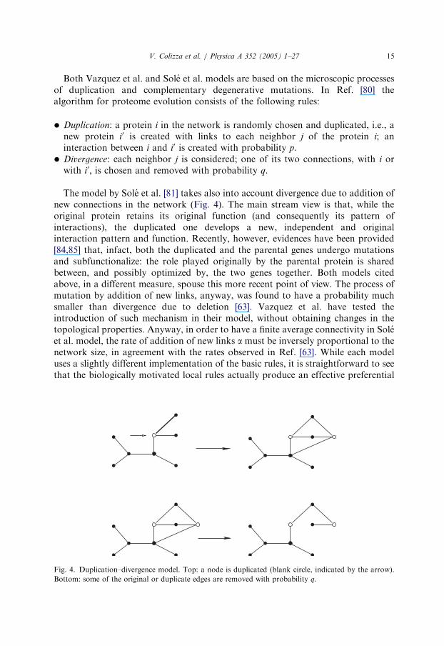
Both Vazquez et al. and Sole et al. models are based on the microscopic processesof duplication and complementary degenerative mutations. In Ref. [80] thealgorithm for proteome evolution consists of the following rules:
Fi
Bo
Duplication: a protein i in the network is randomly chosen and duplicated, i.e., anew protein i0 is created with links to each neighbor j of the protein i; aninteraction between i and i0 is created with probability p.
Divergence: each neighbor j is considered; one of its two connections, with i orwith i0; is chosen and removed with probability q.The model by Sole et al. [81] takes also into account divergence due to addition ofnew connections in the network (Fig. 4). The main stream view is that, while theoriginal protein retains its original function (and consequently its pattern ofinteractions), the duplicated one develops a new, independent and originalinteraction pattern and function. Recently, however, evidences have been provided[84,85] that, infact, both the duplicated and the parental genes undergo mutationsand subfunctionalize: the role played originally by the parental protein is sharedbetween, and possibly optimized by, the two genes together. Both models citedabove, in a different measure, spouse this more recent point of view. The process ofmutation by addition of new links, anyway, was found to have a probability muchsmaller than divergence due to deletion [63]. Vazquez et al. have tested theintroduction of such mechanism in their model, without obtaining changes in thetopological properties. Anyway, in order to have a finite average connectivity in Soleet al. model, the rate of addition of new links a must be inversely proportional to thenetwork size, in agreement with the rates observed in Ref. [63]. While each modeluses a slightly different implementation of the basic rules, it is straightforward to seethat the biologically motivated local rules actually produce an effective preferential
g. 4. Duplication–divergence model. Top: a node is duplicated (blank circle, indicated by the arrow).
ttom: some of the original or duplicate edges are removed with probability q.

ARTICLE IN PRESS
V. Colizza et al. / Physica A 352 (2005) 1–2716
attachment. Indeed, the probability that a node of the network with degree k gainsone more link is given by the probability that one of its neighbors is duplicated (k=N)times the probability of its new link not to be removed (1� q). We therefore obtainthat, ignoring self-interactions and correlations, each protein has an effectiveprobability of getting new edges given by
PðkÞ ð1� qÞk=N , (8)
readily disclosing the presence of an emergent preferential attachment in the networkdynamics. Indeed, it is interesting to mention that similar copy mechanisms havebeen implemented also in other domains such as the modeling of the WWW network[86–88], providing a microscopic description of their growth dynamics and the originof the preferential attachment principle.PIN models can be analytically studied using a mean-field approximation. Here
we follow Ref. [80], since the two approaches lead to analogous results in terms ofthe respective parameters introduced. The average degree hkiNþ1 of the network withN þ 1 nodes can be expressed as
hkiNþ1 ¼NhkiN þ 2p þ ð1� 2qÞhkiN
N þ 1, (9)
where 2p represents the gain in average degree due to self-interacting link and�2qhkNi the loss corresponding to removed connections for divergence process. Inthe continuum limit for large N, one obtains a differential equation whose solutionshows two distinct behaviors, depending on the rate q. For q41=2; a finite averageconnectivity is reached, i.e., hki ¼ k1 ¼ 2p=ð2q � 1Þ; while for qo1=2; hki divergeswith N as N1�2q: At q ¼ q1 ¼ 1=2 a phase transition occurs. Networks obtaineddisplay multifractal connectivity properties, with a scale-free behavior characterizedby an infinite set of scaling exponents, a features that seems to be related to localinheritance mechanisms [89].Other relevant quantities have been investigated in Ref. [80], such as the
clustering coefficient which displays the correct behavior, reaching a finitevalue for increasing network size. Once the values of the two rates p and q are setto have clustering coefficient and square coefficient values consistent withthose of the protein–protein interaction network of the yeast S. cerevisiae, themodel is able to reproduce other quantities, such as average degree anddegree distribution together with tolerance against random and selective deletionof nodes, which are in good agreement with experimental results (Fig. 5). InRef. [81], approximate values of the rates d and b are found by imposing theexperimental value of the average degree of the yeast, together with estimations ofthe ratio a=d from Ref. [63]. The degree distribution PðkÞ obtained for networks ofsize comparable to yeast PINs (Fig. 6) can be fitted by a power-law with anexponential cut off, Eq. (3), already used by Jeong et al. [20] to analyze theconnectivity distribution of S. cerevisiae. The fit parameters, g ¼ 2:5� 0:1 and kc ’
28 are in good agreement with those found in Ref. [20]. Other quantities, such asclustering coefficient, average path length and size of the giant component, werequite well reproduced by the model.

ARTICLE IN PRESS
0 0.5 1 1.5log10k
0
log 1
0P(k
)
PINDD
0 0.5 1
f
0
0.5
1
P(f
)
PIN randomDD random
PIN selectiveDD selective
-2
-4
-6
Fig. 5. Duplication–divergence (DD) model results. Left: Connectivity distribution of the protein
interaction network (PIN) compared to DD model with optimized rates; error bars represent standard
deviations on a single realization. The straight line is a power-law with exponent 2.5. Right: Fraction of
nodes Pðf Þ ¼ Nðf Þ=N belonging to the giant component after a fraction f of nodes has been deleted.
Comparison of DD model curves (averaged over 100 realizations) with experimental results.
k
100
100 101 102
10-1
10-2
10-3
10-4
10-5
P(k)
N=103
γ = 2.5
Fig. 6. Degree distribution PðkÞ for the model [81], averaged over 104 realizations of networks with size
N ¼ 103:
V. Colizza et al. / Physica A 352 (2005) 1–27 17
Finally, it is worth noticing that it is easy to imagine how the duplication anddivergence mechanisms produces a relevant number of proteins that, when comparedto a RG or a graph obtained by preferential attachment only, share a large numberof common partners, as observed by Samantha and Liang [90]. We will return on the

ARTICLE IN PRESS
V. Colizza et al. / Physica A 352 (2005) 1–2718
relevance of this fact when correlation topology–function and methods for functionprediction will be discussed in the next section.
5. Proteins functional characterization
With the advent of high-throughput methods, the traditional view of proteinfunction as a task performed by a single protein independently from the others hasbeen substituted by a more general context [91,92], in which interactions betweenproteins play crucial roles when performing their activities and several cellularprocesses are the outcome of complex interactions between proteins. The underlyingnetwork of interactions thus assumes a deeper meaning in terms of functionalrelationships between proteins, representing cooperative participation in performingfunctional tasks.
5.1. Topology/functionality correlations
In the work by von Mering et al. [24] about the quality of different proteininteraction data sets in terms of accuracy and coverage, it was shown that in highlyaccurate data sets functionally related proteins are more likely linked to each other.This feature is usually exploited in function prediction models (see Section 5.2) toinfer functional annotation of unclassified proteins from classified neighbors. Theauthors computed the distribution of interactions according to functional categoriesand represented the results in terms of a matrix M whose generic element Mðsi;sjÞ
represents the fraction of links between pairs of proteins performing, respectively,functions si and sj : They found that the reference set adopted shows considerablyhigher values along the matrix diagonal, thus in correspondence of shared functionsbetween proteins.Here we would like to go further in the investigation about the correlation between
the pattern of interactions among proteins and their functionalities, with the purposeof reaching a deepened understanding of biological significance of networkarchitecture. The protein–protein interaction networks of the S. Cerevisiae we haveconsidered are those already investigated in Section 3 from a topological point ofview, i.e., (I) the two-hybrid data in Refs. [11,12], (II) the data set obtained with anexperiment of TAP [13], (III) a heterogeneous collection of interactions detected bydifferent techniques, documented at the Database of Interacting Proteins (DIP) [49].The functional classification was extracted from the MIPS database [93]; the finestfunctional classification scheme consists of 424 different functional classes, while thecoarse-grained one contains only 18 functional categories. The number of proteins ineach data set with no defined functional classification (i.e., belonging to thecategories named ‘‘classification not yet clear-cut’’ and ‘‘unclassified proteins’’) is,respectively: 638 out of 2152 in (I), 279 out of 1361 in (II) and 1665 out of 4713in (III).In order to review the likelihood that functionally related proteins are directly
connected in the PIN, we compute the rate of interacting protein pairs sharing at

ARTICLE IN PRESS
V. Colizza et al. / Physica A 352 (2005) 1–27 19
least one functional category, in all three data sets examined. Only classified proteinsare taken into account here, together with the whole set of functions they perform.The values obtained adopting the coarse-grained level of functional classification are83% of interactions between proteins with at least one function in common in two-hybrid data, 83% for TAP and 72% for DIP data set (III). This seems to confirmprevious observations, although, the sensitive decrease observed in (III) respect to (I)and (II) highlights the need for caution when interpreting DIP data, since it mightindicate the presence of a large amount of false positives.To determine the actual significance of these results, we compare them with the
rates of shared functionalities obtained in two distinct null models, compatible withthe constraints embodied by the number of proteins belonging to each functionalcategory. The first null model (NM1) consists in a functional rewiring of the network.Starting from the PINs considered, we choose at random two proteins pi and pj andexchange their annotations. Unclassified proteins are also considered in the rewiringand the underlying network is not modified. The procedure is repeated a number oftimes large enough to obtain a network sufficiently ‘‘scrambled’’ but still preservingthe composition of each multi-functional annotation. The second null model (NM2),instead, is based on a random functional assignment on the empty network. Startingfrom the network of interactions with no functional annotation, we randomly assignfunctions to proteins extracted with uniform probability, following three constraints:(a) the number of proteins belonging to each functional category must be globallyconserved; (b) a protein cannot be assigned the same function twice; and (c) thenumber of unclassified proteins must be conserved.Performing 100 realizations of each null model, we obtain the average values of
the rate of interactions between proteins having at least one function in common,together with their standard deviations (see Table 2).We notice that the random rates of shared functionalities between interacting
proteins obtained in the two null models are similar and are both considerably lowerthan the corresponding real values (exp in Table 2) computed on experimental data.These observations indicate the emergence of a marked correlation between physicallink and functional association in protein–protein interaction networks.The results shown are obtained considering the whole set of classified proteins,
independently of their degrees. In order to investigate a possible dependence of theshared functional rate on degree, we have computed the same quantity for low- and
Table 2
Rates of interacting protein pairs sharing at least one functional category. Results obtained from the three
networks (exp) are compared with the values averaged over 100 realizations of the two null models, NM1
and NM2, described in the text
Ratelink!fcommon (I) (%) (II) (%) (III) (%)
exp 82.90 82.89 72.36
NM1 ð60:55� 0:19Þ ð65:35� 0:22Þ ð49:28� 0:15ÞNM2 ð60:64� 0:20Þ ð64:05� 0:20Þ ð49:62� 0:16Þ

ARTICLE IN PRESS
Table 3
Comparison among the rates of interacting protein pairs sharing at least one functional category
computed on: the whole set of links (link(8k; 8k)); link(ksmall ; ksmall) between proteins with small degree;
link(klarge; klarge) between proteins with large degree; link(ksmall ; klarge) between proteins having,
respectively, small and large degree, with the average connectivity hki being the separation value. For
comparison, we report also the values obtained with the two null models—NM1 and NM2
Ratelink!fcommon (I) (%) (II) (%) (III) (%)
link(8k; 8k) 82.90 82.89 72.36
link(ksmall ; ksmall) 80.85 81.16 63.71
link(klarge; klarge) 88.50 85.33 78.76
link(ksmall ; klarge) 75.30 79.70 63.17
NM1 ð60:55� 0:19Þ ð65:35� 0:22Þ ð49:28� 0:15ÞNM2 ð60:64� 0:20Þ ð64:05� 0:20Þ ð49:62� 0:16Þ
V. Colizza et al. / Physica A 352 (2005) 1–2720
high-connectivity proteins. The average connectivity hki is the separation value:ksmall indicates low degrees (ksmallohki), klarge refers to high degrees (klarge4hki). InTable 3 we report results obtained from the experimental data corresponding tonetworks (I), (II) and (III) and compare them with results from the null models. Nodistinctions based on protein connectivity are considered in the null models, sincefunctional annotation is by definition uncorrelated with topology.A common behavior can be observed in all networks: the rate of functional
commonality between interacting proteins increases when considering two proteinswith large degree klarge; while it is considerably lower when the connected pair iscomposed at least by a protein with small degree ksmall : The lowest value is assumedin correspondence of the type of links (ksmall ; klarge). We have also investigated therole of peripheral proteins (k ¼ 1), being affected by false interactions with higherprobability. We have thus computed the same quantities as before without includingperipheral proteins among those with ksmall : The observed trend is unchanged,showing a deeper correlation of functional characterization with topology, whichshould be investigated in the next future. As we will see in the next section thisgeneral feature will be exploited in protein function prediction methods.
5.2. Function prediction methods
Despite the impressive progresses performed during the last years in genomesequencing and high-throughput proteomics techniques, a great amount of encodedproteins per completely sequenced genome is still functionally uncharacterized [94],so that the development of bioinformatics methods for function prediction assumes acrucial importance in order to fully exploit genome data. The list of available in silicoapproaches to protein function prediction is extensive and includes methods basedon sequence similarity, clustering patterns of co-regulated genes [95,96], phylogeneticprofiles [97] and analysis of protein complexes [13,14]. In this section we will focus on

ARTICLE IN PRESS
V. Colizza et al. / Physica A 352 (2005) 1–27 21
those methods [11,12,90,98–101] that more closely rely on the assumed correlationbetween function and topology for which evidences have been discussed in theprevious section.The basic strategy usually relies on the assumption that two proteins will be more
likely functionally related when they are close to each other, rather than if they arefar away in the network. This assumption has been first exploited in the ‘‘majorityrule’’ method [98,99], whose functional assignment for an uncharacterized protein isobtained by looking at the functional annotations of its classified neighbors. Theassignment proposed consists of the most common function(s) among the onesperformed by the classified binding partners.Capitalizing on the majority rule method, Samantha and Liang [90] assumes that
couples of proteins that share a large number of common neighbors are most likely tobe functionally related and therefore the function of one of the two member of thecouple can be deduced if that of the other is known. The mechanism of duplicationand divergence discussed earlier provides a rationale to this method: proteins sharing alarge number of common partners are also those that more recently diversified from acommon ancestor and therefore those whose functions have emerged as a subfunctionof the common parent’s function. A further modification of the basic majority rulestrategy is proposed in Ref. [101] where predictions are made on the basis of a tree-likenetwork of interactions constructed with the introduction of a new definition of‘‘functional’’ distance between proteins. The latter takes into account not only theshortest path between the proteins but even their number of common partners.All the methods above offer insights on the structure of protein network
functionality from a different point of view and have their own range of applicability.A method that appears to have a more general and automatic applicability is the‘‘global optimization model’’ (GOM) described in Ref. [100]. Leveraging on the sameassumptions on which the ‘‘majority rule’’ [99] is based on, it addresses one of its mainweakness, namely the fact that the majority rule completely disregards the role playedby the unclassified proteins in the function assignment process. In poorly annotatedinteractomes, infact, the potential information conveyed by the unknown proteinscould easily overweight that from the un-annotated ones. More specifically, thefunctional assignment GOM gives to an unclassified protein depends on theannotations of the entire set of its binding partners, considering both classified andunclassified proteins. The method is thus able to provide an assignment for alluncharacterized proteins in the network, in a self-consistent way, taking into accountalso the functional information carried by connections between proteins of unknownfunction. A score E is associated to each functional assignment, by looking at sharedfunctionalities between connected proteins:
E ¼ �X
ioj
Jijdsi ;sj�
X
i
hiðsiÞ , (10)
where Jij ¼ 1 only if the two proteins i and j are both unclassified and directlyconnected, otherwise Jij ¼ 0; si is the function of protein i, dl;m is the discrete deltafunction and hiðsiÞ represents the number of classified binding partners of protein i
sharing the same function si: The score assigns value �1 to each connection between

ARTICLE IN PRESS
V. Colizza et al. / Physica A 352 (2005) 1–2722
unclassified proteins or between classified and unclassified proteins when sharing acommon function. The functional assignment proposed by the method is the one thatminimizes the cost function E over the whole network, thus performing a globaloptimization, while majority rule algorithm, ignoring connections between proteinswith unknown functions, is obtained by minimizing the second term only of the right-hand side of Eq. (10). Interestingly, the optimization process leads to several equivalentor nearly equivalent minima corresponding to equal or very close values of the scorefunction. Indeed, the computational problem is frustrated because of the presence ofthe boundary conditions imposed by the classified proteins, which do not allow tosatisfy all requested shared functionalities between interacting proteins. The functionalannotations proposed by the GOM are those of the optimal configurations whichcorrespond to the minima of the score function evaluated on the whole network,therefore allowing in a natural way for multiple assignments.Several quantities have been investigated in order to test the functional predictions
proposed by the global optimization method. The predictive accuracy has beenevaluated by testing the method on a set of classified proteins and quantified by theintroduction of a measure of the rate of successful predictions, which corresponds tothe percentage of proposed assignments actually recovering correct annotations ofthe test proteins. The accuracy of the method depends on several factors, includingthe classification scheme adopted, the particular network considered and on thefraction of classified proteins present, but is surprisingly reliable. Results show thataccuracy increases with the degree of the unclassified protein under considerationand approach the maximum achievable when the coarsest functional scheme andwell connected proteins are considered. Also, the method proved very robust againstthe presence of false positives/negatives links by which data sets are certainlyaffected. The promising results obtained with GOM have recently motivated furtherstudies in this direction aimed at modifying the score function in Eq. (10) in order totake more throughly into account the topological structure of the network and theobserved correlation between interacting proteins [102].
6. Outlook
As most often in the biological context, results concerning the analysis of PINS arenot exempt from various caveats. Nevertheless, the global statistical analysis of theever increasing number of data sets available provides a general conceptualframework in which eventually many questions concerning evolution, biologicalfunction and the network topology may be addressed. Indeed, several groups havestarted to obtain interesting findings concerning the modular architecture of PINsand its role in the evolutionary process. For instance, specific topological motifs ofthe protein interaction networks might be associated with functional modulesproviding a first step in connecting the biological networks topological architectureto their detailed function and evolution. At the same time, genome sequencing andPINs data gathering of different organisms open exciting opportunities forcomparative analysis and evolutionary studies [6,21–23]. The detection of the

ARTICLE IN PRESS
V. Colizza et al. / Physica A 352 (2005) 1–27 23
particular role at the evolutionary level of proteins belonging to specifictopological motifs finds an immediate impact in evolutionary models for the PINbased on duplication–divergence mechanisms. As well, the relevance of specificmotifs and local configuration analysis aimed at identifying cellular functionmodules may enrich local and global algorithm for protein function assignmentbased on the PIN. In this perspective, the results reviewed in this paper mightrepresent a relevant contribution toward the answer of specific and detailedquestions about biological complexity.
Acknowledgements
We thank A. Vazquez and M.Vergassola for discussion and data sharing. A.V. ispartially funded by the EC-FOP COSIN-IST-2001-33555.
References
[1] L.H. Hartwell, et al., From molecular to modular cell biology, Nature 402 (1999) C47.
[2] A. Wagner, D.A. Fell, The small world inside large metabolic networks, Proc. Roy. Soc. London
Ser. B 268 (2001) 1803.
[3] Y.I. Wolf, G.P. Karev, E.V. Koonin, Scale-free networks in biology: new insights into the
fundamentals of evolution?, Bioessays 24 (2002) 105.
[4] E.V. Koonin, Y.I. Wolf, G.P. Karev, The structure of the protein universe and genome evolution,
Nature 420 (2002) 218.
[5] U. Alon, Biological networks: the tinkerer as an engineer, Science 301 (2003) 1866.
[6] A.-L. Barabasi, Z.N. Oltvai, Network biology: understanding the cell’s functional organization,
Nat. Rev. Genet. 5 (2004) 101.
[7] R. Albert, A.-L. Barabasi, Statistical mechanics of complex networks, Rev. Mod. Phys.
74 (2002) 47.
[8] M. Newman, The structure and function of complex networks, SIAM Rev. 45 (2003) 167.
[9] S.N. Dorogovtsev, J.F.F. Mendes, Evolution of Networks, Oxford Univesrity Press, Oxford, 2003.
[10] R. Pastor-Satorras, A. Vespignani, Evolution and Structure of the Internet, Cambridge Univesrity
Press, Cambridge, 2004.
[11] P. Uetz, et al., A comprehensive analysis of protein–protein interactions in Saccharomyces cerevisiae,
Nature 403 (2000) 623.
[12] T. Ito, et al., A comprehensive two-hybrid analysis to explore the yeast protein interactome, Proc.
Natl. Acad. Sci. USA 98 (2001) 4569.
[13] A.C. Gavin, et al., Functional organization of the yeast proteome by systematic analysis of protein
complexes, Nature 415 (2002) 141.
[14] Y. Ho, et al., Systematic identification of protein complexes in Saccharomyces cerevisiae by mass
spectrometry, Nature 415 (2002) 180.
[15] L. Giot, et al., A protein interaction map of Drosophila melanogaster, Science 302 (2003) 1727.
[16] S.Y. Yook, Z.N. Oltvai, A.-L. Barabasi, Functional and topological characterization of protein
interaction networks, Proteomics 4 (2004) 928.
[17] E. Ravasz, A.-L. Barabasi, Hierarchical organization in complex networks, Phys. Rev. E 67 (2003)
026112.
[18] S. Maslov, K. Sneppen, Specificity and stability in topology of protein newtorks, Science 296 (2002)
210.
[19] A.-L. Barabasi, R. Albert, Emergence of scaling in random networks, Science 286 (1999) 509.

ARTICLE IN PRESS
V. Colizza et al. / Physica A 352 (2005) 1–2724
[20] H. Jeong, S.P. Mason, A.-L. Barabasi, Z.N. Oltvai, Lethality and centrality in protein networks,
Nature 411 (2001) 41.
[21] H.B. Fraser, et al., Evolutionary rate in the protein interaction network, Science 296 (2002) 750.
[22] H.B. Fraser, et al., A simple dependence between protein evolution rate and the number of
protein–protein interactions, BMC Evol. Biol. 3 (2003) 11.
[23] S. Wuchty, et al., Evolutionary conservation of motif constituents in the yeast protein interaction
network, Nat. Genet. 35 (2003) 176.
[24] C. von Mering, R. Krause, B. Snel, M. Cornell, S.G. Oliver, S. Fields, P. Bork, Comparative
assessment of large-scale data sets of protein–protein interactions, Nature 417 (2002) 399.
[25] J.D. Bloom, C. Adami, Apparent dependence of protein evolutionary rate on number of interactions
is linked to biases in protein–protein interaction data sets, BMC Evol. Biol. 3 (2003) 21.
[26] I.K. Jordan, et al., No simple dependence between protein evolution rate and the number of
protein–protein interactions, BMC Evol. Biol. 3 (2003) 1.
[27] T. Dandekar, B. Snel, M. Huynen, P. Bork, Conservation of gene order: a fingerprint of proteins
that physically interact, Trends Biochem. Sci. 23 (1998) 324.
[28] H. Ge, Z. Liu, G.M. Church, M. Vidal, Correlation between transcriptome and interactome
mapping data from Saccharomyces cerevisiae, Nat. Genet. 29 (2001) 482.
[29] R.J. Cho, et al., A genome-wide transcriptional analysis of the mitotic cell cycle, Molecular Cell. 2
(1998) 65.
[30] T.R. Hughes, et al., Functional discovery via a compendium of expression profiles, Cell 102 (2000)
109.
[31] A.H. Tong, et al., Systematic genetic analysis with ordered arrays of yeast deletion mutants, Science
294 (2001) 2364.
[32] H.W. Mewes, et al., MIPS: a database for genomes and protein sequences, Nucleic Acids Res. 30
(2002) 31.
[33] A.J. Enright, I. Iliopoulos, N.C. Kyrpides, C.A. Ouzounis, Protein interaction maps for complete
genomes based on gene fusion events, Nature 402 (1999) 86.
[34] E.M. Marcotte, M. Pellegrini, H.L. Ng, D.W. Rice, D.O. Yeates, D. Eisenberg, Detecting protein
function and protein–protein interactions from genome sequences, Science 285 (1999) 751.
[35] R. Overbeek, M. Fonstein, M. D’Souza, G.D. Pusch, N. Maltsev, The use of gene clusters to infer
functional coupling, Proc. Natl. Acad. Sci. USA 96 (1999) 2896.
[36] M. Pellegrini, E.M. Marcotte, M.J. Thompson, D. Eisenberg, T.O. Yeates, Assigning protein
functions by comparative genome analysis: protein phylogenetic profiles, Proc. Natl. Acad. Sci.
USA 96 (1999) 4285.
[37] M.A. Huynen, P. Bork, Measuring genome evolution, Proc. Natl. Acad. Sci. USA 95 (1998)
5849.
[38] J. Ma, M. Ptashne, A new class of yeast transcriptional activators, Cell 51 (1987) 113.
[39] S. Fields, O.-K. Song, A novel genetic system to detect protein–protein interactions, Nature 340
(1989) 245.
[40] G. Rigaut, A. Shevchenko, B. Rutz, M. Wilm, M. Mann, B. Seraphin, A generic protein purification
method for protein complex characterization and proteome exploration, Nat. Biotechnol. 17 (1999)
1030.
[41] A. Valencia, F. Pazos, Computational methods for the prediction of protein inetarctions, Curr.
Opin. Struct. Biol. 12 (2002) 368.
[42] T. Gaasterland, M.A. Ragan, Microbial genescapes: phyletic and functional patterns of ORF
distribution among prokaryotes, Microb. Comp. Genomics 3 (1998) 199.
[43] J. Tamames, G. Casari, C. Ouzounis, A. Valencia, Conserved clusters of functionally related genes
in two bacterial genomes, J. Mol. Evol. 44 (1997) 66.
[44] P. Novick, B.C. Osmond, D. Botstein, Suppressors of yeast actin mutations, Genetics 121 (1989)
659.
[45] L. Guarente, Synthetic enhancement in gene interaction—a genetic tool come of age, Trends Genet.
9 (1993) 362.
[46] I. Xenarios, D. Eisenberg, Protein interaction databases, Curr. Opin. Biotechnol. 12 (2001) 334.

ARTICLE IN PRESS
V. Colizza et al. / Physica A 352 (2005) 1–27 25
[47] G.D. Bader, C.W.V. Hogue, Analyzing yeast protein–protein interaction data obtained from
different sources, Nat. Biotechnol. 20 (2002) 991.
[48] C.M. Deane, L. Salwinski, I. Xenarios, D. Eisenberg, Protein interactions—two methods
for assessment of the reliability of high throughout observations, Mol. Cell. Proteomics 1
(2002) 349.
[49] Database of Interacting Proteins, http://dip.doe-mbi.ucla.edu/.
[50] D.J. Watts, S.H. Strogatz, Nature 393 (1998) 440.
[51] M.E.J. Newman, Random graphs as models of networks, in: S. Bornholdt, H.G. Schuster (Eds.),
Handbook of Graphs and Networks: from the Genome to the Internet, Wiley-VCH, Berlin, 2003,
pp. 35–68.
[52] P. Erdos, A. Renyi, On random graphs, Publicationes Mathematicae 6 (1959) 290.
[53] B. Bollobas, Random Graphs, Cambridge University Press, Cambridge, 2001.
[54] A. Vazquez, R. Pastor-Satorras, A. Vespignani, Large-scale topological and dynamical properties of
Internet, Phys. Rev. E 65 (2002) 066130.
[55] S.N. Dorogovtsev, A.V. Goltsev, J.F.F. Mendes, Pseudofractal scale-free web, Phys. Rev. E 65
(2002) 066122.
[56] S. Jung, S. Kim, B. Kahng, Geometric fractal growth model for scale-free networks, Phys. Rev. E 65
(2002) 056101.
[57] G. Caldarelli, R. Pastor-Satorras, A. Vespignani, Structure of cycles and local ordering in complex
networks, Eur. Phys. J. B 36 (2003) 203.
[58] G. Bianconi, A. Capocci, Number of loops of size h in growing scale-free networks, Phys. Rev. Lett.
90 (2003) 078701.
[59] E. Ravasz, A.L. Somera, D.A. Mongru, Z. Oltvai, A.-L. Barabasi, Hierarchical organization of
modularity in metabolic networks, Science 297 (2002) 1551.
[60] A. Vazquez, Growing network with local rules: preferential attachment, clustering hierarchy, and
degree correlations, Phys. Rev. E 67 (2003) 056104.
[61] R. Pastor-Satorras, A. Vazquez, A. Vespignani, Dynamical and correlation properties of the
Internet, Phys. Rev. Lett. 87 (2001) 258701.
[62] M.E.J. Newman, Assortative mixing in networks, Phys. Rev. Lett. 89 (2002) 208701.
[63] A. Wagner, The yeast protein interaction network evolves rapidly and contains few redundant
duplicate genes, Mol. Biol. Evol. 18 (2001) 1283.
[64] H.A. Simon, On a class of skew distribution functions, Biometrika 42 (1955) 425.
[65] D.J. de, S. Price, A general theory of bibliometric and other cumulative advantage processes, J. Am.
Soc. Inform. Sci. 27 (1976) 292.
[66] A.-L. Barabasi, R. Albert, H. Jeong, Mean-field theory for scale-free random networks, Physica A
272 (1999) 173.
[67] P.L. Krapivsky, S. Redner, F. Leyvraz, Connectivity of growing random networks, Phys. Rev. Lett.
85 (2000) 4629.
[68] S.N. Dorogovtsev, J.F.F. Mendes, A.N. Samukhin, Structure of growing networks with preferential
linking, Phys. Rev. Lett. 85 (2000) 4633.
[69] P.L. Krapivsky, S. Redner, Organization of growing random networks, Phys. Rev. E 63 (2001)
066123.
[70] S.N. Dorogovtsev, J.F.F. Mendes, A.N. Samukhin, Principles of statistical mechanics of
uncorrelated random networks, Nucl. Phys. B 666 (2003) 396.
[71] S.N. Dorogovtsev, J.F.F. Mendes, Scaling behaviour of developing and decaying networks,
Europhys. Lett. 52 (2000) 33.
[72] P.L. Krapivsky, S. Redner, A statistical physics perspective on Web growth, Comput. Netw. 39
(2002) 261.
[73] G. Bianconi, A.-L. Barabasi, Bose-Einstein condensation in complex networks, Phys. Rev. Lett. 86
(2001) 5632.
[74] G. Bianconi, A.-L. Barabasi, Competition and multiscaling in evolving networks, Europhys. Lett. 54
(2001) 436.
[75] G. Ergun, G.J. Rodgers, Growing random networks with fitness, Physica A 303 (2002) 261.

ARTICLE IN PRESS
V. Colizza et al. / Physica A 352 (2005) 1–2726
[76] H. Jeong, Z. Neda, A.-L. Barabasi, Measuring preferential attachment in evolving networks,
Europhys. Lett. 61 (2003) 567.
[77] M.E.J. Newman, Clustering and preferential attachment in growing networks, Phys. Rev. E 64
(2001) 025102.
[78] S. Ohono, Evolution by Gene Duplication, Springer, Berlin, 1970.
[79] K.H. Wolfe, D.C. Shields, Molecular evidence for an ancient duplication of the entire yeast genome,
Nature 387 (1997) 708.
[80] A. Vazquez, A. Flammini, A. Maritan, A. Vespignani, Modeling of protein interaction networks,
ComPlexUs 1 (2003) 38.
[81] R.V. Sole, R. Pastor-Satorras, E. Smith, T.B. Kepler, A model of large-scale proteome evolution,
Adv. Complex Systems 5 (2002) 43.
[82] A. Bhan, D.J. Galas, T.G. Dewey, A duplication growth model of gene expression networks, J. Mol.
Biol. 18 (2002) 1486.
[83] A. Wagner, How the global structure of protein interaction networks evolves, Proc. Roy. Soc.
London B 270 (2003) 457.
[84] A. Force, M. Lynch, F.B. Pickett, A. Amores, Y.-l. Yan, J. Postlethwait, Preservation of duplicate
genes by complementary, degenerative mutations, Genetics 151 (1999) 1531.
[85] M. Lynch, A. Force, The probability of duplicate gene preservation by subfunctionalization,
Genetics 154 (2000) 459.
[86] J.M. Kleinberg, R. Kumar, P. Raghavan, S. Rajagopalan, A.S. Tomkins, The web as a graph:
measurements models and methods, Proceedings of the 32nd Annual ACM Symposium on Theory
of Computing, 2000, p. 163.
[87] R. Kumar, P. Raghavan, S. Rajagopalan, D. Sivakumar, A.S. Tomkins, E. Upfal, Stochastic models
for the web graph, Proceedings of the 41st Annual IEEE Symposium on Foundations of Computer
Science, 2000, p. 57.
[88] A. Broder, R. Kumar, F. Maghoul, P. Raghavan, S. Rajagopalan, R. Stata, A. Tomkins, J. Wiener,
Graph structure in the web, Comput. Netw. 33 (2000) 309.
[89] S.N. Dorogovtsev, A.N. Samukhin, J.F.F. Mendes, Multifractal properties of growing networks,
Europhys. Lett. 57 (2002) 334.
[90] M.P. Samanta, S. Liang, Predicting protein functions from redundancies in large-scale protein
interaction networks, Proc. Natl. Acad. Sci. USA 100 (2003) 12579.
[91] T.C. Hodgman, A historical perspective on gene/protein functional assignment, Bioinformatics 16
(2000) 10.
[92] D. Eisenberg, E.M. Marcotte, I. Xenarios, T.O. Yeates, Protein function in post-genomic era,
Nature 405 (2000) 823.
[93] The MIPS Comprehensive Yeast Genome Database (CYGD), http://mips.gsf.de/proj/yeast/CYGD/
db/.
[94] H.W. Mewes, K. Albermann, K. Heumann, S. Liebl, F. Pfeiffer, MIPS: a database for
protein sequences, homology data and yeast genome information, Nucleic Acids Res. 25
(1997) 28.
[95] M.Q. Zhang, Promoter analysis of co-regulated genes in the yeast genome, Comput. Chem. 23
(1999) 233–250.
[96] H.C. Harrington, C. Rosenow, J. Relief, Monitoring gene expression using DNA microarrays, Curr.
Opin. Microbiol. 3 (2000) 285–291.
[97] M. Pellegrini, E. Marcotte, M.J. Thompson, D. Eisemberg, T.O. Yeates, Assigning protein functions
by comparative analysis: protein phylogenetic profile, Proc. Natl. Acad. Sci. USA 96 (1999)
4285–4288.
[98] B. Schwikowski, P. Uetz, S. Fields, A network of protein–protein interactions in yeast, Nat.
Biotechnol. 18 (2000) 1257.
[99] H. Hishigaki, K. Nakai, T. Ono, A. Tanigami, T. Tagaki, Assessment of prediction accuracy of
protein function from protein–protein interaction data, Yeast 18 (2001) 523.
[100] A. Vazquez, A. Flammini, A. Maritan, A. Vespignani, Global protein function prediction from
protein–protein interaction network, Nat. Biotechnol. 21 (2003) 697.

ARTICLE IN PRESS
V. Colizza et al. / Physica A 352 (2005) 1–27 27
[101] C. Brun, F. Chevenet, D. Martin, J. Wojcik, A. Guenoche, B. Jacq, Functional classification of
proteins for the prediction of cellular function from a protein–protein interaction network, Genome
Biol. 5 (2003) R6.
[102] V. Colizza, P. De Los Rios, A. Flammini, A. Maritan, Protein function prediction from
protein–protein interaction data, under review of Genome Biology.
Copyright © 2022 FDOKUMEN