Sites of Interaction between Aldolase and Thrombospondin-related Anonymous Protein in Plasmodium
11
Molecular Biology of the Cell Vol. 14, 4947– 4957, December 2003 Sites of Interaction between Aldolase and Thrombospondin-related Anonymous Protein in Plasmodium Carlos A. Buscaglia,* † Isabelle Coppens, ‡ Wim G. J. Hol, § and Victor Nussenzweig* *Michael Heidelberg Division of Immunology, Department of Pathology, New York University School of Medicine, New York, New York 10016; ‡ Infectious Diseases Section, Department of Internal Medicine, Yale University School of Medicine, New Haven, Connecticut 06520-8022; and § Department of Biochemistry, Howard Hughes Medical Institute, Biomolecular Structure Center, University of Washington, Seattle, Washington 98195 Submitted June 2, 2003; Accepted July 29, 2003 Monitoring Editor: Paul Matsudaira Gliding motility and host cell invasion by apicomplexan parasites are empowered by an acto-myosin motor located underneath the parasite plasma membrane. The motor is connected to host cell receptors through trans-membrane invasins belonging to the thrombospondin-related anonymous protein (TRAP) family. A recent study indicates that aldolase bridges the cytoplasmic tail of MIC2, the homologous TRAP protein in Toxoplasma, and actin. Here, we confirm these unexpected findings in Plasmodium sporozoites and identify conserved features of the TRAP family cytoplasmic tail required to bind aldolase: a subterminal tryptophan residue and two noncontiguous stretches of negatively charged amino acids. The aldolase substrate and other compounds that bind to the active site inhibit its interaction with TRAP and with F-actin, suggesting that the function of the motor is metabolically regulated. Ultrastructural studies in salivary gland sporozoites localize aldolase to the periphery of the secretory micronemes containing TRAP. Thus, the interaction between aldolase and the TRAP tail takes place during or preceding the biogenesis of the micronemes. The release of their contents in the anterior pole of the parasite upon contact with the target cells should bring simultaneously aldolase, TRAP and perhaps F-actin to the proper subcellular location where the motor is engaged. INTRODUCTION The phylum Apicomplexa is composed of unicellular eu- karyotic parasites that include several major pathogens to humans and/or livestock such as Plasmodium, Toxoplasma, Cryptosporidium, Eimeria, and Babesia. They display a com- mon set of organelles known as rhoptries, micronemes, and dense granules that discharge their content in a defined order during the host cell attachment/invasion processes (Preiser et al., 2000; Morrissette and Sibley, 2002). In addi- tion, the apicomplexan invasive forms (termed zoites) are surrounded by a pellicle composed of the plasma membrane and underneath it a continuous layer of flattened vesicles defining the inner membrane complex (IMC). Even though zoites do not possess cilia or flagella, they exhibit a unique kind of locomotion termed gliding motility, defined by the lack of obvious modification in the moving cell shape, and the need for a supporting substrate (King, 1988). Several lines of evidence indicate that gliding motility and cell invasion are related mechanisms empowered by the same acto-myosin motor located in the cortical space in between the plasma membrane and the IMC (King, 1988; Dobrowolski and Sibley, 1996; Meissner et al., 2002b; Wetzel et al., 2003). According to the current model, trans-membrane proteins displaying adhesive extracellular domains and an- chored to the motor trigger either forward locomotion or penetration into the host cells (King, 1988; Menard, 2001; Opitz and Soldati, 2002). One such molecule is the throm- bospondin-related anonymous protein (TRAP), whose ex- pression is restricted to Plasmodium sporozoites (Rogers et al., 1992; Robson et al., 1997). The essential role of TRAP on gliding motility and inva- sion has been ascertained by genetic approaches (Sultan et al., 1997; Kappe et al., 1999; Matuschewski et al., 2002). TRAP is the founding member of a family of apicomplexan mem- brane proteins, which includes the TgMIC2 protein of Tox- oplasma gondii among others (Menard, 2001; Meissner et al., 2002a). These proteins are stored in the micronemes and rapidly translocated to the cell surface upon parasite activa- tion, likely by Ca 2 signaling (Carruthers and Sibley, 1999; Gantt et al., 2000). The similarities among the 45 amino acid-long carboxyl cytoplasmic tail of TRAP family proteins are restricted to a subterminal tryptophan and a high con- tent of acidic residues (Menard, 2001). However, replace- ment of the cytoplasmic tail of TRAP by that of TgMIC2 did not affect motility or infectivity of P. berghei sporozoites, indicating that notwithstanding their diverse sequence the cytoplasmic tail of these molecules have the same functional Article published online ahead of print. Mol. Biol. Cell 10.1091/ mbc.E03– 06 – 0355. Article and publication date are available at www.molbiolcell.org/cgi/doi/10.1091/mbc.E03– 06 – 0355. † Corresponding author. E-mail address: [email protected]. Abbreviations used: F1,6P, fructose 1,6-phosphate; IMC, inner membrane complex; PfAldo, Plasmodium falciparum aldolase; TRAP, thrombospondin-related anonymous protein. © 2003 by The American Society for Cell Biology 4947
-
Upload
johnshopkins -
Category
Documents
-
view
0 -
download
0
Transcript of Sites of Interaction between Aldolase and Thrombospondin-related Anonymous Protein in Plasmodium

Molecular Biology of the CellVol. 14, 4947–4957, December 2003
Sites of Interaction between Aldolase andThrombospondin-related Anonymous Protein inPlasmodiumCarlos A. Buscaglia,*† Isabelle Coppens,‡ Wim G. J. Hol,§ andVictor Nussenzweig*
*Michael Heidelberg Division of Immunology, Department of Pathology, New York University Schoolof Medicine, New York, New York 10016; ‡Infectious Diseases Section, Department of InternalMedicine, Yale University School of Medicine, New Haven, Connecticut 06520-8022; and §Departmentof Biochemistry, Howard Hughes Medical Institute, Biomolecular Structure Center, University ofWashington, Seattle, Washington 98195
Submitted June 2, 2003; Accepted July 29, 2003Monitoring Editor: Paul Matsudaira
Gliding motility and host cell invasion by apicomplexan parasites are empowered by an acto-myosin motor locatedunderneath the parasite plasma membrane. The motor is connected to host cell receptors through trans-membraneinvasins belonging to the thrombospondin-related anonymous protein (TRAP) family. A recent study indicates thataldolase bridges the cytoplasmic tail of MIC2, the homologous TRAP protein in Toxoplasma, and actin. Here, we confirmthese unexpected findings in Plasmodium sporozoites and identify conserved features of the TRAP family cytoplasmic tailrequired to bind aldolase: a subterminal tryptophan residue and two noncontiguous stretches of negatively chargedamino acids. The aldolase substrate and other compounds that bind to the active site inhibit its interaction with TRAP andwith F-actin, suggesting that the function of the motor is metabolically regulated. Ultrastructural studies in salivary glandsporozoites localize aldolase to the periphery of the secretory micronemes containing TRAP. Thus, the interactionbetween aldolase and the TRAP tail takes place during or preceding the biogenesis of the micronemes. The release of theircontents in the anterior pole of the parasite upon contact with the target cells should bring simultaneously aldolase, TRAPand perhaps F-actin to the proper subcellular location where the motor is engaged.
INTRODUCTION
The phylum Apicomplexa is composed of unicellular eu-karyotic parasites that include several major pathogens tohumans and/or livestock such as Plasmodium, Toxoplasma,Cryptosporidium, Eimeria, and Babesia. They display a com-mon set of organelles known as rhoptries, micronemes, anddense granules that discharge their content in a definedorder during the host cell attachment/invasion processes(Preiser et al., 2000; Morrissette and Sibley, 2002). In addi-tion, the apicomplexan invasive forms (termed zoites) aresurrounded by a pellicle composed of the plasma membraneand underneath it a continuous layer of flattened vesiclesdefining the inner membrane complex (IMC).
Even though zoites do not possess cilia or flagella, theyexhibit a unique kind of locomotion termed gliding motility,defined by the lack of obvious modification in the movingcell shape, and the need for a supporting substrate (King,1988). Several lines of evidence indicate that gliding motilityand cell invasion are related mechanisms empowered by the
same acto-myosin motor located in the cortical space inbetween the plasma membrane and the IMC (King, 1988;Dobrowolski and Sibley, 1996; Meissner et al., 2002b; Wetzelet al., 2003). According to the current model, trans-membraneproteins displaying adhesive extracellular domains and an-chored to the motor trigger either forward locomotion orpenetration into the host cells (King, 1988; Menard, 2001;Opitz and Soldati, 2002). One such molecule is the throm-bospondin-related anonymous protein (TRAP), whose ex-pression is restricted to Plasmodium sporozoites (Rogers etal., 1992; Robson et al., 1997).
The essential role of TRAP on gliding motility and inva-sion has been ascertained by genetic approaches (Sultan etal., 1997; Kappe et al., 1999; Matuschewski et al., 2002). TRAPis the founding member of a family of apicomplexan mem-brane proteins, which includes the TgMIC2 protein of Tox-oplasma gondii among others (Menard, 2001; Meissner et al.,2002a). These proteins are stored in the micronemes andrapidly translocated to the cell surface upon parasite activa-tion, likely by Ca2� signaling (Carruthers and Sibley, 1999;Gantt et al., 2000). The similarities among the �45 aminoacid-long carboxyl cytoplasmic tail of TRAP family proteinsare restricted to a subterminal tryptophan and a high con-tent of acidic residues (Menard, 2001). However, replace-ment of the cytoplasmic tail of TRAP by that of TgMIC2 didnot affect motility or infectivity of P. berghei sporozoites,indicating that notwithstanding their diverse sequence thecytoplasmic tail of these molecules have the same functional
Article published online ahead of print. Mol. Biol. Cell 10.1091/mbc.E03–06–0355. Article and publication date are available atwww.molbiolcell.org/cgi/doi/10.1091/mbc.E03–06–0355.
† Corresponding author. E-mail address: [email protected] used: F1,6P, fructose 1,6-phosphate; IMC, innermembrane complex; PfAldo, Plasmodium falciparum aldolase;TRAP, thrombospondin-related anonymous protein.
© 2003 by The American Society for Cell Biology 4947

properties (Kappe et al., 1999). Several parasite mutants inthe TRAP C terminus were either nonmotile or displayedabnormal gliding motility, denoting that this portion of themolecule must interact directly or indirectly with the molec-ular motor (Kappe et al., 1999). Direct binding of TRAP tocytoskeletal proteins, however, has not been demonstratedso far (Opitz and Soldati, 2002). An indirect kind of TRAP–motor interaction via adapter molecule(s) and/or conforma-tional changes regulated by extracellular ligands, such asthat defined for integrins and selectins (Serrador et al., 2002;Liddington and Ginsberg, 2002), seems the most likely pos-sibility.
Indeed, recent evidence indicates that the cytoplasmic tailof both TgMIC2 and TRAP are bound to actin via the tet-rameric glycolytic enzyme aldolase (Jewett and Sibley,2003). It is known that mammalian aldolases bind to actinthrough defined amino acid motifs (O’Reilly and Clarke,1993; Wang et al., 1996) and that this binding affects both theenzyme activity and cell contraction/motility (Schindler etal., 2001). Here, we confirm in Plasmodium these unforeseenfindings and attempt to define the structural basis and theregulation of the TRAP–aldolase interaction.
MATERIALS AND METHODS
AldolasesHistidine-tagged P. falciparum aldolase (PfAldo) was expressed in Escherichiacoli and purified to crystallographic purposes by means of three chromatog-raphy steps, including immobilized metal ion adsorption, ion exchange, andsize-exclusion columns (Kim et al., 1998). Partial clones from P. yoelii aldolaseobtained from the TIGR P. yoelii Gene Index were assembled by geneticprocedures to obtain the full-length sequence in frame with the glutathioneS-transferase (GST) protein of the pGEX 4T-1 vector (Amersham Biosciences,Piscataway, NJ). Rabbit aldolase A and spinach aldolase were from Sigma-Aldrich (St. Louis, MO).
Aldolase Activity AssaysAldolase activity was assayed by either coupling the fructose 1,6-phosphate(F1,6P) cleavage to the triose phosphate isomerase/�-glycerophosphate de-hydrogenase reaction and continuous measuring of NADH consumption at340 nm (Dobeli et al., 1990) or by using the aldolase kit (Sigma-Aldrich).Carbohydrates and aldolase inhibitors were from Sigma-Aldrich. Kineticvalues were determined from double-reciprocal plots by using the least-squares method.
TRAP ProteinsThe entire cytoplasmic TRAP C terminus containing 45 residues was ampli-fied from P. berghei (NK65 strain) genomic DNA by using the primers PbTC-tfor (5�-ggcGAATTCtataattttatagcaggaagtagcgc-3�) and PbTC-t rev (5�-agcGTCGACtctagattagttccagtcattatcttcagg-3�). The TRAP�W and TRAP�acidmolecules were constructed using the PbTC-t for primer along with thereverse primers PbTC-tW (5�-agcGTCGACtctagattagttcgcgtcattatcttcaggta-3�)and PbTC-tA (5�-agcGTCGACtctagattagttccaggcattacctgcaggtaatttaaac-3�), re-spectively. The TRAP25 mutant molecule was constructed using the primersPbTC-tGLUT for (5�-cgGAATTCgatgtaatggcagatgatga-3�) and PbTC-t rev.The amplicons were treated with EcoRI/SalI, purified, and cloned into pGEX4T-1. Constructs were confirmed by DNA sequence analysis. GST-fusionproteins expressed in E. coli were purified by affinity chromatography onglutathione (GSH)-Sepharose columns (Amersham Biosciences) as describedpreviously (Buscaglia et al., 1998).
PeptidesPeptides derived from P. falciparum TRAP (Rogers et al., 1992) or P. bergheialdolase (Cloonan et al., 2001) were custom-synthesized by Genemed SynthesisInc. (San Francisco, CA). Their sequences are as follows: TRAP13mer, N-PEQ-FRLPEENEWN-C; TRAP25mer, N-ETLGEEDKDLDEPEQFRLPEENEWN-C;TRAP34mer, N-CYAGEPAPFDETLGEEDKDLDEPEQFRLPEENEWN-C; andAldolase C-t, N-GAGGSTAGASLYEKKYVYC-C. In some cases, a cysteine resi-due (denoted in bold) not present in the original sequence was added for directconjugation to Imject maleimide-activated keyhole limpet hemocyanin (KLH)and bovine serum albumin (BSA) (both from Pierce Chemical, Rockford, IL)following manufacturer’s guidelines.
AntiseraGoat anti-rabbit aldolase antibody was from Chemicon International (Te-mecula, CA). Anti-GST antibody was from Amersham Biosciences. An anti-serum to PfAldo was obtained by immunization of BALB/c mice with 30 �gof enzyme emulsified in complete Freund’s adjuvant by the intraperitonealroute, followed by two booster injections of 10 �g in incomplete Freund’sadjuvant every 15 d. Two rabbit antisera against P. berghei TRAP were used,one recognizing the cytoplasmic tail and other directed toward the amino acidrepeats (Sultan et al., 1997). The specificity of these antisera was confirmed bylack of reactivity toward TRAP null parasites. In addition, an antiserum to thecytoplasmic tail of P. falciparum TRAP was raised in mice immunized asdescribed above with the TRAP34mer peptide coupled to KLH. Anti-KLH-Aldolase C-t peptide antiserum was raised in rabbits (Covance, Denver, PA).The specificity of the two latter antisera was assessed by reactivity toward thecorresponding BSA-coupled peptide by enzyme-linked immunosorbent assay(ELISA).
IgG and Fab PurificationIgG was obtained by protein A-Sepharose chromatography (Amersham Bio-sciences) following manufacturer’s guidelines. To obtain the Fab fragments,IgG was incubated overnight at 37°C with 0.02 mg/ml papain (Sigma-Al-drich) in phosphate-buffered saline (PBS) containing 20 mM EDTA and 20mM l-cysteine. Reaction was stopped with 100 �l of 0.3 M iodoacetamide inPBS followed by buffer exchange using NAP-10 desalting columns (Amer-sham Biosciences). Samples were applied onto protein A-Sepharose columnsand the flow-through fractions (containing the Fab) were collected.
ImmunoprecipitationP. berghei sporozoites (5 � 106) were resuspended in 2 ml of 25 mM HEPES,pH 7.3, 1 mM EDTA, 1 mM MgCl2, 50 mM KCl, 0.5% Tween 20, and aprotease inhibitor cocktail (Sigma-Aldrich) and subjected to two bursts ofsonication (20 sec each) on ice. Every subsequent step was carried out at 4°C.Tubes were kept on ice for 20 min and centrifuged at 14,000 rpm for 20 min.Supernatant was centrifuged again and preadsorbed for 1 h with 200 �l ofprotein G-Sepharose (Amersham Biosciences) equilibrated in resuspensionbuffer. Aliquots (500 �l) of the supernatant were incubated for 4 h with 100 �gof the indicated IgG. Protein G-Sepharose (100 �l) was then added, andsamples incubated for 1 h. Resins were washed five times in 1 ml each ofresuspension buffer and stripped at 100°C in loading buffer (50 mM Tris-HCl,pH 6.8, 2% SDS, 10% glycerol, 10% 2-mercaptoethanol, and 0.1% bromphenolblue).
Pull-Down AssaysOne hundred micrograms of either PfAldo or as a control rabbit glyceralde-hyde-3-phosphate dehydrogenase (Sigma-Aldrich) was mixed with 150 �g ofBSA and preadsorbed for 1 h with GSH-Sepharose equilibrated in buffer A (10mM imidazole acetate, pH 7.3, 50 mM KCl, and 0.2% Tween 20). Supernatantswere incubated with 100 �l of GSH-Sepharose loaded with 50 �g of theindicated GST-fusion protein for 3 h at 4°C. Pellets were washed five times in1 ml each of buffer A and finally stripped at 100°C in loading buffer.
F-Actin Co-sedimentation AssaysActin was purified from rabbit muscle acetone powder (Sigma-Aldrich) asdescribed previously (Pardee and Spudich, 1982). Nonpolymerized actin(G-actin) was diluted in buffer G (5 mM Tris-HCl, pH 8, 0.2 mM ATP, 0.2 mMdithiothreitol, and 0.2 mM CaCl2) up to 100 �g/ml and centrifuged to removedenatured protein. Actin polymerization was carried out for 2 h at 4°C by theaddition of 50 mM KCl, 2 mM MgCl2, and 0.8 mM ATP (Pardee and Spudich,1982). Twenty microliters of either GST-fusion protein (300 �g/ml) wasmixed with 1.5 �l of PfAldo (4 mg/ml) and 180 �l of polymerized actin(F-actin) and incubated at room temperature for 45 min. In some cases,different carbohydrates (100 �M) were added. F-Actin was precipitated at65,000 � g for 2 h in a Beckman ultracentrifuge. Supernatants were collectedand pellets washed twice with 1 ml of buffer G before solubilization in 200 �lof loading buffer diluted 1:5 in 8 M urea. Equivalent aliquots from both pelletand supernatant fractions were analyzed in SDS-PAGE followed by Coomas-sie Brilliant Blue staining.
Protein BiotinylationPfAldo and normal rabbit IgG (2 mg/ml) extensively dialyzed against PBSwere labeled for 2 h at room temperature with 1 mM EZ-link Sulfo-NHS-Biotin (Pierce Chemical). Reaction was stopped with 15 �l of 1 M Tris-HCl,pH 7.2. Samples were applied onto NAP-10 columns to remove the excess offree biotin and stored at 4°C with sodium azide (0.01%). The extent ofbiotinylation was determined by the 2-hydroxyazobenzene-4�-carboxylic aciddye method (Bayer and Wilchek, 1990).
C.A. Buscaglia et al.
Molecular Biology of the Cell4948
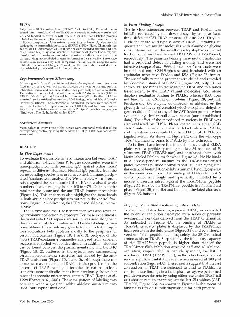
ELISAPolystyrene ELISA microplates (NUNC A/S, Roskilde, Denmark) werecoated with 1 nmol/well of the TRAP34mer peptide in carbonate buffer, pH9.5, and blocked in buffer A with 5% BSA for 3 h. Biotin-labeled proteinsdiluted in the same buffer were incubated for 3 h in the presence of theindicated compounds. After extensive washings in buffer A, NeutrAvidinconjugated to horseradish peroxidase (HRPO) (1:5000; Pierce Chemical) wasadded for 1 h. Absorbance values at 405 nm were recorded after the additionof 2,2�-azino-bis(3-ethylbenzthiazoline-6-sulfonic acid) (Pierce Chemical) andtransformed to protein concentration by using a calibration curve of thecorresponding biotin-labeled protein performed in the same plate. Percentageof inhibition displayed by each compound was calculated using the samecalibration curves and taking as 0% inhibition the mean value recorded for thebiotin-labeled protein without further additions.
Cryoimmunoelectron MicroscopySalivary glands from P. yoelii-infected Anopheles stephensi mosquitoes werefixed for 2 d at 4°C with 8% paraformaldehyde in 0.25 M HEPES, pH 7.4,infiltrated, frozen, and sectioned as described previously (Folsch et al., 2001).The sections were labeled first with mouse anti-PfAldo antibodies (1:100 inPBS 1% fish skin gelatin) followed by anti-mouse IgG antibodies and 5-nmprotein A-gold particles (Department of Cell Biology, Medical School, UtrechtUniversity, Utrecht, The Netherlands). Afterward, sections were incubatedwith rabbit anti-TRAP repeats antibodies (1:10) followed by 10-nm proteinA-gold particles before examination with a Philips 410 electron microscope(Eindhoven, The Netherlands) under 80 kV.
Statistical AnalysisMean values in every point of the curves were compared with that of thecorresponding control by using the Student’s t test. p � 0.05 was consideredsignificant.
RESULTS
In Vivo ExperimentsTo evaluate the possible in vivo interaction between TRAPand aldolase, extracts from P. berghei sporozoites were im-munoprecipitated with purified IgG against either TRAPrepeats or different aldolases. Normal IgG purified from thecorresponding species was used as control. Immunoprecipi-tated fractions were analyzed by Western blot. An antiserumraised against the cytoplasmic tail of TRAP reveals a discretenumber of bands ranging from �100 to �75 kDa in both thetotal parasite lysate and the anti-TRAP immunoprecipitate(Figure 1A). This antiserum also highlights the same bandsin both anti-aldolase precipitates but not in the control frac-tions (Figure 1A), indicating that TRAP and aldolase interactin vivo.
The in vivo aldolase–TRAP interaction was also revealedby cryoimmunoelectron microscopy. For these experiments,the rabbit anti-TRAP repeats antiserum was used along withthe mouse anti-PfAldo antiserum. Double labeling of sec-tions obtained from salivary glands from infected mosqui-toes colocalizes both proteins mostly to the periphery ofcertain micronemes (Figure 1B, 1 and 3). Sixty-six of 165(40%) TRAP-containing organelles analyzed from differentsections are labeled with both antisera. In addition, aldolasecan be found between the plasma membrane and the IMC(Figure 1B, 2), scattered in the cytosol, and surroundingcertain microneme-like structures not labeled by the anti-TRAP antiserum (Figure 1B, 1 and 3). Although these mi-cronemes may not contain TRAP, it is also possible that theabsence of TRAP staining is technical in nature. Indeed,using the same antibodies it has been previously shown thatmost of sporozoite micronemes contain TRAP (Kappe et al.,1999; Bhanot et al., 2003). The same pattern of labeling wasobtained when a goat anti-rabbit aldolase antiserum wasused (our unpublished data).
In Vitro Binding AssaysThe in vitro interaction between TRAP and PfAldo wasinitially evaluated by pull-down assays by using as baitsthree different GST-TRAP proteins (Figure 2A). They in-clude the entire wild-type P. berghei TRAP C-terminus se-quence and two mutant molecules with alanine or glycinesubstitutions in either the penultimate tryptophan or the lasttract of acidic residues (termed TRAP�W and TRAP�acid,respectively). The parasites bearing these mutant moleculeshad a profound defect in gliding motility and were notinfective (Kappe et al., 1999). These TRAP constructs wereimmobilized onto GSH-Sepharose and incubated with anequimolar mixture of PfAldo and BSA (Figure 2B, input).The specifically retained proteins were eluted and revealedby Coomassie-stained SDS-PAGE (Figure 2B, output). Asshown, PfAldo binds to the wild-type TRAP and to a muchlesser extent to the TRAP variant molecules. GST alonedisplays negligible binding to PfAldo, whereas BSA doesnot bind to the GST-fusion proteins (Figure 2B, output).Furthermore, the enzyme downstream of aldolase on theglycolytic pathway (glyceraldehyde-3-phosphate dehydro-genase) did not bind to any of the GST-fusion proteins whenevaluated by similar pull-down assays (our unpublisheddata). The effect of the introduced mutations in TRAP wasalso evaluated by ELISA. Plates coated with either GST-TRAP molecule were incubated with biotin-labeled PfAldo,and the interaction revealed by the addition of HRPO-con-jugated avidin. As shown in Figure 2C, only the wild-typeTRAP significantly binds to PfAldo by this technique.
To further characterize this interaction, we coated ELISAplates with a peptide spanning the last 34 residues of P.falciparum TRAP (TRAP34mer) and incubated them withbiotin-labeled PfAldo. As shown in Figure 3A, PfAldo bindsin a dose-dependent manner to the TRAP34mer-coatedplates, whereas purified normal rabbit IgG displaying sim-ilar extent of biotinylation exhibits minimal binding capacityin the same conditions. The binding of PfAldo to TRAP-coated plates is strongly and specifically inhibited by amouse antiserum raised against the TRAP34mer peptide(Figure 3B, top), by the TRAP34mer peptide itself in the fluidphase (Figure 3B, middle) and by nonbiotinylated aldolases(Figure 3B, bottom).
Mapping of the Aldolase-binding Site in TRAPTo map the aldolase-binding region in TRAP, we evaluatedthe extent of inhibition displayed by a series of partiallyoverlapping peptides derived from the TRAP C terminus.As indicated in Figure 4A, the binding of PfAldo toTRAP34mer-coated plates is displaced by the TRAP34meritself present in the fluid phase (Figure 3B), and by a shorterversion of this peptide spanning solely the 25 C-terminalamino acids of TRAP. Surprisingly, the inhibitory capacityof the TRAP25mer peptide is higher than that of theTRAP34mer (50% inhibition achieved at 5 and 40 �M con-centration, respectively). A peptide spanning the last 13residues of TRAP (TRAP13mer), on the other hand, does notrender significant inhibition even when assayed at 100 �Mconcentration (Figure 4A). These results suggest that the last25 residues of TRAP are sufficient to bind to PfAldo. Toconfirm these findings in a fluid-phase assay, we performedpull-down experiments by using either the entire TRAP tailor a shorter version spanning just the last 25 residues (GST-TRAP25; Figure 2A). As shown in Figure 4B, the extent ofbinding to PfAldo is indistinguishable for both proteins.
Aldolase-TRAP Interaction in Plasmodium
Vol. 14, December 2003 4949

Mapping of the TRAP-binding Site in Aldolase
Compounds That Bind to the Enzyme Active Site InhibitAldolase–TRAP Interaction To map the TRAP bindingsite(s) on Plasmodium aldolase, we initially tried to obtain inE. coli distinct truncated versions of the P. yoelii enzyme.Unfortunately, all of these proteins, except for the full-lengthmolecule, were poorly expressed or targeted to inclusionbodies (our unpublished data).
We then analyzed the effect of catalysis on the aldolase–TRAP interaction. The substrate F1,6P and the products ofthe reaction catalyzed by aldolase (dihydroxyacetone phos-phate and glyceraldehyde-3-phosphate) inhibit at low mi-cromolar concentration the binding of the enzyme to TRAP(Table 1). These values are in the range of the Km for PfAldo(Table 1), suggesting that this inhibition is mediated byoccupation of the active site. Related compounds such asfructose 1-phosphate and fructose 6-phosphate display a
Figure 1. TRAP and aldolase interact invivo. (A) Lysates from P. berghei sporozoiteswere immunoprecipitated using the IgG indi-cated above each lane and probed with a rab-bit anti-TRAP C-terminus antiserum (1:1000).Molecular markers (in kilodaltons) are indi-cated on the right. (B) Immunoelectron mi-croscopy of P. yoelii sporozoites doubly la-beled with mouse anti-PfAldo (5-nm goldparticles) and rabbit anti-TRAP repeats(10-nm gold particles). 1, a longitudinal sec-tion of the parasite identifies the TRAP-con-taining micronemes (mi), some of which areperipherally associated with aldolase. Somemicroneme-like structures are solely labeledby aldolase (arrows). 2, localization of aldo-lase between the IMC and the plasma mem-brane (pm) is highlighted by arrows. 3, atransversal section of the parasite pinpointsthe intense and specific labeling of mi-cronemes with TRAP and aldolase; few mol-ecules of aldolase are observed in the parasitecytosol. The arrow in the inset of 3 shows thepresence of TRAP and aldolase in a singlemicroneme. sg, salivary gland tissue. Bars,150 nm.
C.A. Buscaglia et al.
Molecular Biology of the Cell4950

significantly impaired inhibitory activity (Table 1). The con-centrations required to reach 50% of inhibition of the PfAl-do–TRAP interaction and 50% inhibition of PfAldo activityon F1,6P are very similar (millimolar range), further sup-porting the role of the active site occupation in the formereffect. Neither fructose nor glucose 6-phosphate inhibitsPfAldo–TRAP interaction or PfAldo cleavage of F1,6P (Table1). We also tested the effect of competitive inhibitors ofaldolase in our assay. As shown in Table 1, both suraminand d-myo-inositol 1,4,5-phosphate (but not d-myo-inositol)inhibit both PfAldo–TRAP interaction and F1,6P consump-tion at roughly similar concentration.
Overall, these results suggest that TRAP is binding eitherdirectly to the active site or to another region of the enzymewhose conformation is changed upon occupation of theactive site. However, neither the TRAP34mer nor theTRAP25mer inhibit PfAldo enzymatic activity even whenassayed at 100 �M concentration (Table 1).
The C Terminus of Aldolase Is Not Involved in the Bindingto TRAP Previous site-directed mutagenesis and crystallo-graphic studies (Itin et al., 1993; Kim et al., 1998) indicate thatthe last �20 amino acids of PfAldo play a critical role in thecatalytic reaction. It has been speculated that this species-
specific and flexible segment modulates the PfAldo activityby burrowing into the active site (Kim et al., 1998). In Plas-modium aldolases, this sequence is rich in positively chargedresidues (Cloonan et al., 2001), suggesting that it might me-diate the interaction with the acidic TRAP tail. However, wefound that the Aldolase C-t peptide spanning the last 19residues of P. berghei aldolase (90% identical to the PfAldosequence) does not inhibit the PfAldo-TRAP binding even at100 �M concentration (our unpublished data). Furthermore,Fab fragments prepared from an antiserum raised againstthis peptide are also inactive in preventing PfAldo–TRAPinteraction (Figure 5A), although they recognize and par-tially inhibit PfAldo enzymatic activity (Figure 5B). Overall,our data suggest that the C-terminal region of PfAldo is notcritical for its interaction with TRAP.
The Aldolase–TRAP Complex Interacts with F-ActinThe actin-binding site of mammalian aldolases includes aseries of residues (O’Reilly and Clarke, 1993; Wang et al.,1996) highly conserved in the Plasmodium aldolases (Figure6A) (Cloonan et al., 2001), but the direct interaction of theparasite aldolase with actin has never been demonstrated.Therefore, we first evaluated the in vitro interaction ofPfAldo and F-actin by cosedimentation assays. As shown in
Figure 2. TRAP cytoplasmic tail interacts withPfAldo in vitro. (A) Schematic representation ofGST-TRAP variant molecules. Different con-structs derived from P. berghei TRAP werecloned in-frame at the C terminus of GST by theunique EcoRI and SalI restriction sites. Aminoacid replacements inserted in each construct areshaded. (B) Equal amounts of each GST-fusionprotein (indicated above) immobilized ontoGSH-Sepharose were incubated with 100 �g ofPfAldo and 150 �g of BSA (input). Specificallyretained proteins were evaluated by CoomassieBlue-stained SDS-PAGE (output). The identityof the revealed bands is indicated on the leftand the molecular markers (in kilodaltons) onthe right. (C) ELISA plates coated with 100 ng ofthe indicated GST-fusion protein were incu-bated with biotin-labeled PfAldo and revealedby the addition of HRPO-avidin followed by acolorimetric substrate. Samples were tested intriplicate. Mean values of absorbance at 405 nmare indicated (SD did not exceed 5% of meanvalues; our unpublished data). Asterisks denotesignificant differences (p � 0.01) with the corre-sponding GST values.
Aldolase-TRAP Interaction in Plasmodium
Vol. 14, December 2003 4951

Figure 6B, PfAldo coprecipitates with actin microfilaments.This interaction is partially inhibited by the aldolase sub-strate F1,6P and by the aldolase products dihydroxyacetonephosphate and glyceraldehyde-3-phosphate but not by fruc-tose 6-phosphate or fructose (Figure 6B). BSA and �-actininwere used as internal negative and positive F-actin–copre-
cipitating controls, respectively (Figure 6B) (Hohaus et al.,2002). Additional cosedimentation assays were performed todemonstrate the specific formation of a ternary complex(F-actin–PfAldo–TRAP). As depicted in Figure 6C, GST-TRAP, but not GST, is brought down into the F-actin pelletwhen in the presence of PfAldo, indicating that the ternary
Figure 3. Specificity of PfAldo-TRAP inter-action. (A) ELISA plates coated with theTRAP34mer peptide were probed with eitherbiotin-labeled PfAldo or biotin-labeled rabbitIgG as in legend to Figure 2C. (B) Inhibition ofTRAP34mer-biotin-labeled PfAldo (6 �g/ml)interaction displayed by different mouse an-tisera (top), peptides (middle), or nonbiotiny-lated aldolases (bottom). Samples were testedin triplicate and the mean values of absor-bance at 405 nm are indicated (SD did notexceed 5% of mean values; our unpublisheddata). Asterisks denote significant differences(p � 0.01) with the mean value recorded forthe biotin-labeled PfAldo without further ad-ditions. One of three experiments with similarresults is shown. UP, peptide unrelated toTRAP.
Figure 4. Mapping of aldolase-binding site on TRAP. (A) Peptides derived from the P. falciparum TRAP C-terminus sequence were testedfor their ability to interfere with the binding between biotin-labeled PfAldo and the TRAP34mer peptide as indicated in legend to Figure 3.The sequence, length, and concentration required to obtain 50% inhibition (IC50%) are shown. (B) Equal amounts of each GST fusion proteinwere tested for their ability to interact with PfAldo by pull-down assays as described in Figure 2B.
C.A. Buscaglia et al.
Molecular Biology of the Cell4952

complex is obtained even though TRAP per se is unable tobind F-actin. The intensity of the revealed PfAldo and actinbands in both the supernatant and pellet fractions is verysimilar in the presence of either GST or GST-TRAP (Figure6C). Therefore, neither the F-actin–PfAldo interaction, northe F-actin/G-actin ratio is affected by GST-TRAP.
Pull-down and ELISA assays revealed the key role of boththe penultimate tryptophan and the terminal stretch ofacidic residues of TRAP in its association to PfAldo (Figure2, B and C). Indeed, all of wild-type TRAP is associated viaPfAldo to the F-actin pellet, whereas a fraction of bothTRAP�W and TRAP�acid molecules remain in the super-natant (unbound) fraction as revealed by Western blot byusing an anti-GST antibody (Figure 6D).
DISCUSSION
Current evidence strongly suggests that gliding motility andhost cell invasion by apicomplexan parasites are empoweredby the same acto-myosin motor, a unique machinery re-stricted to this phylum and highly conserved among itsmembers (Menard, 2001; Opitz and Soldati, 2002). Definingthe identity and mode of interaction of the components ofthis machinery may provide new tools for the prophylaxis ortreatment of a wide range of human and livestock diseases.The overall arrangement of the motor and several of its keyconstituents such as actin (Dobrowolski and Sibley, 1996;Wetzel et al., 2003), myosin A (Pinder et al., 1998; Matus-chewski et al., 2001; Meissner et al., 2002b), and a family ofsurface invasins related to TRAP (Menard, 2001; Matus-chewski et al., 2002) have been already characterized. Recentdata also indicate participation in the machinery of a proteinthat binds to the tail of myosin A (Herm-Gotz et al., 2002;Bergman et al., 2003), of a Toxoplasma myosin docking pro-tein (Beckers, personal communication), and of aldolase
(Jewett and Sibley, 2003). This latter molecule probablytransduces the signals from the parasite surface to the motorby bridging the cytoplasmic tail of invasins to actin.
Here, we use different approaches to study the in vivo andin vitro interaction of Plasmodium aldolase and TRAP. Weshow that the last 25 residues of the TRAP tail are sufficientto mediate this binding (Figure 4). This region of the TRAPtail harbors most of its charged residues, whereas the re-maining sequence close to the trans-membrane domain isbiased toward glycines and hydrophobic residues (valines,alanines, prolines, tyrosines; Figure 2A) and promotes theintracellular sorting of the molecule (Bhanot et al., 2003).Within the defined aldolase-binding site on TRAP, our find-
Table 1. Correlation between inhibition of PfAldo activity andPfAldo-TRAP interaction
Compound IC50%a K1
b
Fructose 1,6-phosphate 4 �Mc N.A.dDihydroxyacetone phosphate 5 �M N.A.Glyceraldehyde 3-phosphate 5 �M N.A.Fructose 1-phosphate 1 mM 5 mMFructose 6-phosphate 5 mM 3 mMGlucose 6-phosphate �50 mM �50 mMFructose �50 mM �50 mMSuramin 500 �M 100 �Md-Myo-inositol �10 mM �10 mMd-Myo-inositol 1,4,5-phosphate 200 �M 500 �MTRAP34mer 40 �M �100 �MTRAP25mer 5 �M �100 �M
Each compound was tested at serial dilutions for the inhibition ofbiotin-labeled PfAldo-TRAP34mer peptide interaction by ELISAand for the PfAldo inhibition in activity assays using fructose 1,6-phosphate as substrate.a IC50%, concentration required to obtain 50% inhibition of PfAldo-TRAP interaction.b Ki, concentration required to obtain 50% inhibition of PfAldoactivity. Ki was calculated by plotting 1/Vel against the compoundconcentration.c Km value for PfAldo � 9 �M.d N.A., non-applicable.
Figure 5. Effect of aldolase C terminus (c-t) on its interaction withTRAP. (A) The inhibition of biotin-labeled PfAldo-TRAP34mer pep-tide interaction displayed by different Fab fractions (0.3 mg/ml) wasevaluated as indicated in legend to Figure 3. One of two experi-ments with similar results is shown. (B) PfAldo activity was con-tinuously monitored at 340 nm in the presence of the indicatedantisera (1:250 dilution) or Fab fragments (1:50 dilution). Sampleswere tested in duplicate and the mean values of absorbance areindicated (SD did not exceed 3% of mean values; our unpublisheddata). Asterisks denote significant differences (p � 0.01) with themean value recorded for the PfAldo without further additions. Oneof two experiments with similar results is shown.
Aldolase-TRAP Interaction in Plasmodium
Vol. 14, December 2003 4953

ings reveal the critical role of the penultimate tryptophan inthe enzyme recognition (Figures 2, B and C, and 6D). Theseresults are in close agreement with those reported by study-ing TgMIC2/TRAP and either rabbit or T. gondii aldolase(Jewett and Sibley, 2003), except that we detect a minorbinding of aldolase to the TRAP�W molecule, which isprobably of relevance (see below). In addition, we demon-strated that the acidic last stretch of 13 residues of the TRAPtail is required but not sufficient for optimal binding toaldolase (Figures 2, B and C, 4, and 6D). Our binding studiesusing partially overlapping peptides demonstrate the pres-ence of additional aldolase-contacting residues upstream ofthe last 13 amino acids of TRAP. Indeed, as shown in Figure4A, this sequence cannot displace the interaction betweenaldolase and a longer version of the TRAP tail (TRAP34merpeptide), whereas a 25mer peptide displays full inhibition.
Overall, these data concur with previous in vivo experi-ments by using TRAP mutants. P. berghei sporozoites har-boring a TRAP molecule deleted on its last 14 residues arenoninfective but still display residual motility (Kappe et al.,1999). A similar phenomenon was verified for parasitesbearing TRAP molecules mutated in either the penultimate
tryptophan or the last tract of acidic residues (Kappe et al.,1999). Though aberrant, this residual motility suggests thatthe tethering of TRAP mutant molecules to the motor issomehow taking place. The comparison of the aldolase-binding sequence in TRAP with the structures of the aldo-lase-binding motifs of other proteins provides additionalsupporting evidence for our hypothesis. All of these mole-cules display a consensus sequence composed of two acidicstretches flanking a single highly hydrophobic residue (Fig-ure 7). Deletion of either acidic tract or posttranslationalmodification of the hydrophobic residue correlates with adecreased affinity of these molecules for aldolase (Itin et al.,1993; Volker and Knull, 1997; Eisenmesser and Post, 1998).The participation of ionic bonds in the aldolase-TRAP inter-action is also supported by the inhibitory effect of salts(IC50% reached at 60 mM) and/or ionic detergents (ourunpublished data).
Thus, it is very likely that the two noncontiguous acidicstretches in the TRAP tail (Figure 7) interact with positivelycharged regions scattered throughout the surface of thealdolase. The enzyme active site itself comprises a cluster ofbasic residues, some of which are involved in the anchoring
Figure 6. An actin–aldolase–TRAP ternarycomplex is formed in vitro. (A) Sequencealignment of the actin-binding site of rabbitaldolase A, and the corresponding residues inPlasmodium-derived aldolases. The relativeposition in the primary sequence for each res-idue is indicated. (B) Left, PfAldo was ultra-centrifuged in the presence or absence of F-actin and both supernatant (S) and pellet (P)fractions analyzed by Coomassie-stainedSDS-PAGE. The identity of the revealedbands is indicated on the left. The addition ofdifferent compounds (100 �M) before centrif-ugation is indicated below each lane. DHAP,dihydroxyacetone phosphate; G3P, glyceral-dehyde-3-phosphate; F6P, fructose 6-phos-phate; F, fructose. Right, fractionation of BSAand �-actinin in F-actin cosedimentation as-says. Both pellet (P) and supernatant (S) frac-tions were analyzed as described in A. (C)GST or GST-TRAP molecules were mixedwith F-actin in the presence (left) or absence(right) of PfAldo and subjected to ultracentrif-ugation. Equivalent supernatant (S, top) andpellet (P, bottom) fractions were analyzed asin A. (D) TRAP variants (indicated above eachlane) were assayed for their cosedimentationcapacity with F-actin in the presence ofPfAldo. Equivalent pellet (P, top) and super-natant fractions (S, bottom) were analyzed byCoomassie staining and Western blot by us-ing an anti-GST antibody (1:500), respectively.The identity of the revealed bands is indi-cated on the left.
C.A. Buscaglia et al.
Molecular Biology of the Cell4954

of the phosphate groups of the substrate/products (Kim etal., 1998). A direct binding of the TRAP tail to the aldolasecatalytic region is therefore conceivable. In this case, TRAPand F-actin should be stably accommodated in a small sur-face to form the ternary complex because both the catalyticpocket and the actin-binding site on PfAldo are closelyapposed (Figure 8). This scenario would explain the inhibi-tory role of aldolase substrate, products, and competitiveinhibitors in this interaction (Table 1). Indeed, the associa-tion to aldolase of other sequences structurally related toTRAP such as those displayed in Figure 7 is also displacedby aldolase substrate/products (Schneider and Post, 1995;Volker and Knull, 1997; Kao et al., 1999). However, this ideais not supported by the observation that neither theTRAP34mer nor the TRAP25mer peptide inhibited the invitro PfAldo activity even when tested at 100 �M concen-tration (Table 1).
Although other explanations can be invoked, it is conceiv-able that TRAP tail is not interacting with the aldolase activesite itself but with another positively charged region whose
structure is modified upon the occupancy of the catalyticcenter. The last �20 residues of PfAldo define a highlyflexible and positively charged structure that is presumed tomodulate the enzymatic activity by its reversible burrowinginto the active site (Kim et al., 1998) (Figure 8). We failed,however, to demonstrate a direct interaction between TRAPand the C terminus of PfAldo (Figure 5). If successful, on-going cocrystallization experiments between PfAldo and theTRAP tail peptide will shed some light on this puzzlingissue.
Here, we demonstrated for the first time the specific as-sociation of F-actin, aldolase, and TRAP in a ternary com-plex (Figure 6C). The binding of aldolase to both F-actin andTRAP is, however, inhibited during substrate turnover (Ta-ble 1 and Figure 6B). How can the motor bridging propertiesand the catalytic role of aldolase be reconciled in vivo?Aldolase is a tetramer made up of four identical subunitsand F-actin is a multimer that can be assembled in bundles.We favor the idea that actin–aldolase–TRAP form large dis-sociable dynamic complexes with the rest of the motor be-neath the plasma membrane, and that enzymatic turnoverwill not disrupt all of these links simultaneously. The pos-sibility that the concentration of the aldolase substrate/products in the cortical cytoplasm is lower than that re-quired to inhibit the interaction of the enzyme with TRAP,or even that glycolysis does not take place in the spacewhere the motor is engaged cannot be ruled out. In this case,the ATP required for the assembly and translocation of actinmicrofilaments should be generated outside the cortical cy-toplasm. Further studies dealing with the mechanisms ofenergy conversion in this parasite stage and/or the in vivostructure/function of the gliding motility machinery mayclarify these issues.
It has been previously shown that the aldolase–MIC2complex is present on the surface of T. gondii tachyzoites(Jewett and Sibley, 2003). Our ultrastructural studies onsalivary gland (resting) Plasmodium sporozoites indicate thatthe formation of this complex occurs at a previous step. Asdemonstrated for other apicomplexan invasins (Soldati et al.,2001), TRAP is likely to be stored in the micronemes as anintegral type I membrane protein, with its adhesive N ter-minus protruding toward the micronemal lumen and theC-terminus tail facing to the cytoplasm. As shown in Figure1B, aldolase is localized on the surface of micronemes con-taining TRAP, most likely bound to its cytoplasmic tail.These observations provide a plausible explanation for thesorting of aldolase (and probably actin) to the narrow, con-strained space in between the IMC and the plasma mem-brane (Ngo et al., 2000). On parasite activation, TRAP-con-taining micronemes fuse with the parasite membrane(Carruthers and Sibley, 1999; Gantt et al., 2000) bringing theadhesive domains of TRAP to the parasite surface, andpiggy back to its tail the aldolase–actin complex. This hy-pothesis is in agreement with the observation that polymer-
Figure 7. Sequence comparison of aldolase-binding motifs present in different proteins.Aldolase-binding motifs present in humanglucose transporter 4 (GLUT4), bovine �-tu-bulin, and the anion exchanger of humanerythrocyte membrane (band 3) were alignedwith the last 25 residues of P. falciparum TRAPcytoplasmic tail (Pf TRAP). Overall acidic re-gions are indicated by boxes.
Figure 8. Proximity of the actin-binding site and the catalytic centerin PfAldo. Ribbon diagram of the PfAldo monomer structure based oncrystallographic data showing the substrate (fructose 1,6-phosphate)anchored to the active site, the sequence 38DESTQTIKKRFDN50 span-ning the core of the actin-binding motif (in green) and the flexible Cterminus (in violet) apposed to the catalytic pocket.
Aldolase-TRAP Interaction in Plasmodium
Vol. 14, December 2003 4955

ization of actin in Apicomplexa takes place initially in theanterior parasite pole (Wetzel et al., 2003). During glidingmotility and cell invasion, the aldolase–TRAP complex (andperhaps other molecules) would be moved along the locallyformed and tightly regulated actin filaments (Jewett andSibley, 2003), propelled by the myosin A molecules tetheredto the IMC (Bergman et al., 2003).
ACKNOWLEDGMENTS
We thank Drs. Ute Frevert (New York University School of Medicine, NewYork, NY) and Ali Sultan (Department of Immunology and Infectious Dis-eases, Harvard School of Public Health, Boston, MA) for some of the anti-TRAP antibodies and Drs. Karine Kaiser and Stefan Kappe (New York Uni-versity) for additional Plasmodium peptides and genomic DNA samples.Critical reading of the manuscript by Dr. Stefan Kappe is also appreciated. P.yoelii aldolase clones were kindly provided by Dr. Lawrence Bergman (DrexelUniversity, Philadelphia, PA). We are also indebted to Dr. Matthieu Schapira(New York University), for modeling the aldolases, Dr. Ruth Nussenzweig(New York University) for the sporozoites, Nelly Camargo (New York Uni-versity) for the purified salivary glands of mosquitoes, Dr. Defne Yarar(Department of Molecular and Cell Biology, University of California at Berke-ley) for advice on actin purification, and Brian Krumm (Department ofBiochemistry, University of Washington, Seattle, WA) for preparing PfAldo.Our work was supported by a grant from the National Institutes of Health toV.N. C.A.B. is a recipient of a Bernard Levine fellowship.
REFERENCES
Bayer, E.A., and Wilchek, M. (1990). Protein biotinylation. Methods Enzymol.184, 138–160.
Bergman, L.W., Kaiser, K., Fujioka, H., Coppens, I., Daly, T.M., Fox, S.,Matuschewski, K., Nussenzweig, V., and Kappe, S.H. (2003). Myosin A taildomain interacting protein (MTIP) localizes to the inner membrane complexof Plasmodium sporozoites. J. Cell Sci. 116, 39–49.
Bhanot, P., Frevert, U., Nussenzweig, V., and Persson, C. (2003). Defectivesorting of the thrombospondin-related anonymous protein (TRAP) inhibitsPlasmodium infectivity. Mol. Biochem. Parasitol. 126, 263–273.
Buscaglia, C.A., Campetella, O., Leguizamon, M.S., and Frasch, A.C. (1998).The repetitive domain of Trypanosoma cruzi trans-sialidase enhances the im-mune response against the catalytic domain. J. Infect. Dis. 177, 431–436.
Carruthers, V.B., and Sibley, L.D. (1999). Mobilization of intracellular calciumstimulates microneme discharge in Toxoplasma gondii. Mol. Microbiol. 31,421–428.
Cloonan, N., Fischer, K., Cheng, Q., and Saul, A. (2001). Aldolase genes ofPlasmodium species. Mol. Biochem. Parasitol. 113, 327–330.
Dobeli, H., Trzeciak, A., Gillesen, D., Matile, H., Srivastava, I., Perrin, L.,Jakob, P., and Certa, U. (1990). Expression, purification, biochemical charac-terization and inhibition of recombinant Plasmodium falciparum aldolase. Mol.Biochem. Parasitol. 41, 259–268.
Dobrowolski, J.M., and Sibley, L.D. (1996). Toxoplasma invasion of mammaliancells is powered by the actin cytoskeleton of the parasite. Cell 84, 933–939.
Eisenmesser, E.Z., and Post, C.B. (1998). Insights into tyrosine phosphoryla-tion control of protein-protein association from the NMR structure of a band3 peptide inhibitor bound to glyceraldehyde-3-phosphate dehydrogenase.Biochemistry 37, 867–877.
Folsch, H., Pypaert, M., Schu, P., and Mellman, I. (2001). Distribution andfunction of AP-1 clathrin adaptor complexes in polarized epithelial cells.J. Cell Biol. 152, 595–606.
Gantt, S., Persson, C., Rose, K., Birkett, A.J., Abagyan, R., and Nussenzweig,V. (2000). Antibodies against thrombospondin-related anonymous protein donot inhibit Plasmodium sporozoite infectivity in vivo. Infect. Immun. 68, 3667–3673.
Herm-Gotz, A., Weiss, S., Stratmann, R., Fujita-Becker, S., Ruff, C., Meyhofer,E., Soldati, T., Manstein, D. J., Geeves, M. A., and Soldati, D. (2002). Toxo-plasma gondii myosin A and its light chain: a fast, single-headed, plus-end-directed motor. EMBO J. 21, 2149–2158.
Hohaus, A., Person, V., Behlke, J., Schaper, J., Morano, I., and Haase, H.(2002). The carboxyl-terminal region of ahnak provides a link between cardiacL-type Ca2� channels and the actin-based cytoskeleton. FASEB J. 16, 1205–1216.
Itin, C., Burki, Y., Certa, U., and Dobeli, H. (1993). Selective inhibition ofPlasmodium falciparum aldolase by a tubulin derived peptide and identificationof the binding site. Mol. Biochem. Parasitol. 58, 135–143.
Jewett, T.J., and Sibley, L.D. (2003). Aldolase forms a bridge between cellsurface adhesins and the actin cytoskeleton in apicomplexan parasites. Mol.Cell. 11, 885–894.
Kao, A.W., Noda, Y., Johnson, J.H., Pessin, J.E., and Saltiel, A.R. (1999).Aldolase mediates the association of F-actin with the insulin-responsive glu-cose transporter GLUT4. J. Biol. Chem. 274, 17742–17747.
Kappe, S., Bruderer, T., Gantt, S., Fujioka, H., Nussenzweig, V., and Menard,R. (1999). Conservation of a gliding motility and cell invasion machinery inapicomplexan parasites. J. Cell Biol. 147, 937–944.
Kim, H., Certa, U., Dobeli, H., Jakob, P., and Hol, W.G. (1998). Crystalstructure of fructose-1, 6-bisphosphate aldolase from the human malariaparasite Plasmodium falciparum. Biochemistry 37, 4388–4396.
King, C.A. (1988). Cell motility of sporozoan protozoa. Parasitol. Today 4,315–319.
Liddington, R.C., and Ginsberg, M.H. (2002). Integrin activation takes shape.J. Cell Biol. 158, 833–839.
Matuschewski, K., Mota, M.M., Pinder, J.C., Nussenzweig, V., and Kappe,S.H. (2001). Identification of the class XIV myosins Pb-MyoA and Py-MyoAand expression in Plasmodium sporozoites. Mol. Biochem. Parasitol. 112, 157–161.
Matuschewski, K., Nunes, A.C., Nussenzweig, V., and Menard, R. (2002).Plasmodium sporozoite invasion into insect and mammalian cells is directedby the same dual binding system. EMBO J. 21, 1597–1606.
Meissner, M., Reiss, M., Viebig, N., Carruthers, V., Toursel, C., Tomavo, S.,Ajioka, J., and Soldati, D. (2002a). A family of transmembrane micronemeproteins of Toxoplasma gondii contain EGF-like domains and function asescorters. J. Cell Sci. 115, 563–574.
Meissner, M., Schluter, D., and Soldati, D. (2002b). Role of Toxoplasma gondiimyosin A in powering parasite gliding and host cell invasion. Science 298,837–840.
Menard, R. (2001). Gliding motility and cell invasion by Apicomplexa: in-sights from the Plasmodium sporozoite. Cell Microbiol. 3, 63–73.
Morrissette, N.S., and Sibley, L.D. (2002). Cytoskeleton of apicomplexan par-asites. Microbiol. Mol. Biol. Rev. 66, 21–38.
Ngo, H.M., Hoppe, H.C., and Joiner, K.A. (2000). Differential sorting andpost-secretory targeting of proteins in parasitic invasion. Trends Cell Biol. 10,67–72.
O’Reilly, G., and Clarke, F. (1993). Identification of an actin binding region inaldolase. FEBS Lett. 321, 69–72.
Opitz, C., and Soldati, D. (2002). ‘The glideosome’: a dynamic complexpowering gliding motion and host cell invasion by Toxoplasma gondii. Mol.Microbiol. 45, 597–604.
Pardee, J.D., and Spudich, J.A. (1982). Purification of muscle actin. MethodsEnzymol. 85, 164–181.
Pinder, J., Fowler, R., Dluzewski, A., Bannister, L., Lavin, F., Mitchell, G.,Wilson, R.J., and Gratzer, W.B. (1998). Actomyosin motor in the merozoite ofthe malaria parasite, Plasmodium falciparum: implications for red cell invasion.J. Cell Sci. 111, 1831–1839.
Preiser, P., Kaviratne, M., Khan, S., Bannister, L., and Jarra, W. (2000). Theapical organelles of malaria merozoites: host cell selection, invasion, hostimmunity and immune evasion. Microbes Infect. 2, 1461–1477.
Robson, K.J., Naitza, S., Barker, G., Sinden, R.E., and Crisanti, A. (1997).Cloning and expression of the thrombospondin related adhesive protein geneof Plasmodium berghei. Mol. Biochem. Parasitol. 84, 1–12.
Rogers, W., Malik, A., Mellouk, S., Nakamura, K., Rogers, M., Szarfman, A.,Gordon, D., Nussler, A., Aikawa, M., and Hoffman, S.L. (1992). Characteriza-tion of Plasmodium falciparum sporozoite surface protein 2. Proc. Natl. Acad.Sci. USA 89, 9176–9180.
Schindler, R., Weichselsdorfer, E., Wagner, O., and Bereiter-Hahn, J. (2001).Aldolase-localization in cultured cells: cell-type and substrate-specific regu-lation of cytoskeletal associations. Biochem. Cell Biol. 79, 719–728.
Schneider, M.L., and Post, C.B. (1995). Solution structure of a band 3 peptideinhibitor bound to aldolase: a proposed mechanism for regulating binding bytyrosine phosphorylation. Biochemistry 34, 16574–16584.
Serrador, J.M., Urzainqui, A., Alonso-Lebrero, J.L., Cabrero, J.R., Montoya,M.C., Vicente-Manzanares, M., Yanez-Mo, M., and Sanchez-Madrid, F. (2002).A juxta-membrane amino acid sequence of P-selectin glycoprotein ligand-1 is
C.A. Buscaglia et al.
Molecular Biology of the Cell4956

involved in moesin binding and ezrin/radixin/moesin-directed targeting atthe trailing edge of migrating lymphocytes. Eur. J. Immunol. 32, 1560–1566.
Soldati, D., Dubremetz, J.F., and Lebrun, M. (2001). Microneme proteins:structural and functional requirements to promote adhesion and invasion bythe apicomplexan parasite Toxoplasma gondii. Int. J. Parasitol. 31, 1293–1302.
Sultan, A.A., Thathy, V., Frevert, U., Robson, K.J., Crisanti, A., Nussenzweig,V., Nussenzweig, R.S., and Menard, R. (1997). TRAP is necessary for glidingmotility and infectivity of Plasmodium sporozoites. Cell 90, 511–522.
Volker, K.W., and Knull, H. (1997). A glycolytic enzyme binding domain ontubulin. Arch. Biochem. Biophys. 338, 237–243.
Wang, J., Morris, A.J., Tolan, D.R., and Pagliaro, L. (1996). The molecularnature of the F-actin binding activity of aldolase revealed with site-directedmutants. J. Biol. Chem. 271, 6861–6865.
Wetzel, D.M., Hakansson, S., Hu, K., Roos, D., and Sibley, L.D. (2003). Actinfilament polymerization regulates gliding motility by apicomplexan parasites.Mol. Biol. Cell 14, 396–406.
Aldolase-TRAP Interaction in Plasmodium
Vol. 14, December 2003 4957