ECG delineation techniques and applications in Cobra venom interacted patients
Upload
independentCategory
view
0download
0
Delineation of a unique protein–protein interactionsite on the surface of the estrogen receptorEric H. Kong*†, Nina Heldring†‡, Jan-Åke Gustafsson‡, Eckardt Treuter‡, Roderick E. Hubbard*, and Ashley C. W. Pike*§
*Structural Biology Laboratory, Chemistry Department, University of York, York YO10 5YW, United Kingdom; and ‡Department of Biosciences at Novum,Karolinska Institutet, S-14157 Huddinge, Sweden
Edited by Pierre Chambon, Institut de Genetique et de Biologie Moleculaire et Cellulaire, Strasbourg, France, and approved January 24, 2005 (received forreview September 28, 2004)
Recent studies have identified a series of estrogen receptor (ER)-interacting peptides that recognize sites that are distinct from theclassic coregulator recruitment (AF2) region. Here, we report thestructural and functional characterization of an ER�-specific pep-tide that binds to the liganded receptor in an AF2-independentmanner. The 2-Å crystal structure of the ER�peptide complexreveals a binding site that is centered on a shallow depression onthe �-hairpin face of the ligand-binding domain. The peptide bindsin an unusual extended conformation and makes multiple contactswith the ligand-binding domain. The location and architecture ofthe binding site provides an insight into the peptide’s ER subtypespecificity and ligand interaction preferences. In vivo, an engi-neered coactivator containing the peptide motif is able to stronglyenhance the transcriptional activity of liganded ER�, particularly inthe presence of 4-hydroxytamoxifen. Furthermore, disruption ofthis binding surface alters ER’s response to the coregulator TIF2.Together, these results indicate that this previously unknowninteraction site represents a bona fide control surface involved inregulating receptor activity.
coregulator � phage display � structure
The estrogen receptor (ER) functions as a ligand-activatedtranscription factor and is responsible for mediating the phys-
iological effects of the steroid hormone 17�-estradiol (E2). Tran-scriptional activation is facilitated by two distinct activation func-tions (AF), the constitutively active AF1 located at the N terminusof the receptor and a ligand-dependent AF2 that resides in theC-terminal ligand-binding domain (LBD) (1), which recruit a rangeof coregulatory protein complexes to the DNA-bound receptor (2).ER agonists promote association with coactivators, whereas antag-onists favor recruitment of corepressors.
The LBD serves as the major interaction point between nuclearreceptors (NRs) and coregulatory proteins. Coactivator recruit-ment is mediated by short �-helical, leucine-rich LxxLL consensusmotifs (NR-box) that bind to the AF2 region of the LBD (3–5).Structural analysis of NR LBDs has established that agonist bindingstabilizes a receptor conformation in which elements of AF2 forma hydrophobic groove that can accommodate the LxxLL motifsfound within coactivators (6–8). In contrast, ER antagonists affectthe positioning of the AF2’s mobile C-terminal helix (H12), therebydisrupting the LxxLL-binding site and preventing coactivator re-cruitment (7, 9). The resultant antagonist-induced conformationfavors recruitment of corepressor complexes (10). Although thedetails of corepressor binding to ERLBD are not known, antago-nist-induced displacement of H12 in other NRs promotes core-pressor recruitment to an interaction surface that overlaps with theAF2 groove (11–13).
Although this conformational mechanism can be used to ratio-nalize the differential recruitment of coactivators and corepressorsto a common surface on ER, it is not immediately apparent how thisalone can account for the diverse pharmacology of selective ERreceptor modulators (SERMs). SERMs, such as raloxifene and4-hydroxytamoxifen (OHT), are a class of synthetic ER ligands thatexhibit tissue-dependent pharmacology, imitating the action of
estrogens in certain tissues while opposing their action in others(14). These compounds appear to elicit their effects by inducingdistinct conformational changes in ER that favor interaction withspecific subsets of coactivators (15). The mechanisms of suchcoactivator binding are unclear but are likely to involve recognitionsurfaces, which are unrelated to AF2, that are revealed in responseto a particular modulator.
The screening of random peptide libraries is a powerful methodfor isolating motifs that mimic protein–protein interaction surfaces.Phage display techniques using both focused and random peptidelibraries have been used to investigate the interaction of coregula-tors with liganded ER (13, 16–19). These studies have isolated anumber of short peptides with characteristic sequence motifs thatspecifically recognize different liganded states of ER. Such pep-tides, in addition to acting as conformational probes (16), have beenshown to interfere with receptor activity both in vitro and incell-based assays (18, 19). These observations have led to thesuggestion that such motifs represent biologically relevant interac-tion modules or, alternatively, that they mimic receptor–cofactorinteractions. Although some of the isolated motifs are directed tothe region around ER’s AF2 binding groove, several possesspreviously undescribed consensus sequences and appear to interactwith distinct regions of the receptor.
To address whether ERLBD harbors additional protein–proteininteraction sites, we have studied an ER interaction motif that bindsspecifically to ER�. The 11-residue peptide antagonist, referred toas �II, was initially isolated from a random peptide phage displaylibrary by using E2- or OHT-bound ER� and found to interact inthe presence of a broad spectrum of receptor modulators (16). Inthis study, we have used a combination of techniques to bothdelineate the �II binding site on ER�LBD and demonstrate thatthis surface is involved in ER’s response to certain ligands.
Materials and MethodsProtein Expression and Purification. The H12 truncated mutant(ER��H12) used for structural analysis was generated by insertinga stop codon after Val-533 in a pET15b-ER�LBD construct (20).ER��H12 was expressed in Escherichia coli strain C41 (DE3).ER��H12 was extracted from inclusion bodies with a buffer (150mM NaCl�1 mM EDTA�2 mM DTT�10% glycerol�1 mMPMSF�50 mM Tris, pH 8.0) containing 1% (wt�vol) Zwittergent3-12. The detergent was removed by dialysis, and the extract wasapplied to an E2–Sepharose column. The E2-affinity matrix wasprepared as described (21). Bound ER��H12 was carboxymethy-lated overnight with 10 mM iodoacetic acid and eluted with 50 �M
This paper was submitted directly (Track II) to the PNAS office.
Abbreviations: ER, estrogen receptor; E2 17�-estradiol; AF, activation function; LBD, li-gand-binding domain; NR, nuclear receptor; OHT, 4-hydroxytamoxifen; SPR, surface plas-mon resonance.
Data deposition: The atomic coordinates have been deposited in the Protein Data Bank,www.pdb.org (PDB ID code 2BJ4).
†E.H.K. and N.H. contributed equally to this work.
§To whom correspondence should be addressed. E-mail: [email protected].
© 2005 by The National Academy of Sciences of the USA
www.pnas.org�cgi�doi�10.1073�pnas.0407189102 PNAS � March 8, 2005 � vol. 102 � no. 10 � 3593–3598
BIO
CHEM
ISTR
Y

OHT in a buffer containing 1 mM DTT, 1 mM EDTA, 250 mMNaSCN, and 25 mM Tris (pH 8.5). ER��H12 was further purifiedby using ion exchange chromatography. Pooled fractions containingER��H12 were combined with a 1.5-fold molar excess of �IIpeptide and concentrated to 10 mg�ml. Additional peptide wasadded to obtain a final peptide�LBD molar ratio of 3:1.
Crystallization and Structure Determination. Hanging drops com-posed of equal volumes of ER��H12-OHT-�II complex andreservoir solution [20% (wt�vol) polyethylene glycol (PEG) 2000monomethyl ether (Mme)�620 mM sodium formate�3% (wt�vol)glucose in 100 mM Tris, pH 8.0] were equilibrated against reservoirsolution at 19°C. Crystals were cryoprotected in mother liquorcontaining 32.5% (wt�vol) PEG 2000 Mme and vitrified in liquidnitrogen. Crystals belong to space group P212121 and have a singleLBD homodimer per asymmetric unit. Diffraction data, collectedto a resolution of 2 Å on beamline ID29 at the European Synchro-tron Radiation Facility (Grenoble, France), were indexed, reduced,and scaled by using the HKL suite of programs (22). The structurewas solved by molecular replacement using AMORE (23). Initialelectron density maps clearly indicated the position of OHT and the�II peptide. The structure was refined with REFMAC5 (24) using allavailable data. All model building was carried out with QUANTA(Accelrys, San Diego). The final model has an Rcryst of 18.7% anda Rfree of 21.9%. Data collection and refinement statistics are shownin Table 1, which is published as supporting information on thePNAS web site.
Surface Plasmon Resonance (SPR). Measurements were performedby using a BIAcore X instrument and streptavidin-coated sensorchips. Experiments were carried out as described (8). Biotinylated�II peptide (SGSGLTSRDFGSWYA) was immobilized to variableresponses (60–160 relative units). Qualitative binding experimentswith liganded ER���LBD and LBD mutants were performed byflowing each complex (1 �M dimeric concentration) over the �IIchip for 2 min at 5 �l�min�1. A sensor chip comprising an immo-bilized biotinylated SRC2–2 peptide (EKHKILHRLLQDS) (8)was used as a control binding surface to assess the structuralintegrity of the LBD mutants. Methods used for construction of theLBD mutants, the kinetic analysis and preparation of protein usedfor SPR studies are described in Supporting Text, which is publishedas supporting information on the PNAS web site.
Mammalian Two-Hybrid and Reporter Assays. Details of the ER andTIF2 expression plasmids used are described in Supporting Text.Mammalian two-hybrid experiments were carried out as described(19). For the transcriptional assays with the engineered TIF2coactivator, Cos-7 cells were cotransfected with expression vectors(pSG5) for wild-type ER� and either TIF2-NR12 or TIF2�IItogether with the estrogen responsive 3� ERE-TATA-luc reporter.Experiments were performed in triplicate and contained 30 ng ofER� construct, 30 ng of �-gal construct, 150 ng of reporterconstruct, and 150 ng of TIF2 expression plasmid. After transfec-tion, cells were treated with 10 nM E2, 100 nM OHT, or dimeth-ylsulphoxide for 16 h before analyzing luciferase and �-gal activity.For the transcriptional assays comparing wild-type and mutant(ER� G442H) receptor activity, Cos-7 cells were cotransfected withexpression vectors (pSG5 or Gal4DBD) for wild-type ER�, ER�G442H, Gal4-tagged ER� DEF�H12, or Gal4-tagged ER�DEFG442H�H12 and either empty expression vector (pSG5) orTIF2wt expression vector together with either the estrogen respon-sive 3� ERE-TATA-luc reporter or the Gal4 responsive reporter5� UAS-TATA-luc. Each triplicate contained 30 ng of ER�construct, 30 ng of �-gal construct, 150 ng of reporter construct, and30 ng of TIF2 expression plasmid. After transfection, cells weretreated with 100 nM OHT or dimethylsulphoxide for 16 h beforeanalyzing luciferase and �-gal activity.
Results�II Peptide Motif Interacts Specifically with Liganded ER�LBD. Pre-vious reports of �II binding to ER� showed that the interactionoccurs within the C-terminal LBD region between residues 282 and535 (18). To confirm that �II interacts with the alkylated LBD usedfor crystallization (9), we carried out real-time analysis of thebinding between ERLBD and the �II interaction motif by usingSPR. A biotinylated �II peptide immobilized onto a streptavidin-coated sensor chip can specifically recognise various liganded formsof ER�LBD (Fig. 5A, which is published as supporting informationon the PNAS web site). The �II peptide interacts with ER�LBDbound to both agonists (E2, diethylstilbestrol, genistein) and anAF2 antagonist (OHT). The ligand interaction preference is similarto that observed for full-length ER� (16). As previously reported(18), �II binding is not dependent on AF2, because an LBD mutantlacking the C-terminal activation helix H12 (ER��H12) retains itsability to bind to the �II peptide (Fig. 5B). Kinetic analysisdemonstrates that the �II peptide binds to ER�LBD with moderateaffinity and has an apparent dissociation constant (Kd) of 34 �M(Fig. 5C).
Structure of the ER�LBD-�II Peptide Complex. To get a betterunderstanding of the nature and location of the �II-binding site onER�, we cocrystallized ER�LBD with an 11-residue peptidecorresponding to the �II interaction motif (LTSRDFGSWYA).Initial crystallization trials with various ER�LBD-�II complexesdid not yield crystals suitable for structural analysis. Based on ourobservations that �II binding was independent of H12, we also setup trials using the truncated ER��H12 mutant. The resultantcomplex, liganded with OHT, produced well diffracting crystals.The overall ER��12 mutant structure is very similar to that seenpreviously for ER�LBD in complex with either E2 (9) or OHT (7)(rms deviation for �-carbons of 0.51 and 0.69 Å, respectively).Removal of H12 and the preceding loop has no discernable effecton the integrity of the LBD fold.
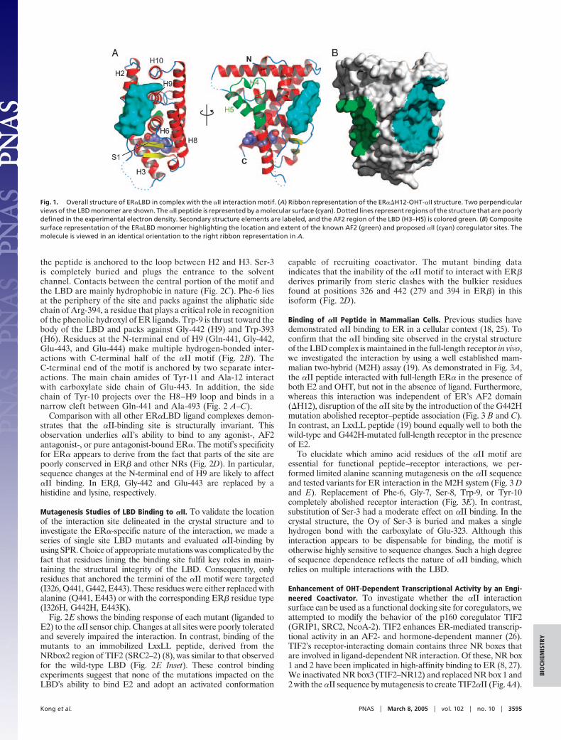
Peptide-Binding Site. The ER��H12-OHT-�II peptide complexcrystallises with a single LBD homodimer in the asymmetric unit.Each monomer within the homodimer interacts with a peptidemotif (Fig. 6A, which is published as supporting information on thePNAS web site). Difference Fourier maps revealed well definedelectron density for the peptide, allowing us to model the entire11-residue �II sequence (Fig. 6B). The �II motif binds across the�-hairpin face of the LBD (Fig. 1A). The interaction site is centeredon a shallow depression between H2, H6, and H9 that surrounds theentrance of the solvent-filled channel that leads to the buriedhormone-binding cavity. This surface lies diametrically opposite thereceptor’s AF2 coactivator recruitment site (Fig. 1B). Residuesfrom H2, the H2–H3 loop, H6, the S1–S2 �-hairpin, H8–H9 loop,H9, and H10 contribute to an extensive binding surface (10 Å by 24Å long).
The �II peptide adopts an extended conformation that closelymatches the surface topology of the LBD (Fig. 2A). ResiduesLeu-1–Phe-6 run parallel to the S1 strand with residues Ser-3–Asp-5adopting a 310 helical conformation. At Gly-7, the peptide backboneturns 90° and climbs toward H10. This bent conformation isstabilized by an intramolecular interaction between the backbonecarbonyl of Phe-6 and the indole nitrogen of Trp-9. Gly-7 appearsto fulfil a structural role because of the strained backbone dihedralangles required at this position.
LBD recognition is mediated by an extensive network ofhydrogen-bonded and nonpolar interactions. No single interactionappears to dominate, and the observed peptide conformation isreliant on multiple intra- and intermolecular contacts. Binding ofthe �II motif buries 650 Å2 of predominantly hydrophobic surfacearea on the LBD. Residues from the peptide make a total of eightdirect hydrogen bonds with the LBD (Fig. 2B). The N terminus of
3594 � www.pnas.org�cgi�doi�10.1073�pnas.0407189102 Kong et al.
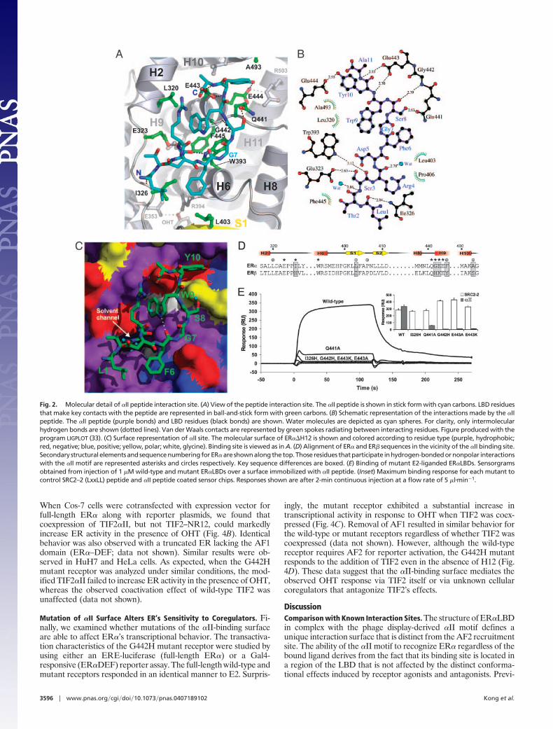
the peptide is anchored to the loop between H2 and H3. Ser-3is completely buried and plugs the entrance to the solventchannel. Contacts between the central portion of the motif andthe LBD are mainly hydrophobic in nature (Fig. 2C). Phe-6 liesat the periphery of the site and packs against the aliphatic sidechain of Arg-394, a residue that plays a critical role in recognitionof the phenolic hydroxyl of ER ligands. Trp-9 is thrust toward thebody of the LBD and packs against Gly-442 (H9) and Trp-393(H6). Residues at the N-terminal end of H9 (Gln-441, Gly-442,Glu-443, and Glu-444) make multiple hydrogen-bonded inter-actions with C-terminal half of the �II motif (Fig. 2B). TheC-terminal end of the motif is anchored by two separate inter-actions. The main chain amides of Tyr-11 and Ala-12 interactwith carboxylate side chain of Glu-443. In addition, the sidechain of Tyr-10 projects over the H8–H9 loop and binds in anarrow cleft between Gln-441 and Ala-493 (Fig. 2 A–C).
Comparison with all other ER�LBD ligand complexes demon-strates that the �II-binding site is structurally invariant. �hisobservation underlies �II’s ability to bind to any agonist-, AF2antagonist-, or pure antagonist-bound ER�. The motif’s specificityfor ER� appears to derive from the fact that parts of the site arepoorly conserved in ER� and other NRs (Fig. 2D). In particular,sequence changes at the N-terminal end of H9 are likely to affect�II binding. In ER�, Gly-442 and Glu-443 are replaced by ahistidine and lysine, respectively.
Mutagenesis Studies of LBD Binding to �II. To validate the locationof the interaction site delineated in the crystal structure and toinvestigate the ER�-specific nature of the interaction, we made aseries of single site LBD mutants and evaluated �II-binding byusing SPR. Choice of appropriate mutations was complicated by thefact that residues lining the binding site fulfil key roles in main-taining the structural integrity of the LBD. Consequently, onlyresidues that anchored the termini of the �II motif were targeted(I326, Q441, G442, E443). These residues were either replaced withalanine (Q441, E443) or with the corresponding ER� residue type(I326H, G442H, E443K).
Fig. 2E shows the binding response of each mutant (liganded toE2) to the �II sensor chip. Changes at all sites were poorly toleratedand severely impaired the interaction. In contrast, binding of themutants to an immobilized LxxLL peptide, derived from theNRbox2 region of TIF2 (SRC2–2) (8), was similar to that observedfor the wild-type LBD (Fig. 2E Inset). These control bindingexperiments suggest that none of the mutations impacted on theLBD’s ability to bind E2 and adopt an activated conformation
capable of recruiting coactivator. The mutant binding dataindicates that the inability of the �II motif to interact with ER�derives primarily from steric clashes with the bulkier residuesfound at positions 326 and 442 (279 and 394 in ER�) in thisisoform (Fig. 2D).
Binding of �II Peptide in Mammalian Cells. Previous studies havedemonstrated �II binding to ER in a cellular context (18, 25). Toconfirm that the �II binding site observed in the crystal structureof the LBD complex is maintained in the full-length receptor in vivo,we investigated the interaction by using a well established mam-malian two-hybrid (M2H) assay (19). As demonstrated in Fig. 3A,the �II peptide interacted with full-length ER� in the presence ofboth E2 and OHT, but not in the absence of ligand. Furthermore,whereas this interaction was independent of ER’s AF2 domain(�H12), disruption of the �II site by the introduction of the G442Hmutation abolished receptor–peptide association (Fig. 3 B and C).In contrast, an LxxLL peptide (19) bound equally well to both thewild-type and G442H-mutated full-length receptor in the presenceof E2.
To elucidate which amino acid residues of the �II motif areessential for functional peptide–receptor interactions, we per-formed limited alanine scanning mutagenesis on the �II sequenceand tested variants for ER interaction in the M2H system (Fig. 3 Dand E). Replacement of Phe-6, Gly-7, Ser-8, Trp-9, or Tyr-10completely abolished receptor interaction (Fig. 3E). In contrast,substitution of Ser-3 had a moderate effect on �II binding. In thecrystal structure, the O� of Ser-3 is buried and makes a singlehydrogen bond with the carboxylate of Glu-323. Although thisinteraction appears to be dispensable for binding, the motif isotherwise highly sensitive to sequence changes. Such a high degreeof sequence dependence reflects the nature of �II binding, whichrelies on multiple interactions with the LBD.
Enhancement of OHT-Dependent Transcriptional Activity by an Engi-neered Coactivator. To investigate whether the �II interactionsurface can be used as a functional docking site for coregulators, weattempted to modify the behavior of the p160 coregulator TIF2(GRIP1, SRC2, NcoA-2). TIF2 enhances ER-mediated transcrip-tional activity in an AF2- and hormone-dependent manner (26).TIF2’s receptor-interacting domain contains three NR boxes thatare involved in ligand-dependent NR interaction. Of these, NR box1 and 2 have been implicated in high-affinity binding to ER (8, 27).We inactivated NR box3 (TIF2–NR12) and replaced NR box 1 and2 with the �II sequence by mutagenesis to create TIF2�II (Fig. 4A).
Fig. 1. Overall structure of ER�LBD in complex with the �II interaction motif. (A) Ribbon representation of the ER��H12-OHT-�II structure. Two perpendicularviews of the LBD monomer are shown. The �II peptide is represented by a molecular surface (cyan). Dotted lines represent regions of the structure that are poorlydefined in the experimental electron density. Secondary structure elements are labeled, and the AF2 region of the LBD (H3–H5) is colored green. (B) Compositesurface representation of the ER�LBD monomer highlighting the location and extent of the known AF2 (green) and proposed �II (cyan) coregulator sites. Themolecule is viewed in an identical orientation to the right ribbon representation in A.
Kong et al. PNAS � March 8, 2005 � vol. 102 � no. 10 � 3595
BIO
CHEM
ISTR
Y
When Cos-7 cells were cotransfected with expression vector forfull-length ER� along with reporter plasmids, we found thatcoexpression of TIF2�II, but not TIF2–NR12, could markedlyincrease ER activity in the presence of OHT (Fig. 4B). Identicalbehavior was also observed with a truncated ER lacking the AF1domain (ER�–DEF; data not shown). Similar results were ob-served in HuH7 and HeLa cells. As expected, when the G442Hmutant receptor was analyzed under similar conditions, the mod-ified TIF2�II failed to increase ER activity in the presence of OHT,whereas the observed coactivation effect of wild-type TIF2 wasunaffected (data not shown).
Mutation of �II Surface Alters ER’s Sensitivity to Coregulators. Fi-nally, we examined whether mutations of the �II-binding surfaceare able to affect ER�’s transcriptional behavior. The transactiva-tion characteristics of the G442H mutant receptor were studied byusing either an ERE-luciferase (full-length ER�) or a Gal4-responsive (ER�DEF) reporter assay. The full-length wild-type andmutant receptors responded in an identical manner to E2. Surpris-
ingly, the mutant receptor exhibited a substantial increase intranscriptional activity in response to OHT when TIF2 was coex-pressed (Fig. 4C). Removal of AF1 resulted in similar behavior forthe wild-type or mutant receptors regardless of whether TIF2 wascoexpressed (data not shown). However, although the wild-typereceptor requires AF2 for reporter activation, the G442H mutantresponds to the addition of TIF2 even in the absence of H12 (Fig.4D). These data suggest that the �II-binding surface mediates theobserved OHT response via TIF2 itself or via unknown cellularcoregulators that antagonize TIF2’s effects.
DiscussionComparison with Known Interaction Sites. The structure of ER�LBDin complex with the phage display-derived �II motif defines aunique interaction surface that is distinct from the AF2 recruitmentsite. The ability of the �II motif to recognize ER� regardless of thebound ligand derives from the fact that its binding site is located ina region of the LBD that is not affected by the distinct conforma-tional effects induced by receptor agonists and antagonists. Previ-
Fig. 2. Molecular detail of �II peptide interaction site. (A) View of the peptide interaction site. The �II peptide is shown in stick form with cyan carbons. LBD residuesthat make key contacts with the peptide are represented in ball-and-stick form with green carbons. (B) Schematic representation of the interactions made by the �IIpeptide. The �II peptide (purple bonds) and LBD residues (black bonds) are shown. Water molecules are depicted as cyan spheres. For clarity, only intermolecularhydrogen bonds are shown (dotted lines). Van der Waals contacts are represented by green spokes radiating between interacting residues. Figure produced with theprogram LIGPLOT (33). (C) Surface representation of �II site. The molecular surface of ER��H12 is shown and colored according to residue type (purple, hydrophobic;red, negative; blue, positive; yellow, polar; white, glycine). Binding site is viewed as in A. (D) Alignment of ER� and ER� sequences in the vicinity of the �II binding site.Secondary structural elements and sequence numbering for ER� are shown along the top. Those residues that participate in hydrogen-bonded or nonpolar interactionswith the �II motif are represented asterisks and circles respectively. Key sequence differences are boxed. (E) Binding of mutant E2-liganded ER�LBDs. Sensorgramsobtained from injection of 1 �M wild-type and mutant ER�LBDs over a surface immobilized with �II peptide. (Inset) Maximum binding response for each mutant tocontrol SRC2–2 (LxxLL) peptide and �II peptide coated sensor chips. Responses shown are after 2-min continuous injection at a flow rate of 5 �l�min�1.
3596 � www.pnas.org�cgi�doi�10.1073�pnas.0407189102 Kong et al.

ous studies have shown that the AF2 region of the LBD serves asthe primary docking site for coactivators and corepressors (4–8, 12,13). The �II site identified here is located on the opposite face ofthe LBD (Fig. 1B). Both surfaces are predominantly hydrophobicin character but differ in topology. The LxxLL binding groove iscompact, encompasses a relatively small surface area (450 Å2), andis conserved in most NRs. In contrast, the �II site covers a largerarea (650 Å2) and is specific to ER�. The concave �II bindingsurface incorporates conserved regions of the LBD that flank theentrance to a channel that leads to the buried hormone bindingcavity. Binding of the �II peptide blocks the entrance to this channeland creates a 300-Å3 water-filled chamber. It is noted this chamber
corresponds to the so-called ‘‘second-binding site’’ that has beenevaluated as an alternative ligand-binding cavity (28). However, noexperimentally validated function has been attributed to this regionof ER�LBD, and its role in ligand-binding remains controversial.Consequently, it is highly unlikely that the �II site is involved inbinding small molecule ligands.
Biological Significance of the �II Sequence. What is the likelihoodthat the �II sequence represents a previously unknown, biologicallyrelevant recruitment motif? In the original phage display study, the�II motif was only observed once and, therefore, no consensusinformation is available (16). Our mutagenesis studies demonstrate
Fig. 3. ER�-peptide interaction in mammalian cells. Interac-tion of GAL4-DBD-tagged �II with VP16-tagged ERs. Full-length ER� (A), ER��H12 (B), and ER�G442H (C). (D and E)Amino acid requirements for �II interaction. (D) Amino acidsequences of peptides used. (E) Mammalian two-hybrid re-ceptor binding assay of GAL4-DBD-tagged wild type and mu-tant peptides to VP16-ER� wild type. Binding of the �II andLxxLL peptides were evaluated both in the absence of ligand(NH) and in the presence of E2 and OHT. Normalized luciferaseactivity is shown.
Fig. 4. Enhancement of tamoxifen-dependent transcrip-tional activity. (A) Schematic representation of NR-boxmodified coregulator TIF2 containing the �II peptide se-quence. (B) Cell-based reporter assay of transcriptional ac-tivity of engineered TIF2 variants in the presence of full-length ER�. (C and D) Reporter activity of wild-type andmutant ER in the presence of 100 nM OHT. Data are pre-sented as percent activation, where the activity of full-length ER� (C) and ER�DEF�H12 (D) are set to 100%.
Kong et al. PNAS � March 8, 2005 � vol. 102 � no. 10 � 3597
BIO
CHEM
ISTR
Y

that its interaction with ER is highly sequence-dependent. Data-base searches with either the �II sequence or a redundant motifbased on the binding mode observed in the crystal structure do notyield any obvious homologies. Based on the apparent lack of keyhotspots within the sequence, we currently favor a model in whichthe �II peptide acts as a structural, rather than sequence mimic ofthe true interaction partner. Consequently, the possibility thatelements of the �II motif are incorporated into a nonlinearrecognition module, formed by discontinuous regions of primarysequence, cannot be ruled out.
�II Interaction Surface: A Possible Coregulator Recruitment Site? Thequestion remains as to whether the �II motif acts as a fortuitousconformational probe or an actual mimic of a bona fide ERinteraction partner? Norris et al. (18) demonstrated that �II is ableto inhibit the partial agonist activity of OHT while having a minimaleffect on E2-mediated transcription. This observation stronglysuggests that �II’s cognate binding surface on ER� represents animportant control site that is involved in regulating receptor activityunder certain conditions. Short peptides that act as antagonists ofprotein-protein interactions have been isolated from naive librariesfor a number of systems. Subsequent structural analysis has shownthat the peptide binding site overlaps with that of a known bindingpartner (29). In the case of �II, the crystal structure of the complexindicates that peptide binding to the isolated LBD does not induceany significant conformational changes. This finding suggests that�II’s antagonistic properties are manifested by a direct steric effectin which the peptide occludes a key binding site. At this stage, it isunclear whether the �II site is a docking surface for another regionof ER (i.e., involved in intramolecular domain signaling) or for anas yet unidentified coregulator. The coactivator engineering exper-iments clearly demonstrate that the �II binding surface can serveas a valid receptor–coregulator interaction site. By replacing theNR-box�LxxLL regions of TIF2 with the �II sequence, we gener-ated a coactivator (TIF2�II) that could be recruited to the �IIsurface by both agonist and antagonist-bound ER in an AF1�AF2-independent fashion.
Our preliminary functional analysis indicates that the �II surfacemay play a role in ER’s sensitivity to coregulators in the presenceof selective ER receptor modulators such as OHT (Fig. 4C). Nodifferences in behavior were observed between wild-type and
�II-mutated ER in the presence of E2. This ligand-dependencymirrors the previously reported antagonistic effects of the �IIpeptide on E2- and OHT-dependent receptor activity (18). Thesurprising result that the TIF2-mediated enhancement of OHTactivity seen for the G442H mutant requires a functional AF2H12�F domain only in the context of the full-length ER, but not inthe context of the ER DEF, reinforces the idea that the functionalinterplay between different ER domains and coregulators deter-mines the transcriptional properties of the wild type receptor (30).Furthermore, our demonstration that disruption of the �II bindingsite enhanced rather than decreased the response to TIF2 does notnecessarily contradict the currently held view that TIF2 acts pri-marily as a coactivator. Although TIF2 coactivator function isusually associated with recruitment to a functional AF1�2 surface,targeting to other surfaces cannot be discounted. Recent studies byYamamoto and coworkers (31, 32) discovered a context-dependentcorepressor function in TIF2, which may indeed account for theeffect seen in our study. Alternatively, TIF2’s role could be indirectand mediated by either endogenous coregulators known to coop-erate with TIF2 (e.g., CBP�p300, CARM-1) or unidentified core-pressors that bind directly to the �II surface. Indeed, although ourcoregulator engineering experiment targeted a coactivator to the�II surface, similar experiments utilizing an engineered corepressorwould probably have illustrated that the �II site may alternativelybe involved in corepressor binding.
In summary, the site identified in this study appears to beinvolved in maintaining a reduced level of receptor activation in thepresence of TIF2. Further studies are required to establish whetherother recognized or unrecognized ER coactivators and corepres-sors also display sensitivity to the disruption of this unique controlsurface.
We thank Benita Katzenellenbogen for providing the ER�LBD expres-sion construct, Geoffrey Greene for advice on the preparation of the E2affinity resin, and Peter O’Brien for synthesis of the epoxypropyl-E2 usedin the resin coupling reaction. We also thank staff on ID29 (EuropeanSynchrotron Radiation Facility, Grenoble, France) for technical supportduring data collection. A.C.W.P. is funded by a Wellcome Trust CareerDevelopment Fellowship, and E.H.K. is funded by the Biotechnologyand Biological Sciences Research Council. J.-Å.G., E.T., and N.H. aresupported by grants from the Swedish Research Council, the SwedishCancer Society, and Karo Bio AB.
1. Tora, L., White, J., Brou, C., Tasset, D., Webster, N., Scheer, E. & Chambon, P.(1989) Cell 59, 477–487.
2. Smith, C. L. & O’Malley, B. W. (2004) Endocr. Rev. 25, 45–71.3. Heery, D. M., Kalkhoven, E., Hoare, S. & Parker, M. G. (1997) Nature 387,
733–736.4. Darimont, B. D., Wagner, R. L., Apriletti, J. W., Stallcup, M. R., Kushner, P. J.,
Baxter, J. D., Fletterick, R. J. & Yamamoto, K. R. (1998) Genes Dev. 12,3343–3356.
5. McInerney, E. M., Rose, D. W., Flynn, S. E., Westin, S., Mullen, T. M., Krones,A., Inostroza, J., Torchia, J., Nolte, R. T., Assa-Munt, N., et al. (1998) Genes Dev.12, 3357–3368.
6. Nolte, R. T., Wisely, G. B., Westin, S., Cobb, J. E., Lambert, M. H., Kurokawa,R., Rosenfeld, M. G., Willson, T. M., Glass, C. K. & Milburn, M. V. (1998) Nature395, 137–143.
7. Shiau, A. K., Barstad, D., Loria, P. M., Cheng, L., Kushner, P. J., Agard, D. A.& Greene, G. L. (1998) Cell 95, 927–937.
8. Warnmark, A., Treuter, E., Gustafsson, J.-Å., Hubbard, R. E., Brzozowski, A. M.& Pike, A. C. W. (2002) J. Biol. Chem. 277, 21862–21868.
9. Brzozowski, A. M., Pike, A. C. W., Dauter, Z., Hubbard, R. E., Bonn, T., Engstrom,O., Ohman, L., Greene, G. L., Gustafsson, J.-Å. & Carlquist, M. (1997) Nature 389,753–758.
10. Shang, Y. F., Hu, X., DiRenzo, J., Lazar, M. A. & Brown, M. (2000) Cell 103,843–852.
11. Hu, X. & Lazar, M. A. (1999) Nature 402, 93–96.12. Xu, H. E., Stanley, T. B., Montana, V. G., Lambert, M. H., Shearer, B. G., Cobb,
J. E., McKee, D. D., Galardi, C. M., Plunket, K. D., Nolte, R. T., et al. (2002)Nature 415, 813–817.
13. Huang, H. J., Norris, J. D. & McDonnell, D. P. (2002) Mol. Endocrinol. 16,1778–1792.
14. Katzenellenbogen, B. S. & Katzenellenbogen, J. A. (2002) Science 295, 2380–2381.15. Shang, Y. F. & Brown, M. (2002) Science 295, 2465–2468.
16. Paige, L. A., Christensen, D. J., Gron, H., Norris, J. D., Gottlin, E. B., Padilla,K. M., Chang, C. Y., Ballas, L. M., Hamilton, P. T., McDonnell, D. P. & Fowlkes,D. M. (1999) Proc. Natl. Acad. Sci. USA 96, 3999–4004.
17. Chang, C. Y., Norris, J. D., Gron, H., Paige, L. A., Hamilton, P. T., Kenan, D. J.,Fowlkes, D. & McDonnell, D. P. (1999) Mol. Cell. Biol. 19, 8226–8239.
18. Norris, J. D., Paige, L. A., Christensen, D. J., Chang, C. Y., Huacani, M. R., Fan, D. J.,Hamilton, P. T., Fowlkes, D. M. & McDonnell, D. P. (1999) Science 285, 744–746.
19. Heldring, N., Nilsson, M., Buehrer, B., Treuter, E. & Gustafsson, J.-Å. (2004)Mol. Cell. Biol. 24, 3445–3459.
20. Carlson, K. E., Choi, I., Gee, A., Katzenellenbogen, B. S. & Katzenellenbogen,J. A. (1997) Biochemistry 36, 14897–14905.
21. Goldstein, S. W., Bordner, J., Hoth, L. R. & Geoghegan, K. F. (2001) Bioconjug.Chem. 12, 406–413.
22. Otwinowski, Z. & Minor, W. (1997) Methods Enzymol. 276, 307–326.23. Collaborative Computational Project No. 4 (1994) Acta Crystallogr. D 50,
760–763.24. Murshudov, G. N., Vagin, A. A. & Dodson, E. J. (1997) Acta Crystallogr. D 53,
240–255.25. Bapat, A. R. & Frail, D. E. (2003) J. Steroid Biochem. Mol. Biol. 86, 143–149.26. Hong, H., Kohli, K., Garabedian, M. J. & Stallcup, M. R. (1997) Mol. Cell. Biol.
17, 2735–2744.27. Warnmark, A., Almlof, T., Leers, J., Gustafsson, J.-Å. & Treuter, E. (2001) J. Biol.
Chem. 276, 23397–23404.28. van Hoorn, W. P. (2002) J. Med. Chem. 45, 584–589.29. Sidhu, S. S., Fairbrother, W. J. & Deshayes, K. (2003) ChemBioChem 4, 14–25.30. Metivier, R., Stark, A., Flouriot, G., Hubner, M. R., Brand, H., Penot, G., Manu,
D., Denger, S., Reid, G., Kos, M., et al. (2002) Mol. Cell 10, 1019–1032.31. Rogatsky, I., Zarember, K. A. & Yamamoto, K. R. (2001) EMBO J. 20,
6071–6083.32. Rogatsky, I., Luecke, H. F., Leitman, D. C. & Yamamoto, K. R. (2002) Proc. Natl.
Acad. Sci. USA 99, 16701–16706.33. Wallace, A. C., Laskowski, R. A. & Thornton, J. M. (1995) Protein Eng. 8, 127–134.
3598 � www.pnas.org�cgi�doi�10.1073�pnas.0407189102 Kong et al.

Corrections
For the following 18 articles, the authors note, in addition to thepartial funding of this study from KaroBio AB, that J.-Å.G. iscofounder, deputy board member, stockholder, and consultantof KaroBio AB.
(i) NEUROSCIENCE. ‘‘Liver X receptors in the central nervoussystem: From lipid homeostasis to neuronal degeneration,’’ byLing Wang, Gertrud U. Schuster, Kjell Hultenby, QinghongZhang, Sandra Andersson, and Jan-Åke Gustafsson, whichappeared in issue 21, October 15, 2002, of Proc. Natl. Acad. Sci.USA (99, 13878–13883; first published October 4, 2002; 10.1073�pnas.172510899);
(ii) DEVELOPMENTAL BIOLOGY. ‘‘An endocrine pathway in theprostate, ER�, AR, 5�-androstane-3�,17�-diol, and CYP7B1,regulates prostate growth,’’ by Zhang Weihua, Richard Lathe,Margaret Warner, and Jan-Åke Gustafsson, which appeared inissue 21, October 15, 2002, of Proc. Natl. Acad. Sci. USA (99,13589–13594; first published October 7, 2002; 10.1073�pnas.162477299);
(iii) MEDICAL SCIENCES. ‘‘Involvement of estrogen receptor � interminal differentiation of mammary gland epithelium,’’ byCarola Forster, Sari Makela, Anni Warri, Silke Kietz, DavidBecker, Kjell Hultenby, Margaret Warner, and Jan-Åke Gustafs-son, which appeared in issue 24, November 26, 2002, of Proc.Natl. Acad. Sci. USA (99, 15578–15583; first published November18, 2002; 10.1073�pnas.192561299);
(iv) NEUROSCIENCE. ‘‘Estrogen receptor (ER)� knockout micereveal a role for ER� in migration of cortical neurons in thedeveloping brain,’’ by Ling Wang, Sandra Andersson, MargaretWarner, and Jan-Åke Gustafsson, which appeared in issue 2,January 21, 2003, of Proc. Natl. Acad. Sci. USA (100, 703–708;first published January 6, 2003; 10.1073�pnas.242735799);
(v) MEDICAL SCIENCES. ‘‘Disruption of the estrogen receptor �gene in mice causes myeloproliferative disease resemblingchronic myeloid leukemia with lymphoid blast crisis,’’ by Gil-JinShim, Ling Wang, Sandra Andersson, Noemi Nagy, LorandLevente Kis, Qinghong Zhang, Sari Makela, Margaret Warner,and Jan-Åke Gustafsson, which appeared in issue 11, May 27,2003, of Proc. Natl. Acad. Sci. USA (100, 6694–6699; firstpublished May 9, 2003; 10.1073�pnas.0731830100);
(vi) MEDICAL SCIENCES. ‘‘Differential effects on bone of estrogenreceptor � and androgen receptor activation in orchidectomizedadult male mice,’’ by Sofia Moverare, Katrien Venken, Anna-Lena Eriksson, Niklas Andersson, Stanko Skrtic, Jon Wergedal,Subburaman Mohan, Phil Salmon, Roger Bouillon, Jan-ÅkeGustafsson, Dirk Vanderschueren, and Claes Ohlsson, whichappeared in issue 23, November 11, 2003, of Proc. Natl. Acad. Sci.USA (100, 13573–13578; first published October 22, 2003;10.1073�pnas.2233084100);
(vii) MEDICAL SCIENCES. ‘‘Autoimmune glomerulonephritis withspontaneous formation of splenic germinal centers in micelacking the estrogen receptor alpha gene,’’ by Gil-Jin Shim,Lorand Levente Kis, Margaret Warner, and Jan-Åke Gustafs-son, which appeared in issue 6, February 10, 2004, of Proc. Natl.Acad. Sci. USA (101, 1720–1724; first published January 26,2004; 10.1073�pnas.0307915100);
(viii) CELL BIOLOGY. ‘‘Estrogen receptor � inhibits 17�-estradiol-stimulated proliferation of the breast cancer cell line T47D,’’ byAnders Strom, Johan Hartman, James S. Foster, Silke Kietz, JayWimalasena, and Jan-Åke Gustafsson, which appeared in issue 6,February 10, 2004, of Proc. Natl. Acad. Sci. USA (101, 1566–1571;first published January 26, 2004; 10.1073�pnas.0308319100);
(ix) INAUGURAL ARTICLE, PHYSIOLOGY. ‘‘Estrogen receptors ER�and ER� in proliferation in the rodent mammary gland,’’ byGuojun Cheng, Zhang Weihua, Margaret Warner, and Jan-ÅkeGustafsson, which appeared in issue 11, March 16, 2004, of Proc.Natl. Acad. Sci. USA (101, 3739–3746; first published February4, 2004; 10.1073�pnas.0307864100);
(x) MEDICAL SCIENCES. ‘‘Estrogen receptor � regulates epithelialcellular differentiation in the mouse ventral prostate,’’ byOtabek Imamov, Andrea Morani, Gil-Jin Shim, Yoko Omoto,Christina Thulin-Andersson, Margaret Warner, and Jan-ÅkeGustafsson, which appeared in issue 25, June 22, 2004, of Proc.Natl. Acad. Sci. USA (101, 9375–9380; first published June 8,2004; 10.1073�pnas.0403041101);
(xi) IMMUNOLOGY. ‘‘Aromatase-deficient mice spontaneouslydevelop a lymphoproliferative autoimmune disease resemblingSjogren’s syndrome,’’ by Gil-Jin Shim, Margaret Warner, Hyun-Jin Kim, Sandra Andersson, Lining Liu, Jenny Ekman, OtabekImamov, Margaret E. Jones, Evan R. Simpson, and Jan-ÅkeGustafsson, which appeared in issue 34, August 24, 2004, of Proc.Natl. Acad. Sci. USA (101, 12628–12633; first published August16, 2004; 10.1073�pnas.0405099101);
(xii) MEDICAL SCIENCES. ‘‘Characterization of the ER��/� mouseheart,’’ by Carola Forster, Silke Kietz, Kjell Hultenby, MargaretWarner, and Jan-Åke Gustafsson, which appeared in issue 39,September 28, 2004, of Proc. Natl. Acad. Sci. USA (101, 14234–14239; first published September 16, 2004; 10.1073�pnas.0405571101);
(xiii) DEVELOPMENTAL BIOLOGY. ‘‘Estrogen receptor � and im-printing of the neonatal mouse ventral prostate by estrogen,’’ byYoko Omoto, Otabek Imamov, Margaret Warner, and Jan-ÅkeGustafsson, which appeared in issue 5, February 1, 2005, of Proc.Natl. Acad. Sci. USA (102, 1484–1489; first published January 21,2005; 10.1073�pnas.0409168102);
(xiv) DEVELOPMENTAL BIOLOGY. ‘‘Early onset of puberty andearly ovarian failure in CYP7B1 knockout mice,’’ by Yoko Omoto,Richard Lathe, Margaret Warner, and Jan-Åke Gustafsson, whichappeared in issue 8, February 22, 2005, of Proc. Natl. Acad. Sci. USA(102, 2814–2819; first published February 14, 2005; 10.1073�pnas.0500198102);
(xv) BIOCHEMISTRY. ‘‘Delineation of a unique protein–proteininteraction site on the surface of the estrogen receptor,’’ by EricH. Kong, Nina Heldring, Jan-Åke Gustafsson, Eckardt Treuter,Roderick E. Hubbard, and Ashley C. W. Pike, which appearedin issue 10, March 8, 2005, of Proc. Natl. Acad. Sci. USA (102,3593–3598; first published February 23, 2005; 10.1073�pnas.0407189102);
(xvi) NEUROSCIENCE. ‘‘Inactivation of liver X receptor � leads toadult-onset motor neuron degeneration in male mice,’’ by San-dra Andersson, Nina Gustafsson, Margaret Warner, and Jan-Åke Gustafsson, which appeared in issue 10, March 8, 2005, ofProc. Natl. Acad. Sci. USA (102, 3857–3862; first publishedFebruary 28, 2005; 10.1073�pnas.0500634102);
(xvii) PHYSIOLOGY. ‘‘Muscle GLUT4 regulation by estrogenreceptors ER� and ER�,’’ by Rodrigo P. A. Barros, Ubiratan F.Machado, Margaret Warner, and Jan-Åke Gustafsson, whichappeared in issue 5, January 31, 2006, of Proc. Natl. Acad. Sci.USA (103, 1605–1608; first published January 19, 2006; 10.1073�pnas.0510391103);
8298–8299 � PNAS � May 23, 2006 � vol. 103 � no. 21 www.pnas.org

and (xviii) PHYSIOLOGY. ‘‘Role of estrogen receptor � in colonicepithelium,’’ by Osamu Wada-Hiraike, Otabek Imamov, HarukoHiraike, Kjell Hultenby, Thomas Schwend, Yoko Omoto, Mar-garet Warner, and Jan-Åke Gustafsson, which appeared in issue
8, February 21, 2006, of Proc. Natl. Acad. Sci. USA (103,2959–2964; first published February 13, 2006; 10.1073�pnas.0511271103).www.pnas.org�cgi�doi�10.1073�pnas.0602780103
PNAS � May 23, 2006 � vol. 103 � no. 21 � 8299
CORR
ECTI
ON
S
Copyright © 2022 FDOKUMEN