Discovery of Selective Probes and Antagonists for G Protein-Coupled Receptors FPR/FPRL1 and GPR30
THE G PROTEIN-COUPLED ESTROGEN RECEPTOR GPER/GPR30 AS A REGULATOR OF CARDIOVASCULAR FUNCTION
Matthias R. Meyer1,2, Eric R. Prossnitz2, and Matthias Barton1,*
1Molecular Internal Medicine, University of Zurich, Zurich, Switzerland 2Department of CellBiology and Physiology, University of New Mexico Health Sciences Center, Albuquerque, NM,United States
AbstractEndogenous estrogens are important regulators of cardiovascular homeostasis in premenopausalwomen and interfere with the development of hypertension and coronary artery disease. Thesehormones act via three different estrogen receptors affecting both gene transcription and rapidsignaling pathways in a complex interplay. In addition to the classical estrogen receptors ERα andERβ, which are known mediators of estrogen-dependent vascular effects, a G protein-coupledestrogen receptor termed GPER that is expressed in the cardiovascular system has recently beenidentified. Endogenous human 17β-estradiol, selective estrogen receptor modulators (SERMs)including tamoxifen and raloxifene, and selective estrogen receptor downregulators (SERDs) suchas ICI 182,780 are all agonists of GPER, which has been implicated in the regulation of vasomotortone and protection from myocardial ischemia/reperfusion injury. As a result, understanding theindividual role of ERα, ERβ, and GPER in cardiovascular function has become increasinglycomplex. With accumulating evidence that GPER is responsible for a variety of beneficialcardiovascular effects of estrogens, this receptor may represent a novel target to develop effectivestrategies for the treatment of cardiovascular diseases by tissue-specific, selective activation ofestrogen-dependent molecular pathways devoid of side effects seen with conventional hormonetherapy.
KeywordsBlood Pressure; Endothelium; Menopause; Nitric Oxide; Vasodilation
1. Estrogen Receptors and Vascular Estrogen SignalingHypertension affects one out of four women worldwide, and its prevalence is particularlyhigh among females over 60 years of age (Kearney et al., 2005). Indeed, the first decadeafter menopause is accompanied by an increase in blood pressure (Burt et al., 1995),pointing to a blood pressure-lowering effect of endogenous estrogens (Barton and Meyer,
© 2011 Elsevier Inc. All rights reserved.*Correspondence to: Matthias Barton, M.D., Professor and Head, Molecular Internal Medicine, University of Zurich, LTK Y44 G22,Winterthurerstrasse 190, CH-8057 Zurich, Switzerland, Tel: +41 77 439 55 54, Fax: +41 44 635 68 75, [email protected]'s Disclaimer: This is a PDF file of an unedited manuscript that has been accepted for publication. As a service to ourcustomers we are providing this early version of the manuscript. The manuscript will undergo copyediting, typesetting, and review ofthe resulting proof before it is published in its final citable form. Please note that during the production process errors may bediscovered which could affect the content, and all legal disclaimers that apply to the journal pertain.Conflict of InterestE.R.P. is an inventor on United States Patent Number 7,875,721.
NIH Public AccessAuthor ManuscriptVascul Pharmacol. Author manuscript; available in PMC 2012 July 5.
Published in final edited form as:Vascul Pharmacol. 2011 ; 55(1-3): 17–25. doi:10.1016/j.vph.2011.06.003.
NIH
-PA Author Manuscript
NIH
-PA Author Manuscript
NIH
-PA Author Manuscript
2009). Importantly, mild elevations in blood pressure can often been found in womenshortly after menopause (Barton and Meyer, 2009) and have been associated with a higherrisk of cardiovascular diseases such as myocardial infarction, stroke, and congestive heartfailure (Vasan et al., 2001). In line with these epidemiological data, a variety of beneficialcardiovascular effects of endogenous estrogens are known based on numerous experimentalstudies, including vasodilator activity and improvement of ischemia/reperfusion-relatedmyocardial injury (Deschamps et al., 2010; Meyer et al., 2006; Meyer and Barton, 2009).Conversely, randomized clinical trials using conjugated equine estrogens andmedroxyprogesterone acetate for postmenopausal hormone therapy could not recapitulatethese beneficial effects but reported increased cardiovascular risk instead (Hulley et al.,1998; Rossouw et al., 2002), indicating that this type of is not suitable for primary orsecondary prevention of cardiovascular diseases.
The controversial outcome of these trials may partly result from the complexity of vascularestrogen signaling (Figure 1), which involves at least three different estrogen receptors.Estrogen receptor α (ERα) was identified in the 1960’s (then termed ER) (Soloff and Szego,1969). In 1996, the less characterized estrogen receptor β (ERβ) was found (Kuiper et al.,1996). ERα and ERβ largely function as transcriptional regulators, but a small fraction mayalso be present at the plasma membrane, where estrogen binding activates intracellularsignaling pathways that mediate acute effects of estrogens, such as rapid vasodilation(Meyer et al., 2006; Meyer et al., 2009). In 1997, a third ER, the seven-transmembrane Gprotein-coupled ER termed GPR30, was cloned from shear-stress exposed humanendothelial cells among other sources (Takada et al., 1997). More recently, GPR30 has beenshown to activate rapid signaling cascades, such as extracellular signal-related kinase (ERK)and phosphatidylinositol-3-kinase (PI3K), after estrogen binding (Filardo et al., 2000;Revankar et al., 2005; Thomas et al., 2005). After establishing GPR30 as a bona fideestrogen-binding receptor, it was renamed GPER by the International Union of Basic andClinical Pharmacology (Alexander et al., 2008). Similar to ERα and ERβ (Meyer et al.,2006), GPER is expressed throughout the cardiovascular system in humans and animals ofboth sexes (Bopassa et al., 2010; Broughton et al., 2010; Deschamps and Murphy, 2009;Ding et al., 2009; Filice et al., 2009; Haas et al., 2007; Haas et al., 2009; Isensee et al., 2009;Lindsey et al., 2009; Ma et al., 2010; Takada et al., 1997). This suggests that in addition toERα and ERβ, GPER also plays a physiological role in regulation of vascular andmyocardial function. With the identification of three different receptors capable ofmediating vascular estrogen signaling, the understanding of the cardiovascular effects ofestrogen has become increasingly complex. This review addresses the individual role of thedifferent ERs in cardiovascular function with a particular focus on its newest member,GPER.
2. Ligands of Vascular Estrogen ReceptorsAlthough a variety of natural and synthetic estrogens are able to induce effects on thecardiovascular system, it is important to separate their ability to regulate ERα, ERβ, andGPER activity. 17β-Estradiol, the major human estrogen lost after menopause, is acombined activator of ERα, ERβ, and GPER, and mediates numerous beneficial vasculareffects. Although 17β-estradiol is primarily synthesized by the ovaries, the enzymearomatase converts androgens into 17β-estradiol and estrone at many sites throughout thebody including the vascular wall, where they may have specialized local effects (Harada etal., 1999). Estrone and other endogenous estrogen-based steroids display a low bindingaffinity for ERα, ERβ, and GPER at physiological concentrations, and their physiologicalactions are less clear (Kuiper et al., 1998; Revankar et al., 2005; Thomas et al., 2005). Onthe other hand, humans are exposed to a variety of natural and man-made estrogeniccompounds at low levels (Jacobs and Lewis, 2002). Natural environmental estrogens are
Meyer et al. Page 2
Vascul Pharmacol. Author manuscript; available in PMC 2012 July 5.
NIH
-PA Author Manuscript
NIH
-PA Author Manuscript
NIH
-PA Author Manuscript
synthesized by plants (phytoestrogens) and include coumestans and isoflavones, whereassynthetic estrogenic compounds (xenoestrogens, known as endocrine disruptors) comprisechemical detergents, pesticides, and plastic monomers. In fact, the isoflavone genistein isable to induce vasodilation (Martin et al., 2008), and has now been shown to not onlyactivate ERα and ERβ, but also GPER (Thomas and Dong, 2006). Some xenoestrogens suchas nonylphenol and DDT have also be implicated in regulation of vascular function (Hsiehet al., 2009; Ruehlmann et al., 1998), and these compounds weakly bind ERα/ERβ, and arealso agonists of GPER (Thomas and Dong, 2006). Taken together, numerous endogenousand exogenous estrogenic compounds implicated in cardiovascular physiology are nowknown to activate all three known estrogen receptors.
Research to improve successful treatment of estrogen-sensitive tumors, breast cancer inparticular, led to the development of drugs that oppose the action of estrogen. Referred to asselective estrogen receptor downregulators (SERDs), compounds such as ICI 182,780(fulvestrant) abolish ERα/ERβ signaling in all tissues (Shanle and Xu, 2010). However, agroup of compounds known as selective estrogen receptor modulators (SERMs) do notuniformly act as estrogen antagonists, but are generally regarded as ER agonists in thecardiovascular system, bone, and liver, and as ER antagonists in breast tissue(Katzenellenbogen and Katzenellenbogen, 2002). Importantly, the SERM tamoxifen as wellas the SERD ICI 182,780 do not solely interact witch ERα and ERβ; they also displaysignificant binding to GPER (Thomas et al., 2005) and induce GPER-dependent signaling inbreast cancer cell lines (Filardo et al., 2000; Revankar et al., 2005). Their role as GPERagonists has been confirmed in several other cell types and tissues (Prossnitz and Barton,2009), indicating that this receptor mediates estrogenic effects even with concomitantblockade of ERα and ERβ. This also indicates that older studies investigating cardiovasculareffects of SERMs and SERDs may in fact point to GPER-dependent effects.
As GPER is known to bind many of the same ligands as ERα and ERβ (Figure 2), selectiveagonists/antagonists were needed to unravel the functional roles of the individual receptors,particularly GPER. This led to the identification of the first GPER-specific agonist G-1(Bologa et al., 2006) and the first GPER-selective antagonist G15 (Dennis et al., 2009). Bothcompounds share a tetrahydro-3H-cyclopenta-[c]quinoline scaffold domain and show anextremely high selectivity for GPER over ERα and ERβ. In addition, agonists for ERα (PPT)and ERβ (DPN) (Harrington et al., 2003) have been used in the past years. The availabilityof these compounds has provided the opportunity to better characterize the role of individualERs, and GPER in particular, in cardiovascular physiology.
3. Mechanisms of Estrogen-Dependent Vasodilation17β-Estradiol is a powerful vasodilator of human blood vessels (Mügge et al., 1993; Silva deSa, Meirelles, 1977), and acute effects on vasomotor tone have been observed in arteries ofboth women and men (Haas et al., 2007; Mügge et al., 1993). Shortly after the seminalobservation that endothelial cells release a relaxing factor in response to acetylcholine(Furchgott and Zawadski, 1980) and its subsequent identification as nitric oxide (NO)(Furchgott, 1988; Ignarro et al., 1987), enhanced endothelium-dependent responses toacetylcholine were reported in estrogen-treated ovariectomized animals (Gisclard et al.,1988). Similarly, flow-mediated vasodilation and NO production correlate with highestrogen levels during the menstrual cycle and increase after estrogen therapy inpostmenopausal women (Miller and Duckles, 2008). These findings support the notion thatestrogen acutely and chronically enhances endothelium-/NO-dependent relaxation of humanarteries, including human coronary arteries (Barton et al., 1998; Miller, Duckles, 2008).Estrogen-stimulated endothelium-dependent NO release is the result of rapid PI3K/Aktactivation and subsequent phosphorylation of endothelial (type III) NO synthase (eNOS)
Meyer et al. Page 3
Vascul Pharmacol. Author manuscript; available in PMC 2012 July 5.
NIH
-PA Author Manuscript
NIH
-PA Author Manuscript
NIH
-PA Author Manuscript

(Haynes et al., 2000; Simoncini et al., 2000), although other mechanisms have also beenproposed (Chow et al., 2010). This effect is believed to be mediated by subpopulations ofERα and possibly ERβ that localize to vesicular invaginations of the plasma membranetermed caveolae (Figure 1) (Chambliss et al., 2000; Chambliss et al., 2002; Simoncini et al.,2000). However, endothelium-dependent relaxation in response to estrogens does notinvolve NO exclusively, but is also mediated by endothelium-derived hyperpolarizingfactors (EDHFs), including epoxyeicosatrienoic acids (EETs), H2O2, potassium, andendothelial gap junction communications (Huang et al., 2004; Luksha et al., 2006; Traupe etal., 2007). In addition, estrogens can act directly on vascular smooth muscle cells to regulatevascular function (White, 2002). These endothelium-independent effects include estrogen-dependent regulation of calcium and potassium fluxes (White, 2002), mainly through theactivity of large-conductance calcium- and voltage-activated potassium (BKCa) channels(Figure 1) (Han et al., 2007; White et al., 1995). Indeed, 17β-estradiol-dependent vasodilatorresponses in human coronary arteries remain effective after removal of endothelial cells(Mügge et al., 1993). Moreover, estrogen also indirectly reduces vasomotor tone byinhibiting the activity of vasoconstrictors, such as prostanoids, endothelin-1, and angiotensinII (Barton and Meyer, 2009; Mügge et al., 1997).
In addition to 17β-estradiol, an agonist of ERα, ERβ, and GPER, there has been greatinterest in defining the role of SERMs in the regulation of vascular tone. Tamoxifen andraloxifene, which are agonists of GPER (Prossnitz and Barton, 2009), activate BKCachannels and evoke acute endothelium-independent vasodilation (Darkow et al., 1997;Hutchison et al., 2001; Leung et al., 2007b). They also induce NO-mediated endothelium-dependent relaxation and increase eNOS phosphorylation in porcine coronary arteries andother vascular beds (Chan et al., 2010; Figtree et al., 1999; Leung et al., 2006; Leung et al.,2007a). Moreover, raloxifene activates eNOS via the PI3K/Akt-pathway in endothelial cells(Simoncini et al., 2002). Recently, the SERM estrogen-dendrimer conjugate was found toactivate eNOS and stimulate growth of endothelial cells (Chambliss et al., 2010). On theother hand, the endogenous SERM and cholesterol metabolite 27-hydroxycholesteroldecreases vascular NOS expression and activity (Umetani et al., 2007). Thus, both 17β-estradiol and several synthetically generated estrogen-like substances affect vascularfunction in distinct ways, although it is often unclear which ER mediates these responses.Defining the role of individual ERs in response to activation by SERMs may help to identifyand selectively target ERs that mediate the beneficial vascular effects of estrogens.
4. Role of ERα and ERβ for Vascular FunctionEstrogens acutely reduce vascular tone in various arteries of different species, yet themolecular mechanisms and the role of individual estrogen receptors in this response are lessclear. Long-term 17β-estradiol treatment increases basal NO production in thoracic aorta ofERβ-, but not of ERα-knockout mice, indicating that ERα mediates NO-dependentvasodilation (Darblade et al., 2002). On the other hand, ERβ-knockout mice developelevated blood pressure as they age, also suggesting a role for ERβ (Zhu et al., 2002).Indeed, disruption of ERβ abrogates the effect of estrogen to attenuate vasoconstriction (Zhuet al., 2002). Conversely, 17β-estradiol lacks vasodilatory effects in perfused carotid orfemoral arteries of either ERα- or ERβ-knockout mice (Guo et al., 2005). In addition, 17β-estradiol stimulates neither ERK nor PI3K activity in carotid arteries of ERα- or ERβ-deficient animals (Guo et al., 2005). This is not only compatible with the notion that bothERα and ERβ are required for estrogen-dependent vasodilation, but also that othermechanisms independent of ERα/ERβ may be involved in these responses. Moreover, it islikely that a functional crosstalk between ERs defines the vasodilator effect of estrogens(Matthews and Gustafsson, 2003). In porcine coronary arteries, selective activation of ERαby PPT evokes a rapid, endothelium- and NO-dependent relaxant response within minutes,
Meyer et al. Page 4
Vascul Pharmacol. Author manuscript; available in PMC 2012 July 5.
NIH
-PA Author Manuscript
NIH
-PA Author Manuscript
NIH
-PA Author Manuscript

followed by a more sustained NO-independent relaxing effect (Traupe et al., 2007). Incontrast, non-selective ER activation by 17β-estradiol and selective activation of ERβ byDPN causes a sustained relaxation only (Traupe et al., 2007). This points to an inhibitoryrole of ERβ on ERα-dependent, NO-mediated acute vasodilation. A similar effect has alsobeen suggested in murine femoral arteries, where 17β-estradiol and PPT induce NO-dependent vasodilation only in animals lacking ERβ, but not in wild-type mice (Cruz et al.,2006). Of note, PPT- and DPN-dependent vasodilator effects differ between distinctvascular beds and species (Al Zubair et al., 2005; Bolego et al., 2005). Taken together,functional interactions of both ERα and ERβ are likely to regulate vasomotor tonedepending on anatomical localization of vascular beds in different species.
Conversely, a number of investigators found that the ERα/ERβ-antagonist (but GPER-agonist (Filardo et al., 2000; Thomas et al., 2005)) ICI 182,780 does not block estrogen-dependent vasodilator effects in several vascular beds of different species (Bracamonte etal., 2002; Scott et al., 2007; Shaw et al., 2000; Sudhir et al., 1995; Teoh et al., 1999).Moreover, we recently demonstrated that ICI 182,780 alone causes a rapid dilation inepicardial porcine coronary arteries, which represent a good model of the human coronaryvasculature with regard to size and mediators of vascular function (Figure 2B) (Meyer et al.,2010). These findings suggest that this vasodilator response does not depend on the activityof ERα and ERβ, but that these responses are mediated by GPER instead (Meyer et al.,2010). In line with these findings, ICI 182,780 causes a rapid, NO-dependent dilation ofpressurized carotid (but not femoral) arteries of ovariectomized mice (Guo et al., 2005), andacts as a vasodilator in rat superior mesenteric arteries (Keung et al., 2011). Interestingly,vasodilator effects in response to ICI 182,780 are absent in carotid arteries of mice lackingERα or ERβ (Guo et al., 2005), which would be compatible with crosstalk between GPER,ERα and ERβ recently suggested (Albanito et al., 2007; Gao et al., 2011). From a clinicalpoint of view, vasodilator effects of ICI 182,780 (fulvestrant, Faslodex®) via GPER mayprovide the first mechanistic explanation for symptomatic hypotension, a common sideeffect of this agent when used as endocrine treatment for advanced breast cancer (Vergoteand Abram, 2006).
5. GPER: A Novel Regulator of Vascular Tone and Blood PressureGPER is an intracellular transmembrane G protein-coupled receptor that mediates rapidestrogen signaling (Filardo et al., 2000; Revankar et al., 2005; Thomas et al., 2005). Thepotential relevance of GPER in cardiovascular physiology was perhaps first suggested byone of the approaches used to clone cDNA encoding GPER in human endothelial cellsexposed to fluid shear stress (Takada et al., 1997). To better define the role of GPER inestrogen-dependent vasodilation, the selective GPER-agonist G-1 (Bologa et al., 2006) aswell as genetically modified animals have been used. G-1 acutely dilates human internalmammary and procine coronary arteries, rat aorta and mesenteric arteries, as well as rat andmurine carotid arteries from males and females, with the effect being less potent in largerconduit arteries (Figure 3) (Broughton et al., 2010; Haas et al., 2009; Lindsey et al., 2009;Lindsey et al., 2011; Meyer et al., 2010). In human internal mammary and murine carotidarteries, the G-1-dependent effect was more pronounced than that of 17β-estradiol, againsuggesting that estrogen-dependent vasodilation involves complex functional crosstalkbetween GPER, ERα, and ERβ (Figure 3A) (Haas et al., 2009). G-1 also inhibits endothelin-(Meyer et al., 2010), angiotensin II- (Lindsey et al., 2009), and serotonin-dependent (Haas etal., 2009) contractions in certain vascular beds. Interestingly, G-1 regulates calcium flux invascular smooth muscle cells, an effect that is inhibited by GPER siRNA treatment (Haas etal., 2009). In fact, GPER activation by G-1 abrogates calcium flux induced by thevasoconstrictor serotonin indicating calcium-antagonistic or desensitizing effects, with
Meyer et al. Page 5
Vascul Pharmacol. Author manuscript; available in PMC 2012 July 5.
NIH
-PA Author Manuscript
NIH
-PA Author Manuscript
NIH
-PA Author Manuscript

intracellular application being much more rapid than extracellular application, indicative ofthe intracellular function of GPER (Haas et al., 2009).
Acute vasodilation in response to G-1 is abolished in animals lacking the GPER gene (Haaset al., 2009) or after pretreatment with the GPER-selective antagonist G15 (Lindsey et al.,2011), providing further evidence for a role of GPER for control of vasomotor tone (Figure3B). In GPER-deficient mice, development of hypertension in females at 9 months of agehas been claimed; however, this assumption was based on blood pressure differences inwild-type mice, and mean blood pressure values were essentially normotensive in all groupsinvestigated (Martensson et al., 2009). On the other hand, intravenous injection of G-1 innormotensive rats acutely reduces mean arterial blood pressure (Figure 3D) (Haas et al.,2009). Moreover, treatment with G-1 for 2 weeks lowers blood pressure only inovariectomized but not in ovary-intact female or in male mRen2.Lewis rats, an estrogen-sensitive model of hypertension (Lindsey et al., 2009). In these animals, angiotensin II type1 receptor and angiotensin-converting enzyme gene expression was reduced by G-1treatment, suggesting that GPER activation lowers blood pressure in part by attenuatingvascular angiotensin II signaling (Lindsey et al., 2009). In line with G-1-dependent effectson the renin-angiotensin-aldosterone system is a recent report suggesting that vascularsignaling of aldosterone may involve GPER-dependent pathways (Gros et al., 2011). Takentogether, these studies clearly indicate a role for GPER activation mediating acute andchronic vasodilation in response to estrogen (Table 1).
6. Mechanisms of GPER-Dependent VasodilationWhile several investigators have reported vasodilator effects of G-1, the mechanismsinvolved are still scarcely understood. In rat aorta, carotid and mesenteric as well as inporcine coronary arteries, G-1-mediated relaxation depends at least partly on the presence ofan intact endothelium and is prevented by the NOS inhibitor L-NAME (Broughton et al.,2010; Lindsey et al., 2011; Meyer et al., 2010). This suggests that release of endothelium-derived NO is involved in GPER-dependent vasodilation in certain vascular beds. However,the exact functional and molecular interactions of GPER with the NO pathway, althoughsuggested in previous studies (Broughton et al., 2010; Filice et al., 2009; Lindsey et al.,2011; Meyer et al., 2010), still need to be substantiated. Of note, although G-1-dependentvasodilation is endothelium-dependent, G-1 treatment of ovariectomized mRen2.Lewis ratsalters neither aortic eNOS gene expression nor endothelium- and NO-dependent relaxationto acetylcholine (Lindsey et al., 2009), both of which unexpectedly remain intact despitesevere hypertension and ovariectomy in this model. On the other hand, preliminary evidenceindicates that GPER activation by G-1 and ICI 182,780 also evokes endothelium-independent coronary vasodilation by regulating potassium efflux via BKCa channels,possibly mediated by ERK and PI3K/Akt signaling pathways (Han et al., 2009; Han et al.,2010).
Although there is now evidence that endothelium-derived NO is an important mediator ofGPER-dependent vasodilation (Broughton et al., 2010; Lindsey et al., 2011; Meyer et al.,2010), it is unknown whether GPER activation also induces estrogen-dependent vasodilatorresponses through release of EDHFs, which has previously been shown for ERβ (Luksha etal., 2006; Traupe et al., 2007). EDHFs are important mediators of endothelium-dependent,NO-independent vasodilation in small resistance arteries, and estrogen therapy as well as aphytoestrogen-rich soy diet improve the EDHF response in mesenteric and uterine arteriesof rodents (Burger et al., 2009; Knock et al., 2006). Based on the fact that G-1 dilatesresistance arteries (Haas et al., 2009; Lindsey et al., 2011), it is plausible that GPERactivation improves EDHF-dependent vasodilation in these small vessels, which may
Meyer et al. Page 6
Vascul Pharmacol. Author manuscript; available in PMC 2012 July 5.
NIH
-PA Author Manuscript
NIH
-PA Author Manuscript
NIH
-PA Author Manuscript
contribute to the observed blood pressure-lowering effect of G-1 (Haas et al., 2009; Lindseyet al., 2009).
GPER may also reduce vascular tone by interfering with constrictor reactive oxygen species(ROS), since G-1 has antioxidative properties inhibiting ROS production (Broughton et al.,2010). Importantly, an excess amount of ROS impairs endothelial function partly byreducing NO bioavailability (Ushio-Fukai, 2009). Moreover, ROS function as secondmessengers regulating signaling of G protein-coupled receptors such as the angiotensin IItype 1 receptor, resulting in enhanced vasoconstriction (Ushio-Fukai, 2009). This mayrepresent an additional mechanism whereby GPER beneficially affect vascular tone,particularly in the presence of impaired endothelial function due to cardiovascular diseases,such as hypertension or atherosclerosis.
In summary, current evidence suggests that ERα, ERβ, and GPER all contribute to estrogen-induced vasodilation, with a role for GPER best described in endothelium-/NO-dependentvasodilation. Selective GPER activation is also likely to induce NO-independentvasodilation and to reduce vascular tone via indirect effects on vasoconstrictors that are yetnot fully understood. These responses depend on anatomical localization of vascular beds indifferent species and the time-course of estrogen administration. In line with its beneficialvasodilator effects, GPER also regulates cell growth and apoptosis of vascular smoothmuscle cells (Ding et al., 2009; Haas et al., 2009), pointing towards a possibleatheroprotective role of this receptor.
7. Protective Effects of GPER on Myocardial FunctionIn view of the estrogen-dependent vasodilator effects and low incidence of coronary arterydisease in premenopausal women (Meyer et al., 2006), a role for ER in protection fromischemic heart disease has been suggested (Deschamps et al., 2010). Endogenous estrogensconvey protection in models of acute myocardial infarction and also have antiarrhythmicactivity mimicking ischemic preconditioning, although the exact mechanisms involved arestill unclear (Das and Sarkar, 2006; Deschamps et al., 2010). The ability of estrogen tostimulate NO generation has been of particular interest given that NO-dependentvasodilation may increase myocardial perfusion and thus contractility, alongside severalother NO-mediated cardioprotective effects (Jones and Bolli, 2006). Of note, NO-mediatedcholinergic vasodilation induced by coronary acetylcholine infusion is absent in patientswith advanced coronary atherosclerosis, where infusion results in paradoxicalvasoconstriction (Ludmer et al., 1986). In line with beneficial effects of estrogen onmyocardial function, acute treatment with the non-selective ER agonist 17β-estradiol beforetemporary coronary artery occlusion reduces infarct size in female and male animals(Delyani et al., 1996; Hale et al., 1996; Hale et al., 1997). In addition, selective activation ofERβ provides cardioprotection that requires the availability of NO (Lin et al., 2009). Acuteand chronic treatment of dogs or rodents with the SERM raloxifene also improves ischemia/reperfusion-related myocardial injury, and again partly involves NO-dependent mechanismsmediated by the PI3K/Akt-pathway (Chung et al., 2010; Nemcsik et al., 2004; Ogita et al.,2002; Ogita et al., 2004). A number of experimental studies on ischemia/reperfusion injurysubsequently found beneficial effects mediated by both ERα and ERβ, yet somecontroversies exist whether both receptors provide acute and chronic cardioprotection(Deschamps et al., 2010). Interestingly, functional crosstalk between ERα and ERβdetermining the estrogen-dependent cardioprotective effects has also been suggested(Babiker et al., 2007).
Although these studies clearly demonstrate a role for both ERα and ERβ in cardiacpathophysiology, it is unclear whether effects are mediated by nuclear or membrane
Meyer et al. Page 7
Vascul Pharmacol. Author manuscript; available in PMC 2012 July 5.
NIH
-PA Author Manuscript
NIH
-PA Author Manuscript
NIH
-PA Author Manuscript

populations of these receptors. Also, several estrogen-dependent effects such as inhibition ofcardiomyocyte cell growth and contraction involve mechanisms independent of ERα andERβ (Mercier et al., 2003; Ullrich et al., 2008). More recently, impaired left-ventricularcontractility and relaxation capacity consistent with left-ventricular dysfunction were notedin male, but not in female GPER-deficient mice (Delbeck et al., 2011). Moreover, G-1ameliorates diastolic dysfunction and reduces left-ventricular hypertrophy in a model of salt-induced hypertensive cardiomyopathy independent of blood pressure (Jessup et al., 2010). Inaddition, involvement of estrogen signaling via GPER in ischemic heart disease has beensuggested based on the observation of increased GPER expression mediated by hypoxia-inducible factor HIF-1α in a murine cardiomyocyte-like cell line in response to mimickinghypoxia (Recchia et al., 2011). These studies clearly indicate a role for GPER inpreservation of cardiac structure and function.
Using a model of cardiac ischemia/reperfusion injury, several groups reported additionalGPER-dependent cardioprotective effects (Table 1). In rat hearts, G-1 treatment reducesinfarct size and postischemic contractile dysfunction independent of sex (Figure 4)(Deschamps and Murphy, 2009; Filice et al., 2009; Patel et al., 2010; Weil et al., 2010),possibly involving a PI3K kinase/Akt-dependent mechanism (Deschamps and Murphy,2009). Similarly, the same treatment reduces infarct size and improves functional recoveryin male mice by inhibiting the mitochondria permeability transition pore opening (Bopassaet al., 2010). In these animals, blocking of ERK signaling prevents the G-1-inducedimprovement in heart function and infarct size (Bopassa et al., 2010). In rats, ERKactivation by G-1 also mediates negative inotropic effects and induces the phosphorylationof eNOS (Filice et al., 2009), while no involvement of ERK signaling on acutecardioprotection was observed in a second report (Deschamps and Murphy, 2009).Interestingly, improved heart function in response to G-1 has been linked to a reducedproduction of proinflammatory cytokines including TNF-α, IL-1β, and IL-6 (Weil et al.,2010).
Taken together and consistent with its dilator effects, GPER activation improves functionalrecovery and infarct size after myocardial ischemia although the precise mechanismsmediating this protection remain to be characterized. Moreover, functional crosstalkbetween ERα, ERβ, and GPER may also determine the cardiac effects of estrogen assuggested by experimental studies using ICI 182,780 (Filice et al., 2009). In line with itsbeneficial effects on the heart, GPER may also be protective in scenarios involving acutehypoxic injury of other organs, such as ischemic stroke (Zhang et al., 2010) and liver injury(Hsieh et al., 2007).
8. Clinical Implications and Need for Further ResearchRecent research has shown that GPER mediates numerous beneficial effects of estrogens onvascular and myocardial function. With the presence of three different cellular estrogentargets, predicting the cardiovascular response to estrogen has become increasingly complexdue to nuclear and extranuclear localization and functional crosstalk between ERα, ERβ, andGPER, resulting in activation of multiple genomic and non-genomic signaling pathways(Figure 1) (Meyer et al., 2009). Moreover, the identification of SERMs such as tamoxifenand raloxifene and SERDs such as ICI 182,780 as GPER agonists (Abdelhamid et al., 2011;Filardo et al., 2000; Revankar et al., 2005; Thomas et al., 2005) suggests that many previousstudies using these compounds as “pure” agonists/antagonists of ERα and ERβ must bereconciled. Evidence for the role of GPER in the cardiovascular system is mainly based onstudies using G-1 as a selective agonist. With the availability of GPER-knockout animals(Prossnitz and Barton, 2009) and the GPER antagonist G15 (Dennis et al., 2009), newexperimental tools are available for more detailed mechanistic studies to better characterize
Meyer et al. Page 8
Vascul Pharmacol. Author manuscript; available in PMC 2012 July 5.
NIH
-PA Author Manuscript
NIH
-PA Author Manuscript
NIH
-PA Author Manuscript
the individual roles of GPER, ERα, and ERβ in the cardiovascular system (Meyer andBarton, 2009).
The complexity of estrogen signaling might in part also explain why clinical trials usingconjugated equine estrogens and medroxyprogesterone acetate for postmenopausal hormonetherapy failed to prove a therapeutic benefit on cardiovascular outcomes (Hulley et al., 1998;Rossouw et al., 2002). In addition, there were significant unfavorable effects mainly relatedto the development of breast cancer and venous thromboembolic events (Hulley et al., 1998;Rossouw et al., 2002). The scientific question therefore remains how to pharmacologicallyseparate the beneficial vascular signaling pathways of estrogens from their harmful actions,which can possibly be achieved by tissue-specific, selective ER targeting devoid of the sideeffects seen with other estrogens. Interestingly, the SERMs raloxifene and lasofoxifene haverecently been reported to reduce the risk of cardiovascular events and breast cancer at leastin younger postmenopausal women (Barrett-Connor et al., 2006; Collins et al., 2009;Cummings et al., 2010; Ensrud et al., 2010). On the other hand, raloxifene does not affectblood pressure of normo- and hypertensive postmenopausal women (Cagnacci et al., 2003;Collins et al., 2009; Morgante et al., 2006; Sumino et al., 2010). Compared to other SERMscurrently available for postmenopausal women, lasofoxifene appears to have the mostfavorable cardiovascular benefit-to-risk profile, although it was primarily found to beeffective when used for the treatment of osteoporosis and breast cancer (Ensrud et al., 2010).Therefore, randomized clinical trials using lasofoxifene in postmenopausal women who areat increased cardiovascular risk are warranted. This also indicates that the cardiovasculareffects of distinct SERMs are likely to differ. Thus, future basic research should furtheraddress the question how ERα, ERβ and GPER are involved in the beneficial cardiovasculareffects observed after treatment with different SERMs. In addition, designing syntheticligands that selectively mimic the beneficial vascular effects of 17β-estradiol but lack itsdeleterious effects at pharmacological doses represents an exciting area of research (Chow etal., 2010). Based on the current evidence, G-1 might be a candidate for such a compound. Infact, with the accumulating evidence that GPER mediates a variety of beneficialcardiovascular effects, this receptor may represent a novel target to develop effectivestrategies to treat cardiovascular diseases based on the clear-cut evidence of beneficialeffects of premenopausal estrogens on the cardiovascular system.
Abbreviations
BKCa channels Large-conductance calcium- and voltage-activated potassium channels
EDHF Endothelium-derived hyperpolarizing factors
eNOS Endothelial nitric oxide synthase
ER Estrogen receptor
ERK Extracellular signal-related kinase
GPER G protein-coupled estrogen receptor
NO Nitric oxide
PI3K Phosphatidylinositol-3-kinase
ROS Reactive oxygen species
SERD Selective estrogen receptor downregulator
SERM Selective estrogen receptor modulator
Meyer et al. Page 9
Vascul Pharmacol. Author manuscript; available in PMC 2012 July 5.
NIH
-PA Author Manuscript
NIH
-PA Author Manuscript
NIH
-PA Author Manuscript
AcknowledgmentsSupported by Swiss National Science Foundation (SNSF) grants 3200-108528/1 and K-33KO-122504/1 (to M.B.)and PBZHP3-135874 (to M.R.M.), and National Institutes of Health (NIH) grants CA116662, CA118743 andCA127731 (to E.R.P.).
ReferencesAbdelhamid R, Luo J, VandeVrede L, Kundu I, Michalsen B, Litosh VA, Schiefer IT, Gherezghiher T,
Yao P, Qin Z, Thatcher GRJ. Benzothiophene selective estrogen receptor modulators provideneuroprotection by a novel GPR30-dependent mechanism. ACS Chem Neurosci. 2011; 2:256–268.[PubMed: 21731800]
Al Zubair K, Razak A, Bexis S, Docherty JR. Relaxations to oestrogen receptor subtype selectiveagonists in rat and mouse arteries. Eur J Pharmacol. 2005; 513:101–108. [PubMed: 15878714]
Albanito L, Madeo A, Lappano R, Vivacqua A, Rago V, Carpino A, Oprea TI, Prossnitz ER, MustiAM, Ando S, Maggiolini M. G protein-coupled receptor 30 (GPR30) mediates gene expressionchanges and growth response to 17beta-estradiol and selective GPR30 ligand G-1 in ovarian cancercells. Cancer Res. 2007; 67:1859–1866. [PubMed: 17308128]
Alexander SP, Mathie A, Peters JA. Guide to Receptors and Channels (GRAC), 3rd edition. Br JPharmacol. 2008; 153 Suppl 2:S1–S209. [PubMed: 18347570]
Babiker FA, Lips DJ, Delvaux E, Zandberg P, Janssen BJ, Prinzen F, van Eys G, Grohe C,Doevendans PA. Oestrogen modulates cardiac ischaemic remodelling through oestrogen receptor-specific mechanisms. Acta Physiol (Oxf). 2007; 189:23–31. [PubMed: 17280554]
Barrett-Connor E, Mosca L, Collins P, Geiger MJ, Grady D, Kornitzer M, McNabb MA, Wenger NK.Effects of raloxifene on cardiovascular events and breast cancer in postmenopausal women. N EnglJ Med. 2006; 355:125–137. [PubMed: 16837676]
Barton M, Cremer J, Mügge A. 17Beta-estradiol acutely improves endothelium-dependent relaxationto bradykinin in isolated human coronary arteries. Eur J Pharmacol. 1998; 362:73–76. [PubMed:9865533]
Barton M, Meyer MR. Postmenopausal hypertension: mechanisms and therapy. Hypertension. 2009;54:11–18. [PubMed: 19470884]
Bolego C, Cignarella A, Sanvito P, Pelosi V, Pellegatta F, Puglisi L, Pinna C. The acute estrogenicdilation of rat aorta is mediated solely by selective estrogen receptor-alpha agonists and is abolishedby estrogen deprivation. J Pharmacol Exp Ther. 2005; 313:1203–1208. [PubMed: 15722404]
Bologa CG, Revankar CM, Young SM, Edwards BS, Arterburn JB, Kiselyov AS, Parker MA,Tkachenko SE, Savchuck NP, Sklar LA, Oprea TI, Prossnitz ER. Virtual and biomolecularscreening converge on a selective agonist for GPR30. Nat Chem Biol. 2006; 2:207–212. [PubMed:16520733]
Bopassa JC, Eghbali M, Toro L, Stefani E. A novel estrogen receptor GPER inhibits mitochondriapermeability transition pore opening and protects the heart against ischemia-reperfusion injury.Am J Physiol Heart Circ Physiol. 2010; 298:H16–H23. [PubMed: 19880667]
Bracamonte MP, Jayachandran M, Rud KS, Miller VM. Acute effects of 17beta -estradiol on femoralveins from adult gonadally intact and ovariectomized female pigs. Am J Physiol Heart CircPhysiol. 2002; 283:H2389–H2396. [PubMed: 12388282]
Broughton BR, Miller AA, Sobey CG. Endothelium-dependent relaxation by G protein-coupledreceptor 30 agonists in rat carotid arteries. Am J Physiol Heart Circ Physiol. 2010; 298:H1055–H1061. [PubMed: 20061543]
Burger NZ, Kuzina OY, Osol G, Gokina NI. Estrogen replacement enhances EDHF-mediatedvasodilation of mesenteric and uterine resistance arteries: role of endothelial cell Ca2+ Am JPhysiol Endocrinol Metab. 2009; 296:E503–E512. [PubMed: 19126786]
Burt VL, Whelton P, Roccella EJ, Brown C, Cutler JA, Higgins M, Horan MJ, Labarthe D. Prevalenceof hypertension in the US adult population. Results from the Third National Health and NutritionExamination Survey, 1988–1991. Hypertension. 1995; 25:305–313. [PubMed: 7875754]
Meyer et al. Page 10
Vascul Pharmacol. Author manuscript; available in PMC 2012 July 5.
NIH
-PA Author Manuscript
NIH
-PA Author Manuscript
NIH
-PA Author Manuscript

Cagnacci A, Zanni AL, Volpe A. Administration of raloxifene does not influence 24-hour ambulatoryblood pressure of postmenopausal women with osteopenia: a double-blind placebo-controlledstudy. Am J Obstet Gynecol. 2003; 188:1278–1282. [PubMed: 12748498]
Chambliss KL, Yuhanna IS, Mineo C, Liu P, German Z, Sherman TS, Mendelsohn ME, Anderson RG,Shaul PW. Estrogen receptor alpha and endothelial nitric oxide synthase are organized into afunctional signaling module in caveolae. Circ Res. 2000; 87:E44–E52. [PubMed: 11090554]
Chambliss KL, Yuhanna IS, Anderson RG, Mendelsohn ME, Shaul PW. ERbeta has nongenomicaction in caveolae. Mol Endocrinol. 2002; 16:938–946. [PubMed: 11981029]
Chambliss KL, Wu Q, Oltmann S, Konaniah ES, Umetani M, Korach KS, Thomas GD, Mineo C,Yuhanna IS, Kim SH, Madak-Erdogan Z, Maggi A, Dineen SP, Roland CL, Hui DY, Brekken RA,Katzenellenbogen JA, Katzenellenbogen BS, Shaul PW. Non-nuclear estrogen receptor alphasignaling promotes cardiovascular protection but not uterine or breast cancer growth in mice. JClin Invest. 2010; 120:2319–2330. [PubMed: 20577047]
Chan YC, Leung FP, Wong WT, Tian XY, Yung LM, Lau CW, Tsang SY, Yao X, Chen ZY, HuangY. Therapeutically relevant concentrations of raloxifene dilate pressurized rat resistance arteriesvia calcium-dependent endothelial nitric oxide synthase activation. Arterioscler Thromb VascBiol. 2010; 30:992–999. [PubMed: 20185791]
Chow RW, Handelsman DJ, Ng MK. Minireview: rapid actions of sex steroids in the endothelium.Endocrinology. 2010; 151:2411–2422. [PubMed: 20392826]
Chung MT, Cheng PY, Lam KK, Chen SY, Ting YF, Yen MH, Lee YM. Cardioprotective effects oflong-term treatment with raloxifene, a selective estrogen receptor modulator, on myocardialischemia/reperfusion injury in ovariectomized rats. Menopause. 2010; 17:127–134. [PubMed:19745773]
Collins P, Mosca L, Geiger MJ, Grady D, Kornitzer M, Amewou-Atisso MG, Effron MB, DowsettSA, Barrett-Connor E, Wenger NK. Effects of the selective estrogen receptor modulator raloxifeneon coronary outcomes in the Raloxifene Use for The Heart trial: results of subgroup analyses byage and other factors. Circulation. 2009; 119:922–930. [PubMed: 19204301]
Cruz MN, Douglas G, Gustafsson JA, Poston L, Kublickiene K. Dilatory responses to estrogeniccompounds in small femoral arteries of male and female estrogen receptor-beta knockout mice.Am J Physiol Heart Circ Physiol. 2006; 290:H823–H829. [PubMed: 16183727]
Cummings SR, Ensrud K, Delmas PD, LaCroix AZ, Vukicevic S, Reid DM, Goldstein S, Sriram U,Lee A, Thompson J, Armstrong RA, Thompson DD, Powles T, Zanchetta J, Kendler D, Neven P,Eastell R. Lasofoxifene in postmenopausal women with osteoporosis. N Engl J Med. 2010;362:686–696. [PubMed: 20181970]
Darblade B, Pendaries C, Krust A, Dupont S, Fouque MJ, Rami J, Chambon P, Bayard F, Arnal JF.Estradiol alters nitric oxide production in the mouse aorta through the alpha-, but not beta-,estrogen receptor. Circ Res. 2002; 90:413–419. [PubMed: 11884370]
Darkow DJ, Lu L, White RE. Estrogen relaxation of coronary artery smooth muscle is mediated bynitric oxide and cGMP. Am J Physiol. 1997; 272:H2765–H2773. [PubMed: 9227556]
Das B, Sarkar C. Similarities between ischemic preconditioning and 17beta-estradiol mediatedcardiomyocyte KATP channel activation leading to cardioprotective and antiarrhythmic effectsduring ischemia/reperfusion in the intact rabbit heart. J Cardiovasc Pharmacol. 2006; 47:277–286.[PubMed: 16495767]
Delbeck M, Golz S, Vonk R, Janssen W, Hucho T, Isensee J, Schäfer S, Otto C. Impaired left-ventricular cardiac function in male GPR30-deficient mice. Mol Med Rep. 2011; 4:37–40.
Delyani JA, Murohara T, Nossuli TO, Lefer AM. Protection from myocardial reperfusion injury byacute administration of 17 beta-estradiol. J Mol Cell Cardiol. 1996; 28:1001–1008. [PubMed:8762038]
Dennis MK, Burai R, Ramesh C, Petrie WK, Alcon SN, Nayak TK, Bologa CG, Leitao A, Brailoiu E,Deliu E, Dun NJ, Sklar LA, Hathaway HJ, Arterburn JB, Oprea TI, Prossnitz ER. In vivo effectsof a GPR30 antagonist. Nat Chem Biol. 2009; 5:421–427. [PubMed: 19430488]
Deschamps AM, Murphy E. Activation of a novel estrogen receptor, GPER, is cardioprotective inmale and female rats. Am J Physiol Heart Circ Physiol. 2009; 297:H1806–H1813. [PubMed:19717735]
Meyer et al. Page 11
Vascul Pharmacol. Author manuscript; available in PMC 2012 July 5.
NIH
-PA Author Manuscript
NIH
-PA Author Manuscript
NIH
-PA Author Manuscript
Deschamps AM, Murphy E, Sun J. Estrogen receptor activation and cardioprotection in ischemiareperfusion injury. Trends Cardiovasc Med. 2010; 20:73–78. [PubMed: 21130949]
Ding Q, Gros R, Limbird LE, Chorazyczewski J, Feldman RD. Estradiol-mediated ERKphosphorylation and apoptosis in vascular smooth muscle cells requires GPR 30. Am J PhysiolCell Physiol. 2009; 297:C1178–C1187. [PubMed: 19741198]
Ensrud K, LaCroix A, Thompson JR, Thompson DD, Eastell R, Reid DM, Vukicevic S, Cauley J,Barrett-Connor E, Armstrong R, Welty F, Cummings S. Lasofoxifene and cardiovascular events inpostmenopausal women with osteoporosis: Five-year results from the Postmenopausal Evaluationand Risk Reduction with Lasofoxifene (PEARL) trial. Circulation. 2010; 122:1716–1724.[PubMed: 20937977]
Figtree GA, Lu Y, Webb CM, Collins P. Raloxifene acutely relaxes rabbit coronary arteries in vitro byan estrogen receptor-dependent and nitric oxide-dependent mechanism. Circulation. 1999;100:1095–1101. [PubMed: 10477535]
Filardo EJ, Quinn JA, Bland KI, Frackelton AR Jr. Estrogen-induced activation of Erk-1 and Erk-2requires the G protein-coupled receptor homolog, GPR30, and occurs via trans-activation of theepidermal growth factor receptor through release of HB-EGF. Mol Endocrinol. 2000; 14:1649–1660. [PubMed: 11043579]
Filice E, Recchia AG, Pellegrino D, Angelone T, Maggiolini M, Cerra MC. A new membrane Gprotein-coupled receptor (GPR30) is involved in the cardiac effects of 17beta-estradiol in the malerat. J Physiol Pharmacol. 2009; 60:3–10. [PubMed: 20065491]
Furchgott RF, Zawadzki JV. The obligatory role of endothelial cells in the relaxation of arterialsmooth muscle by acetylcholine. Nature. 1980; 288:373–376. [PubMed: 6253831]
Furchgott, RF. Studies on relaxation of rabbit aorta by sodium nitrite: the basis for the proposal thatacid-activable inhibitory factor from bovine retractor penis is inorganic nitrite and theendothelium-derived relaxing factor is nitric oxide. In: Vanhoutte, PM., editor. Vasodilation:Vascular Smooth Muscle, Peptides, Autonomic Nerves and Endothelium. New York: Raven Press;1988. p. 401-414.
Gao F, Ma X, Ostmann AB, Das SK. GPR30 activation opposes estrogen-dependent uterine growthvia inhibition of stromal ERK1/2 and estrogen receptor alpha (ERalpha) phosphorylation signals.Endocrinology. 2011; 152:1434–1447. [PubMed: 21303939]
Gisclard V, Miller VM, Vanhoutte PM. Effect of 17 beta-estradiol on endothelium-dependentresponses in the rabbit. J Pharmacol Exp Ther. 1988; 244:19–22. [PubMed: 3121846]
Gros R, Ding Q, Sklar LA, Prossnitz ER, Arterburn JB, Chorazyczewski J, Feldman RD. GPR30expression is required for the mineralocorticoid receptor-independent rapid vascular effects ofaldosterone. Hypertension. 2011:57. published online. [PubMed: 21606389]
Guo X, Razandi M, Pedram A, Kassab G, Levin ER. Estrogen induces vascular wall dilation:mediation through kinase signaling to nitric oxide and estrogen receptors alpha and beta. J BiolChem. 2005; 280:19704–19710. [PubMed: 15764600]
Haas E, Meyer MR, Schurr U, Bhattacharya I, Minotti R, Nguyen HH, Heigl A, Lachat M, Genoni M,Barton M. Differential effects of 17beta-estradiol on function and expression of estrogen receptoralpha, estrogen receptor beta, and GPR30 in arteries and veins of patients with atherosclerosis.Hypertension. 2007; 49:1358–1363. [PubMed: 17452498]
Haas E, Bhattacharya I, Brailoiu E, Damjanovic M, Brailoiu GC, Gao X, Mueller-Guerre L, MarjonNA, Gut A, Minotti R, Meyer MR, Amann K, Ammann E, Perez-Dominguez A, Genoni M, CleggDJ, Dun NJ, Resta TC, Prossnitz ER, Barton M. Regulatory role of G protein-coupled estrogenreceptor for vascular function and obesity. Circ Res. 2009; 104:288–291. [PubMed: 19179659]
Hale SL, Birnbaum Y, Kloner RA. beta-Estradiol, but not alpha-estradiol, reduced myocardial necrosisin rabbits after ischemia and reperfusion. Am Heart J. 1996; 132:258–262. [PubMed: 8701884]
Hale SL, Birnbaum Y, Kloner RA. Estradiol, administered acutely, protects ischemic myocardium inboth female and male rabbits. J Cardiovasc Pharmacol Ther. 1997; 2:47–52. [PubMed: 10684441]
Han G, Ma H, Chintala R, Miyake K, Fulton DJ, Barman SA, White RE. Nongenomic, endothelium-independent effects of estrogen on human coronary smooth muscle are mediated by type I(neuronal) NOS and PI3-kinase-Akt signaling. Am J Physiol Heart Circ Physiol. 2007; 293:H314–H321. [PubMed: 17351066]
Meyer et al. Page 12
Vascul Pharmacol. Author manuscript; available in PMC 2012 July 5.
NIH
-PA Author Manuscript
NIH
-PA Author Manuscript
NIH
-PA Author Manuscript
Han G, Barman SA, White RE. Rapid estrogen signaling via GPR30 in coronary artery smoothmuscle. Faseb J. 2009:23. (Meeting Abstract Supplement), abstract 968.5.
Han G, Ma H, Barman SA, Sellers M, Yu X, Stallone JN, White RE. Rapid estrogen signaling viaGPER in human coronary artery smooth muscle. Faseb J. 2010:24. (Meeting AbstractSupplement), abstract 957.1.
Harada N, Sasano H, Murakami H, Ohkuma T, Nagura H, Takagi Y. Localized expression ofaromatase in human vascular tissues. Circ Res. 1999; 84:1285–1291. [PubMed: 10364566]
Harrington WR, Sheng S, Barnett DH, Petz LN, Katzenellenbogen JA, Katzenellenbogen BS.Activities of estrogen receptor alpha- and beta-selective ligands at diverse estrogen responsivegene sites mediating transactivation or transrepression. Mol Cell Endocrinol. 2003; 206:13–22.[PubMed: 12943986]
Haynes MP, Sinha D, Russell KS, Collinge M, Fulton D, Morales-Ruiz M, Sessa WC, Bender JR.Membrane estrogen receptor engagement activates endothelial nitric oxide synthase via the PI3-kinase-Akt pathway in human endothelial cells. Circ Res. 2000; 87:677–682. [PubMed: 11029403]
Hsieh CY, Miaw CL, Hsieh CC, Tseng HC, Yang YH, Yen CH. Effects of chronic 4-nnonylphenoltreatment on aortic vasoconstriction and vasorelaxation in rats. Arch Toxicol. 2009; 83:941–946.[PubMed: 19533100]
Hsieh YC, Yu HP, Frink M, Suzuki T, Choudhry MA, Schwacha MG, Chaudry IH. G protein-coupledreceptor 30-dependent protein kinase A pathway is critical in nongenomic effects of estrogen inattenuating liver injury after trauma-hemorrhage. Am J Pathol. 2007; 170:1210–1218. [PubMed:17392161]
Huang A, Sun D, Wu Z, Yan C, Carroll MA, Jiang H, Falck JR, Kaley G. Estrogen elicits cytochromeP450--mediated flow-induced dilation of arterioles in NO deficiency: role of PI3K-Aktphosphorylation in genomic regulation. Circ Res. 2004; 94:245–252. [PubMed: 14670845]
Hulley S, Grady D, Bush T, Furberg C, Herrington D, Riggs B, Vittinghoff E. Randomized trial ofestrogen plus progestin for secondary prevention of coronary heart disease in postmenopausalwomen. Heart and Estrogen/progestin Replacement Study (HERS) Research Group. JAMA. 1998;280:605–613. [PubMed: 9718051]
Hutchison SJ, Chou TM, Chatterjee K, Sudhir K. Tamoxifen is an acute, estrogen-like, coronaryvasodilator of porcine coronary arteries in vitro. J Cardiovasc Pharmacol. 2001; 38:657–665.[PubMed: 11602812]
Ignarro LJ, Buga GM, Wood KS, Byrns RE, Chaudhuri G. Endothelium-derived relaxing factorproduced and released from artery and vein is nitric oxide. Proc Natl Acad Sci U S A. 1987;84:9265–9269. [PubMed: 2827174]
Isensee J, Meoli L, Zazzu V, Nabzdyk C, Witt H, Soewarto D, Effertz K, Fuchs H, Gailus-Durner V,Busch D, Adler T, de Angelis MH, Irgang M, Otto C, Noppinger PR. Expression pattern of Gprotein-coupled receptor 30 in LacZ reporter mice. Endocrinology. 2009; 150:1722–1730.[PubMed: 19095739]
Jacobs MN, Lewis DF. Steroid hormone receptors and dietary ligands: a selected review. Proc NutrSoc. 2002; 61:105–122. [PubMed: 12002784]
Jessup JA, Lindsey SH, Wang H, Chappell MC, Groban L. Attenuation of salt-induced cardiacremodeling and diastolic dysfunction by the GPER agonist G-1 in female mRen2.Lewis rats. PLoSOne. 2010; 5:15433.
Jones SP, Bolli R. The ubiquitous role of nitric oxide in cardioprotection. J Mol Cell Cardiol. 2006;40:16–23. [PubMed: 16288777]
Katzenellenbogen BS, Katzenellenbogen JA. Defining the "S" in SERMs. Science. 2002; 295:2380–2381. [PubMed: 11923515]
Kearney PM, Whelton M, Reynolds K, Muntner P, Whelton PK, He J. Global burden of hypertension:analysis of worldwide data. Lancet. 2005; 365:217–223. [PubMed: 15652604]
Keung W, Chan ML, Ho EY, Vanhoutte PM, Man RY. Non-genomic activation of adenylyl cyclaseand protein kinase G by 17beta-estradiol in vascular smooth muscle of the rat superior mesentericartery. Pharmacol Res. 2011 published online.
Knock GA, Mahn K, Mann GE, Ward JP, Aaronson PI. Dietary soy modulates endothelium-dependentrelaxation in aged male rats: Increased agonist-induced endothelium-derived hyperpolarising
Meyer et al. Page 13
Vascul Pharmacol. Author manuscript; available in PMC 2012 July 5.
NIH
-PA Author Manuscript
NIH
-PA Author Manuscript
NIH
-PA Author Manuscript

factor and basal nitric oxide activity. Free Radic Biol Med. 2006; 41:731–739. [PubMed:16895793]
Kuiper GG, Enmark E, Pelto-Huikko M, Nilsson S, Gustafsson JA. Cloning of a novel receptorexpressed in rat prostate and ovary. Proc Natl Acad Sci U S A. 1996; 93:5925–5930. [PubMed:8650195]
Kuiper GG, Lemmen JG, Carlsson B, Corton JC, Safe SH, van der Saag PT, van der Burg B,Gustafsson JA. Interaction of estrogenic chemicals and phytoestrogens with estrogen receptorbeta. Endocrinology. 1998; 139:4252–4263. [PubMed: 9751507]
Leung FP, Yung LM, Leung HS, Au CL, Yao X, Vanhoutte PM, Laher I, Huang Y. Therapeuticconcentrations of raloxifene augment nitric oxide-dependent coronary artery dilatation in vitro. BrJ Pharmacol. 2007a; 152:223–229. [PubMed: 17618301]
Leung HS, Yung LM, Leung FP, Yao X, Chen ZY, Ko WH, Laher I, Huang Y. Tamoxifen dilatesporcine coronary arteries: roles for nitric oxide and ouabain-sensitive mechanisms. Br JPharmacol. 2006; 149:703–711. [PubMed: 17016497]
Leung HS, Seto SW, Kwan YW, Leung FP, Au AL, Yung LM, Yao X, Huang Y. Endothelium-independent relaxation to raloxifene in porcine coronary artery. Eur J Pharmacol. 2007b; 555:178–184. [PubMed: 17113071]
Lin J, Steenbergen C, Murphy E, Sun J. Estrogen receptor-beta activation results in S-nitrosylation ofproteins involved in cardioprotection. Circulation. 2009; 120:245–254. [PubMed: 19581491]
Lindsey SH, Cohen JA, Brosnihan KB, Gallagher PE, Chappell MC. Chronic treatment with the Gprotein-coupled receptor 30 agonist G-1 decreases blood pressure in ovariectomized mRen2.Lewisrats. Endocrinology. 2009; 150:3753–3758. [PubMed: 19372194]
Lindsey SH, Carver KA, Prossnitz ER, Chappell MC. Vasodilation in response to the GPR30 agonistG-1 is not different from estradiol in the mRen2.Lewis female rat. J Cardiovasc Pharmacol. 2011published online.
Ludmer PL, Selwyn AP, Shook TL, Wayne RR, Mudge GH, Alexander RW, Ganz P. Paradoxicalvasoconstriction induced by acetylcholine in atherosclerotic coronary arteries. N Engl J Med.1986; 315:1046–1051. [PubMed: 3093861]
Luksha L, Poston L, Gustafsson JA, Hultenby K, Kublickiene K. The oestrogen receptor betacontributes to sex related differences in endothelial function of murine small arteries via EDHF. JPhysiol. 2006; 577:945–955. [PubMed: 17038424]
Ma Y, Qiao X, Falone AE, Reslan OM, Sheppard SJ, Khalil RA. Gender-specific reduction incontraction is associated with increased estrogen receptor expression in single vascular smoothmuscle cells of female rat. Cell Physiol Biochem. 2010; 26:457–470. [PubMed: 20798531]
Martensson UE, Salehi SA, Windahl S, Gomez MF, Sward K, Daszkiewicz-Nilsson J, Wendt A,Andersson N, Hellstrand P, Grande PO, Owman C, Rosen CJ, Adamo ML, Lundquist I, RorsmanP, Nilsson BO, Ohlsson C, Olde B, Leeb-Lundberg LM. Deletion of the G protein-coupledreceptor 30 impairs glucose tolerance, reduces bone growth, increases blood pressure, andeliminates estradiol-stimulated insulin release in female mice. Endocrinology. 2009; 150:687–698.[PubMed: 18845638]
Martin D, Song J, Mark C, Eyster K. Understanding the cardiovascular actions of soy isoflavones:potential novel targets for antihypertensive drug development. Cardiovasc Hematol Disord DrugTargets. 2008; 8:297–312. [PubMed: 19202595]
Matthews J, Gustafsson JA. Estrogen signaling: a subtle balance between ER alpha and ER beta. MolInterv. 2003; 3:281–292. [PubMed: 14993442]
Mercier I, Mader S, Calderone A. Tamoxifen and ICI 182,780 negatively influenced cardiac cellgrowth via an estrogen receptor-independent mechanism. Cardiovasc Res. 2003; 59:883–892.[PubMed: 14553828]
Meyer MR, Haas E, Barton M. Gender differences of cardiovascular disease: new perspectives forestrogen receptor signaling. Hypertension. 2006; 47:1019–1026. [PubMed: 16651458]
Meyer MR, Barton M. ERalpha, ERbeta, and gpER: novel aspects of oestrogen receptor signalling inatherosclerosis. Cardiovasc Res. 2009; 83:605–610. [PubMed: 19541668]
Meyer MR, Haas E, Prossnitz ER, Barton M. Non-genomic regulation of vascular cell function andgrowth by estrogen. Mol Cell Endocrinol. 2009; 308:9–16. [PubMed: 19549587]
Meyer et al. Page 14
Vascul Pharmacol. Author manuscript; available in PMC 2012 July 5.
NIH
-PA Author Manuscript
NIH
-PA Author Manuscript
NIH
-PA Author Manuscript

Meyer MR, Baretella O, Prossnitz ER, Barton M. Dilation of epicardial coronary arteries by the Gprotein-coupled estrogen receptor agonists G-1 and ICI 182,780. Pharmacology. 2010; 86:58–64.[PubMed: 20639684]
Miller VM, Duckles SP. Vascular actions of estrogens: functional implications. Pharmacol Rev. 2008;60:210–241. [PubMed: 18579753]
Morgante G, Delia A, Musacchio MC, Severi FM, Petraglia F, De Leo V. Effects of raloxifene therapyon plasma renin and aldosterone levels and blood pressure in postmenopausal women. GynecolEndocrinol. 2006; 22:376–380. [PubMed: 16864147]
Mügge A, Riedel M, Barton M, Kuhn M, Lichtlen PR. Endothelium independent relaxation of humancoronary arteries by 17 beta-oestradiol in vitro. Cardiovasc Res. 1993; 27:1939–1942. [PubMed:8287400]
Mügge A, Barton M, Fieguth HG, Riedel M. Contractile responses to histamine, serotonin, andangiotensin II are impaired by 17 beta-estradiol in human internal mammary arteries in vitro.Pharmacology. 1997; 54:162–168. [PubMed: 9127439]
Nemcsik J, Morschl E, Egresits J, Kordas K, Laszlo F, Laszlo FA, Pavo I. Raloxifene lowersischaemia susceptibility by increasing nitric oxide generation in the heart of ovariectomized rats invivo. Eur J Pharmacol. 2004; 495:179–184. [PubMed: 15249168]
Ogita H, Node K, Asanuma H, Sanada S, Liao Y, Takashima S, Asakura M, Mori H, Shinozaki Y,Hori M, Kitakaze M. Amelioration of ischemia- and reperfusion-induced myocardial injury by theselective estrogen receptor modulator, raloxifene, in the canine heart. J Am Coll Cardiol. 2002;40:998–1005. [PubMed: 12225729]
Ogita H, Node K, Asanuma H, Sanada S, Kim J, Takashima S, Minamino T, Hori M, Kitakaze M.Raloxifene improves coronary perfusion, cardiac contractility, and myocardial metabolism in theischemic heart: role of phosphatidylinositol 3-kinase/Akt pathway. J Cardiovasc Pharmacol. 2004;43:821–829. [PubMed: 15167276]
Patel VH, Chen J, Ramanjaneya M, Karteris E, Zachariades E, Thomas P, Been M, Randeva HS. G-protein coupled estrogen receptor 1 expression in rat and human heart: Protective role duringischaemic stress. Int J Mol Med. 2010; 26:193–199. [PubMed: 20596598]
Prossnitz ER, Barton M. Signaling, physiological functions and clinical relevance of the Gproteincoupled estrogen receptor GPER. Prostaglandins Other Lipid Mediat. 2009; 89:89–97.[PubMed: 19442754]
Recchia AG, De Francesco EM, Vivacqua A, Sisci D, Panno ML, Ando S, Maggiolini M. The Gprotein-coupled receptor 30 is up-regulated by hypoxia inducible factor-1{alpha} (HIF-1{alpha})in breast cancer cells and cardiomyocytes. J Biol Chem. 2011; 286:10773, 10782. [PubMed:21266576]
Revankar CM, Cimino DF, Sklar LA, Arterburn JB, Prossnitz ER. A transmembrane intracellularestrogen receptor mediates rapid cell signaling. Science. 2005; 307:1625–1630. [PubMed:15705806]
Rossouw JE, Anderson GL, Prentice RL, LaCroix AZ, Kooperberg C, Stefanick ML, Jackson RD,Beresford SA, Howard BV, Johnson KC, Kotchen JM, Ockene J. Risks and benefits of estrogenplus progestin in healthy postmenopausal women: principal results From the Women's HealthInitiative randomized controlled trial. JAMA. 2002; 288:321–333. [PubMed: 12117397]
Ruehlmann DO, Steinert JR, Valverde MA, Jacob R, Mann GE. Environmental estrogenic pollutantsinduce acute vascular relaxation by inhibiting L-type Ca2+ channels in smooth muscle cells.FASEB J. 1998; 12:613–619. [PubMed: 9576488]
Scott PA, Tremblay A, Brochu M, St-Louis J. Vasorelaxant action of 17 -estradiol in rat uterinearteries: role of nitric oxide synthases and estrogen receptors. Am J Physiol Heart Circ Physiol.2007; 293:H3713–H3719. [PubMed: 17951367]
Shanle EK, Xu W. Selectively targeting estrogen receptors for cancer treatment. Adv Drug Deliv Rev.2010; 62:1265–1276. [PubMed: 20708050]
Shaw L, Taggart MJ, Austin C. Mechanisms of 17 beta-oestradiol induced vasodilatation in isolatedpressurized rat small arteries. Br J Pharmacol. 2000; 129:555–565. [PubMed: 10711355]
Silva de Sa MF, Meirelles RS. Vasodilating effect of estrogen on the human umbilical artery. GynecolInvest. 1977; 8:307–313. [PubMed: 612495]
Meyer et al. Page 15
Vascul Pharmacol. Author manuscript; available in PMC 2012 July 5.
NIH
-PA Author Manuscript
NIH
-PA Author Manuscript
NIH
-PA Author Manuscript
Simoncini T, Hafezi-Moghadam A, Brazil DP, Ley K, Chin WW, Liao JK. Interaction of oestrogenreceptor with the regulatory subunit of phosphatidylinositol-3-OH kinase. Nature. 2000;407:538–541. [PubMed: 11029009]
Simoncini T, Genazzani AR, Liao JK. Nongenomic mechanisms of endothelial nitric oxide synthaseactivation by the selective estrogen receptor modulator raloxifene. Circulation. 2002; 105:1368–1373. [PubMed: 11901050]
Soloff MS, Szego CM. Purification of estradiol receptor from rat uterus and blockade of its estrogen-binding function by specific antibody. Biochem Biophys Res Commun. 1969; 34:141–147.[PubMed: 4178559]
Sudhir K, Chou TM, Mullen WL, Hausmann D, Collins P, Yock PG, Chatterjee K. Mechanisms ofestrogen-induced vasodilation: in vivo studies in canine coronary conductance and resistancearteries. J Am Coll Cardiol. 1995; 26:807–814. [PubMed: 7642876]
Sumino H, Ichikawa S, Kasama S, Takahashi T, Kumakura H, Takayama Y, Kanda T, Murakami M,Kurabayashi M. Effects of raloxifene on the renin-angiotensin-aldosterone system and bloodpressure in hypertensive and normotensive osteoporotic postmenopausal women. GeriatrGerontol Int. 2010; 10:70–77. [PubMed: 20102385]
Takada Y, Kato C, Kondo S, Korenaga R, Ando J. Cloning of cDNAs encoding G protein-coupledreceptor expressed in human endothelial cells exposed to fluid shear stress. Biochem BiophysRes Commun. 1997; 240:737–741. [PubMed: 9398636]
Teoh H, Leung SW, Man RY. Short-term exposure to physiological levels of 17 beta-estradiolenhances endothelium-independent relaxation in porcine coronary artery. Cardiovasc Res. 1999;42:224–231. [PubMed: 10435014]
Thomas P, Pang Y, Filardo EJ, Dong J. Identity of an estrogen membrane receptor coupled to a Gprotein in human breast cancer cells. Endocrinology. 2005; 146:624–632. [PubMed: 15539556]
Thomas P, Dong J. Binding and activation of the seven-transmembrane estrogen receptor GPR30 byenvironmental estrogens: a potential novel mechanism of endocrine disruption. J SteroidBiochem Mol Biol. 2006; 102:175–179. [PubMed: 17088055]
Traupe T, Stettler CD, Li H, Haas E, Bhattacharya I, Minotti R, Barton M. Distinct roles of estrogenreceptors alpha and beta mediating acute vasodilation of epicardial coronary arteries.Hypertension. 2007; 49:1364–1370. [PubMed: 17470727]
Ullrich ND, Krust A, Collins P, MacLeod KT. Genomic deletion of estrogen receptors ERalpha andERbeta does not alter estrogen-mediated inhibition of Ca2+ influx and contraction in murinecardiomyocytes. Am J Physiol Heart Circ Physiol. 2008; 294:H2421–H2427. [PubMed:18441199]
Umetani M, Domoto H, Gormley AK, Yuhanna IS, Cummins CL, Javitt NB, Korach KS, Shaul PW,Mangelsdorf DJ. 27-Hydroxycholesterol is an endogenous SERM that inhibits the cardiovasculareffects of estrogen. Nat Med. 2007; 13:1185–1192. [PubMed: 17873880]
Ushio-Fukai M. Vascular signaling through G protein-coupled receptors: new concepts. Curr OpinNephrol Hypertens. 2009; 18:153–159. [PubMed: 19434053]
Vasan RS, Larson MG, Leip EP, Evans JC, O'Donnell CJ, Kannel WB, Levy D. Impact of high-normal blood pressure on the risk of cardiovascular disease. N Engl J Med. 2001; 345:1291–1297. [PubMed: 11794147]
Vergote I, Abram P. Fulvestrant, a new treatment option for advanced breast cancer: tolerability versusexisting agents. Ann Oncol. 2006; 17:200–204. [PubMed: 16251200]
Weil BR, Manukyan MC, Herrmann JL, Wang Y, Abarbanell AM, Poynter JA, Meldrum DR.Signaling via GPR30 protects the myocardium from ischemia/reperfusion injury. Surgery. 2010;148:436–443. [PubMed: 20434187]
White RE, Darkow DJ, Lang JL. Estrogen relaxes coronary arteries by opening BKCa channelsthrough a cGMP-dependent mechanism. Circ Res. 1995; 77:936–942. [PubMed: 7554147]
White RE. Estrogen and vascular function. Vascul Pharmacol. 2002; 38:73–80. [PubMed: 12379953]Zhang B, Subramanian S, Dziennis S, Jia J, Uchida M, Akiyoshi K, Migliati E, Lewis AD,
Vandenbark AA, Offner H, Hurn PD. Estradiol and G1 reduce infarct size and improveimmunosuppression after experimental stroke. J Immunol. 2010; 184:4087–4094. [PubMed:20304826]
Meyer et al. Page 16
Vascul Pharmacol. Author manuscript; available in PMC 2012 July 5.
NIH
-PA Author Manuscript
NIH
-PA Author Manuscript
NIH
-PA Author Manuscript

Zhu Y, Bian Z, Lu P, Karas RH, Bao L, Cox D, Hodgin J, Shaul PW, Thoren P, Smithies O,Gustafsson JA, Mendelsohn ME. Abnormal vascular function and hypertension in mice deficientin estrogen receptor beta. Science. 2002; 295:505–508. [PubMed: 11799247]
Meyer et al. Page 17
Vascul Pharmacol. Author manuscript; available in PMC 2012 July 5.
NIH
-PA Author Manuscript
NIH
-PA Author Manuscript
NIH
-PA Author Manuscript
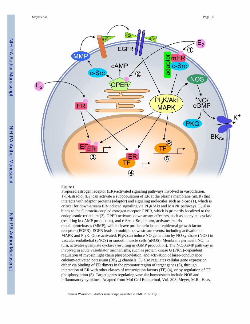
Figure 1.Proposed estrogen receptor (ER)-activated signaling pathways involved in vasodilation.17β-Estradiol (E2) can activate a subpopulation of ER at the plasma membrane (mER) thatinteracts with adaptor proteins (adaptor) and signaling molecules such as c-Src (1), which iscritical for down-stream ER-induced signaling via PI3K/Akt and MAPK pathways. E2 alsobinds to the G protein-coupled estrogen receptor GPER, which is primarily localized to theendoplasmic reticulum (2). GPER activates downstream effectors, such as adenylate cyclase(resulting in cAMP production), and c-Src. c-Src, in turn, activates matrixmetalloproteinases (MMP), which cleave pro-heparin-bound-epidermal growth factorreceptors (EGFR). EGFR leads to multiple downstream events, including activation ofMAPK and PI3K. Once activated, PI3K can induce NO generation by NO synthase (NOS) invascular endothelial (eNOS) or smooth muscle cells (nNOS). Membrane permeant NO, inturn, activates guanylate cyclase (resulting in cGMP production). The NO/cGMP pathway isinvolved in acute vasodilator mechanisms, such as protein kinase G (PKG)-dependentregulation of myosin light chain phosphorylation, and activation of large-conductancecalcium-activated potassium (BKCa) channels. E2 also regulates cellular gene expressioneither via binding of ER dimers in the promotor region of target genes (3), throughinteraction of ER with other classes of transcription factors (TF) (4), or by regulation of TFphosphorylation (5). Target genes regulating vascular homeostasis include NOS andinflammatory cytokines. Adapted from Mol Cell Endocrinol, Vol. 308, Meyer, M.R., Haas,
Meyer et al. Page 18
Vascul Pharmacol. Author manuscript; available in PMC 2012 July 5.
NIH
-PA Author Manuscript
NIH
-PA Author Manuscript
NIH
-PA Author Manuscript
E., Prossnitz, E.R., Barton M, Non-genomic regulation of vascular cell function and growthby estrogen, Pages 9-16, ©2009, with permission from Elsevier.
Meyer et al. Page 19
Vascul Pharmacol. Author manuscript; available in PMC 2012 July 5.
NIH
-PA Author Manuscript
NIH
-PA Author Manuscript
NIH
-PA Author Manuscript

Figure 2.Agonists and antagonists of GPER and plasma membrane-associated subpopulations of ERαand ERβ involved in rapid vascular estrogen signaling. Green arrows indicate activation, redarrows indicate inhibition, and the orange arrow indicates tissue-dependent activation orinhibition. SERM, selective estrogen receptor modulator; SERD, selective estrogen receptordownregulator.
Meyer et al. Page 20
Vascul Pharmacol. Author manuscript; available in PMC 2012 July 5.
NIH
-PA Author Manuscript
NIH
-PA Author Manuscript
NIH
-PA Author Manuscript
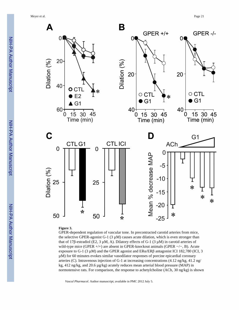
Figure 3.GPER-dependent regulation of vascular tone. In precontracted carotid arteries from mice,the selective GPER-agonist G-1 (3 µM) causes acute dilation, which is even stronger thanthat of 17β-estradiol (E2, 3 µM, A). Dilatory effects of G-1 (3 µM) in carotid arteries ofwild-type mice (GPER +/+) are absent in GPER-knockout animals (GPER −/−, B). Acuteexposure to G-1 (3 µM) and the GPER agonist and ERα/ERβ antagonist ICI 182,780 (ICI, 3µM) for 60 minutes evokes similar vasodilator responses of porcine epicardial coronaryarteries (C). Intravenous injection of G-1 at increasing concentrations (4.12 ng/kg, 41.2 ng/kg, 412 ng/kg, and 20.6 µg/kg) acutely reduces mean arterial blood pressure (MAP) innormotensive rats. For comparison, the response to achetylcholine (ACh, 30 ng/kg) is shown
Meyer et al. Page 21
Vascul Pharmacol. Author manuscript; available in PMC 2012 July 5.
NIH
-PA Author Manuscript
NIH
-PA Author Manuscript
NIH
-PA Author Manuscript
(D). CTL, solvent control (ethanol 0.3%). Panels A, B, and D are reproduced from Haas, E.,Bhattacharya, I., Brailoiu, E., Damjanovic, M., Brailoiu, G.C., Gao, X., Mueller-Guerre, L.,Marjon, N.A., Gut, A., Minotti, R., Meyer, M.R., Amann, K., Ammann, E., Perez-Dominguez, A., Genoni, M., Clegg, D.J., Dun, N.J., Resta, T.C., Prossnitz, E.R., Barton, M.,Regulatory role of G protein-coupled estrogen receptor for vascular function and obesity,Circ Res, 104(3), 288-291, ©2009 by the American Heart Association, with permission ofthe publisher. Panel C is adapted from Meyer, M.R., Baretella, O., Prossnitz, E.R., Barton,M., Dilation of epicardial coronary arteries by the G protein-coupled estrogen receptoragonists G-1 and ICI 182,780, Pharmacology, 86(1), 58-64, ©2010, with permission from S.Karger AG, Basel, Switzerland.
Meyer et al. Page 22
Vascul Pharmacol. Author manuscript; available in PMC 2012 July 5.
NIH
-PA Author Manuscript
NIH
-PA Author Manuscript
NIH
-PA Author Manuscript
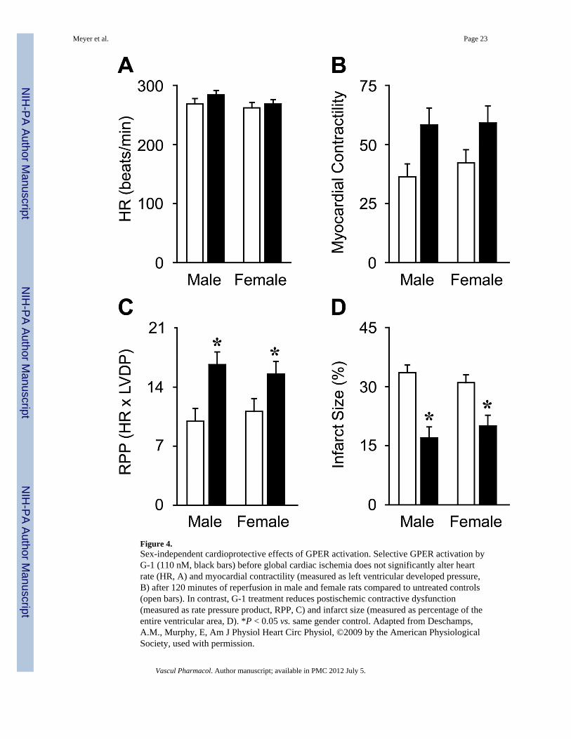
Figure 4.Sex-independent cardioprotective effects of GPER activation. Selective GPER activation byG-1 (110 nM, black bars) before global cardiac ischemia does not significantly alter heartrate (HR, A) and myocardial contractility (measured as left ventricular developed pressure,B) after 120 minutes of reperfusion in male and female rats compared to untreated controls(open bars). In contrast, G-1 treatment reduces postischemic contractive dysfunction(measured as rate pressure product, RPP, C) and infarct size (measured as percentage of theentire ventricular area, D). *P < 0.05 vs. same gender control. Adapted from Deschamps,A.M., Murphy, E, Am J Physiol Heart Circ Physiol, ©2009 by the American PhysiologicalSociety, used with permission.
Meyer et al. Page 23
Vascul Pharmacol. Author manuscript; available in PMC 2012 July 5.
NIH
-PA Author Manuscript
NIH
-PA Author Manuscript
NIH
-PA Author Manuscript
NIH
-PA Author Manuscript
NIH
-PA Author Manuscript
NIH
-PA Author Manuscript
Meyer et al. Page 24
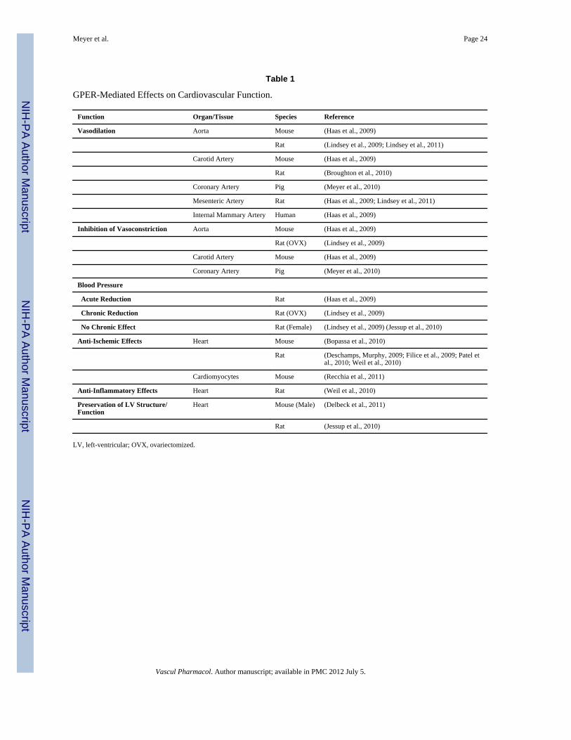
Table 1
GPER-Mediated Effects on Cardiovascular Function.
Function Organ/Tissue Species Reference
Vasodilation Aorta Mouse (Haas et al., 2009)
Rat (Lindsey et al., 2009; Lindsey et al., 2011)
Carotid Artery Mouse (Haas et al., 2009)
Rat (Broughton et al., 2010)
Coronary Artery Pig (Meyer et al., 2010)
Mesenteric Artery Rat (Haas et al., 2009; Lindsey et al., 2011)
Internal Mammary Artery Human (Haas et al., 2009)
Inhibition of Vasoconstriction Aorta Mouse (Haas et al., 2009)
Rat (OVX) (Lindsey et al., 2009)
Carotid Artery Mouse (Haas et al., 2009)
Coronary Artery Pig (Meyer et al., 2010)
Blood Pressure
Acute Reduction Rat (Haas et al., 2009)
Chronic Reduction Rat (OVX) (Lindsey et al., 2009)
No Chronic Effect Rat (Female) (Lindsey et al., 2009) (Jessup et al., 2010)
Anti-Ischemic Effects Heart Mouse (Bopassa et al., 2010)
Rat (Deschamps, Murphy, 2009; Filice et al., 2009; Patel etal., 2010; Weil et al., 2010)
Cardiomyocytes Mouse (Recchia et al., 2011)
Anti-Inflammatory Effects Heart Rat (Weil et al., 2010)
Preservation of LV Structure/Function
Heart Mouse (Male) (Delbeck et al., 2011)
Rat (Jessup et al., 2010)
LV, left-ventricular; OVX, ovariectomized.
Vascul Pharmacol. Author manuscript; available in PMC 2012 July 5.
Copyright © 2022 FDOKUMEN