Training - overtraining: performance, and hormone - British ...

GPR30 and estrogen receptor expression: New insights intohormone dependence of inflammatory breast cancer
Hugo Arias-Pulido1,8,*, Melanie Royce2,*,#, Yun Gong3,8, Nancy Joste4, Lesley Lomo4,Sang-Joon Lee5, Nabila Chaher6, Claire Verschraegen2, Juanita Lara3, Eric R. Prossnitz7,and Massimo Cristofanilli3,#1Translational Therapeutics Laboratory, The University of New Mexico Cancer Center,Albuquerque, NM, USA2Division of Hematology/Oncology, The University of New Mexico Cancer Center, Albuquerque,NM, USA3Department of Pathology, and Breast Medical Oncology-The Morgan Welch Inflammatory BreastCancer Research Program and Clinic, The University of Texas MD Anderson Cancer Center,Houston, TX, USA4Department of Pathology, The University of New Mexico Cancer Center, Albuquerque, NM, USA5Division of Epidemiology and Biostatistics, The University of New Mexico Cancer Center,Albuquerque, NM, USA6Dep. of Pathology, Centre Pierre et Marie Curie, 1, Avenue Battendier, Place May 1st, Algiers,Algeria7Cell Biology and Physiology, The University of New Mexico Cancer Center, Albuquerque, NM, USA
AbstractBackground—GPR30 is a novel G protein-coupled estrogen receptor (ER) associated withmetastases in breast cancer (BC) and poor survival in endometrial and ovarian tumors. Theassociation of GPR30 expression with inflammatory breast cancer (IBC), an aggressive andcommonly hormone-independent form of BC, has not been studied.
Methods—GPR30, ER, progesterone receptor (PR), epidermal growth factor receptor (EGFR) andHER-2 expression were assessed by immunohistochemistry (and FISH for HER-2) in 88 primaryIBCs. GPR30 expression was correlated with patient overall survival (OS), disease free survival(DFS), pathologic variables, and other biomarkers.
Results—GPR30 expression was found in 69% of IBC cases. ER, PR, HER-2 and EGFR werefound in 43%, 35%, 39%, and 34% of IBC cases, respectively. GPR30 expression correlated inverselywith ER expression (P=0.02). Co-expression of ER and GPR30 was found in 24% of IBC samples;19% expressed only ER and 46% expressed only GPR30. Univariate analysis showed no associationbetween GPR30 expression and OS or DFS. However, co-expression of ER and GPR30 wasassociated with improved OS (P<0.03) and marginally with DFS (P<0.06); the absence of both ERand GPR30 was associated with worse OS and DFS (P=0.03 for both). Multivariate analysisidentified ER as an independent prognostic factor of OS (P=0.008) and DFS (P=0.02).
8These authors contributed equally to this work.#These authors equally contributed as senior investigators in this study.*Corresponding authors: Hugo Arias-Pulido; Fax: 505-272-8352; Phone: 505-272-5404; [email protected]; Melanie Royce, Fax:505-272-2841; Phone: 505-272-6057, [email protected] work was partially presented at the First International Inflammatory Breast Cancer Conference (Houston, TX; December 5-7, 2008).
NIH Public AccessAuthor ManuscriptBreast Cancer Res Treat. Author manuscript; available in PMC 2011 August 1.
Published in final edited form as:Breast Cancer Res Treat. 2010 August ; 123(1): 51–58. doi:10.1007/s10549-009-0631-7.
NIH
-PA Author Manuscript
NIH
-PA Author Manuscript
NIH
-PA Author Manuscript
Conclusions—The majority of IBC tumors are GPR30-positive, suggesting that estrogen signalingmay be active in ER-negative IBC patients. These findings suggest potential new therapeutic targetsfor IBC such as novel endocrine agents or direct modulation of GPR30.
KeywordsInflammatory breast cancer; GPR30; Hormone receptors; growth factor receptors; overall survival;disease-free survival
IntroductionInflammatory breast cancer (IBC) is a rare but aggressive and lethal form of breast cancer withclinical and biological characteristics of a rapidly proliferating disease [1,2]. While theincidence of non-IBC (NIBC) has decreased based on the Surveillance, Epidemiology, andEnd Results (SEER) registries [3], that of IBC has increased throughout the 1990s [4]. IBCpatients typically present with clinical signs mimicking an inflammatory process such asdiffuse breast erythema, peau d’orange, skin induration, and warmth. Tumor emboli are oftenidentified in dermal lymphatics, although these are not always seen on skin biopsy [2,5].Although multidisciplinary approaches and multimodality therapy, especially theadministration of neoadjuvant chemotherapy, have improved the outcomes and survival ratesfor locally advanced breast cancer, the prognosis of IBC remains poor [6,7]. This is in partrelated to the fact that knowledge of the molecular mechanism(s) underlying IBC still lagsbehind that of NIBC. At the molecular level, most IBC tumors are associated with features ofpoorer prognosis such as lack of hormone receptor expression, HER-2 amplification and over-expression of EGFR, nuclear factor kB (NF-kB), E-cadherin, and caveolin-1/2, as well asmutations of p53 [8,9]. However, these markers are not specific for IBC. Although RhoCexpression is considered a genetic determinant for IBC and coordinated changes in RhoC andWISP3 expression are fundamental to the pathogenesis of IBC [10], there are no useful clinicalpredictive markers in IBC. Therefore, elucidating the role of novel individual molecules andtheir associated pathways is important as they might provide insights into the biology of andpotential new therapeutic targets for IBC.
GPR30, a recently characterized seven-transmembrane receptor belonging to the G-protein-coupled receptor family, binds estrogen with high affinity, and functions independently fromthe traditional nuclear estrogen receptors (ERα and ERβ) to regulate cellular and physiologicalresponsiveness to estrogen [11,12]. Activation of GPR30 leads to multiple intracellularresponses related to growth, differentiation, and proliferation [13-15]. Therefore, GPR30-mediated activity is of particular interest for patients receiving hormone treatment since it mayrepresent a new mechanism for resistance to classical endocrine therapy by allowing tumorcells to adapt to signaling pathways that can circumvent the classical ER-pathway [16].Tamoxifen, an ER inhibitor widely used in the clinic, stimulates GPR30, which in turn trans-activates the EGFR signaling pathway [11,13,17]. This observation has led investigators topostulate that GPR30 modulates the effects of endocrine therapy by providing an alternativesurvival mechanism either directly or indirectly through cross-talk with growth receptors andother signaling molecules [18]. Furthermore, high expression of GPR30 in breast, endometrialand ovarian tumors has been associated with metastases and poor survival [19-21]. To definethe role of GPR30 as a functional estrogen-related biomarker in IBC, we analyzed GPR30expression in tissue samples of IBC patients that have completed neoadjuvant therapy andundergone surgery. We sought to evaluate the level of GPR30 expression, correlate it with theexpression of hormone receptors, growth factors receptors and other known histopathologicalvariables of IBC and assess the impact of expression on prognosis.
Arias-Pulido et al. Page 2
Breast Cancer Res Treat. Author manuscript; available in PMC 2011 August 1.
NIH
-PA Author Manuscript
NIH
-PA Author Manuscript
NIH
-PA Author Manuscript
MethodsClinical specimens
Eighty-eight patients with primary IBC who were treated at The University of Texas M. D.Anderson Cancer Center (MDACC) from September 1994 to August 2004 were included inthis study. For the purposes of this study, patients were identified as having sufficient amountof residual tumor after neoadjuvant chemotherapy. Patients with pathological completeresponse, minimal residual tumor tissue, missing tissue or information were not included. Thisstudy was approved by the University of New Mexico Cancer Center (UNMCC) and MDACCInstitutional Review Boards. Formalin-fixed paraffin-embedded tissues were obtained frommastectomy specimens and used to build tissue microarrays (TMAs) as described elsewhere[22]. This implies that all cases included had experienced clinical response to neoadjuvantchemotherapy but still displayed persistent disease at the time of surgery. IBC was diagnosedon the basis of clinical signs such as rapid progression (i.e., clinical evolution of less than 3months) of diffuse skin erythema, peau d’orange, tenderness, induration, and warmth, with orwithout an underlying palpable mass, and a histologic proof of invasive breast carcinoma withor without evidence of dermal lymphatic invasion. The modified Black’s nuclear gradingsystem was used to grade invasive tumor cells. All patients received multimodal treatment,including preoperative chemotherapy, surgery, and radiation therapy. The chemotherapy wasgiven according to protocols described previously [23], consisting of four to six cycles of ananthracycline-based regimen that included doxorubicin (50 mg/m2), cyclophosphamide (500mg/m2), and 5-fluorouracil (500 mg/m2) given every 21 days. The majority of the patients(89%) also received paclitaxel and 3 patients received trastuzumab. Endocrine therapy(Tamoxifen or aromatase inhibitor) was given to patients with ER-positive and/or PR-positivetumors.
Evaluation of ER, PR, HER-2, EGFR and GPR30 expressionER, PR and HER-2 protein expression were determined by immunohistochemical staining(IHC) and HER-2 gene amplification was detected using fluorescent in situ hybridization(FISH) at MDACC; EGFR and GPR30 protein expression were assessed by IHC at UNMCC.When only limited tumor sample existed in the TMA core or the core tissue was lost duringthe IHC procedure, a whole section from the original block was used for the study.
ER, PR and HER2 expression levels were evaluated using standard procedures with themodified avidin–biotin complex method in a DAKO auto-stainer (DAKO, Carpinteria, CA)using primary antibodies against ER (clone 6F11; Novocastra, Burlingame, CA; Dilution,1:50), PR (clone 1A6; Novocastra, Burlingame, CA; Dilution, 1:30) and HER-2 (clone AB8;Neomarker/Labvision Corporation, Fremont, CA; Dilution, 1:100) as previously described[24]. Normal endometrial (ER, PR) and breast (HER-2) tissues were used as positive controls;the same tissues, incubated with an isotype-matched antibody, were used as negative controls.
EGFR expression was detected with the 31G7 clone (Zymed, Carlsbad, CA; Dilution 1:50)using the Ventana XT Benchmark auto-stainer as described by the manufacturer (VentanaMedical Systems, Inc., Tucson, AZ). Briefly, tissue sections were baked, deparaffinized inxylene, rehydrated in graded ethanol (100% and 95%), and rinsed in water. The slides werethen incubated in fresh 3% hydrogen peroxide in Dulbecco’s phosphate-buffered saline (DPBS:KCl/KH2PO4/NaCl/Na2HPO4-7H2O, 2.67/1.47/133.93/8.06 mM) for 20 min followed bythree 5-min rinses in DPBS. The slides were loaded on to the Benchmark auto-stainer anddetection was performed using the iVIEW™ DAB detection system (Ventana MedicalSystems, Inc., Tucson, AZ).
Arias-Pulido et al. Page 3
Breast Cancer Res Treat. Author manuscript; available in PMC 2011 August 1.
NIH
-PA Author Manuscript
NIH
-PA Author Manuscript
NIH
-PA Author Manuscript
GPR30 was detected as described elsewhere [20,21] using a detection protocol similar to EGFRexcept that it was carried out manually; antibody retrieval was applied for 20 min in 10 mMcitrate buffer (pH 6.0), and the incubation with an antibody against the GPR30 C-terminus wasperformed overnight. For both EGFR and GPR30 IHC assays, normal placenta was used as apositive control; normal placenta, incubated with an isotype-matched antibody was used as anegative control.
Detection of HER-2 gene copy number by fluorescent in situ hybridization (FISH)FISH was performed as previously described [25] using the PathVysion HER-2SpectrumOrange/CEP17 SpectrumGreen kit (Vysis, Downers Grove, IL).
Scoring of IHC resultsPositive status for ER and PR was defined as having nuclear staining in at least 10% of invasivetumor cells. HER-2 positive status was defined as an IHC score of 3+ or a gene copy ratio ofHER-2:CEP17 of ≥ 2.0. The tumors with an IHC score of 1+ or 2+ were confirmed by FISH.
For EGFR, signal intensity was scored as 0 (negative), 1+ (weak), 2+ (moderate), and 3+(strong). Any complete membranous staining (>1+) was considered positive. The GPR30protein levels were scored as previously described [21] using an H-scoring system obtainedby multiplying the staining intensity (graded as 0 negative; 1+, weak; 2+, moderate and 3+,strong) by the percentage of epithelial tumor cells with GPR30-positive cytoplasmic staining(0 to 100%). The GPR30 cut-off was initially determined in 60 normal breast epithelial biopsies(8 biopsies from non-cancer patients and 52 histologically normal breast biopsies collectedfrom a distant site of the invasive breast tumor biopsy of the same patient). The median obtainedin the normal breast samples was selected as the cut-off, and for statistical analysis as well asto reduce observer variability, the samples were grouped into negative (H ≤ median) or positive(H > median) populations.
Statistical analysisPrimary outcomes for this study were overall survival (OS) and disease-free survival (DFS).OS was calculated from the date of diagnosis with death scored as an event and censoring ofother patients at the date of last follow-up or of non-disease-related death. The DFS intervalwas calculated from the date of diagnosis to development of first recurrence. Patients withoutrecurrence were censored at the time of last follow-up or death. Chi-square and Fisher’s exacttests were used to compare demographic, clinical, and pathological data between IBC patientsand IHC results between samples. The Spearman test was used to assess correlations betweenGPR30 expression and ER, PR, HER2, and EGFR status. For the analysis of ER, PR, HER2,EGFR, and GPR30 status, patients were divided into two groups (positive and negative) asdescribed above. OS and DFS for the groups defined by ER, PR, HER2, EGFR, and GPR30status and other variables (age, nodal status, lymphovascular invasion, nuclear grade, andhormone treatment) were plotted using Kaplan–Meier curves and compared using the log-ranktest. The Cox proportional hazard model with single covariates was used to obtain the hazardratios (HRs) and associated 95% confidence intervals (CIs) for the groups compared. Theprimary multivariable analyses were performed using the Cox model, with adjustments madefor age (<50, ≥ 50 years), number of positive nodes (≤ 3, ≥ 4), lymphovascular invasion (yes,no), nuclear grade (1, 2 vs. 3), hormone treatment (yes, no) and ER, PR, HER-2, EGFR andGPR30 status. Those variables found to be statistically significant in univariate analyses wereconsidered for inclusion in the multivariate model. Inclusion was based on the significance ofeach variable as assessed by a likelihood ratio test comparing the multivariate model to thereduced model obtained by deleting variable(s) from the full model [26]. For each ordinalvariable, the lowest value was used as the reference in computing hazard ratios (HR). Survival
Arias-Pulido et al. Page 4
Breast Cancer Res Treat. Author manuscript; available in PMC 2011 August 1.
NIH
-PA Author Manuscript
NIH
-PA Author Manuscript
NIH
-PA Author Manuscript

rates and HRs are presented with their 95% CIs. Wald tests were used to test for significanceof HRs. Two-tailed P values less than 0.05 were considered statistically significant.
ResultsPatient characteristics
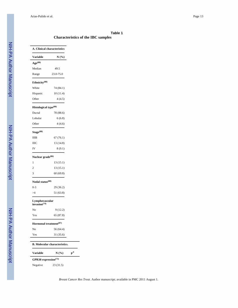
Of the 88 patients included in this study, 84.1 % were non-Hispanic whites, 11.4% Hispanics,and 4.5% Blacks and Asians. The median age was 49.5 years (range, 23 to 75 years). Themajority of cases were infiltrating ductal carcinomas (IDC; 88.6%), stage IIIB (76.1%),modified Black’s nuclear grade 3 (69.8%), with more than 4 positive nodes (63.8%) andpositive for lymphovascular invasion (87.8%) at time of surgery, consistent with expectationsfor a cohort of patients at high risk of disease recurrence. Chemotherapy and hormone treatmentwere given to 95.5% and 35.6% of patients, respectively. The characteristics of patients andtumors are summarized in Table 1. The median follow-up interval was 45.7 months (range 0.2to 164.2 months). At the time of analysis, 52 patients had died (61.2%); the OS was 48.6% andthe DFS was 61.0% (Fig. 1).
GPR30 expression in IBC samplesGPR30 was expressed in epithelial cells from normal breast biopsies (N=60; median H-score=125; range 0 to 300). In normal breast tissue, ductal and lobular epithelia displayed acytoplasmic staining pattern with varying intensities (Fig. 2A and B). As in normal breast, IBCtissue exhibited a cytoplasmic staining pattern with staining intensity ranging from negativeto strong (N=73; median H-score=160; range 0 to 300; P=0.043 for normal vs. IBC specimens;Fig. 2D-F). In several samples, staining of stromal tissue, inflammatory cells, myoepitheliaand stromal fibroblasts was observed but neither was used in the grading scheme.
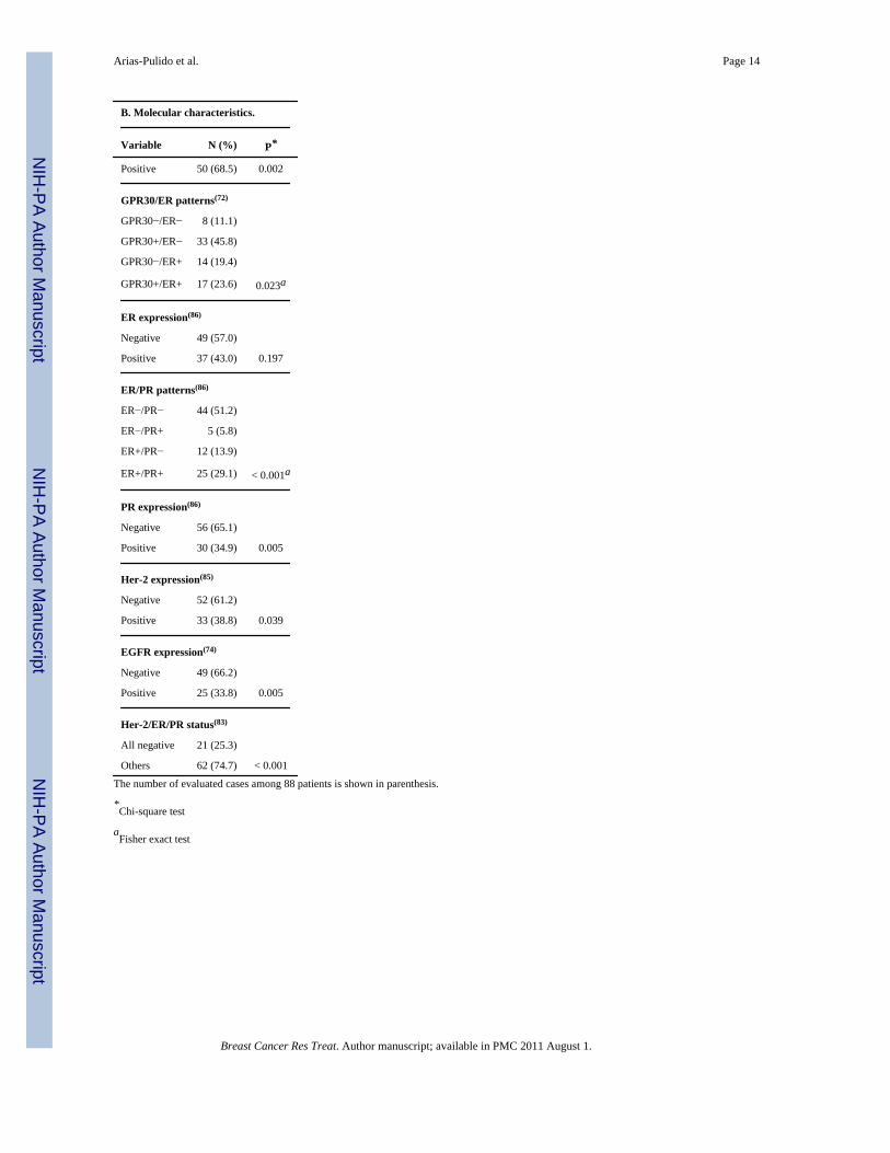
The majority of IBC cases were positive for GPR30 (68.5%, Table 1B). Approximately 24%of the tumors co-expressed ER and GPR30, 19% expressed only ER, 11% were negative forboth ER and GPR30, and interestingly, GPR30 was expressed in 46% of tumors that failed toexpress ER (Table 1B). GPR30 expression was found to be inversely correlated with ER (P =0.02), and hormone treatment but the latter association was not significant (P=0.11); GPR30expression did not correlate with PR, EGFR or HER-2 (P > 0.1 for all) (data not shown).
ER, PR, EGFR, and HER-2 expression in IBC samplesThe majority of samples were independently negative for ER, PR, HER-2, and EGFR. Fiftyone percent of samples lacked expression of both ER and PR and 25% of the samples were“triple negative” (ER/PR/HER-2 negative) (Table 1B).
GPR30 expression and OS and DFSGPR30 expression was not associated with OS or DFS (Fig. 3A, B and Supplemental Table1). However, patients who were positive for GPR30 expression showed a higher median DFSas compared to the median DFS of the whole population (122 vs. 61 months, respectively)(SeeFig. 1 and 3B and Supplemental Table 1). Furthermore, co-expression of GPR30 and ER(GPR30/ER patterns) was associated with OS (P=0.033) but only marginally associated withDFS (P=0.057; Supplemental Table 1 and Supplemental Fig. 1A,B). In particular, tumors thatexpressed both GPR30 and ER showed a trend towards better OS and DFS than otherexpression patterns (Supplemental Table 1 and Supplemental Fig. 1A, B). Of note, patientsshowing no expression of both GPR30 and ER (GPR30/ER status) were associated withsignificantly decreased OS and DFS (P=0.03 for both; Supplemental Table 1 and SupplementalFig. 2A, B).
Arias-Pulido et al. Page 5
Breast Cancer Res Treat. Author manuscript; available in PMC 2011 August 1.
NIH
-PA Author Manuscript
NIH
-PA Author Manuscript
NIH
-PA Author Manuscript
ER, PR, EGFR, HER-2 expression, clinical parameters and OS and DFSER expression was positively correlated with PR and hormone treatment (P<0.001 for both),and an inverse correlation was observed with EGFR (P<0.001) and HER-2 (P=0.02)expression. EGFR expression showed an inverse correlation with hormone treatment (P<0.05)but no correlation with HER-2 expression (P=0.60; Data not shown).
Of the clinico-pathological variables (age, number of positive nodes, lymphovascular invasion,nuclear grade, and hormone treatment) and biomarkers analyzed, hormone treatment, ERexpression, and ER/PR expression patterns were associated with improved OS and DFS(Supplemental Table 1). Specifically, ER, either alone or co-expressed with PR, wassignificantly associated with an improved OS and DFS as shown by the Kaplan-Meier survivalcurves (Supplemental Table 1 and Supplemental. Fig. 3A, B).
“Triple negative status” was associated with poor OS and DFS (P<0.01 for both; SupplementalTable 1). The lack of GPR30 expression in these IBC cases was marginally associated withworse OS (P=0.06) but not with DFS (P=0.11; data not shown). Expression of EGFR wasassociated with decreased OS and DFS (P<0.05 for both; Supplemental Table 1).
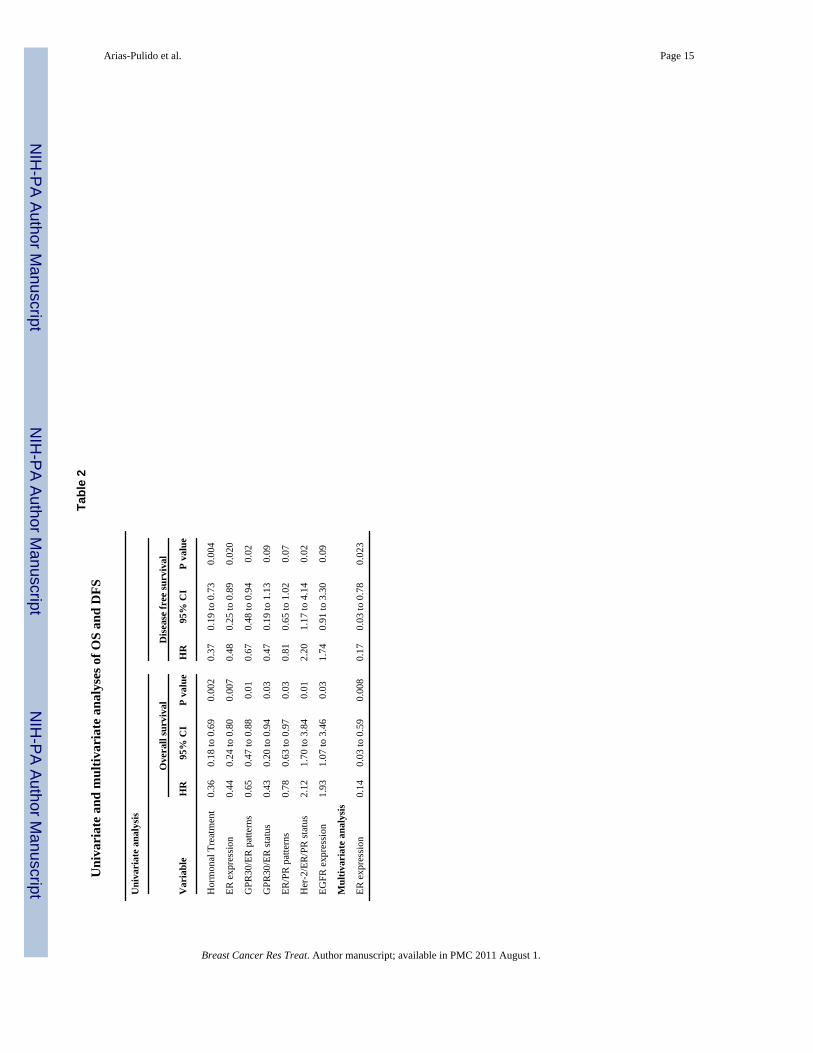
Univariate and Multivariate analysisIn the univariate analysis, hormone treatment, ER expression, GPR30/ER patterns, GPR30/ERstatus, and ER/PR patterns were associated with improved OS and DFS (Table 2). Triplenegative status and EGFR over-expression were associated with worse OS and DFS (Table 2).However, the multivariate analysis identified ER expression as the only independent prognosticfactor associated with improved OS (HR=0.14; 95%CI 0.03 to 0.59; P=0.008) and DFS(HR=0.17; 95%CI 0.03 to 0.78; P=0.02) (Table 2).
DiscussionIBC is an aggressive disease associated with resistance to standard multimodality therapytranslating into a dismal prognosis [7]. The biological features of IBC have not been clearlydefined but the disease is typically characterized by biomarkers of aggressive phenotypes suchas high nuclear grade, lack of hormone receptor expression and HER-2 amplification [8-10].Those characteristics have limited the use of treatment modalities otherwise effective in non-IBC, such as hormone treatment. Endocrine therapy has been employed in treatment andprevention of breast cancer and its efficacy is usually predicted on the basis of ER expressionlevel. More recent research indicates a lack of concordance between tamoxifen responsivenessand ER expression [27], and suggests the existence of alternate routes critical for the growthand survival of breast cancer cells [16,28]. GPR30 has been identified as an alternate routetriggering activation of the EGFR and downstream molecules such as MAP kinases, as wellas up-regulation of Bcl-2 and cyclin D2 [13-15]. We have previously shown that GPR30 isexpressed in 48% of ovarian and 50% of endometrial tumors [20,21]. In endometrial tumors,GPR30 expression is predictive of many parameters associated with adverse outcome (highgrade and biologically aggressive histologic subtypes, cervical involvement, and advancedstage) and is associated with lower survival rates [20]. The same association with poor outcomeis observed for GPR30 in ovarian cancer [21]. In non-IBC specimens, conflicting associationshave been reported. In one study, GPR30 expression was shown to be a significant predictorof tumor size and metastases [19]. However, in a subsequent report, GPR30 expression wasnot associated with either HER-2 expression or survival [29]. While, the former studyemployed immunohistochemistry to assess GPR30 expression exclusively in epithelial tumorcells, the later investigation reported GPR30 expression from total RNA without isolatingepithelial tumor cells. The use of different techniques may explain the discrepancies betweenthe results of these two studies.
Arias-Pulido et al. Page 6
Breast Cancer Res Treat. Author manuscript; available in PMC 2011 August 1.
NIH
-PA Author Manuscript
NIH
-PA Author Manuscript
NIH
-PA Author Manuscript
This study represents the first to investigate the role of GPR30 in relation to other knownbiomarkers and its impact on prognosis in IBC patients. While we found no association betweenGPR30 expression and outcome, it needs to be highlighted that the patients that were includedin the study already had a dismal prognosis because they had extensive residual disease afterneoadjuvant chemotherapy. The study confirmed the expression of hormone and growth factorreceptors within the range of published data, indicating that the tumors analyzed in this studywere representative of those included in other studies. Furthermore, we demonstrate severalimportant biological correlations that may translate into significant clinical implications. First,we found that the majority (68.5%) of IBC tumors were GPR30-positive whereas ER was foundin only 43% of tumors. There was no difference in incidence between the tumors that co-expressed ER and GPR30 (23.6%) and those ER-positive tumors that failed to express GPR30(19.4%). However, twice as many (46%) of the ER-negative IBC tumors retained GPR30(Table 1B), suggesting that ER and GPR30 expression in IBC is not interdependent and. Onecan further postulate that it may be possible to exploit novel endocrine therapies that workthrough non-classical estrogen-dependent pathways as potential therapies for IBC tumorsdespite the lack of detectable ER. Second, in contrast to NIBC [19], there was no correlationbetween GPR30 expression and HER-2 or between GPR30 and EGFR, as we previouslyobserved in endometrial cancer [20]. These findings suggest that GPR30 may not use the EGFRand/or HER-2 routes as alternate pathways to promote tumor cell growth in IBC and/or it ispossible that other yet unidentified routes may be activated by GPR30. Another explanationof these findings may lie in the intrinsic nature of IBC tumors, which are thought to bemetastatic at the onset of the disease [8,10] and, therefore, EGFR and/or HER-2 routes maynot be critical or are bypassed in IBC tumors. This contrasts other studies performed in pre-clinical models and tissue specimens [19] and therefore required confirmatory studies. Third,we found that patients with GPR30 over-expression had prolonged disease control asdocumented by the fact the DFS was twice that of the studied population although it did notreach statistical significance. It should be noted that this result is opposite to what we havepreviously reported in endometrial and ovarian cancers, where GPR30 over-expression was aprognostic factor of poor survival [20,21]. GPR30 over-expression seems to be associated withimproved OS and DFS in ER-positive IBC patients, raising important biological questionsrelated to the interplay between these two important receptors and their regulation of hormone-dependence in IBC.
Tamoxifen behaves as an agonist in GPR30-positive cells, resulting in activation of pro-survival signaling pathways such as ERK and PI3K/Akt [11,13,17]. Therefore, on one hand,this activation may be detrimental for GPR30-positive patients receiving tamoxifen. On theother hand, a brain tumor cell line model demonstrated that the biological effect of tamoxifenis dose-dependent with high doses of tamoxifen resulting in an increase of glioma cellapoptosis, suppression of tumor growth in vivo and improvement of survival in gliomaxenograft models as a result of activation of pro-apoptotic genes, including NF-κB and itstargets genes [30]. Furthermore, GPR30 over-expression inhibits urothelial cell proliferationas a result of decreased cyclin D1expression [31]. Given the protective effect of GPR30expression observed in our study, we performed a subset analysis in our ER/PR-positivepatients that received tamoxifen to analyze the effect of GPR30 expression in those patients.Although we found a trend for improved OS and DFS in GPR30-positive as compared toGPR30-negative patients, it did not reach statistical significance (data not shown) probablydue to the small size of samples. Further analyses in a larger population are needed to confirmwhether GPR30 may activate pro-apoptotic or anti-proliferative mechanisms that may resultin a better response in ER/PR-positive patients treated with tamoxifen. Whether the same NF-κB or cyclin D pathways are activated in IBC patients remains unknown; ongoing studies inour group will clarify these associations.
Arias-Pulido et al. Page 7
Breast Cancer Res Treat. Author manuscript; available in PMC 2011 August 1.
NIH
-PA Author Manuscript
NIH
-PA Author Manuscript
NIH
-PA Author Manuscript
A word of caution should be taken when analyzing these results: our study has the drawbacksinherent to retrospective studies and the small size of samples. Notwithstanding theselimitations, the description by our group of a specific GPR30 agonist (G1) and antagonist (G15)[32,33] fuels our enthusiasm to further assess the effects and mechanisms of action of bothagents in IBC cell lines and tumor xenografts.
Supplementary MaterialRefer to Web version on PubMed Central for supplementary material.
AcknowledgmentsSupported in part by NIH grant CA116662 (E.R.P.) and by New Mexico State IBC funding (New Mexico Senate Bill532) and the State of Texas Inflammatory Breast Cancer Rider. We would like to thank the MDACC (Houston, TX)Human Tissue Repository for providing tissue samples and clinical data; Dr. A. Chakerian (EPL, Pathology, UNM)and T. Howard (Cell Biology and Physiology, UNM) for technical support with the IHC assays.
References1. Levine PH, Veneroso C. The epidemiology of inflammatory breast cancer. Semin Oncol 2008;35:11–
16. [PubMed: 18308141]2. Singletary SE, Cristofanilli M. Defining the clinical diagnosis of inflammatory breast cancer. Semin
Oncol 2008;35:7–10. [PubMed: 18308140]3. Kerlikowske K, Miglioretti DL, Buist DSM, Walker R, Carney PA. Declines in Invasive Breast Cancer
and Use of Postmenopausal Hormone Therapy in a Screening Mammography Population. J NatlCancer Inst 2007;99:1335–1339. [PubMed: 17698950]
4. Hance KW, Anderson WF, Devesa SS, Young HA, Levine PH. Trends in Inflammatory BreastCarcinoma Incidence and Survival: The Surveillance, Epidemiology, and End Results Program at theNational Cancer Institute. J Natl Cancer Inst 2005;97:966–975. [PubMed: 15998949]
5. Molckovsky A, Fitzgerald B, Freedman O, Heisey R, Clemons M. Approach to inflammatory breastcancer. Can Fam Physician 2009;55:25–31. [PubMed: 19155362]
6. Panades M, Olivotto IA, Speers CH, Shenkier T, Olivotto TA, Weir L, Allan SJ, Truong PT. EvolvingTreatment Strategies for Inflammatory Breast Cancer: A Population-Based Survival Analysis. J ClinOncol 2005;23:1941–1950. [PubMed: 15774787]
7. Dawood S, Ueno N, Cristofanilli M. The medical treatment of inflammatory breast cancer. SeminOncol 2008;35:64–71. [PubMed: 18308147]
8. Charafe-Jauffret E, Tarpin C, Viens P, Bertucci F. Defining the molecular biology of inflammatorybreast cancer. Semin Oncol 2008;35:41–50. [PubMed: 18308145]
9. Gong Y. Pathologic Aspects of Inflammatory Breast Cancer: Part 2. Biologic Insights Into ItsAggressive Phenotype. Seminars in Oncology 2008;35:33–40. [PubMed: 18308144]
10. Ventura AC, Merajver SD. Genetic determinants of aggressive breast cancer. Annu Rev Med2008;59:199–212. [PubMed: 17914925]
11. Revankar CM, Cimino DF, Sklar LA, Arterburn JB, Prossnitz ER. A Transmembrane IntracellularEstrogen Receptor Mediates Rapid Cell Signaling. Science 2005;307:1625–1630. [PubMed:15705806]
12. Thomas P, Pang Y, Filardo EJ, Dong J. Identity of an Estrogen Membrane Receptor Coupled to a GProtein in Human Breast Cancer Cells. Endocrinology 2005;146:624–632. [PubMed: 15539556]
13. Filardo EJ, Quinn JA, Bland KI, Frackelton AR Jr. Estrogen-Induced Activation of Erk-1 and Erk-2Requires the G Protein-Coupled Receptor Homolog, GPR30, and Occurs via Trans-Activation of theEpidermal Growth Factor Receptor through Release of HB-EGF. Mol Endocrinol 2000;14:1649–1660. [PubMed: 11043579]
14. Kanda N, Watanabe S. 17beta-estradiol inhibits oxidative stress-induced apoptosis in keratinocytesby promoting Bcl-2 expression. J Invest Dermatol 2003;121:1500–1509. [PubMed: 14675202]
Arias-Pulido et al. Page 8
Breast Cancer Res Treat. Author manuscript; available in PMC 2011 August 1.
NIH
-PA Author Manuscript
NIH
-PA Author Manuscript
NIH
-PA Author Manuscript
15. Kanda N, Watanabe S. 17beta-estradiol stimulates the growth of human keratinocytes by inducingcyclin D2 expression. J Invest Dermatol 2004;123:319–328. [PubMed: 15245432]
16. Prossnitz ER, Arterburn JB, Smith HO, Oprea TI, Sklar LA, Hathaway HJ. Estrogen signaling throughthe transmembrane G protein-coupled receptor GPR30. Annu Rev Physiol 2008;70:165–190.[PubMed: 18271749]
17. Filardo EJ, Quinn JA, Frackelton AR Jr. Bland KI. Estrogen Action Via the G Protein-CoupledReceptor, GPR30: Stimulation of Adenylyl Cyclase and cAMP-Mediated Attenuation of theEpidermal Growth Factor Receptor-to-MAPK Signaling Axis. Mol Endocrinol 2002;16:70–84.[PubMed: 11773440]
18. Prossnitz ER, Sklar LA, Oprea TI, Arterburn JB. GPR30: a novel therapeutic target in estrogen-relateddisease. Trends Pharmacol Sci 2008;29:116–123. [PubMed: 18262661]
19. Filardo EJ, Graeber CT, Quinn JA, Resnick MB, Giri D, DeLellis RA, Steinhoff MM, Sabo E.Distribution of GPR30, a Seven Membrane-Spanning Estrogen Receptor, in Primary Breast Cancerand its Association with Clinicopathologic Determinants of Tumor Progression. Clin Cancer Res2006;12:6359–6366. [PubMed: 17085646]
20. Smith HA, Leslie K, Singh M, Qualls CR, Revankar CM, Joste N, Prossnitz ER. GPR-30: a novelindicator of poor survival in endometrial carcinoma. Am J Obstet Gynecol 2007;196:386.e381–389.[PubMed: 17403429]
21. Smith H, Arias-Pulido H, Kuo D, Howard T, Qualls C, Lee S-J, Verschraegen C, Hathaway H, JosteN, Prossnitz E. GPR30 predicts poor survival for ovarian cancer. Gyn Oncol 2009;114:465–471.
22. Ginestier C, Charafe-Jauffret E, Bertucci F, Eisinger F, Geneix J, Bechlian D, Conte N, Adelaide J,Toiron Y, Nguyen C, et al. Distinct and Complementary Information Provided by Use of Tissue andDNA Microarrays in the Study of Breast Tumor Markers. Am J Pathol 2002;161:1223–1233.[PubMed: 12368196]
23. Cristofanilli M, Buzdar AU, Hortobagyi GN. Update on the Management of Inflammatory BreastCancer. Oncologist 2003;8:141–148. [PubMed: 12697939]
24. Cabioglu N, Gong Y, Islam R, Broglio KR, Sneige N, Sahin A, Gonzalez-Angulo AM, Morandi P,Bucana C, Hortobagyi GN, et al. Expression of growth factor and chemokine receptors: new insightsin the biology of inflammatory breast cancer. Ann Onc 2007;18:1021–1029.
25. Gong Y, Booser D, Sneige N. Comparison of HER-2 status determined by fluorescence in situhybridization in primary and metastatic breast carcinoma. Cancer 2005;103:1763–1769. [PubMed:15786420]
26. Lachin, J. Biostatistical Methods. John Wiley and Sons; NY: 2000. p. 272-285.27. EBCTCG. Effects of chemotherapy and hormonal therapy for early breast cancer on recurrence and
15-year survival: an overview of the randomised trials. The Lancet 2005;365:1687–1717.28. Filardo EJ, Thomas P. GPR30: a seven-transmembrane-spanning estrogen receptor that triggers EGF
release. Trends Endocrinol Metab 2005;16:362–367. [PubMed: 16125968]29. Kuo W, Chang L, Liu D, Hwa H, Lin J, Lee P, Chen C, Lien H, Yuan R, Shun C, et al. The interactions
between GPR30 and the major biomarkers in infiltrating ductal carcinoma of the breast in an Asianpopulation. Taiwan J Obstet Gynecol 2007;46:135–145. [PubMed: 17638621]
30. Hui A-M, Zhang W, Chen W, Xi D, Purow B, Friedman GC, Fine HA. Agents with Selective EstrogenReceptor (ER) Modulator Activity Induce Apoptosis In vitro and In vivo in ER-Negative GliomaCells. Cancer Res 2004;64:9115–9123. [PubMed: 15604281]
31. Teng J, Wang Z-Y, Prossnitz ER, Bjorling DE. The G Protein-Coupled Receptor GPR30 InhibitsHuman Urothelial Cell Proliferation. Endocrinology 2008;149:4024–4034. [PubMed: 18467434]
32. Bologa CG, Revankar CM, Young SM, Edwards BS, Arterburn JB, Kiselyov AS, Parker MA,Tkachenko SE, Savchuck NP, Sklar LA, et al. Virtual and biomolecular screening converge on aselective agonist for GPR30. Nat Chem Biol 2006;2:207–212. [PubMed: 16520733]
33. Dennis M, Burai R, Ramesh C, Petrie W, Alcon S, Nayak T, Bologa C, Leitao A, Brailoiu E, DeliuE, et al. In vivo effects of a GPR30 antagonist. Nat Chem Biol 2009;5:421–427. [PubMed: 19430488]
Arias-Pulido et al. Page 9
Breast Cancer Res Treat. Author manuscript; available in PMC 2011 August 1.
NIH
-PA Author Manuscript
NIH
-PA Author Manuscript
NIH
-PA Author Manuscript

Figure 1.Kaplan-Meier survival curves of OS (continuous line) and DFS (dotted line).
Arias-Pulido et al. Page 10
Breast Cancer Res Treat. Author manuscript; available in PMC 2011 August 1.
NIH
-PA Author Manuscript
NIH
-PA Author Manuscript
NIH
-PA Author Manuscript
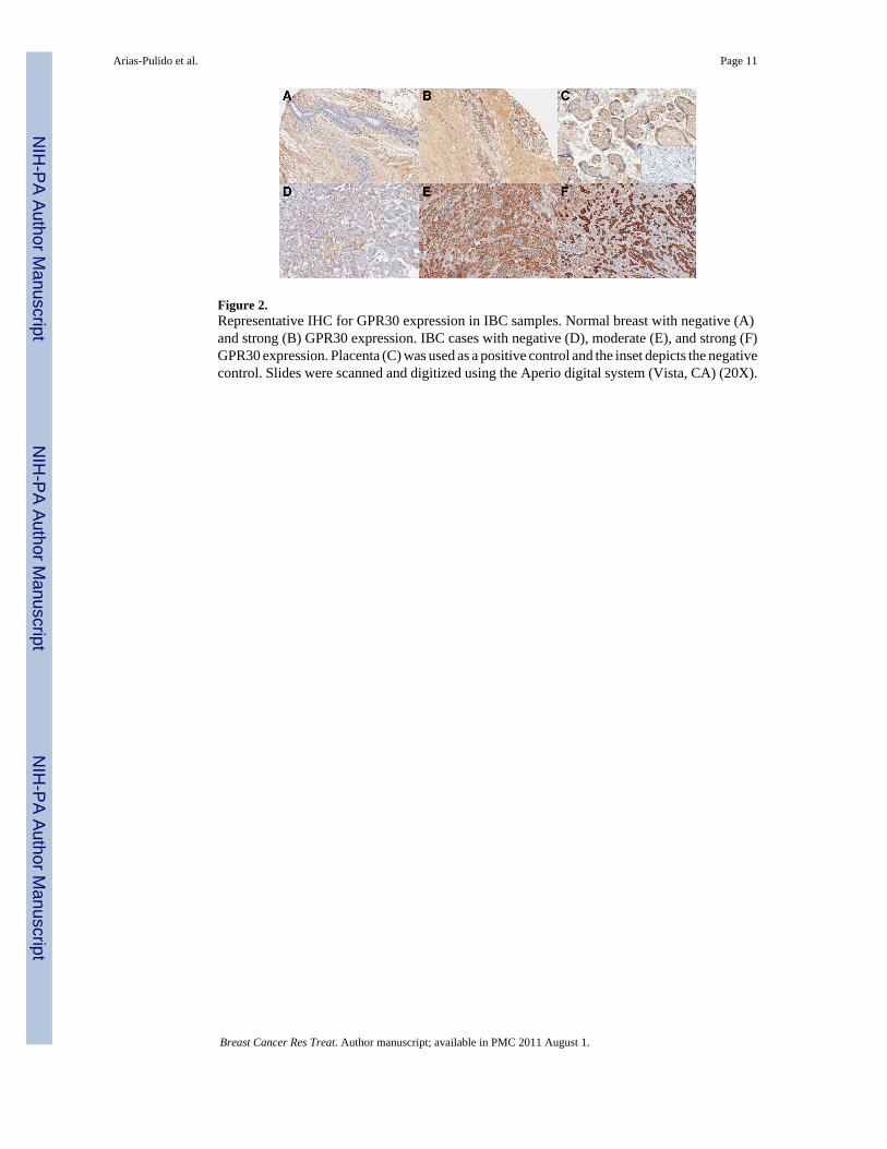
Figure 2.Representative IHC for GPR30 expression in IBC samples. Normal breast with negative (A)and strong (B) GPR30 expression. IBC cases with negative (D), moderate (E), and strong (F)GPR30 expression. Placenta (C) was used as a positive control and the inset depicts the negativecontrol. Slides were scanned and digitized using the Aperio digital system (Vista, CA) (20X).
Arias-Pulido et al. Page 11
Breast Cancer Res Treat. Author manuscript; available in PMC 2011 August 1.
NIH
-PA Author Manuscript
NIH
-PA Author Manuscript
NIH
-PA Author Manuscript
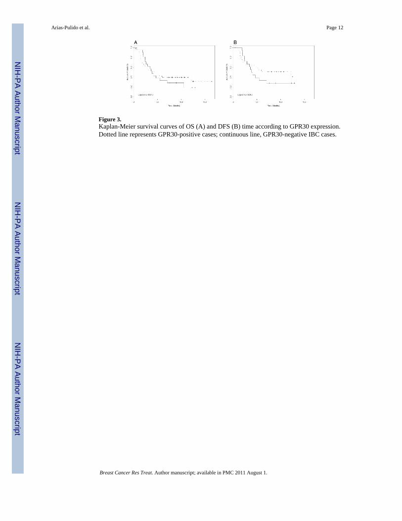
Figure 3.Kaplan-Meier survival curves of OS (A) and DFS (B) time according to GPR30 expression.Dotted line represents GPR30-positive cases; continuous line, GPR30-negative IBC cases.
Arias-Pulido et al. Page 12
Breast Cancer Res Treat. Author manuscript; available in PMC 2011 August 1.
NIH
-PA Author Manuscript
NIH
-PA Author Manuscript
NIH
-PA Author Manuscript
NIH
-PA Author Manuscript
NIH
-PA Author Manuscript
NIH
-PA Author Manuscript
Arias-Pulido et al. Page 13
Table 1Characteristics of the IBC samples
A. Clinical characteristics
Variable N (%)
Age(88)
Median 49.5
Range 23.0-75.0
Ethnicity(88)
White 74 (84.1)
Hispanic 10 (11.4)
Other 4 (4.5)
Histological type(88)
Ductal 78 (88.6)
Lobular 6 (6.8)
Other 4 (4.6)
Stage(88)
IIIB 67 (76.1)
IIIC 13 (14.8)
IV 8 (9.1)
Nuclear grade(86)
1 13 (15.1)
2 13 (15.1)
3 60 (69.8)
Nodal status(80)
0-3 29 (36.2)
>4 51 (63.8)
Lymphovascularinvasion(74)
No 9 (12.2)
Yes 65 (87.8)
Hormonal treatment(87)
No 56 (64.4)
Yes 31 (35.6)
B. Molecular characteristics.
Variable N (%) P*
GPR30 expression(73)
Negative 23 (31.5)
Breast Cancer Res Treat. Author manuscript; available in PMC 2011 August 1.
NIH
-PA Author Manuscript
NIH
-PA Author Manuscript
NIH
-PA Author Manuscript
Arias-Pulido et al. Page 14
B. Molecular characteristics.
Variable N (%) P*
Positive 50 (68.5) 0.002
GPR30/ER patterns(72)
GPR30−/ER− 8 (11.1)
GPR30+/ER− 33 (45.8)
GPR30−/ER+ 14 (19.4)
GPR30+/ER+ 17 (23.6) 0.023a
ER expression(86)
Negative 49 (57.0)
Positive 37 (43.0) 0.197
ER/PR patterns(86)
ER−/PR− 44 (51.2)
ER−/PR+ 5 (5.8)
ER+/PR− 12 (13.9)
ER+/PR+ 25 (29.1) < 0.001a
PR expression(86)
Negative 56 (65.1)
Positive 30 (34.9) 0.005
Her-2 expression(85)
Negative 52 (61.2)
Positive 33 (38.8) 0.039
EGFR expression(74)
Negative 49 (66.2)
Positive 25 (33.8) 0.005
Her-2/ER/PR status(83)
All negative 21 (25.3)
Others 62 (74.7) < 0.001
The number of evaluated cases among 88 patients is shown in parenthesis.
*Chi-square test
aFisher exact test
Breast Cancer Res Treat. Author manuscript; available in PMC 2011 August 1.
NIH
-PA Author Manuscript
NIH
-PA Author Manuscript
NIH
-PA Author Manuscript
Arias-Pulido et al. Page 15
Tabl
e 2
Uni
vari
ate
and
mul
tivar
iate
ana
lyse
s of O
S an
d D
FS
Uni
vari
ate
anal
ysis
Ove
rall
surv
ival
Dis
ease
free
surv
ival
Var
iabl
eH
R95
% C
IP
valu
eH
R95
% C
IP
valu
e
Hor
mon
al T
reat
men
t0.
360.
18 to
0.6
90.
002
0.37
0.19
to 0
.73
0.00
4
ER e
xpre
ssio
n0.
440.
24 to
0.8
00.
007
0.48
0.25
to 0
.89
0.02
0
GPR
30/E
R p
atte
rns
0.65
0.47
to 0
.88
0.01
0.67
0.48
to 0
.94
0.02
GPR
30/E
R st
atus
0.43
0.20
to 0
.94
0.03
0.47
0.19
to 1
.13
0.09
ER/P
R p
atte
rns
0.78
0.63
to 0
.97
0.03
0.81
0.65
to 1
.02
0.07
Her
-2/E
R/P
R st
atus
2.12
1.70
to 3
.84
0.01
2.20
1.17
to 4
.14
0.02
EGFR
exp
ress
ion
1.93
1.07
to 3
.46
0.03
1.74
0.91
to 3
.30
0.09
Mul
tivar
iate
ana
lysi
s
ER e
xpre
ssio
n0.
140.
03 to
0.5
90.
008
0.17
0.03
to 0
.78
0.02
3
Breast Cancer Res Treat. Author manuscript; available in PMC 2011 August 1.
Copyright © 2022 FDOKUMEN