22 Adrenocorticomedullary Hormone - e-PG Pathshala
34
ZOOLOGY Animal Physiology Adrenocorticomedullary Hormones Paper : 06 Animal Physiology Module : 22 Adrenocorticomedullary Hormone Development Team Paper Coordinator: Prof. Rakesh Kumar Seth Department of Zoology, University of Delhi Principal Investigator: Prof. Neeta Sehgal Department of Zoology, University of Delhi Content Writer: Dr. Archana Aggarwal; Dr. Rakhi Gupta Maitreyi College, University of Delhi Content Reviewer: Prof. Neeta Sehgal Department of Zoology, University of Delhi Co-Principal Investigator: Prof. D.K. Singh Department of Zoology, University of Delhi
-
Upload
khangminh22 -
Category
Documents
-
view
0 -
download
0
Transcript of 22 Adrenocorticomedullary Hormone - e-PG Pathshala
ZOOLOGY Animal Physiology
Adrenocorticomedullary Hormones
Paper : 06 Animal Physiology
Module : 22 Adrenocorticomedullary Hormone
Development Team
Paper Coordinator: Prof. Rakesh Kumar Seth Department of Zoology, University of Delhi
Principal Investigator: Prof. Neeta Sehgal Department of Zoology, University of Delhi
Content Writer: Dr. Archana Aggarwal; Dr. Rakhi Gupta Maitreyi College, University of Delhi
Content Reviewer: Prof. Neeta Sehgal Department of Zoology, University of Delhi
Co-Principal Investigator: Prof. D.K. Singh Department of Zoology, University of Delhi
ZOOLOGY Animal Physiology
Adrenocorticomedullary Hormones
Description of Module
Subject Name ZOOLOGY
Paper Name Zool 006 Animal Physiology
Module Name/Title Neuro-endocrine Physiology
Module Id M22 Adrenocorticomedullary Hormone
Keywords Adrenal gland, cortex, medulla, hypothalamus, anterior pituitary,
stress, corticosteroids, catecholamines, aldosterone,
glucocorticoid, epinephrine, norepinephrine, chromaffin cells,
sinusoid capillary, sympathetic nervous system, immune system,
vasoconstriction, lymphocytes, adrenergic receptor
Table of Contents
1. Learning Outcomes
2. Introduction
3. Structure and anatomy of adrenal gland
3.1. Adrenal cortex
3.2. Adrenal medulla
3.3. Blood Supply
4. Corticosteroids
4.1. Biosynthesis
4.2. Transport
4.3. Mechanism of action
4.3.1. Genomic Action
4.3.2. Non-genomic Action
5. Mineralocorticoids – Aldosterone
5.1. Function of aldosterone
5.2. Genomic effects of aldosterone
5.3. Non-genomic effects of aldosterone
5.4. Regulation of aldosterone secretion
6. Glucocorticoids - Cortisol
6.1. Function of cortisol
6.2. Regulation of cortisol secretion
7. Adrenal androgens
8. Medullary hormones
8.1. Synthesis of catecholamines
8.2. Release of catecholamines
ZOOLOGY Animal Physiology
Adrenocorticomedullary Hormones
8.3. Mechanism of action
8.4. Functions of epinephrine (EP) and norepinephrine (NE)
8.5. Functions of dopamine
9. Pathophysiology of adrenal gland
9.1. Adrenal insufficiency
9.1.1. Primary adrenocortical insufficiency/ Addison’s disease
9.1.2. Secondary adrenocortical insufficiency
9.1.3. Congenital adrenal hyperplasia (CAH)
9.2. Hyperadrenalism
9.2.1. Cushing’s Syndrome
9.2.2. Primary hyperaldosteronism/Conn’s syndrome
9.2.3. Adrenal androgen hypersecretion/Adrenogenital Syndrome
9.3. Pheochromocytomas
10. Summary
ZOOLOGY Animal Physiology
Adrenocorticomedullary Hormones
After studying this module, you shall be able to understand the,
Location, structure, and anatomy of the adrenal glands.
Structure of the cortical and medullary hormones.
Synthesis, regulation, mechanism, and function of the adrenal hormones.
Disorders associated with any impairment in the functioning of the gland.
Causes, symptoms and treatment for various disorders.
Adrenal glands (Ad = Upon + Renal = Kidney) are pyramid shaped paired endocrine glands
located at the superior poles of the two kidneys. Also known as suprarenal glands, they lie
beneath the peritoneum (retroperitoneal), and are enclosed in a capsule made up of dense
irregular connective tissue.
Adrenal glands are composed of two structurally and functionally distinct endocrine zones:
an outer adrenal cortex (20%) and an inner adrenal medulla (80%). Adrenal cortex and
medulla are present in the same organ and receive a common blood supply, but they have
different embryological precursors, structure, and function.
Adrenal cortex is derived from mesoderm and divided into three anatomically distinct layers,
each secreting different hormones (mineralocorticoids, glucocorticoids, and androgens) under
the influence of ACTH from anterior pituitary. Hormones of adrenal cortex are collectively
called corticosteroids, synthesized from steroid cholesterol, and have almost same basic
chemical formulas. However, slight differences in their molecular structure give them
different but very important functions. Corticosteroids are essential for life and depletion in
the level of hormones leads to several pathological conditions due to dehydration and
electrolyte imbalances.
Adrenal medulla is derived from ectoderm and source of three catecholamine hormones
(norepinephrine, epinephrine, and dopamine) in response to stress. Hormones are secreted in
2. Introduction
1. Learning Outcomes
ZOOLOGY Animal Physiology
Adrenocorticomedullary Hormones
response to sympathetic stimulation and functionally give same effects as related to the
sympathetic nervous system (SNS).
Collectively the hormones from adrenal gland regulate metabolism, immune system, and the
salt-water balance in the bloodstream; they also play instrumental role in preparing the body
for any kind of stress.
Each adrenal gland is composed of two distinct anatomic tissues: outer cortex and the inner
medulla.
3.1. Adrenal cortex
Multiple layers of lipid-storing cells subdivide the adrenal cortex into three concentric zones
called zona glomerulosa, zona fasciculata, and zona reticularis.
Zona glomerulosa is the outmost region of the adrenal cortex, composed of irregular
ovoid clumps of cells containing few lipid droplets and surrounded by sinusoidal
capillaries. The cells of the zona glomerulosa are the site of mineralocorticoid production,
mainly aldosterone and helps in regulation in the concentration of electrolytes (sodium
and potassium), blood pressure and blood volume.
Zona fasciculata is the largest zone of cortex exhibiting cells arranged in vertical
columns or bundles between which are found capillaries and fine connective tissue fibres.
Cells are light stained and secrete glucocorticoids (cortisol and cortisone) to augment
stress and stimulate protein, fat and carbohydrate metabolism to maintain glucose
homeostasis. Suppress inflammatory response by reducing the number of circulating
lymphocytes and decreasing antibody production.
Zona reticularis is the innermost zone, made up of small cells arranged in the form of
cords and clumps surrounded by sinusoidal capillaries. Cells of zona reticularis are
believed to secrete sex steroids which are weak androgens (dehydroepiandrosterone) and
have little physiological significance.
3. Structure and anatomy of adrenal gland
ZOOLOGY Animal Physiology
Adrenocorticomedullary Hormones
3.2. Adrenal Medulla
Central part of the adrenal gland, composed of special cells called chromaffin cells, which are
organized in clusters around blood vessels. Medulla is a modified neuroendocrine tissue and
cells are modified postganglionic sympathetic neurons that have lost their axons and
dendrites during development. The preganglionic fibre innervates the cells and medullary
cells under the influence of impulses from hypothalamus synthesize and secrete
catecholamines (epinephrine - 80% and norepinephrine – 20%). As a result, the release of
amine hormones is under direct control of the sympathetic nervous system.
3.3. Blood Supply
The adrenal glands like thyroid glands have rich supply of blood capillaries, and are highly
vascularized gland of the body and supplied by three arteries:
The superior suprarenal arteries - multiple small branches from the inferior phrenic
artery.
Middle suprarenal artery - direct branch from the abdominal aorta.
An inferior suprarenal artery - from the renal artery on each side.
The direction of blood flow in the arteries is from cortex to medulla so that each zone is
encountering increasing levels of steroids especially medulla, which requires high cortisol
levels to stimulate enzymes required for epinephrine biosynthesis.
Adrenal hormones are released into the circulation via the left and right suprarenal veins to
the left renal vein or directly to the inferior vena cava on the right side.
ZOOLOGY Animal Physiology
Adrenocorticomedullary Hormones
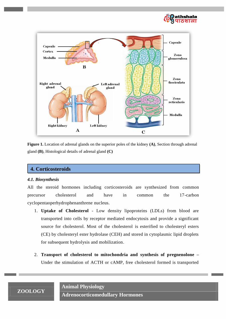
Figure 1. Location of adrenal glands on the superior poles of the kidney (A), Section through adrenal
gland (B), Histological details of adrenal gland (C)
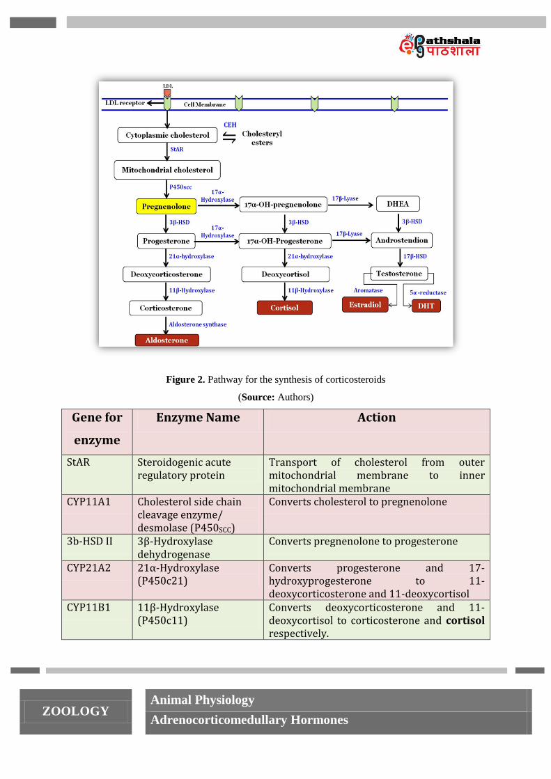
4.1. Biosynthesis
All the steroid hormones including corticosteroids are synthesized from common
precursor cholesterol and have in common the 17-carbon
cyclopentaoperhydrophenanthrene nucleus.
1. Uptake of Cholesterol - Low density lipoproteins (LDLs) from blood are
transported into cells by receptor mediated endocytosis and provide a significant
source for cholesterol. Most of the cholesterol is esterified to cholesteryl esters
(CE) by cholesteryl ester hydrolase (CEH) and stored in cytoplasmic lipid droplets
for subsequent hydrolysis and mobilization.
2. Transport of cholesterol to mitochondria and synthesis of pregnenolone –
Under the stimulation of ACTH or cAMP, free cholesterol formed is transported
4. Corticosteroids
ZOOLOGY Animal Physiology
Adrenocorticomedullary Hormones
from the outer mitochondrial membrane to the inner mitochondrial membrane with
the help of steroidogenic acute regulatory protein (StAR). In the mitochondria, a
cytochrome P450 side chain cleavage enzyme (P450SCC) converts cholesterol to
pregnenolone. The fate of pregnenolone depends on the enzymes that are present
in that tissue.
3. Synthesis of DHEA and androstenedione - Pregnenolone is converted to
progesterone or 17α-hydroxypregnenolone by 3β-HSD enzyme and
P450c17/Cyp17 respectively. Cyp17 or P450c17 (17α-hydroxylase/c 17-20 lyase)
is a single bi-functional enzyme and has two activities. First it converts
pregnenolone and progesterone to 17α-hydroxypregnenolone and 17α-
hydroxyprogesterone respectively. Secondly, 17α-hydroxypregnenolone and 17α-
hydroxyprogesterone again acted on by P450c17 (c 17-20 lyase) to yield
dehydroepiandrosterone (DHEA) and androstenedione, respectively.
4. Synthesis of cortisol and aldosterone - Progesterone and 17-
hydroxyprogesterone are converted to 11-deoxycorticosterone and 11-
deoxycortisol, respectively via hydroxylation catalyzed by P450c21 (21-
hydroxylase). P450c11 (11β-hydroxylase) catalyzes the final step in the synthesis
of mineralocorticoids and glucocorticoids and bring about conversion of 11-
deoxycorticosterone and 11-deoxycortisol to corticosterone and cortisol
respectively. Corticosterone is finally converted to aldosterone by enzyme
aldosterone synthase.
5. Synthesis of testosterone and estradiol - 17β-HSD uses androstenedione as
substrate to produce testosterone. Testosterone is further metabolized to 17β -
estradiol (E2) and dehydrotestosterone (DHT) by aromatase (Cyp19) and 5α-
reductase respectively.
P450SCC, P450c11 (11-β-hydroxylase), and aldosterone synthase are mitochondrial enzyme.
ZOOLOGY Animal Physiology
Adrenocorticomedullary Hormones
Figure 2. Pathway for the synthesis of corticosteroids
(Source: Authors)
Gene for
enzyme
Enzyme Name Action
StAR Steroidogenic acute regulatory protein
Transport of cholesterol from outer mitochondrial membrane to inner mitochondrial membrane
CYP11A1 Cholesterol side chain cleavage enzyme/ desmolase (P450SCC)
Converts cholesterol to pregnenolone
3b-HSD II 3β-Hydroxylase dehydrogenase
Converts pregnenolone to progesterone
CYP21A2 21α-Hydroxylase (P450c21)
Converts progesterone and 17-hydroxyprogesterone to 11-deoxycorticosterone and 11-deoxycortisol
CYP11B1 11β-Hydroxylase (P450c11)
Converts deoxycorticosterone and 11-deoxycortisol to corticosterone and cortisol respectively.
ZOOLOGY Animal Physiology
Adrenocorticomedullary Hormones
CYP11B2 Aldosterone synthase Corticosterone converted to aldosterone
CYP17 17α-Hydroxylase/17, 20 lyase (P450c17)
Converts pregnenolone and progesterone to 17α-hydroxypregnenolone and 17α-hydroxyprogesterone respectively.
17α-hydroxypregnenolone and 17α-hydroxyprogesterone are converted to dehydroepiandrosterone (DHEA) and androstenedione
17b-HSD 17β-Hydroxysteroid dehydrogenase
Converts androstenedione to testosterone
CYP19 Aromatase Converts testosterone to estradiol
Table 1. Details of enzymes involved in biosynthesis of corticosteroids
4.2. Transport
Approximately 5% of cortisol and 40% of aldosterone circulate in the free form; the
remainder is bound to corticosteroid-binding globulin (CBG) called transcortin and albumin.
Transcortin along with the hormone binds to the target cell, and facilitates the entry of
hormone to the target cell. Most of the aldosterone is present in a free state in the blood and
therefore metabolized faster. Cortisol has longer half-life of 60-90 minutes as compared to
aldosterone having 20 minutes. Corticosteroids are metabolized in liver by forming soluble
salts of sulphates and glucuronic acid and then excreted via urine, bile, or feces. Pathological
conditions where liver and kidney functions are impaired leads to decreased rate of
corticosteroids inactivation and excretion of metabolized products
4.3. Mechanism of action
On the basis of receptor localization, the mechanism of hormone action is categorized into
two categories:
Hormones that binds to intracellular receptors follow genomic pathway of action
Hormone that binds to cell surface receptors follow non genomic pathway of action
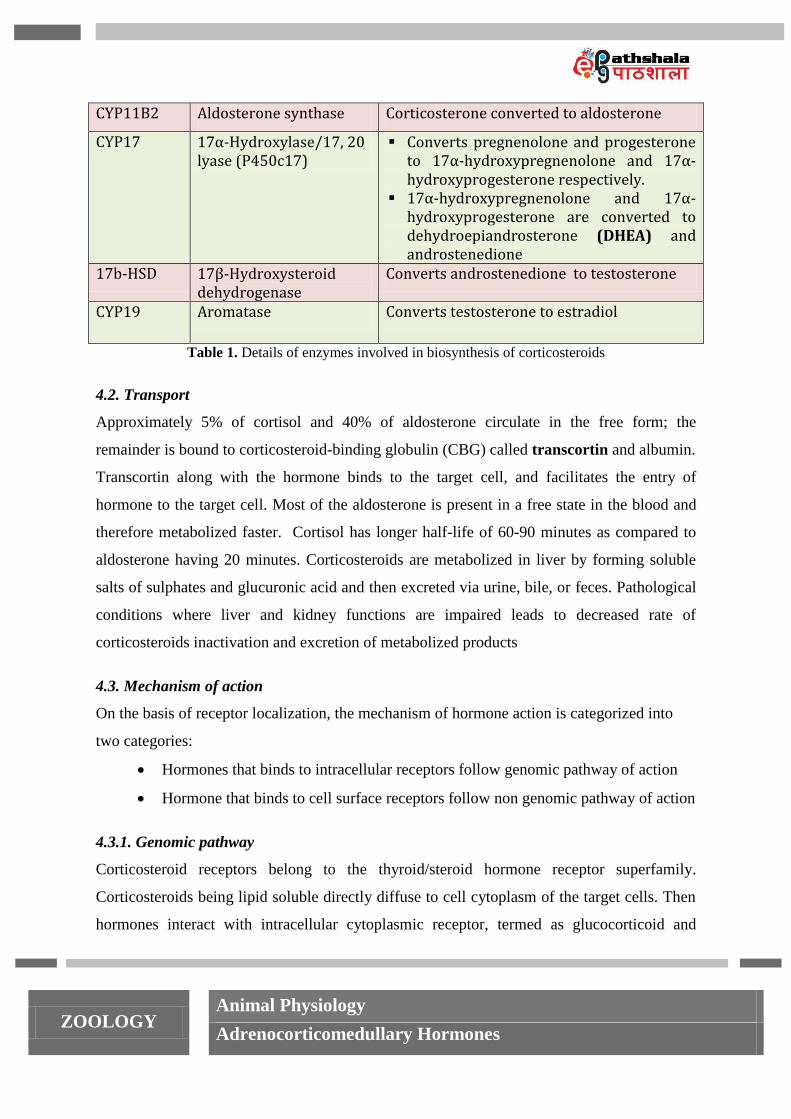
4.3.1. Genomic pathway
Corticosteroid receptors belong to the thyroid/steroid hormone receptor superfamily.
Corticosteroids being lipid soluble directly diffuse to cell cytoplasm of the target cells. Then
hormones interact with intracellular cytoplasmic receptor, termed as glucocorticoid and
ZOOLOGY Animal Physiology
Adrenocorticomedullary Hormones
mineralocorticoid receptors (GR and MR) forming a hormone-receptor complex. Unoccupied
receptors have chaperone proteins bound to them and inhibit the cellular response.
Dissociation of chaperons (HSP 90 and HSP 70) from receptors leads to activation of
hormone-receptor complex. After translocation to the nucleus, dimerized hormone-receptor
complex interact with specific DNA sequences in the promoter regions of target genes called
hormone response element (HRE). This association induces the expression of respective
genes to synthesize RNA and protein.
Figure 3. Genomic pathway for action of corticosteroids
(Source: Authors)
4.3.2. Non-genomic action
Besides having genomic action (60-90 minutes) corticosteroids have rapid non-genomic
(seconds to few minutes) action on the target cell without any requirement for the synthesis
of new proteins. These non-genomic actions are insensitivity towards inhibitors of
ZOOLOGY Animal Physiology
Adrenocorticomedullary Hormones
transcription & translation and are mediated by high affinity cell membrane receptors coupled
to rise in second messenger concentration and activation of protein kinase cascades with
subsequent phosphorylation and activation of target proteins.
Aldosterone was first isolated by Simpson and Tait (1953), is a very potent
mineralocorticoid and accounts for about 90 percent of all mineralocorticoid activity.
5.1. Function of aldosterone
The name mineralocorticoid is derived from its role in regulating concentrations of minerals,
particularly sodium and potassium in extracellular fluids.
Aldosterone, the principle mineralocorticoid acts on cells of distal tubule, collecting
tubule and collecting duct of the kidney, to stimulate the Na+ retention accompanied
by corresponding excretion of K+, H
+ and NH4
+ ions.
Increased retention of sodium ion leads to obligatory absorption of water to maintain
osmotic balance thus controlling extracellular fluid volume as well. Thus, maintaining
blood pressure indirectly.
Aldosterone assists in attaining acid-base balance by controlling the level of hydrogen
ion.
Aldosterone has related effects on sweat glands, salivary glands and the colon where
it helps in conservation of sodium chloride and water.
Mineralocorticoids are life saving corticosteroids and reduced rate of secretion leads to fatal
consequences if remain untreated with salt therapy or hormone administration. Absence of
mineralocorticoid causes retention of potassium ion and depletion in the level of sodium and
chloride ions thus decreasing extracellular volume and blood volume. Decrease in blood
volume further reduces cardiac output and blood pressure, producing a shock like state. Also
increases level of potassium causes serious cardiac toxicity, including weakness of heart
contraction and development of arrhythmia
High level of aldosterone causes hypokalemia, a condition of decrease in potassium ion
concentration affecting normal electrical excitability of nerve and muscle fibers, preventing
5. Mineralocorticoids - Aldosterone
ZOOLOGY Animal Physiology
Adrenocorticomedullary Hormones
action potential propagation. Increased excretion of hydrogen ion along with hypokalemia
cause pH to increase, a condition called mild alkalosis.
Aldosterone secretion follows a cyclical pattern having peak secretion occurs in morning and
lowest point at night.
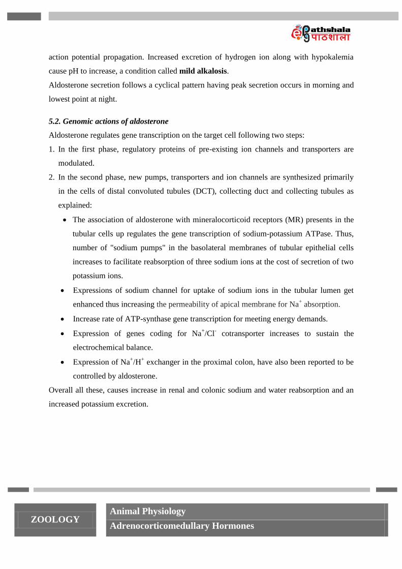
5.2. Genomic actions of aldosterone
Aldosterone regulates gene transcription on the target cell following two steps:
1. In the first phase, regulatory proteins of pre-existing ion channels and transporters are
modulated.
2. In the second phase, new pumps, transporters and ion channels are synthesized primarily
in the cells of distal convoluted tubules (DCT), collecting duct and collecting tubules as
explained:
The association of aldosterone with mineralocorticoid receptors (MR) presents in the
tubular cells up regulates the gene transcription of sodium-potassium ATPase. Thus,
number of "sodium pumps" in the basolateral membranes of tubular epithelial cells
increases to facilitate reabsorption of three sodium ions at the cost of secretion of two
potassium ions.
Expressions of sodium channel for uptake of sodium ions in the tubular lumen get
enhanced thus increasing the permeability of apical membrane for Na+ absorption.
Increase rate of ATP-synthase gene transcription for meeting energy demands.
Expression of genes coding for Na+/Cl
- cotransporter increases to sustain the
electrochemical balance.
Expression of Na+/H
+ exchanger in the proximal colon, have also been reported to be
controlled by aldosterone.
Overall all these, causes increase in renal and colonic sodium and water reabsorption and an
increased potassium excretion.
ZOOLOGY Animal Physiology
Adrenocorticomedullary Hormones
Figure 4. Genomic action of aldosterone
(Source: Authors)
5.3. Non-genomic effects of aldosterone
Aldosterone has been shown to increase formation of cAMP via second messenger system in
vascular smooth muscle cells and in epithelial cells of the renal tubules rapidly. Aldosterone
stimulation increases intracellular calcium ion concentration along with rapid stimulation of
proton conductance of plasma membrane and increase pH of cytosol. Cumulatively all these
factors enhance the number of Na+/H
+ exchanger. In medullary thick ascending limb, there is
a decrease in HCO3- absorption by increase in aldosterone stimulation. The other non-
genomic effects of aldosterone are arterial vasodilation mediated by increase in NO level.
5.4. Regulation of aldosterone secretion
Aldosterone secretion by the zona glomerulosa is regulated by five major factors as follows:
1. The major stimulus for aldosterone synthesis is angiotensin II or renin-angiotensin-
aldosterone system (RAAS) - The renin-angiotensin-aldosterone system is the major
regulator of aldosterone secretion and stimuli for RAAS activation is Na+ deficiency,
dehydration or haemorrhage resulting in low blood volume and thus low blood pressure.
These factors stimulate juxtaglomerular cells to secrete the enzyme renin, a very specific
ZOOLOGY Animal Physiology
Adrenocorticomedullary Hormones
protease synthesized by the juxtaglomerular apparatus (JGA) in the kidney. Renin
converts the angiotensinogen (a precursor glycoprotein synthesized and released by the
liver) to the bioinactive decapeptide angiotensin I. Angiotensin I is acted on by
angiotensin-converting enzyme (ACE) in the lungs and other tissues to angiotensin II
(octapeptide). Angiotensin II stimulate aldosterone synthesis by increasing the activity of
P450scc activity. Long-lasting stimulation up regulates the level of all the enzymes of
glomerulosa required for aldosterone synthesis.
Binding of aldosterone to the cytosolic mineralocorticoid receptor and further molecular
changes results in increased Na+ absorption and K
+ & H
+ secretion by the renal tubules.
Angiotensin II and aldosterone also stimulates contraction of smooth muscle of
arterioles. The resulting vasoconstriction of the arterioles increases blood pressure.
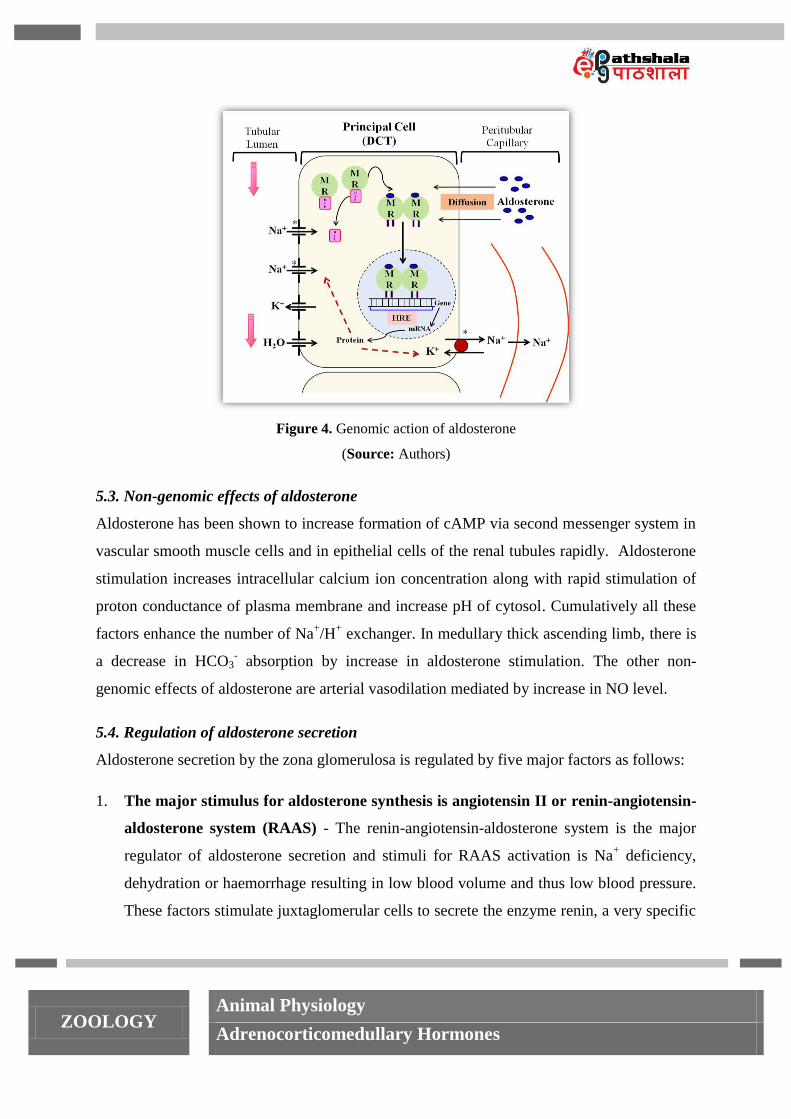
The resultant increase in plasma Na+, decrease in plasma K
+ and elevated blood pressure
provide a feedback mechanism for suppressing renin and, subsequently, aldosterone
secretion (Fig. 5).
Figure 5. Control of aldosterone secretion by renin-angiotensin-aldosterone system (RAAS)
(Source: Authors)
ZOOLOGY Animal Physiology
Adrenocorticomedullary Hormones
2. Increased potassium ion concentration in the extracellular fluid significantly
augments aldosterone secretion - Increase in potassium ion concentration sends
positive signal for the synthesis of aldosterone mediated by increased intra-adrenal
production of angiotensin II. Resultant increase in aldosterone acts on renal tubules to
excrete the surplus potassium ions and bringing the RAAS system to normal.
3. Decreased sodium ion concentration stimulates aldosterone slightly - With 10 to 20
percent decreases in extracellular fluid sodium ion concentration can double the rate of
aldosterone synthesis. The effect of sodium is mediated primarily by increased
angiotensin II and secondary by increased renin release from the kidney.
4. ACTH from the anterior pituitary gland is necessary for aldosterone secretion but
has little effect in controlling the rate of secretion - The role of ACTH
(adrenocorticotropic hormone, a polypeptide tropic hormone secreted by anterior
pituitary gland) in controlling the secretion of aldosterone is minor. Maintained high
levels of ACTH are required to stimulate the aldosterone production. Total absence of
ACTH reduces aldosterone secretion and causes atrophy of the tissue.
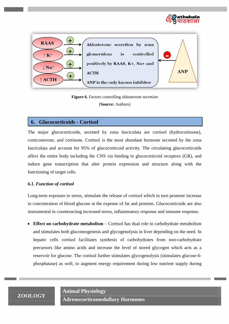
5. Atrial natriuretic peptide (ANP) is the only inhibitor for the synthesis of
aldosterone - Atrial natriuretic peptide (ANP) is a peptide hormone secreted by cells of
atria in response to elevated blood pressure and volume. Increased ANP inhibits release
of renin by the JGA and aldosterone synthesis by adrenal gland. ANP being a vasodilator
is a known to reduce blood pressure.
ZOOLOGY Animal Physiology
Adrenocorticomedullary Hormones
Figure 6. Factors controlling aldosterone secretion
(Source: Authors)
The major glucocorticoids, secreted by zona fasciculata are cortisol (hydrocortisone),
corticosterone, and cortisone. Cortisol is the most abundant hormone secreted by the zona
fasciculata and account for 95% of glucocorticoid activity. The circulating glucocorticoids
affect the entire body including the CNS via binding to glucocorticoid receptors (GR), and
induce gene transcription that alter protein expression and structure along with the
functioning of target cells.
6.1. Function of cortisol
Long-term exposure to stress, stimulate the release of cortisol which in turn promote increase
in concentration of blood glucose at the expense of fat and proteins. Glucocorticoids are also
instrumental in counteracting increased stress, inflammatory response and immune response.
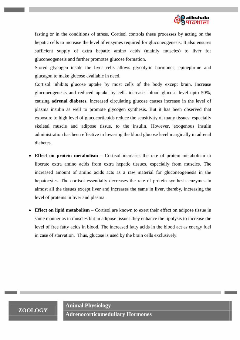
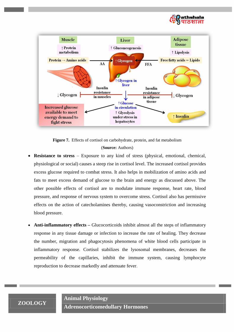
Effect on carbohydrate metabolism – Cortisol has dual role in carbohydrate metabolism
and stimulates both gluconeogenesis and glycogenolysis in liver depending on the need. In
hepatic cells cortisol facilitates synthesis of carbohydrates from non-carbohydrate
precursors like amino acids and increase the level of stored glycogen which acts as a
reservoir for glucose. The cortisol further stimulates glycogenolysis (stimulates glucose-6-
phosphatase) as well, to augment energy requirement during low nutrient supply during
6. Glucocorticoids - Cortisol
ZOOLOGY Animal Physiology
Adrenocorticomedullary Hormones
fasting or in the conditions of stress. Cortisol controls these processes by acting on the
hepatic cells to increase the level of enzymes required for gluconeogenesis. It also ensures
sufficient supply of extra hepatic amino acids (mainly muscles) to liver for
gluconeogenesis and further promotes glucose formation.
Stored glycogen inside the liver cells allows glycolytic hormones, epinephrine and
glucagon to make glucose available in need.
Cortisol inhibits glucose uptake by most cells of the body except brain. Increase
gluconeogenesis and reduced uptake by cells increases blood glucose level upto 50%,
causing adrenal diabetes. Increased circulating glucose causes increase in the level of
plasma insulin as well to promote glycogen synthesis. But it has been observed that
exposure to high level of glucocorticoids reduce the sensitivity of many tissues, especially
skeletal muscle and adipose tissue, to the insulin. However, exogenous insulin
administration has been effective in lowering the blood glucose level marginally in adrenal
diabetes.
Effect on protein metabolism – Cortisol increases the rate of protein metabolism to
liberate extra amino acids from extra hepatic tissues, especially from muscles. The
increased amount of amino acids acts as a raw material for gluconeogenesis in the
hepatocytes. The cortisol essentially decreases the rate of protein synthesis enzymes in
almost all the tissues except liver and increases the same in liver, thereby, increasing the
level of proteins in liver and plasma.
Effect on lipid metabolism – Cortisol are known to exert their effect on adipose tissue in
same manner as in muscles but in adipose tissues they enhance the lipolysis to increase the
level of free fatty acids in blood. The increased fatty acids in the blood act as energy fuel
in case of starvation. Thus, glucose is used by the brain cells exclusively.
ZOOLOGY Animal Physiology
Adrenocorticomedullary Hormones
Figure 7. Effects of cortisol on carbohydrate, protein, and fat metabolism
(Source: Authors)
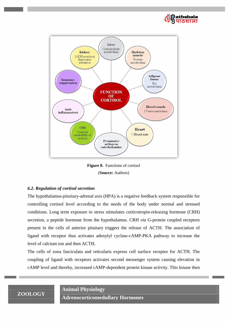
Resistance to stress – Exposure to any kind of stress (physical, emotional, chemical,
physiological or social) causes a steep rise in cortisol level. The increased cortisol provides
excess glucose required to combat stress. It also helps in mobilization of amino acids and
fats to meet excess demand of glucose to the brain and energy as discussed above. The
other possible effects of cortisol are to modulate immune response, heart rate, blood
pressure, and response of nervous system to overcome stress. Cortisol also has permissive
effects on the action of catecholamines thereby, causing vasoconstriction and increasing
blood pressure.
Anti-inflammatory effects – Glucocorticoids inhibit almost all the steps of inflammatory
response in any tissue damage or infection to increase the rate of healing. They decrease
the number, migration and phagocytosis phenomena of white blood cells participate in
inflammatory response. Cortisol stabilizes the lysosomal membranes, decreases the
permeability of the capillaries, inhibit the immune system, causing lymphocyte
reproduction to decrease markedly and attenuate fever.
ZOOLOGY Animal Physiology
Adrenocorticomedullary Hormones
Although, high doses are known to retard tissue repair, slow down the process of healing
and cause mental disturbances. Glucocorticoids are very useful in the treatment of chronic
inflammatory disorders such as rheumatoid arthritis.
Depression of immune responses - Cortisol suppresses the immune response by affecting
most of the cells (cellular and humoral immunity) that participate in immune reactions.
The rate of lymphocyte proliferation goes down along with increase in rate of apoptosis.
The effectiveness of glucocorticoids (prednisone, prednisolone, etc.) in suppressing
immune system makes them an ideal drug to be prescribed during organ transplant
surgeries to limit the chances of tissue rejection.
Effects on cardiovascular system: Cortisol is a known vasoconstrictor and by contracting
smooth muscles of blood vessels it increases blood pressure. Strength of cardiac muscle is
increased by virtue of the direct effect of cortisol in regulating sodium and potassium
concentration in heart cells.
Effects on central nervous system: Cortisol affects the behavior, mood, and electrical
excitability of the neurons. It has been studied that any imbalance in the levels of cortisol
causes symptoms of adrenal fatigue leading to moodiness, decreased tolerance, decreased
clarity of thought and decreased memory. Hippocampus of temporal lobe is responsible
for memory formation and has many glucocorticoid receptors and sensitive to stress, thus
glucocorticoids play an important role in memory as well.
Effects on water and electrolyte metabolism: Deficiency causes increased production of
ADH which can decrease in glomerular filtration rate causing water retention in the body.
Thus, the overall effect of cortisol is to inhibit tissue building while stimulating the
breakdown of stored nutrients to maintain adequate fuel supplies.
ZOOLOGY Animal Physiology
Adrenocorticomedullary Hormones
Figure 8. Functions of cortisol
(Source: Authors)
6.2. Regulation of cortisol secretion
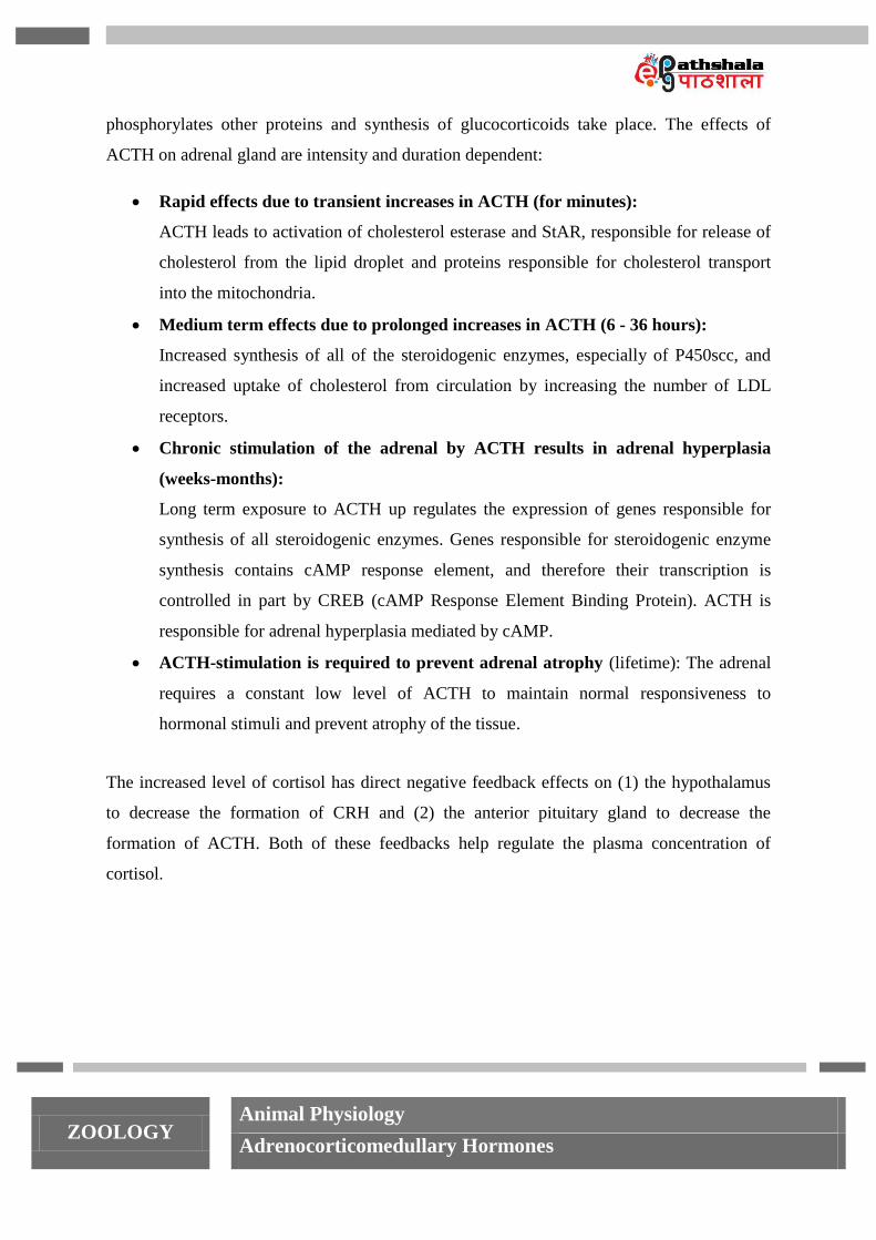
The hypothalamus-pituitary-adrenal axis (HPA) is a negative feedback system responsible for
controlling cortisol level according to the needs of the body under normal and stressed
conditions. Long term exposure to stress stimulates corticotropin-releasing hormone (CRH)
secretion, a peptide hormone from the hypothalamus. CRH via G-protein coupled receptors
present in the cells of anterior pituitary triggers the release of ACTH. The association of
ligand with receptor thus activates adenylyl cyclase-cAMP-PKA pathway to increase the
level of calcium ion and then ACTH.
The cells of zona fasciculata and reticularis express cell surface receptor for ACTH. The
coupling of ligand with receptors activates second messenger system causing elevation in
cAMP level and thereby, increased cAMP-dependent protein kinase activity. This kinase then
ZOOLOGY Animal Physiology
Adrenocorticomedullary Hormones
phosphorylates other proteins and synthesis of glucocorticoids take place. The effects of
ACTH on adrenal gland are intensity and duration dependent:
Rapid effects due to transient increases in ACTH (for minutes):
ACTH leads to activation of cholesterol esterase and StAR, responsible for release of
cholesterol from the lipid droplet and proteins responsible for cholesterol transport
into the mitochondria.
Medium term effects due to prolonged increases in ACTH (6 - 36 hours):
Increased synthesis of all of the steroidogenic enzymes, especially of P450scc, and
increased uptake of cholesterol from circulation by increasing the number of LDL
receptors.
Chronic stimulation of the adrenal by ACTH results in adrenal hyperplasia
(weeks-months):
Long term exposure to ACTH up regulates the expression of genes responsible for
synthesis of all steroidogenic enzymes. Genes responsible for steroidogenic enzyme
synthesis contains cAMP response element, and therefore their transcription is
controlled in part by CREB (cAMP Response Element Binding Protein). ACTH is
responsible for adrenal hyperplasia mediated by cAMP.
ACTH-stimulation is required to prevent adrenal atrophy (lifetime): The adrenal
requires a constant low level of ACTH to maintain normal responsiveness to
hormonal stimuli and prevent atrophy of the tissue.
The increased level of cortisol has direct negative feedback effects on (1) the hypothalamus
to decrease the formation of CRH and (2) the anterior pituitary gland to decrease the
formation of ACTH. Both of these feedbacks help regulate the plasma concentration of
cortisol.
ZOOLOGY Animal Physiology
Adrenocorticomedullary Hormones
Figure 9. Regulation of cortisol secretion - The hypothalamus-pituitary-adrenal axis (HPA)
(Source: Authors)
Adrenal androgens are weak androgens, synthesized by zona reticularis of adrenal cortex.
The major androgens secreted are dehydroepiandrosterone (DHEA) and androstenedione.
Adrenal androgens are produced in response to ACTH and converted to testosterone or
estrogen in peripheral tissues. During prepubertal phase, androgens stimulate growth of
axillary and pubic hair in boys and girls. After puberty the major sources of androgens are
from gonads thus, the role of adrenal androgen is not very significant. However, in adult
females adrenal androgens promote sex drive. After menopause, when ovarian secretion of
estrogens ceases, adrenal androgens become the major source of all the estrogen required by
the system.
8.1. Synthesis of catecholamines
As discussed earlier adrenal medulla is neuroendocrine tissue composed of postganglionic
sympathetic nervous system (SNS) neurons and under the influence of stimulus from
8. Medullary hormones
7. Adrenal androgens
ZOOLOGY Animal Physiology
Adrenocorticomedullary Hormones
hypothalamus synthesize and secrete catecholamines. They are commonly named as
catecholamines due to presence of catechol nucleus and an amine group in their structures.
Chromaffin cells of adrenal medulla synthesize catecholamines from amino acid tyrosine
catalyzed by various enzymes using following steps:
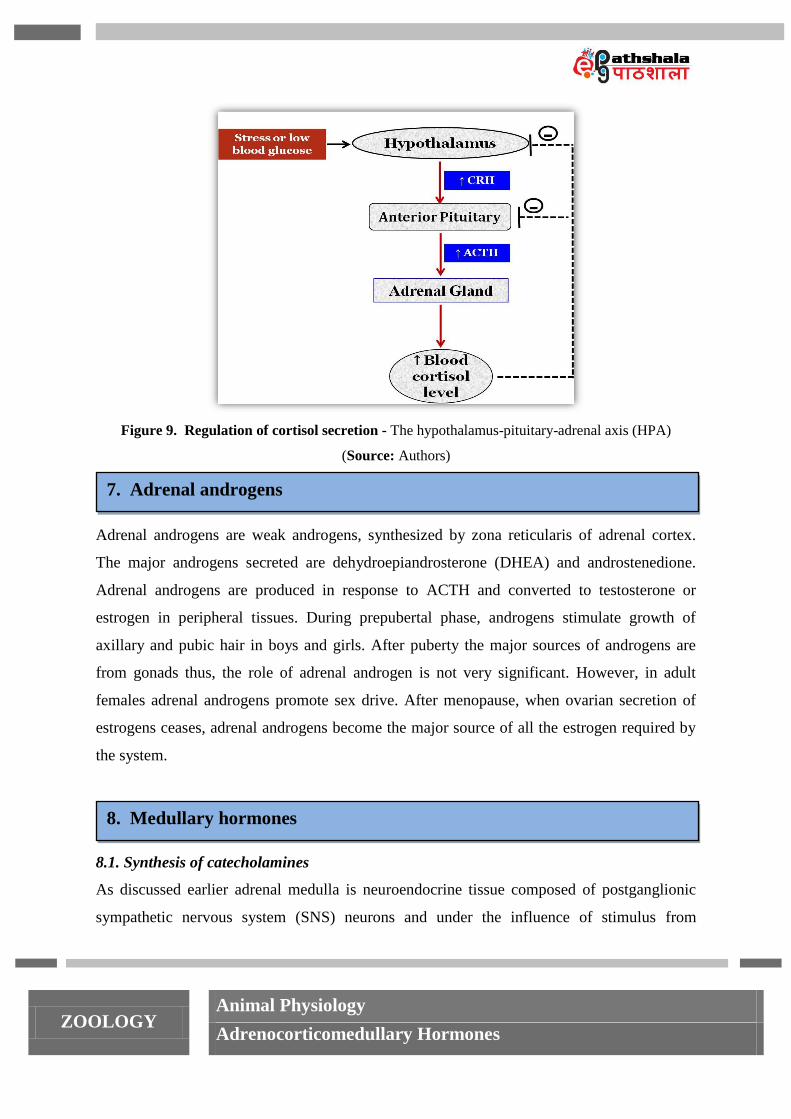
Step 1: Tyrosine hydroxylase (TH) catalyzes the rate-limiting step in catecholamine
synthesis and catalyzes the addition of a hydroxyl group to the tyrosine, thus forming 3, 4-
dihydroxy-l-phenylalanine (l-DOPA). (TH can also hydroxylate phenylalanine to form
tyrosine, which is then converted to l-DOPA; this alternative synthetic route may be of
significance in patients affected with phenylketonuria, a condition in which phenylalanine
hydroxylase activity is depressed).
Step 2: DOPA decarboxylase, a pyridoxine-dependent enzyme catalyzes the synthesis of first
catecholamine i.e. dopamine from l-DOPA by decarboxylation.
Step 3: Dopamine β-hydroxylase (DBH) further catalyzes the addition of hydroxyl group to
the β carbon on the side chain of dopamine to synthesize norepinephrine and use copper as
cofactor.
Step 4: NE synthesized in the granules of chromaffin cells diffuse to cytoplasm and
methylated by phenylethanolamine N-methyltransferase (PNMT). PNMT catalyzes the
addition of methyl group to the nitrogen of norepinephrine (NE) forming a third amine,
epinephrine.
Figure 10. Pathway for catecholamine synthesis in adrenal medulla
(Source: Authors)
ZOOLOGY Animal Physiology
Adrenocorticomedullary Hormones
8.2. Release of catecholamines
Once synthesized, catecholamines are stored in secretory granules of chromaffin cells.
Acetylcholine released from preganglionic sympathetic fibers as a result of stress (physical or
psychological or both) stimulates nicotinic cholinergic receptors and causes depolarization of
the adrenomedullary chromaffin cell. Depolarization leads to activation of voltage-gated Ca2+
channels resulting in exocytosis of secretory vesicle contents. A Ca2+
-sensing receptor
appears to be involved in the process of exocytosis causing release of catecholamines to the
circulation where they circulate in the bound form with albumin or other serum proteins. The
half-life of catecholamine is very short (~ 2 minutes) and thereafter degraded by catechol-O-
methyltransferases (COMT) or by deamination by monoamine oxidases (MAO) via
methylation or deamination respectively.
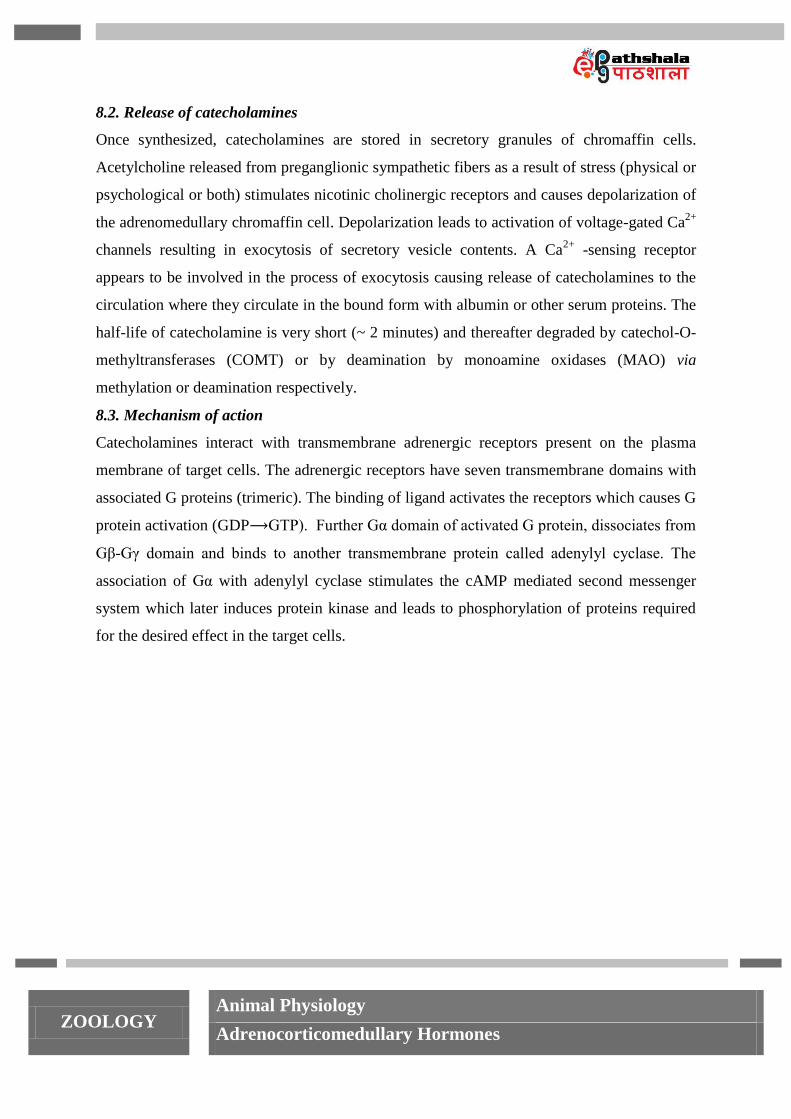
8.3. Mechanism of action
Catecholamines interact with transmembrane adrenergic receptors present on the plasma
membrane of target cells. The adrenergic receptors have seven transmembrane domains with
associated G proteins (trimeric). The binding of ligand activates the receptors which causes G
protein activation (GDP⟶GTP). Further Gα domain of activated G protein, dissociates from
Gβ-Gγ domain and binds to another transmembrane protein called adenylyl cyclase. The
association of Gα with adenylyl cyclase stimulates the cAMP mediated second messenger
system which later induces protein kinase and leads to phosphorylation of proteins required
for the desired effect in the target cells.
ZOOLOGY Animal Physiology
Adrenocorticomedullary Hormones
Figure 11. Cyclic AMP mediated pathway of action for catecholamine on target cell having
adrenergic receptors
(Source: Authors)
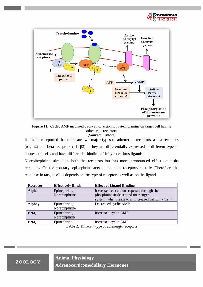
It has been reported that there are two major types of adrenergic receptors, alpha receptors
(α1, α2) and beta receptors (β1, β2). They are differentially expressed in different type of
tissues and cells and have differential binding affinity to various ligands.
Norepinephrine stimulates both the receptors but has more pronounced effect on alpha
receptors. On the contrary, epinephrine acts on both the receptors equally. Therefore, the
response in target cell is depends on the type of receptor as well as on the ligand.
Receptor Effectively Binds Effect of Ligand Binding
Alpha1 Epinephrine,
Norepinphrine
Increase free calcium (operate through the
phosphoinositide second messenger
system, which leads to an increased calcium (Ca2+
)
Alpha2 Epinephrine,
Norepinphrine
Decreased cyclic AMP
Beta1 Epinephrine,
Norepinphrine
Increased cyclic AMP
Beta2 Epinephrine Increased cyclic AMP
Table 2. Different type of adrenergic receptors
ZOOLOGY Animal Physiology
Adrenocorticomedullary Hormones
8.4. Functions of epinephrine (EP) and norepinephrine (NE)
Epinephrine and norepinephrine synthesized and released by adrenal medulla under the
influence of impulses from hypothalamus through sympathomedullary (SAM) pathway.
These amines have long lasting effect, and prepare the body to fight any kind of stress as
done by SNS. Advantage of having these hormones along with SNS is that they circulate in
the blood and can send the signals to the cells that are not directly innervated by nerve fibers.
The following are the effects of EP and NE:
Increased rate and force of contraction of the heart muscle: catecholamines have
both positive inotropic affect (cardiac contraction) and chronotropic effects (heart
rate) on heart. Predominantly these effects are controlled by epinephrine through beta
receptors. Epinephrine directly stimulates myocardial cells to increase the strength of
ventricular contraction thus increased heart rate. Also causes vasoconstriction in the
vessels of the skin, mucosa, and kidney.
Constriction of blood vessels: norepinephrine causes vasoconstriction, resulting in
increased arterial blood pressure and therefore blood flow to the skeletal muscles and
central nervous system is increased. The action of vasoconstriction is counter
balanced by vasodilator action of alpha 2 receptors to some extent.
Dilation of bronchioles: epinephrine is a powerful bronchio-dilatior and thus assists
in pulmonary ventilation.
Stimulation of lipolysis in adipose tissues: this provides fatty acids for energy
production in many tissues and aids in conservation of dwindling reserves of blood
glucose.
Increased metabolic rate: oxygen consumption and heat production increase
throughout the body in response to epinephrine to increase the rate of metabolism.
Medullary hormones also promote glycogenolysis in skeletal muscle and liver to
provide glucose for supplementing energy requirement.
Dilation of the pupils: important in low intensity of light.
ZOOLOGY Animal Physiology
Adrenocorticomedullary Hormones
Inhibition of certain "non-essential" processes: an example is inhibition of
gastrointestinal secretion and motor activity.
8.5. Functions of dopamine
In the brain, dopamine functions as a neurotransmitter, activating dopamine receptors.
Dopamine has important role in cognition, movement, flow of information to frontal lobes
from other areas of brain, and regulation prolactin secretion. Besides this, dopamine is
associated with the pleasure system of the brain, providing us feelings of enjoyment and
motivation. Dopamine system has also been strongly linked to parkinson’s disease, psychosis
and schizophrenia.
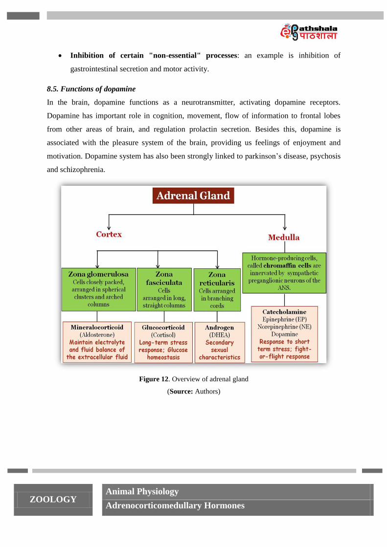
Figure 12. Overview of adrenal gland
(Source: Authors)
ZOOLOGY Animal Physiology
Adrenocorticomedullary Hormones
Several disorders are caused by the dysregulation of the hormones produced by the adrenal
glands. Hypersecretion of corticosteroids causes cushing’s disease characterized by high
blood glucose levels and the accumulation of lipid deposits on the face and neck. In contrast,
the hyposecretion of corticosteroids can result in adrenal insufficiency, characterized by low
blood glucose levels and low blood sodium levels.
9.1. Adrenal insufficiency
Corticoid insufficiency arises from three main reasons:
1. Primary - destruction or dysfunction of the adrenal cortex known as Addison’s disease
2. Secondary - dysfunction of HPA axis causing low levels of ACTH
3. Congenital adrenal hyperplasia
9.1.1. Primary adrenocortical insufficiency/ Addison’s disease
Addison’s disease is named after Thomas Addison, the British physician who first described
the condition in 1855. A life-threatening condition caused by atrophy or dysgenesis of the
adrenal gland. The primary cause of addison’s disease leading to hyposecretion of corticoids
is due to autoimmune attack on the adrenal. Antibodies cause adrenal cortex destruction or
block binding of ACTH to its receptors. Besides this, tuberculosis, and cancer are other
possible causes for addison’s disease.
The signs and symptoms of addison’s disease appear until 90% of the adrenal cortex has been
destroyed. Symptoms include mental lethargy, anorexia, nausea and vomiting, weight loss,
hypoglycemia, muscular weakness, and cravings for salty food. The addison’s disease is
manifested by various disturbances as follows:
Mineralocorticoid deficiency/ hypoaldosteronism
Low levels of aldosterone inhibit the sodium ion and water absorption from renal tubules
with concomitant excretion of potassium and hydrogen ions. All these factors cause decrease
in blood volume and pressure manifesting three conditions:
Hypotension – low blood pressure due to overall decrease in blood volume
9. Pathophysiology of adrenal gland
ZOOLOGY Animal Physiology
Adrenocorticomedullary Hormones
Hyponatremia - low level of sodium in the blood
Hyperkalemia – high level of potassium in the blood
Also there is decrease in cardiac output, fluid volume and increase in red blood cell count
(polycythemia).
Glucocorticoid deficiency
The deficiency of glucocorticoids is characterized by difficulty in mobilization of energy
stores, rapid fatigue, and inability to fight stress. Loss of cortisol levels causes hypoglycemia,
weakness of muscle, weight loss, hyponatremia, increased pigmentation, abnormal metabolic
functions, irritability and mental sluggishness. Lack of adequate glucocorticoid secretion also
makes a person with addison’s disease highly incapable of fighting any kind of stress and a
small bacterial or viral infection can cause death of the individual. If remain untreated patient
with addison’s disease die in few days to few weeks. Treatment consists of administrating
glucocorticoids and mineralocorticoids and increasing sodium in the diet.
9.1.2. Secondary adrenocortical insufficiency
Low levels of corticosteroid secretion caused by disorder of HPA axis leading to diminished
level of ACTH and ultimately low stimulation of adrenal cortex. Besides disturbance in HPA
axis infection, pituitary tumors and irradiation also affect the synthesis of ACTH by anterior
pituitary gland.
9.1.3. Congenital adrenal hyperplasia (CAH)
Congenital adrenal hyperplasia as the name suggests is a congenital disorder caused by
mutation in the genes for the enzymes (especially 21-hydroxylase catalyze the final steps of
mineralocorticoids and glucocorticoids synthesis, but not androgens) required for synthesis of
steroid hormone. This causes decreased synthesis of glucocorticoid and lead to
malfunctioning of HPA axis. Due to low levels of cortisol negative feedback is interrupted
and therefore, compensatory high levels of CRH and ACTH are synthesized by hypothalamus
and anterior pituitary respectively. The increased ACTH causes hyperplasia of the adrenal
and (usually) elevated levels of other adrenal steroid products. The most common enzyme
mutated in CAH is 21-hydroxylase, catalyses the final steps of mineralocorticoids and
glucocorticoids synthesis, but not androgens. Therefore, ACTH stimulation of the adrenal
ZOOLOGY Animal Physiology
Adrenocorticomedullary Hormones
cortex induces the release of excessive amounts of adrenal androgens, which can lead to the
development of ambiguous genitalia and secondary sex characteristics.
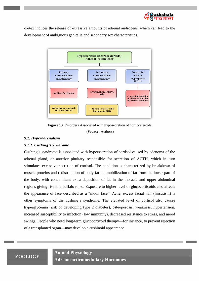
Figure 13. Disorders Associated with hyposecretion of corticosteroids
(Source: Authors)
9.2. Hyperadrenalism
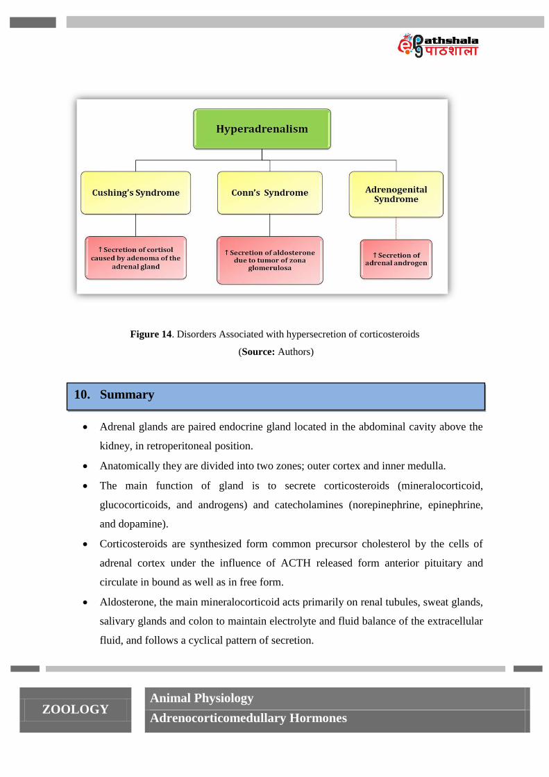
9.2.1. Cushing’s Syndrome
Cushing’s syndrome is associated with hypersecretion of cortisol caused by adenoma of the
adrenal gland, or anterior pituitary responsible for secretion of ACTH, which in turn
stimulates excessive secretion of cortisol. The condition is characterized by breakdown of
muscle proteins and redistribution of body fat i.e. mobilization of fat from the lower part of
the body, with concomitant extra deposition of fat in the thoracic and upper abdominal
regions giving rise to a buffalo torso. Exposure to higher level of glucocorticoids also affects
the appearance of face described as a ―moon face‖. Acne, excess facial hair (hirsutism) is
other symptoms of the cushing’s syndrome. The elevated level of cortisol also causes
hyperglycemia (risk of developing type 2 diabetes), osteoporosis, weakness, hypertension,
increased susceptibility to infection (low immunity), decreased resistance to stress, and mood
swings. People who need long-term glucocorticoid therapy—for instance, to prevent rejection
of a transplanted organ—may develop a cushinoid appearance.
ZOOLOGY Animal Physiology
Adrenocorticomedullary Hormones
Treatment of Cushing’s syndrome consists of removing an adrenal tumor surgically or
destroyed by radiation, followed by administration of adrenal steroids to supplement for
decrease level of hormones. Drugs that block steroidogenesis, such as metyrapone,
ketoconazole, and aminoglutethimide, or that inhibits ACTH secretion (serotonin antagonists
and GABA-transaminase inhibitors) are prescribed to avoid surgery.
9.2.2. Primary hyperaldosteronism/ Conn’s syndrome
Excess aldosterone secretion, due to bilateral idiopathic cortical hyperplasia or tumor of zona
glomerulosa is associated with sodium retention and potassium depletion. High rate of
sodium ion (Hypernatremia) and water retention causes high blood pressure (hypertension)
associated with hypokalemia. Low extracellular potassium concentration causes muscle
paralysis due to depressive effect on action potential transmission by the nerve fibers. Due to
negative feedback of increased aldosterone level renin secretion is inhibited by JGA. (85%)
Hyperaldosteronism treatment includes surgery and administration of aldosterone
antagonists.
9.2.3. Adrenal androgen hypersecretion/Adrenogenital Syndrome
Adrenocortical tumor also causes excess androgen secretion by adrenal gland causing intense
masculinising effects throughout the body. In females over production of testosterone leads to
overdeveloped male characteristics such as hirsutism, deepening voice and more muscular
arms and legs. Increases androgen levels before puberty manifested by development of
premature secondary sex characteristics such as deep voice, beard, enlarged penis and a sex
drive although gonads are not functioning and there is no sperm production.
9.3. Pheochromocytomas
Tumor of the chromaffin cells of adrenal medulla is known as pheochromocytomas and leads
to hypersecretion of catecholamines causing rapid heart rate (cardiac arrhythmias), high
blood pressure (hypertension), high levels of glucose in blood and urine, an elevated basal
metabolic rate (BMR), flushed face, nervousness, sweating, and decreased gastrointestinal
motility. Surgical removal of the gland is practiced if the lesions are localized. a-Methyl-p-
tyrosine (an inhibitor of tyrosine hydroxylase, the rate-limiting enzyme in catecholamine
biosynthesis) may be used to decrease catecholamine secretion from the tumor.
ZOOLOGY Animal Physiology
Adrenocorticomedullary Hormones
Figure 14. Disorders Associated with hypersecretion of corticosteroids
(Source: Authors)
Adrenal glands are paired endocrine gland located in the abdominal cavity above the
kidney, in retroperitoneal position.
Anatomically they are divided into two zones; outer cortex and inner medulla.
The main function of gland is to secrete corticosteroids (mineralocorticoid,
glucocorticoids, and androgens) and catecholamines (norepinephrine, epinephrine,
and dopamine).
Corticosteroids are synthesized form common precursor cholesterol by the cells of
adrenal cortex under the influence of ACTH released form anterior pituitary and
circulate in bound as well as in free form.
Aldosterone, the main mineralocorticoid acts primarily on renal tubules, sweat glands,
salivary glands and colon to maintain electrolyte and fluid balance of the extracellular
fluid, and follows a cyclical pattern of secretion.
10. Summary

ZOOLOGY Animal Physiology
Adrenocorticomedullary Hormones
Cortisol helps in fight against long term stress through its direct control on
carbohydrate, protein, and fat metabolism. Besides this cortisol depress immune
system and inflammatory responses of the body.
The HPA axis is a negative feedback system, responsible for controlling cortisol level
according to the needs of the body under normal and stressed conditions.
Catecholamines are synthesized from tyrosine in the chromaffin cells of adrenal
medulla following specific pathway and released in the circulation under the influence
of stimulus form preganglionic fibers.
Catecholamines have specific adrenergic receptors on the target cells and prepare the
body to fight against short term stress.
Several disorders are associated with dysregulation of the hormones produced by
adrenal glands. Hypersecretion of corticosteroids causes cushing’s disease
characterized by high blood glucose levels and the accumulation of lipid deposits on
the face and neck. In contrast, the hyposecretion of corticosteroids can result in
adrenal insufficiency, characterized by low blood glucose levels and low blood
sodium levels.
Low levels of catecholamines do not show much affect as their role is counter
balanced by sympathetic nervous system however, hypersecretion causes cardiac
arrhythmias, hypertension, high levels of glucose in blood and urine, an elevated basal
metabolic rate etc.