Discovery of Selective Probes and Antagonists for G Protein-Coupled Receptors FPR/FPRL1 and GPR30
17
Discovery of Selective Probes and Antagonists for G Protein- Coupled Receptors FPR/FPRL1 and GPR30 Jeffrey B. Arterburn 1,4 , Tudor I. Oprea 2,4 , Eric R. Prossnitz 3,4 , Bruce S. Edwards 5,6 , and Larry A. Sklar 4,6 1 Department of Chemistry and Biochemistry, New Mexico State University, Las Cruces, NM 88003 2 Division of Biocomputing, Department of Biochemistry and Molecular Biology, University of New Mexico Health Sciences Center, Albuquerque, NM, USA 3 Department of Cell Biology and Physiology, School of Medicine, University of New Mexico Health Science Center, Albuquerque, NM 87131 4 UNM Cancer Center, University of New Mexico Health Science Center, Albuquerque, NM 87131 5 University of New Mexico Center for Molecular Discovery, University of New Mexico, Albuquerque, NM 87131 6 Department of Pathology, School of Medicine, University of New Mexico Health Science Center, Albuquerque, NM 87131 Abstract Recent technological advances in flow cytometry provide a versatile platform for high throughput screening of compound libraries coupled with high-content biological testing and drug discovery. The G protein-coupled receptors (GPCRs) constitute the largest class of signaling molecules in the human genome with frequent roles in disease pathogenesis, yet many examples of orphan receptors with unknown ligands remain. The complex biology and potential for drug discovery within this class provide strong incentives for chemical biology approaches seeking to develop small molecule probes to facilitate elucidation of mechanistic pathways and enable specific manipulation of the activity of individual receptors. We have initiated small molecule probe development projects targeting two distinct families of GPCRs: the formylpeptide receptors (FPR/FPRL1) and G protein- coupled estrogen receptor (GPR30). In each case the assay for compound screening involved the development of an appropriate small molecule fluorescent probe, and the flow cytometry platform provided inherently biological rich assays that enhanced the process of identification and optimization of novel antagonists. The contributions of cheminformatics analysis tools, virtual screening, and synthetic chemistry in synergy with the biomolecular screening program have yielded valuable new chemical probes with high binding affinity, selectivity for the targeted receptor, and potent antagonist activity. This review describes the discovery of novel small molecule antagonists of FPR and FPRL1, and GPR30, and the associated characterization process involving secondary assays, cell based and in vivo studies to define the selectivity and activity of the resulting chemical probes Keywords flow cytometry; fluorescent; GPCR; formylpeptide receptor; inflammation; GPR30; GPER; estrogen; nongenomic; cancer; antidepressant * Address correspondence to: Jeffrey B. Arterburn, [email protected], (575) 646-2738. NIH Public Access Author Manuscript Curr Top Med Chem. Author manuscript; available in PMC 2010 June 15. Published in final edited form as: Curr Top Med Chem. 2009 ; 9(13): 1227–1236. NIH-PA Author Manuscript NIH-PA Author Manuscript NIH-PA Author Manuscript
Transcript of Discovery of Selective Probes and Antagonists for G Protein-Coupled Receptors FPR/FPRL1 and GPR30
Discovery of Selective Probes and Antagonists for G Protein-Coupled Receptors FPR/FPRL1 and GPR30
Jeffrey B. Arterburn1,4, Tudor I. Oprea2,4, Eric R. Prossnitz3,4, Bruce S. Edwards5,6, andLarry A. Sklar4,61 Department of Chemistry and Biochemistry, New Mexico State University, Las Cruces, NM 880032 Division of Biocomputing, Department of Biochemistry and Molecular Biology, University of NewMexico Health Sciences Center, Albuquerque, NM, USA3Department of Cell Biology and Physiology, School of Medicine, University of New Mexico HealthScience Center, Albuquerque, NM 871314 UNM Cancer Center, University of New Mexico Health Science Center, Albuquerque, NM 871315 University of New Mexico Center for Molecular Discovery, University of New Mexico, Albuquerque,NM 871316 Department of Pathology, School of Medicine, University of New Mexico Health Science Center,Albuquerque, NM 87131
AbstractRecent technological advances in flow cytometry provide a versatile platform for high throughputscreening of compound libraries coupled with high-content biological testing and drug discovery.The G protein-coupled receptors (GPCRs) constitute the largest class of signaling molecules in thehuman genome with frequent roles in disease pathogenesis, yet many examples of orphan receptorswith unknown ligands remain. The complex biology and potential for drug discovery within thisclass provide strong incentives for chemical biology approaches seeking to develop small moleculeprobes to facilitate elucidation of mechanistic pathways and enable specific manipulation of theactivity of individual receptors. We have initiated small molecule probe development projectstargeting two distinct families of GPCRs: the formylpeptide receptors (FPR/FPRL1) and G protein-coupled estrogen receptor (GPR30). In each case the assay for compound screening involved thedevelopment of an appropriate small molecule fluorescent probe, and the flow cytometry platformprovided inherently biological rich assays that enhanced the process of identification andoptimization of novel antagonists. The contributions of cheminformatics analysis tools, virtualscreening, and synthetic chemistry in synergy with the biomolecular screening program have yieldedvaluable new chemical probes with high binding affinity, selectivity for the targeted receptor, andpotent antagonist activity. This review describes the discovery of novel small molecule antagonistsof FPR and FPRL1, and GPR30, and the associated characterization process involving secondaryassays, cell based and in vivo studies to define the selectivity and activity of the resulting chemicalprobes
Keywordsflow cytometry; fluorescent; GPCR; formylpeptide receptor; inflammation; GPR30; GPER;estrogen; nongenomic; cancer; antidepressant
*Address correspondence to: Jeffrey B. Arterburn, [email protected], (575) 646-2738.
NIH Public AccessAuthor ManuscriptCurr Top Med Chem. Author manuscript; available in PMC 2010 June 15.
Published in final edited form as:Curr Top Med Chem. 2009 ; 9(13): 1227–1236.
NIH
-PA Author Manuscript
NIH
-PA Author Manuscript
NIH
-PA Author Manuscript
Flow Cytometry for Gpcr Probe DiscoveryThe G protein-coupled receptors (GPCRs) constitute the largest class of signaling moleculesin the human genome with frequent roles in disease pathogenesis that provide targets for alarge proportion of currently marketed drugs. The critical role of GPCRs in transmittingextracellular signals into cells via intracellular G protein heterotrimers, relatively few of whichhave been characterized, and the existence of large number of GPCRs termed “orphanreceptors” for which native ligands are not known, have positioned this class prominently inthe arena of drug discovery. The complex biology associated with GPCR-mediated signalingprovides additional motivation for the discovery of new small molecule ligands that exhibitselective binding and functional control of specific receptors. These chemical probes can serveas valuable tools for elucidating fundamental biological mechanisms in cell-based and invivo studies, and help to establish foundations for drug discovery initiatives that exploit thenovel targets and pathways that are revealed.
Flow cytometry has matured as a platform for the quantitative multi-parameter measurementof cell fluorescence and has numerous applications including drug discovery [1-6]. Theversatility of flow cytometry accommodates the display of virtually any molecular assemblyor cell response. Novel bead-based assay systems allow studies of real-time interactionsbetween solubilized receptors, ligands, protein-protein interactions and molecular signalingcomponents that recapitulate and extend measurements in intact cells. Recent innovationsallow faster serial processing of bulk cell samples. Homogeneous discrimination of free andcell-bound fluorescent probe eliminates wash steps to streamline sample processing. Flowcytometry is particularly convenient for alternately assessing both cellular and molecularactivities of small molecules. By using suspension arrays of particles, which can be color-codedmolecules, nanoparticles, beads or complex cell populations the resulting assays and responsescan be highly multiplexed to maximize sample analysis throughput and generatemultidimensional biological output. These new developments are coupled with the widespreadavailability of instrumentation to position flow cytometry as an attractive analysis platform forhigh-throughput, high-content biological testing and drug discovery.
The University of New Mexico Center for Molecular Discovery was established with supportfrom the NIH Roadmap Molecular Libraries Screening Network (MLSCN) and subsequentlythrough the Molecular Libraries Probe (MLP) program. The NMCMD consists of amultidisciplinary academic research team with assembled expertise from biomedical,biophysical, chemical, computational, instrumentation and engineering fields. This specialtycenter has established flow cytometry as a versatile platform for drug discovery, proteomics,and real-time analysis of molecular interactions while incorporating a novel sampling approach(HyperCyt®) [7], associated technology, assay design, informatics and analysis tools thatsupport high throughput screening to identify small molecule probes in a biological informationcontent-rich format. Small molecule probe development projects incorporating virtualscreening, cheminformatics and synthetic chemistry focused on two distinct families of GPCRshave yielded valuable new chemical probes. In each case the screening application requiredthe development of an appropriate small molecule fluorescent probe, and the flow cytometryplatform provided inherently biological rich assays that facilitated the identification andoptimization of novel antagonists. This review describes the discovery of novel small moleculeantagonists of FPR and FPRL1, and GPR30, and the associated characterization studies thatemploy secondary assays, cell based and in vivo studies to define the selectivity and activityof the resulting chemical probes.
Arterburn et al. Page 2
Curr Top Med Chem. Author manuscript; available in PMC 2010 June 15.
NIH
-PA Author Manuscript
NIH
-PA Author Manuscript
NIH
-PA Author Manuscript

Formyl Peptide Receptors FPR/FPRL1The G protein-coupled formylpeptide receptors (FPRs) have been implicated in a variety ofinflammatory and immunological responses, and provide an important area for the applicationof chemical biology approaches to elucidate the complex interplay and fundamental biologyof this important class of receptors, and to explore potential therapeutic applications [8-11].FPR is expressed in a variety of cell types including astrocytes, hepatocytes, immature dendriticcells, microglial cells, neutrophils and the tunica media of coronary arteries. Two other variantshave been described: FPRL1 and FPRL2 that share 69% and 56% primary sequence identityto FPR respectively, and 83% identity between the pair. FPRL1 occurs in an even greatervariety of cell types than FPR including astrocytoma cells, epithelial cells, hepatocytes,microvascular endothelial cells, neuroblastoma cells, and phagocytic leukocytes. FPRL2 isexpressed only in monocytes. Recent clinical interest has associated FPR with antibacterialinflammation and malignant glioma cell metastasis, while FPRL1 has been linked to chronicinflammation in systemic amyloidosis, Alzheimer's disease, and prion diseases as well asovarian cancer metastasis. N-Formylated peptides constitute the most commonly studied classof FPR activators that trigger a variety of biological activities in myeloid cells, includingchemokinesis, chemotaxis, cytokine production and superoxide generation. Many of thesepeptide ligands are only weak activators of FPRL1, however, a number of host-derived agonistshave been identified in association with pathophysiological processes. At the time this studywas undertaken there was only one report of small molecule antagonist of FPR [12], and onereport of an FPRL1 antagonist was described during the course of this investigation [13].
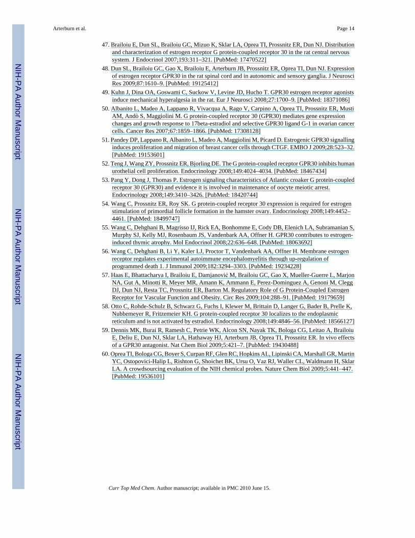
Development of a Cross-Reactive Fluorescent Ligand for FPR/FPRL1The conceptual approach for a screening assay to identify novel small molecule antagonistsfocused on achieving the simultaneous measurement of binding interactions with both FPRand FPRL1 receptors. This strategy has the potential to improve overall screening efficiency,an advantage of multiplexing, and generates data that reflects receptor specificity as an inherentfeature of the screen. This approach required a fluorescent probe with high affinity for bothreceptors FPR and FPRL1 that could simultaneously report ligand-binding interactions in asingle assay. N-Formylated peptides such as E. coli derived N-formyl-Met-Leu-Phe (fMLF)are known to be high-affinity ligands for FPR that initiate a variety of biological activities.Many potent formylpeptide FPR agonists only weakly activate FPRL1. The fMLFK-FITC hasbeen used as a fluorescent reporter for FPR with a Kd = 3 nM, but does not bind FPRL1 [14].Peptides possessing amino terminal tryptophan (W) residues were identified as selective highaffinity ligands for FPRL1 [15], and substitutions at the second amino acid residues within theW-peptide series were tolerated without significantly compromising FPRL1 binding [16]. Weinvestigated the hexapeptide probe H2N-Trpl-Lys(FL)-Tyr-Met-Val-(D)Met-amide(compound WK(FL)YMVm Fig (1)) incorporating a fluorescein dye attached to the lysineresidue [17]. Equilibrium binding experiments with WK(FL)YMVm revealed Kd values of1.21 ± 0.36 nM and 1.82 ± 0.78 nM for FPR and FPRL1 respectively. The affinity of the parenthexapeptide was ∼100 fold higher for FPRL1 than FPR, thus the attachment of fluorescein tothe lysine residue enhanced the affinity of the resulting probe for FPR diproportionately. Theobservation of enhanced binding is consistent with the proposed contribution of a largehydrophobic region towards the binding interaction of FPRL1 [18]. This fluorescent probeWK(FL)YMVm represents the first cross-reactive high affinity reporter ligand, demonstratingspecific ligand interactions with both FPR and FPRL1 expressing cell lines, and havingsatisfied the performance criteria as a fluorescent ligand probe was used to develop a flowcytometric assay.
Arterburn et al. Page 3
Curr Top Med Chem. Author manuscript; available in PMC 2010 June 15.
NIH
-PA Author Manuscript
NIH
-PA Author Manuscript
NIH
-PA Author Manuscript
Identificiation of FPR/FPRL1-Selective AntagonistsA flow-cytometry-based high- throughput screening (HTS) approach was used to identify smallmolecules that exhibit high binding affinity with individual selectivity for FPR and FPRL1.The screening assay measured the ability of test compounds to competitively displace thefluorescent peptide WK(FL)YMVm from FPR, FPRL1, or both. In this assay U937 cellsexpressing FPR and rat basophil leukemia (RBL) cells expressing FPRL1 were tested togetherin a duplex format. The FPR-expressing U937 cells were color coded by incubation with red-fluorescent dye FuraRed™ in order to easily distinguish and separately gate these cells fromthe RBL/FPRL1 cells during the flow cytometric analysis. Compounds, cells, and fluorescentligand were sequentially combined without intervening wash steps. The high-throughputHyperCyt™ flow cytometry platform was used to sample cells from 384-well microplates forintroduction to the flow cytometer [6,19]. Fluorescence was excited at 488nm and detectedwith optical bandpass filters matched for the two reporter dyes. The time-resolved data fileswere analyzed with IDLeQuery software to identify active compounds in each well. Theprimary screen of 24,304 NIH Small Molecule Repository revealed 253 compounds withinhibition >30%, representing 181 for FPR, 72 for FPRL1 and 26 cross-reactive compoundsfor both receptors. Subsequent dose response studies identified a total of 40 compounds withselective binding inhibition constants Ki ≤ 4 μM, including 34 for FPR and 6 for FPRL1. Nocross-reactive compounds with Ki ≤ 4 μM were found. Cheminformatic analysis classifiedcritical chemical scaffolds or chemotype families that correlated with activity, and werecompared to several chemotypes earlier reported by our group [12]. Computational screeningand two-dimensional substructure searching of a commercial ChemDiv compound collectionwas used to identify an additional 1,446 structurally related compounds for activityoptimization. The focused set of compounds selected for expanded screening included 1,276for FPR, and a separate set of 170 for FPRL1. The single concentration HTS assay revealedstrong correlation of activity for the targeted receptor with over 10-fold increase in thepercentage of tested compounds with Ki ≤ 4 μM for FPR, and over 100-fold increase in thepercentage of potent actives for FPRL1, providing clear confirmation of the hypothesizedstructure activity relationships. The most active compounds identified were resynthesized,fully characterized to confirm structure and purity, and retested to quantify activity [20]. Themost potent compound against FPR was the 4H-chromen-4-one derivative 3570-0208, withKi = 95 +/- 10 nM. A clear structure-activity relationship was observed in association withsubstitution of aryl and heterocyclic groups at the 3-position in this series, culminating in themaximum inhibitory activity associated with the benzimidazole derivative 3570-0208. Inseparate duplex competition assays this compounds affinity for FPR exceeded that for FPRL1by more than 100-fold. The compound with the greatest activity for FPRL1 was the imidazo[1,2-a]pyrimidine derivative BB-V-115 with Ki = 270 +/- 51 nM. Structure-activityoptimization focused on the carboxamide group derived from methoxyaniline in this series andachieved maximum inhibition of FPRL1 in the derivative possessing a 2-methylpropanamidegroup. Compound BB-V-115 exhibited receptor selectivity for FPRL1 ∼20-fold greater thanfor FPR. This compound was also compared with the recently reported small molecule FPRL1antagonist C7, a 3-amino-2,3-dihydroquinazolin-4(1H)-one, which was found to exhibit ∼10-fold lower affinity for FPRL1 than BB-V-115 [13].
These compounds were evaluated in secondary assays for their ability to block the intracellularcalcium response induced by high affinity peptide agonists for their respective target receptors.The calcium response elicited by WKYMVm in RBL/FPRL1 cells was blocked by the FPRL1antagonist BB-V-115. The response induced by fMLFF in U937/FPR cells was similarlyblocked by the FPR antagonist 3570-0208. Each of these antagonists demonstrated selectivityfor their targeted receptor, exhibiting the inability to block the activity of the respective peptideagonists for the off-target receptor even at increased concentrations up to 100 μM. To date,
Arterburn et al. Page 4
Curr Top Med Chem. Author manuscript; available in PMC 2010 June 15.
NIH
-PA Author Manuscript
NIH
-PA Author Manuscript
NIH
-PA Author Manuscript
these two probes represent the most potent, selective small molecule antagonist reported fortheir respective receptor.
The resulting selective antagonist probes developed in this study using high throughput flowcytometry to screen large compound libraries are expected to find applications in delineatingpathophysiological processes associated with the FPR and FPRL1 receptors. The duplex formatemploying color-coded cells expressing the respective receptors reduced the overall assay time,minimized reagent requirements, and provided selectivity information at every screening stage,thus proving to be an efficient approach for identifying selective small molecule GPCR receptorligands. The color-coding approach can be readily adapted to higher degrees of assaymultiplexing to encompass even greater biological content and further enhance screeningefficiency. The incorporation of in silico computational methods and substructure searchingto populate an expanded set of chemotype families for characterization of structure-activityduring the probe optimization stage significantly enhanced the percentage of active compoundsand improved the efficiency with which highly selective antagonist probes were identified forboth receptors.
Probe Development Targeting the G Protein-Coupled Estrogen ReceptorGPR30
The G protein-coupled estrogen receptor GPR30 has emerged as an intriguing signalingmolecule enmeshed in the complex pathways through which estrogens regulate diversephysiological processes [21-26]. Estrogen (compound E2 Fig (2)) initiates rapid biochemicaland physiological responses that are not accounted for by changes in gene expression mediatedby the nuclear estrogen receptors (ERα/β). Membrane-associated classical ERα has beenimplicated in the initiation of signaling cascades through direct interactions with membranestructures that include G proteins, caveolins, and receptor tyrosine kinases. Non-classicalmodels attribute this activity to other membrane-associated estrogen-binding proteins,including recent studies that have identified GPR30 as a prime candidate. High affinity estrogenbinding to GPR30 results in activation of stimulatory G proteins, transactivation of epidermalgrowth factor receptor, increased adenylyl cyclase activity, and rapid intracellular calciummobilization, occurring within seconds to minutes, and accumulation of phosphatidylinositol3,4,5-triphosphate in the nucleus that occurs following ∼15 minutes of ligand inducedstimulation [27,28]. However, the experimental difficulty involved in distinguishing thedetailed pathways through which estrogen induces these multiple cellular and physiologicalresponses has presented a primary challenge for advancing the field. GPR30 expression inprimary breast adenocarcinoma has been associated with pathological parameters commonlyused to assess breast cancer progression, including the development of extramammarymetastases [29]. GPR30 expression is linked to lower survival rates in endometrial and ovariancancer [30,31]. Despite the common affinity for E2, differences in binding to syntheticestrogens, phytoestrogens and xenoestrogens have been recognized [32]. The antiestrogenstamoxifen and ICI 182,780 that have high binding affinity for GPR30 but elicit opposite activityprofiles in comparison with ERα/β may have relevance for tumorigenesis and cancer therapy.No examples of GPR30-selective ligands had been reported prior to the initiation of the studiesdescribed herein. Using a combined approach integrating chemical and molecular biology wehave developed the first generation of GPR30-selective probes that provide the tools forelucidating the biological roles of GPR30 from the classical nuclear estrogen receptors.
Cellular studies employing transfected COS7 monkey kidney fibroblast cells expressingGPR30 or ERα/β provide complete experimental control of receptor content that isexceptionally valuable for investigating the associated activation pathways. One particularlyfascinating result from the initial study was the identification of GPR30 as an intracellulartransmembrane estrogen receptor localized in the endoplasmic reticulum [28]. Identical
Arterburn et al. Page 5
Curr Top Med Chem. Author manuscript; available in PMC 2010 June 15.
NIH
-PA Author Manuscript
NIH
-PA Author Manuscript
NIH
-PA Author Manuscript
localization patterns are revealed in cancer cell lines that endogenously express GPR30,including MCF-7, SKBr3, JEG and Hec50co. These provocative findings have broadimplications towards understanding the fundamental biological function of GPCR's and forclinical applications of estrogen therapeutics. The postulate that estrogen activates GPR30 frominside the cell is consistent with the hydrophobic structure of estrogen, which contributesmembrane permeability and provides ready access to the intracellular compartment. Thenecessary machinery for GPCR's to initiate signaling is expected to be present in theendoplasmic reticulum and the intermediate signaling molecules are present in the cytoplasm.The capacity for plasma membrane G protein-coupled receptors to translocate to other cellularstructures and establish ligand-responsive signaling complexes has only been recognizedrelatively recently, and represents a fascinating new arena for pharmacology and drugdiscovery. The possibility of estrogen-stimulated GPR30 signaling from an intracellularlocation is consistent with reports of other functional intracellular GPCRs associated withlipophilic ligands and receptor kinases [36-39].
Development of a Fluorescent Estrogen LigandIn order to further investigate this intriguing scenario, we designed and synthesized a series of17α-[4-aminomethyl-phenylethynyl]-estra-1, 3, 5(10)-triene-3,17β-diol derivatives asfluorescent and functional probes to characterize the localization and functionality of GPR30in comparison with the classical nuclear receptors ERα/β [28]. The selection of the 17α-positionof substitution on the steroid scaffold combined with the rigid hydrophobic linkage were criticalfactors that enabled the synthesis of probes that retained high affinity for both classes ofreceptors. We constructed fluorescent estrogen ligands employing a pendant approach to attacha dye to a derivatized estrogen ligand, as represented by the conjugate (compound E2-Alexa546 Fig (2)). This approach provides a high degree of versatility for probe design due tothe commercial availability of a wide variety of fluorescent dyes with different structuralcharacteristics and photophysical properties that can be matched with specific excitation andemission requirements for experiments employing multiple fluorophores. The structures ofmost fluorescent dyes contain large hydrophobic polycyclic aromatic ring systems adornedwith polar and often ionizable functional groups that provide some measure of water solubility,and this modification results in major differences in steric volume and physicochemicalproperties in comparison with the original receptor ligand. Interestingly, E2-Alexa546 did notexhibit observable binding to the plasma membrane in the GPR30-expressing cell linesinvestigated. The ionic charge of the AlexaFluor dyes rendered the corresponding estrogenconjugates membrane-impermeable over the time course of rapid signaling events studied andno staining was observed unless the cells were permeabilized with saponin,. A nuclear stainingpattern with E2-Alexa546 was observed in transfected cells expressing a fusion protein ofgreen fluorescent protein (GFP) and ERα, (ERα-GFP), as expected for the ligand binding stateof nuclear receptors. Colocalization of this probe with GPR30-GFP was observed in theendoplasmic reticulum, analogous to results obtained using markers and GPR30-targetedantibodies. This binding was competitive with E2, and revealed a Ki = 6.6 nM for GPR30. Theunnatural biologically inactive diastereomer 17α-estradiol (compound 17α-E2 Fig (2)) wasunable to displace the fluorescent ligand, providing further verification of the selectivity ofestrogen binding. These results strongly support the model for estrogen binding to GPR30 inthe endoplasmic reticulum, but cannot exclude the possibility for differential activity ofreceptor localized in the plasma membrane.
Development of Permeability-Restricted Estrogen ProbesTo further investigate the model in which GPR30 initiates signaling from an intracellularlocation, we synthesized a series of related small molecule probes possessing either neutral orcharged appendages, thereby providing a simple parameter with which to control membrane
Arterburn et al. Page 6
Curr Top Med Chem. Author manuscript; available in PMC 2010 June 15.
NIH
-PA Author Manuscript
NIH
-PA Author Manuscript
NIH
-PA Author Manuscript
permeability through passive diffusion [40]. We hypothesized that if GPR30 was functional atthe cell surface, both charged (E2-NMe3
+) and neutral (E2-NB) probes should have access tothe receptor and be capable of activating the rapid signaling pathways, while differences inactivation would be expected if the alternative scenario involving GPR30 activation from anintracellular site. This approach provided a complementary design for permeability-restrictedestrogens that have employed macromolecular conjugates to bovine serum albumin [41], orpoly(amido)amine dendrimer conjugates [42,43], with the technical advantages associatedwith using fully characterized, discrete small molecules. The binding properties of these probeswere evaluated using transfected COS-7 cells for both ERα and GPR30 in competitive bindingassays employing the fluorescent ligand E2-Alexa633. This experimental design involvedmembrane permeabilization with saponin to provide entry to both the charged fluorescentconjugate and the neutral or ionic estrogen probes, thereby allowing comparison of theirrelative binding affinities. Cells expressing ERα revealed a Ki = 0.65 nM for E2, andcorresponding values for E2-NB (Ki = 2.7 nM), and E2-NMe3
+ (Ki = 1.4 nM). Analogousexperiments using GPR30-transfected cells revealed Ki = 9 nM for E2, and the neutral E2-NB (Ki = 16 nM), and charged compound E2-NMe3
+ (Ki = 17 nM). The estrogen-probesexhibited only slightly lower affinities compared with E2 for each receptor type, associatedwith the strategic positioning of the arylethynyl appendages at the 17α-position, but there wereno significant differences in binding affinities between the probes associated with the presenceof charged or neutral hydrophobic substituents.
Two functional signaling assays were then used to evaluate the activation properties of thesynthetic probes in intact, non-permeabilized cell lines. The neutral compound E2-NB initiatedcalcium fluxes at 10 nM concentration that were indistinguishable from those of E2 using thevery rapid intracellular calcium mobilization assay in both transfected cell lines expressingeither ERα or GPR30. In contrast, the charged compound E2-NMe3
+ failed to stimulatemeasureable calcium fluxes during the time course of these experiments, even at concentrations100-fold higher than those needed to provide maximal activity of E2. The compounds werethen assessed for PIP3 production in transfected cell lines expressing either ERα or GPR30.This assay requires a slightly longer time frame (∼15 min) for stimulation, but revealedanalogous results demonstrating rapid action by the neutral compound E2-NB, while thecharged compound E2-NMe3
+ gave virtually no response. The same activation properties wereobtained using SKBr3 cells that endogenously express GPR30 but not ERα/β, providingadditional confirmation for the validity of results obtained using transfected cell lines. Theseresults are entirely consistent with the model of estrogen stimulated GPR30 signaling from anintracellular location, and the reduced ability of the cationic compounds E2-NMe3
+ to enterthe cell by passive diffusion. The possibility that GPR30 is capable of initiating differentsignaling events from distinct cell surface or intracellular locations, and the factors that regulateGPR30 localization in normal or disease states are intriguing questions with potentialpharmacological implications for further investigation.
Discovery of a GPR30-Selective Agonist G-1Distinguishing the biological roles of GPR30-mediated signaling from the classical estrogenreceptors ERα/β is essential for understanding the fundamental mechanisms of estrogen action.This opportunity required the discovery of new ligands with high selectivity for GPR30, andthe capacity to specifically activate or inhibit receptor-mediated signaling. The absence ofdetailed structural data for GPR30 detracts from the possible application of structure-guidedcomputational approaches. The observed cross-reactivity of E2 and estrogen-derived probesfor both classes of receptors provides a common scaffold for a ligand-based virtual screeningapproach to identify estrogen-like structures of interest for biological screening. A combinationof 2D and 3D similarity approaches were used for virtual screening of a library of 10,000molecules that was constructed using concept of GPCR-privileged substructures [44]. The
Arterburn et al. Page 7
Curr Top Med Chem. Author manuscript; available in PMC 2010 June 15.
NIH
-PA Author Manuscript
NIH
-PA Author Manuscript
NIH
-PA Author Manuscript
combined similarity score attributed 40% weight to 2D fingerprints, 40% to the shape-basedsimilarity and 20% to pharmacophore-based similarity to rank the top 100 molecules selectedby composite score for biomolecular screening.
A competitive ligand binding assay using the fluorescent estrogen probe E2-Alexa633 wasdeveloped using a high throughput flow cytometric platform (HyperCyt®) for the primaryscreen of the focused library compounds selected from virtual screening. The assay usedtransfected COS7 cells, which do not display detectable specific binding of the fluorescentestrogen in the absence of exogenous GPR30 or ER expression, with E2 and 17a-estradiol aspositive and negative controls for specific E2-Alexa633 binding. A GPR30-GFP fusion proteinwas expressed and cells were gated for high levels of green fluorescence, correlating withGPR30 expression, in order to maximize the bound fluorescent signal. As GPR30 expressionis localized to the endoplasmic reticulum and not the plasma membrane, cells werepermeabilized with saponin to permit access of the charged E2-Alexa633 to GPR30.Biomolecular screening was carried out using 17β-estradiol E2 as a positive control to blockspecific binding of E2-Alexa633 to GPR30. A single compound, identified as G-1 in Fig (3),consistently inhibited the binding of E2-Alexa633 to GPR30.[45] This compound possessesa tetrahydro-3H-cyclopenta[c]quinoline core structure. The compound was resynthesizedusing the trifluoroacetic acid catalyzed Povarov cyclization, and characterized as the racemic,but diastereomerically pure syn- or endo-isomer. The binding properties of G-1 to GPR30 weredetermined with Ki = 11 nM, whereas the corresponding Ki = 6 nM for E2 as previouslydescribed. The Hill coefficient for G-1 binding to GPR30 was 0.6, which suggests thepossibility that there are two binding sites with similar affinity.
Treatment of GPR30-expressing COS7 cells with G-1 produced a comparable maximumcalcium concentration (t1/2 ∼ 30 s) but was slower than the very rapid response from E2 (t1/2< 2 s). Dose response studies revealed an EC50 for calcium mobilization of approximately 2nM, while the corresponding EC50 for E2 was 0.3 nM. G-1 did not produce a response in non-transfected control cells, or those expressing either ERα or ERβ, both of which respondedrapidly to E2. The complementary assay based on PI3K activation and determination of nuclearaccumulation of PIP3 also demonstrated the capacity of G-1 to activate the same signalingpathways as estrogen through GPR30, while no response was produced in cells expressingERα/β.
In order to evaluate the ability of G-1 to selectively target GPR30 in an environment in whichboth classes of receptors were present, cells were cotransfected with GPR30-monomeric redfluorescent protein-1 (GPR30-mRFP1) and ERα-GFP, which distinctly localize to theendoplasmic reticulum and nucleus respectively. The presence of G-1 selectively blockedstaining of E2-Alexa633 in the endoplasmic reticulum associated with GPR30, but had noeffect on the nuclear staining associated with ERα/β. The presence of G-1 initiated nuclearaccumulation of PIP3 in SKBr3 breast cancer cells, which endogenously express GPR30 butnot ERα/β. Similar results were obtained from G-1 treated MCF7 cells, which express GPR30,high levels of ERα and low levels of ERβ. G-1 was found to inhibit migration of SKBr3 cellswith an IC50 = 0.7 nM, and MCF7 cells with IC50 = 1.6 nM. These results demonstrate theability of G-1 to selectively bind and activate endogenous GPR30, including expression in acomplex cellular environment containing classical receptors ERα/β.
Additional Biological Characterization of G-1 in Cell-Based and In VivoSystems
Following the initial report in which G-1 was shown to activate calcium mobilization andphosphatidyl inositol-3-kinase (PI3K) in cancer cells [45], this probe has been used to examinethe cellular and physiological actions of GPR30 in a variety of other published studies. Cellular
Arterburn et al. Page 8
Curr Top Med Chem. Author manuscript; available in PMC 2010 June 15.
NIH
-PA Author Manuscript
NIH
-PA Author Manuscript
NIH
-PA Author Manuscript
effects of G-1 include activation of calcium fluxes in LHRH neurons and hypothalamic neurons[46,47], spinal neuron depolarization [48], protein kinase Ce activation [49], gene expression[50,51], proliferation [50,52], oocyte meitotic arrest [53], and primordial follicle formation[54]. The role of GPR30 in vivo has been investigated using G-1 with reported effects thatinclude estrogen-induced thymic atrophy [55], experimental autoimmune encephalomyelitis[56], and vascular regulation [57]. In each of these animal models, the G-1-mediated effectswere absent in GPR30 knockout mice, establishing the selectivity of this compound for GPR30.In one report G-1 did not show any estrogenic effects in vivo using the classical estrogen targetorgans, the uterus and the mammary gland [58]. However, recent studies have revealed G-1causes a moderate increase in uterine epithelial cell proliferation [59].
Discovery of a GPR30-Selective Antagonist G15While the GPR30 agonist G-1 provided a valuable probe for distinguishing cellular andphysiological actions of GPR30 from classical nuclear receptors, further pharmacologicalcharacterization required the development of a GPR30-specific antagonist. General structuralsimilarities between G-1 and E2 provide some degree of overlap in the respectivetetrahydro-3H-cyclopenta[c]quinoline and steroid scaffolds, particularly considering theoxygen atoms that are oriented at the periphery of both polycyclic cores. Hypothesizing thatthe hydrogen bond acceptor properties of the ketone group in G-1 were associated with theobserved agonism associated with GPR30 binding, and seeking a corresponding structuraltrigger to block GPR30-mediated signaling, we employed parallel virtual screening andsynthetic programs to identify new compounds with altered activation profiles. Thetetrahydroquinoline scaffold is synthetically accessible using the three-component imino-DielsAlder or Povarov cyclization that combines an aldehyde, aniline and electron-rich alkenecomponents, and is subject to catalysis by a variety of Bronsted and Lewis acid catalysts.Following the precedent for lanthanide-catalyzed cyclization, we employed Sc(OTf)3 toconstruct a focused library of structural analogs. These conditions afforded rapid reactiontimes, excellent product yields and high endo-diastereoselectivity. Virtual screening was usedto search the NIH Molecular Libraries Small Molecule Repository (MLSMR) in order toidentify structures of interest related to the tetrahydro-3H-cyclopenta[c]quinoline scaffold ofG-1. A substructure search of 144,457 molecules in the MLSMR was performed using a customJAVA program built using the OpenEye OEJava toolkit. This virtual screen prioritized 64molecules, of which 57 were obtained from the MLSMR. The primary biomolecular screenfor GPR30 antagonism was developed using the E2-activated calcium mobilization in GPR30-expressing SKBr3 cells and evaluating compounds for the ability to block this cellularactivation. Primary screening of the 57 compounds representing this scaffold from the MLSMRyielded 8 that showed some inhibition of estrogen-mediated calcium mobilization in cellsexpressing GPR30. Of particular interest was the compound G15 shown in Fig (3) that closelyresembled G-1 but lacked the ethanone moiety of that molecule.[59]
Chemically synthesized G15 was subjected to multiple cellular and physiological assays inorder to characterize its biological effects. Competitive binding assays using a radioiodinatedligand G40 demonstrated that G15 binds to GPR30 with an affinity of approximately 20 nM.This compares to the affinity for G-1, utilizing the same assay, of approximately 7 nM, similarto our previously reported affinity of G-1 for recombinant GPR30 of 11 nM and reportedaffinities for 17β-estradiol between 3-6 nM. Thus removal of the ethanone moiety resulted ina decrease in relative binding affinity of 2-3 fold. Additional competitive binding studies toassess interactions with ERα and ERβ revealed that similar to G-1, G15 displays little bindingto ERα or ERβ at concentrations up to 10 μM, where E2 exhibits sub-nanomolar Ki values.These results reveal that G15, like G-1, displays high affinity for GPR30 with minimal bindingto ERα and ERβ (Ki > 10 μM).
Arterburn et al. Page 9
Curr Top Med Chem. Author manuscript; available in PMC 2010 June 15.
NIH
-PA Author Manuscript
NIH
-PA Author Manuscript
NIH
-PA Author Manuscript
The receptor activity of G15 was evaluated using the functional assays employed previously.G15 did not induce the rapid calcium mobilization response in GPR30 expressing SKBr3 breastcancer cells. The pre-treatment of cells with G15 in fact caused a substantial reduction in theresponse produced by subsequent exposure to G-1 or E2, which confirms the ability of G15to block the action of these GPR30 agonists. There was no inhibition of the calcium responsemediated by ATP through endogenous purinergic receptors, indicating the antagonistic effectis specific to GPR30. The inhibition of agonist stimulated calcium mobilization in SKBr3 cellsby G15 was dose-dependent, yielding an IC50 = 185 nM for blocking G-1, which comparesclosely with a similar IC50 = 190 nM for inhibiting the E2 response. Characterization of theeffect of G15 on the PI3K activation pathway in the endogenous GPR30 expressing SKBr3cells of GPR30-transfected COS7 cells revealed analogous results, producing no stimulationof this response and demonstrating the consistent ability of pre-treatment with G15 to reducethe response resulting from stimulation by G-1 and E2. In contrast, G15 had no effect on theE2-stimulated response in cells expressing ERα/β, providing confirmation of the ability ofG15 to specifically block only the GPR30-mediated pathway.
The antagonist properties of G15 were then evaluated in three well-established, physiologicallyrelevant in vivo models of estrogen action. Estrogen treatment in ovariectomized mice producesa uterine response with increasing water content (imbibition) and increased epithelial cellproliferation. Neither G-1 or G15 affected the uterine wet weight or imbibition in this modelas measured by uterine weight and microscopic evaluation of histologic sections. A singleinjection of E2 resulted in a 17-fold increase in the proliferative index of uterine epithelia asmeasured by immunodetection of Ki-67 protein. The GPR30 agonist G-1 also produced amoderate (∼4-fold) increase in proliferation compared to control. The administration of G15had no effect on proliferation. However, when these mice were treated with both G15 andE2 the relative estrogen-induced proliferation was reduced by ∼50% in a dose-dependentresponse. Similarly, the combination treatment with G15 blocked the G-1 stimulatedproliferation in a dose-dependent manner. These results suggest that GPR30 does contributeto the proliferative response in the uterus but not towards imbibition, which appears to bemediated exclusively by ERα.
The tail suspension test serves as a convenient animal model for evaluating antidepressants bymeasuring their effect on the duration of immobility. Treatment with the antagonist G15 hadno effect on the immobility time, while both E2 and G-1 and the tricyclic dibenzazepineantidepressant desipramine markedly reduced immobility time in comparison to control.Pretreatment of mice with G15 resulted in significant attenuation of the effect of both E2 andG-1, but had no effect on desipramine treatment. Desipramine potentiates neurotransmissionby selectively blocking reuptake of norepinephrine from the neural synapse, and appears toimpair serotonin transport with minor anticholinergic activity through its affinity to muscarinicreceptors. These results implicate a neurological role for GPR30 in the regulation of depressionthat may ultimately provide a viable target for the development of new antidepressants.
ConclusionThese results demonstrate the successful application of high throughput flow cytometricscreening for small molecule probe discovery. The development of the first cross reactive highaffinity fluorescent probe WK(FL)YMVm was used in a duplex assay that simultaneouslyevaluated the binding interactions with individual cell lines expressing FPR and FPRL1. Anovel 4H-chromen-4-one derivative 3570-0208 antagonist for FPR was identified with Ki =95 +/- 10 nM. The imidazo[1,2-a]pyrimidine derivative BB-V-115 was found to be a potentantagonist for FPRL1 with Ki = 270 +/- 51 nM. Both compounds were able to block theintracellular calcium response induced by the respective high affinity peptide agonists for theirtarget receptors, and demonstrated selectivity by their inability to block the activity of the
Arterburn et al. Page 10
Curr Top Med Chem. Author manuscript; available in PMC 2010 June 15.
NIH
-PA Author Manuscript
NIH
-PA Author Manuscript
NIH
-PA Author Manuscript
respective peptide agonists for the off-target receptor even at increased concentrations up to100 μM. This process was facilitated by the duplex format of the assay which provided valuableinformation relating to selectivity, and the application of in silico computational methods andsubstructure searching of chemotype families significantly enhanced the percentage of activecompounds and improved the efficiency with which highly selective antagonist probes wereidentified for both receptors.
A pendant approach was used to develop a fluorescent estrogen ligand E2-Alexa546 whichwas used to confirm the intracellular localization of GPR30, corroborating results fromexpression of GPR30-GFP in the endoplasmic reticulum, and also served as a fluorescent ligandfor a flow cytometric competitive binding assay. A series of permeability-restricted estrogenswas developed to further investigate the site of functional GPR30, including the impermeantcharged derivative (E2-NMe 3+) and structural related neutral (E2-NB) estrogen probes. Acombined virtual and biomolecular screening approach identified the tetrahydro-3H-cyclopenta[c]quinoline compound G-1 as a potent GPR30 agonist. Additional biologicalcharacterization of G-1 in cell based and in vivo systems has demonstrated the capacity forstimulating GPR30 responses in a variety of cell types, and in animal models. A structurallyrelated compound G15 that lacks the ethanone group, hypothesized to be associated with theagonist ligand bound conformation of GPR30, was in fact found to be an effective antagonistof GPR30 response in cell based and physiologically relevant in vivo models of estrogen action.These results suggest that GPR30 contributes to the proliferative response in the uterus but nottowards imbibition, which appears to be mediated exclusively by ERα, and implicate a putativeneurological role for GPR30 in the regulation of depression that may ultimately provide a viabletarget for the development of new antidepressants. The identification of this antagonist isexpected to facilitate the further evaluation of the physiological and pharmacological roles ofGPR30. In a recent evaluation of the NIH chemical probes, the following probes: the FPRantagonist 3570-0208, the FPRL1 antagonist BB-V-115, the GPR30 agonist G-1 and theGPR30 antagonist G15, respectively, were give a vote of “high confidence”, indicative of theirpotential usefulness in a lead and drug discovery program [60].
AcknowledgmentsGrant Sponsor: NIH; Grant numbers CA127731, CA118743, R03 MH076381, R01 AI36357, U54MH074425 TheNew Mexico Molecular Libraries Screening Center, U54084690 The University of New Mexico Center for MolecularDiscovery, The University of New Mexico Shared Flow Cytometry Resource, Cancer Research and Treatment Center(CA118100), The University of New Mexico Cancer Center Animal Models & Imaging Core, The New MexicoTobacco Settlement fund, The Oxnard Foundation, The Stranahan Foundation, and The New Mexico Cowboys forCancer Research.
References1. Sklar LA, Carter MB, Edwards BS. Flow cytometry for drug discovery, receptor pharmacology and
high-throughput screening. Curr Opin Pharmacol 2007;7:527–34. [PubMed: 17652026]2. Edwards BS, Oprea TI, Prossnitz ER, Sklar LA. Flow cytometry for high-throughput, high-content
screening. Curr Opin Chem Biol 2004;8:392–8. [PubMed: 15288249]3. Nolan JP, Sklar LA. The emergence of flow cytometry for sensitive, real-time measurements of
molecular interactions. Nat Biotechnol 1998;16:633–8. [PubMed: 9661195]4. Sklar LA, Edwards BS, Graves SW, Nolan JP, Prossnitz ER. Flow cytometric analysis of ligand-
receptor interactions and molecular assemblies. Annu Rev Biophys Biomol Struct 2002;31:97–119.[PubMed: 11988464]
5. Ramirez S, Aiken CT, Andrzejewski B, Sklar LA, Edwards BS. High-throughput flow cytometry:validation in microvolume bioassays. Cytometry A 2003;53:55–65. [PubMed: 12701132]
6. Kuckuck FW, Edwards BS, Sklar LA. High throughput flow cytometry. Cytometry 2001;44:83–90.[PubMed: 11309812]
Arterburn et al. Page 11
Curr Top Med Chem. Author manuscript; available in PMC 2010 June 15.
NIH
-PA Author Manuscript
NIH
-PA Author Manuscript
NIH
-PA Author Manuscript
7. Edwards BS, Young SM, Ivnitsky-Steele I, Ye RD, Prossnitz ER, Sklar LA. High-content screening:flow cytometry analysis. Methods Mol Biol 2009;486:151–65. [PubMed: 19347622]
8. Ye RD, Boulay F, Wang JM, Dahlgren C, Gerard C, Parmentier M, Serhan CN, Murphy PM.International Union of Basic and Clinical Pharmacology. LXXIII. Nomenclature for the formyl peptidereceptor (FPR) family. Pharmacol Rev 2009;61:119–61. [PubMed: 19498085]
9. Panaro MA, Acquafredda A, Sisto M, Lisi S, Maffione AB, Mitolo V. Biological role of the N-formylpeptide receptors. Immunopharmacol Immunotoxicol 2006;28:103–27. [PubMed: 16684671]
10. Blakeney JS, Fairlie DP. Nonpeptide ligands that target peptide-activated GPCRs in inflammation.Curr Med Chem 2005;12:3027–42. [PubMed: 16378503]
11. Dalpiaz A, Scatturin A. Peptide derivatives as agonists or antagonists of formylpeptide receptors:analysis of their effects on neutrophils. Mini Rev Med Chem 2003;3:167–73. [PubMed: 12570833]
12. Edwards BS, Bologa C, Young SM, Balakin KV, Prossnitz ER, Savchuck NP, Sklar LA, Oprea TI.Integration of virtual screening with high-throughput flow cytometry to identify novel small moleculeformylpeptide receptor antagonists. Mol Pharmacol 2005;68:1301–10. [PubMed: 16118363]
13. Zhou C, Zhang S, Nanamori M, Zhang Y, Liu Q, Li N, Sun M, Tian J, Ye PP, Cheng N, Ye RD,Wang MW. Pharmacological characterization of a novel nonpeptide antagonist for formyl peptidereceptor-like 1. Mol Pharmacol 2007;72:976–83. [PubMed: 17652444]
14. Edwards BS, Young SM, Oprea TI, Bologa CG, Prossnitz ER, Sklar LA. Biomolecular screening offormylpeptide receptor ligands with a sensitive, quantitative, high-throughput flow cytometryplatform. Nat Protoc 2006;1:59–66. [PubMed: 17406212]
15. Baek SH, Seo JK, Chae CB, Suh PG, Ryu SH. Identification of the peptides that stimulate thephosphoinositide hydrolysis in lymphocyte cell lines from peptide libraries. J Biol Chem1996;271:8170–5. [PubMed: 8626507]
16. Bae YS, Song JY, Kim Y, He R, Ye RD, Kwak JY, Suh PG, Ryu SH. Differential activation of formylpeptide receptor signaling by peptide ligands. Mol Pharmacol 2003;64:841–7. [PubMed: 14500740]
17. Strouse JJ, Young SM, Mitchell HD, Ye RD, Prossnitz ER, Sklar LA, Edwards BS. A novelfluorescent cross-reactive formylpeptide receptor/formylpeptide receptor-like 1 hexapeptide ligand.Cytometry A 2009;75:264–70. [PubMed: 19006074]
18. Wan HX, Zhou C, Zhang Y, Sun M, Wang X, Yu H, Yang X, Ye RD, Shen JK, Wang MW. Discoveryof Trp-Nle-Tyr-Met as a novel agonist for human formyl peptide receptor-like 1. Biochem Pharmacol2007;74:317–26. [PubMed: 17517377]
19. Ramirez S, Aiken CT, Andrzejewski B, Sklar LA, Edwards BS. High-throughput flow cytometry:validation in microvolume bioassays. Cytometry A 2003;53:55–65. [PubMed: 12701132]
20. Young SM, Bologa CM, Fara D, Bryant BK, Strouse JJ, Arterburn JB, Ye RD, Oprea TI, ProssnitzER, Sklar LA, Edwards BS. Duplex high-throughput flow cytometry screen identifies two novelformylpeptide receptor family probes. Cytometry A 2009;75:253–63. [PubMed: 18785269]
21. Filardo EJ, Quinn JA, Sabo E. Association of the membrane estrogen receptor, GPR30, with breasttumor metastasis and transactivation of the epidermal growth factor receptor. Steroids 2008;73:870–3. [PubMed: 18289622]
22. Prossnitz ER, Arterburn JB, Smith HO, Oprea TI, Sklar LA, Hathaway HJ. Estrogen signaling throughthe transmembrane G protein-coupled receptor GPR30. Annu Rev Physiol 2008;70:165–90.[PubMed: 18271749]
23. Raz L, Khan MM, Mahesh VB, Vadlamudi RK, Brann DW. Rapid estrogen signaling in the brain.Neurosignals 2008;16:140–53. [PubMed: 18253054]
24. Watson CS, Alyea RA, Jeng YJ, Kochukov MY. Nongenomic actions of low concentration estrogensand xenoestrogens on multiple tissues. Mol Cell Endocrinol 2007;274:1–7. [PubMed: 17601655]
25. Thomas P, Dressing G, Pang Y, Berg H, Tubbs C, Benninghoff A, Doughty K. Progestin, estrogenand androgen G-protein coupled receptors in fish gonads. Steroids 2006;71:310–6. [PubMed:16289637]
26. Filardo EJ, Thomas P. GPR30: a seven-transmembrane-spanning estrogen receptor that triggers EGFrelease. Trends Endocrinol Metab 2005;16:362–7. [PubMed: 16125968]
27. Thomas P, Pang Y, Filardo EJ, Dong J. Identity of an estrogen membrane receptor coupled to a Gprotein in human breast cancer cells. Endocrinology 2005;146:624–32. [PubMed: 15539556]
Arterburn et al. Page 12
Curr Top Med Chem. Author manuscript; available in PMC 2010 June 15.
NIH
-PA Author Manuscript
NIH
-PA Author Manuscript
NIH
-PA Author Manuscript
28. Revankar CM, Cimino DF, Sklar LA, Arterburn JB, Prossnitz ER. A transmembrane intracellularestrogen receptor mediates rapid cell signaling. Science 2005;307:1625–30. [PubMed: 15705806]
29. Filardo EJ, Graeber CT, Quinn JA, Resnick MB, Giri D, DeLellis RA, Steinhoff MM, Sabo E.Distribution of GPR30, a seven membrane-spanning estrogen receptor, in primary breast cancer andits association with clinicopathologic determinants of tumor progression. Clin Cancer Res2006;12:6359–66. [PubMed: 17085646]
30. Smith HO, Leslie KK, Singh M, Qualls CR, Revankar CM, Joste NE, Prossnitz ER. GPR30: a novelindicator of poor survival for endometrial carcinoma. Am J Obstet Gynecol 2007;196(386):e1–11.
31. Smith HO, Arias-Pulido H, Kuo DY, Howard T, Qualls CR, Lee SJ, Verschraegen CF, HathawayHJ, Joste NE, Prossnitz ER. GPR30 predicts poor survival for ovarian cancer. Gynecol Oncol. 2009Jun 5; [Epub ahead of print] PMID: 19501895.
32. Prossnitz ER, Sklar LA, Oprea TI, Arterburn JB. GPR30: a novel therapeutic target in estrogen-relateddisease. Trends Pharmacol Sci 2008;29:116–23. [PubMed: 18262661]
33. Boivin B, Vaniotis G, Allen BG, Hébert TE. G protein-coupled receptors in and on the cell nucleus:a new signaling paradigm? J Recept Signal Transduct Res 2008;28:15–28. [PubMed: 18437627]
34. Goetzl EJ. Diverse pathways for nuclear signaling by G protein-coupled receptors and their ligands.FASEB J 2007;21:638–42. [PubMed: 17194692]
35. Hanyaloglu AC, von Zastrow M. Regulation of GPCRs by endocytic membrane trafficking and itspotential implications. Annu Rev Pharmacol Toxicol 2008;48:537–68. [PubMed: 18184106]
36. Gobeil F, Fortier A, Zhu T, Bossolasco M, Leduc M, Grandbois M, Heveker N, Bkaily G, ChemtobS, Barbaz D. G-protein-coupled receptors signalling at the cell nucleus: an emerging paradigm. CanJ Physiol Pharmacol 2006;84:287–97. [PubMed: 16902576]
37. Zhu T, Gobeil F, Vazquez-Tello A, Leduc M, Rihakova L, Bossolasco M, Bkaily G, Peri K, VarmaDR, Orvoine R, Chemtob S. Intracrine signaling through lipid mediators and their cognate nuclearG-protein-coupled receptors: a paradigm based on PGE2, PAF, and LPA1 receptors. Can J PhysiolPharmacol 2006;84:377–91. [PubMed: 16902584]
38. Marrache AM, Gobeil F, Zhu T, Chemtob S. Intracellular signaling of lipid mediators via cognatenuclear G protein-coupled receptors. Endothelium 2005;12:63–72. [PubMed: 16036317]
39. Lo HW, Hung MC. Nuclear EGFR signalling network in cancers: linking EGFR pathway to cell cycleprogression, nitric oxide pathway and patient survival. Br J Cancer 2006;94:184–8. [PubMed:16434982]
40. Revankar CM, Mitchell HD, Field AS, Burai R, Corona C, Ramesh C, Sklar LA, Arterburn JB,Prossnitz ER. Synthetic estrogen derivatives demonstrate the functionality of intracellular GPR30.ACS Chem Biol 2007;2:536–44. [PubMed: 17655271]
41. Stevis PE, Deecher DC, Suhadolnik L, Mallis LM, Frail DE. Differential effects of estradiol andestradiol-BSA conjugates. Endocrinology 1999;140:5455–8. [PubMed: 10537181]
42. Harrington WR, Kim SH, Funk CC, Madak-Erdogan Z, Schiff R, Katzenellenbogen JA,Katzenellenbogen BS. Estrogen dendrimer conjugates that preferentially activate extranuclear,nongenomic versus genomic pathways of estrogen action. Mol Endocrinol 2006;20:491–502.[PubMed: 16306086]
43. Kim SH, Katzenellenbogen JA. Hormone-PAMAM dendrimer conjugates: polymer dynamics andtether structure affect ligand access to receptors. Angew Chem Int Ed Engl 2006;45:7243–8.[PubMed: 17024710]
44. Savchuk, NP.; Tkachenko, SE.; Balakin, KV. Rational Design of GPCR-Specific CombinationalLibraries Based on the Concept of Privileged Substructures. In: Oprea, TI., editor. Chemoinformaticsin Drug Discovery. Wiley VCH; Weinheim: 2005. p. 287-313.
45. Bologa CG, Revankar CM, Young SM, Edwards BS, Arterburn JB, Kiselyov AS, Parker MA,Tkachenko SE, Savchuck NP, Sklar LA, Oprea TI, Prossnitz ER. Virtual and biomolecular screeningconverge on a selective agonist for GPR30. Nat Chem Biol 2006;2:207–212. [PubMed: 16520733]
46. Noel SD, Keen KL, Baumann DI, Filardo EJ, Terasawa E. Involvement of G-Protein CoupledReceptor 30 (GPR30) in Rapid Action of Estrogen in Primate LHRH Neurons. Mol Endocrinol2009;23:349–59. [PubMed: 19131510]
Arterburn et al. Page 13
Curr Top Med Chem. Author manuscript; available in PMC 2010 June 15.
NIH
-PA Author Manuscript
NIH
-PA Author Manuscript
NIH
-PA Author Manuscript
47. Brailoiu E, Dun SL, Brailoiu GC, Mizuo K, Sklar LA, Oprea TI, Prossnitz ER, Dun NJ. Distributionand characterization of estrogen receptor G protein-coupled receptor 30 in the rat central nervoussystem. J Endocrinol 2007;193:311–321. [PubMed: 17470522]
48. Dun SL, Brailoiu GC, Gao X, Brailoiu E, Arterburn JB, Prossnitz ER, Oprea TI, Dun NJ. Expressionof estrogen receptor GPR30 in the rat spinal cord and in autonomic and sensory ganglia. J NeurosciRes 2009;87:1610–9. [PubMed: 19125412]
49. Kuhn J, Dina OA, Goswami C, Suckow V, Levine JD, Hucho T. GPR30 estrogen receptor agonistsinduce mechanical hyperalgesia in the rat. Eur J Neurosci 2008;27:1700–9. [PubMed: 18371086]
50. Albanito L, Madeo A, Lappano R, Vivacqua A, Rago V, Carpino A, Oprea TI, Prossnitz ER, MustiAM, Andò S, Maggiolini M. G protein-coupled receptor 30 (GPR30) mediates gene expressionchanges and growth response to 17beta-estradiol and selective GPR30 ligand G-1 in ovarian cancercells. Cancer Res 2007;67:1859–1866. [PubMed: 17308128]
51. Pandey DP, Lappano R, Albanito L, Madeo A, Maggiolini M, Picard D. Estrogenic GPR30 signallinginduces proliferation and migration of breast cancer cells through CTGF. EMBO J 2009;28:523–32.[PubMed: 19153601]
52. Teng J, Wang ZY, Prossnitz ER, Bjorling DE. The G protein-coupled receptor GPR30 inhibits humanurothelial cell proliferation. Endocrinology 2008;149:4024–4034. [PubMed: 18467434]
53. Pang Y, Dong J, Thomas P. Estrogen signaling characteristics of Atlantic croaker G protein-coupledreceptor 30 (GPR30) and evidence it is involved in maintenance of oocyte meiotic arrest.Endocrinology 2008;149:3410–3426. [PubMed: 18420744]
54. Wang C, Prossnitz ER, Roy SK. G protein-coupled receptor 30 expression is required for estrogenstimulation of primordial follicle formation in the hamster ovary. Endocrinology 2008;149:4452–4461. [PubMed: 18499747]
55. Wang C, Dehghani B, Magrisso IJ, Rick EA, Bonhomme E, Cody DB, Elenich LA, Subramanian S,Murphy SJ, Kelly MJ, Rosenbaum JS, Vandenbark AA, Offner H. GPR30 contributes to estrogen-induced thymic atrophy. Mol Endocrinol 2008;22:636–648. [PubMed: 18063692]
56. Wang C, Dehghani B, Li Y, Kaler LJ, Proctor T, Vandenbark AA, Offner H. Membrane estrogenreceptor regulates experimental autoimmune encephalomyelitis through up-regulation ofprogrammed death 1. J Immunol 2009;182:3294–3303. [PubMed: 19234228]
57. Haas E, Bhattacharya I, Brailoiu E, Damjanović M, Brailoiu GC, Gao X, Mueller-Guerre L, MarjonNA, Gut A, Minotti R, Meyer MR, Amann K, Ammann E, Perez-Dominguez A, Genoni M, CleggDJ, Dun NJ, Resta TC, Prossnitz ER, Barton M. Regulatory Role of G Protein-Coupled EstrogenReceptor for Vascular Function and Obesity. Circ Res 2009;104:288–91. [PubMed: 19179659]
58. Otto C, Rohde-Schulz B, Schwarz G, Fuchs I, Klewer M, Brittain D, Langer G, Bader B, Prelle K,Nubbemeyer R, Fritzemeier KH. G protein-coupled receptor 30 localizes to the endoplasmicreticulum and is not activated by estradiol. Endocrinology 2008;149:4846–56. [PubMed: 18566127]
59. Dennis MK, Burai R, Ramesh C, Petrie WK, Alcon SN, Nayak TK, Bologa CG, Leitao A, BrailoiuE, Deliu E, Dun NJ, Sklar LA, Hathaway HJ, Arterburn JB, Oprea TI, Prossnitz ER. In vivo effectsof a GPR30 antagonist. Nat Chem Biol 2009;5:421–7. [PubMed: 19430488]
60. Oprea TI, Bologa CG, Boyer S, Curpan RF, Glen RC, Hopkins AL, Lipinski CA, Marshall GR, MartinYC, Ostopovici-Halip L, Rishton G, Shoichet BK, Ursu O, Vaz RJ, Waller CL, Waldmann H, SklarLA. A crowdsourcing evaluation of the NIH chemical probes. Nature Chem Biol 2009;5:441–447.[PubMed: 19536101]
Arterburn et al. Page 14
Curr Top Med Chem. Author manuscript; available in PMC 2010 June 15.
NIH
-PA Author Manuscript
NIH
-PA Author Manuscript
NIH
-PA Author Manuscript
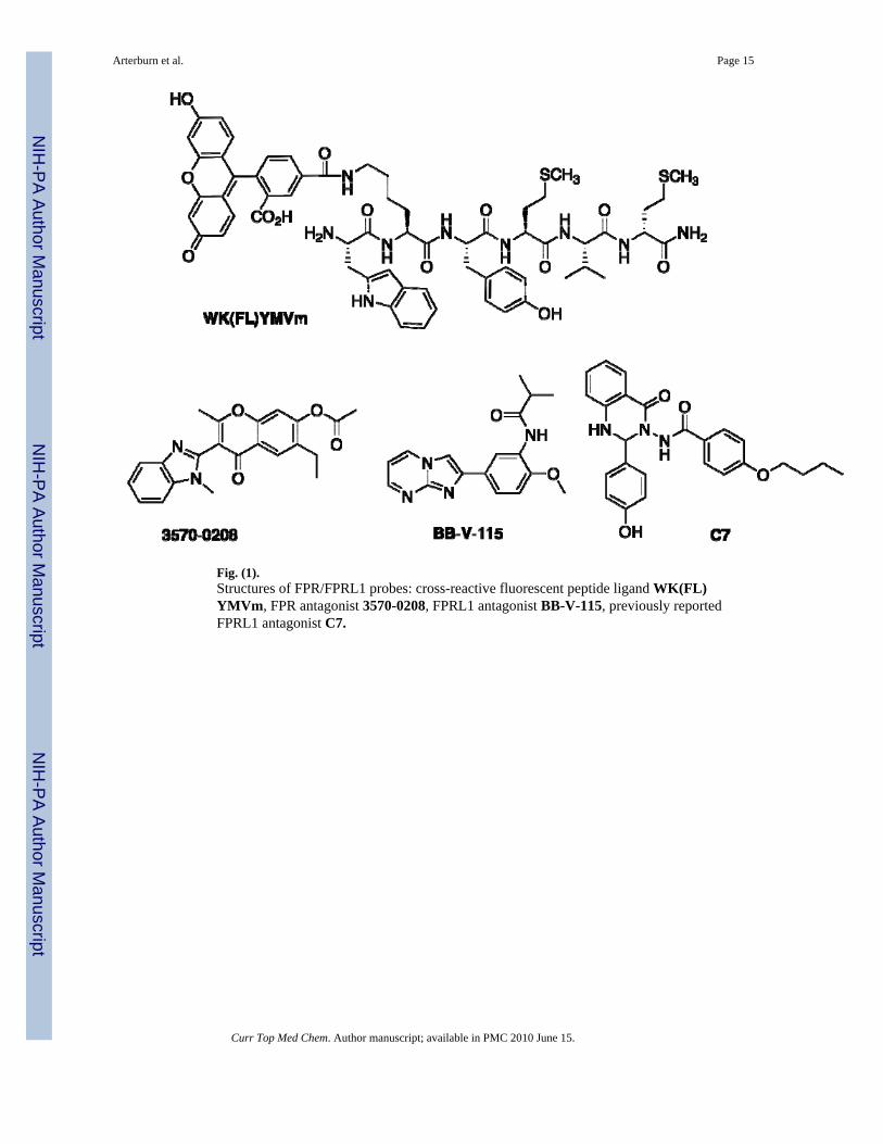
Fig. (1).Structures of FPR/FPRL1 probes: cross-reactive fluorescent peptide ligand WK(FL)YMVm, FPR antagonist 3570-0208, FPRL1 antagonist BB-V-115, previously reportedFPRL1 antagonist C7.
Arterburn et al. Page 15
Curr Top Med Chem. Author manuscript; available in PMC 2010 June 15.
NIH
-PA Author Manuscript
NIH
-PA Author Manuscript
NIH
-PA Author Manuscript
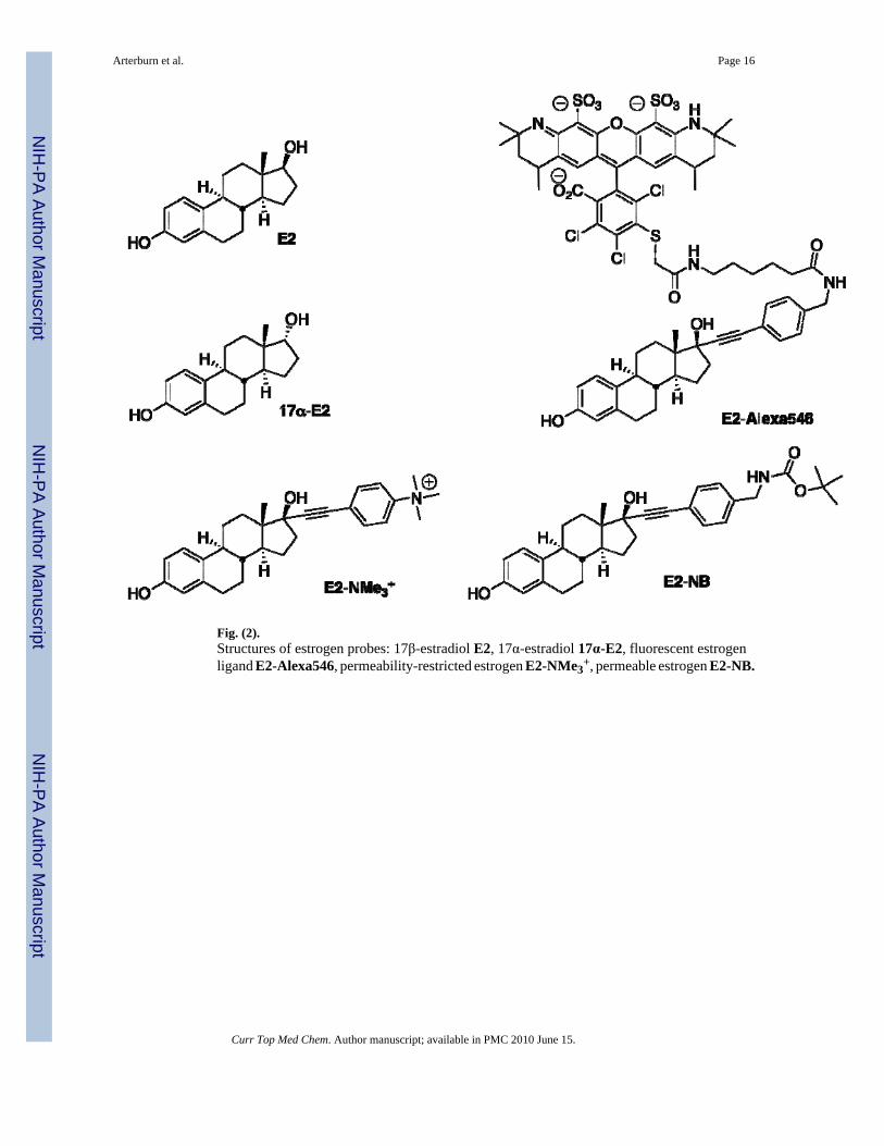
Fig. (2).Structures of estrogen probes: 17β-estradiol E2, 17α-estradiol 17α-E2, fluorescent estrogenligand E2-Alexa546, permeability-restricted estrogen E2-NMe3
+, permeable estrogen E2-NB.
Arterburn et al. Page 16
Curr Top Med Chem. Author manuscript; available in PMC 2010 June 15.
NIH
-PA Author Manuscript
NIH
-PA Author Manuscript
NIH
-PA Author Manuscript
Fig. (3).Structures of the GPR30-selective probes: agonist G-1, antagonist G15, I-125 radiotracerG40.
Arterburn et al. Page 17
Curr Top Med Chem. Author manuscript; available in PMC 2010 June 15.
NIH
-PA Author Manuscript
NIH
-PA Author Manuscript
NIH
-PA Author Manuscript