
An Extended Clq-Binding Assay Using Lactoperoxidase- and Chloramine-T-Iodinated Clq

Synthesis and Characterization of Iodinated TetrahydroquinolinesTargeting the G Protein-coupled Estrogen Receptor GPR30
Chinnasamy Ramesh1, Tapan K. Nayak2,4, Ritwik Burai1, Megan K. Dennis2, Helen J.Hathaway2,3, Larry A. Sklar3,5, Eric R. Prossnitz2,3, and Jeffrey B. Arterburn1,3,*1 Department of Chemistry and Biochemistry, New Mexico State University, Las Cruces, NM 880032 Department of Cell Biology and Physiology, School of Medicine, University of New Mexico HealthScience Center, Albuquerque, NM 871313 UNM Cancer Center, University of New Mexico Health Science Center, Albuquerque, NM 871314 College of Pharmacy, University of New Mexico Health Science Center, Albuquerque, NM 871315 Department of Pathology, School of Medicine, University of New Mexico Health Science Center,Albuquerque, NM 87131
AbstractA series of iodo-substituted tetrahydro-3H-cyclopenta[c]quinolines was synthesized as potentialtargeted imaging agents for the G protein-coupled estrogen receptor GPR30. The affinity andspecificity of binding to GPR30 versus the classical estrogen receptors ERα/β and functionalresponses associated with ligand-binding were determined. Selected iodo-substituted tetrahydro-3H-cyclopenta[c]quinolines exhibited IC50 values lower than 20 nM in competitive binding studies withGPR30-expressing human endometrial cancer cells. These compounds functioned as antagonists ofGPR30 and blocked estrogen-induced PI3K activation and calcium mobilization. The tributylstannylprecursors of selected compounds were radiolabeled with 125I using the iodogen method. In vivobiodistribution studies in female ovariectomized athymic (NCr) nu/nu mice bearing GPR30-expressing human endometrial tumors revealed GPR30-mediated uptake of the radiotracer ligandsin tumor, adrenal and reproductive organs. Biodistribution and quantitative SPECT/CT studiesrevealed structurally-related differences in the pharmacokinetic profiles, target tissue uptake andmetabolism of the radiolabeled compounds as well as differences in susceptibility to deiodination.The high lipophilicity of the compounds adversely affects the in vivo biodistribution and clearanceof these radioligands, and suggests that further optimization of this parameter may lead to improvedtargeting characteristics.
IntroductionEstrogen is a critical hormone that regulates a multitude of biological processes. The nuclearestrogen hormone receptors (ERα and ERβ) are best characterized for their regulation of geneexpression and consequently are important targets in many disease states that include cancer,skeletal, neurological and immunological conditions. New evidence of estrogen’s role in non-genomic signal transduction pathways has expanded the classical paradigm of hormonefunction and suggests corresponding significance for mammalian biology.1,2 The discovery ofthe G protein-coupled estrogen receptor GPR30 (IUPHAR designation: GPER), a seven
*Address correspondence to: Jeffrey B. Arterburn, [email protected], (575) 646-2738.Supporting Information Available. Additional synthetic procedures and characterization data, HPLC-MS data for 8, 9, 21, 22, and SPECT/CT images of 8* and 9*. This material is available free of charge via the Internet at http://pubs.acs.org.
NIH Public AccessAuthor ManuscriptJ Med Chem. Author manuscript; available in PMC 2011 February 11.
Published in final edited form as:J Med Chem. 2010 February 11; 53(3): 1004. doi:10.1021/jm9011802.
NIH
-PA Author Manuscript
NIH
-PA Author Manuscript
NIH
-PA Author Manuscript

transmembrane GPCR, has introduced an entirely new class of receptor to the milieu of non-genomic and genomic estrogen-mediated signaling.3–6 Significant overlap exists between thecellular and physiological aspects of GPR30 function and that of the classical estrogenreceptors,7 as well as in their ligand specificity and pharmacological profiles.8 Studies withbreast, ovarian and endometrial cancers indicate roles for both ERα/β and GPR30 intumoregenesis and suggest the potential for clinical diagnostic and prognostic applicationsbased on receptor expression.9,10 The development of drugs that are capable of differentiatingthe pharmacology of classical estrogen receptors, which have different tissue distributionprofiles and distinct patterns of gene regulation, by selectively modulating the activity of theindividual receptor subtypes ERα/β is widely recognized as an important strategy for obtainingimproved therapeutics.11,12 Unraveling the pharmacological profiles and specificities of thesethree estrogen receptors will contribute towards understanding the interrelated physiologicalroles of each receptor and facilitate the development of the next generation of receptor-specificdrugs.
We combined virtual and biomolecular screening to identify the first GPR30-selective agonist1-[4-(6-bromo-benzo[1,3]dioxol-5-yl)-3a,4,5,9b-tetrahydro-3H-cyclopenta[c]quinolin-8-yl]-ethanone (G-1, 1, Figure 1).13 This compound has found application as a molecular probe forin vitro and in vivo characterization of GPR30-mediated effects.14–25 A focused effortincluding synthetic chemistry, virtual and biomolecular screening through the New MexicoMolecular Libraries Screening Center recently provided the complementary GPR30-selectiveantagonist 4-(6-bromo-benzo[1,3]dioxol-5-yl)-3a,4,5,9b-tetrahydro-3H-cyclopenta[c]quinoline (G-15, 2, Figure 1).26 This compound is capable of blocking cellular activation byestrogen in cells expressing GPR30, but has no effect on estrogen-stimulated intracellularcalcium mobilization or nuclear accumulation of PIP3 induced through ERα or ERβ. Thisintriguing pair of compounds, which share the tetrahydro-3H-cyclopenta[c]quinoline scaffold,have the potential to further investigations of fundamental questions regarding GPR30physiology, including assessment of potential clinical roles for this receptor in diseaseprogression and therapeutic response.
There remain significant opportunities for further delineating the individual biological roles ofthese estrogen receptors through the application of radiolabeled GPR30-selective ligands. Thecommercial availability of [3H]-17β-estradiol has facilitated the characterization of receptordistribution and ligand binding of the classical estrogen receptors using cellular extracts, cellculture and in vivo models. The development of estrogen receptor ligands radiolabeled withpositron- or gamma-emitting halogen isotopes for PET and SPECT imaging applications, aswell as potential therapeutic applications based on estrogen receptor targeting have beenintensively studied over the past 30 years.27–31 Clinical oncologists have successfully used[18F]-FES for staging and visualizing primary and metastatic carcinomas.32,33 Thequantification of ERα and ERβ levels affords predictive value for determining outcomes ofhormone therapy in breast cancer.34,35 The development of radiolabeled 17α-iodovinylestradiols has progressed to clinical assessment of 123I-labeled 11β-methoxy-iodovinylestradiol for estrogen receptor imaging in breast cancer.36 Radiolabeled analogsincorporating Auger-emitting isotopes 125I and 123I have potential as therapeutic agents forestrogen receptor expressing tumors.30,37 The high specific activity and sensitivity of detectionpossible using the γ-emitting isotope 125I offers practical advantages for receptor bindingstudies in the laboratory, and allows efficient determination of receptor content in tissues andconvenient detection and quantification of images. The development of GPR30-selectiveradiotracers would have significant value for characterizing receptor binding properties andinvestigations of imaging applications based on targeting this receptor. The desiredperformance characteristics of these agents should include high selectivity for GPR30, lowaffinity for ERα/β, effective receptor-mediated uptake and retention accompanied by rapid
Ramesh et al. Page 2
J Med Chem. Author manuscript; available in PMC 2011 February 11.
NIH
-PA Author Manuscript
NIH
-PA Author Manuscript
NIH
-PA Author Manuscript

clearance from non-target tissue and organs in order to provide images that directly correspondto GPR30 expression levels with minimum background detection levels.
Herein we report the synthesis of a series of iodo-substituted quinoline derivatives 3–9 asselective ligands and potential targeted imaging agents for GPR30 that meet some of theperformance characteristics outlined above. These compounds were evaluated against a panelof functional and competitive ligand binding assays using GPR30 and ERα/β transfected COS7as well as Hec50 and SKBr3 cells, which endogenously express only GPR30, to evaluatereceptor selectivity and potential cross-reactivity. Selected derivatives 8 and 9 weredemonstrated to be GPR30-selective antagonists. The corresponding tributylstannanederivatives were radiolabeled with 125I and used to determine GPR30 receptor binding affinityin cell culture, and to evaluate biodistribution and in vivo imaging in a mouse tumor model.
Results and DiscussionSynthetic Chemistry
We designed a focused series of iodinated analogs using structure-activity considerationsobtained during our development of the GPR30-selective agonist and antagonist compounds1 and 2 (Figure 1).13,26 The compounds illustrated in Figure 2 possess either direct halogenationof the aryl-tetrahydro-3H-cyclopenta[c]quinoline scaffold (3–6), or conjugation of an iodinatedappendage (7–9). The iodinated analog 3 of 1 was a promising candidate due to the relativelyminor increase in steric volume resulting from iodide substitution. The positions 6–8 of thetetrahydrocyclopenta[c]quinoline scaffold were also identified as possible sites for introducinghalogen substitutions. Series 4–6 have iodo substitutions at positions 6–8 of the 1,2,3,4-tetrahydroquinoline. The other derivatives (7, 8) contain iodophenyl appendages connected atthe C8 position with a flexible N-ethylcarboxamide or urea linkages respectively. Hydrazone8 was prepared directly from 1.
The tetrahydro-3H-cyclopenta[c]quinoline scaffold is synthetically accessible using the three-component Povarov cyclization.38 We evaluated a series of reaction conditions employing avariety of protic and Lewis acid catalysts in order to optimize reaction rate, yield, anddiastereoselectivity for the construction of the compounds in this series. Our optimizedprocedure was based on the application of lanthanide salts as catalysts for the Povarovcyclization,39 and we employed Sc(OTf)3 in acetonitrile, which generally provided rapidreaction times, high product yield and diastereoselective formation of the syn- or endo-products(Scheme 1). This approach provided a direct route for the preparation of the desired iodides3–6. In several cases the endo diastereomer ratio was further enriched by crystallization. Allcompounds used in biological studies were evaluated as racemic mixtures consisting of >95%endo diastereomer.
The synthesis of 4 from 1-(4-amino-3-iodophenyl)ethanone, 6-bromopiperonal andcyclopentadiene was accomplished in moderate yield as illustrated in Scheme 1. This derivativepossesses an iodide substituent at the 6-position of the tetrahydro-3H-cyclopenta[c]quinolinesystem, and retains the ketone group of 1. Monitoring the reaction revealed that both theintermediate imine formation and subsequent cyclization were slow in comparison with theother aniline substrates, attributed to increased steric hindrance of the ortho-iodide, and theyield was not improved by attempted cyclization of the pre-formed imine. The related iodinatedcompounds 5 and 6 substituted at the 7- and 8-positions were prepared analogously from thecorresponding m- or p-iodoanilines, respectively. The derivative 10, possessing a pendantaminoethyl functional group, was prepared using tBoc-protected aminophenethylamine45 andwas isolated in 95% yield after deprotection with TFA/CH2Cl2. The amine 10 was coupledwith p-iodobenzoic acid to provide benzamide 7 as shown in Scheme 2. The related urea 8 wasprepared by coupling 10 with meta-iodophenylisocyanate (Scheme 2).
Ramesh et al. Page 3
J Med Chem. Author manuscript; available in PMC 2011 February 11.
NIH
-PA Author Manuscript
NIH
-PA Author Manuscript
NIH
-PA Author Manuscript

The synthesis of 9 required 5-iodopyridin-2-yl hydrazine, 40 which was prepared bysubstitution of commercially available 2-chloro-5-iodopyridine with hydrazine hydrate inpyridine as shown in Scheme 3. The desired hydrazone 9 was obtained by melting the hydrazinewith 1 at 170–180 °C, followed by preparative reverse phase chromatography.
Assessment of the lipophilicity and solubility were additional considerations for selectingpossible imaging agents. The log Po/w values for the selected iodides 3–9 were calculated andtabulated in Table 1. All seven iodinated compounds exhibited increased lipophilicity relativeto 1. These log Po/w values paralleled the qualitative experimental observations of the solubilityof the compounds. Compound 7 was poorly soluble in alcohols (MeOH and EtOH),halogenated organic solvents (CH2Cl2, CHCl3), and acetonitrile, but was soluble in DMSOwith heating. The urea-linked derivative 8 exhibited a greater range of solubility, includingalcoholic, halogenated solvents, acetonitrile and DMF. The hydrazone 9 was soluble in avariety of protic and aprotic solvents.
With the iodinated compounds 3–9 in hand, we proceeded to synthesize the tributyltin-substituted analogs in preparation for radiolabeling experiments.41 The accessibility of thecorresponding stannane, and efficiency of subsequent iodine exchange combined with analysisof receptor binding and activation properties (vida infra) were factors considered for theselection of compounds that were advanced for radiolabeling experiments and in vivo imagingstudies. Compound 3 is a close structural analog of 1, however attempts to prepare thecorresponding stannane 12 directly by palladium-catalyzed exchange of 1 or 3 withhexabutyldistannane were unsuccessful (Scheme 4). The benzylic amine functionality issusceptible to cyclopalladation and likely results in catalyst degradation.42 An alternative routeto 12 was attempted using the SnBu3-substituted piperonal 11, prepared from 6-bromopiperonal by Pd-catalyzed stannylation, however, the cyclization of 11 was unsuccessfulusing either our optimized procedure or various other protic and Lewis acid catalysts. Attemptsto form the intermediate imine 14 under a variety of forcing conditions were also unsuccessful,resulting in unreacted 11 and aminoacetophenone. The failure to form imine 14 contrasts withthe facile formation of the brominated analog 13, and can be attributed to the increased stericdemands of the stannyl group of 11 in comparison with 6-bromopiperonal. Attemptedstannylation of brominated imine 13 to form 14 was also unsuccessful.
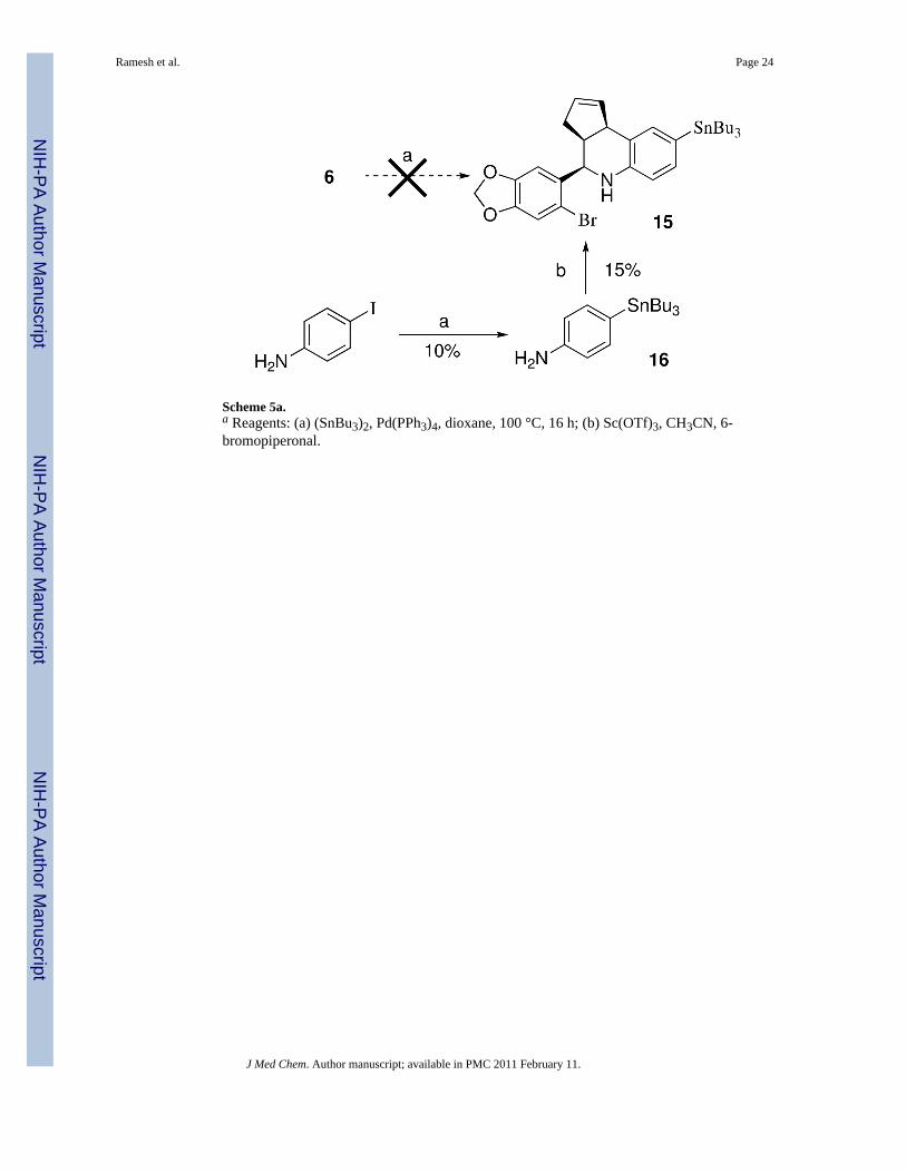
While the direct Pd-catalyzed tributylstannylation of iodides 4–9 appears feasible, in practicewe were unable to effect this transformation due to the competing reactivity of the 6-bromobenzo[1,3]dioxol-5-yl discussed previously. The attempted stannylation of 6 to obtain15 was unsuccessful, therefore an alternative stepwise approach was investigated as shown inScheme 5. The Pd-catalyzed exchange of 4-iodoaniline with (SnBu3)2 to produce 16 requiredextended reaction times and heating, and proceeded in low yield (~10%) accompanied byunreacted starting material under these conditions, presumably due to increased electrondensity from the para-amine substituent. The attempted cyclization of 16 under a variety ofconditions resulted in poor yields of 15, and significant amounts of the proto-dehalogenatedmaterial were observed. Preliminary initial iodination experiments of 15 with iodogendemonstrated poor incorporation of iodide, and proteo-destannylation as the primarycompeting reaction. The poor preparative yield of tributylstannane 15 and the subsequentinefficient iodination of this derivative were recognized as significant disadvantages for aputative imaging agent. In contrast, the three-component cyclization of the meta-tributylstannylaniline 17 proceeded efficiently to provide 18 (Scheme 6). This result confirmsthe positional dependence of substitution on the aniline ring that was problematic for para-substituted analog 16.
The primary aliphatic amine of 4-(2-aminoethyl)aniline reacted preferentially with meta-iodophenylisocyanate to give the urea 19 (Scheme 7). Palladium catalyzed exchange with bis
Ramesh et al. Page 4
J Med Chem. Author manuscript; available in PMC 2011 February 11.
NIH
-PA Author Manuscript
NIH
-PA Author Manuscript
NIH
-PA Author Manuscript

(tributyltin) gave the tributyltin derivative 20. Scandium(III) catalyzed cyclization of 20 with6-bromopiperonal and cyclopentadiene gave the desired product 21 in good yield.
Stannylation of 2-chloro-5-iodopyridine provided an efficient route for the preparation of 2-chloro-5-(tributylstannyl)pyridine. This compound was previously synthesized by halogen-metal exchange with n-butyl lithium followed by stannylation with tributylstannyl chloride.43 Nucleophilic substitution of chloride with hydrazine gave the desired 2-hydrazinyl-5-(tributylstannyl)pyridine (Scheme 8). Hydrazone formation with 1 occurred upon melting, andthe resulting product 22 was purified by preparative reverse phase chromatography.
Receptor binding and functional propertiesThe iodinated compounds 3–9 were evaluated in cell-based binding and functional assays todetermine the potency and type of signaling response (agonism or antagonism) associated withligand-induced GPR30- and ERα/β-mediated signaling (summarized in Table 1 with numericalbinding data provided in the Supporting Information). These compounds were profiled usingtransfected COS7 cells expressing GPR30, ERα or ERβ, in addition to Hec50 and SKBr3 cells,which endogenously express only GPR30. Functional characterization of the compounds usingeither the extent (or inhibition of E2-mediated increase) in intracellular calcium or theactivation of PI3K as measured by production of PIP3 in the nucleus, as previously described.5,13,26 The specificity of the response in these assays was demonstrated in control COS7 cellsthat do not endogenously express GPR30 or ERα/β. The comparison of the observed ligand-induced responses across cells expressing individual receptors in comparison to estrogen-induced responses, in addition to comparison of differential binding to receptors, provides aprofile of the selectivity/cross-reactivity of the compounds.
We have previously shown the specificity of binding of the GPR30-selective ligand 1 to GPR30versus the classical estrogen receptors ERα/β using cell-based competitive binding assays.13
In order to characterize the selectivity of iodinated compounds 3–9, the same cell based assayemploying transfected COS7 cells was used to evaluate binding of these compounds to theclassical estrogen receptors. These assays confirmed that compounds 3, 5 and 8, like 1, do notexhibit binding to either of the classical estrogen receptors ERα/β. Compounds 4, 6 and 7exhibited minimal binding to ERβ at high concentrations (blocking less than 35% of E2-Alexa633 binding when present at 10 μM). Hydrazone 9 was the only iodinated derivative toshow any detectable binding to ERα, which was also minimal. These low levels of competitionat 10 μM compare to an IC50 for estrogen in the same assay of approximately 0.4 nM. 13
Cell-based functional assays for compounds 3–9 were carried out in parallel to binding studiesin order to assess the functional characteristics of this series of compounds. Stimulation of cellsexpressing ERα or GPR30 results in the receptor-dependent activation of PI3K which can bevisualized using the pleckstrin homology (PH)-domain of Akt fused to RFP (PH-RFP), whichserves as a reporter of PIP3 localization. Receptor activation leads to the translocation of thisreporter from a cytoplasmic or plasma membrane-associated localization to the nucleus.5,13
We evaluated the ability of compounds 3–9 alone to induce PI3K activation or the ability ofthese compounds to block estrogen-stimulated PI3K activation to determine the agonist andantagonist properties of each compound in transfected COS7 cells expressing ERα or GPR30;additionally COS7 cells transfected with PH-RFP reporter alone were treated with allcompounds to confirm that the results in receptor-transfected cells were receptor-dependent.As expected due to their lack of binding to ERα, compounds 3–8 were unable to induce eitheractivation of PI3K or inhibit estrogen-induced PI3K activation in ERα transfected COS7 cells.Compound 9, which exhibited very weak binding to ERα, was also unable to influence PI3Kactivity in ERα transfected COS7 cells. Additionally, none of compounds 3–9 was able toactivate PI3K in COS7 cells lacking estrogen receptor expression. Thus, none of the seveniodinated derivatives shows any functional activity mediated through ERα or any ability to
Ramesh et al. Page 5
J Med Chem. Author manuscript; available in PMC 2011 February 11.
NIH
-PA Author Manuscript
NIH
-PA Author Manuscript
NIH
-PA Author Manuscript

activate PI3K via other mechanisms. Compound 3 showed strong GPR30-dependent activationof PI3K as expected for an isostructural analog of 1. A regioisomeric dependence of iodidesubstitution on GPR30-mediated PI3K activation was observed for compounds 4–7.Compounds 4 and 7 exhibited partial agonist properties on PI3K, acting as agonists whenapplied to cells in the absence of estrogen but also demonstrating the ability to inhibit the fullactivation of PI3K by estrogen. Compound 5 was a weaker agonist of PI3K than 3, andcompound 6 had no functional effect. Compounds 8 and 9 were able to block the estrogen-induced PI3K activation via GPR30 and thus appear to function as antagonists of GPR30function in this context.
As rapid signaling via GPR30 results in increased intracellular calcium levels,5 we investigatedwhether compounds 3–9 had similar agonist and antagonist properties in a cell-based calciumassay using SKBr3 cells, which endogenously express only GPR30. In these cells, stimulationwith GPR30 agonists, including estrogen and 1, elicits a rapid increase in intracellular calciumlevels that can be easily monitored using the cell-permeant fluorescent calcium indicatorIndo-1. Consistent with the PI3K activation profiles, compounds 8 and 9 blocked estrogen-induced calcium mobilization and thus functioned as antagonists of estrogen-mediated GPR30function. Additionally, compound 4 appears to weakly antagonize estrogen-mediated calciummobilization. Compounds 3, 5–7 can all be characterized as partial agonists in the context ofthis assay, as all compounds mobilize calcium when applied alone and all four compoundsdisplay some degree of inhibition of estrogen-mediated calcium mobilization. However, as inthe PI3K assay, compound 3 is a potent agonist of calcium mobilization similar to 1.
Some variability in compound profiles exists between the two GPR30 functional assays (i.e.compound 6 was inactive in the PI3K assay but a partial agonist of GPR30 in the calcium assayand compound 3 is a potent agonist in the PI3K assay and a partial, though still potent, agonistin the calcium assay). This is potentially due to the different cellular systems used in the assaysand also may be related to assay sensitivity and the differing time courses of the assays. ThePI3K assay employs ectopically expressed receptors in cells which may lack required cofactorsfor full compound activity, thus some compounds may elicit weaker or no responses in thissystem than in a system in which cells with endogenous receptor are used, such as the calciumassay used to profile the compounds. Additionally, the PI3K assay is a phenotypic assay inwhich many fields of cells must be analyzed and an average response determined relative tocontrol stimulated cells, whereas the calcium assay is a more quantitative assay in whichsmaller changes in signaling are more likely to be observed. Importantly, compounds 8 and9 displayed consistent activity as antagonists of GPR30 function in both functional assays.
I-125 Radiolabeling and GPR30 bindingThe iodides 8 and 9 were selected for radiolabeling based on the results of the binding andfunctional assays combined with considerations of their chemical properties and syntheticaccessibility of the tributylstannylated precursors. Although compound 3 exhibited a favorableGPR30-selective binding and functional profile, efforts to prepare the tributylstannyl precursor12 were unsuccessful. The tributylstannyl precursor, N-{2-[4-(6-bromo-benzo[1,3]dioxol-5-yl)-3a,4,5,9b-tetrahydro-3H-cyclopenta[c]quinolin-8-yl]-ethyl}-4-tributylstannyl-benzamidefor 7 was synthesized but not selected for I-125 labeling due to the poor solubilitycharacteristics. Radioiodinated 8* and 9* were prepared using the iodogen method fromtributylstannyl precursors 21 and 22 respectively. A 1:1 stoichiometry of I-125:tributylstannylprecursor was used for radiolabeling and greatly simplified the purification process by avoidingthe need to separate excess precursor. Typical radiochemical yields ranged from 40–55 %.Over 99.9 % unincorporated I-125 and unlabeled ligands were successfully separated from theradioiodinated 8* and 9* using solid phase extraction as determined by thin layerchromatography. The experimental Log P(o/w) of 7.0 ± 0.3 and 6.0± 0.2 for 8* and 9*
Ramesh et al. Page 6
J Med Chem. Author manuscript; available in PMC 2011 February 11.
NIH
-PA Author Manuscript
NIH
-PA Author Manuscript
NIH
-PA Author Manuscript

determined by the shake-tube method were similar to the corresponding calculated values of7.00 and 6.35 and followed the same relative ordering of lipophilicity 8 > 9.
Competition binding studies were performed on adherent monolayers of GPR30-expressinghuman endometrial Hec50 cancer cells. The IC50 values of 6, 7, 8 and 9 were 1.7 nM, 16.2nM, 8.4 nM and 1.7 nM respectively (Table 1). This binding assay was verified by evaluatingthe receptor binding affinity for the GPR30 agonist 1 (IC50 = ~7 nM), which compares similarlyto the value obtained using a competitive binding assay with a fluorescent estrogen ligand andrecombinant GPR30 (IC50 = ~11 nM),13 and reported values for 17β-estradiol (IC50 = ~3–6nM).5,6 The recently described GPR30 antagonist 2 exhibited slightly weaker binding (IC50 =~20 nM) relative to 1.26
In Vivo studiesIn vivo biodistribution studies were performed on female ovariectomized athymic (NCr) nu/nu mice bearing GPR30-expressing human endometrial Hec50 tumors by intravenouslyinjecting trace amounts of radioiodinated 8* and 9*. Both the radiotracers 8* and 9* wererapidly metabolized and eliminated via the hepatobiliary system (gall bladder uptake of over30 % ID/g). Radiotracer 8* was rapidly cleared from the intestines, kidney and urinary bladder(Table 2).
However, increased uptake and retention was observed in the uterus, adrenal and mammaryglands and tumor, indicative of receptor-mediated uptake. Blocking studies also revealedGPR30-mediated uptake of 8* in tumor, adrenal and reproductive organs, sites of known orexpected GPR30 expression. Radiotracer 9* demonstrated a significantly differentpharmacokinetic profile than radiotracer 8* (Table 2). The blood uptake values of 9* werenearly three fold that of 8* at 1 hr PI (14.3 % ID/g vs. 3.2 %ID/g), indicative of high plasma-protein binding. However, blocking studies also revealed GPR30-mediated uptake of 9* intumor, adrenal and reproductive organs. In spite of receptor-mediated uptake, the tumor wasnot visualized by a whole body 80 s/projections SPECT/CT studies due to poor targetingcharacteristics of both the radiotracers (Supporting information: S10). Quantitative SPECT/CT studies revealed further disparities between 8* and 9*. The thyroid uptake of 9* was almostseven times greater than that of 8* (4.1 % ID vs. 0.6 % ID). Whereas the gall bladder (withbile content) uptake of 9* was nearly three times lower than that of 8* (1.8 % ID vs. 5.7 %ID). For 8*, the organ with the largest uptake was intestine (over 70 % ID) whereas for 9* itwas the stomach (over 15 % ID). The imaging studies suggest that 8* is less susceptible to invivo dehalogenation as compared to 9*, but was also more susceptible to metabolism due toincreased gall bladder activity. The moderate increase in kidney uptake of compound 8* uponcoadministration of excess 1 could be due changes in metabolism or excretion kinetics in thepresence of large doses of blocking agent. The high stomach uptake of 9* could presumablybe associated with the presence of free iodide, which would also correspond with the highthyroid levels observed. The blocking studies using the structurally related agonist 1 verifiedthe critical involvement of GPR30-mediated uptake of the radiotracers; however, thecorresponding displacement by E2 was not characterized. This represents a limitation of thepresent study since endogenous estrogens constitute the dominant competing ligands in intact(not ovariectomized) animals and, although challenging to address experimentally, should beincorporated into the evaluation of next generation agents that exhibit improved targetingcharacteristics.
ConclusionsIn this study, we developed a series of iodinated compounds that exhibit high affinity andselectivity towards GPR30 versus the classical estrogen receptors. The synthetic compoundswere characterized using cell-based competitive binding and functional assays to identify
Ramesh et al. Page 7
J Med Chem. Author manuscript; available in PMC 2011 February 11.
NIH
-PA Author Manuscript
NIH
-PA Author Manuscript
NIH
-PA Author Manuscript

binding affinity, specificity and agonist or antagonist response associated with receptor-mediated signaling pathways. Two compounds incorporating either conjugated phenylurea orhydrazone linkages, respectively, were selected for radiolabeling with 125I to provide 8* and9*. These compounds functioned as antagonists of GPR30-mediated calcium mobilization,and were used for competitive ligand binding assays in cell culture. Comparative in vivo studieswere used to evaluate the biodistribution, pharmacokinetics and the potential for GPR30-targeted tumor imaging in an animal model. Although both radiotracers 8* and 9* exhibitedreceptor-mediated uptake in tumor, adrenal and reproductive organs, neither was effective fortumor imaging due to poor targeting characteristics, high background and issues with plasmaprotein binding and rapid metabolism. The striking differences in the in vivo characteristics of8* and 9* reflect the structural, physicochemical and metabolic differences associated with theiodinated appendages used. Dehalogenation, high plasma-protein binding and uptake in thestomach were problematic for the meta-iodophenylurea 9*. The iodo-pyridin-2yl-hydrazone8* was less susceptible to dehalogenation, but exhibited increased metabolism and high uptakein the intestine. Both compound classes were significantly more lipophilic than either estradiolor the GPR30-selective probes 1 and 2, which likely contributes towards the observed non-target tissue uptake. The in vivo data from 8* and 9* provide valuable insight for the designof the next generation of GPR30-targeted radiotracers. The iodide 3 exhibited favorablecharacteristics including reduced lipophilicity and high selectivity as a GPR30 agonist;however, the inaccessibility of the tributylstannyl precursor through the synthetic routesinvestigated prevented further evaluation of the radioiodinated compound. It may be possibleto incorporate the less sterically-demanding trimethylstannyl group, which could serve as aprecursor for I-125 labeling to yield this promising agent.44 We have previouslydescribed 99mTc-labeled 17β-estradiol derivatives that incorporate pyridin-2-yl-hydrazinechelates for SPECT imaging applications.45,46 These studies suggest that incorporatingthis 99mTc-imaging modality into the tetrahydro-cyclopenta[c]quinoline scaffold could resultin GPR30-selective agents with decreased lipophilicity, increased chemical and metabolicstability, reduced plasma-protein binding, and overall improvements in targetingcharacteristics for in vivo imaging applications.
Experimental SectionGeneral Methods
Reagents and solvents were obtained from commercial sources and used without furtherpurification. Chromatographic separations were performed using medium pressure flashchromatography and ethyl acetate/hexanes or methanol/dichloromethane as eluent. Reversephase chromatography employed C-18 columns and water-acetonitrile or water-methanolmobile phase. NMR spectra were acquired at ambient temperatures (18 ± 2 °C) unless otherwisenoted. Reactions were monitored by thin-layer chromatography on silica gel (60 Å pore size,5–17 μm) polyester backed sheets that were visualized under a UV lamp, iodine vapor,phosphomolybdic acid, or anisaldehyde. The 1H NMR spectra in CDCl3 were referenced toTMS unless otherwise noted. The 13C {1H} NMR spectra were recorded at 75 or 100 MHzand referenced relative to the 13C {1H} peaks of the solvent. Spectra are reported as (ppm),(multiplicity, coupling constants (Hz), and number of protons). Melting points are uncorrected.The purity of all compounds used in biological studies was determined to be >95% by analyticalHPLC equipped with Photodiode Array (PDA) and ESI-MS detection. The compounds (1mg/mL CH3CN, 20 μL) were injected into a Waters Symmetry® C18 5μm 3.0 × 150 mm columnand eluted as specified. Reported binding and functional characterization values are based ona minimum of 3 measurements.
Ramesh et al. Page 8
J Med Chem. Author manuscript; available in PMC 2011 February 11.
NIH
-PA Author Manuscript
NIH
-PA Author Manuscript
NIH
-PA Author Manuscript

General procedure for Sc(OTf)3 mediated cyclizationA catalytic amount of Sc(OTf)3 (10 mol%) in anhydrous acetonitrile (0.5 mL) was added tothe mixture of 6-bromopiperonal (1 eq), aniline derivative (1 eq), and cyclopentadiene (5 eq)in acetonitrile (4 mL). The reaction mixture was stirred at ambient temperature (~23 °C) for2–5 h with monitoring the product formation by thin layer chromatography using ethyl acetate/hexanes as eluent. The volatiles were removed in vacuo. The crude product was purified asspecified.
1-[4-(6-Iodo-benzo[1,3]dioxol-5-yl)-3a,4,5,9b-tetrahydro-3H-cyclopenta[c]quinolin-8-yl]-ethanone (3)
According to the general procedure 6-iodopiperonal (0.055 g, 0.2 mmol), p-aminoacetophenone (0.027 g, 0.2 mmol), cyclopentadiene (0.066 g, 1.0 mmol) and Sc(OTf)3(0.010 g, 0.5 mmol) were combined with a reaction time of 2 h. The volatiles were removedin vacuo. The residue was purified by preparative silica gel column chromatography usingethyl acetate/hexanes (10:90) to provide 3 (0.085 g, 92%) as a colorless solid. mp: 106–109 °C; IR (KBr, cm−1) 3320, 2880, 1665, 1575, 1242; 1H NMR (400 MHz, CDCl3) δ 7.71 (d, J =1.9 Hz, 1H), 7.62 (dd, J = 8.4, 1.9 Hz, 1H), 7.28 (s, 1H), 7.07(s, 1H), 6.61 (d, J = 8.4 Hz, 1H),6.01 (d, J = 1.1 Hz, 1H), 5.99 (d, J = 1.1 Hz, 1H), 5.96-5.94 (m, 1H), 5.68-5.66 (m, 1H), 4.82(d, J = 3.1Hz, 1H), 4.14 (d, J = 8.8 Hz, 1H), 4.06 (bs, 1H), 3.19-3.13 (m, 1H), 2.56-2.44 (m,4H), 1.83-1.79 (m, 1H); 13C NMR (75 MHz, CDCl3) δ 196.5, 149.8, 148.6, 147.8, 136.3, 133.7,130.4, 130.0, 128.6, 127.6, 125.1, 118.9, 115.2, 107.9, 101.7, 60.8, 45.3, 42.2, 31.2, 26.0;HPLC-MS: Elution with 60–90% CH3CN (gradient 1.5% min−1) in H2O, exhibited a singlepeak at 12.38 min. ESI-MS m/z [ES+] calcd for C21H18INO3 [M+H]+ 460.03; found 460.09.
1-(4-(6-bromobenzo[d][1,3]dioxol-5-yl)-6-iodo-3a,4,5,9b-tetrahydro-3H-cyclopenta[c]quinolin-8-yl)ethanone (4)
According to the general procedure 6-bromopiperonal (0.115 g, 0.5 mmol), 1-(4-amino-3-iodo-phenyl)-ethanone (0.140 g, 0.53 mmol), cyclopentadiene (0.165 g, 2.5 mmol), and Sc(OTf)3(0.0246 g, 0.5 mmol) were combined with a reaction time of 3 h. The volatiles were removedin vacuo. The residue was purified by preparative silica gel column chromatography usingethyl acetate/hexanes (08:92) to provide 4 (0.151 g, 56%) as a white solid. mp: 110–114 °C;IR (KBr, cm−1): 3447, 2920, 1670, 1588, 1476, 1242; 1H NMR (300 MHz, CDCl3) δ 8.11 (d,J = 2.0 Hz, 1H), 7.65 (d, J= 2.0 Hz, 1H), 7.14 (s, 1H), 7.06 (s, 1H), 6.03 (d, J = 1.3 Hz, 1H),6.02 (d, J = 1.3 Hz, 1H), 5.92-5.88 (m, 1H), 5.68-5.66 (m, 1H), 4.99 (d, J = 3.5 Hz, 1H), 4.53(bs, 1 H), 4.14 (d, J = 8.2 Hz, 1H), 3.25-3.19(m, 1H), 2.52-2.42 (m, 4H), 1.87-1.77 (m,1H); 13C NMR (75 MHz, CDCl3) δ: 195.1, 148.9, 147.7, 147.6, 137.3, 133.4, 132.9, 130.7,129.6, 129.6, 125.6, 113.0, 107.4, 101.8, 85.4, 77.2, 56.6, 45.9, 42.3, 31.4, 25.9; HPLC-MS:Elution with 60–90% CH3CN in H2O (gradient 1.5 % min−1), exhibited a single peak at 17.40min. HRMS: calcd for C21H17BrINO3 [M+H]+ 537.9515; found 537.9525.
4-(6-Bromo-benzo[1,3]dioxol-5-yl)-7-iodo-3a,4,5,9b-tetrahydro-3H-cyclopenta[c]quinoline(5)
According to the general procedure 6-bromopiperonal (0.229 g, 1 mmol), m-iodoaniline (0.219g, 1 mmol), cyclopentadiene (0.330 g, 5 mmol), and Sc(OTf)3 (0.0492 g, 0.1 mmol) werecombined with a reaction time of 2 h. The volatiles were removed in vacuo. The residue waspurified by preparative silica gel column chromatography using ethyl acetate/hexanes (10:90)to provide 5 (0.354 g, 72%) as a white solid. mp: 195–198 °C; IR (KBr, cm−1) 3432, 2881,1573, 1496, 1217; 1H NMR (300 MHz, CDCl3) δ 7.01 (s, 1H), 7.05 (dd, J = 8.0, 2.0 Hz, 1H),7.03 (s, 1H), 6.97 (d, J = 2.0 Hz, 1H), 6.77 (d, J = 8.0 Hz, 1H), 6.00 (d, J = 1.5 Hz, 1H), 5.98(d, J = 1.5 Hz, 1H), 5.83-5.79 (m, 1H), 5.69-5.64 (m, 1H), 4.87 (d, J = 3.3 Hz, 1H), 4.03 (d,J = 9 Hz, 1H), 3.55 (bs, 1H), 3.21-3.09 (m, 1H), 2.58-2.49 (m, 1H), 1.84-1.75 (m, 1H); 13C
Ramesh et al. Page 9
J Med Chem. Author manuscript; available in PMC 2011 February 11.
NIH
-PA Author Manuscript
NIH
-PA Author Manuscript
NIH
-PA Author Manuscript

NMR (75 MHz, CDCl3) δ 147.5, 147.3, 146.8, 133.9, 133.4, 130.6, 130.5, 128.1, 125.7, 124.4,113.0, 112.9, 107.8, 101.7, 101.7, 90.9, 56.3, 45.6, 40.9, 31.3; HPLC-MS: Elution with 73:27CH3CN/H2O containing 0.01% formic acid, exhibited a single peak Rt = 22.38 min. HRMS:calcd for C19H15BrINO2 [M+H]+ 495.9409; found 495.9412.
4-(6-Bromo-benzo[1,3]dioxol-5-yl)-8-iodo-3a,4,5,9b-tetrahydro-3H-cyclopenta[c]quinoline(6)
According to the general procedure 6-bromopiperonal (0.460 g, 2 mmol), p-iodoaniline (0.438g, 2 mmol), cyclopentadiene (0.66 g, 10 mmol), and Sc(OTf)3 (0.098 g, 0.2 mmol, 10 mol%)were combined with a reaction time of 2 h. The product 6 (0.890 g, 90%) was isolated byfiltration as a white solid. mp: 175–178 °C; IR (KBr, cm−1) 3435, 2887, 1588, 1481,1239; 1H NMR (300 MHz, CDCl3) δ 7.33 (d, J = 2.0 Hz, 1H), 7.23 (dd, J = 8.4, 2.0 Hz, 1H),7.11 (s, 1H), 7.02 (s, 1H), 6.38 (d, J = 8.4 Hz, 1H), 5.99 (d, J = 1.2 Hz, 1H), 5.98 (d, J = 1.2Hz, 1H), 5.84-5.80 (m, 1H), 5.68-5.66 (m, 1H), 4.85 (d, J = 3.3 Hz, 1H), 4.05 (d, J = 9.0 Hz,1H), 3.5 (bs, 1H), 3.20-3.09 (m, 1H), 2.59-2.48 (m, 1H), 1.84- 1.74 (m, 1H); 13C NMR (75MHz, CDCl3) δ 147.5, 147.3, 145.0, 137.5, 134.3, 134.0, 133.4, 130.7, 128.8, 118.5, 113.0,112.9, 107.9, 101.7, 80.5, 56.4, 45.7, 41.9, 31.3; HPLC-MS: Elution with 73:27 CH3CN/H2Ocontaining 0.01% formic acid, exhibited a single peak Rt = 22.10 min. HRMS: calcd forC19H15BrINO2 [M+H]+ 495.9409; found 495.9407.
N-{2-[4-(6-Bromo-benzo[1,3]dioxol-5-yl)-3a,4,5,9b-tetrahydro-3H-cyclopenta[c]quinolin-8-yl]-ethyl}-4-iodo-benzamide (7)
The amine 10 (0.082 g, 0.2 mmol) and 4-iodobenzoic acid (0.049 g, 0.2 mmol) were taken indry DMF (1 mL), disopropylethylamine (0.025 g, 0.2 mmol) was added, followed by theaddition of benzotriazol-1-yl-oxytripyrrolidinophosphonium hexafluorophosphate (0.103 g,0.2 mmol) and allowed to stir at ambient temperature for 20 h. The precipitate formed wasfiltered and washed with water and dried in vacuo to provide 10 (0.100 g, 78%) as a whitesolid. mp: 217–220 °C; IR (KBr, cm−1) 3338, 1631, 1481, 1244, 1038; 1H NMR (300 MHz,DMSO-d6) δ 8.58 (t, J = 5.4 Hz, 1H), 7.85 (d, J = 8.4 Hz, 2H), 7.61 (d, J = 8.4 Hz, 2H), 7.24(s, 1H), 7.14(s, 1H), 6.84 (m, 1H), 6.77 (dd, J = 8.1, 1.7 Hz, 1H), 6.63 (d, J = 8.1 Hz, 1H), 6.09(s, 1H), 6.07 (s, 1H), 5.82-5.77 (m, 1H), 5.58-5.55 (m, 1H), 5.47 (bs, 1H), 4.62 (d, J = 3.0 Hz,1H), 3.96 (d, J = 8.9 Hz, 1H), 3.34-3.38 (m, 2H), 3.04-2.96 (m, 1H), 2,67 (t, J = 7.3Hz, 2H),2.45-2.38 (m, 1H), 1.68-1.60 (m, 1H); 13C NMR (75 MHz, DMSO-d6) δ 165.3, 147.0, 146.9,144.3, 137.5, 137.0, 134.3, 134.0, 131.0, 129.4, 129.1, 128.7, 126.1, 124.8, 116.1, 112.3, 112.1,108.5, 101.8, 98.5, 56.0, 45.54, 41.8, 41.3, 34.3, 31.1; HPLC-MS: Elution with 60–90%CH3CN in H2O (gradient 1.5 % min−1), exhibited a single peak at 15.98 min. HRMS: calcdfor C28H24BrIN2O3 [M+H]+ 664.9913; found 664.9916.
1-{2-[4-(6-Bromo-benzo[1,3]dioxol-5-yl)-3a,4,5,9b-tetrahydro-3H-cyclopenta[c]quinolin-8-yl]-ethyl}-3-(3-iodo-phenyl)-urea (8)
A solution of 3-iodophenylisocyanate (0.10 g, 0.4 mmol) in dry DMF (1 mL) was added to theamine 10 (0.082 g, 0.2 mmol) and Et3N (0.02 g, 0.2 mmol) in dry DMF (1 mL) in an ice bathand allowed to stir overnight at ambient temperature. The reaction mixture washed with water(15 mL) and the product was extracted with methylene chloride (30 mL), the organic layer wasdried over anhydrous sodium sulfate and evaporated under reduced pressure. The residue waspurified by silica gel column chromatography using ethyl acetate/hexanes (25:75) to provide8 (0.09 g, 67%) as a white solid. mp: 210–213 °C; IR (KBr, cm−1) 3341, 2870, 1636, 1580,1474; 1H NMR (300 MHz, DMSO-d6) δ 8.60 (s, 1H), 7.98-7.97(m, 1H), 7.25-7.20 (m, 3H),7.14 (s, 1H), 7.01-6.96 (m, 1H), 6.85 (s, 1H), 6.76 (dd, J = 8.2, 1.76 Hz, 1H), 6.65 (d J = 8.1,1H), 6.11-6.05 (m, 3H), 5.87-5.83 (m, 1H), 5.58-5.53(m, 1H), 5.45 (bs, 1H), 4.63 (d, J = 2.8Hz, 1H), 3.97 (d, J = 8.8 Hz, 1H), 3.28-3.22 (m, 2H), 3.05-2.95 (m, 1H), 2.62-2.54 (m, 2H),
Ramesh et al. Page 10
J Med Chem. Author manuscript; available in PMC 2011 February 11.
NIH
-PA Author Manuscript
NIH
-PA Author Manuscript
NIH
-PA Author Manuscript

2.45-2.40 (m, 1H), 1.69-1.60 (m, 1H); 13C NMR (75 MHz, DMSO-d6) δ 154.7, 147.0, 146.9,144.3, 142.0, 134.6, 134.3, 130.58, 129.4, 129.3, 128.6, 126.1, 125.6, 124.9, 116.7, 116.1,112.3, 112.1, 108.5, 101.8, 94.7, 56.0, 45.5, 41.8, 40.8, 40.3, 35.0, 31.1; HPLC-MS: Elutionwith 63–93 % CH3CN in H2O containing 0.01% formic acid (gradient 1 % min−1), exhibiteda two peaks at Rt = 26.40 min (exo) and 27.88 min (endo). HRMS: calcd forC28H25BrIN3O3 [M+H]+ 658.0202; found 658.0193.
N-{1-[4-(6-Bromo-benzo[1,3]dioxol-5-yl)-3a,4,5,9b-tetrahydro-3H-cyclopenta[c]quinolin-8-yl]-ethylidene}-N′-(5-iodo-pyridin-2-yl)-hydrazine (9)
The (5-iodo-pyridin-2-yl)-hydrazine (0.047 g, 0.2 mmol) was heated with 1 (0.082 g, 0.2 mmol)at 170–180 °C for 5 min in vacuo (2 mmHg). The crude hydrazone was purified by reversephase column chromatography using acetonitrile/water (75:25) to provide 9 (0.101 g, 85%) asa pale yellow solid. mp: 128–131 °C. IR (KBr, cm−1) 3351, 2919, 1611, 1578, 1493; 1H NMR(300 MHz, CDCl3) δ 8.26 (d, J = 1.8 Hz, 1H), 7.98 (s, 1H), 7.79 (dd, J = 8.8, 2.0 Hz, 1H),7.43-7.40 (m, 2H), 7.24-7.18 (m, 1H), 7.13 (s, 1H), 7.02 (s, 1H), 6.61 (d, J = 8.2 Hz, 1H), 5.98(d, J = 1.3 Hz, 1H), 5.97 (d, J = 1.3 Hz, 1H) 5.93-5.899 (m, 1H), 5.68-5.63 (m, 1H), 4.89 (d,J = 3.1 Hz, 1H), 4.13(d, J = 8.8 Hz, 1H), 3.69 (s, 1H), 3.23-3.16 (m, 1H), 2.60-2.51 (m, 1H),2.16 (s, 3 H), 1.84-1.76 (m, 1H); 13C NMR (75 MHz, CDCl3) δ 156.2, 153.0, 147.4, 147.2,146.0, 145.5, 144.5, 134.1, 133.9, 130.3, 129.7, 126.5, 125.6, 124.1, 115.9, 113.0, 112.8, 109.8,107.9, 101.7, 79.2, 56.5, 45.9, 42.1, 31.3, 12.3; HPLC-MS: Elution with 73:27 CH3CN/H2Ocontaining 0.01% formic acid exhibited two peaks at Rt = 17.18 min (exo) and 22.33 min(endo). HRMS: calcd for C26H22BrIN4O2 [M+H]+ 629.0049; found 628.9939.
1-[2-(4-Amino-phenyl)-ethyl]-3-(3-iodo-phenyl)-urea (19)A solution of 3-iodophenylisocyanate (0.490 g, 2.0 mmol) in dry dichloromethane (10 mL)was added to 4-aminophenethylamine (0.272 g, 2.0 mmol) in an ice bath and subsequentlyallowed to stir for 2 h at ambient temperature. The white solid precipitate was separated bycentrifugation and dried in vacuo to provide 19 (0.703 g, 92%) as a colorless solid. mp: 161–163 °C; IR (KBr, cm−1) 3312, 1628, 1577, 1516, 1472; 1H NMR (300 MHz, DMSO-d6) δ 8.60(s, 1H), 7.96 (t, J = 1.76 Hz, 1H), 7.24-7.18 (m, 2H), 6.98 (t, J = 8.0 Hz, 1H), 6.86 (d, J = 8.3Hz, 2H), 6.50 (d, J = 8.3 Hz, 2H), 6.09-6.05 (m, 1H), 4.84 (s, 2H), 3.25-3.18 (m, 2H), 2,54 (t,J = 7.19 Hz, 2H); 13C NMR (75 MHz, DMSO-d6) δ 154.7, 147.7, 142.0, 130.6, 129.3, 128.9,126.1, 125.5, 116.7, 114.0, 94.7.
1-[2-(4-Amino-phenyl)-ethyl]-3-(3-tributylstannanyl-phenyl)-urea (20)A mixture of 1-[2-(4-amino-phenyl)-ethyl]-3-(3-iodo-phenyl)-urea (0.381 g, 1 mmol),bistributyltin (1.16 g, 2 mmol) and tetrakis(triphenylphosphine)palladium (0.115 g, 0.10mmol) was refluxed in dry dioxane at 100 °C for 3 h under argon. The volatiles were removedin vacuo. The residue was purified by preparative silica gel column chromatography usingethyl acetate/hexanes (40:60) to provide 20 (0.331 g, 61%) as a colorless liquid. IR (KBr,cm−1) 3410, 2920, 2912, 1632, 1238; 1H NMR (300 MHz, CDCl3) δ 7.30 (d, J = 1.4 Hz, 1H),7.23-7.20 (m, 2H), 7.14-7.12(m, 1H), 6.92 (d, J = 8.3 Hz, 2H), 6.80 (s, 1H), 6.56 (d, J = 8.3Hz, 2H), 5.13 (t, J = 5.7 Hz, 1H), 3.48 (bs, 2H), 3.37 (q, J = 6.0 Hz, 2H), 2.65 (t, J = 7.0 Hz,2H), 1.56-1.46 (m, 6H), 1.36-1.24 (m, 6H). 1.06-1.00 (m, 6H), 0.86 (t, J = 7.4 Hz, 9H); 13CNMR (75 MHz, CDCl3) δ 156.0, 144.6, 143.1, 138.1, 131.6, 129.5, 128.8, 128.5, 128.4, 120.7,115.3, 41.6, 35.3, 29.1, 27.3, 13.6, 9.5; HPLC-MS: Elution with 60–90% CH3CN in H2O(gradient 1 % min−1) exhibited a single peak Rt = 24.60 min. ESI-MS m/z (ES+) calcd forC27H43N3OSn [M+Na]+ 568.23; found 568.09.
Ramesh et al. Page 11
J Med Chem. Author manuscript; available in PMC 2011 February 11.
NIH
-PA Author Manuscript
NIH
-PA Author Manuscript
NIH
-PA Author Manuscript

(1-{2-[4-(6-Bromo-benzo[1,3]dioxol-5-yl)-3a,4,5,9b-tetrahydro-3H-cyclopenta[c]quinolin-8-yl]-ethyl}-3-(3-tributylstannyl-phenyl)-urea) (21)
According to the general procedure, 6-bromopiperonal (0.034 g, 0.147 mmol), 20 (0.080 g,0.147 mmol), cyclopentadiene (0.048 g, 0.735 mmol) and Sc(OTf)3 (0.007 g, 0.014 mmol)were combined with a reaction time of 8 h. The volatiles were removed in vacuo. The residuewas purified by preparative silica gel column chromatography using ethyl acetate/hexanes(35:65) to provide 21 (0.090 g, 75%) as a white solid. mp 125–127 °C; IR (KBr, cm−1): 3336,2955, 2924, 1635, 1564, 1241; 1H NMR (300 MHz, CDCl3) δ 7.25-7.16 (m, 6H), 7.02 (s, 1H),6.68(s, 1H), 6.80 (dd, J = 7.7, 1.4 Hz, 1H), 6.55 (d, J = 8.0 Hz, 1H), 6.22 (bs, 1H), 5.99 (d, J= 1.3 Hz, 1H), 5.98 (d, J = 1.3 Hz, 1H), 5.82-5.79 (m, 1H), 5.66-5.60 (m, 1H), 4.84 (d, J = 2.8Hz, 1H), 4.78 (bs, 1H), 4.05 (d, J = 8.2 Hz, 1H), 3.47 (q, J = 5.8 Hz, 2H), 3.21-3.10 (m, 1H),2.71 (t, J = 6.9 Hz, 2H), 2.60-2.51 (m, 1H), 1.83-1.74 (m, 1H), 1.57-1.47 (m, 6H), 1.37-1.25(m, 6H), 1.06-1.01 (m, 6H), 0.87 (t, J = 7.4 Hz, 9H); 13C NMR (75 MHz, CDCl3) δ 155.9,147.4, 147.2, 143.6, 143.3, 138.0, 134.4, 133.9, 131.9, 130.2, 129.2, 128.87, 128.85, 128.5,126.6, 126.3, 121.0, 116.3, 113.0, 112.8, 108.0, 101.7, 56.7, 45.9, 42.0, 41.7, 35.4, 31.2, 29.0,27.3, 13.6, 9.5; HPLC-MS: Elution with 93:7 CH3CN/H2O containing 0.01% formic acid,exhibited a single peak Rt = 22.67 min. ESI-MS m/z [ES+] calcd for C40H52BrN3O3Sn [M+H]+ 822.22; found 822.20.
N-{1-[4-(6-Bromo-benzo[1,3]dioxol-5-yl)-3a,4,5,9b-tetrahydro-3H-cyclopenta[c]quinolin-8-yl]-ethylidene}-N′-(5-tributylstannanyl-pyridin-2-yl)-hydrazine (22)
A mixture of 2-chloro-5-tributylstannyl pyridine (0.401 g, 1 mmol) and hydrazine hydrate (3mL) was heated at 120 °C in pyridine (5 mL) for 24 h. The volatiles were removed by rotaryevaporation. Sodium hydroxide (1 N, 25 mL) was added to the residue and the product wasextracted with dichloromethane (3 × 15 mL) and dried over anhydrous Na2SO4. The volatileswere removed in vacuo to provide (5-tributylstannanyl-pyridin-2-yl)-hydrazine. The hydrazinewas heated with 1 (0.144 g, 0.35 mmol) at 170–180 °C for 5 min in vacuo (2 mmHg). Thecrude hydrazone was purified by reverse phase column chromatography using methanol/water(90:10) to provide 22 (0.180 g, 65%) as a pale yellow solid. mp: 114–116 °C; IR (KBr,cm−1) 3351, 2919, 1611, 1578, 1493; 1H NMR (300 MHz, CDCl3) δ 8.09-8.08 (m, 1H), 7.94(bs, 1H), 7.65 (dd, J = 8.2, 1.6 Hz, 1H), 7.48 (d, J = 1.6 Hz, 1H), 7.44 (dd, J = 8.3, 2.0 Hz, 1H),7.37 (dd, J = 8.2, 1.0 Hz, 1H), 7.16 (s, 1H), 7.03(s, 1H), 6.62 (d, J = 8.3 Hz, 1H), 5.99 (d, J =1.4 Hz, 1H), 5.97 (d, J = 1.4 Hz, 1H), 5.94-5.91 (m, 1H), 5.70-5.64 (m, 1H), 4.90 (d, J = 3.0Hz, 1H), 4.15 (d, J = 8.6 Hz, 1H), 3.67 (bs, 1H), 3.25-3.15 (m, 1H), 2.62-2.53 (m, 1H), 2.22(s, 3H), 1.85 (m, 1H), 1.59-1.49 (m, 6H), 1.41-1.25 (m, 6H), 1.08-1.03 (m, 6H), 0.89 (t, J =7.2 Hz, 9H); 13C NMR (75 MHz, CDCl3) δ 157.1, 153.7, 147.5, 147.2, 145.8, 145.7, 143.3,134.2, 133.9, 130.3, 130.1, 126.4, 125.7, 125.4, 124.1, 115.8, 113.0, 112.8, 108.0, 107.9, 101.7,56.6, 45.9, 42.1, 31.3, 29.0, 27.3, 13.6, 12.2, 9.5; HPLC-MS: Elution with 73:27 CH3CN/H2O containing 0.01% formic acid exhibited two peaks at Rt = 8.40 min (exo) and 11.08 min(endo). ESI-MS m/z [ES+] calcd for C38H49BrN4O2Sn [M+H]+ 793.21; found 793.23.
RadioiodinationThe I-125-labeled ligands 8* and 9* were prepared by oxidative radio-iododestannylation ofthe corresponding tributylstannyl-derivatives 21 and 22 using iodogen-coated reaction tubes(Thermo Fisher Scientific, Rockford, IL). The tributylstannyl precursor was dissolved inethanol (5 μL) and allowed to stand at room temperature for 30 minutes with occasional gentlestirring. A 100 μL solution of 10 mM Tris (pH 7) was added to an iodogen-coated tube, followedby addition of sodium I-125 (~5 mCi, cat. # NEZ033L005MC, Perkin Elmer, Waltham, MA),followed by incubation at room temperature for 2–5 minutes. The tributylstannyl precursor (1eq in 5 μL) was added to the iodogen tube and incubated for 5 minutes. The reaction mixturewas transferred to a new iodogen-coated tube and allowed to stand for an additional 1 h with
Ramesh et al. Page 12
J Med Chem. Author manuscript; available in PMC 2011 February 11.
NIH
-PA Author Manuscript
NIH
-PA Author Manuscript
NIH
-PA Author Manuscript

occasional gentle stirring. The product was purified by solid phase extraction using a reversephase C18 Sep-pak column (Waters, Milford, MA). Unreacted I-125 and unlabeled ligand wereeluted in distilled water (weak solvent) and ethanol was used to extract radioiodinated 8* and9*. To assess the iodination efficiency, thin-layer chromatography (TLC) was performed usingsilica gel strips (Gelman Sciences, Inc., Ann Arbor, MI). Ethanol and 0.9% (w/v) sodiumchloride solutions were used as organic and aqueous mobile phases, respectively. Thedeveloped strips were scanned using a thin-layer chromatography-imaging scanner (AR-2000,BioScan, Inc., Washington, DC). Ethanol was evaporated using nitrogen gas to obtainedconcentrated solutions of radioiodinated 8* and 9*. Log P (o/w) were experimentallydetermined using the shake-tube method as previously described.45 For biological experiments,the concentrated ethanol solution was reconstituted in aqueous solutions containing 0.1 %albumin, 0.1 % Tween 20 and 10% ethanol.
Intracellular calcium mobilizationSKBr3 cells (1 × 10⁷/mL) were incubated in HBSS containing 3 μM Indo1-AM (Invitrogen)and 0.05% pluronic acid F-127 for 1 h at RT. Cells were then washed twice with HBSS,incubated at RT for 20 min, washed again with HBSS, resuspended in HBSS at a density of108cells/mL and kept on ice until assay, performed at a density of 2 × 10⁶ cells/mL. Ca++
mobilization was determined ratiometrically using λex 340 nm and λem 400/490 nm at 37°C ina spectrofluorometer (QM-2000-2, Photon Technology International) equipped with amagnetic stirrer. The relative 490nm/400nm ratio was plotted as a function of time. Allexperiments were performed a minimum of three times.
PI3K activationThe PIP3 binding domain of Akt fused to mRFP1 (PH-RFP) was used to localize cellular PIP3.COS7 cells (cotransfected with GPR30-GFP or ERα-GFP and PH-RFP) or SKBr3 (transfectedwith PH-RFP) were plated on coverslips and serum starved for 24 h followed by stimulationwith ligands as indicated. The cells were fixed with 2% PFA in PBS, washed, mounted inVectashield containing DAPI (Vector Labs) and analyzed by confocal microscopy using aZeiss LSM510 confocal fluorescent microscope. All experiments were performed a minimumof three times.
Ligand binding assaysBinding assays for ERα and ERβ were performed as previously described.5 Briefly, COS7 cellswere transiently transfected with either ERα-GFP or ERβ-GFP. Following serum starvationfor 24 h, cells (~5×10⁴) were incubated with compounds 3–9 for 10 min. in a final volume of10 μL prior to addition of 10 μL 20 nM E2-Alexa633 in saponin-based permeabilization buffer.Following 5 min at RT, cells were washed once with 1 mL PBS/2%BSA, resuspended in 200μL and analyzed on a FACS Calibur flow cytometer (BD Biosciences).
For GPR30 binding, 1-{2-[4-(6-Bromo-benzo[1,3]dioxol-5-yl)-3a,4,5,9b-tetrahydro-3H-cyclopenta [c]quinolin-8-yl]-ethyl}-3-(3-iodo(125)-phenyl)-urea(8*), was used. Briefly,Hec50 cells were cultured in phenol-red free DMEM/F-12 containing 10% charcoal-strippedFBS, plated in 24-well tissue culture plates and grown to 80% confluence. Wells were rinsedwith PBS and cells were incubated with competitor (6, 7, 8, 9) for 30 minutes prior to additionof approximately 0.5–1 μCi of radioligand. Wells were incubated at 37°C for 1 hour, rinsedwith PBS and radioactivity collected by ethanol extraction and counted in a Wallac Wizard1480 gamma counter (Perkin Elmer, Gaithersburg, MD). All binding experiments wereperformed a minimum of three times.
Ramesh et al. Page 13
J Med Chem. Author manuscript; available in PMC 2011 February 11.
NIH
-PA Author Manuscript
NIH
-PA Author Manuscript
NIH
-PA Author Manuscript

Cell cultureERα/β-negative and GPR30-negative COS7 cells and ERα/β-negative and GPR30-expressinghuman endometrial carcinoma Hec50 cells were cultured in DMEM medium, fetal bovineserum (10%), 100 units/mL penicillin and 100 μg/mL streptomycin. ERα/β-negative andGPR30-expressing SKBr3 cells were cultured in RPMI-1640 medium, fetal bovine serum(10%), 100 units/mL penicillin and 100 μg/mL streptomycin. Cells were grown as a monolayerat 37 °C, in a humidified atmosphere of 5% CO2 and 95% air.
Animal studiesAll animal experiments were performed in accordance with the NIH Guide for the Care andUse of Laboratory Animals and were approved by the UNM Institutional Animal Care and UseCommittee. A human endometrial carcinoma xenograft tumor model was developed byinjecting 4 million Hec50 cells subcutaneously in 8-weeks-old female ovariectomized athymic(NCr) nu/nu mice (NCI-Frederick, Frederick, MD, USA). After 4–6 weeks, tumors rangingfrom 0.7–0.9 cm in diameter were observed. Once the tumors were formed, biodistribution andSPECT/CT studies were performed. Hec50 tumor-bearing mice were injected intravenously(tail vein) with individual radioiodinated 8* and 9*. To determine GPR30 receptor specificity,in a different set of experiments 5 μg of 1 was co-injected with the radiotracer. At the desiredtime points, animals were sacrificed by CO2 euthanasia and organs were carefully removedand isolated to determine the biodistribution characteristics of the radiotracer. The organsamples were weighed and the corresponding radioactivity was measured using an automatedgamma counter after verifying the counting efficiency with standards. The percent injecteddose per gram of tissue (%ID/g) was calculated by comparison with standards representing theinjected dose per animal.
NanoSPECT/CT Imaging studies were performed using a multi-pinhole NanoSPECT/CTsmall animal imager (Bioscan Inc, Washington DC, USA). Whole body imaging studies werecarried out using 1.5–1.7% isoflurane on a temperature controlled bed after injecting 2.5 MBqof radioiodinated derivatives in tumor-bearing mice.
Statistical analysisAll numerical data were expressed as the mean of the values ± the standard error of mean(S.E.M). Graphpad Prism version 4 (San Diego, CA, USA) was used for statistical analysisand a P value less than 0.05 was considered statistically significant.
Supplementary MaterialRefer to Web version on PubMed Central for supplementary material.
AcknowledgmentsThis work was supported by NIH grants R01 CA127731 (JBA, ERP), CA116662 (ERP), U54MH074425 andU54MH084690 (LAS), the University of New Mexico Cancer Research and Treatment Center (NIH P30 CA118100),the New Mexico Cowboys for Cancer Research Foundation (JBA), Oxnard Foundation (ERP) and the StranahanFoundation (ERP). SPECT/CT images in this paper were generated in the Keck-UNM Small Animal Imaging resourceestablished with funding from the W. M. Keck Foundation (LAS). Images in this paper were generated in the Universityof New Mexico Cancer Center Fluorescence Microscopy Facility, supported as detailed on the webpage:http://hsc.unm.edu/crtc/microscopy/Facility.html. Flow cytometry data were generated in the Flow Cytometry SharedResource Center supported by the University of New Mexico Health Sciences Center and the University of NewMexico Cancer Center.
Ramesh et al. Page 14
J Med Chem. Author manuscript; available in PMC 2011 February 11.
NIH
-PA Author Manuscript
NIH
-PA Author Manuscript
NIH
-PA Author Manuscript

Abbreviations
PI3K phosphoinositide 3-kinase
PIP3 phosphatidylinositol (3,4,5)-trisphosphate
RFP red fluorescent protein
PH pleckstrin homology domain
ERα estrogen receptor subtype alpha
ERβ estrogen receptor subtype beta
SPECT/CT Single photon emission computed tomography with integrated X-ray CTscanner
ID injected dose
References1. Edwards DP. Regulation of signal transduction pathways by estrogen and progesterone. Annu Rev
Physiol 2005;67:335–376. [PubMed: 15709962]2. Lange CA, Gioeli D, Hammes SR, Marker PC. Integration of rapid signaling events with steroid
hormone receptor action in breast and prostate cancer. Annu Rev Physiol 2007;69:171–199. [PubMed:17037979]
3. http://www.iuphar-db.org/GPCR/ReceptorDisplayForward?receptorID=3055.4. Filardo EJ, Quinn JA, Bland KI, Frackelton AR Jr. Estrogen-induced activation of Erk-1 and Erk-2
requires the G protein-coupled receptor homolog, GPR30, and occurs via trans-activation of theepidermal growth factor receptor through release of HB-EGF. Mol Endocrinol 2000;14:1649–1660.[PubMed: 11043579]
5. Revankar CM, Cimino DF, Sklar LA, Arterburn JB, Prossnitz ER. A transmembrane intracellularestrogen receptor mediates rapid cell signaling. Science 2005;307:1625–1630. [PubMed: 15705806]
6. Thomas P, Pang Y, Filardo EJ, Dong J. Identity of an estrogen membrane receptor coupled to a G-protein in human breast cancer cells. Endocrinology 2005;146:624–632. [PubMed: 15539556]
7. Prossnitz ER, Arterburn JB, Smith OH, Oprea TI, Sklar LA, Hathaway HJ. Estrogen signaling throughthe transmembrane G protein-coupled receptor GPR30. Annu Rev Physiol 2008;70:165–190.[PubMed: 18271749]
8. Prossnitz ER, Sklar LA, Oprea TI, Arterburn JB. GPR30: a novel therapeutic target in estrogen-relateddisease. Trends Pharmacol Sci 2008;29:116–123. [PubMed: 18262661]
9. Filardo EJ, Graeber CT, Quinn JA, Resnick MB, Giri D, DeLellis RA, Steinhoff MM, Sabo E.Distribution of GPR30, a seven membrane-spanning estrogen receptor, in primary breast cancer andits association with clinicopathologic determinants of tumor progression. Clin Cancer Res2006;12:6359–6366. [PubMed: 17085646]
10. Smith HO, Leslie KK, Singh M, Qualls CR, Revankar CM, Joste NE, Prossnitz ER. GPR30: a novelindicator of poor survival for endometrial carcinoma. Am J Obstet Gynecol 2007;196:386, e381–389. [PubMed: 17403429]
11. Katzenellenbogen BS, Katzenellenbogen JA. Estrogen receptor transcription and transactivation:Estrogen receptor alpha and estrogen receptor beta: regulation by selective estrogen receptormodulators and importance in breast cancer. Breast Cancer Res 2000;2:335–344. [PubMed:11250726]
12. Ariazi EA, Ariazi JL, Cordera F, Jordan VC. Estrogen receptors as therapeutic targets in breast cancer.Curr Top Med Chem 2006;6:181–202. [PubMed: 16515478]
13. Bologa CG, Revankar CM, Young SM, Edwards BS, Arterburn JB, Kiselyov AS, Parker MA,Tkachenko SE, Savchuck NP, Sklar LA, Oprea TI, Prossnitz ER. Virtual and biomolecular screeningconverge on a selective agonist for GPR30. Nat Chem Biol 2006;2:207–212. [PubMed: 16520733]
Ramesh et al. Page 15
J Med Chem. Author manuscript; available in PMC 2011 February 11.
NIH
-PA Author Manuscript
NIH
-PA Author Manuscript
NIH
-PA Author Manuscript

14. Albanito L, Madeo A, Lappano R, Vivacqua A, Rago V, Carpino A, Oprea TI, Prossnitz ER, MustiAM, Ando S, Maggiolini M. G protein–coupled receptor 30 (GPR30) mediates gene expressionchanges and growth response to 17β-estradiol and selective GPR30 ligand G-1 in ovarian cancercells. Cancer Res 2007;67:1859–1866. [PubMed: 17308128]
15. Brailoiu E, Dun SL, Brailoiu GC, Mizuo K, Sklar LA, Oprea TI, Prossnitz ER, Dun NJ. Distributionand characterization of estrogen receptor G protein–coupled receptor 30 in the rat central nervoussystem. J Endocrinol 2007;193:311–321. [PubMed: 17470522]
16. Pang Y, Dong J, Thomas T. Estrogen signaling characteristics of Atlantic Croaker G protein-coupledreceptor 30 (GPR30) and evidence it is involved in maintenance of oocyte meiotic arrest.Endocrinology 2008;149:3410–3426. [PubMed: 18420744]
17. Albanito L, Sisci D, Aquila S, Brunelli E, Vivacqua A, Madeo A, Lappano R, Pandey DP, Picard D,Mauro L, Ando S, Maggiolini M. Epidermal growth factor induces G protein-coupled receptor 30expression in estrogen receptor-negative breast cancer cells. Endocrinology 2008;149:3799–3808.[PubMed: 18467441]
18. Teng J, Wang ZY, Prossnitz ER, Bjorling DE. The G protein-coupled receptor GPR30 inhibits humanurothelial cell proliferation. Endocrinology 2008;149:4024–4034. [PubMed: 18467434]
19. Sirianni R, Chimento A, Ruggiero C, Luca AD, Lappano R, Ando S, Maggiolini M, Pezzi V. Thenovel estrogen receptor, G protein-coupled receptor 30, mediates the proliferative effects induced by17β-estradiol on mouse spermatogonial GC-1 cell line. Endocrinology 2008;149:5043–5051.[PubMed: 18566133]
20. Wang C, Dehghani B, Magrisso IJ, Rick ER, Bonhomme E, Cody DB, Elenich LA, Subramanian S,Murphy SJ, Kelly MJ, Rosenbaum JS, Vandenbark AA, Offner H. GPR30 contributes to estrogen-induced Thymic Atrophy. Mol Endocrinol 2008;22:636–648. [PubMed: 18063692]
21. Madak-Erdogana Z, Kieser KJ, Kim SH, Komm B, Katzenellenbogen JA, Katzenellenbogen BS.Nuclear and extranuclear pathway inputs in the regulation of global gene Expression by EstrogenReceptors. Mol Endocrinol 2008;22:2116–2127. [PubMed: 18617595]
22. Alyea RA, Laurence SE, Kim SH, Katzenallenbogen BS, Katzenallenbogen JA, Watson CS. Theroles of membrane estrogen receptor subtypes in modulating dopamine transporters in PC-12 cells.J Neurochem 2008;106:1525–1533. [PubMed: 18489713]
23. Kamanga-Sollo E, White ME, Chung KY, Johnson BJ, Dayton WR. Potential role of G-protein-coupled receptor 30 (GPR30) in estradiol-17β-stimulated IGF-I mRNA expression in bovine satellitecell cultures. Domest Anim Endocrinol 2008;35:254–262. [PubMed: 18650055]
24. Otto C, Rohde-Schulz B, Schwarz G, Fuchs I, Klewer M, Brittain D, Langer G, Bader B, Prelle K,Nubbemeyer R, Fritzemeier KH. G protein-coupled receptor 30 localizes to the endoplasmicreticulum and is not activated by estradiol. Endocrinology 2008;149:4846–4856. [PubMed:18566127]
25. Dun SL, Brailoiu GC, Gao X, Brailoiu E, Arterburn JB, Prossnitz ER, Oprea TI, Dun NJ. Expressionof estrogen receptor GPR30 in the rat spinal cord, autonomic and sensory ganglia. J Neurosci Res2009;87:1610–1619. [PubMed: 19125412]
26. Dennis MK, Burai R, Ramesh C, Petrie WK, Alcon SN, Nayak TK, Bologa CG, Leitao A, BrailoiuE, Deliu E, Dun NJ, Sklar LA, Hathaway HJ, Arterburn JB, Oprea TI, Prossnitz ER. In vivo effectsof a GPR30 antagonist. Nat Chem Biol 2009;5:421–427. [PubMed: 19430488]
27. Hochberg RB, Rosner W. Interaction of 16 α-[125I]iodo-estradiol with estrogen receptor and othersteroid-binding proteins. Proc Natl Acad Sci U S A 1980;77:328–332. [PubMed: 6928625]
28. Katzenellenbogen JA, Carlson KE, Heiman DF, Goswami R. Receptor-binding radiopharmaceuticalsfor imaging breast tumors: estrogen-receptor interactions and selectivity of tissue uptake ofhalogenated estrogen analogs. J Nucl Med 1980;21:550–558. [PubMed: 6247466]
29. Cummins CH. Radiolabeled steroidal estrogens in cancer research. Steroids 1993;58:245–259.[PubMed: 8212070]
30. Hanson RN. Synthesis of Auger electron-emitting radiopharmaceuticals. Curr Pharm Design2000;6:1457–1468.
31. Van de Wiele C, De Vos F, Slegers G, Van Belle S, Dierckx RA. Radiolabeled estradiol derivativesin breast cancer: a review. Eur J Nucl Med 2000;27:1421–1433. [PubMed: 11007529]
Ramesh et al. Page 16
J Med Chem. Author manuscript; available in PMC 2011 February 11.
NIH
-PA Author Manuscript
NIH
-PA Author Manuscript
NIH
-PA Author Manuscript

32. McGuire AH, Dehdashti F, Siegel BA, Lyss AP, Brodack JW, Mathias CJ, Mintun MA,Katzenellenbogen JA, Welch MJ. Positron tomographic assessment of 16 α-[18F] fluoro-17β-estradiol uptake in metastatic breast carcinoma. J Nucl Med 1991;32:1526–1531. [PubMed:1869973]
33. Yoshida Y, Kurokawa T, Sawamura Y, Shinagawa A, Okazawa H, Fujibayashi Y, Kotsuji F. Thepositron emission tomography with F18 17β-estradiol has the potential to benefit diagnosis andtreatment of endometrial cancer. Gynecol Oncol 2007;104:764–766. [PubMed: 17156828]
34. Yoo J, Dence CS, Sharp TL, Katzenellenbogen JA, Welch MJ. Synthesis of an estrogen receptor β-selective radioligand: 5-[18F]fluoro-(2R*,3S*)-2,3-bis(4-hydroxyphenyl)pentanenitrile andcomparison of in vivo distribution with 16α-[18F]fluoro-17β-estradiol. J Med Chem 2005;48:6366–6378. [PubMed: 16190762]
35. Linden HM, Stekhova SA, Link JM, Gralow JR, Livingston RB, Ellis GK, Petra PH, Peterson LM,Schubert EK, Dunnwald LK, Krohn KA, Mankoff DA. Quantitative fluoroestradiol positron emissiontomography imaging predicts response to endocrine treatment in breast cancer. J Clin Oncol2006;24:2793–2799. [PubMed: 16682724]
36. Rijks LJM, Boer GJ, Endert E, de Bruin K, Janssen AGM, van Royen EA. The Z-isomer of 11β-methoxy-17α-[123I]iodovinylestradiol is a promising radioligand for estrogen receptor imaging inhuman breast cancer. Nucl Med Biol 1997;24:65–75. [PubMed: 9080477]
37. Fischer T, Schomacker K, Schicha H. Diethylstilbestrol (DES) labeled with Auger emitters: Potentialradiopharmaceutical for therapy of estrogen receptor-positive tumors and their metastases? Int JRadiat Biol 2008;84:1112–1122. [PubMed: 19061136]
38. Kouznetsov VV. Recent synthetic developments in a powerful imino Diels–Alder reaction (Povarovreaction): application to the synthesis of N-polyheterocycles and related alkaloids. Tetrahedron2009;6:2721–2750.
39. Kobayashi S, Sugiura M, Kitagawa H, Lam WWL. Rare-earth metal triflates in organic synthesis.Chem Rev 2002;102:2227–2302. [PubMed: 12059268]
40. Moran DB, Morton GO, Albright JD. Synthesis of (pyridinyl)-1,2,4-triazolo[4,3-a]pyridines. JHeterocycl Chem 1986;23:1071–1077.
41. Hanson RN, Napolitano E, Fiaschi R. Synthesis and evaluation of 11β-substituted 21-choloro/iodo-(17α, 20E/Z)-19-norpregna-1, 3, 5 (10), 20-tetraene-3, 17 β-diols: High affinity ligands for theestrogen receptor. J Med Chem 1998;41:4686–4692. [PubMed: 9822539]
42. Arterburn JB, Rao KB, Perry MC. Novel 17α-ethynylestradiol derivatives: Sonogashira couplingsusing unprotected phenylhydrazines. Tetrahedron Lett 2000;41:839–842.
43. Sirisoma NS, Johnson CR. α-Iodocycloalkenones: Synthesis of (±)- epibatidine. Tetrahedron Lett1998;39:2059–2062.
44. Pimlott S, Stevenson L, Wyper D, Sutherland A. Rapid and efficient radiosynthesis of [123I]I-PK11195, a single photon emission computed tomography tracer for peripheral benzodiazepinereceptors. Nucl Med Biol 2008;35:537–542. [PubMed: 18589297]
45. Ramesh C, Bryant B, Nayak T, Revankar CM, Anderson T, Carlson KE, Katzenellenbogen JA, SklarLA, Norenberg JP, Prossnitz ER, Arterburn JB. Linkage effects on binding affinity and activation ofGPR30 and estrogen receptors ERα/β with tridentate pyridin-2-yl hydrazine tricarbonyl-Re/99mTc(I) chelates. J Am Chem Soc 2006;128:14476–14477. [PubMed: 17090028]
46. Nayak TK, Hathaway HJ, Ramesh C, Arterburn JB, Dai D, Sklar LA, Norenberg JP, Prossnitz ER.Preclinical development of a neutral, estrogen receptor-targeted, tridentate 99mTc(I)-estradiol-pyridin-2-yl hydrazine derivative for imaging of breast and endometrial cancers. J Nucl Med2008;49:978–986. [PubMed: 18483091]
Ramesh et al. Page 17
J Med Chem. Author manuscript; available in PMC 2011 February 11.
NIH
-PA Author Manuscript
NIH
-PA Author Manuscript
NIH
-PA Author Manuscript

Figure 1.Structures of 17β-estradiol (E2), GPR30-selective agonist (G-1) and antagonist (G15).
Ramesh et al. Page 18
J Med Chem. Author manuscript; available in PMC 2011 February 11.
NIH
-PA Author Manuscript
NIH
-PA Author Manuscript
NIH
-PA Author Manuscript

Figure 2.Structure of direct and pendant iodo-substituted tetrahydro-3H-cyclopenta[c]quinolinederivatives.
Ramesh et al. Page 19
J Med Chem. Author manuscript; available in PMC 2011 February 11.
NIH
-PA Author Manuscript
NIH
-PA Author Manuscript
NIH
-PA Author Manuscript

Scheme 1.a Reagents: Sc(OTf)3, CH3CN, 23 °C, 2–5 h
Ramesh et al. Page 20
J Med Chem. Author manuscript; available in PMC 2011 February 11.
NIH
-PA Author Manuscript
NIH
-PA Author Manuscript
NIH
-PA Author Manuscript

Scheme 2a.a Reagents: (a) PyBOP, iPrNEt2, DMF; (b) Et3N, DMF.
Ramesh et al. Page 21
J Med Chem. Author manuscript; available in PMC 2011 February 11.
NIH
-PA Author Manuscript
NIH
-PA Author Manuscript
NIH
-PA Author Manuscript

Scheme 3a.a Reagents: (a) H2NNH2, H2O, pyridine, 120 °C, 5 h; (b) 1, 170–180 °C melt.
Ramesh et al. Page 22
J Med Chem. Author manuscript; available in PMC 2011 February 11.
NIH
-PA Author Manuscript
NIH
-PA Author Manuscript
NIH
-PA Author Manuscript

Scheme 4a.a Reagents: (a) (SnBu3)2, Pd(PPh3)4, dioxane, 100 °C; (b) Sc(OTf)3, CH3CN, cyclopentadiene,4-aminoacetophenone; (c) 4-aminoacetophenone, 170–180 °C melt.
Ramesh et al. Page 23
J Med Chem. Author manuscript; available in PMC 2011 February 11.
NIH
-PA Author Manuscript
NIH
-PA Author Manuscript
NIH
-PA Author Manuscript
Scheme 5a.a Reagents: (a) (SnBu3)2, Pd(PPh3)4, dioxane, 100 °C, 16 h; (b) Sc(OTf)3, CH3CN, 6-bromopiperonal.
Ramesh et al. Page 24
J Med Chem. Author manuscript; available in PMC 2011 February 11.
NIH
-PA Author Manuscript
NIH
-PA Author Manuscript
NIH
-PA Author Manuscript

Scheme 6a.a Reagents: (a) (SnBu3)2, Pd(PPh3)4, dioxane, 100 °C, 15 h; (b) Sc(OTf)3, CH3CN, 6-bromopiperonal, 2h.
Ramesh et al. Page 25
J Med Chem. Author manuscript; available in PMC 2011 February 11.
NIH
-PA Author Manuscript
NIH
-PA Author Manuscript
NIH
-PA Author Manuscript

Scheme 7a.a Reagents: (a) 3-iodophenylisocyanate, CH2Cl2; (b) (SnBu3)2, Pd(PPh3)4, dioxane, 100 °C;(c) Sc(OTf)3, CH3CN, 6-bromopiperonal.
Ramesh et al. Page 26
J Med Chem. Author manuscript; available in PMC 2011 February 11.
NIH
-PA Author Manuscript
NIH
-PA Author Manuscript
NIH
-PA Author Manuscript

Scheme 8a.a Reagents: (a) (SnBu3)2, Pd(PPh3)4, dioxane, 100 °C; (b) H2NNH2, H2O, pyridine, 120 °C,24 h; (c) 1, 170–180 °C melt.
Ramesh et al. Page 27
J Med Chem. Author manuscript; available in PMC 2011 February 11.
NIH
-PA Author Manuscript
NIH
-PA Author Manuscript
NIH
-PA Author Manuscript

NIH
-PA Author Manuscript
NIH
-PA Author Manuscript
NIH
-PA Author Manuscript
Ramesh et al. Page 28
Tabl
e 1
Bin
ding
and
func
tiona
l cha
ract
eriz
atio
n of
GPR
30-ta
rget
ed c
ompo
unds
Com
poun
dC
Log
PE
Rα
bind
ing1
ERβ
bind
ing1
GPR
30 b
indi
ng (I
C50
)G
PR30
ago
nism
(Cal
cium
)2G
PR30
ant
agon
ism
(Cal
cium
)3E
Rα
agon
ism
(PI3
K)4
ERα
anta
goni
sm (P
I3K
)5G
PR30
ago
nism
(PI3
K)4
GPR
30 a
ntag
onis
m (P
I3K
)5
E2
3.37
+++
+++
3–6n
M++
+−
+++
−++
+−
14.
55−
−7n
M++
+−
−−
+++
−
34.
94−
−N
D++
++/−
−−
+++
−
45.
82−
+/−
ND
−+
−−
+/−
+/−
56.
00−
−N
D+
+/−
−−
+/−
−
66.
00−
+/−
1.7n
M+
+/−
−−
−−
76.
71−
+/−
16.2
nM+
+/−
−−
+++/−
87.
00−
−8.
4nM
−++
−−
−++
+
96.
35+/−
−1.
7nM
−+
−−
−+
1 +/−
deno
tes t
hat 1
0μM
com
poun
d bl
ocks
~25
% o
f 10n
M E
2-A
lexa
bin
ding
, − d
enot
es <
10%
inhi
bitio
n of
E2
bind
ing.
2 +++
deno
tes c
alci
um m
obili
zatio
n in
resp
onse
to 1
0 μM
com
poun
d is
80–
100%
of r
efer
ence
com
poun
d (2
00 n
M E
2), +
den
otes
cal
cium
mob
iliza
tion
in re
spon
se to
10μ
M c
ompo
und
is 2
0–40
% o
f ref
eren
ce c
ompo
und
3 ++ d
enot
es 1
0μM
com
poun
d bl
ocks
80–
100%
of 2
00 n
M E
2-in
duce
d ca
lciu
m m
obili
zatio
n, +
den
otes
10μ
M c
ompo
und
bloc
ks 5
0–60
% o
f E2-
indu
ced
calc
ium
mob
iliza
tion,
+/−
den
otes
10μ
M c
ompo
und
bloc
ks le
ss th
an 5
0% o
f E2-
indu
ced
calc
ium
mob
iliza
tion
4 +++
deno
tes a
ctiv
atio
n of
PI3
K si
mila
r by
10μM
com
poun
d si
mila
r to
activ
atio
n in
duce
d by
10n
M re
fere
nce
com
poun
d (E
2), +
+ an
d +/−
deno
te d
ecre
ased
act
ivat
ion
of P
I3K
(qua
litat
ive
assa
y)
5 +++
deno
tes c
ompl
ete
anta
goni
sm o
f 10n
M E
2-in
duce
d PI
3K a
ctiv
atio
n by
10μ
M c
ompo
und,
+ a
nd +
/− d
enot
e le
sser
deg
rees
of a
ntag
onis
m o
f 10n
M E
2-m
edia
ted
PI3K
act
ivat
ion
by 1
0μM
com
poun
d
J Med Chem. Author manuscript; available in PMC 2011 February 11.

NIH
-PA Author Manuscript
NIH
-PA Author Manuscript
NIH
-PA Author Manuscript
Ramesh et al. Page 29
Tabl
e 2
In v
ivo
biod
istri
butio
n (%
ID/g
)of G
PR30
-targ
eted
radi
oiod
inat
ed tr
acer
Org
ans
1 h
PI2
h PI
2 h
PI (B
lock
)a
8*9*
8*9*
8*9*
Hea
rt2.
03 ±
0.3
55.
23 ±
0.5
21.
36 ±
0.0
93.
52 ±
0.0
91.
50 ±
0.3
63.
72 ±
1.1
9
Blo
od3.
18 ±
0.4
514
.29
± 1.
253.
14 ±
0.4
013
.02
± 2.
422.
23 ±
0.4
39.
01 ±
0.5
6
Lung
2.51
± 0
.14
7.97
± 1
.04
2.63
± 0
.51
7.32
± 1
.75
1.77
± 1
.19
5.21
± 1
.30
Live
r6.
31 ±
0.8
17.
01 ±
1.4
65.
88 ±
0.1
84.
83 ±
0.4
26.
74 ±
0.6
05.
20 ±
1.9
4
Sple
en2.
65 ±
0.9
65.
49 ±
0.3
12.
08 ±
0.2
15.
50 ±
1.3
52.
22 ±
0.5
25.
94 ±
1.3
0
Larg
e In
test
ine
26.1
2 ±
5.53
8.04
± 1
.83
8.14
± 0
.87
5.40
± 0
.50
4.99
± 0
.85
9.11
± 2
.48
Smal
l Int
estin
e9.
56 ±
4.9
09.
86 ±
0.8
63.
83 ±
0.8
79.
76 ±
2.0
42.
86 ±
1.0
07.
82 ±
4.1
9
Stom
ach
4.51
± 1
.75
24.0
4 ±
7.39
5.42
± 2
.38
36.3
5 ±
4.24
3.84
± 1
.72
29.9
9 ±
3.34
Kid
ney
6.76
± 2
.71
7.05
± 0
.42
1.90
± 0
.13
5.52
± 0
.52
3.60
± 0
.62
5.30
± 1
.76
Adr
enal
#2.
15 ±
0.1
46.
24 ±
1.0
41.
70 ±
0.1
35.
44 ±
0.4
00.
99 ±
0.1
13.
25 ±
0.5
2
Bon
e0.
76 ±
0.0
31.
86 ±
0.2
60.
48 ±
0.0
92.
72 ±
0.2
50.
69 ±
0.1
42.
03 ±
0.7
5
Mus
cle
1.14
± 0
.56
1.36
± 0
.08
0.52
± 0
.05
1.30
± 0
.14
0.70
± 0
.07
1.42
± 0
.58
Ute
rus#
1.42
± 0
.25
5.33
± 0
.64
2.83
± 0
.51
3.55
± 0
.33
0.67
± 0
.23
1.67
± 0
.10
Mam
mar
y#1.
35 ±
0.2
65.
01 ±
0.6
32.
04 ±
0.1
03.
33 ±
0.2
60.
63 ±
0.1
52.
23 ±
0.6
1
Bra
in0.
14 ±
0.0
20.
52 ±
0.2
70.
10 ±
0.0
40.
41 ±
0.1
00.
08 ±
0.0
70.
64 ±
0.1
5
Urin
ary
Bla
dder
1.72
± 0
.86
7.03
± 1
.01
0.43
± 0
.10
4.00
± 1
.10
0.26
± 0
.07
5.60
± 1
.80
Tum
or#
0.99
± 0
.03
4.41
± 0
.25
1.08
± 0
.15
3.47
± 0
.17
0.56
± 0
.08
1.98
± 0
.50
a Rec
epto
r blo
ckin
g st
udie
s per
form
ed b
y co
-inje
ctin
g 5 μg
1 w
ith th
e ra
diot
race
r
# Upt
ake
valu
es a
t 2 h
r PI a
re si
gnifi
cant
ly d
iffer
ent (
p <
0.05
) fro
m th
e va
lues
at 2
hr P
I whe
n th
e ra
diot
race
r was
co-
inje
cted
with
5 μ
g 1
for b
lock
ing
the
rece
ptor
.
J Med Chem. Author manuscript; available in PMC 2011 February 11.
Copyright © 2022 FDOKUMEN