Resveratrol chemosensitizes breast cancer cells to melphalan by cell cycle arrest
Upload
independentCategory
view
2download
0
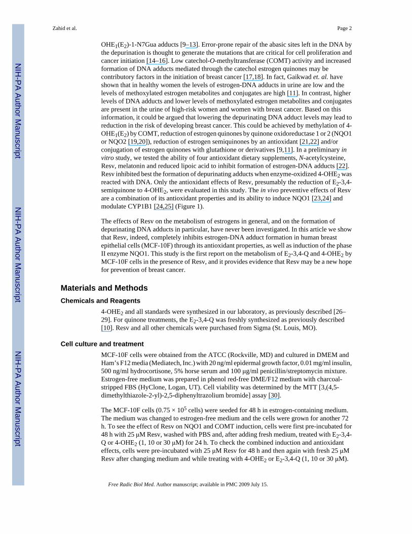
Prevention of estrogen-DNA adduct formation in MCF-10F cells byresveratrol
Muhammad Zahid, Nilesh W. Gaikwad, Mohamed F. Ali, Fang Lu, Muhammad Saeed, LiYang, Eleanor G. Rogan, and Ercole L. Cavalieri*Eppley Institute for Research in Cancer and Allied Diseases, University of Nebraska Medical Center,986805 Nebraska Medical Center, Omaha, NE 68198-6805
AbstractResveratrol (Resv), a natural occurring phytolexin present in grapes and other foods, possesseschemopreventive effects revealed by its striking modulation of diverse cellular events associatedwith tumor initiation, promotion and progression. Catechol estrogens generated in the metabolismof estrogens are oxidized to catechol quinones that react with DNA to form predominantlydepurinating estrogen-DNA adducts. This event can generate the mutations responsible for cancerinitiation. In this regard, Resv acts as both an antioxidant and an inducer of the phase II enzyme NAD(P)H:quinone oxidoreductase 1 (NQO1). In this report, we present the effects of Resv on themetabolism of estrogens in normal breast epithelial cells (MCF-10F) treated with 4-hydroxyestradiol(4-OHE2) or estradiol-3,4-quinone (E2-3,4-Q). Resv induced NQO1 in a dose- and time-dependentmanner, but did not affect the expression of catechol-O-methyltransferase. Ultraperformance liquidchromatography/tandem mass spectrometry was used to determine the effects of Resv on estrogenmetabolism. Preincubation of the cells with Resv for 48 h decreased the formation of depurinatingestrogen-DNA adducts from 4-OHE2 or E2-3,4-Q and increased formation of methoxycatecholestrogens. When Resv was also present with the 4-OHE2 or E2-3,4-Q, even greater increases inmethoxycatechol estrogens were observed, and the DNA adducts were undetectable. We concludethat Resv can protect breast cells from carcinogenic estrogen metabolites, suggesting that it could beused in breast cancer prevention.
IntroductionResveratrol (3,5,4′-trihydroxystilbene, Resv), which is present in red wine and grapes, has beenwidely studied for its antioxidant and biological effects [1]. It has been shown to induceanticancer effects by modulating several cellular pathways related to cancer initiation,promotion and progression [2–4]. Resv inhibits cell proliferation and induces apoptosis tosuppress and prevent a variety of tumors [5]. Resv stops the proliferation of human breastcancer MCF-7 cells and non-transformed MCF-10F cells treated with estrogens [6–8]. Themolecular mechanisms underlying these activities are not completely understood.
Estrogens are metabolized to catechol estrogens and then to catechol estrogen quinones, whichcan act as endogenous carcinogens [9–12] (Figure 1). These quinones react with DNA to formpredominantly the depurinating 4-hydroxyestrone(estradiol) [OHE1(E2)-1-N3Ade] and 4-
*To whom correspondence should be addressed. Ercole L. Cavalieri, Eppley Institute for Research in Cancer and Allied Diseases, UNMC,986805 Nebraska Medical Center, Omaha, NE 68198-6805, phone 402-559-7237, fax 402-559-8068, e-mail, [email protected]'s Disclaimer: This is a PDF file of an unedited manuscript that has been accepted for publication. As a service to our customerswe are providing this early version of the manuscript. The manuscript will undergo copyediting, typesetting, and review of the resultingproof before it is published in its final citable form. Please note that during the production process errors may be discovered which couldaffect the content, and all legal disclaimers that apply to the journal pertain.
NIH Public AccessAuthor ManuscriptFree Radic Biol Med. Author manuscript; available in PMC 2009 July 15.
Published in final edited form as:Free Radic Biol Med. 2008 July 15; 45(2): 136–145.
NIH
-PA Author Manuscript
NIH
-PA Author Manuscript
NIH
-PA Author Manuscript
OHE1(E2)-1-N7Gua adducts [9–13]. Error-prone repair of the abasic sites left in the DNA bythe depurination is thought to generate the mutations that are critical for cell proliferation andcancer initiation [14–16]. Low catechol-O-methyltransferase (COMT) activity and increasedformation of DNA adducts mediated through the catechol estrogen quinones may becontributory factors in the initiation of breast cancer [17,18]. In fact, Gaikwad et. al. haveshown that in healthy women the levels of estrogen-DNA adducts in urine are low and thelevels of methoxylated estrogen metabolites and conjugates are high [11]. In contrast, higherlevels of DNA adducts and lower levels of methoxylated estrogen metabolites and conjugatesare present in the urine of high-risk women and women with breast cancer. Based on thisinformation, it could be argued that lowering the depurinating DNA adduct levels may lead toreduction in the risk of developing breast cancer. This could be achieved by methylation of 4-OHE1(E2) by COMT, reduction of estrogen quinones by quinone oxidoreductase 1 or 2 (NQO1or NQO2 [19,20]), reduction of estrogen semiquinones by an antioxidant [21,22] and/orconjugation of estrogen quinones with glutathione or derivatives [9,11]. In a preliminary invitro study, we tested the ability of four antioxidant dietary supplements, N-acetylcysteine,Resv, melatonin and reduced lipoic acid to inhibit formation of estrogen-DNA adducts [22].Resv inhibited best the formation of depurinating adducts when enzyme-oxidized 4-OHE2 wasreacted with DNA. Only the antioxidant effects of Resv, presumably the reduction of E2-3,4-semiquinone to 4-OHE2, were evaluated in this study. The in vivo preventive effects of Resvare a combination of its antioxidant properties and its ability to induce NQO1 [23,24] andmodulate CYP1B1 [24,25] (Figure 1).
The effects of Resv on the metabolism of estrogens in general, and on the formation ofdepurinating DNA adducts in particular, have never been investigated. In this article we showthat Resv, indeed, completely inhibits estrogen-DNA adduct formation in human breastepithelial cells (MCF-10F) through its antioxidant properties, as well as induction of the phaseII enzyme NQO1. This study is the first report on the metabolism of E2-3,4-Q and 4-OHE2 byMCF-10F cells in the presence of Resv, and it provides evidence that Resv may be a new hopefor prevention of breast cancer.
Materials and MethodsChemicals and Reagents
4-OHE2 and all standards were synthesized in our laboratory, as previously described [26–29]. For quinone treatments, the E2-3,4-Q was freshly synthesized as previously described[10]. Resv and all other chemicals were purchased from Sigma (St. Louis, MO).
Cell culture and treatmentMCF-10F cells were obtained from the ATCC (Rockville, MD) and cultured in DMEM andHam’s F12 media (Mediatech, Inc.) with 20 ng/ml epidermal growth factor, 0.01 mg/ml insulin,500 ng/ml hydrocortisone, 5% horse serum and 100 μg/ml penicillin/streptomycin mixture.Estrogen-free medium was prepared in phenol red-free DME/F12 medium with charcoal-stripped FBS (HyClone, Logan, UT). Cell viability was determined by the MTT [3,(4,5-dimethylthiazole-2-yl)-2,5-diphenyltrazolium bromide] assay [30].
The MCF-10F cells (0.75 × 10⁵ cells) were seeded for 48 h in estrogen-containing medium.The medium was changed to estrogen-free medium and the cells were grown for another 72h. To see the effect of Resv on NQO1 and COMT induction, cells were first pre-incubated for48 h with 25 μM Resv, washed with PBS and, after adding fresh medium, treated with E2-3,4-Q or 4-OHE2 (1, 10 or 30 μM) for 24 h. To check the combined induction and antioxidanteffects, cells were pre-incubated with 25 μM Resv for 48 h and then again with fresh 25 μMResv after changing medium and while treating with 4-OHE2 or E2-3,4-Q (1, 10 or 30 μM).
Zahid et al. Page 2
Free Radic Biol Med. Author manuscript; available in PMC 2009 July 15.
NIH
-PA Author Manuscript
NIH
-PA Author Manuscript
NIH
-PA Author Manuscript
To examine the dose-dependency of the Resv effects, cells were treated with 1, 10 or 30 μMResv, while keeping the 4-OHE2 constant at 10 μM. To keep the concentration of DMSO(0.001%) the same in all experiments, different stock solutions of 4-OHE2 (1–30 mM) andResv (0.1–30 mM) were prepared. Media from four T-150 flasks of MCF-10F cells treatedwith an equal amount of organic solvent were used as controls.
Analysis of estrogen-DNA adductsFollowing the treatments, the media were harvested, supplemented with 2 mM ascorbic acid(to prevent possible decomposition of the compounds) and processed immediately. Samplepreparation and analysis by ultraperformance liquid chromatography/tandem massspectrometry (UPLC-MS/MS) were carried out as follows:
i. Sample preparation—Culture media from four flasks (40 mL) were processed by passingthrough Varian C8 Certify II cartridges (Varian, Harbor City, CA). The cartridges were pre-equilibrated by sequentially passing 1 ml of methanol, distilled water, and potassium phosphatebuffer (100 mM, pH 8) through them. Culture media were adjusted with 1 ml of 1 M potassiumphosphate buffer to pH 8.0 and passed through the cartridges. After washing with 200 μl ofthe phosphate buffer, the analytes were eluted with 1 ml of elution buffer[methanol:acetonitrile:water: trifluoroacetic acid (8:1:1:0.1)] and processed as previouslydescribed [18].
ii. UPLC-MS/MS—Samples were analyzed on an Acquity UPLC system connected to aMicroMass QuattroMicro triple quadrupole mass spectrometer (Waters, Milford, MA) [11].The 10-μl injections were carried out on a Waters Acquity UPLC BEHC18 column (1.7 μm, 1× 100 mm). The instrument was operated in the positive electrospray ionization mode. Allaspects of system operation, data acquisition and processing were controlled using QuanLynxv4.0 software (Waters). The column was eluted starting with 5% acetonitrile in water (0.1%formic acid) for 1 min at a flow rate of 150 μl/min; then a gradient to 55% acetonitrile in 10min was run. Ionization was achieved using the following settings: capillary voltage 3 kV;cone voltage 15–40 V; source block temperature 100 °C; desolvation temperature 200 °C, witha nitrogen flow of 400 l/h. Nitrogen was used as both the desolvation and auxiliary gas. Argonwas used as the collision gas. Three-point calibration curves were run for each standard.Triplicate samples were analyzed for each data point.
The UPLC-MS/MS limit of detection for the estrogen derivatives was in the range of 0.002 to0.06 pmole, which was well below the experimental values obtained for all the compounds,and the standard error for this analysis, calculated from triplicate analyses was 0.00002–0.0066pmole.
UPLC-MS/MS both identified and quantified the estrogen derivatives (Figure 2). For example,in one experiment the adducts were detected as single peaks and pretreatment of the cells withResv decreased the level of 4-OHE2-1-N7Gua from 313 to 70 pg/μl (Figure 2A and B), 4-OHE1-1-N7Gua from 233 to 20 pg/μl (Figure 2C and D), 4-OHE2-1-N3Ade from 220 to 11pg/μl (Figure 2E and F) and 4-OHE1-1-N3Ade from 255 to 47 pg/μl (Figure 2G and H).
ImmunoblottingComprehensive analysis of the expression patterns of the protective phase II enzymes (COMTand NQO1) from control and treated MCF-10F cells at different time points was conducted byimmunoblotting. Whole cell lysates were prepared by suspending cells in RIPA buffer withprotease inhibitor and lysed by three freeze-thaw cycles. After removing cellular debris bycentrifugation, protein concentrations were determined by using a BCA protein assay kit(Pierce Biotechnology, Rockford, IL). The proteins were separated by SDS-PAGE and
Zahid et al. Page 3
Free Radic Biol Med. Author manuscript; available in PMC 2009 July 15.
NIH
-PA Author Manuscript
NIH
-PA Author Manuscript
NIH
-PA Author Manuscript
transferred to PVD membranes for immunodetection as previously described [17]. Dilutionsof the primary anti-COMT (Chemicon International, Temecula, CA) and NQO1 (Abcam,Temecula, CA) antibodies were made in blocking solution (5% non-fat dry milk in PBS). Theblots were incubated for 3 h with primary antibody and for 1 h with secondary antibody coupledto horseradish peroxidase at room temperature. After each step, blots were washed with PBST(PBS and 0.1% Tween-20), incubated with ECL solution (Amersham Biotech, Piscataway,NJ) for 1 min, and visualized with radiographic film. The intensities of the bands werequantified by Alpha DigiDoc 1201 (Alpha Innotech, San Leandro, CA).
Determination of NQO1 activity by UPLC-MS/MSCellular NQO1 (25 μg of whole cell lysate protein) from control and 25 μM Resv-treatedMCF-10F cells was prepared. Triplicate enzyme reactions were carried out in a final volumeof 100 μl of 0.1 M sodium phosphate, pH 7. We used freshly synthesized E2-3,4-Q as thesubstrate and 25 μM NADH as the co-factor in the buffer system containing 0.7 mg/ml BSA.Recombinant NQO1 protein (10 units, Sigma, St. Louis, MO) in the reaction system served asthe positive control. Inhibition studies were carried out with inclusion of a potent inhibitor ofNQO1, dicumarol (10 μM), in the assay mixture. Control assays were carried out withoutenzyme or cofactor. The reaction mixture was pre-incubated for 3 min at room temperatureprior to the addition of substrate. The reaction was then initiated by adding 5 μl of 100 μME2-3,4-Q in acetonitrile and terminated after 27 min at 37 °C by adding 100 μl methanol.Following centrifugation to precipitate proteins, the supernatant was passed through a 5,000M.W. cut-off filter (Millipore, Billerica, MA), and 100 μl of each sample was analyzed for theproduct, 4-OHE2, by UPLC-MS/MS.
Determination of COMT activity by UPLC-MS/MSCellular COMT from control and 25 μM Resv (0–72 h)-treated MCF-10F cells was preparedas a cytosolic extract. Enzyme reactions were carried out in a final volume of 250 μl of 0.1 Msodium phosphate, pH 7.8, containing 1 mM DTT, 5 mM MgCl2, 100 μg cytosolic protein,100 μM 4-OHE2, and 200 μM S-adenosylmethionine (SAM, saturating). The incubationmixture, except for the substrate, was pre-incubated for 3 min at 37 °C. Then, the reaction wasinitiated by adding 5 μl of 4-OHE2 in DMSO and terminated after 27 min by adding 25 μl of4 M perchloric acid. Following centrifugation to precipitate proteins, the supernatant (245 μl)was passed through a 5,000 M.W. cut-off filter (Millipore, Billerica, MA), and 10 μl of eachsample was analyzed by UPLC-MS/MS. Each analysis was conducted in triplicate.
Statistical AnalysisThe statistical significance of the results was determined by Student’s t-test and ANOVAanalysis by using SAS and GraphPad Prism 4.0 software. P<0.05 was considered significant.All data were obtained from triplicate experiments.
ResultsEffect of Resv on NQO1 and COMT expression and activity
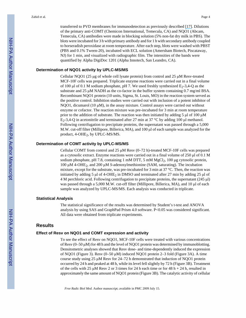
To see the effect of Resv on NQO1, MCF-10F cells were treated with various concentrationsof Resv (0–50 μM) for 48 h and the level of NQO1 protein was determined by immunoblotting.Densitometric analyses showed that Resv dose- and time-dependently induced the expressionof NQO1 (Figure 3). Resv (0–50 μM) induced NQO1 protein 2–3 fold (Figure 3A). A timecourse study using 25 μM Resv for 24–72 h demonstrated that induction of NQO1 proteinoccurred by 24 h and peaked at 48 h, while its level fell slightly by 72 h (Figure 3B). Treatmentof the cells with 25 μM Resv 2 or 3 times for 24 h each time or for 48 h + 24 h, resulted inapproximately the same amount of NQO1 protein (Figure 3B). The catalytic activity of cellular
Zahid et al. Page 4
Free Radic Biol Med. Author manuscript; available in PMC 2009 July 15.
NIH
-PA Author Manuscript
NIH
-PA Author Manuscript
NIH
-PA Author Manuscript
NQO1 in control and Resv-treated cells was determined by an in vitro enzyme activity assayusing UPLC-MS/MS. The levels of the reaction product, 4-OHE2, in treated cells weresignificantly greater than in the untreated cells (p< 0.05), as determined by ANOVA (Figure3C). The level of 4-OHE2 in reaction mixtures containing recombinant NQO1 protein, thepositive control, was 78.5±11.5 pg/μl, whereas the cellular proteins from 25 μM Resv-treatedcells showed a 2-fold increase in enzymatic activity compared with that from control cells (4-OHE2= 123±10.3 pg/μl). The reduction of E2-3,4-Q to 4-OHE2 by NQO1 was subsequentlyconfirmed by adding its potent inhibitor dicumarol (10 μM) to the assay mixture, which almostcompletely inhibited the formation of 4-OHE2. Control assays carried out without enzyme orcofactor had lower levels of 4-OHE2 (4.3±1.2 pg/μl). In the presence of NADH, the level wasslightly higher, 16.7 ± 5.7 pg/μl. The increased NQO1 activity in Resv-treated cellscorresponded well with the induction in NQO1 protein.
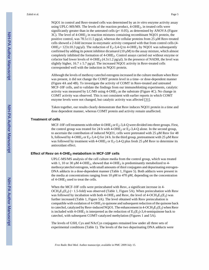
Although the levels of methoxy catechol estrogens increased in the culture medium when Resvwas present, it did not change the COMT protein level in a time- or dose-dependent manner(Figure 4A and 4B). To investigate the activity of COMT in Resv-treated and untreatedMCF-10F cells, and to validate the findings from our immunoblotting experiments, catalyticactivity was measured by LC/MS using 4-OHE2 as the substrate (Figure 4C). No change inCOMT activity was observed. This is not consistent with earlier reports in which COMTenzyme levels were not changed, but catalytic activity was affected [31].
Taken together, our results clearly demonstrate that Resv induces NQO1 protein in a time anddose dependent manner, whereas COMT protein and activity remain unaffected.
Treatment of cellsMCF-10F cell treatments with either 4-OHE2 or E2-3,4-Q were divided into three groups. First,the control group was treated for 24 h with 4-OHE2 or E2-3,4-Q alone. In the second group,to ascertain the contribution of induced NQO1, cells were pretreated with 25 μM Resv for 48h, followed by 4-OHE2 or E2-3,4-Q for 24 h. In the third group, pretreatment with 25 μM Resvwas followed by treatment with 4-OHE2 or E2-3,4-Q plus fresh 25 μM Resv to determine itsantioxidant effect.
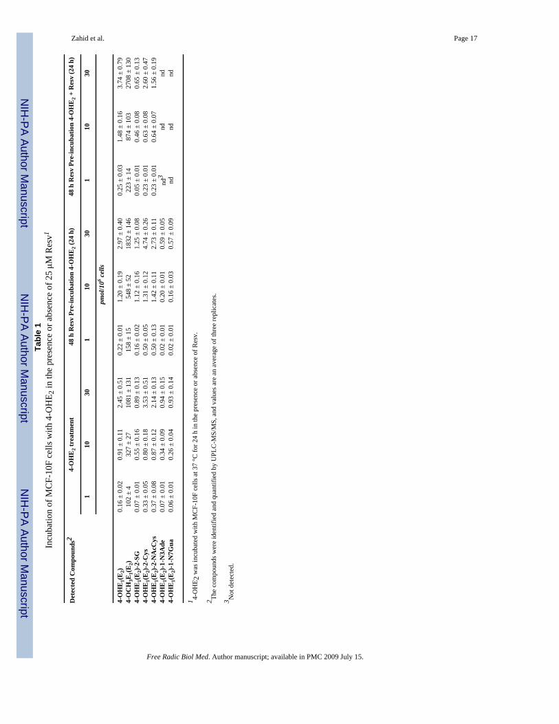
Effect of Resv on 4-OHE2 metabolism in MCF-10F cellsUPLC-MS/MS analysis of the cell culture media from the control group, which was treatedwith 1, 10 or 30 μM 4-OHE2, showed that 4-OHE2 is predominantly metabolized to 4-methoxycatechol estrogens, with small amounts of thiol conjugates and depurinating estrogen-DNA adducts in a dose-dependent manner (Table 1, Figure 5). Both adducts were present inthe media at concentrations ranging from 10 pM to 470 pM, depending on the concentrationof 4-OHE2 used to treat the cells.
When the MCF-10F cells were preincubated with Resv, a significant increase in 4-OCH3E1(E2) (~ 1.5-fold) was observed (Table 1, Figure 5A). When preincubation with Resvwas followed by incubation with both 4-OHE2 and Resv, the level of 4-OCH3E1(E2) wasfurther increased (Table 1, Figure 5A). The level obtained with Resv preincubation iscompatible with oxidation of 4-OHE2 to quinone and subsequent reduction of the quinone backto catechol, catalyzed by Resv-induced NQO1. The enhancement in 4-OCH3E1(E2) when Resvis included with 4-OHE2 is interpreted as the reduction of E1(E2)-3,4-semiquinone back tocatechol, with subsequent COMT-catalyzed methylation (Figures 1 and 5A).
The levels of GSH, Cys and NAcCys conjugates remained low under all three sets ofexperimental conditions (Table 1). The levels of the two depurinating DNA adducts were
Zahid et al. Page 5
Free Radic Biol Med. Author manuscript; available in PMC 2009 July 15.
NIH
-PA Author Manuscript
NIH
-PA Author Manuscript
NIH
-PA Author Manuscript
decreased when the cells were preincubated with Resv and became undetectable when the cellswere preincubated with Resv and incubated with 4-OHE2 plus Resv (Table 1, Figure 5B).
Effect of Resv on E2-3,4-Q metabolism in MCF-10F cellsMuch lower levels of 4-OCH3E1(E2) were observed when the MCF-10F cells were incubatedwith E2-3,4-Q (Table 2) compared to 4-OHE2 (Table 1). When the cells were preincubatedwith Resv, the level of 4-OCH3E1(E2) increased about 30%, and inclusion of Resv withE2-3,4-Q led to a doubling of the levels of 4-OCH3E1(E2) (Table 2, Figure 6A). The levels ofthiol conjugates showed a small dose-response, but were unaffected by preincubation orincubation with Resv (Table 2). The levels of the depurinating DNA adducts were lower withE2-3,4-Q than with 4-OHE2 (Table 1), and they decreased dramatically with Resvpreincubation, once again becoming undetectable when the cells were preincubated with Resvand incubated with E2-3,4-Q + Resv (Table 2, Figure 6B).
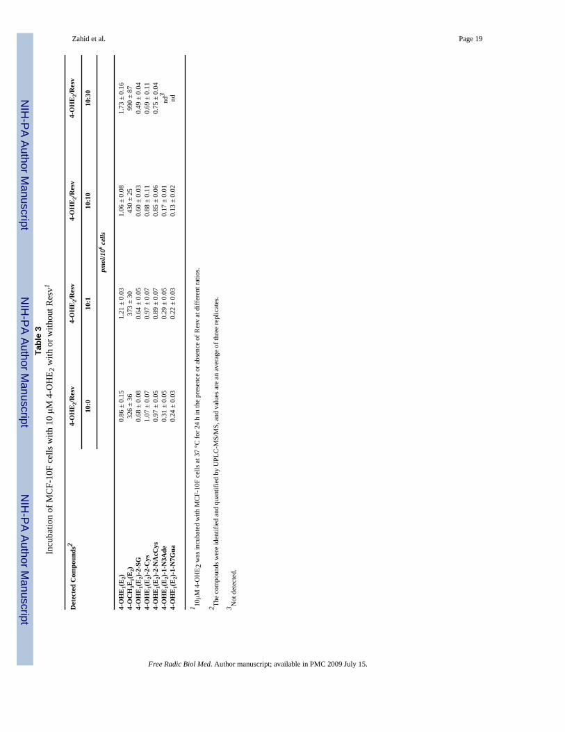
Dose-Response of ResvMCF-10F cells were treated with 10 μM 4-OHE2 and varying concentrations (0, 1, 10, 30μM) of Resv, and the metabolic profile was analyzed by UPLC-MS/MS. There was a dose-dependent increase in the level of 4-methoxycatechol estrogens, with a small increase atequimolar 4-OHE2 and Resv and a 3-fold increase when the concentration of Resv was 3-timesthat of 4-OHE2 (Table 3). There was a marginal, dose-dependent decrease in the correspondinglevels of the 4-OHE1(E2)-2-SG, 4-OHE1(E2)-2-Cys and 4-OHE1(E2)-2-NAcCys conjugatescompared with control (Table 3). Significant inhibition of 4-OHE1(E2)-1-N3Ade and 4-OHE1(E2)-1-N7Gua adduct formation was observed with increased Resv concentrations andthe adducts were not detected with 30 μM Resv (Table 3).
DiscussionResv induced NQO1 protein and activity in MCF-10F cells in a dose- and time- dependentmanner, with maximal induction after 48 h (Figure 3). The induction of NQO1 by Resv occursthrough transcriptional events [24,32,33] and is accompanied by localization of NQO1 in thenucleus [24]. When the cells were pre-incubated with Resv for 48 h, the effects of NQO1induction on the metabolism of 4-OHE2 can be observed from the increasing formation of 4-OCH3E1(E2) and decreasing amounts of 4-OHE1(E2)-1-N3Ade and 4-OHE1(E2)-1-N7Guaadducts (Table 1, Figure 5). The level of methoxycatechol estrogens increased about 50% andthe level of adducts decreased about 2-fold at all three doses of 4-OHE2. A similar effect ofResv was seen when the cells were incubated with E2-3,4-Q, following pre-incubation withResv for 48 h (Table 2, Figure 6). In this case, the levels of 4-OCH3E1(E2) were much lowerthan with 4-OHE2 as the estrogen, but the increase in methoxycatechol estrogens across dosesof E2-3,4-Q was about 30%, while the decrease in depurinating estrogen-DNA adducts wasgreater than 50% and they were no longer detectable at the lowest dose of the estrogen. Marginalchanges were seen in the levels of thiol conjugates (Table 1).
Pre-incubation with Resv had no effect on the expression of COMT as detected by its level ofprotein or catalytic activity (Figure 4). The level of COMT in the MCF-10F cells wassufficiently high to methylate virtually all of the 4-OHE1(E2) present in the cells incubatedwith either 4-OHE2 or E2-3,4-Q (Tables 1 and 2).
To investigate the antioxidant effect of Resv, the MCF-10F cells were pre-incubated with Resvfor 48 h, then the cells were incubated with 4-OHE2 or E2-3,4-Q plus a second dose of Resv(Tables 1 and 2, Figures 5 and 6). Little further induction of NQO1 during this 24 h periodwould be expected, based on the results shown in Figure 3. Thus, the effects of Resv observedcorrespond to its antioxidant properties.
Zahid et al. Page 6
Free Radic Biol Med. Author manuscript; available in PMC 2009 July 15.
NIH
-PA Author Manuscript
NIH
-PA Author Manuscript
NIH
-PA Author Manuscript
With 4-OHE2 as the estrogen, the levels of 4-OCH3E1(E2) were 2–2.5 times those seen with4-OHE2 alone, and no depurinating estrogen-DNA adducts could be detected (Table 1, Figure5). The ratio of 4-OHE2 to Resv affected the estrogen metabolism (Table 3). Little effect wasobserved when the concentration of Resv was 10% that of 4-OHE2; at equimolar doses, themethoxycatechol estrogens increased about 30% and a similar decrease in adducts was seen.When the level of Resv was 3-times that of the catechol estrogen, however, the level of 4-OCH3E1(E2) approximately tripled, and the adducts were not detected (Table 3).
The antioxidant effect of Resv is due to the high energy of resonance of its one-electronoxidized form, namely, the 4′-oxyradical [34]. This chemical property can give Resv thecapacity to reduce the E2-3,4-semiquinone to 4-OHE2, as already observed in vitro whenenzyme-activated 4-OHE2 was reacted with DNA [22]. This reduction has also been observedfor cysteine and N-acetylcysteine [21,22]. With E2-3,4-Q as the estrogen, the levels ofmethoxycatechol estrogens were almost twice those seen with the quinone alone, and theadducts were not detected (Table 2, Figure 6). Inclusion of Resv with E2-3,4-Q had little effecton the formation of thiol conjugates (Table 2).
The ability of Resv to modulate estrogen metabolism may be important in the prevention ofbreast, prostate and other estrogen-initiated cancers. Men with prostate cancer [12] and womenwith breast cancer or at high risk for the disease [11] form significantly higher amounts of thedepurinating estrogen-DNA adducts than healthy men or women. Thus, formation of theseadducts appears to play a critical role in the initiation of these cancers [9,11,12]. The resultsreported here clearly show that Resv can induce NQO1, which serves as a protective enzyme,by reducing the amount of catechol estrogen quinones available to react with DNA and formdepurinating adducts. In addition, Resv inhibits CYP1B1, which catalyzes the formation of 4-OHE1(E2) [24,25].
In summary, the inhibitory effects of Resv are the sum of three factors. One is the inductionof NQO1 that provides a decrease in estrogen quinones and an increase in catechol estrogen[24]. The second factor is the inhibition of CYP1B1 expression, which decreases the formationof 4-catechol estrogens [24,25]. The third factor is the antioxidant properties of Resv that reducethe estrogen semiquinones to catechol estrogens, as indirectly determined in vitro [22]. Thus,Resv may be very effective in preventing cancer initiation by estrogens because it induces theprotective enzyme NQO1, as reported here and in reference 24, it inhibits overexpression ofCYP1B1 [24,25], and it acts as an antioxidant, presumably reducing catechol estrogensemiquinones to catechol estrogens [21,22,34].
Acknowledgements
Grant Support: This research was supported by U.S. Army Breast Cancer Research Program grant DAMD-17-03-0229and by Prevention LLC. Core support of the Eppley Institute was provided by grant P30 CA36727 from the NationalCancer Institute.
AbbreviationsResv
resveratrol
CE catechol estrogen
E2-3 4-Q, estradiol-3,4-quinone
4-OHE1(E2)
Zahid et al. Page 7
Free Radic Biol Med. Author manuscript; available in PMC 2009 July 15.
NIH
-PA Author Manuscript
NIH
-PA Author Manuscript
NIH
-PA Author Manuscript
4-ydroxyestrone(estradiol)
4-OCH3E1(E2) 4-methoxyestrone(estradiol)
NQO1 NAD(P)H quinone xidoreductase 1
COMT Catechol-O-methyltransferase
UPLC-MS/MS Ultraperformance liquid chromatography/tandem mass spectrometry
References1. Agarwal BB, Bhardwaj A, Agarwal RS, Seeram NP, Shishodia S, Takada Y. Role of resveratrol in the
prevention and therapy of cancer: preclinical and clinical studies. Anticancer Res 2004;24:3–53.2. Bhat KP, Pezzuto JM. Cancer chemopreventive activity of resveratrol. Ann NY Acad Sci
2002;957:210–229. [PubMed: 12074974]3. Jang M, Cai L, Udeani GO, Slowing KV, Thomas CF, Beecher CW. Cancer chemopreventive activity
of resveratrol, a natural product derived from grapes. Science 1997;275:218–220. [PubMed: 8985016]4. Tessitone L, Davit A, Sarotto I, Caderni G. Resveratrol depresses the growth of colorectal aberrant
crypt foci by affecting bax and p21(CIP) expression. Carcinogensis 2000;21:1619–1622.5. Aziz MH, Kumar R, Ahmed N. Cancer chemoprevention by resveratrol: in vitro and in vivo studies
and the underlying mechanisms (Review). Int J Oncol 2003;23:17–28. [PubMed: 12792772]6. Kim Y-A, Choi BT, Lee YT, Park DI, Rhee S-H, Park K-Y, Choi YH. Resveratrol inhibits cell
proliferation and induces apoptosis of human breast carcinoma MCF-7 cells. Oncol Rep 2004;11:441–446. [PubMed: 14719081]
7. Pozo-Guisado E, Merino JM, Mulero-Novarro S, Lorenzo-Benayas MJ, Centeno F, Alvarez-BarrientosA, Salguero PMF. Resveratrol induced apoptosis in MCF-7 human breast cancer cells involvescaspase-independent mechanism with down regulation of Bcl-2 and NF-kB. Int J Caner 2005;115:74–84.
8. Mgbonyebi OP, Russo J, Russo IH. Antiproliferative effect of synthetic resveratrol on human breastepithelial cells. Int J Oncol 1998;12:865–869. [PubMed: 9499448]
9. Cavalieri E, Chakravarti D, Guttenplan J, Hart E, Ingle J, Jankowiak R, Muti P, Rogan E, Russo J,Santen R, Sutter T. Catechol estrogen quinones as initiators of breast and other human cancers:implications for biomarkers of susceptibility and cancer prevention. BBA Reviews on Cancer2006;1766:63–78. [PubMed: 16675129]
10. Zahid M, Kohli E, Saeed M, Rogan E, Cavalieri E. The greater reactivity of estradiol-3,4-quinoneversus estradiol-2,3-quinone with DNA in the formation of depurinating DNA adducts. Implicationsfor tumor–initiating activity. Chem Res Toxicol 2006;19:164–172. [PubMed: 16411670]
11. Gaikwad NW, Yang L, Muti P, Meza J, Pruthi S, Ingle J, Rogan EG, Cavalieri EL. The molecularetiology of breast cancer: evidence from biomarkers of risk. Int J Cancer 2008;122:1949–1953.[PubMed: 18098283]
12. Markushin Y, Gaikwad N, Zhang H, Kapke P, Rogan EG, Cavalieri EL, Trock BJ, Pavlovich C,Jankowiak R. Potential biomarker for early risk assessment of prostate cancer. Prostate2006;66:1565–1571. [PubMed: 16894534]
13. Markushin Y, Zhong W, Cavalieri EL, Rogan EG, Small GJ, Yeung ES, Jankowiak R. Spectralcharacterization of catechol estrogen quinone (CEQ)-derived DNA adducts and their identificationin human breast tissue extract. Chem Res Toxicol 2003;16:1107–1117. [PubMed: 12971798]
14. Chakravarti D, Mailander P, Li K-M, Higginbotham S, Zhang HL, Gross ML, Meza JL, CavalieriEL, Rogan EG. Evidence that a burst of DNA depurination in SENCAR mouse skin induces error-prone repair and form mutations in the H-ras gene. Oncogene 2001;20:7945–7953. [PubMed:11753677]
Zahid et al. Page 8
Free Radic Biol Med. Author manuscript; available in PMC 2009 July 15.
NIH
-PA Author Manuscript
NIH
-PA Author Manuscript
NIH
-PA Author Manuscript
15. Mailander PC, Meza JL, Higginbotham S, Chakravarti D. Induction of A.T to G.C mutations byerroneous repair of depurinated DNA following estrogen treatment of the mammary gland of ACIrats. J Steroid Biochem Mol Biol 2006;101:204–215. [PubMed: 16982187]
16. Zhao Z, Kosinska W, Khmelnitsky M, Cavalieri EL, Rogan EG, Chakravarti D, Sacks PG, GuttenplanJB. Mutagenic activity of 4-hydroxyestradiol, but not 2-hydroxyestradiol, in BB rat2 embryonic cells,and the mutational spectrum of 4-hydroxyestradiol. Chem Res Toxicol 2006;19:475–479. [PubMed:16544955]
17. Lu F, Zahid M, Saeed M, Cavalieri EL, Rogan EG. Estrogen metabolism and formation of estrogenDNA-adducts in estradiol-treated MCF-10F cells. The effects of 2,3,7,8-tetrachlorodibenzo-p-dioxininduction and catechol-O-methyltransferase inhibition. J Steroid Biochem Mol Biol 2007;105:150–158. [PubMed: 17582757]
18. Zahid M, Saeed M, Lu F, Gaikwad N, Cavalieri EL, Rogan EG. Inhibition of catechol-O-methyltransferase increases estrogen-DNA adduct formation. Free Radic Biol Med 2007;43:1534–1540. [PubMed: 17964424]
19. Gaikwad NW, Rogan EG, Cavalieri EL. Evidence by ESI-MS for NQO1-catalyzed reduction ofestrogen ortho-quinones. Free Radic Biol Med 2007;43:1289–1298. [PubMed: 17893042]
20. Gaikwad NW, Yang L, Rogan EG, Cavalieri EL. Evidence for NQO2-mediated reduction of thecarcinogenic estrogen ortho-quinones. Free Radic Biol Med. 2008Submitted
21. Samuni AM, Chuang EY, Krishna MC, Stein W, DeGraff W, Russo A, Mitchell JB. Semiquinoneradical intermediate in catecholic estrogen-mediated cytotoxicity and mutagenesis: chemopreventionstrategies with antioxidants. Proc Natl Acad Sci USA 2003;100:5390–5395. [PubMed: 12702779]
22. Zahid M, Gaikwad N, Cavalieri EL, Rogan EG. Inhibition of depurinating estrogen-DNA adducts bynatural compounds. Chem Res Toxicol 2007;20:1947–1953. [PubMed: 18039013]
23. Floreani M, Napoli E, Quintieri L, Palatini P. Oral administration of trans-resveratrol to guinea pigsincreases cardiac DT-diaphorase and catalase activities, and protects isolated atria from menadionetoxicity. Life Sci 2003;24:2741–2750. [PubMed: 12679191]
24. Lu F, Zahid M, Wang C, Saeed M, Cavalieri EL, Rogan EG. Resveratrol prevents estrogen-DNAadduct formation and neoplastic transformation in MCF-10F cells. Cancer Prevention Research.2008Submitted
25. Guengerich FP, Chun YJ, Kim D, Gillam EM, Shimada T. Cytochrome P450 1B1: a target forinhibition in anticarcinogenesis strategies. Mutat Res 2003;523–524:173–182.
26. Li K-M, Todorovic R, Devanesan P, Higginbotham S, Kofeler H, Ramanathan R, Gross ML, RoganEG, Cavalieri EL. Metabolism and DNA binding studies of 4-hydroxyestradiol and estradiol-3,4-quinone in vitro and in female ACI rat mammary gland in vivo. Carcinogenesis 2004;25:289–297.[PubMed: 14578156]
27. Saeed M, Zahid M, Rogan E, Cavalieri E. Synthesis of the catechols of natural and synthetic estrogensby using 2′-iodoxybenzoic acid (IBX) as the oxidizing agent. Steroids 2005;70:173–178. [PubMed:15763595]
28. Stack DE, Byun J, Gross ML, Rogan EG, Cavalieri EL. Molecular characteristics of catechol estrogenquinones in reactions with deoxyribonucleosides. Chem Res Toxicol 1996;9:851–859. [PubMed:8828920]
29. Cao K, Stack DE, Ramanathan R, Gross ML, Rogan EG, Cavalieri EL. Synthesis and structureelucidation of estrogen quinones conjugated with cysteine, N-acetylcysteine, and glutathione. ChemRes Toxicol 1998;11:909–916. [PubMed: 9705753]
30. Denizot F, Lang R. Rapid colorimetric assay for cell growth and survival. Modifications to thetetrazolium dye procedure giving improved sensitivity and reliability. J Immunol Methods1986;89:271–277. [PubMed: 3486233]
31. Dawling S, Roodi N, Mernaugh R, Wang X, Parl FF. Catechol-O-methyltransferase (COMT)-mediated metabolism of catechol estrogens: comparison of wild-type and variant COMT isoforms.Cancer Res 2001;61:6716–6722. [PubMed: 11559542]
32. Li Y, Cao Z, Zhu H. Upregulation of endogenous antioxidants and phase 2 enzymes by the red winepolyphenol, resveratrol in cultured aortic smooth muscle cells leads to cytoprotection againstoxidative and electrophilic stress. Pharmacol Res 2006;53:6–15. [PubMed: 16169743]
Zahid et al. Page 9
Free Radic Biol Med. Author manuscript; available in PMC 2009 July 15.
NIH
-PA Author Manuscript
NIH
-PA Author Manuscript
NIH
-PA Author Manuscript
33. Hsieh TC, Lu X, Wang Z, Wu JM. Induction of quinone reductase NQO1 by resveratrol in humanK562 cells involves the antioxidant response element ARE and is accompanied by nucleartranslocation of transcription factor Nrf2. Med Chem 2006;2:275–285. [PubMed: 16948474]
34. Stivala LA, Savio M, Carafoli F, Perucca P, Bianchi L, Maga G, Forti L, Pagnoni UM, Albini A,Prosperi E, Vannini V. Specific structural determinants are responsible for the antioxidant activityand the cell cycle effects of resveratrol. J Biol Chem 2001;276:22586–22594. [PubMed: 11316812]
Zahid et al. Page 10
Free Radic Biol Med. Author manuscript; available in PMC 2009 July 15.
NIH
-PA Author Manuscript
NIH
-PA Author Manuscript
NIH
-PA Author Manuscript
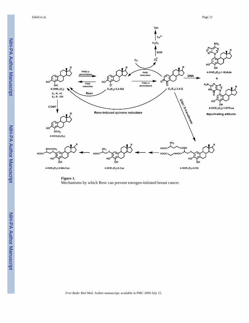
Figure 1.Mechanisms by which Resv can prevent estrogen-initiated breast cancer.
Zahid et al. Page 11
Free Radic Biol Med. Author manuscript; available in PMC 2009 July 15.
NIH
-PA Author Manuscript
NIH
-PA Author Manuscript
NIH
-PA Author Manuscript

Figure 2.UPLC-MS/MS chromatograms of depurinating DNA adducts: 4-OHE2-1-N7Gua (A, B), 4-OHE1-1-N7Gua (C, D), 4-OHE2-1-N3Ade (E, F) and 4-OHE1-1-N3Ade (G, H) in mediumfrom MCF-10F cells treated with 10 μM 4-OHE2 alone (A, C, E and G) or with 10 μM 4-OHE2 plus 10 μM Resv (B, D, F and H), respectively.
Zahid et al. Page 12
Free Radic Biol Med. Author manuscript; available in PMC 2009 July 15.
NIH
-PA Author Manuscript
NIH
-PA Author Manuscript
NIH
-PA Author Manuscript
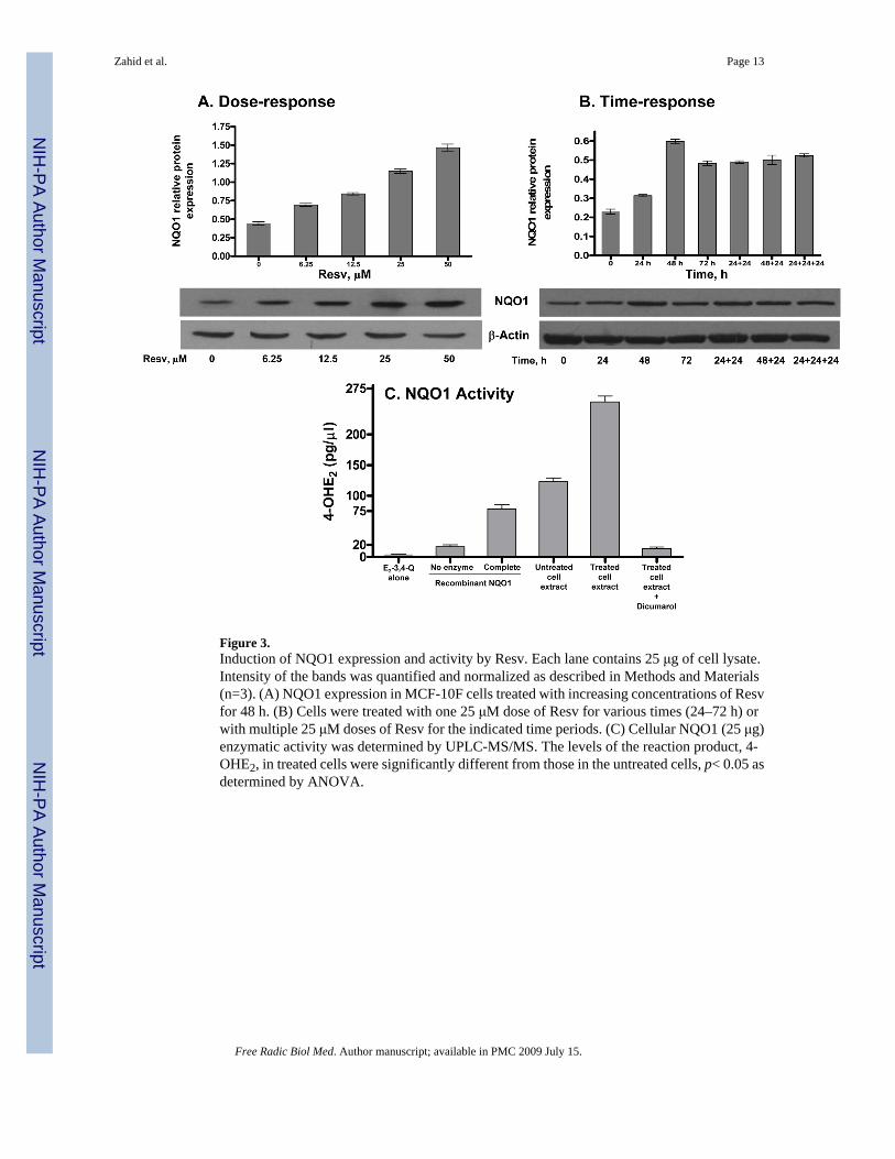
Figure 3.Induction of NQO1 expression and activity by Resv. Each lane contains 25 μg of cell lysate.Intensity of the bands was quantified and normalized as described in Methods and Materials(n=3). (A) NQO1 expression in MCF-10F cells treated with increasing concentrations of Resvfor 48 h. (B) Cells were treated with one 25 μM dose of Resv for various times (24–72 h) orwith multiple 25 μM doses of Resv for the indicated time periods. (C) Cellular NQO1 (25 μg)enzymatic activity was determined by UPLC-MS/MS. The levels of the reaction product, 4-OHE2, in treated cells were significantly different from those in the untreated cells, p< 0.05 asdetermined by ANOVA.
Zahid et al. Page 13
Free Radic Biol Med. Author manuscript; available in PMC 2009 July 15.
NIH
-PA Author Manuscript
NIH
-PA Author Manuscript
NIH
-PA Author Manuscript
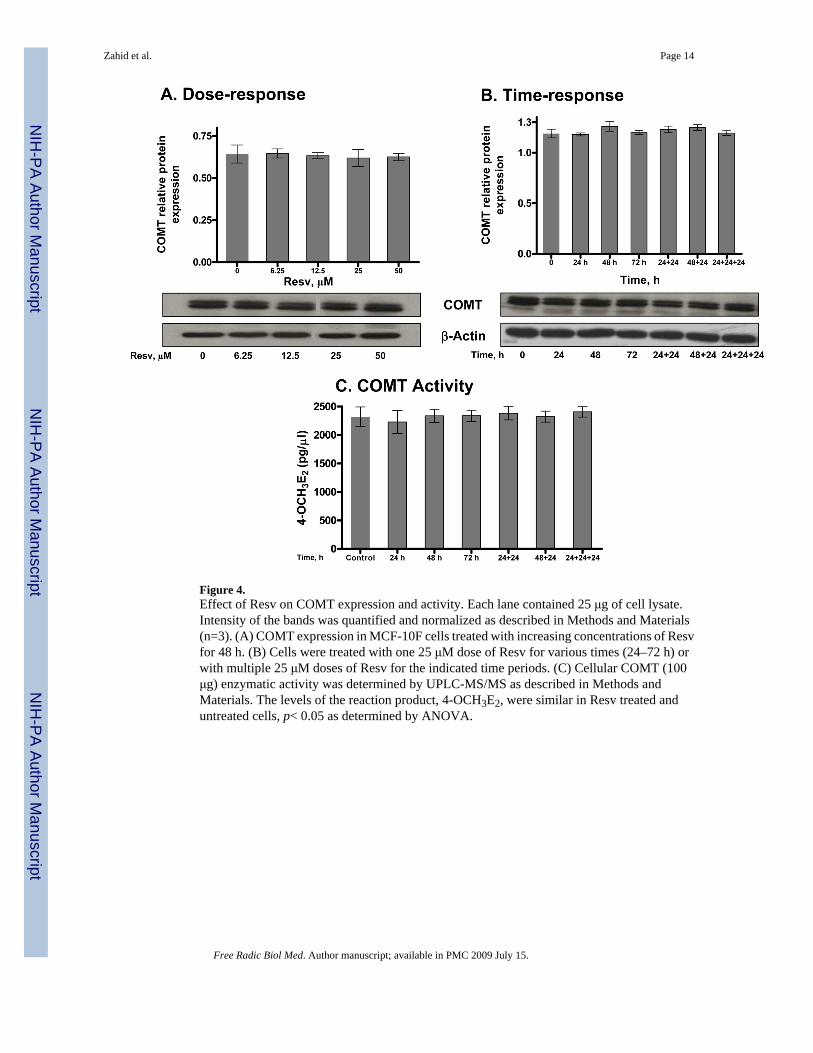
Figure 4.Effect of Resv on COMT expression and activity. Each lane contained 25 μg of cell lysate.Intensity of the bands was quantified and normalized as described in Methods and Materials(n=3). (A) COMT expression in MCF-10F cells treated with increasing concentrations of Resvfor 48 h. (B) Cells were treated with one 25 μM dose of Resv for various times (24–72 h) orwith multiple 25 μM doses of Resv for the indicated time periods. (C) Cellular COMT (100μg) enzymatic activity was determined by UPLC-MS/MS as described in Methods andMaterials. The levels of the reaction product, 4-OCH3E2, were similar in Resv treated anduntreated cells, p< 0.05 as determined by ANOVA.
Zahid et al. Page 14
Free Radic Biol Med. Author manuscript; available in PMC 2009 July 15.
NIH
-PA Author Manuscript
NIH
-PA Author Manuscript
NIH
-PA Author Manuscript
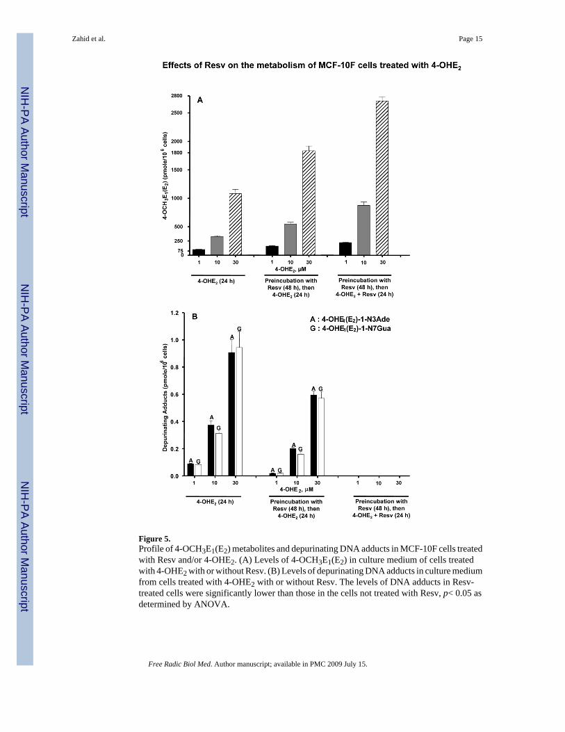
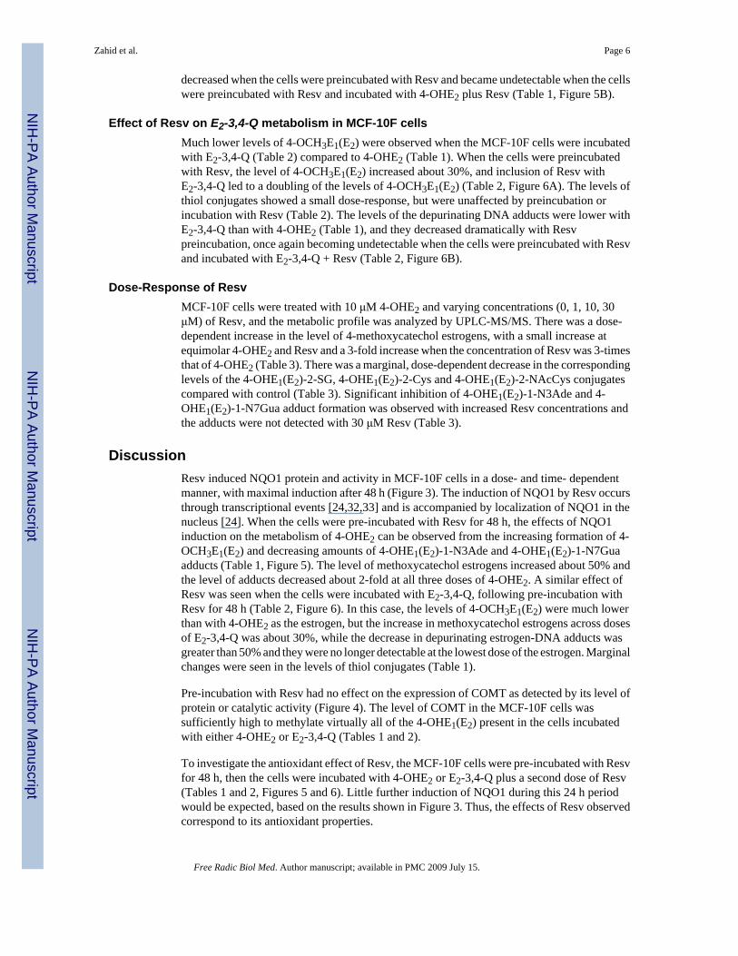
Figure 5.Profile of 4-OCH3E1(E2) metabolites and depurinating DNA adducts in MCF-10F cells treatedwith Resv and/or 4-OHE2. (A) Levels of 4-OCH3E1(E2) in culture medium of cells treatedwith 4-OHE2 with or without Resv. (B) Levels of depurinating DNA adducts in culture mediumfrom cells treated with 4-OHE2 with or without Resv. The levels of DNA adducts in Resv-treated cells were significantly lower than those in the cells not treated with Resv, p< 0.05 asdetermined by ANOVA.
Zahid et al. Page 15
Free Radic Biol Med. Author manuscript; available in PMC 2009 July 15.
NIH
-PA Author Manuscript
NIH
-PA Author Manuscript
NIH
-PA Author Manuscript
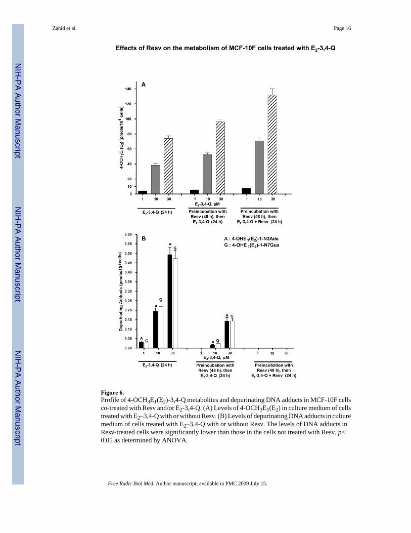
Figure 6.Profile of 4-OCH3E1(E2)-3,4-Q metabolites and depurinating DNA adducts in MCF-10F cellsco-treated with Resv and/or E2-3,4-Q. (A) Levels of 4-OCH3E1(E2) in culture medium of cellstreated with E2–3,4-Q with or without Resv. (B) Levels of depurinating DNA adducts in culturemedium of cells treated with E2–3,4-Q with or without Resv. The levels of DNA adducts inResv-treated cells were significantly lower than those in the cells not treated with Resv, p<0.05 as determined by ANOVA.
Zahid et al. Page 16
Free Radic Biol Med. Author manuscript; available in PMC 2009 July 15.
NIH
-PA Author Manuscript
NIH
-PA Author Manuscript
NIH
-PA Author Manuscript
NIH
-PA Author Manuscript
NIH
-PA Author Manuscript
NIH
-PA Author Manuscript
Zahid et al. Page 17Ta
ble
1In
cuba
tion
of M
CF-
10F
cells
with
4-O
HE 2
in th
e pr
esen
ce o
r abs
ence
of 2
5 μM
Res
v1
Det
ecte
d C
ompo
unds
24-
OH
E2 t
reat
men
t48
h R
esv
Pre-
incu
batio
n 4-
OH
E2 (
24 h
)48
h R
esv
Pre-
incu
batio
n 4-
OH
E2 +
Res
v (2
4 h)
110
301
1030
110
30
pmol
/106 c
ells
4-O
HE
1(E
2)0.
16 ±
0.0
20.
91 ±
0.1
12.
45 ±
0.5
10.
22 ±
0.0
11.
20 ±
0.1
92.
97 ±
0.4
00.
25 ±
0.0
31.
48 ±
0.1
63.
74 ±
0.7
94-
OC
H3E
1(E
2)10
2 ±
432
7 ±
2710
81 ±
131
158
± 15
548
± 52
1832
± 1
4622
3 ±
1487
4 ±
103
2708
± 1
304-
OH
E1(
E2)
-2-S
G0.
07 ±
0.0
10.
55 ±
0.1
60.
89 ±
0.1
30.
16 ±
0.0
21.
12 ±
0.1
61.
25 ±
0.0
80.
05 ±
0.0
10.
46 ±
0.0
80.
65 ±
0.1
34-
OH
E1(
E2)
-2-C
ys0.
33 ±
0.0
50.
80 ±
0.1
83.
53 ±
0.5
10.
50 ±
0.0
51.
31 ±
0.1
24.
74 ±
0.2
60.
23 ±
0.0
10.
63 ±
0.0
82.
60 ±
0.4
74-
OH
E1(
E2)
-2-N
AcC
ys0.
37 ±
0.0
80.
87 ±
0.1
22.
14 ±
0.1
30.
50 ±
0.1
31.
42 ±
0.1
12.
73 ±
0.1
10.
23 ±
0.0
10.
64 ±
0.0
71.
56 ±
0.1
94-
OH
E1(
E2)
-1-N
3Ade
0.07
± 0
.01
0.34
± 0
.09
0.94
± 0
.15
0.02
± 0
.01
0.20
± 0
.01
0.59
± 0
.05
nd3
ndnd
4-O
HE
1(E
2)-1
-N7G
ua0.
06 ±
0.0
10.
26 ±
0.0
40.
93 ±
0.1
40.
02 ±
0.0
10.
16 ±
0.0
30.
57 ±
0.0
9nd
ndnd
1 4-O
HE 2
was
incu
bate
d w
ith M
CF-
10F
cells
at 3
7 °C
for 2
4 h
in th
e pr
esen
ce o
r abs
ence
of R
esv.
2 The
com
poun
ds w
ere
iden
tifie
d an
d qu
antif
ied
by U
PLC
-MS/
MS,
and
val
ues a
re a
n av
erag
e of
thre
e re
plic
ates
.
3 Not
det
ecte
d.
Free Radic Biol Med. Author manuscript; available in PMC 2009 July 15.

NIH
-PA Author Manuscript
NIH
-PA Author Manuscript
NIH
-PA Author Manuscript
Zahid et al. Page 18Ta
ble
2In
cuba
tion
of M
CF-
10F
cells
with
E2-
3,4-
Q w
ith o
r with
out 2
5 μM
Res
v1
Det
ecte
d C
ompo
unds
2E
2-3,
4-Q
trea
tmen
t48
h R
esv
Pre-
incu
batio
n E
2-3,
4-Q
(24
h)48
h R
esv
Pre-
incu
batio
n E
2-3,
4-Q
+ R
esv
(24
h)
110
301
1030
110
30
pmol
/106 c
ells
4-O
HE
1(E
2)0.
30 ±
0.0
40.
75 ±
0.0
91.
34 ±
0.1
60.
22 ±
0.0
70.
70 ±
0.0
91.
17 ±
0.1
00.
18 ±
0.0
91.
48 ±
0.1
61.
02 ±
0.1
34-
OC
H3E
1(E
2)4.
08 ±
0.4
238
.4 ±
3.5
70.0
± 5
.75.
51 ±
0.2
252
.8 ±
3.4
96.0
± 5
.77.
57 ±
0.2
870
.5 ±
6.8
132
± 14
4-O
HE
1(E
2)-2
-SG
0.05
± 0
.01
0.28
± 0
.05
0.57
± 0
.05
0.07
± 0
.01
0.34
± 0
.06
0.73
± 0
.07
0.06
± 0
.01
0.29
± 0
.02
0.61
± 0
.03
4-O
HE
1(E
2)-2
-Cys
0.36
± 0
.06
0.84
± 0
.17
2.60
± 0
.18
0.46
± 0
.04
1.25
± 0
.09
3.41
± 0
.23
0.39
± 0
.06
0.95
± 0
.07
2.92
± 0
.06
4-O
HE
1(E
2)-2
-NA
cCys
0.10
± 0
.01
0.57
± 0
.06
1.26
± 0
.07
0.16
± 0
.01
1.09
± 0
.06
1.61
± 0
.06
0.12
± 0
.01
0.86
± 0
.05
1.39
± 0
.05
4-O
HE
1(E
2)-1
-N3A
de0.
03 ±
0.0
10.
19 ±
0.0
30.
49 ±
0.0
7nd
30.
02 ±
0.0
10.
14 ±
0.0
3nd
ndnd
4-O
HE
1(E
2)-1
-N7G
ua0.
03 ±
0.0
020.
22 ±
0.0
40.
47 ±
0.0
7nd
0.03
± 0
.01
0.14
± 0
.02
ndnd
nd
1 E 2-3
,4-Q
was
incu
bate
d w
ith M
CF-
10F
cells
at 3
7 °C
for 2
4 h
in th
e pr
esen
ce o
r abs
ence
of R
esv.
2 The
com
poun
ds w
ere
iden
tifie
d an
d qu
antif
ied
by U
PLC
-MS/
MS,
and
val
ues a
re a
n av
erag
e of
thre
e re
plic
ates
.
3 Not
det
ecte
d.
Free Radic Biol Med. Author manuscript; available in PMC 2009 July 15.
NIH
-PA Author Manuscript
NIH
-PA Author Manuscript
NIH
-PA Author Manuscript
Zahid et al. Page 19Ta
ble
3In
cuba
tion
of M
CF-
10F
cells
with
10 μM
4-O
HE 2
with
or w
ithou
t Res
v1
Det
ecte
d C
ompo
unds
24-
OH
E2/R
esv
4-O
HE
2/Res
v4-
OH
E2/R
esv
4-O
HE
2/Res
v
10:0
10:1
10:1
010
:30
pmol
/106 c
ells
4-O
HE
1(E
2)0.
86 ±
0.1
51.
21 ±
0.0
31.
06 ±
0.0
81.
73 ±
0.1
64-
OC
H3E
1(E
2)32
6 ±
3637
3 ±
3043
0 ±
2599
0 ±
874-
OH
E1(
E2)
-2-S
G0.
68 ±
0.0
80.
64 ±
0.0
50.
60 ±
0.0
30.
49 ±
0.0
44-
OH
E1(
E2)
-2-C
ys1.
07 ±
0.0
70.
97 ±
0.0
70.
88 ±
0.1
10.
69 ±
0.1
14-
OH
E1(
E2)
-2-N
AcC
ys0.
97 ±
0.0
50.
89 ±
0.0
70.
85 ±
0.0
60.
75 ±
0.0
44-
OH
E1(
E2)
-1-N
3Ade
0.31
± 0
.05
0.29
± 0
.05
0.17
± 0
.01
nd3
4-O
HE
1(E
2)-1
-N7G
ua0.
24 ±
0.0
30.
22 ±
0.0
30.
13 ±
0.0
2nd
1 10μM
4-O
HE 2
was
incu
bate
d w
ith M
CF-
10F
cells
at 3
7 °C
for 2
4 h
in th
e pr
esen
ce o
r abs
ence
of R
esv
at d
iffer
ent r
atio
s.
2 The
com
poun
ds w
ere
iden
tifie
d an
d qu
antif
ied
by U
PLC
-MS/
MS,
and
val
ues a
re a
n av
erag
e of
thre
e re
plic
ates
.
3 Not
det
ecte
d.
Free Radic Biol Med. Author manuscript; available in PMC 2009 July 15.
Copyright © 2022 FDOKUMEN