Adduct of the blistering warfare agent sesquimustard with ...
15
PAPER IN FOREFRONT Adduct of the blistering warfare agent sesquimustard with human serum albumin and its mass spectrometric identification for biomedical verification of exposure Marc-Michael Blum 1 & Annika Richter 2 & Markus Siegert 2,3 & Horst Thiermann 3 & Harald John 3 Received: 10 July 2020 /Revised: 7 August 2020 /Accepted: 25 August 2020 # The Author(s) 2020 Abstract Apart from the well-known sulfur mustard (SM), additional sulfur-containing blistering chemical warfare agents exist. Sesquimustard (Q) is one of them and five times more blistering than SM. It is a common impurity in mustard mixtures and regularly found in old munitions but can also be used in pure form. Compared to the extensive literature on SM, very little experimental data is available on Q and no protein biomarkers of exposure have been reported. We herein report for the first time the adduct of Q with the nucleophilic Cys 34 residue of human serum albumin (HSA) formed in vitro and introduce two novel bioanalytical procedures for detection. After proteolysis of this HSA adduct catalyzed either by pronase or by proteinase K, two biomarkers were identified by high-resolution tandem mass spectrometry (MS/HR MS), namely a dipeptide and a tripeptide, both alkylated at their Cys residue, which we refer to as HETETE-CP and HETETE-CPF. HETETE represents the Q-derived thio- alkyl moiety bearing a terminal hydroxyl group: “hydroxyethylthioethylthioethyl.” Targeting both peptide markers from plasma, a micro liquid chromatography–electrospray ionization tandem mass spectrometry method working in the selected reaction monitoring mode (μLC-ESI MS/MS SRM) was developed and validated as well suited for the verification of exposure to Q. Fulfilling the quality criteria defined by the Organisation for the Prohibition of Chemical Weapons, the novel methods enable the detection of exposure to Q alone or in mixtures with SM. We further report on the relative reactivity of Q compared to SM. Based on experiments making use of partially deuterated Q as the alkylating agent, we rule out a major role for six-membered ring sulfonium ions as relevant reactive species in the alkylation of Cys 34 . Furthermore, the results of molecular dynamics simulations are indicative that the protein environment around Cys 34 allows adduct formation with elongated but not bulky molecules such as Q, and identify important hydrogen bonding interactions and hydrophobic contacts. Keywords Hydroxyethylthioethyl . Protein adduct . Sulfur mustard . Verification . Vesicant Abbreviations Atr-d 3 Atropine, triple deuterated BioPT OPCW biomedical proficiency test CE Collision energy CID Collision-induced dissociation CWC Chemical Weapons Convention ESI Electrospray ionization HETE Hydroxyethylthioethyl moiety HETETE Hydroxyethylthioethylthioethyl moiety HR MS High-resolution mass spectrometry HSA Human serum albumin LC Liquid chromatography LCt 50 Median lethal concentration after inhalation logP Partition coefficient in octanol/water LOI Limit of identification MD Molecular dynamics MS Mass spectrometry MS/MS Tandem mass spectrometry NMR Nuclear magnetic resonance Electronic supplementary material The online version of this article (https://doi.org/10.1007/s00216-020-02917-w ) contains supplementary material, which is available to authorized users. * Harald John [email protected] 1 Blum – Scientific Services, Björnsonweg 70d, 22587 Hamburg, Germany 2 Department of Chemistry, Humboldt-Universität zu Berlin, Brook-Taylor-Straße 2, 12489 Berlin, Germany 3 Bundeswehr Institute of Pharmacology and Toxicology, Neuherbergstraße 11, 80937 Munich, Germany https://doi.org/10.1007/s00216-020-02917-w / Published online: 9 September 2020 Analytical and Bioanalytical Chemistry (2020) 412:7723–7737
-
Upload
khangminh22 -
Category
Documents
-
view
1 -
download
0
Transcript of Adduct of the blistering warfare agent sesquimustard with ...
PAPER IN FOREFRONT
Adduct of the blistering warfare agent sesquimustard with humanserum albumin and its mass spectrometric identificationfor biomedical verification of exposure
Marc-Michael Blum1& Annika Richter2 & Markus Siegert2,3 & Horst Thiermann3
& Harald John3
Received: 10 July 2020 /Revised: 7 August 2020 /Accepted: 25 August 2020# The Author(s) 2020
AbstractApart from the well-known sulfur mustard (SM), additional sulfur-containing blistering chemical warfare agents exist.Sesquimustard (Q) is one of them and five times more blistering than SM. It is a common impurity in mustard mixtures andregularly found in old munitions but can also be used in pure form. Compared to the extensive literature on SM, very littleexperimental data is available on Q and no protein biomarkers of exposure have been reported. We herein report for the first timethe adduct of Q with the nucleophilic Cys34 residue of human serum albumin (HSA) formed in vitro and introduce two novelbioanalytical procedures for detection. After proteolysis of this HSA adduct catalyzed either by pronase or by proteinase K, twobiomarkers were identified by high-resolution tandemmass spectrometry (MS/HRMS), namely a dipeptide and a tripeptide, bothalkylated at their Cys residue, which we refer to as HETETE-CP and HETETE-CPF. HETETE represents the Q-derived thio-alkyl moiety bearing a terminal hydroxyl group: “hydroxyethylthioethylthioethyl.” Targeting both peptide markers from plasma,a micro liquid chromatography–electrospray ionization tandem mass spectrometry method working in the selected reactionmonitoring mode (μLC-ESI MS/MS SRM) was developed and validated as well suited for the verification of exposure to Q.Fulfilling the quality criteria defined by the Organisation for the Prohibition of Chemical Weapons, the novel methods enable thedetection of exposure to Q alone or in mixtures with SM.We further report on the relative reactivity of Q compared to SM. Basedon experiments making use of partially deuterated Q as the alkylating agent, we rule out a major role for six-membered ringsulfonium ions as relevant reactive species in the alkylation of Cys34. Furthermore, the results of molecular dynamics simulationsare indicative that the protein environment around Cys34 allows adduct formation with elongated but not bulky molecules such asQ, and identify important hydrogen bonding interactions and hydrophobic contacts.
Keywords Hydroxyethylthioethyl . Protein adduct . Sulfur mustard . Verification . Vesicant
AbbreviationsAtr-d3 Atropine, triple deuteratedBioPT OPCW biomedical proficiency test
CE Collision energyCID Collision-induced dissociationCWC Chemical Weapons ConventionESI Electrospray ionizationHETE Hydroxyethylthioethyl moietyHETETE Hydroxyethylthioethylthioethyl moietyHR MS High-resolution mass spectrometryHSA Human serum albuminLC Liquid chromatographyLCt50 Median lethal concentration after inhalationlogP Partition coefficient in octanol/waterLOI Limit of identificationMD Molecular dynamicsMS Mass spectrometryMS/MS Tandem mass spectrometryNMR Nuclear magnetic resonance
Electronic supplementary material The online version of this article(https://doi.org/10.1007/s00216-020-02917-w ) contains supplementarymaterial, which is available to authorized users.
* Harald [email protected]
1 Blum – Scientific Services, Björnsonweg 70d,22587 Hamburg, Germany
2 Department of Chemistry, Humboldt-Universität zu Berlin,Brook-Taylor-Straße 2, 12489 Berlin, Germany
3 Bundeswehr Institute of Pharmacology and Toxicology,Neuherbergstraße 11, 80937 Munich, Germany
https://doi.org/10.1007/s00216-020-02917-w
/ Published online: 9 September 2020
Analytical and Bioanalytical Chemistry (2020) 412:7723–7737
OPCW Organisation for the Prohibitionof Chemical Weapons
Q SesquimustardRMSD Root mean square deviationRMSF Root mean square fluctuationRT Room temperatureSM Sulfur mustardSRM Selected reaction monitoringSγ Sulfur atom of cysteine residue side chaintR Retention timeXIC Extracted ion chromatogramμLC Micro liquid chromatography
Introduction
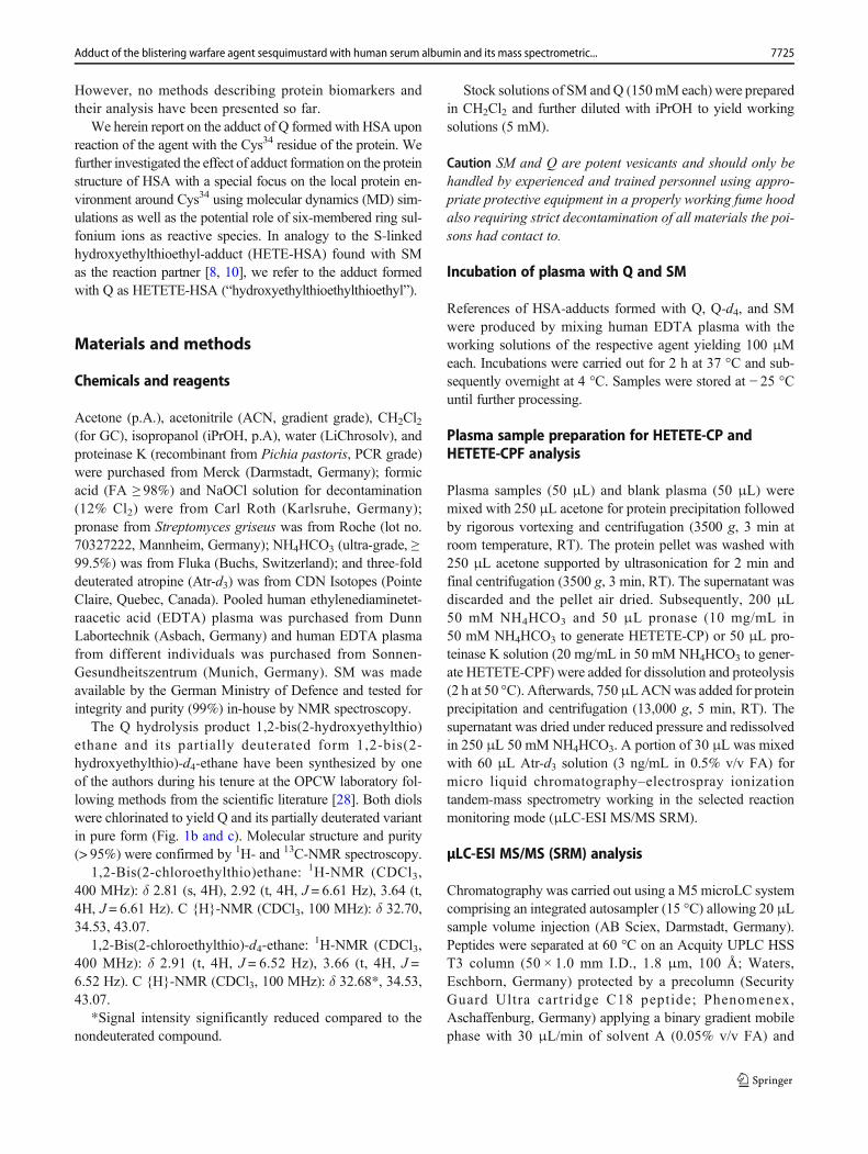
Vesicants, chemicals that cause blistering of the skin, havebeen used as warfare agents for more than 100 years. Themostprominent member of this group is sulfur mustard (SM, bis(2-chloroethyl) sulfide, CAS 505-60-2, Fig. 1a) [1, 2]. It has beenemployed as recently as 2015 and 2016 by the terrorist group“Islamic State” in the Syrian Arab Republic and northern Iraq[3]. SM causes painful blisters and erythema on exposed skinareas characterized by delayed and complicated woundhealing and shows a complex toxicokinetic behavior [1, 4].
Verification of exposure is an important capability in orderto guide medical therapy of casualties but also for generatingevidence that a violation of the international norm on the non-use of chemical weapons has occurred [5]. This norm is laiddown in the Chemical Weapons Convention (CWC), whichentered into force in 1997, and is implemented by theOrganisation for the Prohibition of Chemical Weapons(OPCW) [6]. The OPCW designates laboratories for the anal-ysis of authentic biomedical samples through successful par-ticipation in biomedical proficiency tests (BioPTs). In theseBioPTs, samples of biological fluids spikedwith SM representa regular analytical challenge. The most long-lasting in vivobiomarkers of exposure are protein-adducts such as thoseformed with human serum albumin (HSA). HSA-adducts re-sult from covalent linkage of SM to, e.g., the Cys34 residue,and have emerged as the most prominent biomarkers [7–10].They have been used on several occasions for the analysis ofsamples from OPCW interlaboratory exercises but also fromexposed victims [11–14].
However, SM is not the only blistering agent causing aserious threat for military personnel as well as for civilians.Among the chemicals listed in the Annex on Chemicals of theCWC are a number of higher sulfur mustards, includingsesquimustard (Q, Fig. 1b).
Q (1,2-bis(2-chloroethylthio) ethane, CAS 3563-36-8,Fig. 1b), a white solid (melting point 56.6 °C), is about fivetimes more blistering than SM [15]. As a solid, it represents avery persistent contact hazard but has so far not been used inits pure form. It was produced as a component in the BritishHQ process during World War II resulting in a 70:30 w/wmixture of SM:Q [16]. It is also regularly found in old distilled(HD) and Levinstein (H) mustard. For example, a ton contain-er of Levinstein mustard sampled in the USA after 60 years ofstorage before destruction contained about 10% w/w of Q[17].
Despite its relevance, the number of scientific publica-tions on Q is small, especially when compared with theextensive literature on SM. Q was first reported byBennett and Whincop in 1921 [18]. Its toxic propertiesbut also potential use as an alternative to nitrogen mustardanti-tumor drugs were studied and reported in a few pub-lications thereafter [15, 19, 20]. St. Quintin et al. reportedon the hydrolysis of Q observing six-membered ring sul-fonium ions (Fig. 1f) as long-lived reaction intermediatesin addition to the three-membered ring episulfonium ionsknown from SM (Fig. 1e) adding important insight into thebehavior of Q in aqueous systems [21]. Analysis of Q andits hydrolysis and oxidation products from environmentalsamples by means of gas and liquid chromatography (LC)coupled to mass spectrometry (MS) has been described[22–26]. A method for the quantification of the urinarybiotransformation products of Q using LC coupled to tan-dem mass spectrometry (MS/MS) was also published [27].
a
b
c
d
e
f
Fig. 1 Chemical structures of blister agents and their reactiveintermediates: a sulfur mustard (SM), b sesquimustard (Q), csesquimustard-d4, d reactive episulfonium ions of SM, e reactiveepisulfonium ions of Q, and f the six-membered ring sulfonium ion of Q
7724 Blum M.-M. et al.
However, no methods describing protein biomarkers andtheir analysis have been presented so far.
We herein report on the adduct of Q formed with HSA uponreaction of the agent with the Cys34 residue of the protein. Wefurther investigated the effect of adduct formation on the proteinstructure of HSA with a special focus on the local protein en-vironment around Cys34 using molecular dynamics (MD) sim-ulations as well as the potential role of six-membered ring sul-fonium ions as reactive species. In analogy to the S-linkedhydroxyethylthioethyl-adduct (HETE-HSA) found with SMas the reaction partner [8, 10], we refer to the adduct formedwith Q as HETETE-HSA (“hydroxyethylthioethylthioethyl”).
Materials and methods
Chemicals and reagents
Acetone (p.A.), acetonitrile (ACN, gradient grade), CH2Cl2(for GC), isopropanol (iPrOH, p.A), water (LiChrosolv), andproteinase K (recombinant from Pichia pastoris, PCR grade)were purchased from Merck (Darmstadt, Germany); formicacid (FA ≥ 98%) and NaOCl solution for decontamination(12% Cl2) were from Carl Roth (Karlsruhe, Germany);pronase from Streptomyces griseus was from Roche (lot no.70327222, Mannheim, Germany); NH4HCO3 (ultra-grade, ≥99.5%) was from Fluka (Buchs, Switzerland); and three-folddeuterated atropine (Atr-d3) was from CDN Isotopes (PointeClaire, Quebec, Canada). Pooled human ethylenediaminetet-raacetic acid (EDTA) plasma was purchased from DunnLabortechnik (Asbach, Germany) and human EDTA plasmafrom different individuals was purchased from Sonnen-Gesundheitszentrum (Munich, Germany). SM was madeavailable by the German Ministry of Defence and tested forintegrity and purity (99%) in-house by NMR spectroscopy.
The Q hydrolysis product 1,2-bis(2-hydroxyethylthio)ethane and its partially deuterated form 1,2-bis(2-hydroxyethylthio)-d4-ethane have been synthesized by oneof the authors during his tenure at the OPCW laboratory fol-lowing methods from the scientific literature [28]. Both diolswere chlorinated to yield Q and its partially deuterated variantin pure form (Fig. 1b and c). Molecular structure and purity(> 95%) were confirmed by 1H- and 13C-NMR spectroscopy.
1,2-Bis(2-chloroethylthio)ethane: 1H-NMR (CDCl3,400 MHz): δ 2.81 (s, 4H), 2.92 (t, 4H, J = 6.61 Hz), 3.64 (t,4H, J = 6.61 Hz). C {H}-NMR (CDCl3, 100 MHz): δ 32.70,34.53, 43.07.
1,2-Bis(2-chloroethylthio)-d4-ethane:1H-NMR (CDCl3,
400 MHz): δ 2.91 (t, 4H, J = 6.52 Hz), 3.66 (t, 4H, J =6.52 Hz). C {H}-NMR (CDCl3, 100 MHz): δ 32.68*, 34.53,43.07.
*Signal intensity significantly reduced compared to thenondeuterated compound.
Stock solutions of SM andQ (150mMeach) were preparedin CH2Cl2 and further diluted with iPrOH to yield workingsolutions (5 mM).
Caution SM and Q are potent vesicants and should only behandled by experienced and trained personnel using appro-priate protective equipment in a properly working fume hoodalso requiring strict decontamination of all materials the poi-sons had contact to.
Incubation of plasma with Q and SM
References of HSA-adducts formed with Q, Q-d4, and SMwere produced by mixing human EDTA plasma with theworking solutions of the respective agent yielding 100 μMeach. Incubations were carried out for 2 h at 37 °C and sub-sequently overnight at 4 °C. Samples were stored at − 25 °Cuntil further processing.
Plasma sample preparation for HETETE-CP andHETETE-CPF analysis
Plasma samples (50 μL) and blank plasma (50 μL) weremixed with 250 μL acetone for protein precipitation followedby rigorous vortexing and centrifugation (3500 g, 3 min atroom temperature, RT). The protein pellet was washed with250 μL acetone supported by ultrasonication for 2 min andfinal centrifugation (3500 g, 3 min, RT). The supernatant wasdiscarded and the pellet air dried. Subsequently, 200 μL50 mM NH4HCO3 and 50 μL pronase (10 mg/mL in50 mM NH4HCO3 to generate HETETE-CP) or 50 μL pro-teinase K solution (20 mg/mL in 50 mM NH4HCO3 to gener-ate HETETE-CPF) were added for dissolution and proteolysis(2 h at 50 °C). Afterwards, 750μLACNwas added for proteinprecipitation and centrifugation (13,000 g, 5 min, RT). Thesupernatant was dried under reduced pressure and redissolvedin 250 μL 50 mM NH4HCO3. A portion of 30 μL was mixedwith 60 μL Atr-d3 solution (3 ng/mL in 0.5% v/v FA) formicro liquid chromatography–electrospray ionizationtandem-mass spectrometry working in the selected reactionmonitoring mode (μLC-ESI MS/MS SRM).
μLC-ESI MS/MS (SRM) analysis
Chromatography was carried out using a M5microLC systemcomprising an integrated autosampler (15 °C) allowing 20 μLsample volume injection (AB Sciex, Darmstadt, Germany).Peptides were separated at 60 °C on an Acquity UPLC HSST3 column (50 × 1.0 mm I.D., 1.8 μm, 100 Å; Waters,Eschborn, Germany) protected by a precolumn (SecurityGuard Ultra cartridge C18 peptide; Phenomenex,Aschaffenburg, Germany) applying a binary gradient mobilephase with 30 μL/min of solvent A (0.05% v/v FA) and
7725Adduct of the blistering warfare agent sesquimustard with human serum albumin and its mass spectrometric...
solvent B (ACN/H2O 80:20 v/v, 0.05% v/v FA): t [min]/B[%]: 0/2, 11/60, 11.5/95, 13.5/95, 14/2, 15/2. Using an ESIinterface working in positive mode (5 kV) the QTrap 6500+
mass spectrometer (AB Sciex) was coupled to monitor prod-uct ions after collision-induced dissociation (CID) of analytesusing nitrogen as collision gas. The following MS settingswere applied: declustering potential (DP) 60 V, curtain gas(CUR) 30 psi (2.07 × 10⁵ Pa), temperature 200 °C, sourcegas 1 (GS1) 50 psi (3.45 × 10⁵ Pa), source gas 2 (GS2)60 psi (4.14 × 10⁵ Pa), entrance potential (EP) 10 V, cell exitpotential (CXP) 10 V each, and dwell time 50 ms. The singleprotonated precursor ions of the biomarkers were fragmentedwith a collision energy (CE) of 30 V to generate product ionsby MS/MS working in the SRM mode: HETETE-CP: m/z383.1 >m/z 105.0, m/z 217.1; HETETE-CPF: m/z 530.2 >m/z 105.0, m/z 137.0; HETE-CP: m/z 323.1 >m/z 105.0, m/z137.0; and HETE-CPF: m/z 470.2 >m/z 105.0, m/z 137.0.The internal standard Atr-d3 was monitored as follows: m/z293.3 >m/z 127.1 and m/z 93.1 using a CE of 42 V.
μLC-ESI MS/HR MS (Orbitrap) analysis
Initial high-resolution tandem mass spectrometric (MS/HRMS) detection of HETETE-adducts was carried out to deter-mine the exact masses of product ions. For chromatography ofa 20-μL sample volume, a MicroPro pump (EldexLaboratories, Napa, CA, USA) in combination with anINTEGRITY autosampler and a MISTRAL column oven(both Spark Holland, Emmen, The Netherlands) was used.The stationary and mobile phases were the same as describedabove, but the gradient was as follows: t [min]/B [%]: 0/10;11/60; 11.1/95; 13.9/95; 14/10; 15/10. The system was con-trolled by the Eldex MicroPro 1.0.54 software (EldexLaboratories) and coupled on-line to a QExactive plusOrbitrap mass spectrometer via the HESI II ion source(Thermo Scientific, Bremen, Germany). The following MSparameters were applied to monitor product ions of the singleprotonated precursor ions of HETETE-CP (m/z 383.1), d4-HETETE-CP (m/z 387.1), HETETE-CPF (m/z 530.2), andd4-HETETE-CPF (m/z 534.2) in the parallel reaction monitor-ing (PRM) mode with a mass spectrometric resolution of35,000 FWHM (full width at half maximum at m/z 200):sheath gas flow 23 arbitrary units (a.u.), aux gas flow 8 a.u.,sweep gas flow 1 a.u., spray voltage 3.5 kV, capillary temper-ature 250 °C, S-lens RF level 50 a.u., and aux gas heatertemperature 125 °C. Data were acquired between 3 and15 min. The maximum automated gain control value (AGC)was set to 2 × 10⁵ charges in the Orbitrap analyzer, the max-imum injection time (IT) to 100 ms, the isolation window to2.0m/z, the isolation offset to 0.5m/z, and a fixed first mass to50.0 m/z. For all analytes, a normalized collision energy(NCE, normalized to m/z 200, z = 1) of 25 V was applied.The MS system was controlled by the Excalibur 4.1 software
(Thermo Scientific), and for data processing, the FreeStyle 1.3software was used (Thermo Scientific).
Selectivity
The selectivity of HETETE-CP and HETETE-CPF detectionin plasma samples was investigated by μLC-ESI MS/MS(SRM) analysis of blank plasma (not exposed to any agent)from 6 individuals following the standard protocol for inter-ference detection.
Determination of linear range and lower limit ofidentification
To determine the linearity of HETETE-CP and HETETE-CPFpeak areas plotted against the concentration of the agent ap-plied for incubation with plasma and to estimate the respectivelower limits of identification (LOI), 12 plasma standards wereproduced. Accordingly, the working solution of Q was dilutedin iPrOH yielding 12 differently concentrated solutions. Analiquot of 20 μL was added to 1980 μL plasma each (n = 3)resulting in Q concentrations of 50 μM, 20 μM, 4 μM,800 nM, 400 nM, 160 nM, 80 nM, 32 nM, 16 nM, 6.4 nM,3.2 nM, and 1.28 nM. Samples were incubated in triplicate,prepared, and analyzed by μLC-ESI MS/MS (SRM) as de-scribed above. The LOI was defined as the lowest concentra-tion of the spiked agent still allowing detection of the adductin all three replicates exhibiting the respective ion ratios ofproduct ions obtained from a reference sample (50 μM Q inplasma).
Stability of HETETE-CP and HETETE-CPF in theautosampler
The stability of both peptide-adducts present in the preparedplasma samples stored at 15 °C in the autosampler was deter-mined hourly by μLC-ESI MS/MS (SRM) analysis over a 24-h period. Peak areas obtained from the extracted ion chro-matograms (XIC) of the most intense product ions (m/z105.0 each) were used to follow the relative concentration–time profiles.
Freeze and thaw cycles
Plasma was incubated with Q as described above in concen-trations of 50μMand 20μM. Three aliquots (500μL each) ofboth mixtures were pipetted into separate reaction vials, and50 μL each was analyzed immediately (day 0, n = 3) by μLC-ESI MS/MS (SRM) to monitor HETETE-CP following thestandard protocol. The remaining volumes were frozen andstored at − 20 °C for 24 h prior to thawing and repeated anal-ysis (day 1, n = 3). This cycle of freezing and thawing wasrepeated three times (day 2 and day 3, n = 3, each). Peak areas
7726 Blum M.-M. et al.
obtained fromXICs (m/z 105.0) were determined to follow therelative concentrations of HETETE-CP as a measure ofHETETE-HSA stability.
Co-incubation to characterize the relative reactivity ofQ and SM
Buffered HSA solutions (960 μL, 133 μg/mL in PBS,1.96 μM) were incubated in ultrafiltration (UF) devices(0.5 mL Amicon ultrafiltration unit, molecular weight cutoff10 kDa, Merck-Millipore, Darmstadt, Germany) with (i)100 μM SM, (ii) 100 μM Q, (iii) 100 μM SM plus 100 μMQ, and (iv) 100 μM SM plus 10 μM Q for 2 h at 37 °C.Aliquots of 100 μL were mixed with 300 μL 50 mMNH4HCO3 and 100 μL pronase solution (10 mg/mL in50 mM NH4HCO3) carried out in triplicate. Following prote-olysis for 2 h at 37 °C, samples were subjected to UF (10 minat 12,070 g, 15 °C) and portions of the filtrates (30 μL) werediluted 1:3 with Atr-d3 solution (3 ng/mL in 0.5% v/v FA)prior to μLC-ESI MS/MS (SRM) analysis. Peak areas of theXICs of the product ions at m/z 105.0 were determined as ameasure of HETE-CP and HETETE-CP for relative quantifi-cation of the respective HSA-adducts.
MD simulations
MD simulations were carried out using the GROMACS pack-age [29] (v. 2020.2) employing the GROMOS96 53A7 forcefield [30]. The topology for cysteine adducted by Q(HETETE-Cys34) was created using the AutomatedTopology Builder (ATB) version 3.0 [31], and the resultingvalues (ATBMolecule ID 478917) were used to add the mod-ified amino acid to the force field. Coordinates for HSA wereobtained from the protein database (PDB entry: 1AO6) andused for the simulation of the apo protein. The starting struc-ture for the construction of the HETETE-Cys34 variant ofHSA was extracted from the production simulation (200 ns)of apo HSA and a frame was chosen in which Cys34 is solventaccessible and the HETETE moiety could be constructedwithout steric hindrance of neighboring residues. The Cys34
residue of HSA was modified using UCSF Chimera [32] sothat the HETETE moiety points either into the solvent or fitsinto the groove between the two helices adjacent to Cys34.Apo HSA and variants were centered in a cubic unit cell witha distance between solute and box edge of 1.0 nm that wassubsequently filled with simple point charge (SPC) water [33].Fourteen sodium ions were added to balance the charge.Steepest descent minimization was performed, followed by200 ps of canonical (NVT) equilibration and 200 ps of equil-ibration under an isothermal-isobaric (NPT) ensemble.Production simulations of 200 ns for apo HSA and 100 nsfor the two HETETE-Cys34 variants followed. Positional re-straints were applied to the protein during equilibration and
released for the production run. All bond lengths wereconstrained using the LINCS method, allowing a 2-fs timestep [34]. The Verlet cutoff scheme was applied [35]. Long-range electrostatic interactions were calculated with the parti-cle mesh Ewald (PME) method [36, 37]. Simulations utilizedthe velocity-rescaling thermostat [38] and Parrinello–Rahmanbarostat [39, 40]. Further details can be found in the Electronicsupplementary material (ESM).
Results and discussion
The analysis of biomedical samples for possible exposureto SM is well established. While biotransformation prod-ucts in urine are only present for a few days after expo-sure, protein-adducts can often be detected for severalweeks [4]. Therefore, protein-adducts of SM are generallyconsidered the most valuable biomarkers. As there arecurrently no established protein biomarkers of exposureto Q, a person exposed to this agent will show the typicalclinical signs and symptoms of SM injury, but the bio-medical analysis will turn out negative. Also, the co-exposure to SM and Q due to the presence of Q as aminor component of the mustard preparations will remainundetected. Therefore, protein biomarkers for exposure toQ must be identified and analytical procedures must beestablished following internationally accepted qualitystandards.
The OPCW tests laboratory capabilities throughBioPTs. The regulations applicable for the reporting ofdata in these BioPTs also apply when reporting data fromreal samples collected on missions [41]. Positive identifi-cation of an agent is usually achieved through reporting ofat least two different biomarkers where every biomarker isidentified by an individual analytical method. Markers aredivided into primary and secondary ones. While secondarymarkers are not unambiguous for identification but able tosupport identifications based on primary ones, data has tobe reported from at least one primary biomarker. For theidentification of SM in human plasma samples, the primarybiomarkers are HETE-CP (from HSA), HETE-CPF (fromHSA), N1/N3-HETE-His (from HSA and Globin), andother protein- or amino acid-adducts (e.g., AE230(-HETE)VSKL from HSA [12] and LGM329(-HETE) F from HSA,[9]), while the secondary marker for SM is thiodiglycol[41]. Our aim was to develop methods for the analysisand identification of HETETE-CP and HETETE-CPF asprimary biomarkers of Q in plasma. Analysis of these twopeptides will be sufficient to identify the spiking chemicalQ in OPCW BioPTs and therefore also for the identifica-tion of exposure to Q in real samples. Accordingly, theformation of Q-adducts in plasma corresponding to thoseknown from SM had to be detected and identified.
7727Adduct of the blistering warfare agent sesquimustard with human serum albumin and its mass spectrometric...
Identification of HETETE-CP and HETETE-CPF byMS/HR MS
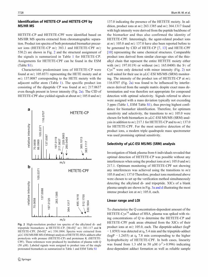
HETETE-CP and HETETE-CPF were identified based onMS/HR MS spectra extracted from chromatographic separa-tion. Product ion spectra of both protonated biomarker precur-sor ions (HETETE-CP m/z 383.1 and HETETE-CPF m/z530.2) are shown in Fig. 2 and the structural assignment ofthe signals is summarized in Table 1 for HETETE-CP.Assignments for HETETE-CPF can be found in the ESM(Table S1).
Characteristic predominant ions of HETETE-CP werefound at m/z 105.0371 representing the HETE moiety and atm/z 137.0087 corresponding to the HETE moiety with theadjacent sulfur atom (Table 1). The specific product ionconsisting of the dipeptide CP was found at m/z 217.0637even though present in lower intensity (Fig. 2a). The CID ofHETETE-CPF also yielded signals at aboutm/z 105.0 andm/z
137.0 indicating the presence of the HETETE moiety. In ad-dition, product ions at m/z 263.1385 and m/z 364.1317 foundwith high intensity were derived from the peptide backbone ofthe biomarker and thus also confirmed the identity ofHETETE-CPF. Interestingly, the agent-related product ionsat m/z 105.0 and m/z 137.0 have also been reported before tobe generated by CID of HETE-CP [7, 13] and HETE-CPF[10] representing the same chemical structures. Comparableproduct ions derived from similar cleavage sites of the thio-alkyl chain that represent the entire HETETE moiety eitherwith (m/z 197.0119) or without (m/z 165.0400) the Sγ ofCys34 were only detected with minor intensity (Fig. 2) notwell suited for their use in μLC-ESI MS/MS (SRM) monitor-ing. The intensity of the product ion of HETETE-CP at m/z116.0707 (Fig. 2a) was found to be influenced by interfer-ences derived from the sample matrix despite exact mass de-termination and was therefore not appropriate for compounddetection with optimal selectivity. Signals referred to abovewere assigned with a mass deviation typically not exceeding3 ppm (Table 1, ESM Table S1), thus proving highest confi-dence for biomarker identification. Therefore, for optimumsensitivity and selectivity, the transitions to m/z 105.0 werechosen for both biomarkers in μLC-ESI MS/MS (SRM) anal-ysis in addition tom/z 217.1 for HETETE-CP and tom/z 137.0for HETETE-CPF. For the most sensitive detection of theproduct ions, a modern triple quadrupole mass spectrometerwas used promising optimal sensitivity.
Selectivity of μLC-ESI MS/MS (SRM) analysis
Investigation of blank plasma from 6 individuals revealed thatoptimal detection of HETETE-CP was possible without anyinterferences when using the product ions atm/z 105.0 andm/z217.1. Optimum monitoring of HETETE-CPF not showingany interferences was achieved using the transitions to m/z105.0 andm/z 137.0 Therefore, product ions mentioned abovewere chosen to set up the verification method simultaneouslydetecting the alkylated di- and tripeptide. XICs of a blankplasma sample are shown in Fig. 3a and d illustrating the mostintense product ion at m/z 105.0, each.
Linear range and LOI
To characterize the Q concentration-dependent amount of theHETETE-Cys34-adduct of HSA, plasma was spiked with ris-ing concentrations of Q to determine the HETETE-CP andHETETE-CPF peak areas obtained from the XICs of theirproduct ions at m/z 105.0, each. The dipeptide-adduct (logP− 1.8593) was detected at tR 5.4 min and the tripeptide-adduct(logP − 1.2455) at tR 7.8 min corresponding to the higherhydrophobicity of HETETE-CPF. In both cases, linearitywas found from 1.3 nM to 50 μM (r2 > 0.996) indicatingdose-dependent adduct formation as well as reliable sample
a
b
Fig. 2 High-resolution product ion spectra of the alkylated di- andtripeptide biomarkers: a HETETE-CP, [M+H]+ m/z 383.117 and bHETETE-CPF, [M+H]+ m/z 530.1804. Spectra were extracted fromμLC-ESIMS/HRMS (Orbitrap) analysis of HETETE-HSA-adducts afterproteolysis with pronase (HETETE-CP) and proteinase K (HETETE-CPF). These references were produced by incubation of plasma with Q(50 μM). Labeled signals were assigned to product ions of the singleprotonated biomarkers as summarized in Table 1 and ESM Table S1
7728 Blum M.-M. et al.
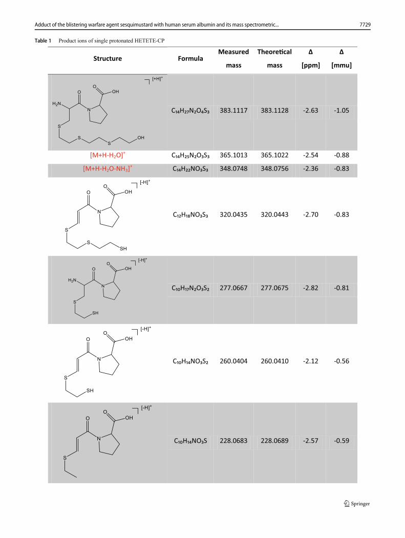
Table 1 Product ions of single protonated HETETE-CP
Structure FormulaMeasured
mass
Theore�cal
mass
Δ
[ppm]
Δ
[mmu]
C₁₄H₂₇N₂O₄S₃ 383.1117 383.1128 -2.63 -1.05
[M+H-H2O]+ C₁₄H₂₅N₂O₃S₃ 365.1013 365.1022 -2.54 -0.88
[M+H-H2O-NH3]+ C₁₄H₂₂NO₃S₃ 348.0748 348.0756 -2.36 -0.83
C₁₂H₁₈NO₃S₃ 320.0435 320.0443 -2.70 -0.83
C₁₀H₁₇N₂O₃S₂ 277.0667 277.0675 -2.82 -0.81
C₁₀H₁₄NO₃S₂ 260.0404 260.0410 -2.12 -0.56
C₁₀H₁₄NO₃S 228.0683 228.0689 -2.57 -0.59
7729Adduct of the blistering warfare agent sesquimustard with human serum albumin and its mass spectrometric...
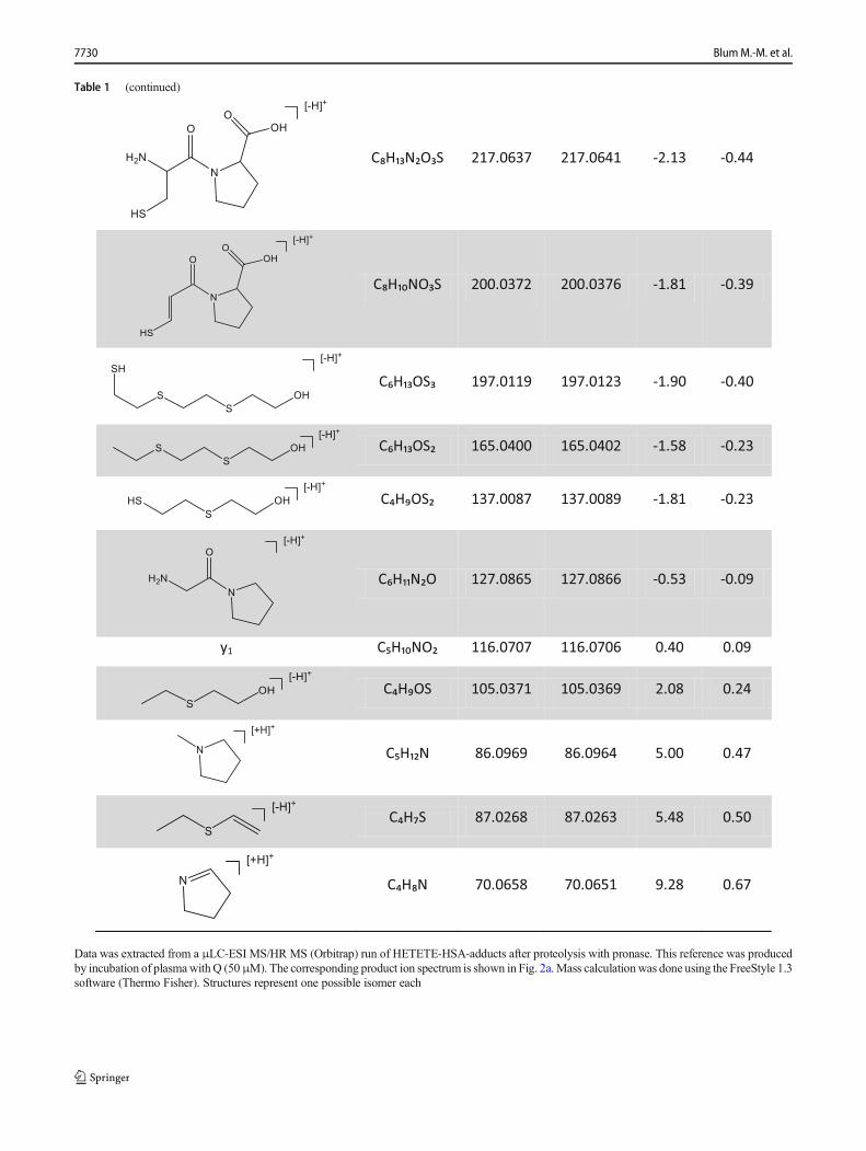
Table 1 (continued)
C₈H₁₃N₂O₃S 217.0637 217.0641 -2.13 -0.44
C₈H₁₀NO₃S 200.0372 200.0376 -1.81 -0.39
C₆H₁₃OS₃ 197.0119 197.0123 -1.90 -0.40
C₆H₁₃OS₂ 165.0400 165.0402 -1.58 -0.23
C₄H₉OS₂ 137.0087 137.0089 -1.81 -0.23
C₆H₁₁N₂O 127.0865 127.0866 -0.53 -0.09
y1 C₅H₁₀NO₂ 116.0707 116.0706 0.40 0.09
C₄H₉OS 105.0371 105.0369 2.08 0.24
C₅H₁₂N 86.0969 86.0964 5.00 0.47
C₄H₇S 87.0268 87.0263 5.48 0.50
C₄H₈N 70.0658 70.0651 9.28 0.67
Data was extracted from a μLC-ESI MS/HR MS (Orbitrap) run of HETETE-HSA-adducts after proteolysis with pronase. This reference was producedby incubation of plasmawithQ (50μM). The corresponding product ion spectrum is shown in Fig. 2a.Mass calculationwas done using the FreeStyle 1.3software (Thermo Fisher). Structures represent one possible isomer each
7730 Blum M.-M. et al.
preparation procedures. The ion ratios of the diverse productions (given in percentage) were calculated from the peak area(A) ratio of the relevant XICs, e.g., 100 × A(m/z 137.0)/A(m/z105.0) abbreviated as (137/105). The following ion ratioswere found for these transitions: HETETE-CP (217/105)50.1% and HETETE-CPF (137/105) 53.3%. According tothe OPCW quality criteria, ion ratios from biomarkers ofany sample had to fit those of a reference for unambiguousidentification. The LOI was defined as the lowest concentra-tion of Q still fulfilling this criterion. Accordingly, the LOI forHETETE-CP detection was found at 32 nM Q in plasma,whereas 6.4 nM was found for HETETE-CPF. XICs (m/z105.0) corresponding to these LOI concentrations are illustrat-ed in Fig. 3b and e. These LOI are in the same range as thoseof the corresponding HSA-adducts of SMHETE-CP (156 nMand 19.5 nM [13]) and HETE-CPF (10 nM [10]). However,such limits strictly depend on the method-specific samplepreparation procedures with respect to concentrating and di-luting working steps as well as to instruments used in terms ofinjected sample amount, ion yield (sensitivity), and mass res-olution (selectivity). Therefore, the mentioned LOI were quitesimilar for all vesicant biomarkers.
Stability in the autosampler
HETETE-CP as well as HETETE-CPF were found to beof sufficient stability in the prepared samples whenstored at 15 °C in the autosampler (results not shown).Only a slight peak area decrease of less than 7% forboth biomarkers indicated a negligible concentration de-crease within the 24-h test period thus documenting
good stability beneficial especially when large sets ofsamples have to be analyzed.
Freeze and thaw cycles
No degradation of HETETE-HSA in frozen and thawed plas-ma was determined by monitoring the HETETE-CP biomark-er (data not shown), thus documenting that plasma samplescan be handled following common rules of plasma sampletreatment without the need for special care.
Co-incubation to characterize the relative reactivity ofSM and Q for Cys34 alkylation
The co-incubation experiment was carried out to investigatethe relative reactivity of SM and Q toward alkylation of Cys34
in HSA. Q and SM competed for the thiol groups of a limitedamount of Cys34 applied as neat HSA instead of plasma tominimize any side reactions with other plasma proteins. Asnumerous amino acids of HSA are expected to be prone toalkylation as exemplarily shown for Glu230 [12] and Met329
[9], a low HSA concentration was applied. After incubation,samples were subjected to pronase-mediated proteolysis tomonitor HETE-CP and HETETE-CP simultaneously as mea-sures of their respective HSA-adducts. For comparison, themaximum peak areas of both biomarkers resulting from singleincubation (100 μM each) were determined. These valuesserved as a maximum reference (100%) to determine any ef-fective competition that would result in reduced areas.Accordingly, under the assumption of the same alkylatingreactivity of SM and Q toward Cys34, a decrease to 50% of
7.8
HETETE-CPF
Inte
nsity [cps]
d e
Time [min] Time [min] 1515 00
1.1 e68.0 e3 6.0 e3
f
Time [min] 150
7.8
5.4
1.6 e5
cblank LOI
5.4
1.4 e4 1.1 e4
HETETE-CP
Inte
nsity [cps]
a breference
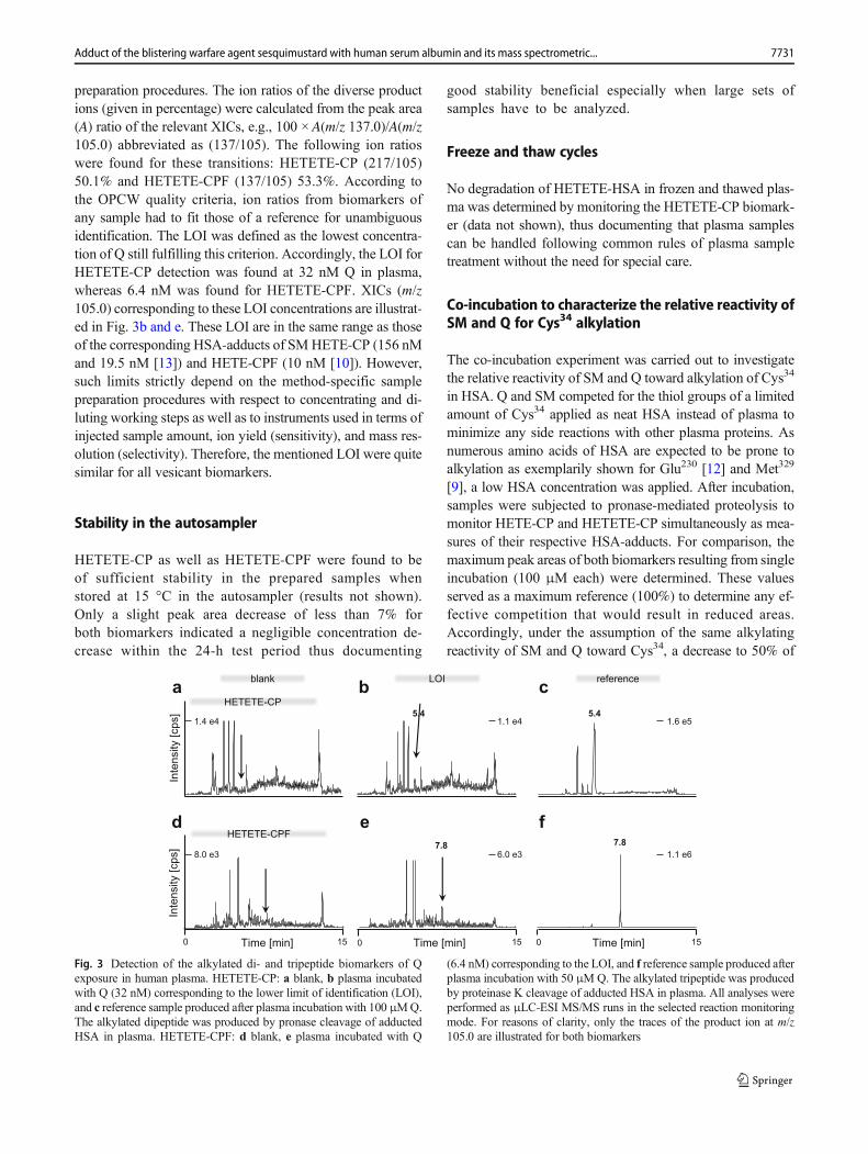
Fig. 3 Detection of the alkylated di- and tripeptide biomarkers of Qexposure in human plasma. HETETE-CP: a blank, b plasma incubatedwith Q (32 nM) corresponding to the lower limit of identification (LOI),and c reference sample produced after plasma incubation with 100 μMQ.The alkylated dipeptide was produced by pronase cleavage of adductedHSA in plasma. HETETE-CPF: d blank, e plasma incubated with Q
(6.4 nM) corresponding to the LOI, and f reference sample produced afterplasma incubation with 50 μMQ. The alkylated tripeptide was producedby proteinase K cleavage of adducted HSA in plasma. All analyses wereperformed as μLC-ESI MS/MS runs in the selected reaction monitoringmode. For reasons of clarity, only the traces of the product ion at m/z105.0 are illustrated for both biomarkers
7731Adduct of the blistering warfare agent sesquimustard with human serum albumin and its mass spectrometric...

the reference level would be expected for both biomarkerswhen incubating with a 1:1-M ratio.
When using 19.6 μMHSA, individual peak areas resultingfrom co-incubation with 100 μM SM and 100 μM Q wereidentical to those from separate incubation, thus documentingno competition. This was most likely due to a large extent ofadditional reactions including simple hydrolysis as well asamino acid alkylations indicating the high reactivity of bothvesicants. Therefore, we repeated the incubations using aHSA concentration of 1.96 μM. This is equal to the maximumconcentration of free Cys34 and was chosen to ensure an ef-fective molar excess of the vesicants when applied at 100 μMconcentrations. Results are shown in Table 2.
Resulting reference values of peak areas from triplicatemeasurements of HETE-CP and HETETE-CP, after individ-ual incubations with 100 μMof SM and Q, respectively, wereidentical (about 4.85 × 10⁵ cts ± 6%, each) thus supporting theidea of an identical molar yield (identical extent of Cys34
alkylation). After co-incubation (100 μM of SM and100 μM of Q), a reasonably higher yield of HETETE-CP(79 ± 3% of the reference) was found, whereas only 59 ± 3%of the reference was detected for HETE-CP, thus indicating ahigher reactivity of Q. The same effect was observed after co-incubation with 100 μMSM and 10 μMQ yielding only 79 ±1% of the HETE-CP and 12 ± 4% of the HETETE-CP refer-ence. This relative concentration of both vesicants (10:1) waschosen to simulate ratios known from mustard ammunitions[17]. The higher total yield of alkylated CP (sum of HETE-CPand HETETE-CP > 100% when co-incubating with 100 μMeach) was presumably due to the higher total vesicant concen-tration (200 μM instead of 100 μM in single incubation) in-creasing the product concentration according to the law ofmass action.
In summary, Q appears somewhat more reactive than SM(factor 1.33) for Cys34 alkylation. This might presumably bedue to, e.g., a higher affinity of Q to amino acids close toCys34 resulting in a favorable binding complex or a moreprecise orientation of the reactive sulfonium ion in a “near-attack” conformation, a higher intrinsic reactivity of the Qsulfonium ion compared to the one of SM, or a smaller extent
of side reactions. The important conclusion of our data, how-ever, supports the observation that HETETE-HSA will beproduced even though it might be present in much smallerconcentrations than SM. This is of high relevance for thepossibility of biomarker detection when Q is only found asan impurity or side-product in a mustard mixture.
Analysis of apo HSA and HETETE-Cys34 variants usingMD simulations
As the most abundant protein in human plasma and its impor-tance as a drug carrier and binding partner for many endogenousmolecules [42], HSA has been subject to intensive studies usingMD simulations [43, 44]. This is also true for the investigation ofthe role of Cys34 that represents the largest pool of free thiolgroups in human plasma and shows an unusually low pKa of8.2 [45–47]. It was shown that relatively long simulation times(around 1 μs) are required for adequate structural sampling, butas we are mainly interested in the changes in the local proteinenvironment around Cys34, simulation times of 200 ns for apoHSA and 100 ns for the HETETE-adduct were considered ap-propriate. Previous MD studies have shown that Cys34 is onlypartially solvent accessible and forms a strong hydrogen bondwith Tyr84 [46]. In the 200-ns simulation of apo HSA, in whichthe backbone root mean square deviation (RMSD) value stabi-lizes after around 80 ns, we observed the same phenomenon.Nevertheless, it was also possible to observe and extract varioussimulation frames in which the Sγ of Cys34 is solvent accessible.We used one such simulation frame to construct the HETETE-adduct in two variants: one with the HETETE moiety pointinginto the solvent and one with the HETETEmoiety fitting into thegroove formed by the two helices adjacent to Cys34. The simu-lation of the “in groove” variant showed a stable backboneRMSD value after about 35 ns and the “in solvent” variant afteraround 60 ns out of a total of 100 ns simulation time, each. HSAconsists of three structurally similar domains (I, II, and III) andeach of them is formed by two subdomains (A andB) [48]. Cys34
is located in subdomain IA (residues 5–107) (Fig. 4). Other HSAamino acid residues potentially subject to alkylation by SM and
Table 2 Co-incubation to characterize the relative reactivity of SM and Q for Cys34 alkylation
Agent incubated and concentration Relative peak area of HETE-CP [%] Relative peak area of HETETE-CP [%] Total of relative peak areas [%]
SM, 100 μM 100 0 100
Q, 100 μM 0 100 100
SM, 100 μM + Q, 100 μM 59 ± 3 79 ± 3 138 ± 6
SM, 100 μM + Q, 10 μM 79 ± 0 12 ± 4 91 ± 4
Relative peak areas were calculated from extracted ion chromatograms of the product ion at m/z 105.0. Incubations were carried out in triplicate each
HETE-CP HSA-derived biomarker obtained after pronase-catalyzed proteolysis of the adduct of HSA and SM, HETETE-CP HSA-derived biomarkerobtained after pronase-catalyzed proteolysis of the adduct of HSA and Q, SM sulfur mustard, Q sesquimustard
7732 Blum M.-M. et al.

Q (E230, M329) are all solvent exposed without the sterical con-straints of Cys34.
Visual inspection of the two MD trajectories of theHETETE-adduct and comparison with the apo protein didnot yield significant differences in the structure of subdomainIA. The two different “in solvent” and “in groove” startingstructures did not result in markedly different trajectories. Inall simulations, subdomain IA remained rather rigid withsmall root mean square fluctuation (RMSF) values per residuecompared to other parts of the protein (see ESM Figs. S1–S3).
We observed that the HETETE moiety of the adductedCys34 showed high flexibility and conformational mobilityas expected due to the large number of rotatable bonds. Thisflexibility was, however, steered and restricted by the twohelices adjacent to Cys34. The HETETE moiety starting atthe Sγ of Cys34 moved toward the protein surface while show-ing an important hydrophobic interaction with the side chainof Leu42, that is in almost constant van der Waals contact(average distance 3.8 Å) with the HETETE moiety. The polarend of the HETETE moiety with the terminal hydroxyl groupwas found to regularly explore the solvent, but for most of thesimulation time, it acted as either a hydrogen donor or accep-tor interacting with nearby amino acid residues (as H donor:Asp38, Lys41, Glu45, Lys73, Thr76, Val77, Leu80; as H acceptor:Lys41, Lys73, Leu80, Arg81). By far, the most important ofthese hydrogen bonding partners are the Glu45 carboxyl groupand the Lys41 amino group. The HETETE hydroxyl groupforms a hydrogen bond most often with Glu45. The Lys41
residue is the secondmost preferred hydrogen bonding partnerapart from the solvent.
This observation is also reinforced by cluster analysisemploying the method of Daura [49] on those parts ofthe MD trajectories showing stable backbone RMSDvalues to reduce the dimensionality of the trajectoriescontaining 10,000 frames each (Fig. 5). The most pop-ulated cluster (56% of frames, Fig. 5a cluster 1) showshydrogen bonding with Glu45 and hydrophobic interac-tions with Lys41 and Leu42. Despite the polar aminogroup, the four methylene groups in the Lys41 sidechain can contribute significantly to hydrophobic inter-actions as well [50]. The second most populated cluster(27% of frames, Fig. 5b cluster 2) shows hydrogenbonding with the terminal amino group of Lys45, whilethe third most populated cluster (5% of frames, Fig. 5ccluster 3) shows again hydrogen bonding with Glu45
and hydrophobic “guidance” by Lys41 and Leu42. Thisis in agreement with the hydrogen bonding analysis de-scribed above.
Cluster 1
Cluster 2
Cluster 3
Lys41
HETETE-Cys34
Glu45
Leu42
Lys41
Glu45
Leu42
HETETE-Cys34
HETETE-Cys34
Glu45
Leu42
Lys41
a
b
c
Fig. 5 Structural environment around HETETE-Cys34 in the three mostpopulated clusters (a–c) after cluster analysis. Important hydrogenbonding partners (Lys41 and Glu45) as well as important hydrophobicinteraction partners (Leu42 and Lys41) are highlighted
Cys34
IA [Res 5-107]
Fig. 4 Protein structure of apo HSA (PDB: 1AO6) subdomain IA ishighlighted in light blue comprising the Cys34 residue that is prone toalkylation by SM and Q resulting in HETE-Cys34- and HETETE-Cys34-adducts, respectively
7733Adduct of the blistering warfare agent sesquimustard with human serum albumin and its mass spectrometric...
In conclusion, the MD simulations show that adduction ofCys34 by Q resulting in HETETE-Cys34 does not disturb theoverall structural rigidity of subdomain IA of HSA, and thatwithin certain steric constraints, the protein can accommodatethe quite big HETETE moiety well. HETETE–protein inter-actions are dominated by hydrogen bonding to Lys41 andGlu45 and hydrophobic interactions with Lys41 and Leu42.
The role of six-membered ring sulfonium ions as re-active species with HSA
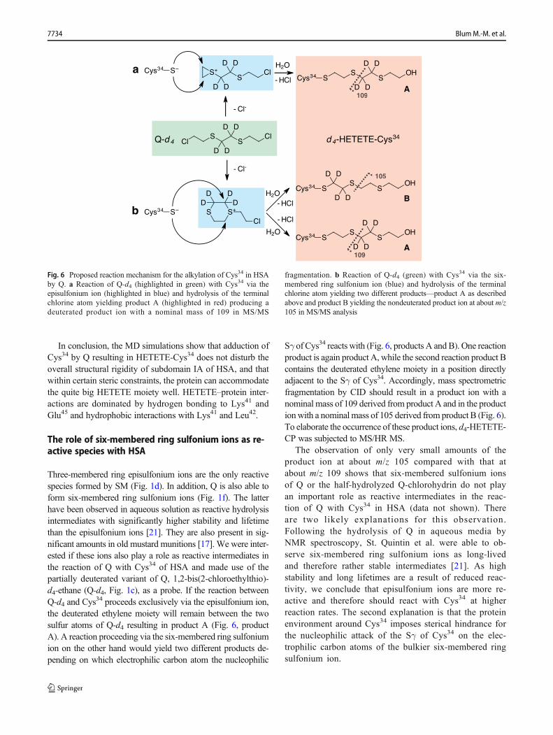
Three-membered ring episulfonium ions are the only reactivespecies formed by SM (Fig. 1d). In addition, Q is also able toform six-membered ring sulfonium ions (Fig. 1f). The latterhave been observed in aqueous solution as reactive hydrolysisintermediates with significantly higher stability and lifetimethan the episulfonium ions [21]. They are also present in sig-nificant amounts in old mustard munitions [17]. We were inter-ested if these ions also play a role as reactive intermediates inthe reaction of Q with Cys34 of HSA and made use of thepartially deuterated variant of Q, 1,2-bis(2-chloroethylthio)-d4-ethane (Q-d4, Fig. 1c), as a probe. If the reaction betweenQ-d4 and Cys
34 proceeds exclusively via the episulfonium ion,the deuterated ethylene moiety will remain between the twosulfur atoms of Q-d4 resulting in product A (Fig. 6, productA). A reaction proceeding via the six-membered ring sulfoniumion on the other hand would yield two different products de-pending on which electrophilic carbon atom the nucleophilic
Sγ of Cys34 reacts with (Fig. 6, products A and B). One reactionproduct is again product A, while the second reaction product Bcontains the deuterated ethylene moiety in a position directlyadjacent to the Sγ of Cys34. Accordingly, mass spectrometricfragmentation by CID should result in a product ion with anominal mass of 109 derived from product A and in the productionwith a nominal mass of 105 derived from product B (Fig. 6).To elaborate the occurrence of these product ions, d4-HETETE-CP was subjected to MS/HR MS.
The observation of only very small amounts of theproduct ion at about m/z 105 compared with that atabout m/z 109 shows that six-membered sulfonium ionsof Q or the half-hydrolyzed Q-chlorohydrin do not playan important role as reactive intermediates in the reac-tion of Q with Cys34 in HSA (data not shown). Thereare two likely explanations for this observation.Following the hydrolysis of Q in aqueous media byNMR spectroscopy, St. Quintin et al. were able to ob-serve six-membered ring sulfonium ions as long-livedand therefore rather stable intermediates [21]. As highstability and long lifetimes are a result of reduced reac-tivity, we conclude that episulfonium ions are more re-active and therefore should react with Cys34 at higherreaction rates. The second explanation is that the proteinenvironment around Cys34 imposes sterical hindrance forthe nucleophilic attack of the Sγ of Cys34 on the elec-trophilic carbon atoms of the bulkier six-membered ringsulfonium ion.
109
109
105
109
109
105
a
b
A
A
B
Fig. 6 Proposed reaction mechanism for the alkylation of Cys34 in HSAby Q. a Reaction of Q-d4 (highlighted in green) with Cys34 via theepisulfonium ion (highlighted in blue) and hydrolysis of the terminalchlorine atom yielding product A (highlighted in red) producing adeuterated product ion with a nominal mass of 109 in MS/MS
fragmentation. b Reaction of Q-d4 (green) with Cys34 via the six-membered ring sulfonium ion (blue) and hydrolysis of the terminalchlorine atom yielding two different products—product A as describedabove and product B yielding the nondeuterated product ion at about m/z105 in MS/MS analysis
7734 Blum M.-M. et al.
Potential forensic application
While verification analysis of biomedical samples generallyaims to prove or disprove exposure of a victim to a certainchemical agent, there is a growing interest in the possibility toconduct source attribution of a chemical agent. Source attribu-tion means to trace the agent back to a specific synthesis meth-od, reaction batch, specific precursor chemicals, or an existingstockpile. The chemical signatures of environmental sampleshave been the subject of academic interest [51–54] but havealso been used in real cases [55]. So far, biomedical sampleshave not been considered for source attribution for several rea-sons: The intact chemical agent and the potential by-productsand impurities are normally no longer present in vivo.Biotransformation processes alter compound compositionsand impurities that might serve as signatures. In addition, suchsignatures are typically present in either extremely low concen-tration or they are biotransformed and excreted from the bodyseriously impeding their bioanalytical detection.
Sulfur mustards exhibit a complex synthesis and degrada-tion chemistry with a significant number of impurities stillbeing potent vesicants [56]. As described above, Q is an im-portant impurity of SM and its relative amounts depend on thesynthesis route, storage conditions, and age. The parallel de-tection of HETE, HETETE, and potential other adducts ofHSA could provide valuable forensic information. In the ab-sence of environmental samples, it might provide cluespointing to the type of mustard used. If a sample of the agentwas available, the comparisonmight allow to conclude wheth-er the exposure was caused by that specific batch of the agent.As adducts with endogenous proteins are regularly detected inthe low ppb range, sufficient sensitivity of the method will bechallenging, especially if the relative amounts of Q or otherblistering impurities are low. Further investigations on humanplasma samples of exposed individuals—including retrospec-tive analysis of stored samples—are required for a detailedassessment.
Acknowledgments We thank the OPCW Laboratory (Rijswijk,The Netherlands) for making 1,2-bis(2-hydroxyethylthio) ethane and1,2-bis(2-hydroxyethylthio)-d4-ethane available for this work. We thankMarina Dentzel (InstPharmToxBw) for her excellent technical assistance.
Funding Open Access funding provided by Projekt DEAL.
Compliance with ethical standards
Conflict of interest The authors declare that they have no conflict ofinterest.
Human and animal rights and informed consent Research was carriedout without human participants and animals.
Open Access This article is licensed under a Creative CommonsAttribution 4.0 International License, which permits use, sharing,
adaptation, distribution and reproduction in any medium or format, aslong as you give appropriate credit to the original author(s) and thesource, provide a link to the Creative Commons licence, and indicate ifchanges weremade. The images or other third party material in this articleare included in the article's Creative Commons licence, unless indicatedotherwise in a credit line to the material. If material is not included in thearticle's Creative Commons licence and your intended use is notpermitted by statutory regulation or exceeds the permitted use, you willneed to obtain permission directly from the copyright holder. To view acopy of this licence, visit http://creativecommons.org/licenses/by/4.0/.
References
1. Kehe K, Szinicz L. Medical aspects of sulfur mustard poisoning.Toxicology. 2005;214:198–209.
2. Johnson NH, Larsen JC, Meek EC. Historical perspective of chem-ical warfare agents. In: Gupta R, editor. Handbook of toxicology ofchemical warfare agents. 3rd ed. San Diego: Elsevier Inc./Academic; 2020. p. 17–26.
3. Quillen C. The Islamic State’s evolving chemical arsenal. StudConfl Terror. 2016;39:1019–30.
4. John H, Balszuweit F, Steinritz D, Kehe K,Worek F, Thiermann H.Toxicokinetic aspects of nerve agents and vesicants. In: Gupta R,editor. Handbook of toxicology of chemical warfare agents. 3rd ed.San Diego: Elsevier Inc./Academic; 2020. p. 875–929.
5. Blum MM, Mamidanna MRVS. Analytical chemistry and the chemi-cal weapons convention. Anal Bioanal Chem. 2014;406:5067–9.
6. Convention on the prohibition of the development, production,stockpiling and use of chemical weapons and on their destruction,current version 27 September 2005, https://www.opcw.org/sites/default/files/documents/CWC/CWC_en.pdf
7. Gandor F, Gawlik M, Thiermann H, John H. Evidence of sulfurmustard exposure in human plasma byLC-ESIMS/MS detection ofthe albumin-derived alkylated HETE-CP dipeptide and chromato-graphic investigation of its cis/trans isomerism. J Anal Toxicol.2015;39:270–9.
8. John H, Gandor F, Gawlik M, Thiermann H. LC-ESI MS/MS de-tection of the alkylated dipeptide HETE-CP to prove sulfur mustardexposure: application to plasma samples provided by theOrganisation for the Prohibition of Chemical Weapons. ToxichemKrimtech. 2015;82:211–7.
9. Siegert M, Gandor F, Kranawetvogl A, Börner H, Thiermann H,John H. Methionine329 in human serum albumin: a novel target foralkylation by sulfur mustard. Drug Test Anal. 2019;11:659–68.
10. Noort D, Hulst A, de Jong L, Benschop H. Alkylation of humanserum albumin by sulfur mustard in vitro and in vivo: mass spec-trometric analysis of a cysteine adduct as a sensitive biomarker ofexposure. Chem Res Toxicol. 1999;12:715–21.
11. Steinritz D, Striepling E, Rudolf KD, Schröder-Kraft C, Püschel K,Hullard-Pulstinger A, et al. Medical documentation, bioanalyticalevidence of an accidental human exposure to sulfur mustard andgeneral therapy recommendations. Toxicol Lett. 2016;244:112–20.
12. John H, Koller M, Worek F, Thiermann H, Siegert M. Forensicevidence of sulfur mustard exposure in real cases of human poison-ing by detection of diverse albumin-derived protein-adducts. ArchToxicol. 2019;93:1881–91.
13. John H, Siegert M, Gandor F, Gawlik M, Kranawetvogl A,Karaghioshoff K, et al. Optimized verification method for detection ofan albumin-sulfur mustard adduct at Cys34 in plasma by μLC-ESIMS/MS using a hybrid quadrupole time-of-flight tandemmass spectrometerafter direct plasma proteolysis. Toxicol Lett. 2016;244:103–11.
7735Adduct of the blistering warfare agent sesquimustard with human serum albumin and its mass spectrometric...
14. John H, Willoh S, Hörmann P, Siegert M, Vondran A, ThiermannH. Procedures for analysis of dried plasma using microsamplingdevices to detect sulfur mustard albumin adducts for verificationof poisoning. Anal Chem. 2016;88:8787–94.
15. Gasson J, McCombie H, Williams AH, Woodward FN. New or-ganic sulphur vesicants. Part IV. 1:2-Di-(2-chloro-ethylthio) ethaneand its analogues. J Chem Soc. 1948:44–6.
16. Timperley CM, Black RM, Bird M, Holden I, Mundy JL, ReadRW. Hydrolysis and oxidation products of the chemical warfareagents 1,2-bis[(2-chloroethyl)thio] ethane Q and 2,2′-bis(2-chloroethylthio) diethyl ether T. Phosphorus Sulfur Silicon RelatElem. 2003;178:2027–46.
17. National Research Council. Interim design assessment for the bluegrass chemical agent destruction pilot plant. Washington, DC: TheNational Academies Press; 2005.
18. Bennett GM,Whincop EM. Some derivatives of monothioethyleneglycol. J Chem Soc. 1921;119:1860–4.
19. Vocci FJ, Ballard TA, Yevich P, Punte CL. Inhalation toxicitystudies with aerosols of sesqui-mustard. Toxicol Appl Pharmacol.1963;5:677–84.
20. Sassenrath EN, Morrison DC, Larson AM, Greenberg DM.Evaluation of the carcinostatic activity of 1,2-bis(ß-chloroethylthio)ethane. Cancer Res. 1955;15:755–9.
21. St. Quintin TD, Leslie DR, Collins JG. Hydrolysis ofsesquimustards. Aust J Chem. 2003;56:309–13.
22. D’Agostino PA, Provost PA. Capillary column isobutane chemicalionization mass spectrometry of mustard and related compounds.Biomed Environ Mass Spectrom. 1988;15:553–64.
23. D ’Agost ino PA, Provost PA. Capi l lary column gaschromatography-ammonia and deuterated ammonia chemical ion-ization mass spectrometry of sulfur vesicants. J Chromatogr A.1992;600:267–72.
24. D’Agostino PA, Provost PA. Capillary column electron impact andammonia chemical ionization gas chromatographic-mass spectrometricand gas chromatographic-tandem mass spectrometric analysis of mus-tard hydrolysis products. J Chromatogr A. 1993;645:283–92.
25. D’Agostino PA, Provost PA, Hancock JR. Analysis of mustard hydro-lysis products by packed capillary liquid chromatography–electrospraymass spectrometry. J Chromatogr A. 1998;808:177–84.
26. Lemire SW, Ash DH, Johnson RC, Barr JR. Mass spectral behaviorof the hydrolysis products of sesqui- and oxy-mustard type chem-ical warfare agents in atmospheric pressure chemical ionization. JAm Soc Mass Spectrom. 2007;18:1364–74.
27. Ash DH, Lemire SW, McGrath SC, McWilliams LG, Barr JR.Multianalyte quantification of five sesqui- and ethyl ether oxy-mustard metabolites in human urine by liquid chromatography-atmospheric pressure chemical ionization-tandem mass spectrome-try. J Anal Toxicol. 2008;32:44–50.
28. Kaushik MP, Rana H. Faci le one-s tep synthesis ofdithiaalkanediols. Org Prep Proced Int. 2005;37:267–72.
29. Abraham MJ, Murtola T, Schulz R, Páll S, Smith JC, Hess B, et al.GROMACS: high performance molecular simulations throughmulti-level parallelism from laptops to supercomputers.SoftwareX. 2015;1–2:19–25.
30. Schmid N, Eichenberger AP, Choutko A, Riniker S, Winger M,Mark AE, et al. Definition and testing of the GROMOS force-field versions 54A7 and 54B7. Eur Biophys J. 2011;40:843–56.
31. Stroet M, Caron B, Visscher KM, Geerke DP, Malde AK, MarkAE. Automated topology builder version 3.0: prediction of solva-tion free enthalpies in water and hexane. J Chem Theory Comput.2018;14:5834–45.
32. Pettersen EF, Goddard TD, Huang CC, Couch GS, Greenblatt DM,Meng EC, et al. UCSF Chimera-a visualization system for explor-atory research and analysis. J Comput Chem. 2004;25:1605–12.
33. Berendsen HJC, Postma JPM, van Gunsteren WF, Hermans J.Interaction models for water in relation to protein hydration. In:
Pullman B, editor. Intermolecular forces. Dordrecht: Reidel;1981. p. 331–42.
34. Hess B. P-LINCS: a parallel linear constraint solver for molecularsimulation. J Chem Theory Comput. 2008;4:116–22.
35. Pall S, Hess B. A flexible algorithm for calculating pair interactions onSIMD architectures. Comput Phys Commun. 2013;184:2641–50.
36. Darden T, York D, Pedersen L. Particle mesh Ewald: an N·log(N)method for Ewald sums in large systems. J Chem Phys. 1993;98:10089–92.
37. Essmann U, Perera L, Berkowitz ML, Darden T, Lee H, PedersenLGA. A smooth particle mesh Ewald method. J Chem Phys.1995;103:8577–93.
38. Bussi G, Donadio D, Perinello M. Canonical sampling throughvelocity rescaling. J Chem Phys. 2007;126:014101.
39. ParrinelloM, RahmanA. Polymorphic transitions in single crystals:a newmolecular dynamics method. J Appl Phys. 1981;52:7182–90.
40. Nosé S, Klein ML. Constant pressure molecular dynamics for mo-lecular systems. Mol Phys. 1983;50:1055–76.
41. Work instruction for the reporting of the results of the OPCW bio-medical proficiency tests. QDOC/LAB/WI/BioPT04. The Hague:The Organisation for the Prohibition of Chemical Weapons(OPCW); 2020.
42. Fasano M, Curry S, Terreno E, Galliano M, Fanali G, Narciso P,et al. The extraordinary ligand binding properties of human serumalbumin. IUBMD Life. 2005;57:787–96.
43. Lambrinidis G, Vallianatou T, Tsantili-Kakoulidou A. In vitro, insilico and integrated strategies for the estimation of plasma proteinbinding. A review. Adv Drug Deliv Rev. 2015;86:27–45.
44. Paris G, Ramseyer C, EnescuM. A principal component analysis ofthe dynamics of subdomains and binding sites in human serumalbumin. Biopolymers. 2014;101:561–72.
45. Fabisiak JP, Sedlov A, Kagan VE. Quantification of oxidative/nitrosative modification of Cys34 in human serum albumin usinga fluorescence-based SDS–PAGE assay. Antioxid Redox Signal.2002;4:855–65.
46. Castellanos MM, Colina CM. Molecular dynamics simulations ofhuman serum albumin and role of disulfide bonds. Phys Chem B.2013;117:11895–905.
47. Bonanata J, Turell L, Antmann L, Ferrer-Sueta G, Botasini S,Méndez E, et al. The thiol of human serum albumin: acidity, mi-croenvironment and mechanistic insights on its oxidation tosulfenic acid. Free Radic Biol Med. 2017;108:952–62.
48. He XM, Carter DC. Atomic structure and chemistry of humanserum albumin. Nature. 1992;358:209–15.
49. Daura X, Gademann K, Jaun B, Seebach D, van Gunsteren WF,Mark AE. Peptide folding: when simulation meets experiment.Angew Chem Int Ed. 1999;38:236–40.
50. Dyson HJ, Wright PE, Scheraga HA. The role of hydrophobicinteractions in initiation and propagation of protein folding. ProcNatl Acad Sci U S A. 2006;103:13057–61.
51. Ovenden SPB, Webster RL, Micich E, McDowall LJ, McGill NW,Williams J, et al. The identification of chemical attribution signa-tures of stored VX nerve agents using NMR, GC-MS, and LC-HRMS. Talanta. 2020;211:120753.
52. Fraga CG, Pérez Acosta GA, Crenshaw MD, Wallace K, MongGM, Colburn HA. Impurity profiling to match a nerve agent to itsprecursor source for chemical forensics applications. Anal Chem.2011;83:9564–72.
53. Holmgren KH, Valdez CA, Magnusson R, Vu AK, Lindberg S,Williams AM, et al. Part 1: tracing Russian VX to its syntheticroutes by multivariate statistics of chemical attribution signatures.Talanta. 2018;186:586–96.
54. Fraga CG, Bronk K, Dockendorff BP, Heredia-Langner A. Organicchemical attribution signatures for the sourcing of a mustard agentand its starting materials. Anal Chem. 2016;88:5406–13.
7736 Blum M.-M. et al.
55. United Nations Security Council. Document S/2017/904, seventhreport of the Organisation for the Prohibition of ChemicalWeapons-United Nations joint investigative mechanism. 2017.https://undocs.org/S/2017/904. Accessed 5 June 2020.
56. Rosenblatt DH, Small MJ, Kimmell TA, Anderson AW.Background chemistry for chemical warfare agents and decontam-ination processes in support of delisting waste streams at the U.S.Army Dugway Proving Ground, Utah. ANL/EAD/TM-56.Argonne National Laboratory; 1996.
Publisher’s note Springer Nature remains neutral with regard to jurisdic-tional claims in published maps and institutional affiliations.
Marc-Michael Blum is an inde-pendent consultant for analyticaland laboratory solutions inCBRN defense and previouslyworked as the head of theLaboratory of the Organisationfor the Prohibition of ChemicalWeapons (OPCW), Rijswijk,The Netherlands. He studiedchemistry at the TechnicalUniversity of Braunschweig,Germany, and obtained his Ph.D.in b iochemis t ry f rom theUn i v e r s i t y o f F r a n k f u r t ,Germany. He also holds an M.A.
in War Studies from King’s College, London, UK. His research interestsinclude analytical methods for the detection of chemical threat agents,chemical signatures for potential source attribution, biomarkers for proofof exposure, and molecular foundations of toxic action.
Annika Richter studied chemistryat the Humboldt-Universität zuBerlin, Germany, and is currentlyfinishing her master’s thesis at theBund e sweh r I n s t i t u t e o fPharmacology and Toxicology inthe working group of Prof. Dr.John with a focus on albumin ad-ducts of sulfurmustards. Soon shewill start her Ph.D. studies at theLeibnitz Institute for MolecularPharmacology in Berlin Buch.
Markus Siegert studied chemis-try at the Ludwig-Maximilians-University, Munich, Germany,and finished his master’s thesisin 2015 in collaboration with theBund e sweh r I n s t i t u t e o fPharmacology and Toxicology inthe working group of Prof. Dr.John. Currently, he is finishinghis Ph.D. studies in the same lab-oratory as a student of theHumboldt-Universität zu Berlin,Germany. Both his master’s thesisand doctorate focus on novelbioanalytical methods for the de-
tection of protein adducts with the chemical warfare agent sulfur mustard.
Horst Thiermann studied medi-c i ne a t t he Un ive r s i t y o fRegensburg, Germany, and theTechnical University of Munich,Germany. He is the director, colo-nel (MC), of the BundeswehrInstitute of Pharmacology andToxicology, Munich, Germany,and Adjunct Professor at theTechnical University of Munich.He specializes in pharmacologyand toxicology as well as in clin-ical pharmacology. His main sub-ject is research in medical defenseagainst chemical weapons. He is
President of the European Association of Poisons Centres and ClinicalToxicologists (EAPCCT) and Vice-Chairman of the German FederalInstitute for Risk Assessment’s (BfR) Committee for the Assessment ofIntoxications.
Harald John is Head of laborato-ry at the Bundeswehr Institute ofPharmacology and Toxicology,Munich, Germany. He studiedchemistry and finished his Ph.D.thesis in analytical chemistry inMünster, Germany. He also stud-ied toxicology at the Faculty ofMedicine at the University ofLeipzig, Germany, and completedhis degree as a scientific specialistfor toxicology. In 2016, he wasappointed as Adjunct Professorfor analytical chemistry at theHumboldt-Universität zu Berlin,
Germany. His work in Munich is focused on the development of forensicmass spectrometry-based methods for post-exposure analysis of biologi-cal samples to verify poisoning with chemical warfare agents and relatedtoxicants with a special focus on protein adducts.
7737Adduct of the blistering warfare agent sesquimustard with human serum albumin and its mass spectrometric...