Resveratrol chemosensitizes breast cancer cells to melphalan by cell cycle arrest
11
Resveratrol Chemosensitizes Breast Cancer Cells to Melphalan by Cell Cycle Arrest Fabiana Casanova, 1 Julia Quarti, 2 Danielly Cristiny Ferraz da Costa, 1 Caroline Arau ´ jo Ramos, 2 Jerson Lima da Silva, 1 and Eliane Fialho 2 * 1 Universidade Federal do Rio de Janeiro, Instituto de Bioquı ´mica Me ´dica, Rio de Janeiro, RJ 21941-590, Brazil 2 Universidade Federal do Rio de Janeiro, Instituto de Nutric ¸a ˜o Josue ´ de Castro, Departamento de Nutric ¸a ˜o Ba ´sica e Experimental, Rio de Janeiro, RJ 21941-590, Brazil ABSTRACT Melphalan (MEL) is a chemotherapeutic agent used in breast cancer therapy; however, MEL’s side effects limit its clinical applications. In the last 20 years, resveratrol (RSV), a polyphenol found in grape skins, has been proposed to reduce the risk of cancer development. The aim of this study was to investigate whether RSV would be able to enhance the antitumor effects of MEL in MCF-7 and MDA-MB-231 cells. RSV potentiated the cytotoxic effects of MEL in human breast cancer cells. This finding was related to the ability of RSV to sensitize MCF-7 cells to MEL-induced apoptosis. The sensitization by RSV involved the enhancement of p53 levels, the decrease of procaspase 8 and the activation of caspases 7 and 9. Another proposed mechanism for the chemosensitization effect of MCF-7 cells to MEL by RSV was the cell cycle arrest in the S phase. The treatment with RSV or MEL increased the levels of p-Chk2. The increase became pronounced in the combined treatments of the compounds. The expression of cyclin A was decreased by treatment with RSV and by the combination of RSV with MEL. While the levels of cyclin dependent kinase 2 (CDK2) remained unchanged by treatments, its active form (Thr 160 -phosphorylated CDK2) was decreased by treatment with RSV and by the combination of RSV with MEL. The activity of CDK7, kinase that phosphorylates CDK2 at Thr 160 , was inhibited by RSV and by the combination of RSV with MEL. These results indicate that RSV could be used as an adjuvant agent during breast cancer therapy with MEL. J. Cell. Biochem. 113: 2586–2596, 2012. ß 2012 Wiley Periodicals, Inc. KEY WORDS: APOPTOSIS; CANCER; CELL CYCLE; MELPHALAN; RESVERATROL C ancer is a growing health problem around the world particularly with the steady rise in life expectancy, increasing urbanization, and the subsequent changes in environmental conditions including lifestyle. According to a recent report [Jemal et al., 2011], cancer accounted for 7.6 million deaths (around 13% of all deaths) in 2008, and deaths from cancer worldwide are projected to continue to rise to over 11 million in 2030. The therapies available to date for cancer treatment are surgery, radiotherapy, and chemotherapy. Chemotherapy is often used as the main regimen in the treatment of most cancers. However, the development of tumor resistance to chemotherapy (chemoresis- tance) presents a major hurdle in cancer therapy [Higgins, 2007]. The use of cancer chemopreventive phytochemicals in combination with chemotherapeutic agents has been shown to be a pragmatic approach to overcome chemoresistance and sensitize cancer cells to apoptosis or growth arrest, while minimizing the side effects arising from the conventional therapy [Garg et al., 2005]. Among the potential chemosensitizers are bioactive compounds such as resveratrol (RSV). RSV is a natural phytoalexin that is present in especially high concentrations in grape skins and, as a consequence, in red wine [Gusman et al., 2001]. Its beneficial health effects include its anti- infective, antioxidant, and cardioprotective functions in addition to its anticancer potential [Baur and Sinclair, 2006; Marques et al., 2009]. Research from in vitro and in vivo studies indicate that RSV can sensitize tumor cells to chemotherapeutic agents by modulating Journal of Cellular Biochemistry ARTICLE Journal of Cellular Biochemistry 113:2586–2596 (2012) 2586 Grant sponsor: Fundac ¸a ˜o de Amparo a ` Pesquisa Carlos Chagas Filho do Estado do Rio de Janeiro (FAPERJ); Grant number: E-26/103.110/2008; Grant sponsor: Cancer Foundation/2009; Grant sponsor: Conselho Nacional de Desenvolvimento Cientı ´fico e Tecnolo ´ gico; Grant sponsor: Coordenac ¸a ˜o de Aperfeic ¸oamento de Pessoal de Nı ´vel Superior. *Correspondence to: Eliane Fialho, PhD, Departamento de Nutric ¸a ˜o Ba ´sica e Experimental, Instituto de Nutric ¸a ˜o Josue ´ de Castro, Centro de Cie ˆncias da Sau ´ de, Universidade Federal do Rio de Janeiro, UFRJ, Caixa Postal 68041, Cidade Universita ´ria, Ilha do Funda ˜o, Rio de Janeiro, CEP 21941-590, Brazil. E-mail: fi[email protected] Manuscript Received: 21 August 2011; Manuscript Accepted: 7 March 2012 Accepted manuscript online in Wiley Online Library (wileyonlinelibrary.com): 13 March 2012 DOI 10.1002/jcb.24134 ß 2012 Wiley Periodicals, Inc.
Transcript of Resveratrol chemosensitizes breast cancer cells to melphalan by cell cycle arrest

Resveratrol Chemosensitizes Breast Cancer Cells toMelphalan by Cell Cycle Arrest
Fabiana Casanova,1 Julia Quarti,2 Danielly Cristiny Ferraz da Costa,1
Caroline Araujo Ramos,2 Jerson Lima da Silva,1 and Eliane Fialho2*1Universidade Federal do Rio de Janeiro, Instituto de Bioquımica Medica, Rio de Janeiro, RJ 21941-590, Brazil2Universidade Federal do Rio de Janeiro, Instituto de Nutricao Josue de Castro,Departamento de Nutricao Basica e Experimental, Rio de Janeiro, RJ 21941-590, Brazil
ABSTRACTMelphalan (MEL) is a chemotherapeutic agent used in breast cancer therapy; however, MEL’s side effects limit its clinical applications. In the
last 20 years, resveratrol (RSV), a polyphenol found in grape skins, has been proposed to reduce the risk of cancer development. The aim of this
study was to investigate whether RSV would be able to enhance the antitumor effects of MEL in MCF-7 and MDA-MB-231 cells. RSV
potentiated the cytotoxic effects of MEL in human breast cancer cells. This finding was related to the ability of RSV to sensitize MCF-7 cells to
MEL-induced apoptosis. The sensitization by RSV involved the enhancement of p53 levels, the decrease of procaspase 8 and the activation of
caspases 7 and 9. Another proposed mechanism for the chemosensitization effect of MCF-7 cells to MEL by RSVwas the cell cycle arrest in the
S phase. The treatment with RSV or MEL increased the levels of p-Chk2. The increase became pronounced in the combined treatments of the
compounds. The expression of cyclin A was decreased by treatment with RSV and by the combination of RSV with MEL. While the levels of
cyclin dependent kinase 2 (CDK2) remained unchanged by treatments, its active form (Thr160-phosphorylated CDK2) was decreased by
treatment with RSV and by the combination of RSV with MEL. The activity of CDK7, kinase that phosphorylates CDK2 at Thr160, was inhibited
by RSV and by the combination of RSV with MEL. These results indicate that RSV could be used as an adjuvant agent during breast cancer
therapy with MEL. J. Cell. Biochem. 113: 2586–2596, 2012. � 2012 Wiley Periodicals, Inc.
KEY WORDS: APOPTOSIS; CANCER; CELL CYCLE; MELPHALAN; RESVERATROL
C ancer is a growing health problem around the world
particularly with the steady rise in life expectancy, increasing
urbanization, and the subsequent changes in environmental
conditions including lifestyle. According to a recent report [Jemal
et al., 2011], cancer accounted for 7.6 million deaths (around 13% of
all deaths) in 2008, and deaths from cancer worldwide are projected
to continue to rise to over 11 million in 2030.
The therapies available to date for cancer treatment are surgery,
radiotherapy, and chemotherapy. Chemotherapy is often used as
the main regimen in the treatment of most cancers. However, the
development of tumor resistance to chemotherapy (chemoresis-
tance) presents amajor hurdle in cancer therapy [Higgins, 2007]. The
use of cancer chemopreventive phytochemicals in combination with
chemotherapeutic agents has been shown to be a pragmatic
approach to overcome chemoresistance and sensitize cancer cells
to apoptosis or growth arrest, while minimizing the side effects
arising from the conventional therapy [Garg et al., 2005]. Among
the potential chemosensitizers are bioactive compounds such as
resveratrol (RSV).
RSV is a natural phytoalexin that is present in especially high
concentrations in grape skins and, as a consequence, in red wine
[Gusman et al., 2001]. Its beneficial health effects include its anti-
infective, antioxidant, and cardioprotective functions in addition to
its anticancer potential [Baur and Sinclair, 2006; Marques et al.,
2009]. Research from in vitro and in vivo studies indicate that RSV
can sensitize tumor cells to chemotherapeutic agents by modulating
Journal of CellularBiochemistry
ARTICLEJournal of Cellular Biochemistry 113:2586–2596 (2012)
2586
Grant sponsor: Fundacao de Amparo a Pesquisa Carlos Chagas Filho do Estado do Rio de Janeiro (FAPERJ); Grantnumber: E-26/103.110/2008; Grant sponsor: Cancer Foundation/2009; Grant sponsor: Conselho Nacional deDesenvolvimento Cientıfico e Tecnologico; Grant sponsor: Coordenacao de Aperfeicoamento de Pessoal de NıvelSuperior.
*Correspondence to: Eliane Fialho, PhD, Departamento de Nutricao Basica e Experimental, Instituto de Nutricao Josuede Castro, Centro de Ciencias da Saude, Universidade Federal do Rio de Janeiro, UFRJ, Caixa Postal 68041, CidadeUniversitaria, Ilha do Fundao, Rio de Janeiro, CEP 21941-590, Brazil. E-mail: [email protected]
Manuscript Received: 21 August 2011; Manuscript Accepted: 7 March 2012
Accepted manuscript online in Wiley Online Library (wileyonlinelibrary.com): 13 March 2012
DOI 10.1002/jcb.24134 � � 2012 Wiley Periodicals, Inc.

apoptotic pathways, down-regulating drug transporters, and down-
modulating proteins involved in tumor cell proliferation. In
addition, RSV has also been shown to overcome chemoresistance
by inhibiting the NF-kB and STAT3 pathways [Athar et al., 2009;
Gupta et al., 2011].
Chan et al. [2008], using HCT116 human colon cancer cells,
showed that RSV exerted synergistic effects with 5-FU in a caspase-
6 dependent manner. RSV has also been shown to enhance the
chemosensitivity of tumor cells by arresting the cells at different
stages of the cell cycle and by downregulating genes involved in cell
proliferation. Gatouillat et al. [2010] showed that RSV enhanced
doxorubicin induced cytotoxicity in a chemoresistant B16 melano-
ma by downregulating cyclin D1. Combined treatment with RSVwas
also associated with an increase in cell cycle arrest at the G(1)-S
phase.
Interestingly, this bioactive compound has also been reported to
suppress apoptosis induced by paclitaxel, vincristine, and daunoru-
bicin in some tumor cells [Ahmad et al., 2004; Mao et al., 2010]. The
potential mechanisms underlying this dual effect are discussed.
Overall, studies suggest that RSV can be used to sensitize tumors to
specific cancer chemotherapeutics. No study has been conducted
demonstrating the effect of the combination of RSV with melphalan
(MEL) in breast cancer cells.
MEL is a well-known alkylating agent presently employed as an
antineoplastic agent in humans, and is often the drug of choice in
the treatment of metastatic melanoma, ovarian, and breast cancer.
MEL, especially when used at a high dose, shows a diversity of toxic
side effects. The most common side effect occurring during therapy
is bone marrow suppression leading to leukopenia and thrombocy-
topenia [Dollery, 1991]. Therefore, the aim of this study was to
investigate whether RSV would be able to enhance the cytotoxic
effects of MEL in breast cancer cells. The improvement of the
cytotoxic effects of MEL on tumor cells could result in clinical
practice in reduction of the dose used of this compound, and this
would provide a reduction of side effects.
MATERIALS AND METHODS
MATERIALS
RSV (>99% pure), MEL, protease inhibitor cocktail, propidium
iodide, RNase A and 3-(4,5-Dimethylthiazol-2-yl)-2,5-diphenylte-
trazolium bromide (MTT) were obtained from Sigma Chemical Co.
(St. Louis, MO). Dulbecco’s minimal essential medium (DMEM) and
fetal bovine serum (FBS) used in the cell culture methods were
products of Gibco (USA). All other chemicals were purchased in the
purest form commercially available.
CELL CULTURES
The human breast epithelial cell lines MCF-7, an estrogen receptor-
positive cell line derived from an in situ carcinoma, and MDA-MB-
231, an estrogen receptor-negative cell line derived from a
metastatic carcinoma, were used in this study. Both were obtained
from American Type Culture Collection (ATCC) and were grown in
DMEM supplemented with 10% FBS, 100 units/ml of penicillin,
100mg/ml of streptomycin, and 5mg/ml of insulin; the cells were
kept at 378C in a humidified atmosphere of 5% CO2 in air according
to ATCC recommendations. For all experiments, the cells were
subjected to no more than 20 passages. For treatment, the cells were
counted and plated at the same initial density. When the cultures
reached 70–80% confluence, the cells were treated with various
concentrations of trans-RSV, MEL, or both compounds at the
indicated times.
CELL VIABILITY ASSAY
The cell viability assay was performed using MTT according to the
method described by Carmichael et al. [1987]. After treatment, the
cells were washed with phosphate buffered saline (PBS) and then
incubated for 3 h in 0.5ml of MTT solution (0.5mg/ml of PBS) at
378C in 5% CO2 in an incubator. The medium was removed, and
0.5ml of 0.04mol/L HCl in absolute isopropanol was added to the
attached cells. The absorbance of the converted dye in living cells
was measured at a wavelength of 570 nm. The cell viability of breast
cancer cells cultured in the presence of the studied compounds was
calculated as a percent of the control cells. The experiments were
performed in triplicate. The IC50 values were calculated from dose–
response curves; the IC50 was defined as the concentration of drugs
that reduced the number of viable cells to 50% of the control.
GraphPad Software 5.0 was used for the IC50 calculations.
CELL CYCLE AND CELL DEATH ANALYSIS
Cell cycle distribution was analyzed by flow cytometry as described
previously [Pozo-Guisado et al., 2002]. After treatment, the cells
(5� 105) were harvested, washed twice with PBS, and incubated
with 0.5ml of PBS containing 1mg/ml RNase A, 0.1% Triton X-100,
and 50mg/ml propidium iodide for 30min at room temperature in
the dark. The stained cells were analyzed using a FACScan laser flow
cytometer equipped with Cell Quest software (Becton Dickinson, San
Jose, CA). The percentage of cells in the G1, S, G2/M, and sub-G1
phases was quantified using the WinMDI version 2.9 Software.
PREPARATION OF CELL LYSATES
After treatment for 24 h, the cells were washed with PBS and lysed in
liquid nitrogen. The cells were then scraped in ice-cold lysis buffer
[5mM Tris–HCl, 10mM ethylenediamine tetraacetic acid, 5mM
sodium fluoride, 1mM sodium orthovanadate, 1mM phenylarsine
oxide, 1mM okadaic acid, and 1mM phenylmethylsulfonyl fluoride
(pH 7.4)] with freshly added protease inhibitor cocktail (1.04mM 4-
2-aminoethylbenzenesulfonyl fluoride, 15mM pepstatin A, 14mM
E-64, 40mM bestatin, 20mM leupeptin, and 0.8mM aprotinin). The
lysate was collected, sonicated and cleared by centrifugation at
8,000 rpm for 5min at 48C; the supernatant (total cell lysate) was
then collected, aliquoted, and stored at �808C. The protein
concentration was determined according to Lowry’s method
[1951]; bovine serum albumin was used as standard.
WESTERN BLOT ANALYSIS
Equal amounts of total cellular proteins (100mg) were resolved by
sodium dodecyl sulfate-polyacrylamide gel electrophoresis (SDS–
PAGE) and transferred onto polyvinylidene difluoride (PVDF)
membranes (Immobilon P, Millipore, Bedford, MA). The membranes
were blocked overnight at 48C in Tris-buffered saline containing 1%
Tween 20 (TBS-T), and 5% nonfat milk and incubated for 2 h with
JOURNAL OF CELLULAR BIOCHEMISTRY RESVERATROL SENSITIZES CANCER CELLS TO MELPHALAN 2587
the primary antibody (1:1,000). The membranes were then washed
with TBS-T and incubated with a peroxidase-conjugated secondary
antibody (1:5,000) for 1 h. The antibodies used were as follows: anti-
p53 (DO-1), anti-procaspase 8 (SC7890), and anti-Glyceraldehyde-
3-phosphate dehydrogenase (GAPDH; 0411), from Santa Cruz
Biotechnology, and anti-caspase 9 (#9502), anti-caspase 7 (#9492),
anti-phospho-Chk2 (Thr68) (#2661), anti-p21 (#2947), anti-Cdc25A
(#3252), anti-CDK7 (#2916), anti-cyclin dependent kinase 2 (CDK2)
(#2546), anti-phospho-CDK2(#2561), anti-cyclin A (#4656), anti-
cyclin E (#4132), from Cell Signaling Technology. The immuno-
complexes were visualized with the enhanced chemiluminescence
(ECL) kit (Amersham, UK). For every immunoblot assay, equal
loading of protein was confirmed by stripping the blot and re-
probing with the GAPDH antibody. The quantification of protein
was performed by densitometric analysis of protein bands using
ImageJ 1.42q Software.
IMMUNOPRECIPITATION
Two hundred microliters of cell lysate (containing 200mg total
cellular proteins) were first precleared by incubation with protein A/
G-agarose (20ml, 50% slurry, Santa Cruz Biotechnology) for 30min.
The clarified supernatants were collected by microcentrifugation
and then incubated overnight with the primary antibody at 48C.Then, 20ml of protein A/G-agarose were added to the reaction
mixtures for 3 h at 48C to absorb the immunocomplexes.
Immunoprecipitated proteins were subjected to SDS–PAGE and
then transferred onto a PVDFmembrane. The resulting proteins were
visualized by immunoblotting.
IN VITRO CDK7 KINASE ASSAY
CDK7 kinase activity was determined by an in vitro method
[Agarwal et al., 2004]. Briefly, 200mg of total protein from cell
lysates were precleared with protein A/G-agarose, and the CDK7
protein was then immunoprecipitated using an anti-CDK7 antibody
with protein A/G-agarose beads. After an overnight incubation at
48C, the immunocomplexes were washed three times with washing
buffer containing 50mM HEPES-KOH, pH 7.5, 150mM NaCl, 1mM
EDTA, 2.5mM EGTA, 1mM DTT, 80mM b-glycerophosphate, 1mM
NaF, 0.1mM sodium orthovanadate, 0.1% Tween 20, 10% glycerol,
1mM PMSF, and 10mg/ml of each aprotinin and leupeptin and were
then rinsed with kinase buffer containing 50mM HEPES-KOH, pH
7.5, 2.5mM EGTA, 1mM DTT, 10mM b-glycerophosphate, 10mM
MgCl2, 1mM NaF, and 0.1mM sodium orthovanadate. CDK2
phosphorylation was determined by incubating the immunocom-
plexes with 30ml of the CDK2 kinase solution containing 1mg of
CDK2 substrate protein (Santa Cruz Biotechnology) and 0.1mMATP
in kinase buffer for 30min at 378C. Then, the phosphorylated CDK2
was subjected to SDS–PAGE, transferred, and then visualized by
immunoblotting with antibodies against specific phosphorylated
sites of the CDK2 substrate.
STATISTICAL ANALYSIS
Quantitative data represent the mean values with the respective
standard error (SE) of the mean corresponding to three or more
replicates. Data were analyzed by one-way analysis of variance
(ANOVA) using the post-hoc multiple comparisons Tukey’s test.
Data were considered to be statistically significant at P< 0.05.
RESULTS
CYTOTOXIC EFFECTS OF RSV AND MEL
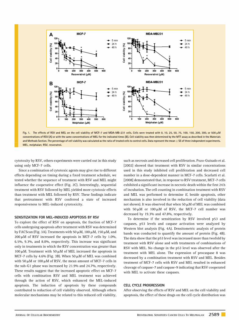
We first investigated the effects of RSV and MEL on the viability of
human breast cancer cells using the MTT assay as previously
described. As shown in Figure 1A,B, RSV and MEL exhibited
cytotoxic effects onMCF-7 andMDA-MB-231 cell growth in a dose-
and time-dependent manner. The IC50 values of RSV and MEL in
MCF-7 cells after 24 h of treatment were �120mM, and �110mM,
respectively. In MDA-MB-231 cells, the IC50 values of RSV and MEL
after 24 h of treatment were �370mM and �305mM, respectively.
These results show that MCF-7 cells were more sensitive to RSV and
MEL than MDA-MB-231 cells. To investigate the cytotoxic effect of
RSV in non-malignant cells, the cell viability assay was performed
in VERO cells, kidney epithelial cells extracted from an African
green monkey, and BHK-21 cells, hamster kidney fibroblast cells
(data not shown). The cytotoxic effect of RSV on the VERO cells was
lower than on the MCF-7 cells, with an IC50 value of �300mM. The
treatment of VERO cells with 200mM of RSV reduced �23% of cell
viability, whereas treatment with the same concentration of RSV
reduced the viability of the MCF-7 cells by �68%. In BHK-21 cells,
no cytotoxic effect was observed in treatments with up to 200mM of
RSV. Aziz et al. [2006] showed that treatment with RSV (0–50mM)
for 24 h resulted in a decrease of cell viability and an induction of
apoptosis in human prostate carcinoma LNCaP cells. The same result
was not observed in normal prostate epithelial HPEC cells. These
observations suggested that RSV may have potential as a
chemotherapeutic agent.
EFFECTS OF THE ASSOCIATION OF RSV WITH MEL ON THE
CELL VIABILITY
Given the results obtained with the use of RSV against carcinoma
cells, we analyzed the effect of RSV in combination with MEL on the
MCF-7 andMDA-MB-231 cells (Fig. 2). After treatment for 24 h with
a combination of 50mM of RSV and 50mM of MEL, MCF-7 and
MDA-MB-231 cell viability decreased by 32.2% and 19.2%,
respectively (Fig. 2A). This decrease in viability was greater than
the sum of the cytotoxic effects of separate treatments with RSV and
MEL at the same concentrations. Other combinations of RSV and
MEL also produced similar results; for example, treatment with
100mM of RSV and 25mM of MEL led to a decrease of viability of
MCF-7 and MDA-MB-231 cells that was 13% and 3%, respectively,
greater than the sum of the effects of these compounds used
individually at the same concentrations. After treatment for 48 h,
combinations of 100mM of RSV and either 25, 50, or 75mM of MEL
were able to reduce the viability of the MCF-7 cells by almost 100%.
As shown by bright field microscopy, the combination of RSV with
MEL promoted more modifications in the morphology of MCF-7
cells than treatment with RSV or MEL alone (Fig. 2B). These data
show that RSV is able to potentiate the cytotoxic effects of MEL in
MCF-7 and MDA-MB-231 cells. Although this increase of cytotoxic
effects of MEL by RSV was greater in MCF-7 cells than MDA-MB-
231 cells. Therefore, in the investigation of enhanced MEL
2588 RESVERATROL SENSITIZES CANCER CELLS TO MELPHALAN JOURNAL OF CELLULAR BIOCHEMISTRY
cytotoxity by RSV, others experiments were carried out in this study
using only MCF-7 cells.
Since a combination of cytotoxic agents may give rise to different
effects depending on timing during a fixed treatment schedule, we
tested whether the sequence of treatment with RSV and MEL might
influence the cooperative effect (Fig. 2C). Interestingly, sequential
treatment with RSV followed by MEL yielded more cytotoxic effects
than treatment with MEL followed by RSV. These findings indicate
that pretreatment with RSV conferred a state of increased
responsiveness to MEL-induced cytotoxicity.
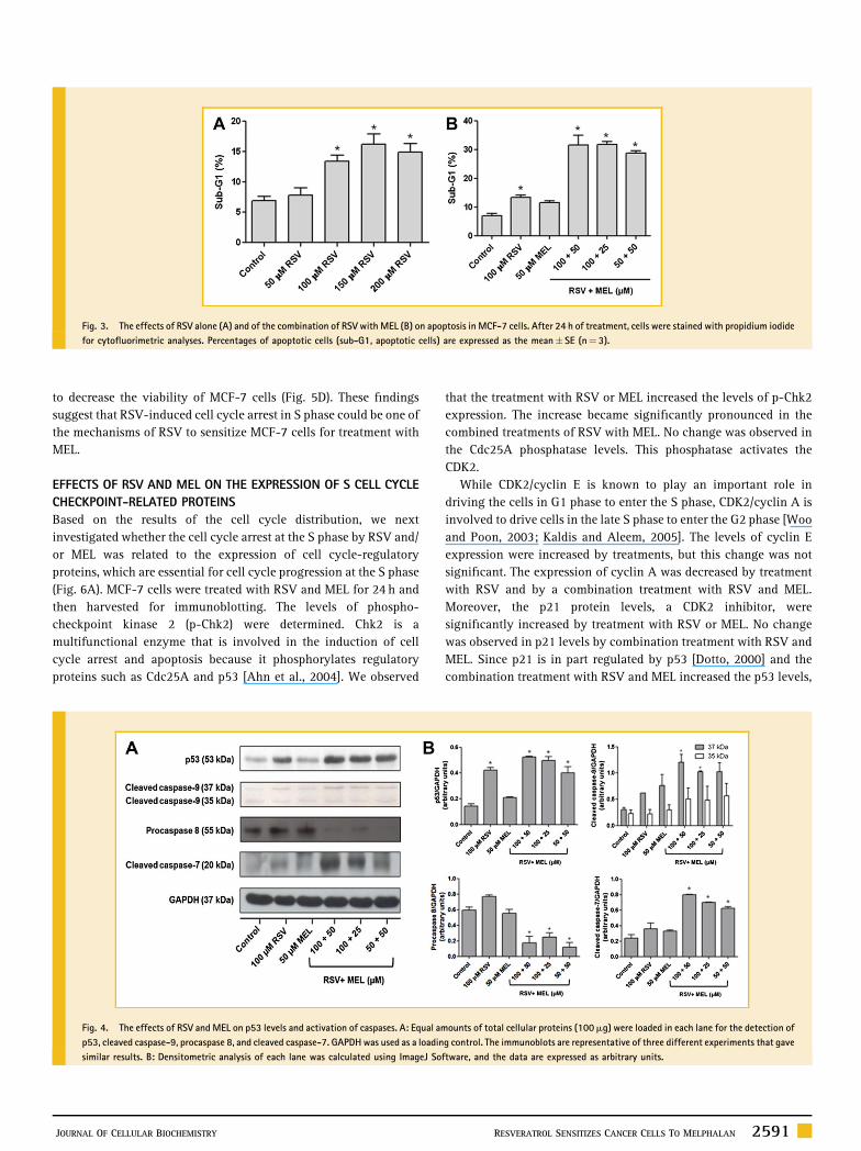
SENSITIZATION FOR MEL-INDUCED APOPTOSIS BY RSV
To explore the effect of RSV on apoptosis, the fraction of MCF-7
cells undergoing apoptosis after treatment with RSV was determined
by FACScan (Fig. 3A). Treatments with 50mM, 100mM, 150mM, and
200mM of RSV increased the apoptosis in MCF-7 cells by 1.0%,
6.5%, 9.3%, and 8.0%, respectively. This increase was significant
only in treatments in which the RSV concentration was greater than
100mM. Treatment with 50mM of MEL increased the apoptosis in
MCF-7 cells by 4.6% (Fig. 3B). When 50mM of MEL was combined
with 50mM or 100mM of RSV, the mean amount of MCF-7 cells in
the sub-G1 phase was increased by 21.9% and 24.7%, respectively.
These results suggest that the increased apoptotic effect on MCF-7
cells with combination RSV and MEL treatment was achieved
through the action of RSV, which enhanced the MEL-induced
apoptosis. The induction of apoptosis by these compounds
contributed to reduction of cell viability observed. Although others
molecular mechanisms may be related to this reduced cell viability,
such as necrosis and decreased cell proliferation. Pozo-Guisado et al.
[2002] showed that treatment with RSV in similar concentrations
used in this study inhibited cell proliferation and decreased cell
number in a dose-dependent manner in MCF-7 cells. Scarlatti et al.
[2008] demonstrated that, in response to RSV treatment, MCF-7 cells
exhibited a significant increase in necrotic death within the first 24 h
of incubation. The cell counting in combination treatment with RSV
and MEL was performed to determine if, beside apoptosis, other
mechanism is also involved in the reduction of cell viability (data
not shown). It was observed that when 50mM of MEL was combined
with 50mM or 100mM of RSV, the MCF-7 cell number was
decreased by 19.3% and 47.8%, respectively.
To determine if the sensitization by RSV involved p53 and
caspases, p53 levels and caspase activation were analyzed by
Western blot analysis (Fig. 4A). Densitometric analysis of protein
bands was conducted to quantify the amount of protein (Fig. 4B).
The data show that the p53 level was increasedmore than twofold by
treatment with RSV alone and with treatments of combinations of
RSV with MEL. No change in the p53 level was observed after the
treatment with MEL alone. The expression of procaspase 8 was
decreased by a combination treatment with RSV and MEL. Besides
treatment of MCF-7 cells with RSV and MEL resulted in enhanced
cleavage of caspase-7 and caspase-9 indicating that RSV cooperated
with MEL to activate these caspases.
CELL CYCLE PROGRESSION
After observing the effects of RSV and MEL on the cell viability and
apoptosis, the effect of these drugs on the cell cycle distribution was
Fig. 1. The effects of RSV and MEL on the cell viability of MCF-7 and MDA-MB-231 cells. Cells were treated with 0, 10, 25, 50, 75, 100, 150, 200, 300, or 500mM
concentrations of RSV (A) or with the same concentrations of MEL for the indicated times (B). Cell viability was then determined by the MTT assay as described in the Materials
andMethods Section. The percentage of cell viability was calculated as the ratio of treated cells to control cells. Data represent the mean� SE of three independent experiments.
MEL, melphalan; RSV, resveratrol.
JOURNAL OF CELLULAR BIOCHEMISTRY RESVERATROL SENSITIZES CANCER CELLS TO MELPHALAN 2589

analyzed by flow cytometry (Fig. 5A,B). Treatment for 24 h with
50mMor 200mMof RSV induced a significant accumulation of cells
in the S phase and a concomitant decrease in the number of cells the
in G1 and G2/M phases (Fig. 5A). In accordance with these data,
Pozo-Guisado et al. [2002] demonstrated that RSV, at the same
concentrations used in this study, inhibited DNA synthesis activity
in MCF-7 cells by measuring the rate of thymidine incorporation.
Treatment for 48 h with 50mM of RSV enhanced the accumulation
of cells in the phase S. Treatment with 25mM, 50mM, or 75mM of
MEL also promoted the accumulation of cells in the S phase, but this
result was not significant (data not shown). When the cells were
treated with combinations of RSV and MEL, a significant increase of
cells in the S phase was observed. This effect was significant only in
treatments in which the concentration of RSVwas 50mM (Fig. 5B). It
was not possible to perform the cell cycle distribution of treatment
with 200mM of RSV alone and with combination of 50mM of RSV
and 75mM of MEL for 48 h due to low cell viability in these
treatments.
To directly test the role of cell cycle progression in increasing
MEL’s cytotoxicity, cells were arrested in the G1, S, or G2/M phase
using the specific cell cycle inhibitors mimosine, thymidine, or
nocodazole and treated concomitantly with MEL (Fig. 5C). Similar to
the treatment with both RSV andMEL, treatment with a combination
of cell cycle inhibitors and MEL also potentiated the ability of MEL
Fig. 2. The effects of the association of RSV with MEL in breast cancer cells. A: The effects of the association of RSV withMEL on the cell viability of MCF-7 andMDA-MB-231
cells for the indicated times. B: Representative bright field microscopy images of MCF-7 cells treated with RSV and MEL. C: Sequence-dependent effect of RSV and MEL on the
cell viability. MCF-7 cells were treated with RSV for 24 h followed byMEL for 24 h (RSVþMEL) or withMEL for 24 h followed by RSV for 24 h (MELþ RSV). The cell viability was
determined by the MTT assay as described in the Materials and Methods Section. The percentage of cell viability was calculated as the ratio of treated cells to control cells. Data
represent the mean� SE of three independent experiments. MEL, melphalan; RSV, resveratrol.
2590 RESVERATROL SENSITIZES CANCER CELLS TO MELPHALAN JOURNAL OF CELLULAR BIOCHEMISTRY
to decrease the viability of MCF-7 cells (Fig. 5D). These findings
suggest that RSV-induced cell cycle arrest in S phase could be one of
the mechanisms of RSV to sensitize MCF-7 cells for treatment with
MEL.
EFFECTS OF RSV AND MEL ON THE EXPRESSION OF S CELL CYCLE
CHECKPOINT-RELATED PROTEINS
Based on the results of the cell cycle distribution, we next
investigated whether the cell cycle arrest at the S phase by RSV and/
or MEL was related to the expression of cell cycle-regulatory
proteins, which are essential for cell cycle progression at the S phase
(Fig. 6A). MCF-7 cells were treated with RSV and MEL for 24 h and
then harvested for immunoblotting. The levels of phospho-
checkpoint kinase 2 (p-Chk2) were determined. Chk2 is a
multifunctional enzyme that is involved in the induction of cell
cycle arrest and apoptosis because it phosphorylates regulatory
proteins such as Cdc25A and p53 [Ahn et al., 2004]. We observed
that the treatment with RSV or MEL increased the levels of p-Chk2
expression. The increase became significantly pronounced in the
combined treatments of RSV with MEL. No change was observed in
the Cdc25A phosphatase levels. This phosphatase activates the
CDK2.
While CDK2/cyclin E is known to play an important role in
driving the cells in G1 phase to enter the S phase, CDK2/cyclin A is
involved to drive cells in the late S phase to enter the G2 phase [Woo
and Poon, 2003; Kaldis and Aleem, 2005]. The levels of cyclin E
expression were increased by treatments, but this change was not
significant. The expression of cyclin A was decreased by treatment
with RSV and by a combination treatment with RSV and MEL.
Moreover, the p21 protein levels, a CDK2 inhibitor, were
significantly increased by treatment with RSV or MEL. No change
was observed in p21 levels by combination treatment with RSV and
MEL. Since p21 is in part regulated by p53 [Dotto, 2000] and the
combination treatment with RSV and MEL increased the p53 levels,
Fig. 3. The effects of RSV alone (A) and of the combination of RSV with MEL (B) on apoptosis in MCF-7 cells. After 24 h of treatment, cells were stained with propidium iodide
for cytofluorimetric analyses. Percentages of apoptotic cells (sub-G1, apoptotic cells) are expressed as the mean� SE (n¼ 3).
Fig. 4. The effects of RSV and MEL on p53 levels and activation of caspases. A: Equal amounts of total cellular proteins (100mg) were loaded in each lane for the detection of
p53, cleaved caspase-9, procaspase 8, and cleaved caspase-7. GAPDHwas used as a loading control. The immunoblots are representative of three different experiments that gave
similar results. B: Densitometric analysis of each lane was calculated using ImageJ Software, and the data are expressed as arbitrary units.
JOURNAL OF CELLULAR BIOCHEMISTRY RESVERATROL SENSITIZES CANCER CELLS TO MELPHALAN 2591

the results suggest that, under such experimental conditions, p21 is
partly regulated in a p53-independent manner. The GAPDH
expression was analyzed to confirm the equal loading of protein;
a representative GAPDH blot is shown.
The effects of RSV and MEL on the expression of CDK2 were
also determined. We found that while the total protein levels of
CDK2 remained largely unchanged by treatments, its active form
(Thr160-phosphorylated CDK2) was decreased by treatment with
RSV and by the combination treatment with RSV and MEL. This
finding suggests that RSV is responsible for decreasing the
phosphorylation of CDK2 at Thr160. The treatment of MEL alone
increased the levels of Thr160-phosphorylated CDK2. Therefore, the
activity of CDK7, the kinase that phosphorylates CDK2 at Thr160, was
analyzed by an in vitro kinase assay. Figure 6B shows a reduction in
Fig. 5. The effects of RSV alone (A) and of the combination of RSV with MEL (B) on the cell cycle distribution in MCF-7 cells. After treatment, the cells were stained with
propidium iodide, and their DNA content was determined by flow cytometry. C: Analysis of the cell cycle after 24 h of treatment with the cell cycle inhibitors mimosine,
thymidine and nocodazole. The percentages of cells in G1, S or G2/M phase are shown. D: The effect of cell cycle inhibitors on MEL-induced cytotoxicity. Cell viability of MCF-7
cells was determined via the MTT assay after concomitant treatment of MEL with 2mM thymidine, 0.4mM mimosine, or 0.4mg/ml nocodazole. The percentage of cell viability
was calculated as the ratio of treated cells to control cells. Data represent the mean� SE of three independent experiments. MEL, melphalan; MMS, mimosine; NOC, nocodazole;
RSV, resveratrol; TMD, thymidine.
2592 RESVERATROL SENSITIZES CANCER CELLS TO MELPHALAN JOURNAL OF CELLULAR BIOCHEMISTRY
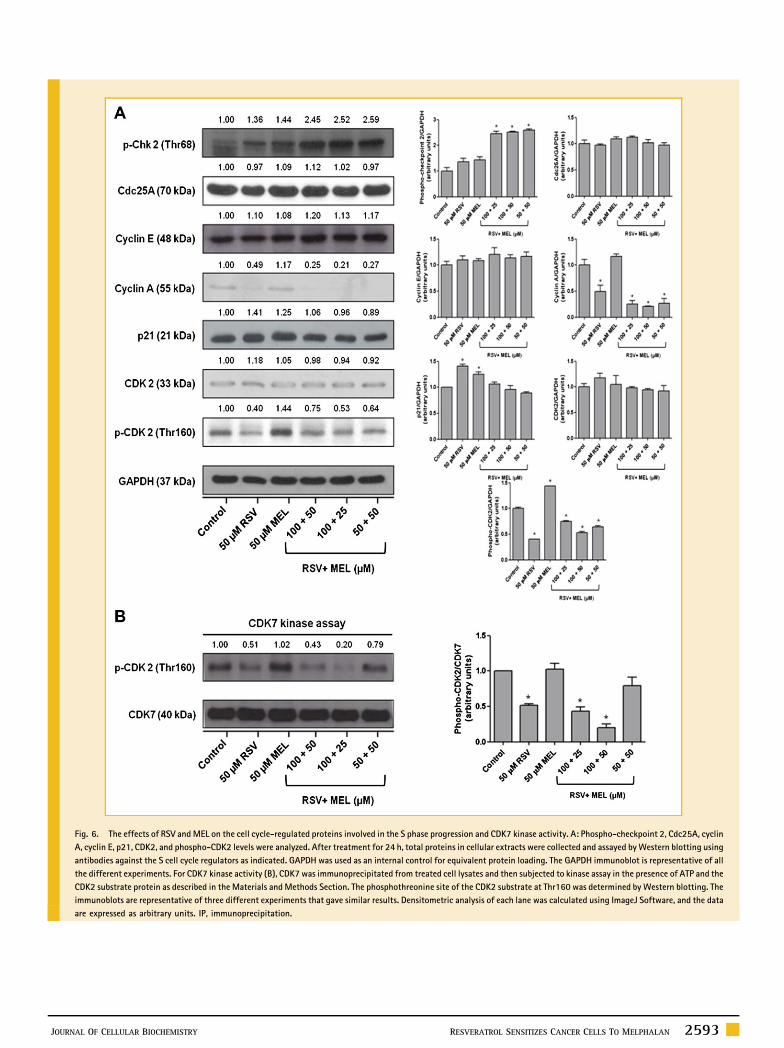
Fig. 6. The effects of RSV and MEL on the cell cycle-regulated proteins involved in the S phase progression and CDK7 kinase activity. A: Phospho-checkpoint 2, Cdc25A, cyclin
A, cyclin E, p21, CDK2, and phospho-CDK2 levels were analyzed. After treatment for 24 h, total proteins in cellular extracts were collected and assayed byWestern blotting using
antibodies against the S cell cycle regulators as indicated. GAPDH was used as an internal control for equivalent protein loading. The GAPDH immunoblot is representative of all
the different experiments. For CDK7 kinase activity (B), CDK7 was immunoprecipitated from treated cell lysates and then subjected to kinase assay in the presence of ATP and the
CDK2 substrate protein as described in the Materials andMethods Section. The phosphothreonine site of the CDK2 substrate at Thr160 was determined by Western blotting. The
immunoblots are representative of three different experiments that gave similar results. Densitometric analysis of each lane was calculated using ImageJ Software, and the data
are expressed as arbitrary units. IP, immunoprecipitation.
JOURNAL OF CELLULAR BIOCHEMISTRY RESVERATROL SENSITIZES CANCER CELLS TO MELPHALAN 2593

the phosphorylation of CDK2 substrate at Thr160; these data support
the above suggestion that RSV inhibited the activity of the CDK7
kinase. The same effect was found in the cells treated with a
combination of RSV and MEL but not in cells that were treated with
MEL alone.
DISCUSSION
MEL is a phenylalanine derivative of nitrogen mustard used in
cancer therapy; it was first synthesized in 1953. This cytotoxic drug
is believed to exert its pharmacologic activity by inducing
interstrand cross-links in the major groove of DNA; this mechanism
represents the toxicity of all alkylation events [Rothbarth et al.,
2004]. Initially, MEL was only used to treat multiple myeloma, but
later, it is also shown to be effective in the treatment of patients with
several other tumors, such as ovarian or breast cancer. MEL,
especially when used at a high dose, shows a diversity of toxic side
effects. The most common side effect occurring during therapy is
bone marrow suppression leading to leukopenia and thrombocyto-
penia. The occurrence of resistance to antineoplastic agents such as
MEL is a major problem in cancer treatment [Dollery, 1991]. Studies
are necessary to improve the efficacy of MEL either by potentiating
the cytotoxicity of MEL or by reducing the resistance against MEL.
Here, we investigated the effect of the combination treatment of
RSV and MEL on human breast cancer MCF-7 and MDA-MB231
cells. RSV is a natural phytoalexin that is present in grape skins and,
as a consequence, in red wine [Gusman et al., 2001]. As early as
1997, RSV was found to be a potent chemopreventive agent, which
blocked the initiation, promotion, and progression of tumors [Jang
et al., 1997]. Since that time, extensive research on its anticancer
activity performed in a wide variety of cellular models suggests a
potential antiproliferative and apoptogenic use of this compound
[Goswami and Das, 2009].
Consistent with earlier observations [Nakagawa et al., 2001;
Pozo-Guisado et al., 2002; Wesierska-Gadek et al., 2008], we found
here that RSV inhibits the viability of human breast cancer MCF-7
and MDA-MB-231 cells while maintaining little cytotoxic effect in
non-malignant cell lines.
We also showed that RSV enhanced the cytotoxic effects of MEL
on MCF-7 and MDA-MB231 cells in vitro. The potentiation of
cytotoxic effects of MEL by RSV was higher in MCF-7 cells than
MDA-MB231 cells. This increase was dependent on the treatment
sequence. Sequential treatment with RSV followed by MEL yielded
more cytotoxic effects than treatment with MEL followed by RSV.
Fulda and Debatin [2004] showed that pretreatment with RSV
enhanced the ability of anticancer agents, such as VP16,
doxorubicin, cytarabine, actinomycin D, taxol, or methotrexate,
to induce apoptosis in SHEP neuroblastoma cells in a dose- and
time-dependent manner. He et al. [2011] demonstrated that RSV
enhanced the antitumor activity of rapamycin in multiple breast
cancer cell lines.
Studies suggest that RSV could act as potent sensitizer for
antitumor drug-induced apoptosis [Kubota et al., 2003; Fulda and
Debatin, 2004; Jazirehi and Bonavida, 2004; Duraj et al., 2006]. A
similar effect was observed in this study. To gain further insight into
the molecular mechanism of RSV induced apoptosis sensitivity, we
investigated key molecules known to regulate apoptosis. The
combination treatment of RSV with MEL resulted in the upregula-
tion of p53, downregulation of procaspase 8, and the activation of
caspases 7 and 9. Gatouillat et al. [2010] reported that RSV enhanced
the chemotherapeutic potential of doxorubicin in chemoresistant
B16 melanoma cells through the upregulation of p53. Interestingly,
in another study, an enhancement in the sensitization effect of RSV
on apoptosis induced by various drugs in cancer cell lines was found
to be p53 independent [Fulda and Debatin, 2004]. Bhardwaj et al.
[2007] have shown that RSV enhanced the apoptotic and anti-
proliferative potential of velcade and thalidomide in multiple
myeloma cells. Such an enhancement was associated with inhibition
of NF-kB and STAT3 activation pathways and with an accumulation
of the sub-G(1) population, an increase in Bax release, and the
activation of caspase-3.
The analysis of cell cycle distribution showed that RSV, both
alone and in combination treatment with MEL, inhibited cell cycle
progression in the S phase. Treatment of MCF-7 cells with specific
cell cycle inhibitors followed by MEL also potentiated the cytotoxic
effect of MEL, suggesting that the cell cycle arrest is necessary for
the chemosensitization of MCF-7 cells to MEL by RSV. The cell cycle
arrest may increase MEL incorporation into the cells. This finding
would explain the improved efficacy of MEL we observed here.
The effect on cell cycle progression was probably caused by
downregulation of cyclin A and phospho-CDK2 (Thr160) by
treatment with RSV and its association with MEL. The treatment
of MEL alone increased the levels of Thr160-phosphorylated CDK2.
This observation is in agreement with the known function of
activated CDK2 (phospo-CDK2) in driving cells in late G1 phase into
the S-phase. Zhou et al. [2009] showed that activation of p-CDK2 is
believed to play a role in the induction of a reversible, non-cytotoxic
S-phase delay in HepG2 cells.
Figure 7 shows proteins related to cell cycle progression in the S
phase. An important mechanism for regulating cell cycle progres-
sion is controlled by the activity of CDKs, which is regulated by
interacting with their respective CDK subunits [Malumbres and
Barbacid, 2005]. Cyclin E associates with CDK2 to regulate
progression from G1 into S phase. Cyclin A binds with CDK2,
and this complex is required during S phase. In addition to cyclin
binding, CDK2 activity is also regulated by phosphorylation on
conserved threonine and tyrosine residues. Dephosphorylation at
tyrosine-15 and threonine-14 sites by the enzyme Cdc25A is
necessary for activation of CDK2 and further progression through
the cell cycle. Full activation of CDK2 requires phosphorylation
of threonine 160, brought about by the CDK7 [Vermeulen et al.,
2003].
Because hypophosphorylation of CDK2 protein was induced in
MCF-7 cells by RSV treatment alone and by combination treatment
withMEL, we analyzed the CDK7 activity and observed an inhibition
of its activity. The CDK7 kinase inhibition maintained CDK2 in its
inactive form and arrested the cell cycle in the S phase. Liang et al.
[2003] demonstrated that RSV induced cell cycle arrest through the
inhibition of CDK7 kinase activity in colon carcinoma HT29 cells.
However, the cell cycle arrest observed by these researchers was at
the G2 phase. CDK7, in addition to controlling the activity of CDK2,
2594 RESVERATROL SENSITIZES CANCER CELLS TO MELPHALAN JOURNAL OF CELLULAR BIOCHEMISTRY
also controls the activity of CDK1, which is an enzyme responsible
for progression at the G2 phase [Vermeulen et al., 2003].
In summary, our results indicate that RSV could be used as an
adjuvant agent during breast cancer therapy with MEL. It is
important to emphasize that this is the first study demonstrating the
effect of the combination of RSV with MEL in human cancer
cells. Despite more than 1,100 publications on the cancer
chemotherapeutic potential of RSV, only about 20 reported on its
chemosensitization potential. Thus, further studies should be
performed in this area to determine effective combinations of
RSV and chemotherapeutic agents, their effects on different types of
cancers and possible mechanisms of action involved in chemo-
sensitization by RSV.
ACKNOWLEDGMENTS
This work was supported by grants from Fundacao de Amparo aPesquisa Carlos Chagas Filho do Estado do Rio de Janeiro (FAPERJ—E-26/103.110/2008); Cancer Foundation/2009 (without grantnumber); Conselho Nacional de Desenvolvimento Cientıfico eTecnologico (CNPq—PhD scholarship); and Coordenacao deAperfeicoamento de Pessoal de Nıvel Superior (CAPES—PhDscholarship).
REFERENCES
Agarwal C, Dhanalakshmi S, Singh RP, Agarwal R. 2004. Inositol hexapho-sphate inhibits growth and induces G1 arrest and apoptotic death ofandrogen-dependent human prostate carcinoma LNCaP cells. Neoplasia6:646–659.
Ahmad KA, Clement MV, Hanif IM, Pervaiz S. 2004. Resveratrol inhibitsdrug-induced apoptosis in human leukemia cells by creating an intracellularmilieu nonpermissive for death execution. Cancer Res 64:1452–1459.
Ahn J, Urist M, Prives C. 2004. The Chk2 protein kinase. DNA Repair 3:1039–1047.
Athar M, Back JH, Kopelovich L, Bickers DR, Kim AL. 2009. Multiplemolecular targets of resveratrol: Anti-carcinogenic mechanisms. Arch Bio-chem Biophys 486:95–102.
Aziz MH, Nihal M, Fu VX, Jarrard DF, Ahmad N. 2006. Resveratrol-causedapoptosis of human prostate carcinoma LNCaP cells is mediated via modu-lation of phosphatidylinositol 30-kinase/Akt pathway and Bcl-2 familyproteins. Mol Cancer Ther 5:1335–1341.
Baur JA, Sinclair DA. 2006. Therapeutic potential of resveratrol: The in vivoevidence. Nat Rev Drug Discov 5:493–506.
Bhardwaj A, Sethi G, Vadhan-Raj S, Bueso-Ramos C, Takada Y, Gaur U, NairAS, Shishodia S, Aggarwal BB. 2007. Resveratrol inhibits proliferation,induces apoptosis, and overcomes chemoresistance through down-regulation of STAT3 and nuclear factor-kappa B-regulated antiapoptoticand cell survival gene products in humanmultiple myeloma cells. Blood 109:2293–2302.
Carmichael J, DegraffW, Gazdar A, Minna J, Mitchell J. 1987. Evaluation of atetrazolinium-based semiautomated colorimetric assay: Assessment of che-mosensitivity testing. Cancer Res 47:936–942.
Chan JY, PhooMS, ClementMV, Pervaiz S, Lee SC. 2008. Resveratrol displaysconverse dose-related effects on 5-fluorouracil-evoked colon cancer cellapoptosis—The roles of caspase-6 and p53. Cancer Biol Ther 7:1305–1312.
Dollery C. 1991. Melphalan, therapeutic drugs. London: ChurchillLivingstone.
Dotto GP. 2000. p21 WAF1/Cip1: More than a break to the cell cycle?Biochim Biophys Acta 1471:M43–M56.
Duraj J, Bodo J, Sulikova M, Rauko P, Sedlak J. 2006. Diverse resveratrolsensitization to apoptosis induced by anticancer drugs in sensitive andresistant leukemia cells. Neoplasma 53:384–392.
Fulda S, Debatin KM. 2004. Sensitization for anticancer drug-inducedapoptosis by the chemopreventive agent resveratrol. Oncogene 23:6702–6711.
Fig. 7. The effects of RSV and its association with MEL on proteins related to the cell cycle progression in the S phase of MCF-7 cells. M, melphalan; R, resveratrol; RþM,
association of RSV with MEL.
JOURNAL OF CELLULAR BIOCHEMISTRY RESVERATROL SENSITIZES CANCER CELLS TO MELPHALAN 2595

Garg AK, Buchholz TA, Aggarwal BB. 2005. Chemosensitization and radio-sensitization of tumors by plant polyphenols. Antioxid Redox Signal 7:1630–1647.
Gatouillat G, Balasse E, Joseph-Pietras D, Morjani H, Madoulet C. 2010.Resveratrol induces cell-cycle disruption and apoptosis in chemoresistantB16 melanoma. J Cell Biochem 110:893–902.
Goswami SK, Das DK. 2009. Resveratrol and chemoprevention. Cancer Lett284:1–6.
Gupta SC, Kannappan R, Reuter S, Kim JH, Aggarwal BB. 2011. Chemo-sensitization of tumors by resveratrol. Ann NY Acad Sci 1215:150–160.
Gusman J, Malonne H, Atassi G. 2001. A reappraisal of the potentialchemopreventive and chemotherapeutic properties of resveratrol. Carcino-genesis 22:1111–1117.
He X, Wang Y, Zhu JH, Orloff M, Eng C. 2011. Resveratrol enhances the anti-tumor activity of themTOR inhibitor rapamycin in multiple breast cancer celllines mainly by suppressing rapamycin-induced AKT signaling. Cancer Lett301:168–176.
Higgins CF. 2007. Multiple molecular mechanisms for multidrug resistancetransporters. Nature 446:749–757.
Jang MS, Cai EN, Udeani GO, Slowing KV, Thomas CF, Beecher CWW, FongHHS, Farnsworth NR, Kinghorn AD, Mehta RG, Moon RC, Pezzuto JM. 1997.Cancer chemopreventive activity of resveratrol, a natural product derivedfrom grapes. Science 275:218–220.
Jazirehi AR, Bonavida B. 2004. Resveratrol modifies the expression ofapoptotic regulatory proteins and sensitizes non-Hodgkin’s lymphomaand multiple myeloma cell lines to paclitaxel-induced apoptosis. Mol CancerTher 3:71–84.
Jemal A, Bray F, Center MM, Ferlay J, Ward E, Forman D. 2011. GlobalCancer Statistics. CA Cancer J Clin 61:69–90.
Kaldis P, Aleem E. 2005. Cell cycle sibling rivalry—Cdc2 vs. Cdk2. Cell Cycle4:1491–1494.
Kubota T, Uemura Y, Kobayashi M, Taguchi H. 2003. Combined effects ofresveratrol and paclitaxel on lung cancer cells. Anticancer Res 23:4039–4046.
Liang YC, Tsai SH, Chen L, Lin-Shiau SY, Lin JK. 2003. Resveratrol-inducedG(2) arrest through the inhibition of CDK7 and p34(CDC2) kinases in coloncarcinoma HT29 cells. Biochem Pharmacol 65:1053–1060.
Lowry OH, Rosebrough NJ, Farr AL, Randall RJ. 1951. Protein measurementwith the folin phenol reagent. J Biol Chem 193:265–275.
Malumbres M, Barbacid M. 2005. Mammalian cyclin-dependent kinases.Trends Biochem Sci 30:630–641.
Mao QQ, Bai Y, Lin YW, Zheng XY, Qin J, Yang K, Xie LP. 2010. Resveratrolconfers resistance against taxol via induction of cell cycle arrest in humancancer cell lines. Mol Nutr Food Res 54:1574–1584.
Marques FZ, Markus MA, Morris BJ. 2009. Resveratrol: Cellular actions of apotent natural chemical that confers a diversity of health benefits. Int JBiochem Cell Biol 41:2125–2128.
Nakagawa H, Kiyozuka Y, Uemura Y, Senzaki H, Shikata N, Hioki K, TsuburaA. 2001. Resveratrol inhibits human breast cancer cell growth and maymitigate the effect of linoleic acid, a potent breast cancer cell stimulator.J Cancer Res Clin Oncol 127:258–264.
Pozo-Guisado E, Alvarez-Barrientos A, Mulero-Navarro S, Santiago-JosefatB, Fernandez-Salguero PM. 2002. The antiproliferative activity of resveratrolresults in apoptosis in MCF-7 but not in MDA-MB-231 human breast cancercells: Cell-specific alteration of the cell cycle. Biochem Pharmacol 64:1375–1386.
Rothbarth J, Koevoets C, Tollenaar R, Tilby MJ, van de Velde CJH, Mulder GJ,Kuppen PJK. 2004. Immunohistochemical detection of melphalan-DNAadducts in colon cancer cells in vitro and human colorectal liver tumoursin vivo. Biochem Pharmacol 67:1771–1778.
Scarlatti F, Maffei R, Beau I, Codogno P, Ghidoni R. 2008. Role of non-canonical Beclin 1-independent autophagy in cell death induced by resvera-trol in human breast cancer cells. Cell Death Differ 15:1318–1329.
Vermeulen K, Van Bockstaele DR, Berneman ZN. 2003. The cell cycle: Areview of regulation, deregulation and therapeutic targets in cancer. CellProlif 36:131–149.
Wesierska-Gadek J, Kramer MP, Maurer M. 2008. Resveratrol modulatesroscovitine-mediated cell cycle arrest of human MCF-7 breast cancer cells.Food Chem Toxicol 46:1327–1333.
Woo RA, Poon RY. 2003. Cyclin-dependent kinases and S phase control inmammalian cells. Cell Cycle 2:316–324.
Zhou R, Fukui M, Choi HJ, Zhu BT. 2009. Induction of a reversible, non-cytotoxic S-phase delay by resveratrol: implications for a mechanism oflifespan prolongation and cancer protection. Br J Pharmacol 158:462–474.
2596 RESVERATROL SENSITIZES CANCER CELLS TO MELPHALAN JOURNAL OF CELLULAR BIOCHEMISTRY