branched-chain amino acid supplementation and resistance ...
Upload
khangminh22Category
view
0download
0
I
Maternal resveratrol supplementation in gestational diabetes prevents
cardio-metabolic disease development and improves cardiac structure in the rat offspring
by
Gabriel Martin Brawerman
A Thesis submitted to the Faculty of Graduate Studies of
The University of Manitoba
in partial fulfillment of the requirements of the degree of
MASTER OF SCIENCE
The Department of Pharmacology and Therapeutics
University of Manitoba
Winnipeg
Copyright © 2018 by Gabriel Brawerman

II
Abstract
Gestational diabetes mellitus (GDM), a common complication of pregnancy, arises during
the third trimester and is characterized by hyperglycemia. GDM increases cardio-metabolic
disease risk in mothers and offspring. Current treatments have disadvantages. Resveratrol (RESV),
a naturally produced polyphenol, has anti-oxidant and positive metabolic health effects. Thus, we
hypothesized that RESV supplementation during the third trimester and lactation would improve
maternal glucose tolerance and prevent cardio-metabolic disease in the offspring.
A diet-induced GDM model was utilized for this thesis. Different metabolic tests were
performed. Echocardiography was used to assess cardiac structure and function. Immunoblotting,
qPCR and cardiomyocyte isolations were performed to study mechanisms.
RESV supplementation prevented maternal glucose intolerance and cardio-metabolic
disease development in the offspring by improving glucose homeostasis and inhibiting cardiac
remodelling.
Supplementing maternal diets with RESV at the onset of GDM may become a newer
intervention to protect mothers and their offspring from GDM-induced short and long-term
consequences.

III
Acknowledgements
Firstly, I must thank my supervisor Dr. Vern Dolinsky. Thank you so much for giving me
the opportunity to work in your laboratory. Thank you for always being available and having your
office door open whenever I needed help or advice with my research. I could not have done this
without your encouragement, feedback, and guidance and I truly appreciate it. I am very glad I did
my MSc. research in your laboratory.
I want to also thank my advisory and examining committee, Dr. Tiina Kauppinen, Dr.
Grant Hatch, and Dr. Joe Gordon for all your advice, criticism and comments during our committee
meetings, and for reviewing my thesis.
Next, I must thank Ms. Stephanie Kereliuk, Mr. Mario Fonseca, and Mrs. Bo Xiang. I could
not have finished this work without the help from all of you, whether it was animal work,
cardiomyocyte isolations, or just troubleshooting western blots. Thank you for always being there
for me.
I could not have done this research without the funding from Research Manitoba/CHRIM,
so thank you so much for your studentship, and I hope other students will get the same benefit I
received in the future.
Last but not least, I want to thank my family and fiancée, Adriana, Claudio, Ariel, and
Amy, for all your support even when you did not fully understand what my research was on. Thank
you for listening to me practice my presentations. I could not have finished this without any of
you. Thank you mom for introducing me to Science, I would not have even considered a career in
Science if it were not for you.

IV
Dedication
“Education is the most powerful weapon which you can use to change the world”
Nelson Mandela

V
Table of Contents
Abstract ......................................................................................................................................................... II
Acknowledgements ...................................................................................................................................... III
Dedication .................................................................................................................................................... IV
Table of Contents .......................................................................................................................................... V
List of Tables ............................................................................................................................................... IX
List of Figures ............................................................................................................................................... X
List of Abbreviations .................................................................................................................................. XII
Chapter 1: .................................................................................................................................................... 1
Introduction ................................................................................................................................................. 1
1.1 The Obesity Epidemic: ....................................................................................................................... 2
1.1.1 Type 2 Diabetes Mellitus (T2D): ................................................................................................. 3
1.1.2 Gestational Diabetes Mellitus: ..................................................................................................... 5
1.2 The Developmental Origins of Health and Disease (DOHaD) Hypothesis: ....................................... 7
1.2.1 Predictive Adaptive Response and the Mismatch Hypothesis: .................................................... 9
1.3 GDM Effects on Mothers and Offspring: ......................................................................................... 10
1.3.1 Diabetic Cardiomyopathy – one of the most common complications affecting GDM-exposed
offspring .............................................................................................................................................. 11
1.3.2 Cardiac Hypertrophy: ................................................................................................................. 14
1.4 Type 2 Diabetes and GDM Treatments: ........................................................................................... 15
1.4.1 Lifestyle Therapy Effects on Maternal and Offspring Health: - Clinical Data .......................... 16
1.4.2 Lifestyle Therapies during Pregnancy in Animal Models: ........................................................ 18
1.5 Therapeutic Approaches: .................................................................................................................. 21
1.5.1 Insulin: ....................................................................................................................................... 22
1.5.2 Metformin: ................................................................................................................................. 23
1.5.3 Glyburide: .................................................................................................................................. 24
1.6 Natural Health Products: ................................................................................................................... 25
1.6.1 Resveratrol: ................................................................................................................................ 26
1.6.2 Maternal Resveratrol Supplementation Studies: ........................................................................ 29
1.6.3 Offspring Studies: ...................................................................................................................... 29
1.7 GDM Animal Model: ........................................................................................................................ 32

VI
1.7.1 GDM Animal Model previously characterized in the Lab:........................................................ 33
1.8 Thesis Objectives ................................................................................................................................. 34
1.9 General Hypotheses: ........................................................................................................................... 36
Materials and Methods: ........................................................................................................................... 37
Diet-Induced GDM Model and Resveratrol Treatment .......................................................................... 37
Assessment of Postnatal Diet Effect on Offspring:................................................................................. 38
Insulin and Glucose Tolerance Tests (ITTs and GTTs): ......................................................................... 40
Assessing Circulating and Tissue Factors: .............................................................................................. 40
Dual-Energy X-ray Absorptiometry Scans: ............................................................................................ 41
Echocardiography Scans: ........................................................................................................................ 41
Liver Histology/pWAT Histology: ......................................................................................................... 42
Western Blot: .......................................................................................................................................... 42
RNA Isolation and qPCR: ....................................................................................................................... 44
Cardiomyocyte Isolations: ...................................................................................................................... 46
MitoSOX Experiment: ........................................................................................................................ 47
MitoTracker Experiment: .................................................................................................................... 48
TMRM Experiment: ............................................................................................................................ 48
Maternal and Offspring Islet Isolations: ................................................................................................. 49
Glucose-Stimulated Insulin Secretion (GSIS) Assays: ........................................................................... 50
Statistical Analysis: ................................................................................................................................. 50
Chapter 2: .................................................................................................................................................. 51
Effects of Resveratrol Supplementation during Pregnancy on Maternal Metabolism. ..................... 51
2.1 Introduction: ...................................................................................................................................... 52
2.2 Materials and Methods – Refer to page 37. ...................................................................................... 55
2.3 Results: .............................................................................................................................................. 55
2.3.1 Maternal RESV supplementation during pregnancy prevented glucose intolerance, restored
blood glucose levels, and improved insulin secretion, without affecting gestational weight gain in the
rat dams: .............................................................................................................................................. 55
2.4 Discussion/Conclusion: ..................................................................................................................... 61
Chapter 3: .................................................................................................................................................. 65
Effects of Resveratrol Supplementation during Pregnancy on the Metabolism of the 15-Week-Old
Young-Adult Rat Offspring ..................................................................................................................... 65
3.1 Introduction: ...................................................................................................................................... 66
3.2 Materials and Methods: Refer to page 37. ........................................................................................ 68

VII
3.3 Fetal Offspring Results: .................................................................................................................... 69
3.3.1 Maternal RESV supplementation was safe during pregnancy and prevented macrosomia in
males but not in females: .................................................................................................................... 69
3.4 15-Week-Old Young-Adult Offspring Results: ................................................................................ 72
3.4.1 Maternal RESV supplementation prevented the increase in obesity in the young adult male rat
offspring, but not in the female offspring: .......................................................................................... 72
3.4.2 A high fat and sucrose postnatal diet resulted in increased fat accumulation in the male rat
offspring regardless of maternal diet, while RESV supplementation increased fat mass in females: 73
3.4.3 Maternal RESV supplementation prevented hepatic steatosis development in the male rat
offspring at 15 weeks of age: .............................................................................................................. 79
3.4.4 Maternal RESV supplementation prevented glucose intolerance and attenuated the insulin
resistance caused by a GDM pregnancy in the young-adult male rat offspring: ................................ 80
3.4.5 Maternal RESV supplementation did not affect the protein expression of proteins involved in
the insulin signalling pathway in the 15 week old male rat offspring: ............................................... 84
3.4.6 GDM induced changes in hepatic gene expression of several genes involved in metabolism, but
maternal RESV supplementation prevented them: ............................................................................. 87
3.5 Conclusion/Discussion ...................................................................................................................... 91
Chapter 4: ................................................................................................................................................ 105
Effects of Resveratrol Supplementation during Pregnancy on the 15-Week-Old Young-Adult Male
Rat Offspring Heart ................................................................................................................................ 105
4.1 Introduction: .................................................................................................................................... 106
4.2 Materials and Methods – Refer to page 37. .................................................................................... 109
4.3 Results: ............................................................................................................................................ 109
4.3.1 Maternal RESV supplementation prevented the development of GDM-induced cardiac
hypertrophy in fetal offspring: .......................................................................................................... 109
4.3.2 GDM-induced cardiac hypertrophy persisted to 15 weeks of age in the male rat offspring, but
was prevented by maternal RESV supplementation in the GDM+RESV-HFS offspring: ............... 110
4.3.3 Cardiac hypertrophy was not associated with a reduction in cardiac function: ....................... 115
4.3.4 GDM-LF male offspring exhibited cardiac steatosis, while this was prevented in the
GDM+RESV-LF offspring at 15 weeks of age: ............................................................................... 115
4.3.5 Maternal RESV supplementation changed the protein expression of several proteins in the
cardiac tissue of the 15 week old male rat offspring: ........................................................................ 118
4.3.6 GDM fetal cardiomyocytes exhibited increased reactive oxygen species, while maternal RESV
supplementation attenuated the increase: .......................................................................................... 122
4.4 Conclusion/Discussion: ................................................................................................................... 127
Chapter 5 ................................................................................................................................................. 137
General Discussion, Conclusions and Future Directions: ............................................................... 138

VIII
References:............................................................................................................................................... 147

IX
List of Tables
Table 1 – Primary antibodies used for immunoblotting analyses-------------------------------------43
Table 2 – Primer sequences used for quantitative real-time PCR-------------------------------------45
Table 3 – Average daily food consumption by mothers from T3 to end of lactation---------------58
Table 4 – Litter Sizes and Sex Distribution---------------------------------------------------------------70
Table 5 – Average daily food consumption and energy intake in the male and female offspring –
grouped by litter----------------------------------------------------------------------------------------------77

X
List of Figures
Figure 1 – Current Interventions for GDM---------------------------------------------------------------21
Figure 2 – Proposed molecular pathways affected by RESV supplementation with a focus on
glucose and lipid metabolism-------------------------------------------------------------------------------28
Figure 3 – Timing of therapy across lifespan and the effect on selected tissue development in
different animal models-------------------------------------------------------------------------------------31
Figure 4 – Amount of different foods people have to consume to achieve 1g/day of Resveratrol--
------------------------------------------------------------------------------------------------------------------31
Figure 5 – Experimental Animal Model------------------------------------------------------------------39
Figure 6 – Maternal RESV supplementation during pregnancy prevents glucose intolerance,
restores blood glucose levels, and improves insulin secretion, without affecting gestational weight
gain------------------------------------------------------------------------------------------------------------59
Figure 7 – Maternal RESV supplementation did not cause adverse developmental effects and
prevented macrosomia in males but not in females------------------------------------------------------71
Figure 8 – Maternal RESV supplementation reduced obesity in the 15 week-old male rat offspring,
but not in the female rat offspring--------------------------------------------------------------------------76
Figure 9 – Consumption of a postnatal HFS diet resulted in increased fat accumulation in the male
rat offspring, while maternal RESV consumption increased fat mass in the female rat offspring at
15 weeks of age-----------------------------------------------------------------------------------------------78
Figure 10 – Maternal RESV supplementation prevented hepatic steatosis development in the
young-adult male rat offspring-----------------------------------------------------------------------------82
Figure 11 – Maternal RESV supplementation prevented glucose intolerance and attenuated the
insulin resistance caused by GDM in the young adult male rat offspring-----------------------------83

XI
Figure 12 – Maternal RESV supplementation did not affect the protein expression of proteins involved in
the insulin signalling pathway in the 15 week old male rat offspring--------------------------------------------86
Figure 13 – GDM modified the expression of several hepatic genes involved in metabolism, but
maternal RESV supplementation prevented some of those changes in the young adult male rat
offspring-------------------------------------------------------------------------------------------------------89
Figure 14 – Maternal RESV supplementation prevented the development of cardiac hypertrophy
caused by a GDM pregnancy in the fetal offspring, without affecting heart function--------------112
Figure 15 – Cardiac hypertrophy persisted at 15 weeks of age in the GDM-HFS male rat offspring,
while it was prevented in the GDM+RESV-HFS male offspring-------------------------------------114
Figure 16 – Cardiac function was not affected by either maternal or postnatal diet at 15 weeks of
age in the male rat offspring-------------------------------------------------------------------------------117
Figure 17 – Maternal RESV supplementation changed protein expression of several targets in the
cardiac tissue of the 15 week-old young-adult male rat offspring------------------------------------121
Figure 18 – GDM fetal cardiomyocytes exhibited increased production of reactive oxygen species,
which was attenuated with maternal RESV supplementation-----------------------------------------124
Figure 19 – GDM fetal cardiomyocytes had a slight increase in mitochondrial number when
compared to Lean and GDM+RESV cardiomyocytes-------------------------------------------------125
Figure 20 – GDM fetal cardiomyocytes exhibited an increase in mitochondrial membrane
potential when compared to both Lean and GDM+RESV fetal cardiomyocytes-------------------126
Figure 21 – Proposed molecular mechanism for cardiac hypertrophy development in the young-
adult male rat offspring------------------------------------------------------------------------------------132
XII
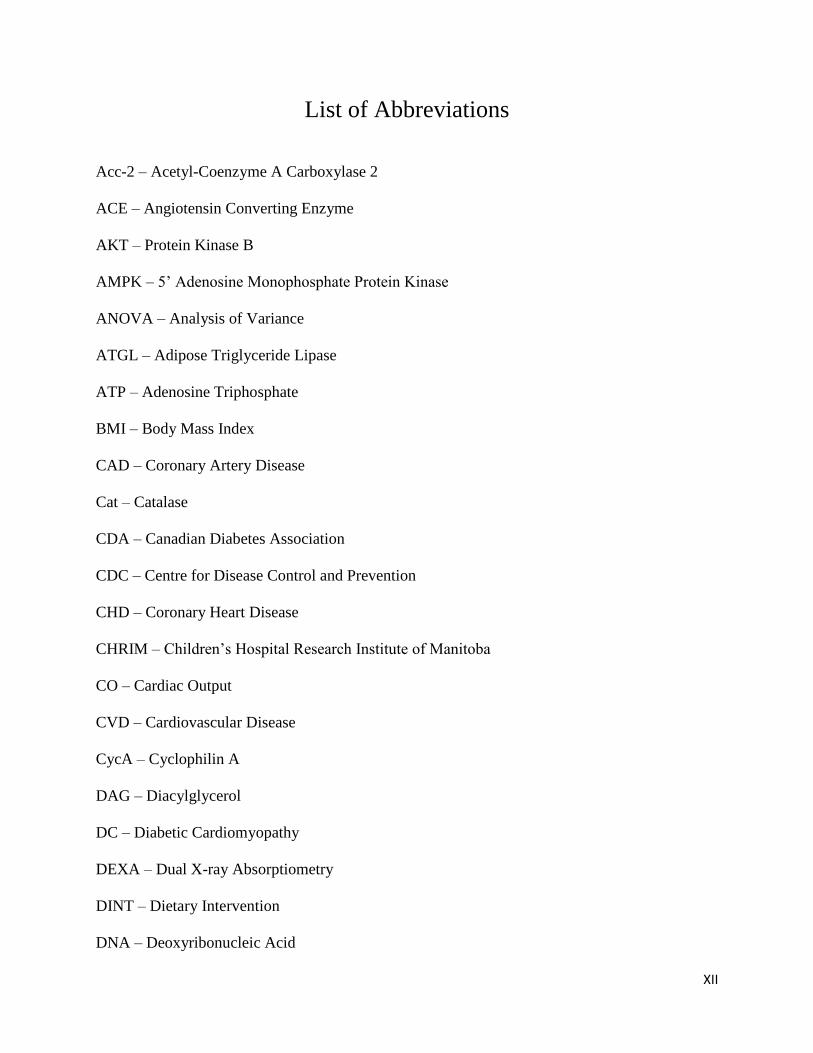
List of Abbreviations
Acc-2 – Acetyl-Coenzyme A Carboxylase 2
ACE – Angiotensin Converting Enzyme
AKT – Protein Kinase B
AMPK – 5’ Adenosine Monophosphate Protein Kinase
ANOVA – Analysis of Variance
ATGL – Adipose Triglyceride Lipase
ATP – Adenosine Triphosphate
BMI – Body Mass Index
CAD – Coronary Artery Disease
Cat – Catalase
CDA – Canadian Diabetes Association
CDC – Centre for Disease Control and Prevention
CHD – Coronary Heart Disease
CHRIM – Children’s Hospital Research Institute of Manitoba
CO – Cardiac Output
CVD – Cardiovascular Disease
CycA – Cyclophilin A
DAG – Diacylglycerol
DC – Diabetic Cardiomyopathy
DEXA – Dual X-ray Absorptiometry
DINT – Dietary Intervention
DNA – Deoxyribonucleic Acid

XIII
DOHaD – Developmental Origins of Health and Disease
eEF2 – Eukaryotic Elongation Factor 2
EF – Ejection Fraction
eIF2α – Eukaryotic translation Initiation Factor 2α
ELISA – Enzyme-Linked Immunosorbent Assay
FAO – Fatty Acid Oxidation
FBS – Fetal Bovine Serum
GDM – Gestational Diabetes Mellitus
GLUT-4 – Glucose Transporter Protein 4
GMC – Genetic Models Center
GPX1 – Glutathione Peroxidase 1
GSIS – Glucose Stimulated Insulin Secretion
GTT – Glucose Tolerance Test
gWAT – Gonadal White Adipose Tissue
GWG – Gestational Weight Gain
G-6-P – Glucose-6-Phosphate
HbA1c – Hemoglobin A1c Levels
H&E – Hematoxylin and Eosin
HFS – High Fat and Sucrose Diet
HG – High Glucose
HOMA-IR – Homeostatic Model Assessment for Insulin Resistance
HW:BW – Heart Weight to Body Weight
HW:TL – Heart Weight to Tibia Length

XIV
IFNα/β – Interferon α/β
IL-6 – Interleukin-6
IRβ – Insulin Receptor β
IRS-1 – Insulin Receptor Substrate-1
ITT – Insulin Tolerance Test
IVRT – Isovolumetric Relaxation Time
IVS – Interventricular Septal Thickness
LF – Low Fat Diet
LKB1 – Liver Kinase B1
LVAW – Left Ventricular Anterior Wall Thickness
LVID – Left Ventricular Interior Diameter
LVPW – Left Ventricular Posterior Wall Thickness
mRNA – Messenger Ribonucleic Acid
mTOR – Mammalian Target of Rapamycin
NAD+ - Nicotinamide Adenine Dinucleotide +
NAFLD – Non-Alcoholic Fatty Liver Disease
NFκB – Nuclear Factor κB
NRf2 – Nuclear Factor-Like 2
OGTT – Oral Glucose Tolerance Test
PBS – Phosphate-Buffered Saline
Pcyt2 – CDP:Phosphoethanolamine Cytidylyltransferase 2
PE – Phosphatidylethanolamine
Pepck/Pck1 – Phosphoenolpyruvate Carboxykinase

XV
PFA – Paraformaldehyde
PGC1α – Peroxisome Proliferator Activated Receptor-γ Co-Activator 1α
PI3K – Phosphatidylinositol 3 Kinase
PPARα – Peroxisome Proliferator-Activated Receptor α
pWAT – Perirenal White Adipose Tissue
p70S6K – Ribosomal Protein p70 S6 Kinase
qPCR – Quantitative Polymerase Chain Reaction
RESV – Resveratrol
RNA – Ribonucleic Acid
ROS – Reactive Oxygen Species
SEM – Standard Error of the Mean
Serca2a – Sarco/endoplasmic Reticulum Calcium ATPase
SGLT2 – Sodium Glucose Co-Transporter 2
SIRT1 – Sirtuin 1
SOD – Superoxide Dismutase
Srebp-1c – Sterol Response Element Binding Protein 1C
TG – Triglycerides
TMRM – Tetramethylrhodamine, Methyl Ester, Perchlorate
TNF-α – Tumor Necrosis Factor α
T1D – Type 1 Diabetes
T2D – Type 2 Diabetes
T3 – Trimester 3

1
Chapter 1:
Introduction

2
1.1 The Obesity Epidemic:
Obesity is now widely recognized as a key risk factor for the development of many
different types of chronic diseases. Obesity is generally defined as the excess accumulation of fat
tissue which can have detrimental consequences for an individual’s health (Corey and Kaplan
2014). This can be the result of an unbalanced food intake and calorie expenditure, leading to
increased fat deposition and increased weight gain, mainly due to a societal switch to fatty food
consumption and lack of exercise (Corey and Kaplan 2014; Lehnert et al. 2013; Swinburn et al.
2011). Clinicians currently use Body Mass Index (BMI) to assess whether their patients are obese
or overweight. This index takes into consideration the person’s weight and height and expresses
the result in kg/m2 (World Health Organization. 2017). The World Health Organization has set a
BMI of 25 kg/m2 or more as being overweight, while more than 30 kg/m2 corresponds to obese
individuals (World Health Organization. 2017). This method is also widely used in children,
however, the values need to be standardized for both age and sex of the child to make proper
comparisons (Adab, Pallan, and Whincup 2018). Other methods that doctors may use to assess fat
distribution and content particularly in children include dual x-ray absorptiometry scans (DEXA),
skinfold thickness, waist circumference, waist to height ratio, waist to hip ratio, and bioelectric
impendence, although BMI is still believed to be the most accurate, as described by (Adab, Pallan,
and Whincup 2018).
Obesity rates are increasing worldwide setting the stage for a global epidemic. Rates of
obesity in the general population have tripled since 1975, and there are currently 2 billion
overweight adults, of which 650 million are obese (World Health Organization. 2017). In addition,
obesity in children has also been on the rise. In 2016, 41 million children were either overweight
or obese (World Health Organization. 2017), and unfortunately these numbers are still expected to

3
continue rising if the issue is not addressed. This becomes particularly significant when these
individuals reach adulthood and the chronic diseases associated with obesity start to emerge
resulting in a reduced quality of life and an increased financial burden on the health care system
(Cawley and Meyerhoefer 2012; Lehnert et al. 2013).
Being overweight or obese puts the individual at a greater risk for developing cardio-
metabolic diseases, such as type 2 diabetes (T2D) and cardiovascular disease (CVD)(Ng et al.
2014). Cardio-metabolic disease/syndrome is a term that encompasses a broad spectrum of
different risk factors associated with altered metabolism (Srivastava 2012). For example, glucose
intolerance, insulin resistance, dyslipidemia and lipotoxicity, hypertension and increased adiposity
are all risk factors which can affect the transition to T2D and/or CVD (Srivastava 2012). Another
complication of obesity is non-alcoholic fatty liver disease (NAFLD), one of the most common
diseases of the liver in the United States (Corey and Kaplan 2014; Ludwig et al. 1980; Vernon,
Baranova, and Younossi 2011). Here, the increasing amount of triglycerides from the diet
accumulate and get stored in the liver causing steatosis and triggering inflammation which can
then damage the function of the liver and can lead to other liver diseases (Corey and Kaplan 2014;
Vernon, Baranova, and Younossi 2011). Additionally, there is an association between hepatic
steatosis and the development of insulin resistance and thus the progression to T2D (Kelley et al.
2003; Samuel and Shulman 2017; B. A. Wicklow et al. 2012).
1.1.1 Type 2 Diabetes Mellitus (T2D):
T2D is a chronic metabolic condition first reported in Egypt almost 3000 years ago (Ahmed
2002). T2D is characterized by glucose intolerance or hyperglycemia, insulin resistance, and
pancreatic β-cell dysfunction (Olokoba, Obateru, and Olokoba 2012). One of the contributors for
T2D development is obesity. Obesity may cause insulin resistance through the abnormal

4
accumulation of lipids in the liver, skeletal muscle and pancreas, which can then lead to whole
body insulin resistance, hyperinsulinemia, glucose intolerance, and eventually β-cell failure (as
reviewed in (van Herpen and Schrauwen-Hinderling 2008). The β-cell of the pancreas is the
endogenous producer of insulin, which is required to take up glucose into tissues to allow for
glucose utilization and energy production (Arshad, Karim, and Hasan 2014). Unlike T2D, type 1
diabetes (T1D) is characterized by a complete destruction of β-cells due to an autoimmune
response, meaning that T1D patients have almost no insulin production (Salsali and Nathan 2006).
T2D is the more common of the two, in fact, according to the Centre for Disease Control and
Prevention (CDC) in the United States, almost 90-95% of diabetes cases belong to T2D group
(Centers for Disease Control and Prevention 2011). In Canada, 29% of the population has diabetes
or prediabetes and by 2025 that number will jump to 33% (Canadian Diabetes Association (CDA)
2015). Currently, there are more than 422 million people living with diabetes, and in 2015, 1.6
million people died from diabetes and its complications (World Health Organization 2017).
Furthermore, diabetes is putting a great financial burden on our health care system as it is costing
the Canadian government $14 billion per year (Canadian Diabetes Association (CDA) 2015).
Consumption of diets with a high level of fat and a shift towards a more sedentary lifestyle, in
addition to the environment and genetics, have influenced the development of T2D in more people
than ever before (Hu et al. 2001; Olokoba, Obateru, and Olokoba 2012).
For a long time, T2D predominantly affected adults, but, more recently, the incidence of
T2D in children has increased rapidly (Dana Dabelea et al. 2014; Todd, Srinivasan, and Pollin
2018). This could be partially explained by the rise in childhood obesity, genetic susceptibility and
the environment, as reviewed by (Todd, Srinivasan, and Pollin 2018). Currently, Manitoba has the
highest rate of T2D of Canadian youth with approximately 20 cases per 100,000 children per year

5
(Sellers, Wicklow, and Dean 2012). When T2D occurs in children, these children may develop
complications of diabetes earlier in their lives (D. Dabelea et al. 2017; Dart et al. 2014; Jaiswal et
al. 2018; Todd, Srinivasan, and Pollin 2018). For example, these children have a higher risk of
developing renal complications than children with T1D (Dart et al. 2012). Additionally, having
T2D increases the prevalence of cardiovascular risk factors and subsequent heart disease
(Rodriguez et al. 2006). A diagnosis of T2D also puts individuals at a greater risk of hypertension,
stroke, heart disease, retinopathy, neuropathy, and amputations (Centers for Disease Control and
Prevention 2011). Current treatments for T2D involve lifestyle therapies such as healthy diets and
regular exercise, the administration of insulin parenterally, or the oral anti-diabetic agents
metformin, glyburide, and others to control glycemic levels (Lipscombe et al. 2018). They all have
advantages and disadvantages and will be described in more detail in a subsequent section in this
thesis.
1.1.2 Gestational Diabetes Mellitus:
As obesity levels increase, more women are entering their pregnancies either obese or
overweight which puts them at an increased risk of developing pregnancy-related complications
that can affect them and their children. Consequently, the cases of gestational diabetes mellitus
(GDM) are increasing as obese women of reproductive age have a greater risk of developing the
disease while pregnant (Simmons 2011). As GDM affects 1-14% of all pregnancies, it is
considered to be a common complications of pregnancy (Ferrara 2007; Kim et al. 2010). GDM is
a condition that usually manifests in the late second or early in the third trimester of pregnancy
and is characterized by hyperglycemia, or high blood glucose levels, insulin resistance, glucose
intolerance, and increased gestational weight gain. Normally, women with GDM will go back to
having normal glucose levels after delivery, however they are at an increased risk of developing

6
T2D later on (American Diabetes Association 2015; Buchanan, Anny H. Xiang, and Page 2012;
Jovanovic and Pettitt 2001; T J Pereira et al. 2015). This is in contrast to T2D where the patient
will have hyperglycemia regardless of whether they are pregnant or not. However, once GDM is
established, the condition is very similar to T2D since hyperglycemia, hyperinsulinemia and
potentially β-cell dysfunction are all present (Buchanan, Anny H. Xiang, and Page 2012).
Importantly, T2D and GDM share many of the same complications, such as cardiac disease and
heart failure (American Diabetes Association 2015; Rodriguez et al. 2006).
Normal physiology dictates that during pregnancy, insulin sensitivity will decrease so that
glucose will be spared for the fetus to ensure its proper development, but in healthy women, the
pancreatic β-cells will produce more insulin to maintain proper blood glucose levels (Arshad,
Karim, and Hasan 2014). However, in GDM, the increased production of insulin by the pancreas
may not be sufficient to compensate for the insulin resistance of pregnancy, or there may be a
dysregulation in the ability of the β-cell to secrete enough insulin when glucose levels are high
(Arshad, Karim, and Hasan 2014; Banerjee 2018).
The diagnosis criteria for GDM has remained controversial in many parts of the world as
different methods are currently being used in the United States, Canada and Europe (M. M.
Agarwal 2015). In addition, the diagnostic criteria used and ethnicity of the patient can play a big
role in determining the risk for GDM for each individual pregnancy (Kim et al. 2010). According
to the Canadian Diabetes Association (CDA), two different approaches are used to determine
whether a pregnant woman has GDM (Canadian Diabetes Association 2013). Firstly, 50 grams
followed by 75 grams of glucose are consumed and then an oral glucose tolerance test (OGTT) is
performed where blood glucose levels are measured at different time intervals. GDM will then be
diagnosed if more than one result is not normal, for example, fasting blood glucose ≥ 5.3 mmol/L,

7
after 1 hour blood glucose ≥10.6 mmol/L, and after 2 hours ≥9.0 mmol/L. The second approach is
to only consume 75 g of glucose followed by an OGTT. In this case, GDM will be diagnosed if
fasting blood glucose ≥5.1 mmol/L, after 1 hour ≥10.0 mmol/L, and after 2 hours ≥8.5 mmol/L
(Canadian Diabetes Association 2013). Even though these methods are the ones used in Canada,
they have been shown to both under and overestimate the incidence of GDM (M. M. Agarwal
2015). The incidence of GDM increases as the rates of obesity also increase. In fact, estimates
indicate that there are 300 million women who are obese worldwide (World Health Organization.
2017). In the United States alone, 60% of women of childbearing age are obese (Flegal et al. 2010).
There are several risk factors for GDM development such as pre-conception obesity and obesity
throughout gestation, being older than 35, ethnicity, increased gestational weight gain, previous
pregnancy complicated with GDM, increased abdominal fat accumulation and a family history of
diabetes (Pons et al. 2015). As GDM frequency continues to rise, the health care system will have
to adapt to treat the potential complications associated with GDM pregnancies in both mothers and
their children, meaning that the financial burden will also be problematic (Poston, Harthoorn, and
Van Der Beek 2011).
1.2 The Developmental Origins of Health and Disease (DOHaD)
Hypothesis:
Dr. Barker’s Developmental Origins of Health and Disease (DOHaD) hypothesis
revolutionized the way we study pregnancy environments and interpret the effects that it has on
short and long-term health outcomes in the offspring. The DOHaD theory, as defined by Barker,
states that any environmental exposure, such as over or under-nutrition and hyperglycemia, in
utero will condition fetal development, thus, increasing the risk for developing chronic diseases in
the offspring later on (D. J P Barker 2007). He studied how maternal undernutrition during

8
pregnancy resulted in obesity, cardio-metabolic disease and CVD in the offspring (D. Barker et al.
1993; D. J P Barker 2007; David J.P. Barker 1997). His main findings came from British hospital
records from the 1920s which showed that maternal undernutrition caused low birthweights and
they were associated with higher rates of ischemic heart disease, coronary artery disease (CAD)
and death as both men and women aged (D. Barker et al. 1993; David J.P. Barker 1997).
On the other hand, not only undernutrition during pregnancy plays a role in disease
development. We recently published a review paper on how intake of diets rich in carbohydrates
and fats during pregnancy can also predispose offspring to cardio-metabolic disease development
via genetic programming through epigenetic mechanisms (S. Kereliuk, Brawerman, and Dolinsky
2017). Epigenetics is a term referring to changes occurring with gene expression without
modifications in the deoxyribonucleic acid (DNA) sequence (P. Gluckman and Hanson 2008). It
is believed that environmental conditions, such as intrauterine environment and nutrition state, can
cause epigenetic modifications causing critical gene expression changes during fetal development
as reviewed by (P. Gluckman and Hanson 2008). Histone methylation, DNA methylation and
acetylation and non-coding ribonucleic acid (RNA) can all affect gene expression, but do not cause
changes in DNA sequences (P. Gluckman and Hanson 2008). Therefore, these epigenetic
modifications may occur early during development, thus predisposing the offspring to different
health outcomes later on (P. Gluckman and Hanson 2008). The fact that diabetes during pregnancy
can result in cardio-metabolic disease in the offspring was further established by Dr. Freinkel and
his “fuel mediated teratogenesis” theory (Freinkel 1980). His theory states that the hyperglycemic
environment the fetus is in can have an impact on the development of many different organs, such
as the liver, pancreas, heart, etc., which are all responsible for maintaining proper metabolic health
in the offspring (Freinkel 1980). Further studies have shown a clear association between

9
hyperglycemia during pregnancy and cardio-metabolic disease development in the offspring, as
summarized in (Kaaja and Rönnemaa 2008).
1.2.1 Predictive Adaptive Response and the Mismatch Hypothesis:
Another important concept is the predictive adaptive response and the mismatch
hypothesis. Here, the developing fetus is believed to undergo several changes in gene expression
to be able to properly adapt to the intrauterine environment it is in. These changes in gene
expression are meant to prepare the offspring for life after birth with the assumption that they will
be exposed to a similar environmental condition (Bateson et al. 2004; P. D. Gluckman 2012; P.
Gluckman and Hanson 2008). However, when the postnatal diet or conditions differ from those
during development, then this mismatch can increase the susceptibility of the offspring to develop
cardio-metabolic disease, such as obesity, T2D and CVD (Bateson et al. 2004; P. D. Gluckman
2012; P. Gluckman and Hanson 2008). This hypothesis was clearly observed in studies that looked
at the Dutch famine and the Leningrad Siege cohorts. During the Second World War, the
Netherlands suffered a famine where food availability was scarce, however, once the war ended
conditions went back to normal (G.-P. Ravelli, Stein, and Susser 1976). Children who were
conceived prior to the famine but were born during the famine years were born restricted, had a
reduction in glucose utilization and tolerance, were more obese and had larger waist
circumferences. They also had hyperlipidemia and an increased risk for developing cardio-
metabolic disease such as T2D and CAD, and increased death rates than children who were born
once the famine ended and nutrition status had gone back to normal, suggesting the importance of
the mismatch hypothesis (Abeelen van et al. 2012; Painter et al. 2006; A. C. Ravelli et al. 1999;
A. C. J. Ravelli et al. 1998; G.-P. Ravelli, Stein, and Susser 1976). On the other hand, studies
looking at the Leningrad Siege cohort did not see any associations between intrauterine

10
environments and cardio-metabolic disease risk in the offspring (S. A. Stanner and Yudkin 2001;
S. a Stanner et al. 1997). In this population, people suffered from starvation both during the war
and after the war ended, therefore the predictive adaptive response may have taken place in utero
to prepare the children to live life with fewer nutrient availability, and thus protect them from
disease development (S. A. Stanner and Yudkin 2001; S. a Stanner et al. 1997; Yudkin and Stanner
1998).
1.3 GDM Effects on Mothers and Offspring:
The DOHaD hypothesis, the predictive adaptive response and the mismatch hypothesis
could all be playing a role in GDM patients and their offspring. Even though GDM only occurs
during pregnancy and usually reverts back to normal conditions after delivery, its effects can have
both short and long-lasting complications for both mother and offspring health (Simmons 2011).
Some of the maternal complications include an increase in caesarean section delivery, eclampsia-
preeclampsia or high blood pressure during pregnancy, lower rates of breast feeding initiation,
increased obesity, and the development of T2D and CVD (Poston, Harthoorn, and Van Der Beek
2011). GDM can also adversely affect the offspring as it increases the risk for large for gestational
age offspring, or macrosomia, primarily due to increased insulin signalling which acts as a growth
factor and to increase storage of fat as a response to hyperglycemia exposure in the placenta
(Arshad, Karim, and Hasan 2014). Having a large for gestational age child can lead to shoulder
dystocia which is a serious complication that may occur during delivery (Hod et al. 1991; Kaaja
and Rönnemaa 2008). Additionally, there is an increased risk of birth defects or malformations in
the digestive, genital, limb, nervous and cardiac systems of children born to overweight or obese
mothers which could potentially turn into a more serious chronic disease or to infant death (Persson
et al. 2017). Furthermore, hypoglycemia, due to increased production of fetal insulin in response

11
to the hyperglycemic environment, and hyperbilirubinemia have also been described as critical
risks associated with GDM (Hod et al. 1991; Kaaja and Rönnemaa 2008). Unfortunately, as these
offspring get older, they can develop obesity, T2D, and CVD earlier than children born to healthy
women (Capobianco et al. 2016).
Therefore, the exposure of the fetus to the high levels of blood sugar during gestation may
play a significant role in generating an adverse fetal environment which could result in disease
development later on.
1.3.1 Diabetic Cardiomyopathy – one of the most common complications affecting
GDM-exposed offspring
Due to an aging population, the obesity epidemic and the increasing number of people with
diabetes, heart failure is becoming the leading cause of death and morbidity worldwide (Jia, Hill,
and Sowers 2018). Individuals living with diabetes experience a higher risk of heart failure and
CVD, so as the incidence of diabetes continues to rise, so will the incidence of heart failure and
CVD (Jia, Hill, and Sowers 2018; Shindler et al. 1996). Diabetic cardiomyopathy (DC) is a
condition where there is myocardial dysfunction without the presence of coronary artery disease
(CAD), valvular disease (i.e. disease affecting cardiac valves which are responsible for blood flow
within the heart), hypertension (i.e. high blood pressure), or dyslipidemia (i.e. high levels of lipids
in the circulation) as reviewed by (Jia, Hill, and Sowers 2018). The term was first coined by Rubler
in 1972 after studying four adults who had both diabetes and heart failure at the time of death, but
did not have any other risk factors (Rubler et al. 1972). It is estimated that 60% of patients with
T2D have DC (Mohammadshahi, Haidari, and Soufi 2014). DC can go undetected for many years,
as symptoms only appear once the heart cannot function properly any longer (Falcão-Pires and
Leite-Moreira 2012). During that time, the heart undergoes several structural and functional

12
changes. For example, there is increased fibrosis and stiffness development, increased ventricular
hypertrophy (i.e. increased thickening of the heart muscle along the ventricle), and diastolic
dysfunction (i.e. abnormal cardiac relaxation which prevents proper filling with blood) that occur
before the development of systolic dysfunction (i.e. abnormal cardiac excitation which causes less
blood to be pumped through the body) and full blown heart failure (Falcão-Pires and Leite-Moreira
2012; Jia, Hill, and Sowers 2018; Mohammadshahi, Haidari, and Soufi 2014).
Many factors are believed to have a role in the etiology of DC including left ventricular
hypertrophy, metabolic dysfunction such as glucose intolerance and insulin resistance, oxidative
stress, mitochondrial dysfunction and apoptosis (Falcão-Pires and Leite-Moreira 2012). However,
the main factor behind the development of DC is believed to be hyperglycemia (Falcão-Pires and
Leite-Moreira 2012). In fact, patients with T1D had a 30% increased risk for heart failure for every
1% increase in glycated hemoglobin A1c levels (HbA1c), which is a measure of how much glucose
is in the bloodstream in a 3 month period (Lind et al. 2011). While T2D patients had an 8% risk
of heart failure for every 1% increase in HbA1c (Stratton et al. 2000). These findings were
independent of obesity, smoking, high blood pressure, dyslipidemia and coronary heart disease
(Lind et al. 2011; Stratton et al. 2000). Also, high blood glucose levels can increase the
concentrations of growth factors and free fatty acids leading to abnormal substrate utilization and
abnormal lipid and calcium homeostasis, which can all lead to oxidative stress and apoptosis
(Falcão-Pires and Leite-Moreira 2012). Additionally, hyperglycemia can cause the mitochondria,
the energy source of cells, to have a dysfunctional electron transport chain resulting in a reduction
in adenosine triphosphate (ATP) generation and increasing levels of reactive oxygen species
(ROS) production which can then damage DNA and proteins increasing apoptotic rates of

13
cardiomyocytes, or heart muscle cells, and in skeletal muscle cells (Duncan 2011; Falcão-Pires
and Leite-Moreira 2012; Petersen et al. 2004).
Studies have found that diabetes-induced hyperglycemia can damage mitochondria
causing a reduction in oxidative phosphorylation and in fatty acid oxidation, which are required
for proper ATP production (Duncan 2011; Petersen et al. 2004). These mitochondrial changes can
also reduce gene expression of the peroxisome-proliferator-activated receptor (PPAR) γ-
coactivator-1α (PGC-1α) which is involved in mitochondrial biogenesis (Duncan 2011). As the
heart prefers fatty acid oxidation (FAO) over glucose oxidation for energy generation, since FAO
produces more energy, a reduction in the process can have detrimental effects on the heart tissue,
as fewer ATP molecules would be produced leading to hypertrophy and cell death (Duncan 2011;
Mazumder et al. 2004). Interestingly, T2D mice had enhanced FAO mainly due to an increase in
fatty acid transporters which increased uptake and further oxidation to produce energy (Carley et
al. 2007). The increased fatty acid uptake can also result in saturated mitochondrial enzymes, thus
the remaining fatty acid will not be oxidized, but rather will get secreted back into circulation, and
will be subsequently converted to triglycerides to be stored in the liver or heart tissue leading to
increased insulin resistance (Aguirre et al. 2014; Carley et al. 2007; Sparks et al. 2005).
The main consequence of having faulty mitochondria is increased oxidative stress which
may have a significant role in the pathogenesis of DC, namely in cardiac hypertrophy development
(Mohammadshahi, Haidari, and Soufi 2014). This increase in ROS can damage cardiomyocytes
causing hypertrophy, fibrosis, stiffness and eventually apoptosis (Mohammadshahi, Haidari, and
Soufi 2014). Another hypothesis is that inflammation may also play a role in DC as it is known
that hyperglycemic environments can be associated with chronic low-grade inflammation (Jia,
Hill, and Sowers 2018). This inflammation can cause changes in gene expression leading to

14
abnormal responses (Jia, Hill, and Sowers 2018). Most of these studies have looked at T2D cases,
however less research has been done on T1D patients and DC development. But, there is some
evidence suggesting that T1D patients could also suffer from DC, the difference is that insulin
therapy may be masking the phenotype making it harder to study, characterize, and diagnose (as
reviewed in (Holscher, Bode, and Bugger 2016).
1.3.2 Cardiac Hypertrophy:
One of the main predictors and features of DC and heart failure is cardiac hypertrophy.
Cardiac hypertrophy, or the thickening of the heart muscle, can be described as being either
physiological or pathological (Chung and Leinwand 2014). In physiological conditions, both
continued exercise training done by athletes for example, and pregnancy will naturally cause the
heart to increase in size and thickness to compensate for the increased blood flow required during
intense bouts of exercise or during pregnancy, however this will result in normal or enhanced heart
function, thus referred to physiological hypertrophy (Chung and Leinwand 2014; Hytten and
Paintin 1963). During pregnancy, the left ventricle of the heart hypertrophies to compensate for
the increased blood volume required to move nutrients from the mother to the fetus (Hytten and
Paintin 1963; Li et al. 2012). This is a complicated process highlighted by changes in gene
expression, signalling pathways and sex hormones, particularly estrogen, as reviewed by (Li et al.
2012). In pathological conditions, such as T2D or hypertension, there is increased pressure and
wall stress for the heart to pump against. In order to compensate, the heart muscle enlarges or
remodels to deal with the increased pressure overload (Chung and Leinwand 2014).
These natural compensations by the heart can result in either concentric hypertrophy, where
the ventricular wall thickness is affected but the chamber size is not, or eccentric hypertrophy
where both walls and chamber size are dilated or enlarged (Chung and Leinwand 2014; Chung,

15
Yeung, and Leinwand 2012). Eventually, these compensatory mechanisms will become
maladaptive and will not be sufficient any longer, so the heart will not be able to generate enough
pumping force to deal with the increased pressure, resulting in a decline in cardiac function and
subsequent heart failure (Chung and Leinwand 2014).
Physiological and pathological hypertrophy can activate different signalling pathways and
will respond to different stimuli which can be used to be able to differentiate between the two
(Chung and Leinwand 2014; Mcmullen et al. 2003). Generally, physiological hypertrophy will
activate beneficial pathways, such as increased angiogenesis, so that more blood vessels are
available to carry more oxygen and nutrients (Chung and Leinwand 2014). However, pathological
hypertrophy will not result in increased angiogenesis and will instead activate the angiotensin
receptor and β-adrenergic signalling making a bad situation worse since these pathways are
associated with increasing blood volume, blood pressure and cardiac contractility (Chung and
Leinwand 2014). Therefore, cardiac hypertrophy development, whether it is physiological or
pathological, under the right conditions, can turn into diabetic cardiomyopathy, which could
eventually progress into heart failure.
1.4 Type 2 Diabetes and GDM Treatments:
Currently, the clinical treatments used in patients that have T2D include lifestyle
therapies and pharmacological interventions. According to the 2018 clinical practice guidelines
from Diabetes Canada, the first line treatment consists of lifestyle changes, namely moderate
exercise and the consumption of a proper and balanced diet, to maintain healthy blood glucose
levels (Lipscombe et al. 2018). If that fails, the next step is to prescribe oral glucose-lowering
drugs such as metformin and glyburide. Metformin therapy is safe, has a low cost, and it may
provide beneficial heart effects as well (Lipscombe et al. 2018). But, if these agents were to fail,

16
then insulin injections may be prescribed, as well as some newer second-line glucose lowering
agents, such as the sodium glucose co-transporter 2 inhibitors (SGLT2, i.e. Empagliflozin) which
were shown to lower blood glucose levels, by directly inhibiting the reabsorption of glucose from
the kidney back into circulation, resulting in increased excretion of glucose through the urine, and
were also shown to be beneficial for the prevention of CVD and mortality (Lipscombe et al. 2018;
Zinman et al. 2015). However, SGLT2 inhibitors have not been tested on GDM patients yet.
Therefore, as GDM shares many of the same features as T2D, clinical treatment approaches for
GDM have been similar to T2D. We recently published a review article detailing the different
GDM treatments available today and how they impact both mother and offspring short and long-
term health outcomes in both animal and human studies (Brawerman and Dolinsky 2018). For this
thesis, a few of the examples provided in the paper will be discussed here.
1.4.1 Lifestyle Therapy Effects on Maternal and Offspring Health: - Clinical Data
It is widely accepted that consuming a healthy diet and exercising regularly will improve
the health status of the individual whether they are pregnant women or not. Many studies have
been performed to assess the effectiveness of diet and exercise on health outcomes of mothers and
their offspring. Of note, the DALI Lifestyle Study followed 436 obese pregnant women from 9
different European countries for two years who were between 20 and 35 weeks of gestation and
were randomly assigned to four different lifestyle interventions (Simmons et al. 2017). The
interventions were healthy eating, exercise, a combination of the two and usual care. This study
found that both eating healthy and exercising during pregnancy reduced gestational weight gain
when compared to controls and to mothers who only ate healthy or exercised (Simmons et al.
2017). However, this study failed to show any improvements in fasting or fed glucose levels,
insulin levels, or the Homeostatic Model Assessment for Insulin Resistance (HOMA-IR), which

17
is a measure of how insulin resistant a person is, suggesting that lifestyle therapy alone is not
enough to prevent GDM (Simmons et al. 2017). Briefly, the HOMA-IR is a mathematical equation
used to assess insulin sensitivity and pancreatic β-cell function which is responsible for insulin
secretion (Wallace, Levy, and Matthews 2004). This equation multiplies fasting insulin blood
levels by fasting blood glucose levels and divides it by a constant (i.e. 22.5) (Wallace, Levy, and
Matthews 2004). A high HOMA-IR value is associated with increased insulin resistance (Wallace,
Levy, and Matthews 2004). In contrast, the RADIEL trial from Finland followed 293 obese
pregnant women and had a similar intervention strategy to that of the previous study (Koivusalo
et al. 2016). Combined lifestyle therapy improved gestational weight gain, but more importantly
it prevented GDM development (Koivusalo et al. 2016). In fact, the incidence of GDM in women
who were treated with lifestyle therapy was 13.9% compared to 21.6% of the controls, suggesting
that lifestyle therapy alone was sufficient to stop GDM development in this cohort (Koivusalo et
al. 2016). The Brazilian PAMELA study looked at the effects of exercise and strength training in
213 pregnant women for a 16 week period during their second and third trimesters while 426
pregnant women did not receive extra training sessions (Ginar Da Silva et al. 2017). They found
that exercise did not reduce gestational weight gain nor prevent GDM development; however
adverse effects were not observed in the newborns (Ginar Da Silva et al. 2017). A study from a
Spanish population of 510 pregnant women that were randomly assigned to three sessions of
supervised moderate exercise per week or sedentary control groups from an earlier point during
pregnancy in the first trimester found that both maternal and offspring health outcomes were
improved (Barakat et al. 2013). Even though exercise did not prevent GDM development, mothers
had a reduction in gestational weight gain, as well as a reduced risk of having a Caesarean delivery
and large for gestational age offspring (Barakat et al. 2013).

18
Thus, it seems that lifestyle therapies have contradictory effects on the prevention of GDM
development. However, a healthy maternal weight at conception reduces the risk of GDM
development (Nelson, Matthews, and Poston 2010). Additionally, exercising during pregnancy
may be associated with potential risks such as uterine contractility stimulation and a reduction of
glucose supply to the fetus which can lead to fetal hypoglycemia (Nelson, Matthews, and Poston
2010). Thus, duration and intensity of exercise should also be assessed to ensure adherence and
prevent potential consequences in either mother or offspring health (Nelson, Matthews, and Poston
2010). Clinical trials have also found that exercise during pregnancy can lower the availability of
glucose for the fetus to develop (Nelson, Matthews, and Poston 2010). This is because the mother
utilizes glucose to produce energy while exercising, so the fetus will not be exposed to a high
glucose concentration, potentially decreasing fetal growth. This could be beneficial for lowering
the risk of macrosomia, but it also increases the risk of fetal hypoglycemia, which could be fatal
(Nelson, Matthews, and Poston 2010). Therefore, duration and intensity of maternal exercise is
crucial to ensure the offspring develops properly.
1.4.2 Lifestyle Therapies during Pregnancy in Animal Models:
Animal models provide a controlled way of studying how different lifestyle therapies can
affect both maternal and offspring health. Furthermore, as the lifespan of some animals used in
scientific studies is shorter to that of humans, we can identity any long-term effects on the
offspring. For example, one study randomly assigned either a chow diet or a high energy
obesogenic diet to female rats before becoming pregnant (Zambrano et al. 2010). Before breeding,
some of the rats consuming the high energy diet were given the control diet instead to determine
whether dietary changes before breeding would have any benefits and in this study they were
defined as the dietary intervention group (DINT). The DINT dams exhibited a lower body weight

19
than the dams that consumed the obesogenic diet (Zambrano et al. 2010). At weaning, the male
offspring from DINT dams had a reduction in leptin, insulin, serum triglycerides levels and fat
mass, while at postnatal day 120, glucose levels, HOMA-IR values and fat cell size were all
reduced (Zambrano et al. 2010). In another study, female rats consumed either a low or a high fat
diet up until the second trimester of pregnancy; however, at the beginning of the third trimester
until birth, some rats had their diets restricted (Giraudo et al. 2010). They found that the 11 week-
old offspring of the high fat fed dams that had their diet restricted had reduced body weights when
compared to the offspring of dams that consumed the high fat diet throughout (Giraudo et al. 2010).
Thus, restricting food intake specifically in the third trimester of pregnancy prevented obesity in
the offspring. Other studies have also shown that obesity development differs between the males
and female offspring of dams fed a high fat and sugar diet during pregnancy. In one specific study,
female offspring were resistant to the intrauterine exposure to a high fat and sugar diet as they did
not gain as much weight as the male offspring did, suggesting females are capable of dealing better
with the additional nutrient supply in utero, primarily due to differences in sex specific genes or
hormones (Gallou-Kabani et al. 2007).
In terms of exercise during pregnancy, many different studies have examined how
moderate exercise affects maternal and offspring health. In one study, female mice were randomly
divided into an exercise group having access to a running wheel, or sedentary group prior to
pregnancy up until weaning (Carter et al. 2012). Importantly, the offspring did not exercise and
their results showed that exercise during pregnancy had positive outcomes on both insulin and
glucose tolerance in both male and females, while males, and not females, had a higher percentage
of lean body mass and reduced fat mass (Carter et al. 2012). In a follow up study looking only at
the female offspring, they found that the female offspring from the dams that exercised during

20
pregnancy were more glucose tolerant, had lower insulin levels, reduced hepatic gluconeogenesis
and higher glucose uptake in skeletal muscle, while the heart had reduced glucose uptake when
compared to the female offspring from the sedentary dams (Carter et al. 2013). Similar findings
were reported in more recent studies from different research groups that used rodent models that
were subjected to different exercise protocols at different time points in pregnancy (Fernandez-
Twinn et al. 2017; Stanford et al. 2015, 2017; Vega et al. 2015). Generally, these studies also found
that offspring of dams that had exercised during pregnancy had reductions in circulating glucose,
insulin, leptin and triglyceride levels, as well as reduced percent body fat, adiposity, and improved
hepatic gluconeogenesis in both male and female offspring when compared to the offspring from
sedentary dams (Fernandez-Twinn et al. 2017; Stanford et al. 2015, 2017; Vega et al. 2015).
Additionally, another study found that exercise throughout pregnancy improved
cardiovascular parameters in the adult male offspring (Beeson et al. 2018). Using a diet-induced
model, obese female mice had access to a treadmill for five days per week from one week before
pregnancy until E17. They did not find any changes in body weights at 8 weeks of age, however,
maternal exercise prevented cardiac hypertrophy development and cardiac dysfunction, but did not
prevent high blood pressure in the male offspring (Beeson et al. 2018). Interestingly, exercise prior
to conception and throughout pregnancy may also affect epigenetic markers in the offspring
suggesting a mechanism for fetal programming (Laker et al. 2014). In the skeletal muscle of the
offspring of mothers that exercised, the promoter of the PGC1α gene was hypomethylated, while
the PGC1α messenger RNA (mRNA) levels were increased, suggesting more mitochondrial
biogenesis (Laker et al. 2014). These offspring also had improved glucose and insulin tolerance
when compared to the offspring of mothers that did not exercise (Laker et al. 2014). Therefore,

21
lifestyle therapy prior and during pregnancy, namely diet restriction and exercise, have positive
health outcomes in the offspring, and seem to be more conclusive than human studies.
1.5 Therapeutic Approaches:
Lifestyle therapies are a good starting point for the treatment of GDM in pregnant women,
and evidence from animal models suggests that these lifestyle therapies are also beneficial to the
long-term health of the offspring. On the other hand, if these lifestyle interventions fail to maintain
proper maternal blood glucose levels, then different therapeutics may be prescribed (Fig. 1).
Fig. 1: Current Interventions for GDM
First line therapies include lifestyle therapies like exercise and diet. Second line therapies include
pharmacological treatments such as insulin, glyburide and metformin, and finally newer therapies can
include natural health products like Resveratrol.

22
1.5.1 Insulin:
Injection of insulin or its analogues, such as NPH, Lispro, Aspart, and Detemir, have all
been studied extensively and were found to be generally safe and effective when given during
pregnancy to lower blood glucose levels (Bergel et al. 2016). Insulin, first purified by Dr. Banting
in Toronto, is naturally secreted from β-cells of the pancreas after glucose enters the cell and
subsequently generates ATP (Wilcox 2005). This ATP will close the potassium-ATP-dependent
channels, depolarizing the cell and opening voltage gated calcium channels. The entry of calcium
allows insulin vesicles to get secreted, so that insulin can bind to insulin receptors in adjacent cells
triggering a signalling cascade resulting in glucose transporter protein 4 (GLUT-4) translocation
and entry of glucose into cells (Wilcox 2005). However, this increase in insulin concentration can
result in hypoglycemia, so using the correct dosage is critical (Bergel et al. 2016; Simmons 2015).
On the other hand, insulin use has been shown to decrease the risk of pregnancy-related
complications like pre-eclampsia, Caesarean section and shoulder dystocia (Simmons 2015). It is
important to emphasize that insulin is a growth factor, thus studies have shown associations
between insulin administration, gestational weight gain and macrosomia, or large for gestational
age offspring (Scholl and Chen 2002). Insulin itself does not cross the placenta as it is a large
molecule, so it is safe to use during pregnancy (Bergel et al. 2016; Simmons 2015). However,
insulin can interact with placental lipid carriers thereby signalling for the accumulation of fat in
the fetus (Ruiz-Palacios et al. 2017). In a study where GDM mothers were treated with insulin,
there was an association with higher expression of endothelial lipase and lower expression of
lipoprotein lipase in the placenta with increased fetal abdominal circumference (Ruiz-Palacios et
al. 2017). This association implies that there was a higher accumulation of fat in the fetus, thus
explaining the macrosomic phenotype observed with insulin therapy (Ruiz-Palacios et al. 2017).

23
Therefore, insulin therapy efficiently lowers blood glucose levels in GDM patients, but the risks
associated with its use are serious and include hypoglycemia and macrosomia.
1.5.2 Metformin:
Metformin is an oral anti-diabetic drug that made its reappearance in the pharmaceutical
market in 1995 when studies suggested that metformin could prevent insulin resistance and could
lower blood glucose levels without causing weight gain (Bailey 2017). Today, metformin is the
gold standard for the treatment of T2D after the 1998 UKPDS study found that it lowered blood
glucose levels, did not cause hypoglycemia or weight gain, and it reduced CVD and death rates in
patients who took the drug long-term (Turner 1998). Metformin works by upregulating a key
energy homeostatic sensor in the cells, the 5’ adenosine monophosphate protein kinase (AMPK),
thereby reducing endogenous gluconeogenesis in the liver and increasing peripheral insulin
sensitivity so more glucose can be taken up (Feig and Moses 2011; Rena, Pearson, and Hardie
2017). The use of metformin during pregnancy was very limited back in the 1970s as it was
unknown what effect it might have on the developing fetus (Lindsay and Loeken 2017). Later
studies, particularly the Metformin in Gestational Diabetes (MiG) Trial, showed that metformin
administration during pregnancy did not cause any significant neonatal complications and there
were fewer hypoglycemic events when compared to mothers who used insulin instead (J.A. Rowan
et al. 2008). These findings provided evidence that metformin may be a good alternative to insulin
therapy. Additionally, metformin treatment of GDM mothers reduced their risk of developing
postpartum T2D by 40% (Aroda et al. 2015). Interestingly, metformin crosses the placenta, but
studies have not reported serious adverse effects on the fetus. One study reported that two year old
children of mothers given metformin therapy for GDM had increased mid upper arm
circumference, larger bicep and subscapular skinfolds compared to the children of insulin-treated

24
mothers (Janet A. Rowan et al. 2011). Additionally, 18 month-old children of mothers given
metformin were heavier and taller than the children of mothers who were treated with insulin (Ijas
et al. 2015). Thus, metformin may be a good alternative to insulin therapy having fewer side effects
than insulin for the mothers, however, it may result in larger children.
1.5.3 Glyburide:
Glyburide or Glibenclamide, belongs to the family of sulfonylureas that target the
pancreatic β-cell to increase insulin secretion by closing the sulfonylurea receptor 1 subunit of the
ATP-sensitive potassium ion channel, thereby depolarizing the membrane, opening voltage gated
calcium ion channels and allowing insulin vesicles to be secreted (Malek and Davis 2016)
therefore lowering blood glucose levels. A systematic review and meta-analysis of randomized
controlled trials found that glyburide treatment during GDM increased the risk of pregnancy
related complications such as macrosomia and large for gestational age offspring, while maternal
and neonatal hypoglycemia were also a concern (Amin et al. 2015). However, no long-term studies
have been done to assess the incidence of post-partum T2D development in mothers. Additionally,
after some contrasting results, glyburide was shown to cross the placenta (Hebert et al. 2009;
Schwartz et al. 2015). In some studies, low dose glyburide did not increase the risk of preterm
birth, malformations or macrosomia (Glover et al. 2016), but large doses increased birth weights
in the offspring, while maternal hypoglycemia, a common complication of glyburide, was not
observed (Díaz et al. 2017; Glover et al. 2016). One breakthrough study examined the effects of
glyburide treatment versus insulin for the treatment of GDM. Glyburide was as efficient as insulin
in lowering blood glucose levels without causing macrosomia associated with insulin (Langer et
al. 2000). Eventually, more studies were designed to compare both treatments and the results were
very controversial as some studies found insulin therapy to be better than glyburide or vice versa

25
in terms of weight gain, hypoglycemic events, neonatal complications, macrosomia and caesarean
sections (Camelo Castillo et al. 2015; Koren et al. 2016; Mirzamoradi et al. 2015). Therefore, even
though glyburide is effective for GDM treatment, maternal and fetal hypoglycemia as well as
macrosomia remain a concern.
In order to better understand what the best choice is for GDM therapy, different studies
have been designed that compared all three therapies during GDM. Recent systematic reviews and
randomized controlled trials have reached the same conclusion that glyburide was more strongly
associated with macrosomia, neonatal hypoglycemia, increased gestational weight gain, and
decreased fasting glucose levels, pre-eclampsia, increased rates of preterm birth and other
complications when compared against insulin and metformin treatments (Balsells et al. 2015;
George et al. 2015). Furthermore, patients taking glyburide tended to fail reaching optimal
glycemic levels and experienced more hypoglycemic events (Nachum et al. 2017). Thus, based on
these comparisons, metformin should be the drug of choice over both insulin and glyburide for the
treatment of GDM since it is associated with the lowest risk of maternal and fetal hypoglycemia,
macrosomia, preeclampsia, and other complications.
1.6 Natural Health Products:
Due to the contradicting results arising from studies comparing insulin, metformin and
glyburide therapies, there is a need for new treatments, particularly ones that do not cause
macrosomia or fetal hypoglycemia. Furthermore, prescribing medications during pregnancy is a
significant concern as there is a shortage of evidence about their direct effects on the developing
fetus as well as their long-term effects on the health outcomes in the offspring. Additionally,
lifestyle therapies may be difficult to comply with and maintain throughout pregnancy. Thus, the

26
use of natural health products and vitamins are gaining popularity for disease prevention and
treatment during pregnancy.
1.6.1 Resveratrol:
Resveratrol (RESV) is a compound naturally produced by plants in response to
environmental threats, such as bacteria and parasites, and is one of the most widely studied
polyphenolic compounds (Zordoky, Robertson, and Dyck 2015). RESV is produced by several
different vegetables and fruits, for example grapes, berries, peanuts, and Japanese knotweed
(Vernon W. Dolinsky and Dyck 2011). RESV was first identified by M. Takaoka in 1939 after its
successful isolation from a plant’s root (Catalgol et al. 2012). However, RESV research became
popular after the “French paradox” was described. In 1992, researchers noticed that coronary heart
disease (CHD) incidence in the French population was low even though the French are known for
consuming diets high in saturated fats (Renaud and de Lorgeril 1992). They argued that the
moderate drinking of red wine, which is highly consumed in France, may be protecting the
population from CHD (Renaud and de Lorgeril 1992). Even though other compounds and
antioxidants are present in red wine, the high RESV concentration led researchers to believe that
RESV could be the protective compound (Catalgol et al. 2012). Further studies have shown RESV
to have pleiotropic activity on multiple signalling pathways and RESV supplementation in animal
models has been reported to improve health outcomes in animals suffering from different chronic
disorders, such as cancer, diabetes, Alzheimer’s disease and CVD, suggesting this compound
could be used as a broad-spectrum treatment for these chronic conditions (Diaz-Gerevini et al.
2016). Furthermore, RESV was shown to activate the same signalling pathways as exercise and
calorie restriction implying that this compound could be an add-on to the lifestyle therapies
prescribed for GDM (V.W. Dolinsky et al. 2012; Timmers et al. 2011) (Fig. 2)

27
RESV can be found in both trans and cis isomers, however the trans form is the most
biologically active and the most stable (Catalgol et al. 2012). Even though RESV has low
bioavailability, many studies have reported that RESV has anti-inflammatory, anti-apoptotic, anti-
oxidant characteristics and can also efficiently lower blood glucose levels (Mohammadshahi,
Haidari, and Soufi 2014; Roberts et al. 2014; Turan, Tuncay, and Vassort 2012), suggesting that
RESV can be a good agent to be used against hyperglycemia, diabetes and GDM. Both, diabetes
and GDM, are characterized by increased oxidative stress and low grade inflammation which can
cause further damage and disease development (Coutinho et al. 2018). RESV studies have reported
increased activation of anti-oxidants, like glutathione peroxidase, catalase, and superoxide
dismutase (SOD), and decreased expression of inflammatory molecules such as Nuclear Factor κB
(NFκB), Nuclear Factor like 2 (NRf2), Tumor Necrosis Factor α (TNFα), Interferon α and β
(IFNα/β), and Interleukin 6 (IL-6) (Coutinho et al. 2018; Mohammadshahi, Haidari, and Soufi
2014). Therefore, RESV could also be used to target the increased ROS and inflammation
associated with diabetes and GDM. Additionally, RESV was shown to prevent cardiac
hypertrophy, a key risk factor for diabetic cardiomyopathy, in animal studies (Vernon W Dolinsky
et al. 2013).
RESV has many cellular targets; however the main signalling pathways involved are
AMPK, an important regulator of energy homeostasis as it controls glucose metabolism and fatty
acid oxidation among other things, and the nicotinamide adenine dinucleotide (NAD) + dependent
histone deacetylase, Sirtuin 1 (SIRT1) , which its activation is also energy state dependent and is
associated with changes in gene expression (C. K. Singh et al. 2013; Turan, Tuncay, and Vassort
2012) (Fig. 2). RESV supplementation is also believed to be safe as few side effects have been

28
reported in human studies, namely constipation or diarrhea, nausea and indigestion only in patients
that consumed more than 1g/day (B. Wicklow et al. 2015).
Fig. 2: Proposed molecular pathways affected by RESV supplementation with a focus on
glucose and lipid metabolism
Several molecules from these pathways were targeted to investigate the effect of GDM on their expression
and whether RESV supplementation rescued their expression in liver and cardiac tissue of the 15 week old
young adult male rat offspring. Arrow indicates activation, while black dot indicates inhibition. NAD+:
Nicotinamide Adenine Dinucleotide; PGC1α: Peroxisome Proliferator Activated Receptor-γ Co-Activator
1α; SIRT1: Sirtuin 1; LKB1: Liver Kinase B1; AMPK: 5’ Adenosine Monophosphate Protein Kinase;
Serca2a: Sarco/Endoplasmic Reticulum Calcium ATPase; G6P: Glucose-6-Phosphate; Pepck:
Phosphoenolpyruvate Carboxykinase; PI3K: Phosphatidylinositol 3 Kinase; AKT: Protein Kinase B; IRβ:
Insulin Receptor β; IRS-1: Insulin Receptor Substrate-1; GLUT-4: Glucose Transporter Protein 4; mTOR:
Mammalian Target of Rapamycin; P70S6K: Ribosomal Protein p70 S6 Kinase; eEF2: Eukaryotic
Elongation Factor 2; SOD: Superoxide Dismutase; Cat: Catalase; GPX1: Glutathione Peroxidase 1; TNFα:
Tumor Necrosis Factor α; NFκB: Nuclear Factor κB; NRf2: Nuclear Factor-Like 2; IL-6: Interleukin-6;
IFNα/β: Interferon α/β; FA: Fatty Acids; TG: Triglycerides; Acc2: Acetyl-CoA Carboxylase 2; ATGL:
Adipose Triglyceride Lipase; Srebp-1c: Sterol Response Element Binding Protein 1c; FAO: Fatty Acid
Oxidation; Pparα: Peroxisome Proliferator-Activated Receptor α; ATP: Adenosine Triphosphate.
Adapted from the following: (Abbasi Oshaghi et al. 2017; Coutinho et al. 2018; V. Dolinsky and Dyck
2014; Mohammadshahi, Haidari, and Soufi 2014; Price et al. 2012; C. K. Singh et al. 2013; Turan, Tuncay,
and Vassort 2012)

29
1.6.2 Maternal Resveratrol Supplementation Studies:
Currently, there is a lack of clinical studies looking at the effects of RESV supplementation
during pregnancy. Two studies found that RESV supplementation in overweight pregnant women
reduced blood pressure and also lowered lipid and blood glucose levels, thereby reducing the
chance of developing GDM (Ding et al. 2017; Malvasi et al. 2017). Recently, more animal research
studies have been performed looking at the effects of RESV supplementation either before or
throughout gestation and lactation on the health of the dams and offspring. In terms of maternal
health, RESV supplementation prior and during pregnancy in Japanese macaques that consumed
a high fat diet prevented gestational weight gain, improved uterine artery blood flow and glucose
homeostasis when compared to the controls that consumed a high fat diet without RESV (Roberts
et al. 2014). In other studies using rodents, such as the db/+ leptin receptor deficient mouse model
consuming a chow (i.e. regular) diet, RESV supplementation improved glucose levels, insulin
sensitivity, reduced triglycerides and circulating cholesterol and increased anti-oxidant expression
(C. K. Singh et al. 2011, 2013; Yao et al. 2015).
1.6.3 Offspring Studies:
The safety of RESV supplementation during pregnancy on fetal and offspring health in
animal models has been assessed. Generally, RESV was found to be safe, well tolerated, and non-
teratogenic (Bourque et al. 2012; C. K. Singh et al. 2011, 2013; Williams et al. 2009). Different
dosages and timing of supplementation have been tested, but generally, with positive results.
RESV supplementation (4g/kg of diet) throughout gestation prevented small for gestational age
offspring exposed to severe hypoxia (Bourque et al. 2012). Other studies using different animal
models, dosages, and timing of intervention found that RESV supplementation increased fetal

30
survivability, decreased obesity, increased hepatic AMPK activity, reduced hepatic
gluconeogenesis and improved insulin sensitivity in both male and female offspring (Yao et al.
2015; Zou et al. 2017). Maternal RESV supplementation during the lactation period reduced
hepatic triglycerides, and hepatic steatosis, inhibited fatty acid synthesis and reduced obesity
without affecting food intake by upregulating AMPK and SIRT1 in the male offspring (Tanaka et
al. 2017). Interestingly, RESV had no effect on offspring from dams that consumed a low fat diet,
but when compared to offspring of dams that had a high fat and sucrose diet supplemented with
RESV, they had reduced body weight and adipose tissue content, suggesting that RESV may not
have a significant role in healthy populations (Ros et al. 2018). Only one study reported a serious
adverse effect of RESV on the offspring when RESV supplementation was started pre-conception
and continued throughout pregnancy. Japanese macaques exposed to the RESV diet had a 42%
enlargement of their fetal pancreatic mass, although islet and β-cell mass were not changed
(Roberts et al. 2014). Also, insulin gene expression was not affected, and even though α-cell mass
was reduced, there was an increase in glucagon expression (Roberts et al. 2014). The authors
suggested there was an increase in α-cell proliferation as ki-67,which is a marker for proliferation,
was up-regulated in these animals (Roberts et al. 2014).
Other problems with RESV include the timing of therapy (whether it occurs prior, during
pregnancy or during lactation) (Fig. 3), and dosage (Fig. 4), as bioavailability is low and high doses
or amounts may be required to reach proper therapeutic levels. Additionally, more research is
needed to elucidate the long-term health outcomes and any potential adverse events in the offspring
as these are currently unknown.
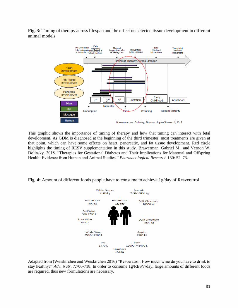
31
Fig. 3: Timing of therapy across lifespan and the effect on selected tissue development in different
animal models
This graphic shows the importance of timing of therapy and how that timing can interact with fetal
development. As GDM is diagnosed at the beginning of the third trimester, most treatments are given at
that point, which can have some effects on heart, pancreatic, and fat tissue development. Red circle
highlights the timing of RESV supplementation in this study. Brawerman, Gabriel M., and Vernon W.
Dolinsky. 2018. “Therapies for Gestational Diabetes and Their Implications for Maternal and Offspring
Health: Evidence from Human and Animal Studies.” Pharmacological Research 130: 52–73.
Fig. 4: Amount of different foods people have to consume to achieve 1g/day of Resveratrol
Adapted from (Weiskirchen and Weiskirchen 2016) “Resveratrol: How much wine do you have to drink to
stay healthy?” Adv. Nutr. 7:706-718. In order to consume 1g/RESV/day, large amounts of different foods
are required, thus new formulations are necessary.

32
1.7 GDM Animal Model:
Due to the ethical concerns of prescribing therapeutics to pregnant women for research,
many different animal models have been designed to study the effects of GDM on both mothers
and their offspring. These animal models provide us with a safer and more ethical way of testing
out different treatments and interventions as well as different timings and dosages which we can
later translate into clinical trials. GDM in humans is characterized by high blood glucose levels,
glucose intolerance, insulin resistance and increased gestational weight gain in mothers and
macrosomia in children with the onset near the end of the second trimester or beginning of the
third (Buchanan, Anny H. Xiang, and Page 2012; Jovanovic and Pettitt 2001; Troy J. Pereira et al.
2015). Most current animal models may manifest some of these phenotypes, but not all. Different
chemicals have been used to induce glucose and insulin intolerance, such as Streptozotocin, which
is used to destroy β-cells of the pancreas, however in GDM β-cell destruction does not occur, thus
this would not be a proper model to use (Pasek and Gannon 2013). Other models may use genetic
manipulations, such as the db/db mouse which has a mutated leptin receptor, thus affecting
appetite, and closely resembles the characteristics of GDM (Pasek and Gannon 2013). However,
the homozygous mutant female is not fertile, and only the heterozygous can be used. But,
clinically, most women do not tend to have this mutation (T J Pereira et al. 2015). Consequently,
many models have been using diets rich in fat to induce obesity, which is a key risk factor for the
development of GDM, and more closely resembles the clinical phenotype of GDM (Pasek and
Gannon 2013).

33
1.7.1 GDM Animal Model previously characterized in the Lab:
Previously in the Dolinsky Lab, an animal model was designed to assess how accurate it
would be at resembling a GDM pregnancy. Female Sprague-Dawley rats were randomly fed either
a low fat diet or a high fat and sucrose diet for 6 weeks prior to mating to induce either a pre-
diabetic and/or obesity state (Troy J. Pereira et al. 2015). They were then mated with lean males
and followed throughout pregnancy. During the third trimester, after the onset of GDM, a glucose
tolerance test (GTT) was performed to ensure that a glucose intolerance phenotype was observed.
Based on this model, the dams that consumed the high fat and sucrose diet had increased
gestational weight gain, impaired glucose tolerance, hyperinsulinemia, and moderate
hyperglycemia mid-gestation (Troy J. Pereira et al. 2015). All of these observations are very
similar to the clinical progression of GDM, suggesting that this model may resemble some of the
features of the disease as well. The offspring also exhibited similar responses that are observed in
children of mothers with GDM, such as increased obesity, macrosomia, insulin resistance, glucose
intolerance, and other early markers of cardio-metabolic disease (Troy J. Pereira et al. 2015).
Therefore, for my thesis work, we selected this model, but with some modifications to allow for
the pregnant dams to consume the RESV diet instead (Refer to Materials and Methods and Fig. 4).
34
1.8 Thesis Objectives
Obesity levels are rising worldwide, and with this increase, more women are entering their
pregnancies either obese or overweight, thus putting them at a greater risk of developing GDM.
Previous research has shown that a GDM pregnancy predisposes mothers to have increased obesity
and an increased risk for developing cardio-metabolic disease later on, such as T2D and CVD.
Similarly, GDM has detrimental effects on the offspring as they are also at a greater risk for cardio-
metabolic disease development. Current treatments include lifestyle therapies, such as diet and
exercise, and therapeutics such as insulin, glyburide and metformin. These treatments can be
efficient at lowering blood glucose levels; however they are all associated with serious adverse
events such as maternal and fetal hypoglycemia, macrosomia and potential delivery complications.
Even though these treatments may work in a subset of the population, failure rates are also rising,
therefore there is the need for new therapies to be researched and brought into the market. Natural
health products such as RESV, which has been shown to lower blood glucose levels and improve
cardio-metabolic parameters, may be a good therapy option for these patients. However,
appropriate dosages, timing of therapy, and the long term effect of RESV supplementation on the
offspring are currently unknown. I used a diet-induced GDM animal model previously established
in the Dolinsky lab, where rats were fed the high fat diet supplemented with RESV during the third
trimester of pregnancy and lactation only. My first aim was to show what effects RESV would
have on maternal metabolism, such as glucose tolerance and gestational weight gain compared to
dams not exposed to RESV. My second aim was to study the effects of RESV supplementation on
the metabolism in the young adult offspring (e.g. at 15 weeks of age) compared to the offspring
from Lean and GDM dams. And finally, since our lab has observed that GDM exposure induced

35
cardiac hypertrophy in the male offspring, my third aim was to assess what effect RESV
supplementation had on cardiac structure and function in the 15 week-old offspring.

36
1.9 General Hypotheses:
I hypothesized that maternal RESV supplementation during the third trimester of pregnancy and
lactation would prevent glucose intolerance and improve metabolic parameters in the dams.
I also hypothesized that maternal RESV supplementation would prevent metabolic health
disorders such as glucose intolerance, obesity and hepatic steatosis in the 15 week-old young
adult rat offspring.
In addition, I hypothesized that maternal RESV supplementation would prevent cardiac
hypertrophy and improve cardiac function in the 15 week old young adult male rat offspring.

37
Materials and Methods:
Diet-Induced GDM Model and Resveratrol Treatment
The University of Manitoba’s Central Animal Care committees approved all animal
procedures. 3 week-old female Sprague-Dawley rats were received from the Genetic Models
Center (GMC) core facility (University of Manitoba) and were housed 2 per cage. They were
randomly fed either a low-fat (LF) diet (10% kcal fat, Research Diets, D12450B) or a high fat and
sucrose (HFS) diet (45% kcal fat, Research Diets, D12451) for 6 weeks to induce obesity prior to
pregnancy. All rats were then mated with chow diet (i.e. regular rodent food) fed males within the
animal facility at the Children’s Hospital Research Institute of Manitoba (CHRIM) (one male per
two female rats) and diets were continued throughout pregnancy and lactation. Pregnancy was
confirmed 10 days after male breeders were separated from females via echocardiography scans.
Pregnant females were then housed alone until birth. Since GDM is typically diagnosed later in
pregnancy, a subgroup of female rats that consumed a HFS diet were switched to a HFS diet
supplemented with Resveratrol (45% kcal fat + 4g/kg RESV, Research Diets, D10020402) at the
beginning of the third trimester (i.e. day 14 of gestation) until birth and throughout lactation. This
dosage, which is equivalent to ~147.6 mg/kg/day based on the amount of food consumed per rat
per day, was previously determined to improve insulin sensitivity, glucose response, exercise
capacity, be safe and non-teratogenic during pregnancy (Bourque et al. 2012; V. W. Dolinsky et
al. 2011; V.W. Dolinsky et al. 2012; Vernon W Dolinsky et al. 2013). Therefore, we created three
different experimental mothers; Lean (fed LF diet), GDM (fed HFS diet) and GDM+RESV (fed
HFS diet + RESV). Pre-breeding, trimester 1, 2, and 3 random and fasted blood glucose levels
were measured from the tail vein using the ACCU-CHEK® Aviva glucose meters and glucose
38
strips (Roche Diagnostics). Serum was collected at all three trimesters for later analysis. Maternal
food consumption and body weights were measured weekly. Fetal echocardiography was done at
e18.5 to assess fetal heart structure and function.
Assessment of Postnatal Diet Effect on Offspring:
Within 24 hours of giving birth, total litter size, sex, pup weight and length were all
assessed, and litters were reduced to eight pups to avoid competition for food. Fetal hearts, livers
and kidneys were collected and placed in liquid nitrogen and subsequently stored away in -80°C
freezer for future studies. After weaning, at 3 weeks of age, offspring were randomly assigned to
consume either a LF or a HFS diet for 12 weeks to examine the interaction between prenatal
exposure and the postnatal diet and were housed 2 per cage when possible. Thus, six experimental
groups were created; Offspring from lean dams consuming either LF or HFS postnatal diet (Lean-
LF or Lean-HFS), offspring from GDM dams consuming either a LF or a HFS postnatal diet
(GDM-LF or GDM-HFS), and finally offspring from a GDM+Resv dam consuming either a LF
or a HFS postnatal diet (GDM+Resv-LF or GDM+Resv-HFS). Offspring food consumption and
body weights were measured weekly. At 14 weeks of age, an insulin tolerance test (ITT) was
performed on the offspring, while at 15 weeks of age a glucose tolerance test (GTT) was done.
Similarly, at 15 weeks of age, dual x-ray absorptiometry scans (DEXA) and echocardiography
were performed. Finally, at 15 weeks of age, rats were fasted for 4 hours prior to sacrifice. Some
rats were injected with saline, while others were injected with Insulin (1 mU/kg) 15 minutes prior
to sacrifice to allow for insulin-activated pathway studies. Rats were then euthanized by an
overdose injection of sodium pentobarbital, while whole blood was collected into Vacutainer®
tubes coated with 10.8 mg EDTA (BD, Franklin Lakes, NJ, USA) via cardiac puncture. These
tubes were then centrifuged at 4000 rpm for 20 minutes at 4°C and the serum was aliquoted into

39
1.5mL Eppendorf tubes and stored at -80°C for later analyses. Hearts, kidneys, livers, lungs, fat
pads, soleus and anterior tibialis muscle were dissected, weighed and placed in liquid nitrogen and
later stored at -80°C for future experiments or in 10% formalin or 4% paraformaldehyde (PFA) to
fix tissues for histological analyses. Another set of rats were sacrificed at e20.5-21 for cardiac
myocyte isolations (Fig. 5).
Fig. 5: Experimental Animal Model
Three different experimental dams were generated by feeding them either a low fat (Lean control), high fat
and sucrose (GDM), or high fat and sucrose + Resveratrol (GDM+RESV). The latter was given at the start
of the third trimester of pregnancy until the end of lactation. Throughout pregnancy, dams had their blood
glucose levels measured, and a T3 glucose tolerance test was performed at ~e18. Fetal echocardiography
was performed at ~e18.5, while fetal cardiomyocytes were isolated at ~e20.5. After a 3 week lactation
period, offspring were randomly assigned to either a low fat or a high fat and sucrose diet until 15 weeks
of age. At 14 weeks of age, insulin tolerance tests were performed. At 15 weeks of age, glucose tolerance
tests, DEXA scans and echocardiography were done. Offspring were then sacrificed and their tissues and
serum collected for later analyses. GDM: Gestational Diabetes Mellitus, RESV: Resveratrol, GTT: Glucose
Tolerance Test, ITT: Insulin Tolerance Test, LF: Low Fat Diet, HFS: High Fat and Sucrose Diet, DEXA:
Dual X-ray Absorptiometry Scans.

40
Insulin and Glucose Tolerance Tests (ITTs and GTTs):
Prior to mating and during the third trimester of pregnancy (i.e. ~e18), female rats were
given a glucose tolerance test to determine glucose response and whether resveratrol offered any
improvements as previously described (V. W. Dolinsky et al. 2011). Briefly, female rats were
fasted overnight for the pre-breeding GTT while pregnant rats were fasted for 4 hours for the
trimester 3 GTT. Fasted blood glucose from the tail vein was measured with the aid of ACCU-
CHEK® Aviva glucose meters and glucose strips (Roche Diagnostics) and serum was then
collected using a microvette® (Sarstedt, Germany). A glucose solution (2g/kg) was
intraperitoneally injected into the rats. Blood glucose levels were measured at times 0, 15, 30, 45,
60, 90, and 120 minutes. In the offspring, at 14 and 15 weeks of age, insulin and glucose tolerance
tests were performed as previously described (V. W. Dolinsky et al. 2011). Offspring were fasted
for 4 hours for the ITT and for 24 hours for the GTT procedure. The GTT was performed as
described above. For the ITT, an insulin solution (Sigma, St. Louis, MO, USA) (2g/kg) was
intraperitoneally injected into the rats. Blood glucose levels were measured at times 0, 30, 45, 60,
90, and 120 minutes. HOMA-IR index was calculated by multiplying fasting blood insulin
concentration and fasting blood glucose levels divided by 22.5, as previously described (Wallace,
Levy, and Matthews 2004).
Assessing Circulating and Tissue Factors:
Circulating concentrations of insulin (ALPCO®, Salem, NH, USA), free fatty acids
(Cayman Chemical, Ann Arbor ,MI, USA), and triglycerides (Wako Diagnostics, Mountain View,
CA, USA) from serum were assessed using colorimetric and fluorometric ELISA (Enzyme-Linked
Immunosorbent Assay) assay kits by following the manufacturer’s instructions.. Liver and cardiac

41
tissue were homogenized to measure triglyceride content via colorimetric assays (Biovision,
Milpitas, CA, USA) by following the manufacturer’s instructions.
Dual-Energy X-ray Absorptiometry Scans:
To further study fat distribution, content, and mass, plus bone mineral content and density,
DEXA (Dual Energy X-Ray Absorptiometry – Hologic Discovery ®) scans were performed on 15
week-old rat offspring at CHRIM (Children’s Hospital Research Institute of Manitoba) facilities.
A technician blinded to the offspring experimental groups took all the images once rats were
anesthetized using 5% Isoflurane gas for induction and 2% Isoflurane gas for maintenance. Rats
were placed back into their cages before they regained consciousness.
Echocardiography Scans:
To study the effects of the prenatal and postnatal diets on cardiomyopathy development in
the offspring exposed to GDM and subsequently with Resveratrol, in vivo echocardiography using
the Vevo 2100 high frequency ultrasound system (Central Animal Care Small Animal Imaging
Core Facility at the U of M) was used. A technician blinded to the experimental groups took all
echocardiography images including data from all rats in utero (e18.5) and in young adulthood
stages (15 weeks of age). I performed a blinded analysis utilizing the Vevo 2100 cardiac
measurement software package to assess cardiac structure and function. Left ventricular wall
thickness, intraventricular septal thickness and left ventricular chamber size and volumes were
used to assess morphological changes, while cardiac output and left ventricular ejection fraction
were measured to assess systolic function of the heart. Isovolumetric relaxation time and mitral
valve E/A ratio were used to assess diastolic function.

42
Liver Histology/pWAT Histology:
Hematoxylin and Eosin (H&E) staining of formalin fixed liver and adipose tissue was
performed by the University of Manitoba Histology Core Platform according to their standard
procedures. Individuals blinded to the offspring diets took liver images using the Zeiss Lab.A1
AX10 microscope from the CHRIM microscopy core platform. Images were taken at both 20x and
40x magnification, and 10 pictures of different sections per slide were taken to assess hepatocyte
size and fat droplet accumulation. Perirenal white adipose tissue (pWAT) was stained using the
same procedure as mentioned above, and images were taken by a blinded individual using the
same microscope as mentioned above. Images were taken at 20x magnification, and 6-10 pictures
of different sections per slide were taken to assess adipocyte number and size. Image analyses was
performed using ImageJ software (NIH, USA).
Western Blot:
For western blotting, frozen cardiac and liver tissue from both saline and insulin injected
animals were homogenized using ice-cold sucrose buffer containing 20mM Tris-HCl at pH 7.4,
50mM NaCl, 50mM NaF, 5mM sodium pyrophosphate, 0.25M sucrose, and supplemented with
protease and phosphatase inhibitor cocktails (Sigma, St. Louis, MO, USA), 1M dithiothreitol
(DTT), and 20mM sodium orthovanadate. Briefly, 20-30mg of tissue was homogenized with
300uL of lysis buffer using a homogenizer and the lysate was then centrifuged at 4000 rpm for 10
minutes at 4°C to remove nuclei and cellular debris. The Bradford protein assay was then used to
calculate protein concentrations (Bio-Rad). 20-25 ug of protein was separated using 8-10% SDS-
PAGE and transferred to a nitrocellulose membrane. Immunoblots were then visualized using the
SuperSignal® West Pico Chemiluminescent Substrate (Thermo Fisher Scientific, Rockford, IL,

43
USA) and a developer. All primary antibodies were purchased from Cell Signalling Technologies
(Danvers, MA, USA), and used at 1:1000, unless otherwise noted (Table 1). Secondary horseradish
peroxidase-conjugated antibodies were purchased from Jackson ImmunoResearch Laboratories
Inc. (West Grove, PA, USA) and were used at concentrations of 1:5000-1:20000. Protein
expression was normalized to either total protein expression or to Tubulin expression and then to
Lean-LF controls. Band intensity was analyzed using ImageJ software (NIH, USA).
Table 1. Primary antibodies used for immunoblotting analyses
Antibody Company Catalog
Number
Working Dilution
p-AKT (Ser473) and AKT Cell Signalling
Technology ®
9271/9272 1:1000
p-PI3K (Tyr458/Tyr199) Cell Signalling
Technology ®
4228 1:500
α-Tubulin Cell Signalling
Technology ®
2144 1:1000
SOD2 Abcam 13534 1:400
Serca2a Cell Signalling
Technology ®
4388 1:1000
ATGL Cell Signalling
Technology ®
2138 1:1000
p-mTOR (Ser2448) and
mTOR
Cell Signalling
Technology ®
2971/2972 1:1000
p-P70S6K (Thr389) and
P70S6K
Cell Signalling
Technology ®
9205/9202 1:1000
Gpx-1 Abcam 22604 1:1000
Catalase (D5N7V)` Cell Signalling
Technology ®
14097 1:1000
p-eEF2 (Thr56) Cell Signalling
Technology ®
2331 1:5000
eEF2 Cell Signalling
Technology ®
2332 1:1000
p-IRβ (Tyr1150/1151)
(19H7)
Cell Signalling
Technology ®
3024 1:2500
IRβ (4B8) Cell Signalling
Technology ®
3025 1:1000
p-IRS-1 (Ser1101) and
IRS-1 (D23G12)
Cell Signalling
Technology ®
2385/3407 1:1000

44
Antibodies and dilutions used for immunoblotting of liver and heart tissue of 15 week old male rat
offspring. AKT: Protein Kinase B, PI3K: Phosphatidylinositol 3 Kinase, SOD2: Superoxide Dismutase 2,
Serca2a: Sarco/endoplasmic Reticulum Calcium ATPase, ATGL: Adipose Triglyceride Lipase, mTOR:
Mammalian Target of Rapamycin, P70S6K: Ribosomal Protein P70 S6 Kinase, Gpx-1: Glutathione
Peroxidase 1, eEF2: Eukaryotic Elongation Factor 2, IRβ: Insulin Receptor β, IRS-1: Insulin Receptor
Substrate 1
RNA Isolation and qPCR:
For RNA isolation, 20-30 mg from saline injected liver tissue were homogenized using 600
uL of lysis buffer provided in the kit, and was further homogenized using a homogenizer column
as described by the manufacturer (Invitrogen, Carlsbad, CA, USA). The PureLink™ RNA Mini
Kit (Ambion Life Technologies, Carlsbad, CA, USA) was used to isolate and purify RNA using
the on-column PureLink® DNase treatment protocol without modifications. RNA integrity was
assessed by running samples on 1% agarose gel and visualized using the FluorChem® HD2 (Alpha
Innotech, Germany). RNA concentration was measured with the NanoDrop 2000 (Thermo Fisher
Scientific, Rockford, IL, USA). 1ug of RNA was converted to cDNA using the ProtoScript® II
First Strand cDNA Synthesis kit (New England BioLabs, Ipswich, MA, USA) according to the
manufacturer’s instructions. The QuantiTect® SYBR® Green PCR Kit (Qiagen) was used to
monitor the amplification of cDNA on the CFX Connect™ Real-Time PCR Detection System
(Bio-Rad). The CFX software was then used to visualize the expression of genes which were plated
in duplicate and analyzed using the 2-ΔΔCT method as described in (Livak and Schmittgen 2001).
Briefly, the target gene Ct was subtracted from the reference genes Ct creating the first ΔCT. This
value was then subtracted from the Ct corresponding to the Lean-LF control gene expression
generating the ΔΔCT. Finally, the following equation was used to calculate the relative fold-
change in expression (2^-ΔΔCT). Additionally, data were normalized to the geometric mean
(geomean) of eukaryotic translation initiation factor 2a (eIF2a) and cyclophilin A (cycA)
expression which were used as the reference genes as they were constant across all groups. Thus,

45
gene expression of target genes was expressed relative to the geomean of the housekeeping genes.
In this method, the geometric average of multiple housekeeping genes was taken resulting in a
more accurate representation than if taking the regular average of those genes alone
(Vandesompele et al. 2002). Therefore, instead of normalizing the data to a single gene, the data
was normalized to two different housekeeping genes, which provides a more accurate way of
normalizing qPCR data (Vandesompele et al. 2002). Data was then normalized to the Lean-LF
expression of each target gene. All primers were obtained from Integrated DNA Technologies
(IDT, Coralville, IA, USA). Primer sequences were validated and are provided in Table 2.
Table 2. Primer sequences used for quantitative real-time PCR
Validated primer sequences used for qPCR in liver tissue of 15 week old male rat offspring. Acc2: Acetyl
CoA-Carboxylase 2, Srebp-1c: Sterol Response Element Binding Protein 1c, Pparα: Peroxisome
Proliferator Activated Receptor α, Pcyt2: CTP:phosphoethanolamine cytidylyltrasferase 2, Sod2:
Superoxide Dismutase 2, PGC1α: Peroxisome Proliferator Activated Receptor γ Coactivator 1α, CycA:
Cyclophilin A, EIF2a: Eukaryotic Translation Initiation Factor 2a, Pepck-1: Phosphoenolpyruvate
Carboxykinase-1, G-6-P: Glucose-6-Phosphate

46
Cardiomyocyte Isolations:
To study the function of cardiac myocytes upon different diet exposures, fetal (e20.5-21)
ventricular cardiomyocytes from offspring of Lean, GDM and GDM+Resv dams were isolated as
described by (Kovacic et al. 2003) with some modifications. Briefly, fetal rat hearts at e20.5-21
were isolated and stored in ice-cold 1 X Phosphate Buffered Saline (PBS) solution. Hearts were
rinsed 3 times with 1x PBS and the atria and any other debris on the hearts were removed. The
ventricles were then minced with curved iris scissors and were transferred to a non-vented T25
tissue culture flask containing 17mL of cold 1x PBS, 1mL of filter sterilized 0.5% DNase/2%
Collagenase and 0.5 mL 2% Trypsin. This flask was agitated on a rotary shaker at 80 rpm for 20
minutes at 37°C to start the first dissociation. The contents were then mixed with 20 mL of pre-
warmed DF20 media (plus 20% fetal bovine serum (FBS), 1% penicillin/streptomycin and
50ug/mL gentamicin) and centrifuged at 800 rpm at room temperature for 2 minutes. The
supernatant was discarded, and 1X PBS, DNase/Collagenase and Trypsin were added to the pellet
and placed in the rotary shaker for another 20 minutes at 37°C to start the second dissociation. The
contents were transferred to a 50mL falcon tube containing 20mL of pre-warmed DF20 media and
spun at 800 rpm at room temperature for 1 minute. The supernatant was collected into another
50mL falcon tube and spun at 1700 rpm at room temperature for 7 minutes, the supernatant was
again removed, and 20 mL of pre-warmed DF20 media was added to the pellet. 1X PBS,
DNase/Collagenase and Trypsin were added to the pellet from the second dissociation to start the
third dissociation by placing the flask on the rotary shaker for another 20 minutes at 37°C. After
the final incubation, all the supernatants were mixed and spun at 1700 rpm at 4°C for 7 minutes.
The supernatant was removed, while the pellet was resuspended with 12mL of pre-warmed plating
media (DMEM/F12 (1:1), Horse Serum, FBS, 1% Penicillin/Streptomycin and 50 ug/mL

47
gentamicin). This was filtered through a 100 uM nylon cell strainer into a vented T75 tissue culture
flask and placed into a 37°C incubator for 1 hour. After 1 hour, the supernatant was transferred
into another T75 flask and incubated for an hour. After incubation, the supernatant was collected
into a 50mL falcon tube and spun at 1000 rpm for 2 minutes at room temperature. The resulting
pellet was then diluted with pre-warmed plating media. After cell counting, the cells were plated
on primaria dishes (Falcon) at a density of 500,000 cells/well. The next day, cells were washed
with 1x PBS as needed.
MitoSOX Experiment:
To assess reactive oxygen species production, namely superoxide, by the mitochondria in
the fetal cardiomyocytes, the molecular dye MitoSOX™ Red (Invitrogen™, #M36008) was
applied to the cells. Briefly, cardiomyocytes were washed twice with warm HBSS media, and
0.5mL of MitoSOX solution (plus Hoescht 1:1000) was added to each well. The plate was covered
with aluminum foil and incubated at 37°C for 20 minutes. The wells were washed with warm
HBSS, then again but for 5 minutes on a rocker at room temperature, and one final wash back in
the biosafety cabinet. Cells were then fixed with 4% para-formaldehyde (PFA) for 15 minutes in
a cold room. Wells were washed 3 times with cold 1x PBS. 0.5mL of cold 1X PBS was added to
the cells, the sides of the plate were parafilmed and the plate was wrapped in foil until imaging
using the Olympus Epifluorescence and Calcium Imaging Microscope (CHRIM) using the Cy3
(510/580 nm) and Dapi (350/461 nm) filters with 40X objective.

48
MitoTracker Experiment:
Similarly, MitoTracker™ Deep Red FM (Invitrogen™, #M22426) dye was used to assess
mitochondrial number within each well. Cells were washed twice with warm 1x PBS. MitoTracker
(plus Hoescht 1:1000) solution was added to the wells, the plate was wrapped in foil and incubated
at 37°C for 30 minutes. After incubation, the wells were washed twice with warm 1x PBS, and
then fixed for 15 minutes in the cold room with 4% PFA. Cells were then washed twice with 1x
PBS. 0.5mL of cold 1x PBS was added to the cells, the sides of the plate were parafilmed and
wrapped in tin foil until imaging using the Olympus Epifluorescence and Calcium Imaging
Microscope with the Cy5 (644/665 nm) and Dapi (350/461 nm) filters with 40X objective.
TMRM Experiment:
Finally, mitochondrial membrane potential was observed by applying TMRM
(Tetramethylrhodamine, Methyl Ester, Perchlorate) (Biotium, #70017, Fremont, CA, USA) dye.
Cells were washed once with warm 1x PBS, and then TMRM solution (plus Hoescht 1:1000) was
added to each well. The plate was covered with aluminum foil and incubated at 37°C for 30
minutes. Wells were then washed once with 1x PBS, and the cells were immediately imaged using
the Olympus Epifluorescence and Calcium Imaging Microscope with Cy3 (548/573 nm) and Dapi
(350/461 nm) filters with 40X objective. Hoescht 33342 (Thermo Scientific™, #62249) dye was
used in all three experiments at a concentration of 1:1000 to stain the nucleus blue.

49
Maternal and Offspring Islet Isolations:
This was performed by members in Dr. Christine Doucette’s Laboratory. After sacrificing
the animal, the pancreas was perfused through the bile duct with cold Collagenase V solution
(Sigma, St. Louis, MO, USA). The pancreas was then removed and placed into a 50mL conical
tube which contained 5mL of cold Collagenase V solution. The tube was placed in a water bath at
37°C for 15 minutes with manual shaking at 2-3 minute intervals to allow for proper pancreatic
digestion. 30-35 mL RPMI (11.1 mM glucose, Invitrogen) + 10% FBS + 1% P/S + 1% L-
glutamine, Invitrogen) was then added to the solution, and the tube was put on ice. In order to
create a homogenous solution, the tube was inverted and centrifuged at 290 x g for 30 seconds.
The supernatant was removed, and 20 mL of cold 1X PBS was used to wash the pellet by
centrifugation at 290 x g for 30 seconds. The pellet was re-suspended and the homogenous solution
was filtered through a 100 mm cell strainer (VWR, Pennsylvania, USA) which was previously
wetted with 1-2 mL of 1X PBS, into a 50 mL tube. The strainer was washed with 3 mL 1X PBS
while tapping to increase liquid flow. Islets trapped on the strainer were washed with 10 mL 1 X
PBS. A sterile 100 mm tissue culture plate was used to capture the islets from the strainer after
rinsing with 15 mL of cold media. A dissection microscope (Olympus SZ61; NCL 150 Illuminator)
was used to manually pick individual healthy looking islets. 200-300 islets were picked and
centrifuged at 290 x g for 30 seconds. The resulting pellet was re-suspended in 300 mL RLT buffer
(Qiagen, Valencia CA, USA) and was placed in the -80°C freezer until needed.

50
Glucose-Stimulated Insulin Secretion (GSIS) Assays:
This was performed by members in Dr. Christine Doucette’s Laboratory. 8-20 islets per
group were washed with cold KRB and were then incubated at 37°C in 2.8 mM glucose for 1 hour.
The tubes were put on ice and the KRB removed. 2.8 or 16.7 mM of cold glucose KRB was added
to the islets which were then incubated at 37°C for 30 minutes. A mouse insulin ELISA kit
(ALPCO, Salem, NH, USA) was used to measure insulin concentration in the media. Total insulin
content in the islets were measured as above and the insulin concentration was normalized to DNA
content of the islets.
Statistical Analysis:
Data are presented as the mean +/- standard error of the mean (SEM). GraphPad Software
(La Jolla, CA, USA) was used for all statistical analyses. One and Two-way Analysis of Variance
(ANOVA) were used to assess differences among maternal and postnatal diets with a Bonferroni
post hoc test to look at differences between specific groups. Any comparisons between two groups
were done using an unpaired t-test. Repeated measures tests were performed when necessary.
p<0.05 was defined as the level of significance.

51
Chapter 2:
Effects of Resveratrol Supplementation during Pregnancy
on Maternal Metabolism.

52
2.1 Introduction:
Worldwide obesity has more than tripled since 1975 and is reaching epidemic levels
(World Health Organization. 2017). Since more individuals are living a sedentary lifestyle, our
diets are becoming fattier and less healthy, and there are increased changes in genetics and in the
environment, then obesity levels will continue to rise, particularly among the younger population
(Corey and Kaplan 2014; Lehnert et al. 2013; Swinburn et al. 2011; World Health Organization.
2017). Obesity can be problematic as many conditions and diseases are known to be partially
caused by the increased fat accumulation, such as cardio-metabolic diseases, namely T2D and
CVD (Ng et al. 2014; Srivastava 2012). As obesity levels rise, more women of child-bearing age
will also be starting their pregnancies either obese or overweight (Simmons 2011). The
Developmental Origins of Health and Disease (DOHaD) theory had been suggested to explain how
the maternal intrauterine environment during development could have serious repercussions on
offspring health outcomes in both the short and long-term as molecular programming may be
taking place, thus increasing their risk for developing chronic diseases later on (D. Barker et al.
1993; D. J P Barker 2007; David J.P. Barker 1997).
Gestational diabetes mellitus (GDM) is considered to be one of the most common
complications of pregnancies (Ferrara 2007; Kim et al. 2010). GDM has been found to affect 1-
14% of pregnancies, and these numbers are expected to rise as obesity levels keep increasing in
the population (Ferrara 2007; Flegal et al. 2010; Kim et al. 2010). GDM is characterized by
increased gestational weight gain, mild hyperglycemia and insulin resistance with the onset at the
beginning of the third trimester of pregnancy (American Diabetes Association 2015). Importantly,
GDM is a transient condition, where blood glucose levels tend to go back to normal after
parturition, however, it can have long-lasting effects on both mothers and their offspring

53
(American Diabetes Association 2015; Buchanan, Anny H. Xiang, and Page 2012; Jovanovic and
Pettitt 2001). GDM during pregnancy has been found to predispose mothers to eclampsia-
preeclampsia, greater risk of caesarean sections, reduction in breast feeding initiation, increased
obesity, and eventually, glucose intolerance and insulin resistance, T2D and CVD development
(Poston, Harthoorn, and Van Der Beek 2011). A GDM pregnancy can also have serious problems
in the health of the offspring, such as macrosomia, birth defects or malformations, hypoglycemia,
and cardio-metabolic disease development (Arshad, Karim, and Hasan 2014; Hod et al. 1991;
Kaaja and Rönnemaa 2008; Persson et al. 2017). These offspring effects will be discussed in the
next chapter.
Current treatments include lifestyle therapies, such as diet and exercise, to maintain proper
glycemic levels during pregnancy and the oral-antidiabetic drugs metformin and glyburide, while
insulin injections may also be required in serious cases where women do not respond to previous
treatments (Lipscombe et al. 2018). These interventions generally work in the population, however
they may increase the risk for macrosomia and both fetal and maternal hypoglycemia which could
cause harm to the mother and her infant, as recently reviewed in (Brawerman and Dolinsky 2018).
Additionally, we do not fully understand the long-term effects that interventions administered
during pregnancy could have on the developing baby. Therefore, newer treatments for GDM
should be explored to circumvent problems associated with existing interventional options (Fig.
1).
Maternal exercise has been shown to have some benefits when done by obese pregnant
women or women who already have GDM as they did not experience as many hypoglycemic
events as women on glyburide or metformin did, and even had lower blood glucose levels when
compared to women that did not exercise (Barakat et al. 2013; Koivusalo et al. 2016). However,

54
exercise intensity and duration is still problematic which explains why exercising during
pregnancy is also associated with poor compliance (Nelson, Matthews, and Poston 2010).
Resveratrol (RESV) is a natural health product that has been found to mimic several of the
beneficial effects that we observe with exercise training on metabolic tissues. This is because
RESV activates similar molecular pathways as exercise, so it may become a potential add-on
therapy (V. Dolinsky and Dyck 2014; Vernon W. Dolinsky and Dyck 2011; Timmers et al. 2011).
Importantly, RESV was reported to efficiently decrease blood glucose levels (Mohammadshahi,
Haidari, and Soufi 2014; Yao et al. 2015). Furthermore, in animal studies, RESV supplementation
during pregnancy further decreased maternal hyperglycemia, improved insulin sensitivity, reduced
triglycerides and cholesterol levels and increased the expression of anti-oxidants, potentially
providing a better intrauterine environment for fetal development (C. K. Singh et al. 2011, 2013;
Yao et al. 2015). Finally, RESV was also found to be safe, well tolerated and non-teratogenic in
both human and animal studies (Bourque et al. 2012; C. K. Singh et al. 2011, 2013; B. Wicklow
et al. 2015; Williams et al. 2009).
To date, no studies have been performed to assess the effects of RESV supplementation
for both mother and offspring short and long-term health outcomes in a diet-induced GDM model
(only in a genetic db/+ model, (Yao et al. 2015)). Additionally, this is the only study that
administered the intervention at the onset of GDM (i.e. third trimester), as most previous studies
supplemented diets with RESV either before gestation, throughout pregnancy and lactation, or
only during lactation which may not be clinically significant (Bourque et al. 2012; Roberts et al.
2014; C. K. Singh et al. 2011, 2013; Tanaka et al. 2017; Yao et al. 2015; Zou et al. 2017).
Our objective was to determine whether 4g/kg (~146 mg/kg/day) of RESV
supplementation in the third trimester of pregnancy, at the onset of GDM, and lactation would

55
prevent maternal glucose intolerance and improve metabolic parameters in pregnant rats. We
hypothesized that RESV supplementation would reduce blood glucose levels, thus improving
maternal intrauterine environment, and preventing GDM-related complications on the mothers.
We showed that RESV supplementation did prevent glucose intolerance, lowered blood glucose
levels in the third trimester and improved insulin secretion. We also showed no significant changes
occurred in litter sizes or sex distribution and no serious adverse events were observed with RESV
supplementation.
2.2 Materials and Methods – Refer to page 37.
2.3 Results:
2.3.1 Maternal RESV supplementation during pregnancy prevented glucose
intolerance, restored blood glucose levels, and improved insulin secretion, without
affecting gestational weight gain in the rat dams:
After 6 weeks on their respective diets, the HFS fed females had a tendency of weighing
more, a 1.17 fold increase, than the lean group (Fig. 6A), however their pre-breeding GTT was not
different (Area under the curve for Lean 1089 ± 29.46 vs. GDM 1189.6 ± 30.14). This is consistent
with the clinical phenotype of GDM where there is no glucose intolerance prior to pregnancy.
Females were then mated with chow fed lean males. During pregnancy, the HFS fed dams
continued gaining weight at a higher pace than the LF fed dams, in turn, their gestational weight
gain (GWG) was significantly higher, exhibiting a 2.36 fold increase, than the Lean dams (Fig.
6B). At the beginning of the third trimester of pregnancy, which is when GDM is diagnosed
clinically, a subgroup of females consuming the HFS diet were supplemented with the RESV diet
(termed GDM+RESV). Consumption of RESV diet did not affect the GWG associated with a HFS
diet because their GWG had a 2.48 fold significant increase when compared to the Lean dams
56
(Fig. 6B). But, maternal food consumption from the third trimester of pregnancy was significantly
lower in the GDM+RESV dams when compared to the Lean dams (Fig. 6C). After calculating the
energy intake per kg of body weight, there were no differences among the maternal diets (Fig. 6D
and Table 3).
Closer to the end of pregnancy (i.e. ~18) a trimester 3 GTT was performed on the pregnant
rats to assess glucose intolerance and GDM development. Even though fasting blood glucose
levels were unchanged, 15 minutes after glucose injection, the GDM dams had a 1.5 times
significant increase in blood glucose concentration when compared to both GDM and
GDM+RESV dams (Fig. 6E and 6F). Additionally, it took 2 hours for the GDM dams to reach
basal levels, while it only took 90 minutes for both Lean and GDM+RESV dams to reach their
basal levels (Fig. 6E). Thus, the GDM dams were more glucose intolerant than both Lean and
GDM+RESV dams (Fig. 6E and 6F), suggesting there was an inability of GDM dams to take up
glucose as efficiently as either Lean or GDM+RESV dams. However, with maternal RESV
supplementation glucose response was normal (Fig. 6E and 6F), implying that these animals can
better respond to the increased sugar levels. Additionally, random blood glucose levels were
measured before mating and at each trimester of pregnancy, and a ~1.6 fold significant increase in
blood glucose in the second trimester for both GDM and GDM+RESV dams was observed when
compared to the Lean dams. Interestingly, during the third trimester (i.e. after the change to RESV
diet), blood glucose levels significantly decreased in the GDM+RESV dams to the same levels
observed in the Lean, while the GDM dams still exhibited blood glucose levels that were 1.33
times higher than both Lean and GDM+RESV dams (Fig. 6G). These findings suggested that
GDM+RESV dams were capable of dealing with the extra sugars better than the GDM dams.

57
As blood glucose levels are closely tied with how much insulin is generated by the β-cells
of the pancreas, the insulin concentration in the maternal serum during the third trimester of
pregnancy was quantified using an ultra-sensitive ELISA kit (ALPCO®). The GDM dams had
almost half the concentration of insulin observed in the Lean and GDM+RESV dams (Fig. 6H).
This result could explain the reduction in blood glucose levels observed in those animals. The
HOMA-IR index, a measure of insulin resistance, showed a similar trend, indicating an increase
of almost double in the GDM+RESV dams compared to the GDM dams (Fig. 6I). Even though
the HOMA-IR was increased in the GDM+RESV dams, they did not show signs of insulin
resistance or impaired glucose metabolism. Additionally, as part of a collaboration with Dr.
Christine Doucette’s laboratory at CHRIM, glucose stimulated insulin secretion (GSIS) assays
were done on the islets that were isolated from pancreas extracted from the dams. In high glucose
(HG) media, insulin secretion was impaired in the islets from GDM dams, as they secreted almost
half of what the Lean dams secreted, whereas GDM+RESV dams were able to secrete 3.87 times
more insulin than the GDM dams (Fig 6J and 6K). However, with low glucose concentrations in
the media, insulin secretion by isolated islets was similar among all three groups of dams (Fig. 6J).
These findings imply that RESV rescued insulin secretion after exposure to glucose in the third
trimester in GDM+RESV dams.

58
Table 3. Average daily food consumption by pregnant mothers (from T3 to end of lactation)
Daily amount of food consumed and energy intake by pregnant mothers from T3 to the end of lactation.
N=3-10. *p values represent significant differences (<0.05) between GDM+RESV and Lean after One
Way ANOVA with Bonferroni post-hoc tests. GDM: Gestational Diabetes Mellitus, RESV: Resveratrol,
T3: Trimester 3

59
Fig. 6: Maternal RESV supplementation during pregnancy prevents glucose intolerance, restores
blood glucose levels, and improves insulin secretion, without affecting gestational weight gain.
Lean GDM GDM+RESV0
500
1000
1500
2000
GT
T A
rea U
nd
er
Cu
rve
I
C D
H
A
0 1 2 3 4 5 6 7 8 90
200
400
600
Weeks
Bo
dy W
eig
ht
(g)
Lean
GDM
GDM+RESV
Pregnant
* *
Lean GDM GDM+Resv0
50
100
150
Gesta
tio
nal W
eig
ht
Gain
(g
)
* *
B
0 30 60 90 1200
5
10
15
20
25
Time (min)
Glu
co
se (
mM
)
Lean
GDM
GDM+RESV
*#
Pre-mating T2 T30
2
4
6
8
10
Glu
co
se (
mM
)
Lean
GDM
GDM+RESV
*# * *
Lean GDM GDM+RESV0
1
2
3
4
5
Insu
lin
Secre
tio
n In
dex
(ng
/mg
DN
A/h
r)
E
LG HG0
5
10
15
20
Insu
lin
Secre
ted
(ng
/mg
DN
A/h
r)
Lean
GDM
GDM+RESV
*
*
Lean GDM GDM+RESV0
1
2
3
Insu
lin
Co
ncen
trati
on
(ng
/mL
)
F
G
p=0.0503
J K
*
*# *# *#
*#
#

60
(A) Change in body weights over time – arrow indicates timing of RESV supplementation, (B) Gestational
Weight Gain, (C) Food Consumption, (D) Energy Intake adjusted to body weight, (E) Trimester 3 GTT,
(F) GTT Area Under the Curve, (G) Blood Glucose Levels pre-pregnancy and throughout pregnancy, (H)
Insulin Concentrations at Trimester 3, (I) HOMA-IR Index, (J) Insulin Secretion, (K) Insulin Secretion
Index. Maternal body weights and gestational weight gain N= 6-9, Maternal food consumption and energy
intake N=3-10, T3 GTT N=6-8, T3 insulin concentration N=4-8, Blood glucose levels N= 2-8, Insulin
assays N= 5-18. p-values represent significance (<0.05) after One Way, Two Way ANOVA, Repeated
Measures ANOVA or multiple T-tests where applicable with Bonferroni post-hoc tests. *p<0.05 vs. Lean.
#p<0.05 vs. GDM+RESV. GTT: Glucose Tolerance Test, HOMA-IR: Homeostatic Model of Assessment
– Insulin Resistance, GDM: Gestational Diabetes Mellitus, RESV: Resveratrol.

61
2.4 Discussion/Conclusion:
The objective was to determine the effects of resveratrol supplementation during the third
trimester of pregnancy and lactation on female Sprague-Dawley rats exposed to GDM. Previous
clinical studies have found that RESV supplementation decreased the risk of developing GDM by
improving both lipid and blood glucose levels, with the addition of also lowering blood pressure
in overweight pregnant women (Ding et al. 2017; Malvasi et al. 2017). Thus, rats consumed a high
fat and sucrose diet for 6 weeks before mating to induce obesity, an important risk factor for the
development of GDM. After 6 weeks on the diet, a pre-breeding GTT was performed to assess
whether these rats were glucose intolerant prior to being mated, and as expected, there were no
differences. Since these animals were not pregnant, the no change in pre-breeding GTT suggests
that consuming a high fat diet is not by itself a trigger for glucose intolerance, but rather a second
hit or stressor (i.e. pregnancy) is required for the onset. This was more clearly observed after
performing a trimester 3 GTT. Since GDM usually occurs at the beginning of the third trimester
of pregnancy, a GTT was done near the end of pregnancy (i.e. ~e18) to determine if at this time
point the rats were glucose intolerant. Indeed, the GDM dams exhibited an abnormal response,
implying that they were not able to clear up the increased glucose load as efficiently as the Lean
dams did. More importantly, the GDM+RESV dams exhibited a very similar response to that of
the Lean, so their glucose tolerance was normal. These improvements in glucose homeostasis after
RESV supplementation were also observed in other animal studies (Roberts et al. 2014; C. K.
Singh et al. 2011, 2013; Yao et al. 2015).
Surprisingly, gestational weight gain was not improved by RESV supplementation. In fact,
the GDM+RESV dams had a similar GWG when compared to the GDM dams, and a significantly
higher GWG when compared to the Lean. This is the opposite of what I was expecting to observe

62
as other research has shown a decrease in GWG (Roberts et al. 2014). However, Roberts et al.
used a Japanese macaque animal model, their RESV dosage was 0.37% mixed in with the diets,
and these animals consumed RESV for 3 months before they were mated and throughout gestation
(Roberts et al. 2014). These differences in dosage, timing and animal model may explain the
different results observed. Interestingly, GDM+RESV dams consumed less food than the other
two groups of dams from the beginning of the third trimester until lactation, however their energy
intake was the same. This suggests that one week on the RESV diet was not enough to counteract
the effects of almost 9 weeks on the HFS diet on weight gain.
More importantly, and as other studies have also shown (C. K. Singh et al. 2011, 2013;
Yao et al. 2015), RESV supplementation reduced blood glucose levels in the third trimester,
suggesting that these animals had a better response to deal with the increase in glucose load. This
further agrees with the T3 GTT results, where more glucose may had been taken up by the cells
from the circulation, reducing the blood glucose levels measured. At the same time, the insulin
concentration in these dams was measured using serum collected at time 0 of the T3 GTT.
GDM+RESV dams had more insulin in the circulation than GDM dams did, which could be a
reason as to why blood glucose levels decreased in these animals. Since the GDM dams had the
least amount of insulin in their serum, it was hypothesized that a defective insulin secretion by the
β-cells of the pancreas in GDM dams caused a reduction in insulin in the circulation which could
have induced hyperglycemia during pregnancy. This hypothesis appeared to be correct as both
human and animal studies have found associations between β-cell failure and GDM development,
as reviewed in (Banerjee 2018). To further elucidate maternal β-cell function, I collaborated with
Dr. Christine Doucette’s laboratory (at CHRIM) to measure the amount of insulin secretion by the
β-cell upon exposure to low levels of glucose (i.e. control levels) or high glucose levels (i.e. after

63
consumption of a meal). They found that when exposed to the high glucose levels, GDM+RESV
dams were able to secrete more insulin than both GDM and Lean dams, while GDM dams had the
least amount of insulin secretion from all three groups. This data suggests that the β-cell may be
dysfunctional in GDM dams, while RESV supplementation may be maintaining proper β-cell
function, and thus proper insulin and glucose response. Other studies have measured circulating
insulin levels and also found them to be increased after RESV supplementation, however they did
not assess maternal β-cell function (C. K. Singh et al. 2011, 2013; Yao et al. 2015). Thus, this
study is unique in that we showed a plausible explanation as to how RESV supplementation
prevented maternal glucose intolerance by increasing insulin secretion from β-cells of the
pancreas.
As RESV supplementation seemed to improve glucose homeostasis in the dams, it is
possible that the fetal environment may be healthier and better for proper fetal development than
the environment found in the GDM dams. Furthermore, no serious adverse events were observed,
suggesting that RESV supplementation during pregnancy is likely to be well tolerated and safe.
The only potential side effect associated with RESV supplementation is gastrointestinal problems,
such as constipation. After dissection of the mothers, we did notice that some constipation may
have occurred in the intestines of these animals. However, the lack of serious side effects was also
observed in other clinical and animal studies (Bourque et al. 2012; C. K. Singh et al. 2011, 2013;
B. Wicklow et al. 2015; Williams et al. 2009). The long-term effects of RESV on the offspring
(up to 15 weeks of age) in this model will be described in the next chapter of this thesis.
Taken together, these maternal data seems to show that RESV supplementation during the
third trimester of pregnancy, at the onset of GDM, and during lactation prevents glucose
intolerance, lowers blood glucose levels and improves β-cell function thereby increasing insulin

64
secretion. Additionally, no serious side effects were observed. Since RESV supplementation had
these positive outcomes in the dams, it is possible that the fetal environment may have also
improved providing protection from GDM-induced complications in the offspring.

65
Chapter 3:
Effects of Resveratrol Supplementation during Pregnancy
on the Metabolism of the 15-Week-Old Young-Adult Rat
Offspring

66
3.1 Introduction: This work is based on a manuscript in preparation for scientific review.
As obesity levels rise worldwide, particularly among women of child-bearing age, so are
the cases of gestational diabetes mellitus (GDM) (Simmons 2011; World Health Organization.
2017). GDM is considered to be one of the most prevalent complications of pregnancy, as
anywhere from 1 to 14 percent of all pregnancies can be affected (Ferrara 2007; Kim et al. 2010).
GDM is characterized by increased gestational weight gain with the onset of hyperglycemia,
glucose intolerance and insulin resistance during pregnancy that is typically diagnosed in the
second or third trimester (American Diabetes Association 2015; Buchanan, Anny H. Xiang, and
Page 2012; Jovanovic and Pettitt 2001; Troy J. Pereira et al. 2015). GDM is very similar to type 2
diabetes (T2D) because the development of both conditions are characterized by obesity, insulin
resistance and the metabolic syndrome (Poston, Harthoorn, and Van Der Beek 2011; Rodriguez et
al. 2006).
A GDM pregnancy is associated with birth complications in newborns such as shoulder
dystocia, macrosomia, neonatal hypoglycemia, and other malformations (Arshad, Karim, and
Hasan 2014; Hod et al. 1991; Kaaja and Rönnemaa 2008; Persson et al. 2017). Consistent with the
Developmental Origins of Health and Disease (DOHaD) theory (D. Barker et al. 1993; D. J P
Barker 2007; David J.P. Barker 1997), human studies have shown an association between exposure
to GDM and later cardio-metabolic disease development in the offspring (B. Wicklow et al. 2018)
and as recently reviewed in (Prasoon Agarwal et al. 2018). These observations have been
confirmed in animal model studies of maternal obesity and GDM, as reviewed in (S. Kereliuk,
Brawerman, and Dolinsky 2017). For example, we recently showed that a diet-induced GDM
pregnancy in rats led to increased obesity, insulin resistance and hepatic steatosis development in

67
the 15 week old rat offspring even after the consumption of a low fat diet postnatally (Troy J.
Pereira et al. 2015). Furthermore, consuming a high fat and sucrose diet postnatally exacerbated
the metabolic effects in the offspring (Troy J. Pereira et al. 2015). Therefore, it is crucial to treat
GDM in order to prevent adverse health outcomes in the offspring.
While insulin, metformin and glyburide are used in pregnant women with GDM and are
generally considered safe, they are associated with some potential adverse effects such as maternal
and fetal hypoglycemia and fetal macrosomia, as reviewed in (Brawerman and Dolinsky 2018).
Maternal exercise interventions during pregnancy have been shown to have some positive effects
on maternal glucose homeostasis, as well as the prevention of fetal programming effects in the
offspring (Beeson et al. 2018; Brawerman and Dolinsky 2018). However, initiation and adherence
to a program of exercise may be difficult for many sedentary and overweight pregnant women.
Resveratrol (RESV) is a natural polyphenolic compound that is produced by plants which
has been found to mimic many of the positive cardio-metabolic effects of exercise (V. Dolinsky
and Dyck 2014). RESV was found to be safe, well tolerated and non-teratogenic in animal studies
of pregnancy (Bourque et al. 2012; C. K. Singh et al. 2011, 2013; Williams et al. 2009). In addition,
RESV reduced body weights, prevented hepatic steatosis and cardiac hypertrophy and improved
glucose homeostasis and insulin sensitivity in the offspring in models of severe hypoxia, obesity
and GDM (Bourque et al. 2012; Tanaka et al. 2017; Yao et al. 2015; Zou et al. 2017). However,
no studies have been performed to date on a diet-induced GDM model to determine what effects
RESV supplementation during pregnancy would have on the short and long-term health outcomes
in the offspring. Additionally, this is the first study that supplemented maternal diets with RESV
specifically in the third trimester of pregnancy, which is when GDM is typically diagnosed, while
other studies supplemented with RESV during different time points throughout gestation when

68
GDM may not necessarily be present (Bourque et al. 2012; Roberts et al. 2014; C. K. Singh et al.
2011, 2013; Tanaka et al. 2017; Yao et al. 2015; Zou et al. 2017).
Thus, the objective of this study was to examine the effects of supplementing maternal
diets with 4g/kg of RESV, a dosage equivalent to 147.6 mg/kg/day, during the third trimester of
pregnancy and lactation on the metabolic health outcome in the 15 week old rat offspring. Also,
we wanted to determine the outcome of consuming either a LF or a HFS postnatal diet on metabolic
syndrome development in the offspring and whether previous maternal RESV consumption would
protect the offspring from disease development (Fig. 5). Based on the work described in the
preceding chapter (i.e. Chapter 2), maternal RESV supplementation prevented glucose intolerance,
improved β-cell function of the pancreas, increased insulin secretion and lowered blood glucose
levels in the dams, potentially providing a better intrauterine environment for proper offspring
development. Therefore, we hypothesized that RESV supplementation would prevent metabolic
health disorders such as obesity, glucose intolerance and hepatic steatosis in the 15 week-old rat
offspring. In this chapter, we showed that RESV supplementation prevented fetal macrosomia,
obesity, and hepatic steatosis, while it also improved glucose homeostasis and insulin sensitivity
in the rat offspring. In addition, the expression of several metabolic genes in the liver of the
offspring were modified by maternal RESV supplementation. We also observed differences
between male and female offspring response to RESV supplementation, particularly in terms of
obesity and fat accumulation.
3.2 Materials and Methods: Refer to page 37.

69
3.3 Fetal Offspring Results:
3.3.1 Maternal RESV supplementation was safe during pregnancy and prevented
macrosomia in males but not in females:
Within 24 hours of offspring birth, pups were sexed, weighed, and their lengths were
measured. No changes in litter sizes or sex distribution were observed (Figs. 7A and 7B, and Table
4), implying that RESV supplementation did not have adverse effects during development. Pups
were then reduced to eight to avoid competition for food, and were left alone with their respective
mothers for the three week lactation period where these pups consumed the same diet their mothers
did (i.e. LF, HFS, or HFS+RESV diets). Surplus neonatal rats were then sacrificed and their hearts,
livers and kidneys were collected for future analyses. Male offspring from GDM dams exhibited
a significant 1.21 fold increase in body weight at birth when compared to the Lean offspring, and
had a 1.11 significant fold increase when compared to GDM+RESV offspring (Fig. 7C). Thus,
GDM neonates were large for gestational age, or macrosomic, a common clinical consequence
observed in GDM pregnancies (Arshad, Karim, and Hasan 2014; Scholl and Chen 2002). On the
other hand, female offspring from both GDM and GDM+RESV dams exhibited a significant 1.23
and 1.18 fold increase respectively when compared to the Lean offspring. However, there were no
changes when GDM female offspring was compared to GDM+RESV offspring (Fig. 7D). Thus,
these changes may imply that different sex specific characteristics may be affecting the response
to RESV providing benefits in males and no effects in females. Additionally, body lengths did not
differ among neonates (Fig. 7E). So, after normalizing body weights to body lengths, no changes
were observed, suggesting that the body weights were not influenced by the size of the animal
(Fig. 7F).

70
Table 4. Litter Sizes and Sex Distribution
Upon 24 hours of offspring birth, litter sizes were calculated and offspring sexed. No significant differences
were observed. Litter Size N=6-11; Sex Distribution N=3-9. One-Way ANOVA with Bonferroni post-hoc
tests was used. GDM: Gestational Diabetes Mellitus, RESV: Resveratrol

71
Fig. 7: Maternal RESV supplementation did not cause adverse developmental effects and
prevented macrosomia in males but not in females.
A B
C D
E F
*# * *

72
(A) Litter Size, (B) Sex Distribution, (C) Male Neonatal Body Weights upon birth, (D) Female Neonatal
Body Weights upon birth, (E) Neonatal offspring body lengths (F) Body Weight to Body Length Ratio.
Litter Size & Sex Distribution by Litter N=3-11, Body Weights N=28-53. p-values represent significance
(<0.05) after One Way ANOVA with Bonferroni post-hoc tests. *p<0.05 vs. Lean, #p<0.05 vs.
GDM+RESV. GDM: Gestational Diabetes Mellitus, RESV: Resveratrol.
3.4 15-Week-Old Young-Adult Offspring Results:
3.4.1 Maternal RESV supplementation prevented the increase in obesity in the
young adult male rat offspring, but not in the female offspring:
After the three week lactation period, offspring were randomly weaned onto either a LF or
a HFS diet for the remainder of the study, until 15 weeks of age, when offspring were sacrificed
and their serum and tissues collected (i.e. important to remember these offspring did not consume
RESV diet postnatally after the lactation period). Body weights and food consumption were
recorded every week. As expected, male offspring that consumed the HFS diet postnatally gained
more weight than the male offspring that consumed the LF diet when comparing offspring from
the same maternal groups, as shown in Fig. 8A. However, Lean-LF offspring gained the least
amount of weight among all offspring groups, while GDM-HFS and Lean-HFS gained the most
(Fig. 8A), showing the impact a HFS diet has on the offspring even when exposed to a LF diet
prenatally, consistent with the mismatch hypothesis. Of note, GDM+RESV-LF and GDM+RESV-
HFS offspring were in the middle of the body weight growth curve, indicating a significant
reduction in their body weights when compared to both GDM-HFS and Lean-HFS (Fig. 8A). Body
weights measured at 15 weeks of age prior to sacrifice showed a significant 1.17 fold increase in
GDM-HFS offspring when compared to Lean-LF, and significant 1.14 and 1.21 fold increases
when compared to GDM-LF and GDM+RESV-HFS, respectively (Fig. 8B). Female offspring
exhibited a completely different response in terms of body weights. In fact, the GDM+RESV-HFS
female offspring were the most obese when compared to all the other groups (Fig. 8C). When body

73
weights were measured at 15 weeks of age prior to sacrifice, the same result was seen, as the
GDM+RESV-HFS female offspring were 1.32 times heavier than the Lean-LF offspring, while all
other groups had similar body weights regardless of maternal or postnatal diets (Fig. 8D), once
again highlighting the sex specific responses that GDM in utero and RESV exposure may have on
metabolism in the offspring. Furthermore, upon examination of food consumption and energy
intake in both male and female offspring, no changes were observed (Figs. 8E, 8F, 8G, and 8H,
Table 5). Thus, differences in metabolic rates and substrate utilization could have led to alterations
in fat storage, which could partially explain the reduction in body weights observed in the
GDM+RESV male offspring even when energy intake among all offspring groups was the same.
3.4.2 A high fat and sucrose postnatal diet resulted in increased fat accumulation in
the male rat offspring regardless of maternal diet, while RESV supplementation
increased fat mass in females:
To further evaluate fat content and distribution in the young adult male rat offspring, dual
energy x-ray absorptiometry (DEXA) scans were performed at the CHRIM facilities by a blinded
and experienced technician. Upon visual examination of the body shape of the offspring, there
appeared to be more abdominal fat in the offspring fed a HFS diet postnatally when compared to
offspring fed a LF diet, regardless of maternal diet (Fig. 9A). DEXA results further showed a
significant 1.5 fold increase in percent body fat in the GDM-HFS offspring when compared to the
Lean-LF offspring, and a significant 1.23 and 1.38 fold increases when compared against Lean-
HFS and GDM-LF offspring, respectively (Fig. 9B). Interestingly, the GDM+RESV-HFS
offspring had 1.3 times more percent body fat than both Lean-LF and GDM+RESV-LF offspring
(Fig. 9B), confirming that the postnatal HFS diet was a significant determinant for body fat
percentage. The increase in abdominal fat mass in the HFS fed offspring was further examined by
weighing fat pads from 15 week-old offspring at sacrifice. Perirenal white adipose tissue (pWAT),

74
or the fat pad surrounding the kidneys, and gonadal white adipose tissue (gWAT), were all heavier
in offspring that consumed the HFS diet postnatally when compared to offspring fed a LF diet
(Fig. 9C). In fact, the GDM-HFS offspring had significant 2.12 and 1.78 fold increases in pWAT
weights when compared to Lean-LF and GDM-LF offspring, respectively (Fig. 9C). Interestingly,
the GDM+RESV-HFS offspring had significant 1.7 and 1.38 fold increases when compared to
Lean-LF and GDM+RESV-LF offspring, respectively (Fig. 9C), showcasing that the HFS
postnatal diet consumption, regardless of the maternal diet, resulted in increased fat accumulation.
Moreover, the combination of GDM and postnatal HFS diet induced an additive increase in pWAT
mass, but RESV decreased this effect by ~25% (P>0.05). Similarly, the GDM+RESV-HFS
offspring had a significant 1.68 and 1.4 fold increases in gWAT weights when compared to Lean-
LF and GDM+RESV-LF offspring, respectively, again highlighting the impact of a HFS postnatal
diet (Fig. 9C). Therefore, this increase in visceral fat pad mass was directly associated with the
percent body fat found in the offspring. The fat mass in these animals, which was another
parameter reported in the DEXA scan, was also dependent on whether animals consumed a HFS
or LF diet postnatally, as the GDM-HFS offspring had a significant 1.59 fold increase in fat mass
when compared to the GDM-LF offspring (Fig. 9D).
When female offspring were assessed, the results were different from males. No changes
were observed in the GDM offspring, however, the GDM+RESV-HFS offspring had a significant
2.33 and 1.92 fold increase in pWAT weights when compared to the Lean-LF and GDM+RESV-
LF offspring, respectively (Fig. 9E). Similarly, the gWAT weight was significantly increased by a
factor of 2.27 when comparing the GDM+RESV-HFS to the Lean-LF offspring (Fig. 9E). This
suggests that in utero RESV exposure had additive effects on fat accumulation in female offspring

75
fed a postnatal HFS diet, and that GDM did not affect the female offspring. Again, this is another
example of how sex specific differences are potentially affecting the results.
Lastly, perirenal adipocyte, or fat cell, diameter and number in male offspring were
calculated based on H&E stained adipocytes looked under a microscope (Fig. 9F). As it was noted
with the previous results, the offspring fed a HFS postnatal diet had adipocytes with the biggest
diameter (Fig. 9G), suggesting increased fat accumulation, regardless of maternal diet. In addition,
and as expected, the adipocyte diameter was inversely related to the adipocyte number per section
(Fig. 9H). In other words, as the adipocyte diameter increased, fewer adipocytes were present per
section.

76
Fig 8: Maternal RESV supplementation reduced obesity in the 15 week-old male rat offspring, but
not in the female rat offspring
A B
C D
E F
G H
/day/week /day/week
/day/week
/day/week

77
(A) Male body weight gain per week of Lean, GDM and GDM+RESV offspring, (B) Male body weight at
sacrifice, (C) Female body weight gain per week of Lean, GDM, and GDM+RESV offspring, (D) Female
body weight at sacrifice, (E) Male offspring food consumption by litter/day/week, (F) Male energy intake
per litter/kg body weight/day/week, (G) Female offspring food consumption by litter/day/week, (H) Female
energy intake per litter/kg body weight/day/week. Male Body Weights N=12-23, Female Body Weights
N=3-8, Male Food Consumption/Energy Intake by Litter N=2-9, Female Food Consumption/Energy Intake
by Litter N=2-4. p-values represent significance (<0.05) after Two-Way or Repeated Measures ANOVA
with Bonferroni post-hoc tests. $p<0.05 postnatal LF vs. HFS diets, #p<0.05 GDM vs. Lean and
GDM+RESV maternal diets, *p<0.05 vs. Lean-LF, §p<0.05 as indicated in the figure. LF: Low Fat Diet,
HFS: High Fat and Sucrose Diet, GDM: Gestational Diabetes Mellitus, RESV: Resveratrol.
Table 5. Average daily food consumption/energy intake in the male and female offspring -
grouped by litter
Average daily food consumption by litter
Lean GDM GDM+RESV
Male
Offspring
LF Diet
(N=9)
HFS Diet
(N=9)
LF Diet
(N=2)
HFS Diet
(N=3)
LF Diet
(N=4)
HFS Diet
(N=4)
Food
Consumption (g)
28.38 +/-
2.01
25.61 +/-
1.42
28.19
+/- 0.10
29.18 +/-
6.22
29.04 +/-
2.10
27.27 +/-
0.74
Energy Intake
(kcal/day)
109.25 +/-
7.73
121.1 +/-
6.74
108.5
+/- 0.26
138.02 +/-
29.42 *
111.8 +/-
8.10
129 +/-
3.51
Energy Intake
(kcal/day/kg
body weight)
171.7 +/-
5.42
174.6 +/-
5.39
164.4
+/- 14.9
176.1 +/-
11.09
170.1 +/-
5.34
178.6 +/-
3.21
Lean GDM GDM+RESV
Female
Offspring
LF Diet
(N=4)
HFS Diet
(N=4)
LF Diet
(N=2)
HFS Diet
(N=2)
LF Diet
(N=4)
HFS Diet
(N=2)
Food
Consumption (g)
19.35 +/-
0.62
16.94 +/-
2.42
20.67 +/-
2.06
17.14 +/-
1.89
18.5 +/-
1.22
18.5 +/-
2.05
Energy Intake
(kcal/day)
74.5 +/-
2.41
80.15 +/-
11.46
79.59 +/-
7.94
81.09 +/-
8.96
71.23 +/-
4.72
87.56 +/-
9.68
Energy Intake
(kcal/day/kg
body weight)
205.6 +/-
17.38
199.1 +/-
8.83
209.3 +/-
4.60
216.6 +/-
9.82
206.3 +/-
10.28
195.7 +/-
1.27
Daily averages of food consumption and energy intake in male and female offspring grouped by litter. Male
Offspring N=2-9, Female Offspring N=2-4. *p-values represent significance (<0.05) between GDM-HFS
and Lean-LF after 2 Way ANOVA with Bonferroni post-hoc tests. LF: Low Fat Diet, HFS: High Fat and
Sucrose Diet, GDM: Gestational Diabetes Mellitus, RESV: Resveratrol

78
Fig. 9: Consumption of a postnatal HFS diet resulted in increased fat accumulation in the male rat
offspring, while maternal RESV consumption increased fat mass in the female rat offspring at 15
weeks of age
Lean-HFS GDM-HFS
Lean-LF GDM-LF GDM+RESV-LF
GDM+RESV-HFS
Lean-HFS GDM-HFS
Lean-LF GDM-LF GDM+Resv-LF
GDM+Resv-HFS
A B
C
D E
F
G
H
*#
*
*
*

79
(A) Representative images of DEXA scans in male offspring at 15 weeks of age, (B) Percent Body Fat of
male offspring, (C) pWAT and gWAT weights of male offspring, (D) Fat Mass of male offspring, (E)
pWAT and gWAT weights of female offspring, (F) Representative images of male adipocytes in pWAT
stained with H&E taken at 20x magnification, (G) Male Adipocyte Diameter, (H) Male Adipocyte Number
per Section. DEXA N=7-13, male pWAT/gWAT N=5-8, female pWAT/gWAT N=3-8, Adipocytes N=4.
p-values represent significance (<0.05) after Two-Way ANOVA with Bonferroni post-hoc tests. *p<0.05
vs. Lean-LF, #p<0.05 Postnatal HFS diet vs. LF diet within the same maternal group, $p<0.05 as indicated
in the figure. DEXA: Dual X-Ray Absorptiometry Scans, pWAT: Perirenal White Adipose Tissue, gWAT:
Gonadal White Adipose Tissue, H&E: Hematoxylin & Eosin, LF: Low Fat Diet, HFS: High Fat and Sucrose
Diet, GDM: Gestational Diabetes Mellitus, RESV: Resveratrol.
3.4.3 Maternal RESV supplementation prevented hepatic steatosis development in
the male rat offspring at 15 weeks of age:
Since RESV attenuated the GDM-induced obesity in the males, next we assessed whether
maternal RESV supplementation also affected hyperlipidemia (i.e. circulating triglycerides and
free fatty acids) as well as the accumulation of triglycerides in the liver (i.e. hepatic steatosis) that
are associated with obesity. Male offspring fed the postnatal HFS diet had a ~50-60% reduction in
serum triglycerides when compared to the LF fed offspring, while the maternal environment did
not influence the values (Fig. 10A). Serum free fatty acids were significantly elevated (2.11 fold
increase) in the GDM-HFS offspring when compared to the GDM-LF offspring, while all other
groups did not differ, although the GDM-HFS offspring had a ~1.4 fold increase in serum free
fatty acids when compared to GDM+RESV-LF and HFS offspring (P>0.05) (Fig. 10B). Finally,
in order to determine whether hepatic steatosis was occurring, liver triglyceride levels were
measured. Lean-HFS offspring exhibited a 1.7 and a 1.53 fold significant increase in hepatic
steatosis when compared to Lean-LF and GDM+RESV-HFS offspring, respectively (Fig. 10C),
while GDM-HFS offspring had a 1.92 and a 1.75 fold significant increase over Lean-LF and
GDM+RESV-HFS offspring, respectively (Fig. 10C), suggesting that maternal RESV
supplementation protected these offspring from accumulating abnormal levels of triglycerides in
the liver.

80
Histologically, hepatocytes stained with H&E, were observed under the microscope and
the images clearly showed an increase in lipid droplets in both Lean-HFS and particularly in GDM-
HFS offspring, while GDM+RESV-LF and GDM+RESV-HFS offspring had fewer lipid droplets
accumulated (Fig. 10D), consistent with prevention of hepatic steatosis development. As hepatic
steatosis is a risk factor for insulin resistance and T2D, Lean-HFS and GDM-HFS offspring may
be at a greater risk for disease development. Interestingly, maternal RESV supplementation
prevented the increase in hepatic triglyceride levels, and therefore could be lowering the risk for
hepatic steatosis and insulin resistance.
3.4.4 Maternal RESV supplementation prevented glucose intolerance and
attenuated the insulin resistance caused by a GDM pregnancy in the young-adult
male rat offspring:
To determine how GDM and RESV influenced the insulin sensitivity of the male offspring,
insulin tolerance tests (ITTs) and glucose tolerance tests (GTTs) were performed at 14 and 15
weeks of age respectively. Fasting blood glucose levels did not differ significantly among the
offspring groups (Fig. 11A). Furthermore, the GDM+RESV-HFS offspring had a significant 2.75
fold increase in plasma insulin levels compared to the Lean-LF offspring (Fig. 11B). Similarly,
the HOMA-IR index was 2.71 times higher in the GDM+RESV-HFS offspring when compared to
the Lean-LF offspring (Fig. 11C), which could suggest increased insulin resistance in those
offspring. However, the GTT showed that the GDM offspring fed a HFS postnatal diet were more
glucose intolerant than the other groups fed the HFS diet, as they had higher glucose levels
throughout the test (Fig. 11D). More importantly, the GDM+RESV offspring exhibited an even
better response than the Lean offspring, suggesting it prevented glucose intolerance and improved
glucose homeostasis (Fig. 11D). Based on the area under the curve graph, the GDM-HFS offspring
were the most glucose intolerant as they had a significant 1.35, 1.17, and 1.28 fold increase when
81
compared to Lean-LF, Lean-HFS, and GDM+RESV-HFS offspring, respectively (Fig. 11E),
suggesting that maternal GDM induced glucose intolerance, while RESV supplementation
prevented it. Lastly, the ITT indicated that the GDM offspring were the most insulin resistant,
while the GDM+RESV offspring seemed to be similar to the Lean offspring, potentially having a
better insulin response than the GDM offspring (Fig. 11F). Based on the area under the curve
graph, the GDM-LF were more insulin resistant than the Lean-LF offspring by a significant fold
change of 1.17, while the GDM-HFS offspring were significantly more insulin resistant than the
Lean-LF and the Lean-HFS offspring by a fold change of 1.24 and 1.14, respectively (Fig. 11G),
indicating that a GDM pregnancy predisposed these offspring to insulin resistance. Additionally,
the GDM-HFS offspring were 1.14 times more insulin resistant than the GDM+RESV-HFS
offspring (p=0.0529) (Fig. 11G), which shows that maternal RESV supplementation may have
attenuated the insulin resistance response.

82
Fig. 10: Maternal RESV supplementation prevented hepatic steatosis development in the young-
adult male rat offspring
(A) Serum Triglycerides, (B) Serum Free Fatty Acids, (C) Liver Triglycerides, (D) Representative images
of H&E stained liver slides taken at 20x magnification. Serum Triglycerides N=5, Serum Free Fatty Acids
N=4-6, Liver Triglycerides N=4-10, Liver Slides N=4-6. p-values represent significance (<0.05) after Two-
Way ANOVA with Bonferroni post-hoc tests. *p<0.05 vs. Lean-LF, #p<0.05 HFS postnatal diet vs. LF
postnatal diet within the same maternal group, $p<0.05 vs. GDM+RESV-HFS. TG: Triglycerides, H&E:
Hematoxylin & Eosin, GDM: Gestational Diabetes Mellitus, RESV: Resveratrol, LF: Low Fat Diet, HFS:
High Fat and Sucrose Diet
Lean-LF
Lean-HFS
GDM-LF
GDM-HFS
GDM+Resv-LF
GDM+Resv-HFS
A B
C D

83
Fig. 11: Maternal RESV supplementation prevented glucose intolerance and attenuated the insulin
resistance caused by GDM in the young adult male rat offspring
(A) Fasted blood glucose levels, (B) Plasma Insulin Concentration, (C) HOMA-IR Index, (D) GTT at 15
weeks of age in HFS postnatal diet groups, (E) GTT Area Under the Curve, (F) ITT at 14 weeks of age in
HFS postnatal diet groups, (G) ITT Area Under the Curve. Glucose, plasma insulin and HOMA-IR N=4-
10. GTT and ITT N=5-8. p-values represent significance (<0.05) after Two-Way ANOVA or Repeated
Measures ANOVA where applicable with Bonferroni post-hoc tests. The litter was used as the unit of
analyses. *p<0.05 vs. Lean-LF, $p<0.05 as indicated in the figure. HOMA-IR: Homeostatic Model of
Assessment-Insulin Resistance, GTT: Glucose Tolerance Test, ITT: Insulin Tolerance Test, LF: Low Fat
Diet, HFS: High Fat and Sucrose Diet, GDM: Gestational Diabetes Mellitus, RESV: Resveratrol.
Lean GDM GDM+RESV0
2
4
6
8
Glu
co
se (
mM
)
A
Lean GDM GDM+RESV0
2
4
6
8
10
Pla
sm
a In
su
lin
(n
g/m
L)
Lean GDM GDM+RESV0
500
1000
1500
2000
2500
HO
MA
-IR
In
dex
LF
HFS
C B
E D
G F
*
*

84
3.4.5 Maternal RESV supplementation did not affect the protein expression of
proteins involved in the insulin signalling pathway in the 15 week old male rat
offspring:
The insulin signalling pathway is a complex molecular pathway that controls liver lipid
metabolism and gluconeogenesis, among other functions (Boucher, Kleinridder, and Kahn 2014).
Thus, we determined the effect of maternal RESV supplementation on the insulin signaling
pathway in the liver tissues of the 15 week-old male rat offspring. Liver tissue samples from insulin
injected male rat offspring were prepared to run on western blots. Unfortunately, significant
differences were not observed between neither the maternal diets (i.e. LF, HFS, HFS+RESV) nor
the postnatal diets (i.e. LF or HFS). However, interesting trends were observed. Phosphorylated
Protein Kinase B (AKT) (Ser473) was not affected by maternal diet nor by postnatal diet, as all
offspring groups had the same level of protein expression when normalized to total AKT and
Tubulin (Fig. 12A). Phosphorylated insulin receptor β (IRβ) (Tyr1150/1151) tended to have
increased expression in the offspring fed HFS diet postnatally when normalized to total IRβ,
regardless of in utero exposure to GDM, suggesting a potential increase in the receptor’s activity
(Fig. 12B). Interestingly, the phosphorylated insulin receptor substrate-1 (IRS-1) (Ser1101)
expression tended to be increased in the GDM-HFS offspring by a factor of 1.32 when compared
to the GDM+RESV-HFS offspring (P>0.05), while GDM and Lean offspring were similar when
normalized to total IRS-1 protein (Fig. 12C). This specific phosphorylation site causes target
inhibition, thus a reduction in phosphorylation actually represents an increase in activity. Next, the
phosphorylated phosphatidyl-inositol-3-kinase (PI3K) (Tyr458) expression was 1.19 times higher
in the GDM+RESV-HFS offspring when compared to the GDM-HFS offspring (P>0.05), and was
1.4 times higher than the Lean-LF offspring (P>0.05) after normalization to Tubulin (Fig. 12D).
Finally, superoxide dismutase 2 (SOD2) expression was also assessed and it was found to be

85
significantly decreased in the Lean-HFS offspring when compared to both GDM-HFS and
GDM+RESV-HFS offspring (1.57 fold reduction) after being normalized to Tubulin (Fig. 12E).

86
Fig. 12: Maternal RESV supplementation did not affect the protein expression of proteins involved in the
insulin signalling pathway in the 15 week old male rat offspring
p-AKT
AKT p-insulin receptor β
Insulin receptor β
p-IRS-1
IRS-1 p-PI3K
Tubulin
SOD2
Tubulin
A B
C
D
E
LF HFS LF HFS LF HFS
Lean GDM GDM+RESV
LF HFS LF HFS LF HFS
Lean GDM GDM+RESV
LF HFS LF HFS LF HFS
Lean GDM GDM+RESV
LF HFS LF HFS LF HFS
Lean GDM GDM+RESV
LF HFS LF HFS LF HFS
Lean GDM GDM+RESV
Tubulin

87
(A) p-AKT (Ser473) protein expression relative to total AKT protein normalized to Tubulin and Lean-LF,
(B) p-IRβ (Tyr1150/1151) protein expression relative to total IRβ normalized to Lean-LF, (C) p-IRS-1
(Ser1101) protein expression relative to total IRS-1 normalized to Lean-LF, (D) p-PI3K (Tyr458) protein
expression relative to Tubulin normalized to Lean-LF, (E) SOD2 protein expression relative to Tubulin and
normalized to Lean-LF. N=4-6. p-values represent significance (<0.05) after Two-Way ANOVA with
Bonferroni post-hoc tests. *p<0.05 vs. Lean-LF, $p<0.05 as indicated in the figure. AKT: Protein Kinase
B, IRβ: Insulin Receptor β, IRS-1: Insulin Receptor Substrate-1, PI3K: Phosphatydyl-inositol 3 Kinase,
SOD2: Superoxide Dismutase 2, LF: Low Fat Diet, HFS: High Fat and Sucrose Diet, GDM: Gestational
Diabetes Mellitus, RESV: Resveratrol.
3.4.6 GDM induced changes in hepatic gene expression of several genes involved
in metabolism, but maternal RESV supplementation prevented them:
To further evaluate the gene expression of specific hepatic genes involved in fat
metabolism and synthesis, mitochondrial biogenesis and anti-oxidants in the young adult male rat
offspring, real-time quantitative polymerase chain reaction (qPCR) was used (Table 2). In order to
study lipid metabolism, the expression of the peroxisome proliferator activated receptor α (pparα),
which is a regulator of fatty acid oxidation, was measured in liver tissue of offspring. The GDM-
HFS offspring had a significant reduction (3.42 fold) in the expression of pparα when compared
to the GDM+RESV-HFS offspring (Fig. 13A). CTP:Phosphoethanolamine cytidylyltransferase 2
(pcyt2), which is the rate limiting enzyme of phosphatidylethanolamine (PE) synthesis, was
reduced in the GDM-HFS offspring but increased in the Lean-HFS and GDM+RESV-HFS livers
by a factor of 3.07 and 2.62, respectively (p>0.05) (Fig. 13B). Another important gene required
for fatty acid production is acetyl-coenzyme A carboxylase 2 (acc-2). qPCR of this gene did not
show any significant changes in gene expression, although the GDM-HFS offspring had a 1.61
fold change increase over Lean-HFS, while GDM+RESV-HFS had a 1.26 fold change increase
over GDM-HFS offspring (P>0.05) (Fig. 13C). Furthermore, no changes were observed in the
ppar-γ co-activator 1α (pgc1α) gene expression (Fig. 13D), which is important for mitochondrial
biogenesis. The sterol response element binding protein 1c (srebp-1c) gene expression, which is
required for cholesterol synthesis, had a significant 5 fold increase in the GDM-LF livers when
88
compared to the Lean-LF offspring, and had a 1.23 fold change when compared to GDM+RESV-
LF offspring (p>0.05) (Fig. 13E). Additionally, the gene expression of sod2 was not changed (Fig.
13F). Lastly, to determine whether gluconeogenesis and glycolysis were affected,
phosphoenolpyruvate carboxykinase (pepck) and glucose-6-phosphate (g-6-p) gene expression
were measured. Pepck gene expression was ~2 times higher in the GDM-HFS offspring livers
when compared to the livers from Lean-LF, Lean-HFS, and GDM+RESV-HFS offspring (Fig.
13G), suggesting that the GDM-HFS offspring had increased hepatic glucose production. On the
other hand, g-6-p gene expression was ~4 times higher in the GDM+RESV-LF livers when
compared to the GDM-LF livers (Fig. 13H), suggesting that there was an increase in hepatic
glycolysis and glucose utilization in those animals. Overall, these data suggest that GDM induced
several changes in hepatic gene expression in the offspring and maternal RESV administration
prevented many of these changes.

89
Fig. 13: GDM modified the expression of several hepatic genes involved in metabolism, but
maternal RESV supplementation prevented some of those changes in the young adult male rat
offspring
C
B A
D
E F
G H
90
(A) pparα gene expression relative to geomean of EIF2a and CycA and normalized to Lean-LF, (B) pcyt2
gene expression relative to geomean of EIF2a and CycA and normalized to Lean-LF, (C) acc2 gene
expression relative to geomean of EIF2a and CycA and normalized to Lean-LF, (D) pgc1α gene expression
relative to geomean of EIF2a and CycA and normalized to Lean-LF, (E) srebp-1c gene expression relative
to geomean of EIF2a and CycA and normalized to Lean-LF, (F) sod2 gene expression relative to geomean
of EIF2a and CycA and normalized to Lean-LF, (G) pepck gene expression relative to geomean of EIF2a
and CycA and normalized to Lean-LF, (H) g-6-p gene expression relative to geomean of EIF2a and CycA
and normalized to Lean-LF. N=5-6. p-values represent significance (<0.05) after Two-Way ANOVA with
Bonferroni post-hoc tests. *p<0.05 vs. Lean-LF, $p<0.05 as indicated in the figure. Pparα: Peroxisome
Proliferator Activated Receptor α, eIF2a: eukaryotic Translation Initiation Factor 2a, CycA: Cyclophilin A,
pcyt2: CDP:Phosphoethanolamine Cytidylyltransferase-2, acc2: Acetyl-CoA Carboxylase-2, pgc1α:
Peroxisome Proliferator Activated Receptor γ Co-Activator 1α, srebp-1c: Sterol Response Element Binding
Protein-1c, sod2: Superoxide Dismutase 2, pepck: Phosphoenolpyruvate Carboxykinase, g-6-p: Glucose-6-
Phosphate, LF: Low Fat Diet, HFS: High Fat and Sucrose Diet, GDM: Gestational Diabetes Mellitus,
RESV: Resveratrol.

91
3.5 Conclusion/Discussion
The objective of this study was to examine whether maternal RESV supplementation to a
HFS diet during the third trimester of pregnancy, highlighted by GDM, and lactation would
prevent the development of the metabolic syndrome in the 15 week-old young adult rat offspring.
Further, we wanted to determine whether consumption of either a LF or a HFS diet postnatally
would either protect or exacerbate the effects on offspring health outcomes. We found that the
male and female offspring from GDM dams were macrosomic, while the male offspring developed
obesity, hepatic steatosis (increased accumulation of triglycerides in the liver), insulin resistance
and glucose intolerance and also had altered expression of several different genes involved in
metabolism. Maternal RESV supplementation in combination with the postnatal diet prevented
many of these effects in the offspring although sex specific differences were observed.
Based on the results described here, GDM induced macrosomia, or large for gestational
age, in both male and female neonatal rat offspring. This is consistent with clinical and animal
studies where GDM was found to predispose offspring to macrosomia (Hod et al. 1991; Yao et al.
2015), as well as previous findings from the Dolinsky lab (Troy J. Pereira et al. 2015). Offspring
hyperinsulinemia and insulin resistance are associated with a pregnancy complicated by GDM,
which is what we observed in the GDM male offspring, and since insulin is also a growth factor,
this could explain why the GDM neonates were larger than both Lean and GDM+RESV male
offspring (Arshad, Karim, and Hasan 2014; Scholl and Chen 2002). Even though insulin does not
cross into the placenta, it can interact with the placenta by activating lipid carriers which will signal
for increased fetal fat storage leading to increased fat accumulation and increased obesity and
macrosomia (Ruiz-Palacios et al. 2017). Additionally, maternal hyperglycemia can result in
increased insulin production by the fetal pancreas, which in turn, will raise fetal insulin secretion

92
resulting in increased fat storage and fetal obesity (Arshad, Karim, and Hasan 2014). However,
our results showed that maternal RESV supplementation during pregnancy prevented the
development of macrosomia in the male, but not in the female offspring. Other studies have also
shown RESV treatment during pregnancy to reduce macrosomic risk in the offspring of a rat model
of severe hypoxia in pregnancy (Bourque et al. 2012). Similarly, this study supplemented maternal
diets with 4g RESV/kg of diet, which is a dosage equivalent to 146mg/kg/day, however, their
timing of therapy was different, as the dams consumed RESV from gestational day 7 to day 21
(Bourque et al. 2012). Additionally, 10 mg/kg/day of RESV supplemented during a GDM
pregnancy in db/+ mice showed a reduction in neonatal body weights (Yao et al. 2015).
Importantly, these findings were consistent with the attenuated macrosomia we observed in the
GDM+RESV neonates. Moreover, the GDM+RESV dams had reduced blood glucose levels when
compared to the GDM dams (Fig. 6G), which may have potentially led to a reduction in fetal
insulin production. Furthermore, RESV crosses the placenta and could have direct effects on the
fetus (Bourque et al. 2012). But, this concept and its consequences require further investigation.
Interestingly, the male neonatal offspring from GDM+RESV dams had a more pronounced
decrease in body weights versus the GDM offspring when compared to the females, suggesting
that the decline in body weights could be sex dependent. The changes observed in male and female
offspring response could be due to RESV interacting with sex-specific hormones, such as estrogen.
In fact, RESV has been previously found to have affinity for the estrogen receptor and acting as
an agonist (Gehm et al. 1997). Estrogen is a hormone which binds to nuclear receptors thereby
affecting the expression of several different genes involved in various physiological functions,
such as, tissue development, reproduction, cell growth, and is also involved during menopause and
for the prevention of diseases such as cancer, coronary heart disease and T2D as it improves insulin

93
sensitivity (Murphy 2011; Velarde 2013). As females tend to have more estrogen than males, either
activating or repressing the receptor could have an effect on the gene expression of key targets
involved in signalling pathways which could determine the different sex specific responses that
were observed in this study and others (Velarde 2013). However, this hypothesis is speculative as
we did not measure hormone levels, therefore more research is needed to further elucidate the
mechanisms that may be involved.
Our study also examined how maternal exposures interacted with postnatal LF or HFS diets
to affect obesity development over 15 weeks of age in the offspring. As expected, the Lean-LF
males gained the least weight, while the GDM-HFS offspring gained the most. Interestingly, the
Lean-HFS males gained as much weight as the GDM-HFS males, suggesting that a mismatch in
maternal and postnatal diet may have predisposed the offspring to increased obesity, consistent
with the mismatch hypothesis (Bateson et al. 2004; P. D. Gluckman 2012). Importantly, the
GDM+RESV male offspring, regardless of postnatal diet, had reduced obesity at 15 weeks of age,
which resembled the Lean offspring body weights. Tanaka et al. have also shown a reduction in
body weights in the male rat offspring after 20mg/kg maternal RESV supplementation during
lactation only (Tanaka et al. 2017). Similarly, 200 mg/kg/day of RESV supplemented to pregnant
mice from the beginning of pregnancy until lactation also decreased body weights in the male
offspring (Zou et al. 2017). Thus, maternal RESV supplementation can efficiently lower the
obesity risk in the male offspring.
Interestingly, female offspring exhibited a completely different response. In fact, there
were no differences at 15 weeks of age when comparing body weights of GDM-LF female
offspring to either Lean-LF or GDM-HFS offspring. In contrast, the GDM+RESV-HFS females
were the most obese at 15 weeks of age when compared to all the other groups, suggesting that

94
RESV supplementation may not be beneficial for females. Even though most animal research
involving RESV is currently being done on male offspring, one other study reported their findings
on the effects of maternal RESV supplementation in female offspring. They found that 2.25
mg/kg/day of RESV supplemented during pregnancy and lactation resulted in a much greater
reduction in body weights in female offspring rats that consumed a postnatal high fat diet when
compared to male offspring consuming the same high fat postnatal diet (Ros et al. 2018). It is
important to point out that this dosage was much lower than the dosage used in this thesis. Thus,
these conflicting results highlight the potential differences that are found between males and
females in terms of metabolism and how they may respond to the different diets. So, more research
is needed to further elucidate the different responses and mechanisms that may occur in male and
female offspring after maternal RESV consumption.
One of the strengths of this study is that we examined whole body fat composition using
DEXA scans and we then compared those results with the amount of fat pads collected from the
offspring to corroborate the results. We found that the male offspring that consumed a HFS
postnatal diet exhibited increased abdominal fat deposition, increased percent body fat, increased
fat mass, increased amounts of pWAT and gWAT and enlarged adipocytes when compared to
male offspring that consumed a LF diet, regardless of maternal diets. These data suggests that the
postnatal diet may have played a big role in determining fat deposition. To our knowledge, this is
the first study which used DEXA scans to assess fat deposition in offspring exposed to GDM that
were also supplemented with RESV during gestation. Other clinical and animal studies have used
DEXA scans to assess the effects of RESV on body composition, fat deposition and bone densities
in obese men with metabolic syndrome and in male rats with bone problems (Chen et al. 2012;

95
Durbin et al. 2012; Kjær et al. 2017; Ornstrup et al. 2014), but none were performed with pregnant
women or pregnant rodents with GDM.
Interestingly, female offspring exhibited a different response again. Here, the
GDM+RESV-HFS offspring had the highest accumulation of fat pads, while all other offspring
groups were similar. This result further showcases that the different responses to the diets may
depend on the sex of the animal.
Another important finding is that RESV supplementation prevented hepatic steatosis in the
young adult male rat offspring. Hepatic steatosis is a common complication of obesity that results
from increased triglyceride accumulation in the liver tissue and is closely associated with insulin
resistance and T2D (Corey and Kaplan 2014; Kelley et al. 2003; Samuel and Shulman 2017;
Vernon, Baranova, and Younossi 2011; B. A. Wicklow et al. 2012). In particular, overweight and
obese young adults who have hepatic steatosis are at an increased risk of having risk factors
associated with T2D, such as insulin resistance and glucose intolerance (B. A. Wicklow et al.
2012). Several studies have reported that offspring from high fat diet-fed dams have hepatic
steatosis (Nguyen et al. 2018; Tanaka et al. 2017; Tiao et al. 2018). Consistent with previous
findings from our group (Troy J. Pereira et al. 2015), we observed that both Lean-HFS and GDM-
HFS male offspring had increased levels of hepatic triglycerides and histological evidence of
hepatic steatosis. The GDM-induced hepatic steatosis was markedly reduced in the GDM+RESV
offspring, in both postnatal diet groups. Other studies have also found that different dosages and
timings of RESV supplementation reduced hepatic triglycerides in the offspring, as reviewed in
(Aguirre et al. 2014), but the narrow window of RESV administration in our study suggests that
this developmental period is very important for conditioning postnatal hepatic lipid levels in the
offspring. Additionally, we observed a significant increase in the serum free fatty acid levels in

96
the GDM-HFS offspring when compared to the GDM-LF animals, while there was a ~1.4 fold
increase in serum free fatty acids when GDM-HFS offspring were compared to GDM+RESV-LF
and HFS offspring (P>0.05). The increase in serum free fatty acids in the GDM-HFS offspring
may be due to a dysfunctional fatty acid oxidation in the mitochondria or an increase in fatty acid
synthesis, thus, any excess that cannot be oxidized, will get secreted back into circulation (Sparks
et al. 2005). The increased amounts of free fatty acids found in circulation in the GDM-HFS
offspring may go to the liver where they get converted back into triglycerides to be stored (Aguirre
et al. 2014). Another study also reported that 20mg/kg/day of maternal RESV supplementation
only during lactation lowered fatty acid synthesis, which led to a reduction in hepatic steatosis in
male rat offspring (Tanaka et al. 2017). Additionally, a recent study found that a maternal and
postnatal high fat diet predisposed the male rat offspring to increased hepatic steatosis, but RESV
(50mg/kg/day) supplementation from birth until 4 months of age resulted in prevention of hepatic
steatosis development (Tiao et al. 2018). Even though this study did not supplement RESV during
pregnancy, it does emphasize that post-partum RESV can prevent maternal high fat diet-induced
hepatic steatosis in the offspring (Tiao et al. 2018). Thus, based on our data, RESV
supplementation in a GDM pregnancy prevented the development of hepatic steatosis in the male
offspring by decreasing free fatty acids in the circulation and preventing triglyceride accumulation
in the liver.
We performed ITTs and GTTs to further elucidate glucose homeostasis and insulin
sensitivity in the offspring. In agreement, with our previous report, the GDM offspring were more
insulin resistant than the Lean offspring, regardless of postnatal diets (Troy J. Pereira et al. 2015).
The GDM+RESV offspring exhibited an attenuation in insulin response and sensitivity when
compared to the GDM offspring. Similar results were observed in another study where 200

97
mg/kg/day of RESV was supplemented from gestation to the end of pregnancy and improved
insulin sensitivity in the male mouse offspring (Zou et al. 2017). In our study, the GDM-HFS
offspring were more glucose intolerant than the other groups, but more importantly, the
GDM+RESV offspring exhibited a better glucose response. This was observed as blood glucose
levels decreased more rapidly in the GDM+RESV offspring after a glucose injection when
compared to both Lean and GDM offspring. This improvement in glucose response was probably
associated with the better insulin sensitivity, as well as a suppression in gluconeogenesis. Taken
together, these results indicate that maternal RESV supplementation prevented glucose intolerance
and attenuated the insulin resistance caused by a GDM pregnancy in male rat offspring, regardless
of what they consumed postnatally.
Since maternal RESV supplementation during pregnancy improved insulin and glucose
tolerance in the offspring we examined its effects on the hepatic insulin signalling pathway in the
young adult male rat offspring. Interestingly, we did not find any significant changes in insulin
signaling cascade in the liver, though we did observe some interesting trends. The signalling
pathway starts when insulin binds to the insulin tyrosine kinase receptor β , which will trigger
several auto-phosphorylation events (Boucher, Kleinridder, and Kahn 2014). We found that there
was a trend for increased protein expression of p-IRβ in the offspring that consumed a HFS diet
postnatally, suggesting that the increased in nutrient availability, namely fat and sugars, may have
stimulated insulin secretion and insulin action in those animals when compared to the animals that
consumed the LF diet instead. Further auto-phosphorylation of the IRβ results in the recruitment
of the IRS-1 which interacts with the ligand-receptor complex and will itself become
phosphorylated (Boucher, Kleinridder, and Kahn 2014). Our results showed that p-IRS-1 protein
expression was increased by a factor of 1.32 in the GDM-HFS offspring when compared to the

98
GDM+RESV-HFS offspring (P>0.05). It is important to point out that the antibody we used was
specific for a serine residue (Ser1101) which causes target inhibition when phosphorylated. Thus,
the reduction in phosphorylation in the GDM+RESV offspring may actually indicate an increase
in receptor activation. Similar results were obtained in a study where Streptozotocin was used to
induce diabetes in male rats. These rats exhibited a reduction in hepatic IRS-1 activity, however
after 20mg/kg/day of RESV supplementation the protein expression was restored (Sadi et al.
2015). Using a high fat diet induced obesity model in male mice, obesity resulted in a reduction in
the phosphorylated IRS-1, but 30 mg/kg/day of RESV was enough to restore its expression (Hong
et al. 2014). Upon IRS-1 phosphorylation and activation, the recruitment of PI3K occurs (Boucher,
Kleinridder, and Kahn 2014). Our analysis showed that the protein expression of p-PI3K was
slightly increased by a factor of 1.19 in the GDM+RESV-HFS offspring when compared to the
GDM-HFS offspring (P>0.05). Other studies have also found an increase in both total and
phosphorylated PI3K protein expression after RESV consumption in males (Hong et al. 2014; Sadi
et al. 2015). Finally, after activation of PI3K and phosphorylation of other downstream proteins,
AKT gets phosphorylated and activated in order to recruit glucose transporters (GLUT) to the
plasma membrane and take up glucose from circulation, thereby reducing blood glucose levels in
response to insulin action (Boucher, Kleinridder, and Kahn 2014). Our results did not show any
trends in p-AKT as the protein expression was the same among all different diets. However, other
studies did find an increase in both total AKT and p-AKT protein expressions after RESV
supplementation, suggesting an increase in GLUT translocation to increase glucose uptake (Hong
et al. 2014; Sadi et al. 2015). Taken together, these results do seem to follow the logical progression
of events that occur in the signalling pathway once insulin binds to its receptor in the presence of
glucose. Even though we did not see any significant differences in insulin signalling in the liver,
99
it is possible that other tissues, such as the skeletal muscle, adipose tissue, or heart, may have had
changes in insulin signalling which led to an enhanced insulin response that could have impacted
whole body insulin sensitivity and glucose tolerance. That could explain why we observed
improvements in insulin sensitivity and glucose tolerance after RESV supplementation in the
offspring.
Since metabolism is a very complicated process involving a vast number of different
pathways, we wanted to study the effect of maternal RESV supplementation and the postnatal diets
on the expression of several different hepatic genes involved in lipid metabolism, phospholipid
and fatty acid synthesis, sterol biosynthesis, gluconeogenesis and glycolysis in the young adult
male rat offspring. Firstly, we looked at the expression of pparα, which is a nuclear transcription
factor and a major regulator of lipid metabolism that gets activated under low energy status (Yoon
2009). We found that pparα expression was significantly reduced in the GDM-HFS offspring when
compared to the GDM+RESV-HFS offspring, consistent with previous studies from our lab (Troy
J. Pereira et al. 2015). Upon activation, this gene promotes the transcription of proteins involved
in the uptake, utilization and transport of fatty acids, it also increases fatty acid β-oxidation in the
mitochondria, and increases the expression of lipoprotein lipase to hydrolyze triglycerides into free
fatty acids to provide more substrates for ATP generation (Yoon 2009). Thus, this finding further
agrees with our preceding results (Fig. 10) showing that free fatty acids and hepatic triglycerides
were lower in the GDM+RESV-HFS offspring, suggesting it could be due to increased pparα
expression. On the other hand, as GDM offspring had reduced pparα expression, it is possible that
it could have led to increased free fatty acid availability and increased hepatic steatosis.

100
Phosphatidylethanolamine (PE) is an important phospholipid found in the cell membrane
and has a significant role during cell growth and development (Pavlovic and Bakovic 2013).
Studies have found that a reduction in PE can result in increased hepatic steatosis and insulin
resistance (Fullerton et al. 2009; Pavlovic and Bakovic 2013). pcyt2, is the rate limiting enzyme
required for PE synthesis (Pavlovic and Bakovic 2013). We found that the expression of this gene
was reduced in the GDM-HFS offspring, but increased in the Lean-HFS and GDM+RESV-HFS
offspring by a factor of 3.07 and 2.62, respectively (P>0.05). Similar results were previously
reported by (Troy J. Pereira et al. 2015). Thus, this result may further explain the reduction in
hepatic steatosis and the improvement in insulin sensitivity observed in the GDM+RESV-HFS
offspring. This is explained by the fact that Pcyt2 is a transferase involved in the CDP-
ethanolamine Kennedy pathway which generates de novo PE from ethanolamine and
diacylglycerol (DAG) through a series of different chemical reactions (Pavlovic and Bakovic
2013). In order to synthesize more PE, triglycerides may be broken down into DAG, thereby
reducing the amount of triglycerides that may accumulate in the liver (Pavlovic and Bakovic 2013).
Additionally, excess free fatty acids may also be esterified to produce PE (Pavlovic and Bakovic
2013; R. K. Singh et al. 2012). Importantly, as PE levels decline due to reduced Pcyt2 expression,
as we observed in the GDM-HFS offspring, the availability of DAGs and free fatty acids will
increase promoting the formation of triglycerides and thus the increase in hepatic steatosis as
reviewed in (Pavlovic and Bakovic 2013; R. K. Singh et al. 2012). Therefore, it is possible that the
increase in pcyt2 in the GDM+RESV-HFS offspring could lead to increased PE synthesis which
would then decrease the amount of available DAG and free fatty acids, thus preventing hepatic
steatosis development.

101
We also studied the expression of srebp-1c, which is a transcription factor required for
sterol and fatty acid biosynthesis (Yecies et al. 2011). Our results showed a significant increase in
the expression of this gene in the GDM-LF offspring when compared to the Lean-LF offspring,
while there was a 1.23 fold change between GDM-LF and GDM+RESV-LF offspring (P>0.05).
These results may suggest that there could be a decline in both cholesterol and lipid synthesis in
the GDM+RESV-LF offspring. This finding was similar to what was previously found by Pereira
et al, although their GDM offspring that consumed the HFS diet postnatally had a higher
expression than the ones that consumed the LF diet (Troy J. Pereira et al. 2015). Similarly, the
effects of RESV on srebp-1c gene expression were also observed by (Tanaka et al. 2017). Thus,
changes in srebp-1c could further explain the reduction in serum free fatty acid levels and the
prevention of hepatic steatosis we observed in the GDM+RESV offspring (Fig. 10).
Finally, pepck and g-6-p gene expression were measured to assess whether RESV had any
effect on hepatic gluconeogenesis or glycolysis respectively. We found that the GDM-HFS
offspring hepatic gene expression of pepck was ~2 times higher than the Lean-LF, Lean-HFS and
GDM+RESV-HFS offspring, suggesting that the GDM-HFS offspring were experiencing
increased glucose production by their livers, which could explain their poor glucose tolerance.
Additionally, the gene expression of g-6-p was reduced in the GDM-LF offspring by 4 times when
compared to the GDM+RESV-LF offspring, indicating that these animals were not undergoing
glycolysis, and therefore they may have had increased glycogen storage and poor glucose
utilization. The opposite was observed in the GDM+RESV offspring where they had increased
glycolysis and decreased gluconeogenesis, suggesting better glucose utilization. Similar results
were also obtained in another study where they found that 10mg/kg/day of RESV supplementation
throughout pregnancy reduced hepatic glucose production and improved glycolysis in their mice

102
(Yao et al. 2015). Thus, maternal RESV supplementation may inhibit hepatic gluconeogenesis and
may promote glycolysis, thereby improving glucose utilization and energy generation.
In addition to the research findings, described above, adaptations by the endocrine pancreas
of the offspring to GDM and maternal RESV administration could also influence offspring
phenotypes. Using isolated islets, our lab has recently observed that GDM impaired glucose-
stimulated insulin secretion (P. Agarwal et al. 2018). To further examine whether maternal RESV
rescued the effects of GDM on the pancreas, islets were also isolated from the 15 week old
offspring reported in this chapter of the thesis. All analyses of glucose stimulated insulin secretion
were conducted in Dr. Christine Doucette’s laboratory. Their results showed that insulin secretion
from the islets were reduced in the GDM offspring when exposed to either basal or high glucose
levels, suggesting an impairment in the secretion mechanism which could help explain the reduced
plasma insulin levels and the adverse results we observed during the GTT in these offspring.
Furthermore, in the absence of adequate insulin secretion when blood glucose levels were high,
gluconeogenesis remained unchecked, since insulin would normally inhibit new glucose
production, and this could explain why pepck gene expression was increased in the GDM rat
offspring. On the other hand, the GDM+RESV offspring exhibited an increase in insulin secretion
when exposed to either basal or high glucose levels and also had a higher insulin content than the
GDM offspring. This may further explain the increase in plasma insulin levels and the
improvements in glucose tolerance we observed in the GDM+RESV offspring. Additionally, β-
cell area, but not number, was increased in the GDM+RESV animals which could also explain the
increase in insulin content and secretion, suggesting that RESV may have increased β-cell size in
utero to compensate for the hyperglycemic conditions caused by a GDM pregnancy. As opposed
to the GDM offspring, the GDM+RESV offspring had a reduction in hepatic gluconeogenesis,

103
since pepck gene expression was reduced, and this could have been a consequence of higher insulin
secretion from the β-cells of the pancreas which would then inhibit hepatic glucose production.
It is important to point out that we did not encounter any adverse effects with RESV
supplementation in the short and long-term health outcome in the offspring. However, one study
performed on macaques where they supplemented their diets with 0.37% RESV for 3 months prior
to pregnancy and throughout gestation, did find a serious side effect where the fetal exocrine
pancreas mass was increased by 42% compared to the controls (Roberts et al. 2014). They found
that the insulin gene expression, and hence, β-cell mass, were not changed, however, the glucagon
gene expression, which increases blood glucose levels, was increased. Additionally, they
suggested that these changes were due to increased ki-67, which is a marker for proliferation, and
decreased Bcl-2 which is a marker for cell death (Roberts et al. 2014). Nevertheless, other studies
have shown RESV to be safe during pregnancy (Bourque et al. 2012; C. K. Singh et al. 2011, 2013;
Williams et al. 2009), meaning that the animal model as well as the timing of therapy may be
critical in preventing adverse effects in the offspring (Fig. 3).
As it was mentioned before, timing of therapy and the onset of disease could determine
whether the therapy will be successful or not. In fact, RESV supplementation did not have any
effect on the offspring from dams that consumed a low fat diet (Ros et al. 2018). However, the
offspring of dams that had a high fat and sucrose diet supplemented with 2.25mg/kg/day of RESV,
had reduced body weight and adipose tissue content (Ros et al. 2018). These results suggest that
RESV may not have a significant beneficial role in healthy populations (Ros et al. 2018). Thus,
this highlights the importance of the need to supplement maternal diets with RESV at the onset of
GDM and not before, which is what we did in our study and why our study is unique.

104
One important caveat is that more research is needed to further elucidate the effects of
GDM and RESV supplementation on female offspring as we found significant differences between
males and females in terms of obesity and fat distribution but we did not study the mechanisms
that may be involved in the females. However, taken together, all of these findings seem to suggest
that maternal RESV supplementation during the third trimester of a GDM pregnancy and lactation
in rats is safe and it provides beneficial health outcomes which may prevent the development of
the metabolic syndrome in the young adult offspring later in their lives.

105
Chapter 4:
Effects of Resveratrol Supplementation during Pregnancy
on the 15-Week-Old Young-Adult Male Rat Offspring Heart

106
4.1 Introduction:
Heart failure is one of the leading causes of death and morbidity worldwide. Individuals with
diabetes are at an increased risk for the development of cardiovascular disease (CVD) (Jia, Hill,
and Sowers 2018; Levelt et al. 2018; Shindler et al. 1996). Diabetic cardiomyopathy (DC) is a
complication of CVD as a consequence of diabetes and poorly controlled glycemia. In fact, almost
60% of people with type 2 diabetes (T2D) have DC (Mohammadshahi, Haidari, and Soufi 2014).
DC is described as myocardial dysfunction with no coronary artery disease (CAD), high blood
pressure, or dyslipidemia, as reviewed by (Jia, Hill, and Sowers 2018). DC is difficult to diagnose,
however, echocardiography can detect several of the structural and functional changes that the
heart may undergo (Negishi 2018). The etiology of DC is controversial, but hyperglycemia is
believed to have an important role in the development of this condition (Falcão-Pires and Leite-
Moreira 2012; Marwick et al. 2018). As a matter of fact, there is a 30% increased risk for heart
failure for every 1% increase in glycated HbA1c levels in patients with T1D, while that risk
decreases to 8% in patients with T2D for every 1% increase in HbA1c (Lind et al. 2011; Stratton
et al. 2000).
One of the main features of DC and risk factors for heart failure is cardiac hypertrophy
(Marwick et al. 2018). Cardiac hypertrophy, or thickening of the heart muscle, can either be
physiological or pathological depending on how it occurs and can be described as being concentric
or eccentric (Chung and Leinwand 2014; Chung, Yeung, and Leinwand 2012). Cardiac
hypertrophy is initially adaptive in order to maintain cardiac output. Unfortunately, sustained
hypertrophy can eventually become maladaptive and lead to the development of DC and ultimately
heart failure (Marwick et al. 2018). Increased ROS production by the mitochondria is believed to
be a key driving force behind the pathogenesis of cardiac hypertrophy (Marwick et al. 2018;

107
Mohammadshahi, Haidari, and Soufi 2014). This is because increased ROS can cause
cardiomyocytes, or heart muscle cells, to become hypertrophied, fibrotic, stiff, and can eventually
induce cell death (Mohammadshahi, Haidari, and Soufi 2014). Additionally, increased ROS in
combination with hyperglycemia, have also been closely associated with the development of
chronic low-grade inflammation which can further damage tissues resulting in apoptosis and
disease (Jia, Hill, and Sowers 2018; Marwick et al. 2018).
Unfortunately, there are no specific treatments available for DC. The main strategy is to
decrease blood glucose levels, whether it is through lifestyle therapy or by taking glucose lowering
agents like metformin, as well as other pharmaceuticals to decrease hypertension, such as
angiotensin-converting enzyme (ACE) inhibitors, or drugs used to lower lipids, like statins
(Marwick et al. 2018). Furthermore, drugs currently used for heart failure may also be prescribed
depending on the patients’ situation (Marwick et al. 2018). Additionally, new promising research
has been done on sodium glucose co-transporter 2 (SGLT2) inhibitors, such as Empagliflozin,
where results show that it efficiently lowered blood glucose levels and reduced the risk for CVD
and mortality in patients with T2D (Kaplan et al. 2018; Lipscombe et al. 2018; Lytvyn et al. 2017;
Zinman et al. 2015).
Understanding the factors that contribute to the development of cardiac hypertrophy, DC and
heart failure could lead to better treatment and management of patients at risk that could yield
improvements in lifespan and quality of life. Gestational diabetes mellitus (GDM), one of the most
common complications of pregnancy, has been found to be associated with an increased
predisposition of mothers and their offspring to CVD and heart failure (American Diabetes
Association 2015; Rodriguez et al. 2006). Previously in the Dolinsky lab, GDM exposed offspring
exhibited increased hypertrophy development both at fetal and at young adult stages (T.J. Pereira

108
et al. 2013). However, it remains poorly understood how GDM conditions heart disease
development in the offspring and the mechanisms that are involved. In addition, interventions
administered during pregnancy could be key for combating the effects of GDM on cardiac health
in the offspring.
Resveratrol (RESV) is a natural health product which gained popularity in CVD research ever
since the “French Paradox” was described (Renaud and de Lorgeril 1992). Importantly, as RESV
has been shown to increase the expression of anti-oxidant enzymes, lower the expression of
inflammatory mediators as well as lower blood glucose levels, it could potentially be a good agent
to use against DC (Coutinho et al. 2018; Mohammadshahi, Haidari, and Soufi 2014; Roberts et al.
2014; Turan, Tuncay, and Vassort 2012). Additionally, previous research has shown RESV to
prevent the development of cardiac hypertrophy in an animal model of hypertension, as well as
improve cardiac structure and function in males in a model of exercise training (V.W. Dolinsky et
al. 2012; Vernon W Dolinsky et al. 2013). However, none of these studies were performed in a
GDM model, so the effects of RESV supplementation during pregnancy on heart morphology and
function are currently unknown.
Since our group previously reported that GDM-exposed offspring exhibited increased
hypertrophy development both at fetal and at young adult stages (T.J. Pereira et al. 2013), this
study was designed to determine whether RESV supplementation (4g/kg) during the third trimester
of a GDM pregnancy and lactation would prevent the development of cardiac hypertrophy and
improve cardiac function in the 15 week old young adult male rat offspring. Furthermore, to assess
the role of postnatal diets, the offspring were fed either a low fat or a high fat and sucrose diet from
the end of lactation up until 15 weeks of age (Fig. 5). Thus, we hypothesized that RESV
supplementation during a GDM pregnancy would prevent left ventricular hypertrophy as well as

109
DC and heart failure as the offspring age. Our results indicate that RESV supplementation
prevented the development of cardiac hypertrophy in fetal and young adult offspring. Additionally,
we found that the GDM offspring exhibited an increase in cardiac steatosis, which was not
observed in the GDM+RESV offspring. Maternal RESV supplementation also affected the protein
expression of anti-oxidants and molecular regulators of the protein synthesis. Based on isolated
cardiomyocyte experiments, RESV prevented the increase in mitochondrial ROS and reduced the
mitochondrial membrane potential. Finally, no changes in cardiac function were observed at 15
weeks of age.
4.2 Materials and Methods – Refer to page 37.
4.3 Results:
4.3.1 Maternal RESV supplementation prevented the development of GDM-
induced cardiac hypertrophy in fetal offspring:
Once dams gave birth, their litters were reduced to 8 pups, and the rest were sacrificed and
their hearts, among other tissues, were collected. Neonatal pups from GDM-exposed dams
exhibited a significant 14% and 16% larger hearts when compared to Lean and GDM+RESV pups,
respectively (Fig. 14A). Additionally, the heart weight to body weight ratio was significantly
decreased by 14% in the GDM+RESV neonates when compared to Lean neonates (Fig. 14B),
suggesting that these offspring may have had a smaller heart due to having reduced obesity at birth.
Furthermore, the GDM neonates had a significant 1.12 fold increase in heart weight to body length
ratio when compared to GDM+RESV neonates (Fig. 14C), suggesting that their hearts could have
been larger because the animal itself was bigger than the other group.

110
To further assess the effects of RESV on fetal cardiac structure and function,
echocardiography was performed by a blinded and experienced technician on the anesthetized
dams at ~e18.5. Interestingly, we found many different structural parameters to be affected by a
GDM pregnancy, while maternal RESV consumption prevented those effects. The interventricular
septal (IVS) thickness was ~1.4 times larger in the GDM fetal offspring when compared to both
Lean and GDM+RESV offspring (Fig. 14D). The left ventricular interior diameter (LVID) did not
differ among all three groups (Fig. 14E). However, the left ventricular posterior wall (LVPW)
thickness, which is a marker for cardiac hypertrophy, was dramatically increased by a fold of 1.44
in the GDM fetal offspring when compared to the Lean, and by a fold of 1.48 when compared to
the GDM+RESV fetal offspring (Fig. 14F). Accordingly, the left ventricular mass exhibited a 1.78
fold increase in the GDM fetal offspring when compared to the Lean, and a 1.58 fold increase
when compared to the GDM+RESV offspring (Fig. 14G). There were no clear differences in left
ventricular volume (Fig. 14H) or in cardiac output (Fig. 14I) at this developmental stage in these
animals, suggesting that cardiac function was not altered.
4.3.2 GDM-induced cardiac hypertrophy persisted to 15 weeks of age in the male
rat offspring, but was prevented by maternal RESV supplementation in the
GDM+RESV-HFS offspring:
Upon evaluation of heart tissue at 15 weeks of age (i.e. at the end of the study), there were
no significant changes in either male or female heart weights (Fig. 15A and 15B). To further
determine whether GDM-induced cardiac hypertrophy persisted to 15 weeks of age, we performed
echocardiography in the male rat offspring. Interestingly, we found that the LVPW thickness was
still significantly enlarged in the GDM-HFS offspring by a ~1.2 fold when compared to the Lean-
HFS and the GDM+RESV-HFS offspring, suggesting that a GDM pregnancy predisposed
offspring to increased hypertrophy, while maternal RESV supplementation prevented it (Fig. 15C).

111
Additionally, the left ventricular anterior wall (LVAW) thickness was not different among the
groups (Fig. 15D), neither was the LVID (Fig. 15E) nor the IVS thickness (Fig. 15F). Consistent
with the development of cardiac hypertrophy and diabetic cardiomyopathy, the left ventricular
mass was significantly increased in the GDM-HFS offspring by a 1.48 and a 1.28 fold increase
when compared to the Lean-HFS and GDM+RESV-HFS offspring, respectively. Additionally, the
GDM-HFS offspring had a 1.3 fold increase in left ventricular mass over the GDM-LF offspring,
suggesting that the postnatal HFS diet exacerbated the effects of GDM. Interestingly, the
GDM+RESV-LF offspring had a 1.24 fold increase in left ventricular mass when compared to the
Lean-LF offspring (Fig. 15G).

112
Fig. 14: Maternal RESV supplementation prevented the development of cardiac hypertrophy
caused by a GDM pregnancy in the fetal offspring, without affecting heart function
G
H I
A B
*
C
D E
F

113
(A) Neonatal Heart Weights, (B) Neonatal Heart Weights to Body Weights Ratio, (C) Neonatal Heart
Weights to Body Length Ratio, (D) Intraventricular Septal Thickness, (E) Left Ventricular Interior
Diameter, (F) Left Ventricular Posterior Wall Thickness, (G) Left Ventricular Mass, (H) Left Ventricular
Volume, (I) Cardiac Output. Fetal Heart Weights N=29-56, Fetal Echo N=5-10. p-values represent
significance (<0.05) after One-Way ANOVA with Bonferroni post-hoc tests. *p<0.05 vs. Lean, #p<0.05
vs. GDM+RESV. GDM: Gestational Diabetes Mellitus, RESV: Resveratrol, IVS: Intraventricular Septal
Thickness, LVID: Left Ventricular Interior Diameter, LVPW: Left Ventricular Posterior Wall, LV: Left
Ventricular, Vol: Volume, CO: Cardiac Output.

114
Fig. 15: Cardiac hypertrophy persisted at 15 weeks of age in the GDM-HFS male rat offspring,
while it was prevented in the GDM+RESV-HFS male offspring
A B
D
E F
G H
C

115
(A) 15-Week Old Male Heart Weights, (B) 15-Week Old Female Heart Weights, (C) Left Ventricular
Posterior Wall Thickness, (D) Left Ventricular Anterior Wall Thickness, (E) Left Ventricular Interior
Diameter, (F) Intraventricular Septal Thickness, (G) Left Ventricular Mass, (H) Male Cardiac Triglycerides
Concentration. Male Heart Weight N=5-27, Female Heart Weight N=3-8, Cardiac Triglycerides N=3-5,
Echo N=8-22. p-values represent significance (<0.05) after a Two-Way ANOVA with Bonferroni post-hoc
tests. *p<0.05 vs. Lean-LF, #p<0.05 HFS postnatal diet vs. LF postnatal diet within the same maternal
group, $p<0.05 as indicated in the figure. LF: Low Fat Diet, HFS: High Fat and Sucrose Diet, GDM:
Gestational Diabetes Mellitus, RESV: Resveratrol, LVPW: Left Ventricular Posterior Wall, LVAW: Left
Ventricular Anterior Wall, LVID: Left Ventricular Interior Diameter, IVS: Intraventricular Septal
Thickness, LV: Left Ventricular.
4.3.3 Cardiac hypertrophy was not associated with a reduction in cardiac function:
Even though a GDM pregnancy predisposed the offspring to increased cardiac hypertrophy
development, neither diastolic nor systolic cardiac function parameters were changed among the
groups at 15 weeks of age. For example, the mitral valve E/A ratio (MV E/A) (Fig. 16A), the
isovolumetric relaxation time (IVRT) (Fig. 16B), the left ventricular myocardial performance
index (LV MPI) (Fig. 16C), the ejection fraction (EF) (Fig, 16D), and the cardiac output (CO)
(Fig. 16E) were similar in all groups.
4.3.4 GDM-LF male offspring exhibited cardiac steatosis, while this was prevented
in the GDM+RESV-LF offspring at 15 weeks of age:
As previously shown in chapter 3 of this thesis (Fig. 10), Lean-HFS and GDM-HFS male
offspring had an increased accumulation of hepatic triglycerides, while the development of hepatic
steatosis in the GDM+RESV-HFS offspring was prevented. Thus, we wanted to determine whether
there was any abnormal accumulation of triglycerides in the cardiac tissue of the GDM male
offspring as well. This observation could be indicative of a maladaptive response as the heart is
not known for storing fats, but rather prefers to utilize them for energy (Carley et al. 2007).
Additionally, cardiac steatosis is also a feature of DC (Djouadi et al. 1998, 1999). Using a standard
biochemical kit to measure triglyceride concentrations, it was calculated that the GDM-LF
offspring hearts had a 2.4 fold increase in the accumulation of triglycerides when compared to the

116
GDM+RESV-LF offspring (Fig. 15H), suggesting that RESV prevented the abnormal cardiac
steatosis development in those offspring. Interestingly, the GDM-HFS offspring had similar levels
to the GDM+RESV-HFS offspring, suggesting that the HFS postnatal diet may have had a bigger
impact than maternal diet alone in those groups of animals (Fig. 15H).

117
Fig. 16: Cardiac function was not affected by either maternal or postnatal diet at 15 weeks of age
in the male rat offspring
(A) Mitral Valve E/A Ratio, (B) Isovolumetric Relaxation Time, (C) Left Ventricle Myocardial
Performance Index, (D) Ejection Fraction, (E) Cardiac Output. N=8-22. No significant differences (<0.05)
were observed after Two Way ANOVA with Bonferroni post-hoc tests. LF: Low Fat Diet, HFS: High Fat
and Sucrose Diet, GDM: Gestational Diabetes Mellitus, RESV: Resveratrol, MV E/A: Mitral Valve E/A
Ratio, IVRT: Isovolumetric Relaxation Time, LVMPI: Left Ventricle Myocardial Performance Index, EF:
Ejection Fraction, CO: Cardiac Output.
A B
C D
E

118
4.3.5 Maternal RESV supplementation changed the protein expression of several
proteins in the cardiac tissue of the 15 week old male rat offspring:
In order to better understand which molecular pathways might had been involved in
protecting the heart from developing cardiac hypertrophy and cardiac steatosis in the GDM+RESV
offspring, different molecular targets were assessed by western blotting of cardiac tissue from 15-
week-old male rat offspring. The sarco/endoplasmic reticulum calcium ATPase (Serca2a) was
found to be down-regulated after normalization to tubulin by a fold of ~1.3 in the GDM-LF
offspring when compared to the Lean-LF and GDM+RESV-LF offspring. Similarly, the GDM-
HFS offspring protein expression of Serca2a was also reduced by a fold of ~1.3 when compared
to the Lean-LF, Lean-HFS, and GDM+RESV-HFS offspring (Fig. 17A). This suggests that there
may be a dysfunctional calcium reuptake during the relaxation phase of the cardiac cycle in the
GDM offspring, but with RESV supplementation that decrease in expression is prevented. Next,
several different anti-oxidants were tested as high ROS levels are known to increase the risk for
cardiac hypertrophy development, while RESV is known to have anti-oxidant characteristics
(Mohammadshahi, Haidari, and Soufi 2014). Superoxide dismutase 2 (SOD2) was found to be
increased in the GDM-LF offspring 1.38 fold when compared to the GDM+RESV-LF offspring,
while the GDM-HFS offspring had a 1.54 fold increase in SOD2 protein expression when
compared to GDM+RESV-HFS offspring after normalization to Tubulin (Fig. 17B), suggesting
that there may be higher ROS levels in GDM and lower levels in GDM+RESV offspring.
Additionally, a similar result was observed with catalase (Cat) expression where the Lean-HFS
and GDM-HFS offspring exhibited a ~1.35 fold increase in Cat expression over the GDM+RESV-
HFS offspring after normalization to Tubulin (Fig. 17C), suggesting that the GDM+RESV-HFS
offspring may be producing less ROS than both Lean-HFS and GDM-HFS offspring.
Furthermore, there was a trend for glutathione peroxidase 1 (GPX1) protein expression to be

119
increased in the GDM-HFS offspring when compared to the GDM+RESV-HFS offspring and after
normalizing to Tubulin (1.31 fold, P>0.05) (Fig. 17D), again suggesting that the GDM+RESV-
HFS offspring may be dealing with fewer ROS than the GDM-HFS offspring.
In order to identify mechanisms responsible for alterations in cardiac steatosis in the GDM
offspring, adipose triglyceride lipase (ATGL) levels were assessed. GDM-LF offspring had a 1.54
fold increase in ATGL protein expression when compared to the GDM+RESV-LF offspring after
normalization to Tubulin (P=0.07), while GDM-HFS offspring had a 1.53 fold increase in ATGL
protein expression when compared to the GDM+RESV-HFS offspring (P=0.08) (Fig. 17E),
suggesting that the hearts of the GDM offspring may be undergoing increased triglyceride
hydrolysis to compensate for the high triglyceride concentrations.
Lastly, to try to understand what could be the driving force behind the development of
cardiac hypertrophy in GDM offspring and how RESV is preventing its development, we decided
to study the mammalian target of rapamycin (mTOR) pathway which is involved in protein
synthesis. We found no differences in phosphorylated mTOR (Ser2448) protein expression
between GDM and Lean offspring, however there was a 1.29 fold increase in protein expression
in the GDM-LF when compared to the GDM+RESV-LF offspring after normalization to total
mTOR (P>0.05) (Fig. 17F), suggesting a potential inhibition of mTOR and its downstream effects
in the GDM+RESV-LF offspring. The downstream effector, the phosphorylated ribosomal protein
p70 S6 kinase (p70S6K) (Thr389), which is involved in protein synthesis, was found to be slightly
increased by a factor of ~1.14 (P>0.05) in the GDM offspring when compared to the GDM+RESV
offspring, regardless of postnatal diets and after normalization to total P70S6K (Fig. 17G). And
finally, the phosphorylated form of the eukaryotic elongation factor 2 (eEF2) (Thr56), which is
directly associated with protein synthesis when it is not phosphorylated, was found to be increased

120
in the GDM+RESV-HFS offspring by a factor of 1.15 when compared to the GDM-HFS offspring
and after normalization to total eEF2 (Fig. 17H), indicating that the GDM-HFS offspring may have
increased protein synthesis activity that could explain the increase in hypertrophy observed in
those animals.

121
Fig. 17: Maternal RESV supplementation changed protein expression of several targets in the
cardiac tissue of the 15 week-old young-adult male rat offspring
Lean GDM GDM+RESV
LF HFS LF HFS LF LF HFS HFS Serca2a Tubulin
Lean GDM GDM+RESV LF HFS LF HFS LF LF HFS
Tubulin SOD2
Lean GDM GDM+RESV
LF HFS LF HFS LF HFS
GPX1
Tubulin
Lean GDM GDM+RESV
LF HFS LF HFS LF HFS
p-mTOR mTOR
Lean GDM GDM+RESV
LF HFS LF HFS LF HFS p-P70S6K
P70S6K
LF HFS LF HFS LF HFS
Lean GDM GDM+RESV
p-eEF2 eEF2
A B
F
D
G H
Lean GDM GDM+RESV
LF HFS LF HFS LF HFS
Catalase
Tubulin
C
Lean GDM GDM+RESV
LF HFS LF HFS LF HFS
ATGL
Tubulin
E

122
(A) Serca2a protein expression relative to Tubulin and normalized to Lean-LF, (B) SOD2 protein
expression relative to Tubulin and normalized to Lean-LF, (C) Catalase protein expression relative to
Tubulin and normalized to Lean-LF, (D) GPX1 protein expression relative to Tubulin and normalized to
Lean-LF, (E) ATGL protein expression relative to Tubulin and normalized to Lean-LF, (F) p-mTOR
(Ser2448) protein expression relative to total mTOR and normalized to Lean-LF, (G) p-P70S6K (Thr389)
protein expression relative to total P70S6K and normalized to Lean-LF, (H) p-eEF2 (Thr56) protein
expression relative to total eEF2 and normalized to Lean-LF. N=4-6 by group. P-values represent
significance (<0.05) after Two-Way ANOVA with Bonferroni post-hoc tests. *p<0.05 vs. Lean-LF,
$p<0.05 as indicated in the figure. Serca2a: Sarco/Endoplasmic Reticulum Calcium ATPase 2a, SOD2:
Superoxide Dismutase 2, GPX1: Glutathione Peroxidase 1, ATGL: Adipose Triglyceride Lipase, mTOR:
Mammalian Target of Rapamycin, P70S6K: Ribosomal Protein P70 S6 Kinase, eEF2: Eukaryotic
Elongation Factor 2, LF: Low Fat Diet, HFS: High Fat and Sucrose Diet, GDM: Gestational Diabetes
Mellitus, RESV: Resveratrol.
4.3.6 GDM fetal cardiomyocytes exhibited increased reactive oxygen species,
while maternal RESV supplementation attenuated the increase:
In addition to potentially having increased protein synthesis in the GDM offspring driving the
development of cardiac hypertrophy, we investigated other plausible explanations about how
GDM conditioned cardiomyocyte hypertrophy in these animals. We isolated fetal cardiomyocytes
from fetal pups at ~e20.5 and applied different molecular dyes to test for mitochondrial ROS
production, mitochondrial number, and mitochondrial membrane potential. As increased
mitochondrial ROS production is known to affect cardiac hypertrophy development
(Mohammadshahi, Haidari, and Soufi 2014), we applied MitoSOX dye to cardiomyocytes which
fluoresces in the presence of ROS (Fig. 18A). The GDM-exposed fetal cardiomyocytes exhibited
a significant 25% increase in fluorescence, indicating an increase in mitochondrial ROS
production, when compared to the Lean cardiomyocytes, and a 22% increase in fluorescence when
compared to the GDM+RESV cardiomyocytes (P=0.07) (Fig. 18B). To determine whether these
changes were due to different amounts of mitochondria present in the wells, MitoTracker dye was
used which fluoresces inside active mitochondria (Fig. 19A). There was a slight increase in
fluorescence in the GDM fetal cardiomyocytes when compared to both Lean (1.12 fold change)
and GDM+RESV (1.22 fold change) cardiomyocytes (P>0.05) (Fig. 19B), suggesting that

123
mitochondrial number may had been slightly increased in the GDM fetal cardiomyocytes. Finally,
TMRM, which is a dye to look at mitochondrial membrane potential, was utilized (Fig. 20A).
TMRM fluorescence was significantly increased in the GDM fetal cardiomyocytes by 10% and
17% when compared to both Lean and GDM+RESV cardiomyocytes, respectively (Fig. 20B),
indicating that the GDM cardiomyocytes were more hyperpolarized and potentially more active
than the cardiomyocytes from Lean and GDM+RESV pups.

124
Fig. 18: GDM fetal cardiomyocytes exhibited increased production of reactive oxygen species,
which was attenuated with maternal RESV supplementation
(A) Representative images of MitoSOX, Hoechst, and Overlay Staining with 40X magnification, (B)
Relative MitoSOX Fluorescence normalized to Lean. N=4-8. *p values represent significant differences
(<0.05) between GDM vs. Lean after One-Way ANOVA with Bonferroni post-hoc tests. p = 0.07 GDM
vs. GDM+RESV. GDM: Gestational Diabetes Mellitus, RESV: Resveratrol.
CY3 - MitoSox HOECHST - Nucleus OVERLAY
Lean
GDM
GDM+Resv
A
B

125
Fig. 19: GDM fetal cardiomyocytes had a slight increase in mitochondrial number when compared
to Lean and GDM+RESV cardiomyocytes
(A) Representative images of MitoTracker, Hoechst, and Overlay staining with 40X magnification, (B)
Relative MitoTracker Fluorescence normalized to Lean. N= 3-8. No significance (<0.05) was observed
after One Way ANOVA with Bonferroni post-hoc tests. GDM: Gestational Diabetes Mellitus, RESV:
Resveratrol.
CY5 - MitoTracker HOECHST - Nucleus OVERLAY
Lean
GDM
GDM+Resv
A
B

126
Fig. 20: GDM fetal cardiomyocytes exhibited an increase in mitochondrial membrane potential
when compared to both Lean and GDM+RESV fetal cardiomyocytes
(A) Representative images of TMRM, Hoechst and Overlay Staining with 40X magnification, (B) Relative
TMRM Fluorescence normalized to Lean. N=4-16. *p<0.05 vs. Lean, #p<0.05 vs. GDM+RESV after One-
Way ANOVA with Bonferroni post-hoc tests. TMRM: Tetramethylrhodamine, methyl ester perchlorate,
GDM: Gestational Diabetes Mellitus, RESV: Resveratrol.
CY3 - TMRM HOECHST - Nucleus OVERLAY
Lean
GDM
GDM+Resv
A
B *#

127
4.4 Conclusion/Discussion:
The purpose of this study was to determine whether maternal RESV supplementation
(4g/kg) in addition to the postnatal diet would protect the 15 week-old young adult male rat
offspring from developing cardiac hypertrophy and cardiac dysfunction. Currently, to our
knowledge, this is the only study that has examined the effects of maternal RESV supplementation
during a GDM pregnancy on offspring cardiac health. Previous work in the Dolinsky lab
established that GDM-exposed young adult offspring experienced increased cardiac hypertrophy
compared to Lean offspring (S. M. Kereliuk et al. 2016; T.J. Pereira et al. 2013). Thus, we were
interested in determining whether RESV supplementation during the third trimester of pregnancy
and lactation, at the onset of GDM, would provide any benefits. Our main finding was that
maternal RESV supplementation prevented the GDM-induced cardiac hypertrophy by reducing
protein synthesis and ROS production in the male offspring. Furthermore, we also found that
RESV supplementation reduced the triglyceride accumulation in the hearts of the male rat
offspring.
To begin with, we found that the neonatal hearts of the GDM offspring were larger than
the hearts from both Lean and GDM+RESV pups. This increase in neonatal heart size and weight
in the GDM-exposed pups was an early indicator of cardiac hypertrophy development and that
these offspring could potentially be at a high risk of developing cardiac disease later on. At 15
weeks of age, the offspring were sacrificed and their heart tissue was analyzed. Even though heart
weights did not differ at this time point in either male or female offspring, there was a trend for a
larger heart in the GDM-HFS male offspring. Currently, there is a lack of research on the effects
of supplementing RESV during pregnancy and its effects on the offspring heart. However,
previous studies found that RESV supplementation of adult rodents with underlying cardiovascular

128
conditions, including hypertension (Vernon W Dolinsky et al. 2013), aortic constriction (Gupta,
DiPette, and Supowit 2014; Juric et al. 2007), high salt diet (Rimbaud et al. 2011), and high fat
diets (V. W. Dolinsky et al. 2011; Shah et al. 2017), prevented increases in heart weight and
improved heart function. In fact, 146mg/kg/day of RESV reduced the heart weight to tibia length
(HW:TL) ratio in hypertensive rats and mice (V. Dolinsky et al. 2015; Vernon W Dolinsky et al.
2013). In addition, using a SIRT1 deficient mice model, the authors found that these animals had
increased heart weight to body weight (HW:BW) ratios, but 100mg/kg/day of RESV was sufficient
to reduce heart weight to control levels, suggesting that these effects of RESV on the heart were
SIRT1-independent (Sulaiman et al. 2010). However, one study did not find any improvements in
heart weights when they supplemented their mice with 5mg/kg/day of RESV (Mohammadshahi,
Haidari, and Soufi 2014). This may be due to the lower dosage of RESV that these authors used
for their study. Thus, our data agrees with the literature showing that RESV supplementation
prevents or attenuates cardiac hypertrophy; however, unlike these studies, our research shows that
maternal supplementation with RESV during GDM has long-term effects on the offspring. A
possible theory to explain how RESV might be protecting the offspring long-term is that RESV
may be acting through SIRT1, a histone deacetylase, to change epigenetic markers and thus
program different gene expression that is responsible for regulating heart size, cardiac metabolism,
and anti-oxidative gene expression in the offspring. In fact, a previous study found that 10µM
RESV treatment of cells that were exposed to oxidative stress or inflammation was enough to
increase SIRT1 expression and restore methylation patterns (Maugeri et al. 2018), which suggests
that RESV may be affecting the epigenetics of the cells. On the other hand, it is also possible that
RESV supplementation during pregnancy affected maternal metabolism and the expression of anti-
oxidative genes which may had provided a better intrauterine environment for fetal development

129
and thus protected the offspring from developing cardiac hypertrophy and disease later on.
However, all of these hypotheses are highly speculative, and additional research is needed to prove
or disprove these possibilities.
Since increased heart weight can be considered to be a sign for cardiac hypertrophy
development, we performed echocardiography analyses at both fetal (i.e. ~e18.5) and young adult
stages (i.e. 15 weeks of age) to determine cardiac morphometry and to test whether RESV
supplementation would provide any benefits. At both stages of life, the LVPW thickness, which is
a surrogate marker for cardiac hypertrophy, was much thicker in the GDM offspring, while RESV
supplementation completely prevented the increase. Interestingly, the Lean-HFS offspring had a
reduced LVPW thickness when compared to the GDM-HFS offspring, which implies that a GDM
pregnancy predisposed the offspring to increased hypertrophy development. Importantly, this risk
was prevented after RESV supplementation as the GDM+RESV-HFS offspring had reduced
hypertrophy development, thereby potentially reducing their risk for DC. Accordingly, the left
ventricular mass was increased at both stages of life in the GDM offspring when compared to the
GDM+RESV offspring. As expected, the LVID was not different in any of the groups in either
fetal stages or at 15 weeks of age. These findings show that the development of concentric
hypertrophy, a condition where the left ventricular muscle wall is thickened but the chamber size
remains the same, occurred early on before birth and was still present at 15 weeks of age in the
GDM offspring. Concentric hypertrophy may be the result of a compensatory mechanism taking
place during fetal development as a response to stress caused by increased ROS production and
hyperglycemia, and is a key risk factor for the development of cardiac disease later on (Chung and
Leinwand 2014; Chung, Yeung, and Leinwand 2012). Previous studies performed using a rat
model of hypertension and using the same RESV dosage as this experiment found similar results

130
where the LVPW thickness, the left ventricular mass and the IVS thickness were all reduced after
RESV supplementation, while the LVID remained unchanged (Vernon W. Dolinsky et al. 2009;
Vernon W Dolinsky et al. 2013). Thus, based on these data, RESV supplementation during
pregnancy prevented the GDM-induced cardiac hypertrophy in utero and into young adulthood,
which may potentially protect the offspring from heart dysfunction and heart disease later on in
life.
Since we observed that RESV supplementation during pregnancy prevented cardiac
hypertrophy in the offspring, we hypothesized that cardiac function would also be improved.
However, we did not observe any significant differences in either diastolic or systolic functional
parameters when the different offspring groups were compared. The lack of changes could be
explained by the fact that these offspring were not old enough to exhibit a decline in cardiac
function since the GDM offspring group did not have impaired function at 15 weeks of age. We
can speculate that if we continued to age the offspring, we would have observed a decline in cardiac
function with the GDM offspring, while the GDM+RESV offspring would be protected. In a DC
study, where diabetes was induced by Streptozotocin injection, mice exhibited a decline in
diastolic cardiac function as shown by an increase in the left ventricular end diastolic diameter and
a reduction in the fractional shortening at 3 months of age, but, 100 mg/kg/day of RESV
supplementation completely prevented the cardiac dysfunction (Sulaiman et al. 2010). However,
this was actually a T1D model, and no other studies have looked at the effects of RESV
supplementation during a GDM pregnancy on offspring cardiac function.
Since GDM-induced cardiac hypertrophy was a major phenotypic characteristic of the
offspring and cellular hypertrophy requires increased protein synthesis (A. Y. M. Chan et al. 2004),
we investigated how GDM affected the mTOR/P70S6K/eEF2 pathway in the offspring. mTOR is

131
a serine/threonine protein kinase which is involved in cellular growth and proliferation, amino acid
utilization and increased protein synthesis among other pathways (Capobianco et al. 2016; Mao
and Zhang 2018). Our data showed that the GDM+RESV-LF offspring tended to have a reduced
expression of the activated phosphorylated mTOR (Ser2448), while all the other groups were
similar. The phosphorylation of the downstream effector P70S6K (Thr389) also tended to be
slightly increased in the GDM offspring when compared to either Lean or GDM+RESV offspring,
which may imply greater protein synthesis in the GDM-exposed animals. And finally, the
phosphorylated eEF2 (Thr56) was decreased in the GDM-HFS offspring, but increased in the
GDM+RESV-HFS offspring. It is important to point out that phosphorylation of eEF2 results in
protein inhibition and a reduction in protein synthesis (A. Y. Chan et al. 2008). Therefore, the
GDM-HFS offspring may be experiencing increased protein synthesis, which might explain the
increased cardiac hypertrophy development observed in these animals. Similarly, other studies
have shown that RESV supplementation protected against the development of hypertrophy by
inhibiting the activities of mTOR, P70S6K and eEF2 via AMPK activation, thereby reducing
protein synthesis (A. Y. Chan et al. 2008; Kovacic et al. 2003; Mao and Zhang 2018). Thus, our
findings suggest that maternal RESV supplementation during a GDM pregnancy elicits sustained
effects on molecular pathways that regulate protein synthesis in the offspring (Fig. 21).

132
Fig. 21: Proposed molecular mechanism for cardiac hypertrophy development in the young-adult
male rat offspring.
In GDM offspring, as phosphorylated levels of mTOR increase due to some hypertrophic signal (ex.
Glucose or ROS), so will phosphorylation of the downstream effector P70S6K. This will then result in
phosphorylation of an eEF2 kinase, which will inactivate it, resulting in decreased phosphorylation of eEF2
and increased activity of the eEF2 leading to increased protein synthesis and hypertrophy development.
The opposite occurs in the GDM+RESV offspring. mTOR: mammalian target of rapamycin; P70S6K:
ribosomal protein P70 S6 kinase; eEF2: eukaryotic elongation factor 2; GDM: Gestational Diabetes
Mellitus; RESV: Resveratrol, ROS: Reactive Oxygen Species.

133
Since cardiac steatosis is a major feature of DC, we measured the amount of triglycerides
that were accumulated in the heart tissue. The GDM-LF offspring exhibited the greatest
accumulation of triglycerides, while GDM-HFS and GDM+RESV-HFS offspring had similar
concentrations. This finding is consistent with the mismatch hypothesis, where the prenatal and
postnatal diets differed, thus potentially predisposing the offspring to disease (Bateson et al. 2004;
P. D. Gluckman 2012; P. Gluckman and Hanson 2008). The GDM+RESV-LF offspring had the
least accumulation among all groups. These findings were interesting because the heart has a low
capacity for fat storage since fatty acids are typically utilized immediately to generate the energy
required for contraction through oxidation (Carley et al. 2007). In diabetes, cardiac mitochondrial
fatty acid oxidation is increased due to insulin resistance which prevents glucose uptake to generate
energy (Djouadi et al. 1999; Fukushima and Lopaschuk 2016). This increase in oxidation may lead
to increased oxygen consumption, a reduction in cardiac pumping, and eventually cardiac
dysfunction (Fukushima and Lopaschuk 2016). Thus, it seems that the GDM-LF offspring were
more prone to have increased triglyceride accumulation, indicating the presence of a maladaptive
response by the heart, and poor energy utilization, potentially due to a dysfunctional mitochondria.
Additionally, the HFS postnatal diet had a big effect as RESV supplementation alone was not able
to cause a reduction in triglycerides in the GDM+RESV-HFS offspring. However, based on the
data, RESV supplementation plus a healthy postnatal diet, i.e. LF diet, prevented the development
of cardiac steatosis, suggesting that a healthy diet may also protect against the abnormal
accumulation of triglycerides in the heart.
To better understand why the GDM heart had increased steatosis, we measured expression
of ATGL. ATGL is an enzyme required to hydrolyze triglycerides into free fatty acids and
diacylglycerol for energy production (Zimmermann et al. 2004). We found that the GDM

134
offspring, specifically the one fed a LF diet postnatally, tended to have an increased expression of
ATGL protein when compared to the GDM+RESV-LF offspring (P=0.07). This could be due to a
compensatory mechanism to deal with the increased triglyceride accumulation in the hearts of the
GDM-LF offspring, thus, to generate more energy, ATGL protein expression may be increased to
hydrolyze the extra triglycerides. In contrast, since the GDM+RESV-LF offspring had less
triglyceride accumulation, ATGL expression may have been reduced (P=0.07).
In order to elaborate on potential mechanisms that may be affected by RESV
supplementation, western blotting was used to study different molecular pathways and targets that
may be protecting the heart from cardiac hypertrophy and diabetic cardiomyopathy in the 15 week-
old male rat offspring. Serca2a is responsible for pumping calcium ions into the sarcoplasmic
reticulum from the cytoplasm during diastolic relaxation to allow the heart to fill up with blood
(Sulaiman et al. 2010). We found that in the GDM offspring Serca2a expression was down-
regulated compared to both Lean and GDM+RESV offspring. However, the GDM+RESV
offspring had similar levels as the Lean offspring, suggesting improved calcium transport during
the cardiac relaxation phase, which would allow for more blood to be collected and later pumped.
Studies have shown that animal models of T1D, T2D and DC have reduced levels of Serca2a
expression leading to a greater risk for cardiac dysfunction development (Belke, Swanson, and
Dillmann 2004; Choi et al. 2002; L. Pereira et al. 2006; Zhong et al. 2001). Previous work done in
diabetic mice found that 100 mg/kg/day RESV supplementation increased SIRT1 activity, which
is a Serca2a activator, thus increasing the expression of Serca2a, and improving cardiac function
(Sulaiman et al. 2010). Therefore, RESV supplementation may rescue calcium transport allowing
for proper cardiac contractility and potentially preventing cardiac dysfunction.

135
Next, we assessed whether RESV supplementation during pregnancy conferred oxidative
stress resistance in the cardiomyocytes of the offspring, as RESV is known to have anti-oxidant
capabilities. We found that the GDM fetal cardiomyocytes had increased ROS production when
compared to both Lean and GDM+RESV cells, based on the MitoSOX dye. This data seems to
suggest that maternal RESV supplementation prevented the increase in ROS potentially by up-
regulating the expression of endogenous anti-oxidants as other studies have shown
(Mohammadshahi, Haidari, and Soufi 2014; Turan, Tuncay, and Vassort 2012). But, in this study,
we found that SOD2, Cat and GPX1 protein expression were decreased in the 15 week old
GDM+RESV male offspring when compared to the GDM offspring. These findings could imply
that the GDM offspring may have increased ROS production, as it was observed prior in the
cardiomyocytes, therefore requiring compensatory up-regulation of anti-oxidant mechanisms to
deal with the increased oxidative stress. In contrast, the GDM+RESV offspring may have a
reduction in antioxidant enzyme expression since there is less oxidative stress in this group of
offspring at this age. Another study used fetal endothelial cells in utero that were exposed to a
GDM pregnancy. These cells had increased mitochondrial ROS production and reduced anti-
oxidant expression (Cheng et al. 2013), which is the opposite of what we observed in our GDM
animals where both ROS and anti-oxidants were increased. However, there are no other studies
showing the effects of RESV on cardiomyocytes exposed to a GDM pregnancy. Based on our
findings, it is possible that the GDM+RESV offspring exhibited increased oxidative stress early
during development as they were exposed to the HFS diet only, but upon RESV supplementation,
those anti-oxidants could have been up-regulated in order to decrease ROS back to normal levels.
Therefore, at 15 weeks of age, there was no need to have those enzymes up-regulated any longer
as ROS levels declined to appropriate levels. In fact, several studies have found RESV to up-

136
regulate anti-oxidants to prevent ROS accumulation and prevent oxidative stress as reviewed in
(Turan, Tuncay, and Vassort 2012). Also, using a rat model of DC, 5mg/kg of RESV prevented
oxidative stress by up-regulating anti-oxidant expression (Mohammadshahi, Haidari, and Soufi
2014). However, no other studies have looked at the effects of RESV supplementation during a
GDM pregnancy and its effects on anti-oxidant expression in the offspring.
Finally, the mitochondrial membrane potential, as shown using the TMRM dye, was
increased in the GDM cardiomyocytes, and decreased in both Lean and GDM+RESV cells. The
hyperpolarized mitochondria observed in the GDM cells may have resulted in increased
mitochondrial activity leading to a potential increase in ATP generation, however, this could have
increased ROS production and apoptosis as well, which could eventually cause heart disease
(Gergely et al. 2002). Thus, this could further explain the increase in ROS observed in the GDM
animals.
Taken together, these data showed that maternal RESV consumption during the third
trimester of a GDM pregnancy and lactation prevented cardiac hypertrophy development in both
fetal and young adult stages in the male rat offspring. We also identified several potential
mechanisms that may be involved including reduced ROS production and inhibition of the mTOR
signalling pathway thereby reducing protein synthesis. Therefore, RESV consumption during a
GDM pregnancy may prevent the development of cardiac disease in the male offspring later on,
although additional experiments are required to be able to reach that conclusion.

137
Chapter 5

138
General Discussion, Conclusions and Future Directions:
Since prescribing drug treatments during pregnancy is controversial and there is a low level
of adherence to reducing calorie intake and/or initiating an exercise regimen during pregnancy, we
examined how consumption of a natural health product, resveratrol, specifically during the third
trimester of a GDM pregnancy and lactation, affected the health of both mother and offspring. For
this work, we used a GDM animal model previously established in the Dolinsky lab which shared
many of the same phenotypes as seen in mothers with GDM (Troy J. Pereira et al. 2015).
Additionally, GDM induced adverse health consequences for the offspring, such as macrosomia,
obesity, glucose intolerance, and insulin resistance (Troy J. Pereira et al. 2015) that are also
observed clinically (Arshad, Karim, and Hasan 2014; Buchanan, Anny H. Xiang, and Page 2012;
Hod et al. 1991; Kaaja and Rönnemaa 2008). Thus, we utilized an established experimental animal
model and modified the protocol to test the effect of RESV supplementation on mother and
offspring health.
As obesity levels continue to rise in the population due to unhealthy diets and sedentary
lifestyles, more women of child-bearing age are beginning their pregnancies either obese or
overweight, and as a result the incidence of GDM is also increasing (Corey and Kaplan 2014;
Lehnert et al. 2013; Simmons 2011; Swinburn et al. 2011). This is problematic not only because
GDM can predispose both mothers and their offspring to cardio-metabolic disease and CVD
(Lehnert et al. 2013; Poston, Harthoorn, and Van Der Beek 2011), but also because this trend will
undoubtedly reduce quality of life and increase the burden on the health care system (Cawley and
Meyerhoefer 2012; Lehnert et al. 2013).
139
RESV is a natural health product which has been shown to have positive health outcomes
in many different animal models and in many different diseases (Diaz-Gerevini et al. 2016). More
importantly, in both clinical and animal studies, RESV has been shown to be safe during
pregnancy, well tolerated and non-teratogenic to the offspring (Bourque et al. 2012; C. K. Singh
et al. 2011, 2013; B. Wicklow et al. 2015; Williams et al. 2009). RESV supplementation
throughout pregnancy has been shown to improve maternal glucose homeostasis and insulin
sensitivity in different diabetic animal models using nonhuman primates, rats, and mice (Roberts
et al. 2014; C. K. Singh et al. 2011, 2013; Yao et al. 2015). One important caveat to consider is
that current studies have used different dosages, timings and delivery methods of RESV to
counteract the low bioavailability of the compound (Pangeni et al. 2014). Some studies, for
example, fed RESV to female rats prior to pregnancy and then during gestation and lactation
(Roberts et al. 2014; Yao et al. 2015), while others chose to give RESV at different time points
during pregnancy (Bourque et al. 2012; C. K. Singh et al. 2011, 2013; Zou et al. 2017), or only
during lactation (Tanaka et al. 2017). To our knowledge, this study was the first to supplement
maternal diets with RESV from the third trimester of pregnancy, at the onset of disease, until the
end of lactation. In chapter 2, RESV supplementation also prevented maternal glucose intolerance,
lowered blood glucose levels in the third trimester and improved pancreatic insulin secretion which
were all altered by a GDM pregnancy. Importantly, we did not observe adverse events during the
course of pregnancy in the dams and the litter sizes and newborn rat sizes were normal, consistent
with the preceding reports. Since current therapeutics for GDM (e.g. insulin, glyburide and
metformin) are associated with health risks for mothers and their offspring, (e.g. increased
caesarean deliveries, macrosomia and hypoglycemia) as reviewed by (Brawerman and Dolinsky
2018), it is important to develop and bring to market new and safer treatments. Thus, our data is

140
an important contribution to the knowledge about GDM therapies because it suggests that a narrow
window of RESV therapy merits research in primates and then human pregnancy.
In our study, we did not have a maternal group fed a low fat diet that was supplemented
with RESV. This is because some studies have shown that a maternal low fat diet supplemented
with RESV throughout pregnancy did not have any effect on offspring health (Ros et al. 2018). In
contrast, maternal RESV supplementation in dams that consumed a high fat and sucrose diet
resulted in a decrease in body weight in the offspring (Ros et al. 2018). Therefore, we suggest that
RESV, in the context of pregnancy, should be utilized as a treatment rather than as a preventative
intervention. This is because it may not have a significant beneficial effect in healthy populations
since the data largely suggest that it has its greatest beneficial effect only once the disease is
established.
Few studies have examined the long-term effects of RESV administration during a GDM
pregnancy on the offspring. In chapter 3, we found that maternal RESV supplementation induced
sustained effects on the metabolic health of the offspring, at least up to 15 weeks of age. These
included a reduction in obesity and hepatic steatosis. Additionally, glucose homeostasis and insulin
sensitivity were improved in the offspring, while several genes associated with metabolism had
their gene expression modified by maternal RESV supplementation. Similarly, other animal
studies reported RESV supplementation during pregnancy and lactation prevented obesity,
decreased fat accumulation and glucose levels (Ros et al. 2018), increased insulin sensitivity (Zou
et al. 2017) and decreased glucose production (Yao et al. 2015) in the offspring. Additionally, a
study supplemented maternal diets with RESV during lactation only and found that the offspring
had reduced body weights and reduced hepatic steatosis development (Tanaka et al. 2017). The
previous studies fed the offspring either a chow or a high fat diet. However, our study is different

141
because we supplemented the maternal diet with RESV at the onset of GDM, at the third trimester,
and we also analyzed the effect of postnatal diet on the offspring health and its interaction with
biological sex. We found that the sex of the offspring played a significant role in determining
health outcomes as males and females exhibited different responses to the diets. In fact, in our
study, it seemed that RESV supplementation was beneficial for the male offspring, but detrimental
for the female offspring. In contrast, only one study reported their findings comparing male and
female offspring and also found different responses to maternal diet and RESV supplementation
with female offspring being more affected than the males (Ros et al. 2018). Thus, due to the
contradicting results, more research is required to assess the effects of RESV on female offspring.
Nonetheless, our findings suggest that even a narrow window of RESV administration to the dams
was sufficient to affect the health of the offspring. These findings are important because they
suggest a strategy to reduce the potential adverse effects of RESV administration in pregnancy by
reducing the duration of treatment. Moreover, in contrast to the preceding studies that utilized
RESV as more of a GDM “prevention” strategy, the timing of RESV supplementation in the third
trimester in our study was truly a RESV “treatment” for GDM and is more in line with the clinical
utilization of therapeutics for GDM, as recently reviewed in (Brawerman and Dolinsky 2018).
Furthermore, we emphasized that RESV administration in the third trimester could prevent the
adverse effects of GDM on the offspring metabolic health.
Even though RESV has been shown to be safe for the offspring in most studies, one
particular study described a concerning result. Japanese macaques were supplemented with 0.37%
RESV for 3 months prior to pregnancy and then throughout gestation. Although they found
beneficial health outcomes in the mothers, the offspring had a 42% increase in fetal pancreatic
mass, suggesting a potential toxic effect of RESV (Roberts et al. 2014). Based on work by other

142
members of our lab and in collaboration with Dr. Christine Doucette’s laboratory, we observed
that in our model, RESV did not affect fetal β-cell mass and in fact prevented GDM-induced
defects in insulin secretion by the isolated islets from the 15 week-old male offspring. Thus, the
timing of therapy may be critical in determining the safety and efficacy of RESV treatments.
However, since our study and other studies did not find any significant adverse effects, it is
possible that the dosage, duration or the animal model, may have contributed to that outcome
(Bourque et al. 2012; C. K. Singh et al. 2011, 2013; B. Wicklow et al. 2015; Williams et al. 2009).
The effects of maternal RESV supplementation on offspring metabolic health could have
been a consequence of a better intrauterine environment (e.g. changes in circulating insulin and
other hormones or reduced levels of excess nutrients) and better conditions for fetal growth and
development (e.g. reduced placental oxidative stress or inflammation) when compared to the GDM
environment. On the other hand, RESV could have affected epigenetic factors at the fetal stage,
such as DNA methylation or protein acetylation, which may have long-term effects of
programming gene expression in the offspring. Consistent with this concept, we observed that
several genes were regulated in a GDM-dependent manner in the offspring and RESV attenuated
these effects of GDM. For example, the gene expression of pparα was significantly reduced in the
GDM offspring, but RESV supplementation completely prevented that reduction, which could
have improved fatty acid β-oxidation and thus, better substrate utilization to generate energy.
Additionally, pepck gene expression was increased and g-6-p gene expression was reduced in the
GDM offspring, implying that they had increased hepatic gluconeogenesis and reduced glycolysis
which could have affected their glucose sensitivity and response. However, the GDM+RESV
offspring had reduced hepatic gluconeogenesis and increased g-6-p gene expression, suggesting
better glucose utilization and better glucose homeostasis. A recent study used a human retinal

143
pigment epithelial cell line to research retinal neurodegenerative diseases. The authors found that
when these cells were exposed to either oxidative stress or inflammation, the expression and
activities of DNA methyltransferases and SIRT1 were reduced (Maugeri et al. 2018). In addition,
the methylation of LINE-1, which is a marker for global methylation, was also reduced (Maugeri
et al. 2018). They treated the cells with 10 µM RESV and found that this dosage did not affect cell
viability, but it reduced ROS production. Additionally, these authors found that RESV restored the
expression and activity of both SIRT1 and DNA methyltransferase as well as restored the
methylation in LINE-1 in the cells exposed to both oxidative stress and increased inflammation
(Maugeri et al. 2018). Thus, RESV supplementation may potentially affect epigenetic markers
during fetal development, programming the offspring and providing protection against chronic
disease in the future. Hence, as a future direction for this project, we will perform RNA sequencing
of fetal and neonatal liver and heart tissues to identify specific genes in the offspring that may be
modified upon GDM exposure and may be affected by RESV supplementation. Future work also
needs to be performed in order to examine these epigenetic factors in relation to sex differences in
the offspring.
Previously in the Dolinsky lab, it was identified that GDM predisposed offspring to have
cardiac hypertrophy (S. M. Kereliuk et al. 2016; T.J. Pereira et al. 2013), which is a key risk factor
for heart disease and heart failure. In fact, cardiac hypertrophy can lead to the development of DC
which is one of the most common complications in people with T2D (Mohammadshahi, Haidari,
and Soufi 2014). Previous studies have reported that RESV administration to adult rodents
prevented cardiac hypertrophy and improved cardiac structure and function in models of
hypertension and exercise training (V.W. Dolinsky et al. 2012; Vernon W Dolinsky et al. 2013).
However, the effects of RESV supplementation during a GDM pregnancy on cardiac function and

144
structure in the offspring had not been previously reported. Therefore, in chapter 4 we chose to
examine whether RESV supplementation during pregnancy could prevent cardiac hypertrophy in
the offspring, thus protecting the offspring from cardiac dysfunction and disease later on. We
found that the GDM male offspring exhibited cardiac hypertrophy at both neonatal and young
adult stages, while RESV supplementation prevented myocardial hypertrophy. Interestingly, when
we compared the GDM-LF to the GDM-HFS male offspring, we found that the GDM-HFS
offspring had increased hypertrophy development. This finding implies that following GDM
exposure, consumption of an unhealthy diet high in fats and sugars may accelerate the progression
of heart disease beyond the levels observed in the offspring of lean mothers. This information has
important public health implications because it suggests that following GDM pregnancies, children
should be screened for potential cardiac disease development and dietary interventions could be
effective in reducing heart disease risk.
Mechanistically, protein synthesis is important for cardiac hypertrophy development (A.
Y. M. Chan et al. 2004). We determined that mTOR, P70S6K and eEF2 could be affected by
RESV supplementation, indicating a potential reduction in protein synthesis in the male hearts.
Other studies also found that RESV inhibited mTOR and its downstream effectors in neonatal
cardiomyocytes resulting in reduced protein synthesis and the prevention of cardiac hypertrophy
(A. Y. Chan et al. 2008; Kovacic et al. 2003; Mao and Zhang 2018). Since RESV crosses the
placenta from the maternal circulation and as a result can enter the fetal circulation (Bourque et al.
2012), it may act directly on the cardiomyocytes to prevent GDM-induced cardiac hypertrophy.
Additionally, we observed that Serca2a expression, which is an important player for the
heart’s contractility and calcium homeostasis, was decreased in the GDM offspring, while RESV
supplementation rescued the expression. Other studies have shown that Serca2a deficiency leads

145
to cardiac dysfunction and cardiac disease (Belke, Swanson, and Dillmann 2004; Choi et al. 2002;
L. Pereira et al. 2006; Zhong et al. 2001), however RESV supplementation resulted in increased
Serca2a expression via SIRT1 activation in another study (Sulaiman et al. 2010). Thus, based on
these results, we hypothesized that RESV supplementation would improve cardiac function in
these animals. However, we did not see any changes in the heart’s functionality at this time point.
This may be due to the fact that this is an early stage in heart disease progression and these rats
have not begun to exhibit overt signs of heart disease. But, since we observed a prevention in
cardiac hypertrophy development, we hypothesized that if we had continued with the experiment,
we would have been able to see a protection later on in these animals, while the GDM offspring
would have shown cardiac dysfunction earlier.
More research is needed to determine the optimal RESV dosage in pregnancy not only for
animal studies, but also for human clinical trials. As described throughout this work, the
GDM+RESV dams consumed 4g/kg of RESV, or 147.6 mg/kg/day of RESV which is a high
dosage, but was consistent with other studies (Bourque et al. 2012; V. W. Dolinsky et al. 2011;
V.W. Dolinsky et al. 2012; Vernon W Dolinsky et al. 2013). This high dose was chosen because
RESV has low bioavailability, so to counteract the effect of first-pass metabolism, a greater
concentration was used to ensure proper levels would reach the systemic circulation (Pangeni et
al. 2014). However, RESV dosages in this thesis ranged from 2.5mg/kg/day to 200mg/kg/day in
animal studies and up to 1g/kg/day in human studies, thus more research is needed to find the
optimal RESV dose. This dose will have to be beneficial without causing serious side effects if the
goal is to market RESV as a potential treatment for GDM. Accordingly, more clinical trials should
be designed as there is a lack of research on the effects of RESV supplementation in women with
GDM and their children.

146
Various studies have shown an association between specific genes (i.e. Myosin Heavy
Chain α and β, and Brain Natriuretic Peptide) and increased cardiac hypertrophy development (V.
Dolinsky et al. 2015; Nakagawa et al. 1995), therefore, future studies should focus on performing
gene expression analyses of cardiac tissue to determine whether these hypertrophic genes are
down-regulated in the GDM+RESV offspring, which could further aid in explaining why there is
a decrease in hypertrophy development in those offspring. Furthermore, more cardiomyocyte
experiments should be conducted to test the hypothesis that acute RESV supplementation will
prevent hypertrophy in isoproterenol-treated cells, as well as rescue calcium signalling (especially
given the effects of RESV on Serca2a expression in this model) and homeostasis in those
cardiomyocytes preventing heart disease.
Taken together, this work was the first to investigate whether maternal diets supplemented
with RESV during the third trimester of a GDM pregnancy and lactation would be beneficial to
maternal and offspring short- and long-term health outcomes. We found RESV to be safe during
pregnancy and to reduce obesity, improve glucose homeostasis, prevent hepatic steatosis and
insulin resistance while also preventing cardiac steatosis and cardiac hypertrophy in the young
adult male rat offspring. All of these results suggest that RESV supplementation during pregnancy
may prevent the development of cardio-metabolic disease in the male offspring, although more
research is required to assess the effects of RESV on female offspring.

147
References:
Abbasi Oshaghi, Ebrahim, Mohammad Taghi Goodarzi, Victoria Higgins, and Khosrow Adeli.
2017. “Role of Resveratrol in the Management of Insulin Resistance and Related
Conditions: Mechanism of Action.” Critical Reviews in Clinical Laboratory Sciences 54(4):
267–93.
Abeelen van, Annet F M et al. 2012. “Famine Exposure in the Young and the Risk of Type 2
Diabetes in Adulthood.” Diabetes 61: 2255–60.
Adab, Peymane, Miranda Pallan, and Peter H. Whincup. 2018. “Is BMI the Best Measure of
Obesity?” BMJ (Online) 360(March): 15–16.
Agarwal, Mukesh M. 2015. “Gestational Diabetes Mellitus: An Update on the Current
International Diagnostic Criteria.” World Journal of Diabetes 6(6): 782–91.
http://www.wjgnet.com/1948-9358/full/v6/i6/782.htm.
Agarwal, P. et al. 2018. “Gestational Diabetes Adversely Affects Pancreatic Islet Structure and
Function in the Rat Offspring.” Diabetes - Submitted.
Agarwal, Prasoon et al. 2018. “Maternal Obesity, Diabetes during Pregnancy and Epigenetic
Mechanisms That Influence the Developmental Origins of Cardiometabolic Disease in the
Offspring.” Critical Reviews in Clinical Laboratory Sciences 55(2): 71–101.
http://ovidsp.ovid.com/ovidweb.cgi?T=JS&PAGE=reference&D=medp&NEWS=N&AN=2
9308692.
Aguirre, Leixuri, Maria Puy Portillo, Elizabeth Hijona, and Luis Bujanda. 2014. “Effects of
Resveratrol and Other Polyphenols in Hepatic Steatosis.” World Journal of

148
Gastroenterology 20(23): 7366–80.
Ahmed, AM. 2002. “History of Diabetes Mellitus.” Saudi Med J 23(4): 373–78.
American Diabetes Association. 2015. “12. Management of Diabetes in Pregnancy.” Diabetes
Care 38(January): S77–79.
Amin, Muhammad, Naeti Suksomboon, Nalinee Poolsup, and Obaidullah Malik. 2015.
“Comparison of Glyburide with Metformin in Treating Gestational Diabetes Mellitus: A
Systematic Review and Meta-Analysis.” Clinical Drug Investigation 35(6): 343–51.
Aroda, V. R. et al. 2015. “The Effect of Lifestyle Intervention and Metformin on Preventing or
Delaying Diabetes among Women with and without Gestational Diabetes: The Diabetes
Prevention Program Outcomes Study 10-Year Follow-Up.” Journal of Clinical
Endocrinology and Metabolism 100(4): 1646–53.
Arshad, Rabia, Nasim Karim, and Jahan Ara Hasan. 2014. “Effects of Insulin on Placental, Fetal
and Maternal Outcomes in Gestational Diabetes Mellitus.” Pakistan Journal of Medical
Sciences 30(2): 240–44.
Bailey, Clifford J. 2017. “Metformin: Historical Overview.” Diabetologia 60.
http://link.springer.com/10.1007/s00125-017-4318-z.
Balsells, Montserrat et al. 2015. “Glibenclamide, Metformin, and Insulin for the Treatment of
Gestational Diabetes: A Systematic Review and Meta-Analysis.” BMJ (Clinical research
ed.) 350(January): h102. http://dx.doi.org/doi:10.1136/bmj.h102.
Banerjee, Ronadip R. 2018. “Piecing Together the Puzzle of Pancreatic Islet Adaptation in
Pregnancy.” Annals of the New York Academy of Sciences 1411(1): 120–39.

149
Barakat, Ruben et al. 2013. “Exercise during Pregnancy and Gestational Diabetes-Related
Adverse Effects: A Randomised Controlled Trial.” British Journal of Sports Medicine 47:
630–36. http://bjsm.bmj.com/lookup/doi/10.1136/bjsports-2012-091788.
Barker, D. J P. 2007. “The Origins of the Developmental Origins Theory.” Journal of Internal
Medicine 261(5): 412–17.
Barker, David J.P. 1997. “Maternal Nutrition, Fetal Nutrition, and Disease in Later Life.”
Nutrition 13(9): 807–13.
Barker, DJP et al. 1993. “Fetal Nutrition and Cardiovascular Disease in Adult Life.” The Lancet
341: 938–41.
Bateson, Patrick et al. 2004. “Developmental Plasticity and Human Health.” Nature 430(6998):
419–21.
http://www.nature.com/nature/journal/v430/n6998/abs/nature02725.html%5Cnhttp://www.n
cbi.nlm.nih.gov/pubmed/15269759.
Beeson, Jessica H. et al. 2018. “Maternal Exercise Intervention in Obese Pregnancy Improves
the Cardiovascular Health of the Adult Male Offspring.” Molecular Metabolism: 1–10.
https://doi.org/10.1016/j.molmet.2018.06.009.
Belke, Darrell D, Eric a Swanson, and Wolfgang H Dillmann. 2004. “Contractility in Diabetic
Db / Db Mouse Heart.” Diabetes 53: 3201–8.
Bergel, Riki, Eran Hadar, Yoel Toledano, and Moshe Hod. 2016. “Pharmacological Management
of Gestational Diabetes Mellitus.” Current Diabetes Reports 16(11): 1–9.
http://dx.doi.org/10.1007/s11892-016-0802-y.

150
Boucher, Jérémie, André Kleinridder, and Ronald Kahn. 2014. “Insulin Receptor Signaling in
Normal and Insulin-Resistant States.” Cold Spring Harb Perspect Biol 2014 6: a009191.
Bourque, S L, V W Dolinsky, J R B Dyck, and S T Davidge. 2012. “Maternal Resveratrol
Treatment during Pregnancy Improves Adverse Fetal Outcomes in a Rat Model of Severe
Hypoxia.” Placenta 33(5): 449–52. http://www.ncbi.nlm.nih.gov/pubmed/22321195.
Brawerman, Gabriel M., and Vernon W. Dolinsky. 2018. “Therapies for Gestational Diabetes
and Their Implications for Maternal and Offspring Health: Evidence from Human and
Animal Studies.” Pharmacological Research 130: 52–73.
http://linkinghub.elsevier.com/retrieve/pii/S1043661817313488.
Buchanan, Thomas A., Anny H. Xiang, and Kathleen A. Page. 2012. “Gestational Diabetes
Mellitus: Risks and Management during and after Pregnancy.” Nat Rev Endocrinol 8(11):
639–49.
Camelo Castillo, Wendy et al. 2015. “Association of Adverse Pregnancy Outcomes With
Glyburide vs Insulin in Women With Gestational Diabetes.” JAMA Pediatrics 169(5): 452–
58. http://www.ncbi.nlm.nih.gov/pubmed/25822253.
Canadian Diabetes Association. 2013. “Diabetes and Pregnancy.” Canadian Journal of Diabetes
37: S343–46. http://www.canadianjournalofdiabetes.com/article/S1499-2671(13)00086-
5/pdf.
Canadian Diabetes Association (CDA). 2015. “Diabetes in Canada.”
http://www.diabetes.ca/how-you-can-help/advocate/why-federal-leadership-is-essential.
Capobianco, Evangelina et al. 2016. “A Novel Rat Model of Gestational Diabetes Induced by

151
Intrauterine Programming Is Associated with Alterations in Placental Signaling and Fetal
Overgrowth.” Molecular and Cellular Endocrinology 422: 221–32.
http://dx.doi.org/10.1016/j.mce.2015.12.020.
Carley, A. N. et al. 2007. “Mechanisms Responsible for Enhanced Fatty Acid Utilization by
Perfused Hearts from Type 2 Diabetic Db/Db Mice.” Archives of Physiology and
Biochemistry 113(2): 65–75.
http://www.tandfonline.com/doi/full/10.1080/13813450701422617.
Carter, Lindsay G., Nathan R. Qi, Rafael De Cabo, and Kevin J. Pearson. 2013. “Maternal
Exercise Improves Insulin Sensitivity in Mature Rat Offspring.” Med Sci Sports Exerc
45(5): 832–40.
Carter, Lindsay G et al. 2012. “Perinatal Exercise Improves Glucose Homeostasis in Adult
Offspring.” American journal of physiology. Endocrinology and metabolism 303: E1061-8.
http://www.pubmedcentral.nih.gov/articlerender.fcgi?artid=3469606&tool=pmcentrez&ren
dertype=abstract.
Catalgol, Betul, Saime Batirel, Yavuz Taga, and Nesrin Kartal Ozer. 2012. “Resveratrol: French
Paradox Revisited.” Frontiers in Pharmacology 3: 1–18.
Cawley, John, and Chad Meyerhoefer. 2012. “The Medical Care Costs of Obesity: An
Instrumental Variables Approach.” Journal of Health Economics 31(1): 219–30.
http://dx.doi.org/10.1016/j.jhealeco.2011.10.003.
Centers for Disease Control and Prevention. 2011. “National Diabetes Fact Sheet: National
Estimates and General Information on Diabetes and Prediabetes in the United States.”
https://www.cdc.gov/diabetes/pubs/pdf/ndfs_2011.pdf.

152
Chan, Anita Y. et al. 2008. “Resveratrol Inhibits Cardiac Hypertrophy via AMP-Activated
Protein Kinase and Akt.” Journal of Biological Chemistry 283(35): 24194–201.
http://www.jbc.org/lookup/doi/10.1074/jbc.M802869200.
Chan, Anita Y. M. et al. 2004. “Activation of AMP-Activated Protein Kinase Inhibits Protein
Synthesis Associated with Hypertrophy in the Cardiac Myocyte.” Journal of Biological
Chemistry 279(31): 32771–79. http://www.jbc.org/lookup/doi/10.1074/jbc.M403528200.
Chen, Lu Lu et al. 2012. “Effect of Catch-up Growth by Various Dietary Patterns and
Resveratrol Intervention on Bone Status.” Experimental Biology and Medicine 237(3): 297–
304.
Cheng, X. et al. 2013. “Gestational Diabetes Mellitus Impairs Nrf2-Mediated Adaptive
Antioxidant Defenses and Redox Signaling in Fetal Endothelial Cells In Utero.” Diabetes
62(12): 4088–97. http://diabetes.diabetesjournals.org/cgi/doi/10.2337/db13-0169.
Choi, Kin M et al. 2002. “Defective Intracellular Ca(2+) Signaling Contributes to
Cardiomyopathy in Type 1 Diabetic Rats.” American journal of physiology. Heart and
circulatory physiology 283(4): H1398–1408.
Chung, Eunhee, and Leslie A. Leinwand. 2014. “Pregnancy as a Cardiac Stress Model.”
Cardiovascular Research 101(4): 561–70.
Chung, Eunhee, Fan Yeung, and Leslie A. Leinwand. 2012. “Akt and MAPK Signaling Mediate
Pregnancy-Induced Cardiac Adaptation.” Journal of Applied Physiology 112(9): 1564–75.
http://jap.physiology.org/lookup/doi/10.1152/japplphysiol.00027.2012.
Corey, Kathleen E., and Lee M. Kaplan. 2014. “Obesity and Liver Disease. The Epidemic of the

153
Twenty-First Century.” Clinics in Liver Disease 18(1): 1–18.
http://dx.doi.org/10.1016/j.cld.2013.09.019.
Coutinho, Diego De Sa, Maria Talita Pacheco, Rudimar Luiz Frozza, and Andressa Bernardi.
2018. “Anti-Inflammatory Effects of Resveratrol : Mechanistic Insights.” International
Journal of Molecular Sciences 19: 1812.
Dabelea, D. et al. 2017. “Association of Type 1 Diabetes vs Type 2 Diabetes Diagnosed during
Childhood and Adolescence with Complications during Teenage Years and Young
Adulthood.” JAMA - Journal of the American Medical Association 317(8): 825–35.
Dabelea, Dana et al. 2014. “Prevalence of Type 1 and Type 2 Diabetes among Children and
Adolescents from 2001 to 2009.” JAMA - Journal of the American Medical Association
311(17): 1778–86.
Dart, Allison B. et al. 2014. “Earlier Onset of Complications in Youth with Type 2 Diabetes.”
Diabetes care 37(2): 436–43. http://www.ncbi.nlm.nih.gov/pubmed/24130346.
Dart, Allison B et al. 2012. “High Burden of Kidney Disease in Youth-Onset Type 2 Diabetes.”
Diabetes Care 35(6): 1265–71.
Diaz-Gerevini, G et al. 2016. “Beneficial Action of Resveratrol: How and Why?” Nutrition 32:
174–78. http://dx.doi.org/10.1016/j.nut.2015.08.017.
Díaz, Paula et al. 2017. “Glyburide Treatment in Gestational Diabetes Is Associated with
Increased Placental Glucose Transporter 1 Expression and Higher Birth Weight.” Placenta
57: 52–59. http://dx.doi.org/10.1016/j.placenta.2017.05.016.
Ding, Jian, Yan Kang, Yuqin Fan, and Qi Chen. 2017. “Efficacy of Resveratrol to Supplement

154
Oral Nifedipine Treatment in Pregnancy-Induced Preeclampsia.” Endocrine Connections
6(8): 595–600. http://www.endocrineconnections.com/lookup/doi/10.1530/EC-17-0130.
Djouadi, F. et al. 1999. “The Role of the Peroxisome Proliferator-Activated Receptor Alpha
(PPAR Alpha) in the Control of Cardiac Lipid Metabolism.” Prostaglandins, leukotrienes,
and essential fatty acids 60(5–6): 339–43. http://www.ncbi.nlm.nih.gov/pubmed/10471118.
Djouadi, F et al. 1998. “A Gender-Related Defect in Lipid Metabolism and Glucose Homeostasis
in Peroxisome Proliferator- Activated Receptor Alpha- Deficient Mice.” The Journal of
clinical investigation 102(6): 1083–91. http://www.ncbi.nlm.nih.gov/pubmed/9739042.
Dolinsky, V. W. et al. 2011. “Continued Postnatal Administration of Resveratrol Prevents Diet-
Induced Metabolic Syndrome in Rat Offspring Born Growth Restricted.” Diabetes 60(9):
2274–84. http://diabetes.diabetesjournals.org/cgi/doi/10.2337/db11-0374.
Dolinsky, V.W. et al. 2012. “Improvements in Skeletal Muscle Strength and Cardiac Function
Induced by Resveratrol during Exercise Training Contribute to Enhanced Exercise
Performance in Rats.” Journal of Physiology 590(11): 2783–99.
http://doi.wiley.com/10.1113/jphysiol.2012.230490.
Dolinsky, Vernon et al. 2015. “Resveratrol Prevents Pathological but Not Physiological Cardiac
Hypertrophy.” Journal of Molecular Medicine 93(4): 413–25.
Dolinsky, Vernon, and Jason Dyck. 2014. “Experimental Studies of the Molecular Pathways
Regulated by Exercise and Resveratrol in Heart, Skeletal Muscle and the Vasculature.”
Molecules 19(9): 14919–47. http://www.mdpi.com/1420-3049/19/9/14919/.
Dolinsky, Vernon W. et al. 2009. “Resveratrol Prevents the Prohypertrophic Effects of Oxidative

155
Stress on Lkb1.” Circulation 119(12): 1643–52.
Dolinsky, Vernon W., and Jason R.B. Dyck. 2011. “Calorie Restriction and Resveratrol in
Cardiovascular Health and Disease.” Biochimica et Biophysica Acta 1812(11): 1477–89.
http://dx.doi.org/10.1016/j.bbadis.2011.06.010.
Dolinsky, Vernon W et al. 2013. “Resveratrol Prevents Hypertension and Cardiac Hypertrophy
in Hypertensive Rats and Mice.” Biochimica et Biophysica Acta (BBA) - Molecular Basis of
Disease 1832(10): 1723–33. http://dx.doi.org/10.1016/j.bbadis.2013.05.018.
Duncan, Jennifer J. G. 2011. “Mitochondrial Dysfunction in Diabetic Cardiomyopathy.” Biochim
Biophys Acta. 1813(7): 1351–59.
Durbin, Stephanie M. et al. 2012. “Resveratrol Supplementation Influences Bone Properties in
the Tibia of Hindlimb-Suspended Mature Fisher 344 × Brown Norway Male Rats.” Applied
Physiology, Nutrition, and Metabolism 37(6): 1179–88.
http://www.nrcresearchpress.com/doi/abs/10.1139/h2012-099.
Falcão-Pires, Inês, and Adelino F. Leite-Moreira. 2012. “Diabetic Cardiomyopathy:
Understanding the Molecular and Cellular Basis to Progress in Diagnosis and Treatment.”
Heart Failure Reviews 17(3): 325–44. http://link.springer.com/10.1007/s10741-011-9257-z.
Feig, Denice S., and Robert G. Moses. 2011. “Metformin Therapy during Pregnancy: Good for
the Goose and Good for the Gosling Too?” Diabetes Care 34(10): 2329–30.
Fernandez-Twinn, Denise S. et al. 2017. “Exercise Rescues Obese Mothers’ Insulin Sensitivity,
Placental Hypoxia and Male Offspring Insulin Sensitivity.” Scientific Reports 7(March):
44650. http://www.nature.com/articles/srep44650.

156
Ferrara, Assiamira. 2007. “Increasing Prevalence of Gestational Diabetes Mellitus: A Public
Health Perspective.” Diabetes Care 30(SUPPL. 2): S141–46.
Flegal, Katherine M, Margaret D Carroll, Cynthia L Ogden, and Lester R Curtin. 2010.
“Prevalence and Trends in Obesity Among US Adults, 1999-2008.” Journal of American
Medical Association 303(3): 235–41.
Freinkel, N. 1980. “Banting Lecture 1980. Of Pregnancy and Progeny.” Diabetes 29(12): 1023–
35.
Fukushima, Arata, and Gary D. Lopaschuk. 2016. “Acetylation Control of Cardiac Fatty Acid β-
Oxidation and Energy Metabolism in Obesity, Diabetes, and Heart Failure.” Biochimica et
Biophysica Acta (BBA) - Molecular Basis of Disease 1862(12): 2211–20.
http://linkinghub.elsevier.com/retrieve/pii/S0925443916301880.
Fullerton, MD, F Hakimuddin, A Bonen, and M Bakovic. 2009. “The Development of a
Metabolic Disease Phenotype in CTP:Phosphoethanolamine Cytidylyltransferase-Deficient
Mice.” J Biol Chem (284): 25704–25713.
Gallou-Kabani, Catherine et al. 2007. “Resistance to High-Fat Diet in the Female Progeny of
Obese Mice Fed a Control Diet during the Periconceptual, Gestation, and Lactation
Periods.” American journal of physiology. Endocrinology and metabolism 292(4): E1095–
1100.
Gehm, B D, J M McAndrews, P Y Chien, and J L Jameson. 1997. “Resveratrol, a Polyphenolic
Compound Found in Grapes and Wine, Is an Agonist for the Estrogen Receptor.”
Proceedings of the National Academy of Sciences of the United States of America 94(25):
14138–43. http://www.ncbi.nlm.nih.gov/pubmed/9391166.

157
George, Anne et al. 2015. “Comparison of Neonatal Outcomes in Women with Gestational
Diabetes with Moderate Hyperglycaemia on Metformin or Glibenclamide - A Randomised
Controlled Trial.” Australian and New Zealand Journal of Obstetrics and Gynaecology
55(1): 47–52.
Gergely, Peter Jr. et al. 2002. “Mitochondrial Hyperpolarization and ATP Depletion in Patients
With Systemic Lupus Erythematosus.” Arthritis Rheum 46(1): 175–90.
Ginar Da Silva, Shana et al. 2017. “A Randomized Controlled Trial of Exercise during
Pregnancy on Maternal and Neonatal Outcomes: Results from the PAMELA Study.”
International Journal of Behavioral Nutrition and Physical Activity 14(175): 1–11.
Giraudo, Silvia Q. et al. 2010. “Maternal High Fat Feeding and Gestational Dietary Restriction:
Effects on Offspring Body Weight, Food Intake and Hypothalamic Gene Expression over
Three Generations in Mice.” Pharmacology Biochemistry and Behavior 97(1): 121–29.
http://dx.doi.org/10.1016/j.pbb.2010.04.017.
Glover, Angelica V., Alan Tita, Joseph R. Biggio, and Lorie M. Harper. 2016. “Examining the
Starting Dose of Glyburide in Gestational Diabetes.” American Journal of Perinatology
33(2): 214–20.
Gluckman, PD, and MA Hanson. 2008. “Developmental and Epigenetic Pathways to Obesity:
An Evolutionary-Developmental Perspective.” International journal of obesity 32: S62–71.
http://www.ncbi.nlm.nih.gov/pubmed/19136993.
Gluckman, Peter D. 2012. “Epigenetics, the Life-Course and Metabolic Disease.” Nature
Reviews Endocrinology 8(2): 74–76. http://dx.doi.org/10.1038/nrendo.2011.226.

158
Gupta, Prakash K., Donald J. DiPette, and Scott C. Supowit. 2014. “Protective Effect of
Resveratrol against Pressure Overload-Induced Heart Failure.” Food Science & Nutrition
2(3): 218–29. http://doi.wiley.com/10.1002/fsn3.92.
Hebert, MF et al. 2009. “Are We Optimizing Gestational Diabetes Treatment with Glyburide?
The Pharmacologic Basis for Better Clinical Practice.” Clin Pharmacol Ther 85(6): 607–14.
van Herpen, N. A., and V. B. Schrauwen-Hinderling. 2008. “Lipid Accumulation in Non-
Adipose Tissue and Lipotoxicity.” Physiology and Behavior 94(2): 231–41.
Hod, M et al. 1991. “Gestational Diabetes-Mellitus - a Survey of Perinatal Complications in the
1980S.” Diabetes 40(2): 74–78.
Holscher, Maximilian E, Christoph Bode, and Heiko Bugger. 2016. “Diabetic Cardiomyopathy:
Does the Type of Diabetes Matter?” International Journal of Molecular Sciences 17(12):
2136.
Hong, Hyun Ju et al. 2014. “Effects of Resveratrol on the Insulin Signaling Pathway of Obese
Mice.” Journal of Veterinary Science 15(2): 179–85.
Hu, Frank B et al. 2001. “Diet, Lifestyle, and the Risk of Type 2 Diabetes Mellitus in Women.”
The New England Journal of Medicine 345(11): 790–97.
Hytten, FE, and DB Paintin. 1963. “Increase in Plasma Volume during Normal Pregnancy.” J
Obstet Gynaecol Br Emp June(70): 402–7.
Ijas, H et al. 2015. “A Follow-up of a Randomised Study of Metformin and Insulin in
Gestational Diabetes Mellitus: Growth and Development of the Children at the Age of 18
Months.” BJOG: An International Journal of Obstetrics and Gynaecology 122(7): 994–

159
1000.
Jaiswal, Mamta et al. 2018. “Cardiovascular Autonomic Neuropathy in Adolescents and Young
Adults with Type 1 and Type 2 Diabetes: The SEARCH for Diabetes in Youth Cohort
Study.” Pediatric Diabetes 19(4): 680–89.
Jia, Guanghong, Michael A. Hill, and James R. Sowers. 2018. “Diabetic Cardiomyopathy An
Update of Mechanisms Contributing to This Clinical Entity.” Circulation Research 122(4):
624–38.
Jovanovic, Lois, and David J. Pettitt. 2001. “Gestational Diabetes Mellitus.” Journal of
American Medical Association 286(20): 2516–18.
Juric, Danijel, Peter Wojciechowski, Dipak K. Das, and Thomas Netticadan. 2007. “Prevention
of Concentric Hypertrophy and Diastolic Impairment in Aortic-Banded Rats Treated with
Resveratrol.” American Journal of Physiology-Heart and Circulatory Physiology 292(5):
H2138–43. http://www.physiology.org/doi/10.1152/ajpheart.00852.2006.
Kaaja, Risto, and Tapani Rönnemaa. 2008. “Gestational Diabetes: Pathogenesis and
Consequences to Mother and Offspring.” Review of Diabetic Studies 5(4): 194–202.
Kaplan, Abdullah et al. 2018. “Direct Cardiovascular Impact of SGLT2 Inhibitors: Mechanisms
and Effects.” Heart Failure Reviews 23(3): 419–37.
Kelley, David E. et al. 2003. “Fatty Liver in Type 2 Diabetes Mellitus: Relation to Regional
Adiposity, Fatty Acids, and Insulin Resistance.” American Journal of Physiology-
Endocrinology and Metabolism 285(4): E906–16.
http://www.physiology.org/doi/10.1152/ajpendo.00117.2003.

160
Kereliuk, S.M. et al. 2016. “Gestational Diabetes Mellitus Adversely Programs Mitochondrial
Dysfunction and Impared Cardiac Function in the Offspring - Presented at the Canadian
Cardiovascular Congress, Montreal, QC.” Can. J. Cardiol 32: S82-83.
Kereliuk, Stephanie, Gabriel Brawerman, and Vernon Dolinsky. 2017. “Maternal Macronutrient
Consumption and the Developmental Origins of Metabolic Disease in the Offspring.”
International Journal of Molecular Sciences 18(7): 1451. http://www.mdpi.com/1422-
0067/18/7/1451.
Kim, Shin Y. et al. 2010. “Percentage of Gestational Diabetes Mellitus Attributable to
Overweight and Obesity.” American Journal of Public Health 100(6): 1047–52.
Kjær, Thomas Nordstrøm et al. 2017. “No Beneficial Effects of Resveratrol on the Metabolic
Syndrome: A Randomized Placebo-Controlled Clinical Trial.” The Journal of Clinical
Endocrinology & Metabolism 102(5): 1642–51. https://academic.oup.com/jcem/article-
lookup/doi/10.1210/jc.2016-2160.
Koivusalo, Saila B. et al. 2016. “Gestational Diabetes Mellitus Can Be Prevented by Lifestyle
Intervention: The Finnish Gestational Diabetes Prevention Study (RADIEL): A
Randomized Controlled Trial.” Diabetes Care 39(1): 24–30.
Koren, Ronit, Eran Ashwal, Moshe Hod, and Yoel Toledano. 2016. “Insulin Detemir versus
Glyburide in Women with Gestational Diabetes Mellitus.” Gynecological endocrinology :
the official journal of the International Society of Gynecological Endocrinology
3590(September): 1–4. http://www.ncbi.nlm.nih.gov/pubmed/27597308.
Kovacic, Suzanne et al. 2003. “Akt Activity Negatively Regulates Phosphorylation of AMP-
Activated Protein Kinase in the Heart.” Journal of Biological Chemistry 278(41): 39422–

161
27.
Laker, Rhianna C. et al. 2014. “Exercise Prevents Maternal High-Fat Diet-Induced
Hypermethylation of the Pgc-1a Gene and Age-Dependent Metabolic Dysfunction in the
Offspring.” Diabetes 63(5): 1605–11.
Langer, Oded et al. 2000. “A Comparison of Glyburide and Insulin in Women with Gestational
Diabetes Mellitus.” The New England Journal of Medicine 343(16): 1134–38.
Lehnert, Thomas et al. 2013. “Economic Costs of Overweight and Obesity.” Best Practice and
Research: Clinical Endocrinology and Metabolism 27(2): 105–15.
Levelt, Eylem, Gaurav Gulsin, Stefan Neubauer, and Gerry P. McCann. 2018. “Diabetic
Cardiomyopathy: Pathophysiology and Potential Metabolic Interventions State of the Art
Review.” European Journal of Endocrinology 178(4): R127–39.
Li, Jingyuan et al. 2012. “New Frontiers in Heart Hypertrophy during Pregnancy.” American
journal of cardiovascular disease 2(3): 192–207.
http://www.ncbi.nlm.nih.gov/pubmed/22937489%5Cnhttp://www.pubmedcentral.nih.gov/ar
ticlerender.fcgi?artid=PMC3427979.
Lind, Marcus et al. 2011. “Glycaemic Control and Incidence of Heart Failure in 20 985 Patients
with Type 1 Diabetes: An Observational Study.” The Lancet 378(9786): 140–46.
Lindsay, Robert S., and Mary R. Loeken. 2017. “Metformin Use in Pregnancy: Promises and
Uncertainties.” Diabetologia 60: 1612–19. http://link.springer.com/10.1007/s00125-017-
4351-y.
Lipscombe, Lorraine et al. 2018. “Diabetes Canada Clinical Practice Guidelines Expert

162
Commitee - 2018 Clinical Practice Guidelines, Pharmacologic Glycemic Management of
Type 2 Diabetes in Adults.” Canadian Journal of Diabetes 42: S88–103.
http://guidelines.diabetes.ca/docs/cpg/Ch13-Pharmacologic-Glycemic-Management-of-
Type-2-Diabetes-in-Adults.pdf.
Livak, Kenneth J., and Thomas D. Schmittgen. 2001. “Analysis of Relative Gene Expression
Data Using Real-Time Quantitative PCR and the 2−ΔΔCT Method.” Methods 25(4): 402–8.
http://linkinghub.elsevier.com/retrieve/pii/S1046202301912629.
Ludwig, J, TR Viggiano, DB McGill, and BJ Oh. 1980. “Nonalcoholic Steatohepatitis: Mayo
Clinic Experiences with a Hitherto Unnamed Disease.” Mayo Clin Proc 55(7): 434–38.
Lytvyn, Yuliya et al. 2017. “Sodium Glucose Cotransporter-2 Inhibition in Heart Failure:
Potential Mechanisms, Clinical Applications, and Summary of Clinical Trials.” Circulation
136(17): 1643–58.
Malek, Rana, and Stephen N Davis. 2016. “Pharmacokinetics, Efficacy and Safety of Glyburide
for Treatment of Gestational Diabetes Mellitus.” Expert Opinion on Drug Metabolism &
Toxicology 12(6): 691–99. http://dx.doi.org/10.1080/17425255.2016.1187131.
Malvasi, A. et al. 2017. “Can Trans Resveratrol plus D-Chiro-Inositol and Myo-Inositol Improve
Maternal Metabolic Profile in Overweight Pregnant Patients?” Clinica Terapeutica 168(4):
e240–47.
Mao, Zhuo, and Weizhen Zhang. 2018. “Role of MTOR in Glucose and Lipid Metabolism.”
International Journal of Molecular Sciences 19(7): 2043. http://www.mdpi.com/1422-
0067/19/7/2043.

163
Marwick, Thomas H., Rebecca Ritchie, Jonathan E. Shaw, and David Kaye. 2018. “Implications
of Underlying Mechanisms for the Recognition and Management of Diabetic
Cardiomyopathy.” Journal of the American College of Cardiology 71(3): 339–51.
Maugeri, Andrea et al. 2018. “Resveratrol Modulates SIRT1 and DNMT Functions and Restores
LINE-1 Methylation Levels in ARPE-19 Cells under Oxidative Stress and Inflammation.”
International Journal of Molecular Sciences 19(7): 2118. http://www.mdpi.com/1422-
0067/19/7/2118.
Mazumder, Pradip K et al. 2004. “Impaired Cardiac Efficiency and Increased Fatty Acid
Oxidation in Insulin-Resistant Ob/Ob Mouse Hearts.” Diabetes 53: 2366–74.
Mcmullen, Julie R et al. 2003. “Phosphoinositide 3-Kinase(P110␣) Plays a Critical Role for the
Induction of Physiological, but Not Pathological, Cardiac Hypertrophy.” PNAS 100(21):
12355–60.
Mirzamoradi, M et al. 2015. “Comparison of Glyburide and Insulin in Women with Gestational
Diabetes Mellitus and Associated Perinatal Outcome: A Randomized Clinical Trial.” Acta
Medica Iranica 53(2): 97–103. https://www.scopus.com/inward/record.uri?eid=2-s2.0-
84923607428&partnerID=40&md5=157fe819abca17fb3530a5469991f819.
Mohammadshahi, Majid, Fatemeh Haidari, and Farhad Ghadiri Soufi. 2014. “Chronic
Resveratrol Administration Improves Diabetic Cardiomyopathy in Part by Reducing
Oxidative Stress.” Cardiology Journal 21(1): 39–46.
http://czasopisma.viamedica.pl/cj/article/view/29664.
Murphy, E. 2011. “Estrogen Signaling and Cardiovascular Disease.” Circulation Research
109(6): 687–96. http://circres.ahajournals.org/cgi/doi/10.1161/CIRCRESAHA.110.236687.

164
Nachum, Zohar et al. 2017. “Glyburide Versus Metformin and Their Combination for the
Treatment of Gestational Diabetes Mellitus: A Randomized Controlled Study.” Diabetes
care 40(March): dc162307. http://care.diabetesjournals.org/lookup/doi/10.2337/dc16-
2307%5Cnhttp://www.ncbi.nlm.nih.gov/pubmed/28077460.
Nakagawa, O et al. 1995. “Gene Regulation of Brain Natriuretic Peptide in Cardiocyte
Hypertrophy by Alpha1-Adrenergic Stimulation.” Clin Exp Pharmacol Physiol Suppl 22(1):
S183-185.
Negishi, Kazuaki. 2018. “Echocardiographic Feature of Diabetic Cardiomyopathy: Where Are
We Now?” Cardiovascular Diagnosis and Therapy 8(1): 47–56.
http://www.ncbi.nlm.nih.gov/pubmed/29541610%0Ahttp://www.pubmedcentral.nih.gov/art
iclerender.fcgi?artid=PMC5835643%0Ahttp://cdt.amegroups.com/article/view/18210/1860
2.
Nelson, Scott M., Phillippa Matthews, and Lucilla Poston. 2010. “Maternal Metabolism and
Obesity: Modifiable Determinants of Pregnancy Outcome.” Human Reproduction Update
16(3): 255–75.
Ng, M et al. 2014. “Global, Regional and National Prevalence of Overweight and Obesity in
Children and Adults 1980-2013: A Systematic Analysis.” Lancet 384(9945): 766–81.
Nguyen, Long The et al. 2018. “SRT1720 Attenuates Obesity and Insulin Resistance but Not
Liver Damage in the Offspring Due to Maternal and Postnatal High-Fat Diet Consumption.”
American Journal of Physiology-Endocrinology and Metabolism 315(2): E196–203.
https://www.physiology.org/doi/10.1152/ajpendo.00472.2017.
Olokoba, Abdulfatai B, Olusegun A Obateru, and Lateefat B Olokoba. 2012. “Type 2 Diabetes

165
Mellitus: A Review of Current Trends.” Oman medical journal 27(4): 269–73.
Ornstrup, Marie Juul et al. 2014. “Resveratrol Increases Bone Mineral Density and Bone
Alkaline Phosphatase in Obese Men: A Randomized Placebo-Controlled Trial.” The
Journal of Clinical Endocrinology & Metabolism 99(12): 4720–29.
https://academic.oup.com/jcem/article-lookup/doi/10.1210/jc.2014-2799.
Painter, Rebecca C et al. 2006. “Early Onset of Coronary Artery Disease after Prenatal Exposure
to the Dutch Famine.” Am J Clin Nutr 84: 322–27.
Pangeni, Rudra et al. 2014. “Resveratrol: Review on Therapeutic Potential and Recent Advances
in Drug Delivery.” Expert opinion on drug delivery (MAY): 1–14.
http://www.ncbi.nlm.nih.gov/pubmed/24830814.
Pasek, Raymond C., and Maureen Gannon. 2013. “Advancements and Challenges in Generating
Accurate Animal Models of Gestational Diabetes Mellitus.” Am J Physiol Endocrinol
Metab 305(11): E1327–38.
Pavlovic, Z, and M Bakovic. 2013. “Regulation of Phosphatidylethanolamine Homeostasis;The
Critical Role of CTP:Phosphoethanolamine Cytidylyltransferase (Pcyt2).” Int J Mol Sci
14(2): 2529–50.
Pereira, Laetitia et al. 2006. “Cardiomyopathy of Db / Db Type 2 Diabetic Mice.” Diabetes 55:
608–15.
Pereira, T.J., M.A. Fonseca, J.M. McGavock, and V.W. Dolinsky. 2013. “Early Life
Development of Cardiac Hypertrophy in Rat Offspring from Pregnancies Complicated by
Gestational Diabetes Mellitus - Presented at the Joint Meeting of the Canadian Diabetes

166
Association and the Canadian Cardiovascular Congress - Montreal, QC.” Can. J. Diabetes
37: S63-64.
Pereira, T J, B L Moyce, S M Kereliuk, and V W Dolinsky. 2015. “Influence of Maternal
Overnutrition and Gestational Diabetes on the Programming of Metabolic Health Outcomes
in the Offspring: Experimental Evidence.” Biochem Cell Biol 93(5): 438–51.
http://www.ncbi.nlm.nih.gov/pubmed/25673017.
Pereira, Troy J. et al. 2015. “Maternal Obesity Characterized by Gestational Diabetes Increases
the Susceptibility of Rat Offspring to Hepatic Steatosis via a Disrupted Liver Metabolome.”
The Journal of Physiology 593(14): 3181–97. http://doi.wiley.com/10.1113/JP270429.
Persson, Martina et al. 2017. “Risk of Major Congenital Malformations in Relation to Maternal
Overweight and Obesity Severity: Cohort Study of 1.2 Million Singletons.” BMJ 357: 1–8.
http://www.bmj.com/content/357/bmj.j2563.
Petersen, Kitt Falk et al. 2004. “Impaired Mitochondrial Activity in the Insulin-Resistant
Offspring of Patients with Type 2 Diabetes.” New England Journal of Medicine 350(7):
664–71. http://www.nejm.org/doi/abs/10.1056/NEJMoa031314.
Pons, R S et al. 2015. “Risk Factors for Gestational Diabetes Mellitus in a Sample of Pregnant
Women Diagnosed with the Disease.” Diabetology and Metabolic Syndrome 7(Suppl 1):
36–37.
http://www.embase.com/search/results?subaction=viewrecord&from=export&id=L6158884
52%0Ahttp://sfx.bibl.ulaval.ca:9003/sfx_local?sid=EMBASE&issn=17585996&id=doi:&at
itle=Risk+factors+for+gestational+diabetes+mellitus+in+a+sample+of+pregnant+women+d
iagnosed+w.

167
Poston, Lucilla, Lucien F. Harthoorn, and Eline M. Van Der Beek. 2011. “Obesity in Pregnancy:
Implications for the Mother and Lifelong Health of the Child. A Consensus Statement.”
Pediatric Research 69(2): 175–80.
Price, Nathan L et al. 2012. “SIRT1 Is Required for AMPK Activation and the Beneficial Effects
of Resveratrol on Mitochondrial Function.” Cell Metabolism 15(5): 675–90.
Ravelli, A C J et al. 1998. “Glucose-Tolerance in Adults after Prenatal Exposure to Famine.” The
Lancet 351: 173–77. http://fox.novo.dk/netacgi/getref.pl?ref=C-980111330.
Ravelli, Anita CJ et al. 1999. “Obesity at the Age of 50 Years in Men and Women Exposed to
Famine Prenatally.” Am J Clin Nutr 70: 811–16.
Ravelli, Gian-Paolo, Zena A. Stein, and Mervyn W. Susser. 1976. “Obesity in Young Men after
Famine Exposure in Utero and Early Infancy.” The New England Journal of Medicine
295(7): 349–53.
Rena, G., E.R. Pearson, and D.G.H. Hardie. 2017. “The Mechanisms of Action of Metformin.”
Diabetologia in press 60: 1577–85.
Renaud, S., and M. de Lorgeril. 1992. “Wine, Alcohol, Platelets, and the French Paradox for
Coronary Heart Disease.” Lancet 339(8808): 1523–26.
Rimbaud, Stéphanie et al. 2011. “Resveratrol Improves Survival, Hemodynamics and Energetics
in a Rat Model of Hypertension Leading to Heart Failure” ed. Leon J. de Windt. PLoS ONE
6(10): e26391. http://dx.plos.org/10.1371/journal.pone.0026391.
Roberts, Victoria H J et al. 2014. “Beneficial and Cautionary Outcomes of Resveratrol
Supplementation in Pregnant Nonhuman Primates.” FASEB Journal 28: 2466–77.

168
Rodriguez, Beatriz L. et al. 2006. “Prevalence of Cardiovascular Disease Risk Factors in U.S.
Children and Adolescents with Diabetes: The SEARCH for Diabetes in Youth Study.”
Diabetes Care 29(8): 1891–96.
Ros, Purificación et al. 2018. “Resveratrol Intake During Pregnancy and Lactation Modulates the
Early Metabolic Effects of Maternal Nutrition Differently in Male and Female Offspring.”
Endocrinology 159(2): 810–25.
http://www.ncbi.nlm.nih.gov/pubmed/29186387%0Ahttp://academic.oup.com/endo/advanc
e-article/doi/10.1210/en.2017-00610/4657269.
Rowan, J.A. et al. 2008. “Metformin versus Insulin for the Treatment of Gestational Diabetes.”
The New England Journal of Medicine 358(19): 2003–15.
http://www.nejm.org/doi/full/10.1056/NEJMoa0707193.
Rowan, Janet A. et al. 2011. “Metformin in Gestational Diabetes: The Offspring Follow-Up.”
Diabetes Care 34: 2279–84.
Rubler, Shirley et al. 1972. “New Type of Cardiomyopathy Associated with Diabetic
Glomerulosclerosis.” The American Journal of Cardiology 30(6): 595–602.
Ruiz-Palacios, Maria et al. 2017. “Insulin Treatment May Alter Fatty Acid Carriers in Placentas
from Gestational Diabetes Subjects.” International Journal of Molecular Sciences 18(6).
Sadi, Gökhan et al. 2015. “Resveratrol Improves Hepatic Insulin Signaling and Reduces the
Inflammatory Response in Streptozotocin-Induced Diabetes.” Gene 570(2): 213–20.
Salsali, Afshin, and Muriel Nathan. 2006. “A Review of Types 1 and 2 Diabetes Mellitus and
Their Treatment with Insulin.” American journal of therapeutics 13(4): 349–61.

169
http://www.ncbi.nlm.nih.gov/pubmed/16858171.
Samuel, Varman T., and Gerald I. Shulman. 2017. “Nonalcoholic Fatty Liver Disease as a Nexus
of Metabolic and Hepatic Diseases.” Cell Metabolism 27(1): 22–41.
https://doi.org/10.1016/j.cmet.2017.08.002.
Scholl, Theresa O., and Xinhua Chen. 2002. “Insulin and the ‘thrifty’ Women: The Influence of
Insulin during Pregnancy on Gestational Weight Gain and Postpartum Weight Retention.”
Maternal & Child Health Journal 6(4): 255–61.
http://search.ebscohost.com/login.aspx?direct=true&db=c8h&AN=106817751&%5Cnl
ang=ja&site=ehost-live.
Schwartz, Rachelle A., Barak Rosenn, Katarina Aleksa, and Gideon Koren. 2015. “Glyburide
Transport Across the Human Placenta.” Obstetrics & Gynecology 125(3): 583–88.
http://content.wkhealth.com/linkback/openurl?sid=WKPTLP:landingpage&an=00006250-
201503000-00009.
Sellers, Elizabeth A C, Brandy A Wicklow, and Heather J Dean. 2012. “Clinical and
Demographic Characteristics of Type 2 Diabetes in Youth at Diagnosis in Manitoba and
Northwestern Ontario (2006-2011).” Canadian Journal of Diabetes 36(3): 114–18.
Shah, A., A. Quon, J.S. Morton, and S.T. Davidge. 2017. “Postnatal Resveratrol
Supplementation Improves Cardiovascular Function in Male and Female Intrauterine
Growth Restricted Offspring.” Physiol. Rep. 5(2): e13109.
Shindler, Daniel M et al. 1996. “Diabetes Mellitus, a Predictor of Morbidity and Mortality in the
Studies of Left Ventricular Dysfunction (SOLVD) Trials and Registry.” The American
Journal of Cardiology 77: 1017–20.

170
Simmons, David.. et al. 2017. “Effect of Physical Activity and/or Healthy Eating on Gdm Risk:
The Dali Lifestyle Study.” Journal of Clinical Endocrinology and Metabolism 102(3): 903–
13.
Simmons, David. 2015. “Safety Considerations with Pharmacological Treatment of Gestational
Diabetes Mellitus.” Drug Safety 38(1): 65–78.
Simmons, David. 2011. “Diabetes and Obesity in Pregnancy.” Best Practice and Research:
Clinical Obstetrics and Gynaecology 25(1): 25–36.
http://dx.doi.org/10.1016/j.bpobgyn.2010.10.006.
Singh, Chandra K. et al. 2011. “Resveratrol Prevents Embryonic Oxidative Stress and Apoptosis
Associated with Diabetic Embryopathy and Improves Glucose and Lipid Profile of Diabetic
Dam.” Molecular Nutrition and Food Research 55(8): 1186–96.
Singh, Chandra K et al. 2013. “Diabetic Complications in Pregnancy: Is Resveratrol a Solution?”
Exp Biol Med (Maywood) 238(5): 482–90.
Singh, Ratnesh Kumar, Morgan D. Fullerton, Donna Vine, and Marica Bakovic. 2012.
“Mechanism of Hypertriglyceridemia in CTP:Phosphoethanolamine Cytidylyltransferase-
Deficient Mice.” Journal of Lipid Research 53(9): 1811–22.
http://www.jlr.org/lookup/doi/10.1194/jlr.M021881.
Sparks, Lauren M et al. 2005. “A High-Fat Diet Coordinately Downregulates Genes Required for
Mitochondrial Oxidative Phosphorylation in Skeletal Muscle.” Diabetes 54: 1926–33.
Srivastava, A. K. 2012. “Challenges in the Treatment of Cardiometabolic Syndrome.” Indian
Journal of Pharmacology 44(2): 155–56.

171
Stanford, Kristin I.. et al. 2017. “Maternal Exercise Improves Glucose Tolerance in Female
Offspring.” Diabetes: 1–37.
Stanford, Kristin I. et al. 2015. “Exercise Before and During Pregnancy Prevents the Deleterious
Effects of Maternal High-Fat Feeding on Metabolic Health of Male Offspring.” Diabetes
64(2): 427–33. http://diabetes.diabetesjournals.org/lookup/doi/10.2337/db13-1848.
Stanner, S a et al. 1997. “Does Malnutrition in Utero Determine Diabetes and Coronary Heart
Disease in Adulthood? Results from the Leningrad Siege Study, a Cross Sectional Study.”
BMJ 315: 1342–49.
http://www.pubmedcentral.nih.gov/articlerender.fcgi?artid=2127836&tool=pmcentrez&ren
dertype=abstract.
Stanner, Sara A, and John S. Yudkin. 2001. “Fetal Programming and the Leningrad Siege
Study.” Twin research 4(5): 287–92.
Stratton, I. M et al. 2000. “Association of Glycaemia with Macrovascular and Microvascular
Complications of Type 2 Diabetes (UKPDS 35): Prospective Observational Study.” Bmj
321(7258): 405–12. http://www.bmj.com/cgi/doi/10.1136/bmj.321.7258.405.
Sulaiman, M et al. 2010. “Resveratrol, an Activator of SIRT1, Upregulates Sarcoplasmic
Calcium ATPase and Improves Cardiac Function in Diabetic Cardiomyopathy.” American
journal of physiology. Heart and circulatory physiology 298(3): H833–43.
Swinburn, Boyd A. et al. 2011. “The Global Obesity Pandemic: Shaped by Global Drivers and
Local Environments.” The Lancet 378(9793): 804–14. http://dx.doi.org/10.1016/S0140-
6736(11)60813-1.

172
Tanaka, Masato et al. 2017. “Maternal Resveratrol Intake during Lactation Attenuates Hepatic
Triglyceride and Fatty Acid Synthesis in Adult Male Rat Offspring.” Biochemistry and
Biophysics Reports 9: 173–79. http://dx.doi.org/10.1016/j.bbrep.2016.12.011.
Tiao, MM et al. 2018. “Resveratrol Ameliorates Maternal and Post-Weaning High-Fat Diet-
Induced Nonalcoholic Fatty Liver Disease via Renin-Angiotensin System.” Lipids Health
Dis. 17(1): 178.
Timmers, Silvie et al. 2011. “Calorie Restriction-like Effects of 30 Days of Resveratrol
Supplementation on Energy Metabolism and Metabolic Profile in Obese Humans.” Cell
metabolism 14(5): 1–22.
http://www.pubmedcentral.nih.gov/articlerender.fcgi?artid=3880862&tool=pmcentrez&ren
dertype=abstract.
Todd, Jennifer N, Shylaja Srinivasan, and Toni I Pollin. 2018. “Advances in the Genetics of
Youth-Onset Type 2 Diabetes.” Current diabetes reports 18(8): 57.
http://ovidsp.ovid.com/ovidweb.cgi?T=JS&PAGE=reference&D=prem&NEWS=N&AN=2
9931398.
Turan, Belma, Erkan Tuncay, and Guy Vassort. 2012. “Resveratrol and Diabetic Cardiac
Function: Focus on Recent in Vitro and in Vivo Studies.” Journal of bioenergetics and
biomembranes 44(2): 281–96. http://www.ncbi.nlm.nih.gov/pubmed/22437738.
Turner, Robert. 1998. “Effect of Intensive Blood-Glucose Control with Metformin on
Complications in Overweight Patients with Type 2 Diabetes (UKPDS 34).” Lancet
352(9131): 854–65.
Vandesompele, Jo et al. 2002. “Accurate Normalization of Real-Time Quantitative RT-PCR

173
Data by Geometric Averaging of Multiple Internal Control Genes.” Genome Biology
3(711): 34–1.
http://genomebiology.com/2002/3/7/research/0034.1%5Cnhttp://genomebiology.com/2002/
3/7/research/0034.
Vega, Claudia C. et al. 2015. “Exercise in Obese Female Rats Has Beneficial Effects on
Maternal and Male and Female Offspring Metabolism.” International Journal of Obesity
39(4): 712–19.
Velarde, Michael C. 2013. “Pleiotropic Actions of Estrogen: A Mitochondrial Matter.”
Physiological Genomics 45(3): 106–9.
http://www.physiology.org/doi/10.1152/physiolgenomics.00155.2012.
Vernon, G., A. Baranova, and Z. M. Younossi. 2011. “Systematic Review: The Epidemiology
and Natural History of Non-Alcoholic Fatty Liver Disease and Non-Alcoholic
Steatohepatitis in Adults.” Alimentary Pharmacology and Therapeutics 34(3): 274–85.
Wallace, T. M., J. C. Levy, and D. R. Matthews. 2004. “Use and Abuse of HOMA Modeling.”
Diabetes Care 27(6): 1487–95.
http://care.diabetesjournals.org/cgi/doi/10.2337/diacare.27.6.1487.
Weiskirchen, Sabine, and Ralf Weiskirchen. 2016. “Resveratrol: How Much Wine Do You Have
to Drink to Stay Healthy?” Advances in Nutrition: An International Review Journal 7(4):
706–18. http://advances.nutrition.org/lookup/doi/10.3945/an.115.011627.
Wicklow, Brandy. et al. 2018. “Association of Gestational Diabetes and Type 2 Diabetes
Exposure In Utero With the Development of Type 2 Diabetes in First Nations and Non–
First Nations Offspring.” JAMA Pediatrics.

174
http://www.ncbi.nlm.nih.gov/pubmed/29889938%0Ahttp://archpedi.jamanetwork.com/artic
le.aspx?doi=10.1001/jamapediatrics.2018.1201.
Wicklow, Brandy et al. 2015. “Proposed Trial: Safety and Efficacy of Resveratrol for the
Treatment of Non-Alcoholic Fatty Liver Disease (NAFLD) and Associated Insulin
Resistance in Adolescents Who Are Overweight or Obese Adolescents — Rationale and
Protocol 1.” Biochemistry and Cell Biology 93: 522–30.
http://www.nrcresearchpress.com/doi/10.1139/bcb-2014-0136.
Wicklow, Brandy A. et al. 2012. “Metabolic Consequences of Hepatic Steatosis in Overweight
and Obese Adolescents.” Diabetes Care 35(4): 905–10.
Wilcox, Gisela. 2005. “Insulin and Insulin Resistance.” Clin Biochem Rev 26(May): 19–39.
Williams, Lonnie D et al. 2009. “Safety Studies Conducted on High-Purity Trans-Resveratrol in
Experimental Animals.” Food and chemical toxicology : an international journal published
for the British Industrial Biological Research Association 47(9): 2170–82.
http://www.sciencedirect.com/science/article/pii/S0278691509002695 (January 11, 2016).
World Health Organization. 2017. “Obesity and Overweight.”
http://www.who.int/mediacentre/factsheets/fs311/en/.
World Health Organization. 2017. “Diabetes.” http://www.who.int/news-room/fact-
sheets/detail/diabetes.
Yao, Liangqi et al. 2015. “Resveratrol Relieves Gestational Diabetes Mellitus in Mice through
Activating AMPK.” Reproductive Biology and Endocrinology 13: 1–7.
http://www.rbej.com/content/13/1/118.

175
Yecies, Jessica L. et al. 2011. “Akt Stimulates Hepatic SREBP1c and Lipogenesis through
Parallel MTORC1-Dependent and Independent Pathways.” Cell Metabolism 14(1): 21–32.
Yoon, M. 2009. “The Role of PPARalpha in Lipid Metabolism and Obesity: Focusing on the
Effects of Estrogen on PPARalpha Actions.” Pharmacol Res 60(3): 151–59.
Yudkin, John S., and Sara Stanner. 1998. “Prenatal Exposure to Famine and Health in Later
Life.” The Lancet 351: 1360–62.
Zambrano, E., P. M. Martínez-Samayoa, G. L. Rodríguez-González, and P. W. Nathanielsz.
2010. “Dietary Intervention Prior to Pregnancy Reverses Metabolic Programming in Male
Offspring of Obese Rats.” The Journal of physiology 588(10): 1791–99.
http://www.pubmedcentral.nih.gov/articlerender.fcgi?artid=2887995&tool=pmcentrez&ren
dertype=abstract.
Zhong, Y., S. Ahmed, I.L. Grupp, and M.A. Matlib. 2001. “Altered SR Protein Expression
Associated with Contractile Dysfunction in Diabetic Rat Hearts.” Am.J.Physiol Heart Circ
Physiol 281: H1137–H1147. pm:11514280.
Zimmermann, R. et al. 2004. “Fat Mobilization in Adipose Tissue Is Promoted by Adipose
Triglyceride Lipase.” Science 306(5700): 1383–86.
http://www.sciencemag.org/cgi/doi/10.1126/science.1100747.
Zinman, Bernard et al. 2015. “Empagliflozin, Cardiovascular Outcomes, and Mortality in Type 2
Diabetes.” New England Journal of Medicine 373(22): 2117–28.
http://www.nejm.org/doi/10.1056/NEJMoa1504720.
Zordoky, Beshay N.M., Ian M. Robertson, and Jason R.B. Dyck. 2015. “Preclinical and Clinical

176
Evidence for the Role of Resveratrol in the Treatment of Cardiovascular Diseases.”
Biochimica et Biophysica Acta 1852(6): 1155–77.
http://linkinghub.elsevier.com/retrieve/pii/S0925443914003226.
Zou, Tiande et al. 2017. “Resveratrol Supplementation to High Fat Diet-Fed Pregnant Mice
Promotes Brown and Beige Adipocyte Development and Prevents Obesity in Male
Offspring.” The Journal of Physiology 595(5): 1547–62.
http://www.ncbi.nlm.nih.gov/pubmed/27891610%5Cnhttp://doi.wiley.com/10.1113/JP2734
78.
Copyright © 2022 FDOKUMEN