G-Protein–Coupled Receptor 30 and Estrogen Receptor-α Are Involved in the Proliferative Effects...
8
1648 VOLUME 116 | NUMBER 12 | December 2008 • Environmental Health Perspectives Research Atrazine belongs to the 2-chloro-s-triazine family of herbicides (Figure 1) and is the most common pesticide contaminant of ground- water and surface water (Fenelon and Moore 1998; Kolpin et al. 1998; Lode et al. 1995; Miller et al. 2000; Müller et al. 1997; Solomon et al. 1996; Thurman and Cromwell 2000). Among the endocrine-disrupting effects, atrazine interferes with androgen- and estrogen-mediated processes (Babic-Gojmerac et al. 1989; Cooper et al. 1999, 2000; Cummings et al. 2000; Friedmann 2002; Kniewald et al. 1979, 1995; Narotsky et al. 2001; Shafer et al. 1999; Simic et al. 1991; Stoker et al. 1999, 2000). The interference of atrazine with androgen and estrogen action does not occur by direct agonism or antago- nism of cognate receptors for these steroids as shown by binding affinity studies (Roberge et al. 2004; Tennant et al. 1994a, 1994b). In this respect, previous investigations have sug- gested that atrazine reduces androgen synthe- sis and action (Babic-Gojmerac et al. 1989; Kniewald et al. 1979, 1980, 1995; Simic et al. 1991) and stimulates estrogen production (Crain et al. 1997; Heneweer et al. 2004; Keller and McClellan-Green 2004; Sanderson et al. 2000, 2001, 2002; Spano et al. 2004). The latter ability is exerted through at least two mechanisms that converge on increasing aromatase expression and activity. First, inhibiting phosphodiesterase, atrazine up- regulates cAMP, which induces the expression of SF-1, an important regulator of the PII promoter of aromatase gene CYP19. The enhanced transcription of the aromatase gene increases both enzymatic activity of aromatase and estrogen production (Heneweer et al. 2004; Lehmann et al. 2005; Morinaga et al. 2004; Roberge et al. 2004; Sanderson et al. 2000, 2001). Next, atrazine binds to SF-1 and facilitates the recruitment of this factor to the PII promoter of the aromatase gene, further stimulating the biological effects described above (Fan et al. 2007a, 2007b). Epidemiologic studies have associated long-term exposure to triazine herbicides with increased risk of ovarian cancer in female farm workers in Italy (Donna et al. 1989) and breast cancer in the general population of Kentucky in the United States (Kettles et al. 1997). In addition, atrazine leads to tumor development in the mammary gland and reproductive organs of female F344 rats (Pintér et al. 1990), whereas in Sprague-Dawley rats it causes an earlier onset of mammary and pituitary tumors (Wetzel et al. 1994), a typical response to exogenously administered estrogens (Brawer and Sonnenschein 1975). Given the potential ability of atrazine to interfere with reproduction and to cause can- cer, the European Union banned its use. However, the U.S. Environmental Protection Agency has approved the use of atrazine because of the lack of a clear association between the levels of exposure and cancer incidence in pesticide applicators (Gammon et al. 2005; McElroy et al. 2007; Rusiecki et al. 2004; Sass and Colangelo 2006; Young et al. 2005). Regarding the apparent estrogenic effects of atrazine, previous studies have demonstrated that triazine herbicides do not bind or activate the classical estrogen receptor (ER) (Connor et al. 1996; Tennant et al. 1994a, 1994b). In recent years, increasing evidence has demon- strated in different experimental models that steroid hormones, including estrogens, can exert rapid actions interacting with receptors located within or near the cell membrane (Falkenstein et al. 2000; Norman et al. 2004; Revelli et al. 1998). The importance of this signaling mechanism is becoming more widely recognized as steroid membrane receptors have been implicated in a large number of physio- logic functions. Moreover, it has been sug- gested that nongenomic estrogen actions, like genomic ones, are susceptible to interference from environmental estrogens (Thomas 2000). Of note, these compounds compete with [ 3 H]17β-estradiol ([ 3 H]E 2 ) for binding to estrogen membrane receptors (Loomis and Thomas 2000) and exert agonist effects on nongenomic transduction pathways in differ- ent cell contexts (Loomis and Thomas 2000; Nadal et al. 2000; Ruehlmann et al. 1988; Watson et al. 1999). However, the precise identity and function of many steroid mem- brane receptors are still controversial in terms of their specific molecular interactions with endogenous and environmental estrogens. A seven-transmembrane receptor, G-pro- tein–coupled receptor 30 (GPR30), which is structurally unrelated to the nuclear ER, has been recently shown to mediate rapid actions of estrogens (Filardo et al. 2002; Revankar et al. 2005). Recombinant GPR30 protein, produced in ER-negative HEK-293 cells, exhibited all the steroid binding and signaling Address correspondence to M. Maggiolini, Department of Pharmaco-Biology, University of Calabria, 87030 Rende (CS), Italy. Telephone: 390984493076. Fax: 390984493458. E-mail: [email protected] *These authors contributed equally to this work. This research was supported by grants from the Associazione Italiana per la Ricerca sul Cancro, Ministero dell’Università e Ricerca Scientifica e Tecnologica, and Regione Calabria. The authors declare they have no competing financial interests. Received 28 January 2008; accepted 18 July 2008. G-Protein–Coupled Receptor 30 and Estrogen Receptor-α Are Involved in the Proliferative Effects Induced by Atrazine in Ovarian Cancer Cells Lidia Albanito, 1, * Rosamaria Lappano, 1, * Antonio Madeo, 1 Adele Chimento, 1 Eric R. Prossnitz, 2 Anna Rita Cappello, 1 Vincenza Dolce, 1 Sergio Abonante, 1 Vincenzo Pezzi, 1 and Marcello Maggiolini 1 1 Department of Pharmaco-Biology, University of Calabria, Rende, Italy; 2 Department of Cell Biology and Physiology and Cancer Research and Treatment Center, University of New Mexico, Albuquerque, New Mexico, USA BACKGROUND: Atrazine, one of the most common pesticide contaminants, has been shown to up-regulate aromatase activity in certain estrogen-sensitive tumors without binding or activating the estrogen receptor (ER). Recent investigations have demonstrated that the orphan G-protein– coupled receptor 30 (GPR30), which is structurally unrelated to the ER, mediates rapid actions of 17β-estradiol and environmental estrogens. OBJECTIVES: Given the ability of atrazine to exert estrogen-like activity in cancer cells, we evaluated the potential of atrazine to signal through GPR30 in stimulating biological responses in cancer cells. METHODS AND RESULTS: Atrazine did not transactivate the endogenous ERα in different cancer cell contexts or chimeric proteins encoding the ERα and ERβ hormone-binding domain in gene reporter assays. Moreover, atrazine neither regulated the expression of ERα nor stimulated aromatase activity. Interestingly, atrazine induced extracellular signal-regulated kinase (ERK) phosphorylation and the expression of estrogen target genes. Using specific signaling inhibitors and gene silencing, we demonstrated that atrazine stimulated the proliferation of ovarian cancer cells through the GPR30–epidermal growth factor receptor transduction pathway and the involvement of ERα. CONCLUSIONS: Our results indicate a novel mechanism through which atrazine may exert relevant biological effects in cancer cells. On the basis of the present data, atrazine should be included among the environmental contaminants potentially able to signal via GPR30 in eliciting estrogenic action. KEY WORDS: 17β-estradiol, atrazine, estrogen receptor, GPR30, ovarian cancer cells. Environ Health Perspect 116:1648–1655 (2008). doi:10.1289/ehp.11297 available via http://dx.doi.org/ [Online 22 July 2008]
Transcript of G-Protein–Coupled Receptor 30 and Estrogen Receptor-α Are Involved in the Proliferative Effects...
1648 VOLUME 116 | NUMBER 12 | December 2008 • Environmental Health Perspectives
Research
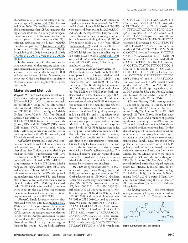
Atrazine belongs to the 2-chloro-s-triazinefamily of herbicides (Figure 1) and is the mostcommon pesticide contaminant of ground-water and surface water (Fenelon and Moore1998; Kolpin et al. 1998; Lode et al. 1995;Miller et al. 2000; Müller et al. 1997;Solomon et al. 1996; Thurman and Cromwell2000). Among the endocrine-disruptingeffects, atrazine interferes with androgen- andestrogen-mediated processes (Babic-Gojmeracet al. 1989; Cooper et al. 1999, 2000;Cummings et al. 2000; Friedmann 2002;Kniewald et al. 1979, 1995; Narotsky et al.2001; Shafer et al. 1999; Simic et al. 1991;Stoker et al. 1999, 2000). The interference ofatrazine with androgen and estrogen actiondoes not occur by direct agonism or antago-nism of cognate receptors for these steroids asshown by binding affinity studies (Robergeet al. 2004; Tennant et al. 1994a, 1994b). Inthis respect, previous investigations have sug-gested that atrazine reduces androgen synthe-sis and action (Babic-Gojmerac et al. 1989;Kniewald et al. 1979, 1980, 1995; Simic et al.1991) and stimulates estrogen production(Crain et al. 1997; Heneweer et al. 2004;Keller and McClellan-Green 2004; Sandersonet al. 2000, 2001, 2002; Spano et al. 2004).The latter ability is exerted through at leasttwo mechanisms that converge on increasingaromatase expression and activity. First,inhibiting phosphodiesterase, atrazine up-regulates cAMP, which induces the expression
of SF-1, an important regulator of the PIIpromoter of aromatase gene CYP19. Theenhanced transcription of the aromatase geneincreases both enzymatic activity of aromataseand estrogen production (Heneweer et al.2004; Lehmann et al. 2005; Morinaga et al.2004; Roberge et al. 2004; Sanderson et al.2000, 2001). Next, atrazine binds to SF-1 andfacilitates the recruitment of this factor to thePII promoter of the aromatase gene, furtherstimulating the biological effects describedabove (Fan et al. 2007a, 2007b).
Epidemiologic studies have associatedlong-term exposure to triazine herbicides withincreased risk of ovarian cancer in female farmworkers in Italy (Donna et al. 1989) and breastcancer in the general population of Kentuckyin the United States (Kettles et al. 1997). Inaddition, atrazine leads to tumor developmentin the mammary gland and reproductiveorgans of female F344 rats (Pintér et al. 1990),whereas in Sprague-Dawley rats it causes anearlier onset of mammary and pituitary tumors(Wetzel et al. 1994), a typical response toexogenously administered estrogens (Brawerand Sonnenschein 1975).
Given the potential ability of atrazine tointerfere with reproduction and to cause can-cer, the European Union banned its use.However, the U.S. Environmental ProtectionAgency has approved the use of atrazinebecause of the lack of a clear associationbetween the levels of exposure and cancer
incidence in pesticide applicators (Gammonet al. 2005; McElroy et al. 2007; Rusieckiet al. 2004; Sass and Colangelo 2006; Younget al. 2005).
Regarding the apparent estrogenic effectsof atrazine, previous studies have demonstratedthat triazine herbicides do not bind or activatethe classical estrogen receptor (ER) (Connoret al. 1996; Tennant et al. 1994a, 1994b). Inrecent years, increasing evidence has demon-strated in different experimental models thatsteroid hormones, including estrogens, canexert rapid actions interacting with receptorslocated within or near the cell membrane(Falkenstein et al. 2000; Norman et al. 2004;Revelli et al. 1998). The importance of thissignaling mechanism is becoming more widelyrecognized as steroid membrane receptors havebeen implicated in a large number of physio-logic functions. Moreover, it has been sug-gested that nongenomic estrogen actions, likegenomic ones, are susceptible to interferencefrom environmental estrogens (Thomas 2000).Of note, these compounds compete with[3H]17β-estradiol ([3H]E2) for binding toestrogen membrane receptors (Loomis andThomas 2000) and exert agonist effects onnongenomic transduction pathways in differ-ent cell contexts (Loomis and Thomas 2000;Nadal et al. 2000; Ruehlmann et al. 1988;Watson et al. 1999). However, the preciseidentity and function of many steroid mem-brane receptors are still controversial in termsof their specific molecular interactions withendogenous and environmental estrogens.
A seven-transmembrane receptor, G-pro-tein–coupled receptor 30 (GPR30), which isstructurally unrelated to the nuclear ER, hasbeen recently shown to mediate rapid actionsof estrogens (Filardo et al. 2002; Revankaret al. 2005). Recombinant GPR30 protein,produced in ER-negative HEK-293 cells,exhibited all the steroid binding and signaling
Address correspondence to M. Maggiolini, Departmentof Pharmaco-Biology, University of Calabria, 87030Rende (CS), Italy. Telephone: 390984493076. Fax:390984493458. E-mail: [email protected]
*These authors contributed equally to this work.This research was supported by grants from the
Associazione Italiana per la Ricerca sul Cancro,Ministero dell’Università e Ricerca Scientifica eTecnologica, and Regione Calabria.
The authors declare they have no competingfinancial interests.
Received 28 January 2008; accepted 18 July 2008.
G-Protein–Coupled Receptor 30 and Estrogen Receptor-α Are Involved in the Proliferative Effects Induced by Atrazine in Ovarian Cancer CellsLidia Albanito,1,* Rosamaria Lappano,1,* Antonio Madeo,1 Adele Chimento,1 Eric R. Prossnitz,2
Anna Rita Cappello,1 Vincenza Dolce,1 Sergio Abonante,1 Vincenzo Pezzi,1 and Marcello Maggiolini1
1Department of Pharmaco-Biology, University of Calabria, Rende, Italy; 2Department of Cell Biology and Physiology and CancerResearch and Treatment Center, University of New Mexico, Albuquerque, New Mexico, USA
BACKGROUND: Atrazine, one of the most common pesticide contaminants, has been shown toup-regulate aromatase activity in certain estrogen-sensitive tumors without binding or activatingthe estrogen receptor (ER). Recent investigations have demonstrated that the orphan G-protein–coupled receptor 30 (GPR30), which is structurally unrelated to the ER, mediates rapid actionsof 17β-estradiol and environmental estrogens.
OBJECTIVES: Given the ability of atrazine to exert estrogen-like activity in cancer cells, we evaluatedthe potential of atrazine to signal through GPR30 in stimulating biological responses in cancer cells.
METHODS AND RESULTS: Atrazine did not transactivate the endogenous ERα in different cancer cellcontexts or chimeric proteins encoding the ERα and ERβ hormone-binding domain in gene reporterassays. Moreover, atrazine neither regulated the expression of ERα nor stimulated aromatase activity.Interestingly, atrazine induced extracellular signal-regulated kinase (ERK) phosphorylation and theexpression of estrogen target genes. Using specific signaling inhibitors and gene silencing, wedemonstrated that atrazine stimulated the proliferation of ovarian cancer cells through theGPR30–epidermal growth factor receptor transduction pathway and the involvement of ERα.
CONCLUSIONS: Our results indicate a novel mechanism through which atrazine may exert relevantbiological effects in cancer cells. On the basis of the present data, atrazine should be included amongthe environmental contaminants potentially able to signal via GPR30 in eliciting estrogenic action.
KEY WORDS: 17β-estradiol, atrazine, estrogen receptor, GPR30, ovarian cancer cells. EnvironHealth Perspect 116:1648–1655 (2008). doi:10.1289/ehp.11297 available via http://dx.doi.org/[Online 22 July 2008]
characteristics of a functional estrogen mem-brane receptor (Thomas et al. 2005; Thomasand Dong 2006). Our studies and others havealso demonstrated that GPR30 mediates therapid response to E2 in a variety of estrogen-responsive cancer cells by activating the epi-dermal growth factor receptor (EGFR)–mitogen-activated protein kinase (MAPK)transduction pathway (Albanito et al. 2007;Bologa et al. 2006; Filardo et al. 2000;Maggiolini et al. 2004; Revankar et al. 2005;Thomas et al. 2005; Vivacqua et al. 2006a,2006b).
In the present study, for the first time wehave demonstrated that atrazine stimulatesgene expression and growth effects in estrogen-sensitive ovarian cancer cells through GPR30and the involvement of ERα. Moreover, weshow that GPR30 mediates the stimulatoryeffects of atrazine in ER-negative SkBr3 breastcancer cells.
Materials and Methods
Reagents. We purchased atrazine [2-chloro-4-(ethylamine)-6-(isopropylamine)-s-triazine],17β-estradiol (E2), N-[2-(p-bromocinnamyl-amino)ethyl]-5-isoquinolinesulfonamidedihydrochloride (H89), wortmannin (WM),and PD98059 (PD) from Sigma-Aldrich(Milan, Italy); AG1478 (AG) from BiomolResearch Laboratories (DBA, Milan, Italy);ICI 182,780 (ICI) from Tocris Chemicals(Bristol, UK); and GF109203X (GFX) fromCalbiochem (VWR International, Milan,Italy). All compounds were solubilized indimethyl sulfoxide (DMSO), except E2 andPD, which were dissolved in ethanol.
Cell culture. Human BG-1 and 2008 ovar-ian cancer cells as well as human Ishikawaendometrial cancer cells were maintained inphenol red–free Dulbecco’s modified Eaglemedium (DMEM) supplemented with 10%fetal bovine serum (FBS). H295R adrenal carci-noma cells were cultured in DMEM/F12 1:1supplemented with 1% ITS Liquid MediaSupplement (Sigma-Aldrich), 10% calf serum,and antibiotics. Human MCF-7 breast cancercells were maintained in DMEM with phenolred supplemented with 10% FBS, and humanSkBr3 breast cancer cells were maintained inphenol red–free RPMI 1640 supplementedwith 10% FBS. Cells were switched to mediumwithout serum the day before experimentsfor immunoblots and reverse transcription-polymerase chain reaction (RT-PCR).
Plasmids. Firefly luciferase reporter plas-mids used were XETL for ERα (Bunone et al.1996) and GK1 for yeast transcription factorGal4 fusion proteins (Webb et al. 1998).XETL contains the estrogen response element(ERE) from the Xenopus vitellogenin A2 gene(nucleotides –334 to –289), the herpes simplexvirus thymidine kinase promoter region(nucleotides –109 to +52), the firefly luciferase
coding sequence, and the SV40 splice andpolyadenylation sites from plasmid pSV232A/L-AA5. Gal4 chimeras Gal-ERα and Gal-ERβwere expressed from plasmids GAL93.ER(G)and GAL.ERβ, respectively. They were con-structed by transferring the coding sequencesfor the hormone-binding domain (HBD) ofERα (amino acids 282–595) from HEG0(Bunone et al. 1996), and for the ERβ HBD(C-terminal 287 amino acids) from plasmidpCMV5-hERβ into the mammalian expres-sion vector pSCTEVGal93 (Seipel et al. 1992).We used the Renilla luciferase expressionvector pRL-TK (Promega, Milan, Italy) as atransfection standard.
Transfection and luciferase assays. BG-1,MCF-7, Ishikawa, and SkBr3 cells (1 × 10⁵)were plated into 24-well dishes with500 µL/well DMEM (BG-1, MCF-7, andIshikawa cells) or RPMI 1640 (SkBr3 cells)containing 10% FBS the day before transfec-tion. We replaced the medium with phenolred–free DMEM or RPMI 1640, both sup-plemented with 1% charcoal-stripped (CS)FBS, on the day of transfection. Transfectionswere performed using FuGENE 6 Reagent asrecommended by the manufacturer (RocheDiagnostics, Mannheim, Germany) with amixture containing 0.3 µg of reporter plas-mid, 1 ng pRL-TK, and 0.1 µg effector plas-mid where applicable. After 5–6 hr, themedium was replaced again with serum-freeDMEM lacking phenol red and supple-mented with 1% CS-FBS; ligands were addedat this point, and cells were incubated for16–18 hr. We measured luciferase activitywith the Dual Luciferase Kit (Promega)according to the manufacturer’s recommen-dations. Firefly luciferase values were normal-ized to the internal transfection controlprovided by Renilla luciferase activity. Thenormalized relative light unit values obtainedfrom cells treated with vehicle were set as1-fold induction, from which the activityinduced by treatments was calculated.
RT-PCR. Using semiquantitative RT-PCRas described previously (Maggiolini et al.1999), we evaluated gene expression for ERα[GenBank accession no. NM 000125 (NationalCenter for Biotechnology Information 2008)],c-fos (NM 005252), progesterone receptor(PR; NM 000926), pS2 (NM 003225),cathepsin D (NM 001909), cyclin A (NM001237), cyclin D1 (NM 053056), cyclin E(NM 001238), and the acid phosphoproteinP0 (36B4) (NM 001002) used as a controlgene. We used the primers 5´-AATTCA-GATAATCGACGCCAG-3´ (ERα forward)and 5´-GTGTTTCAACATTCTCCCTC-CTC-3´ (ERα reverse); 5´-AGAAAAGGA-GAATCCGAAGGGAAA-3´ (c-fos forward)and 5´-ATGATGCTGGGACAGGAAG-TC-3´ (c-fos reverse); 5´-ACACCTTGC-CTGAAGTTTCG-3´ (PR forward) and
5´-CTGTCCTTTTCTGGGGGACT-3´(PR reverse); 5´-TTCTATCCTAATAC-CATCGACG-3´ (pS2 forward) and5´-TTTGAGTAGTCAAAGTCAGAGC-3´(pS2 reverse); 5´-AACAACAGGGTGGGCTTC-3´ (cathepsin D forward), and5´-ATGCACGAAACAGATCTGTGCT-3´(cathepsin D reverse); 5´-GCCATTAGTT-TACCTGGACCCAGA-3´ (cyclin A for-ward) and 5´-CACTGACATGGAAGACAGGAACCT-3´ (cyclin A reverse); 5´-TCTAA-GATGAAGGAGACCATC-3´, (cyclin D1forward) and 5´-GCGGTAGTAGGACAGGAAGTTGTT-3´ (cyclin D1 reverse);5´-CCTGACTATTGTGTCCTGGC-3´(cyclin E forward) and 5´-CCCGCT-GCTCTGCTTCTTAC-3´ (cyclin E reverse);and 5´-CTCAACATCTCCCCCTTCTC-3´(36B4 forward) and 5´-CAAATCCCA-TATCCTCGTCC-3´ (36B4 reverse) to yieldproducts of 345, 420, 196, 210, 303, 354,354, 488, and 408 bp, respectively, with20 PCR cycles for ERα, c-fos, PR, pS2, cathep-sin D, cyclin A, and cyclin E and 15 PCRcycles for both cyclin D1 and 36B4.
Western blotting. Cells were grown in10-cm dishes, exposed to ligands, and thenlysed in 500 µL of 50 mmol/L NaCl, 1.5mmol/L MgCl2, 1 mmol/L EGTA, 10%glycerol, 1% Triton X-100, 1% sodium dode-cyl sulfate (SDS), and a mixture of proteaseinhibitors containing 1 mmol/L aprotinin,20 mmol/L phenylmethylsulfonyl fluoride, and200 mmol/L sodium orthovanadate. We thendiluted samples 10 times and determined pro-tein concentration using Bradford reagentaccording to the manufacturer’s recommenda-tions (Sigma-Aldrich). Equal amounts of wholeprotein extract were resolved on a 10% SDS-polyacrylamide gel and transferred to a nitro-cellulose membrane (Amersham Biosciences,Milan, Italy). Membranes were probedovernight at 4°C with the antibody againstERα (F-10), c-fos (H-125), β-actin (C-2),phosphorylated extracellular signal-regulatedkinase 1/2 (ERK1/2; E-4), and ERK2 (C-14),all purchased from Santa Cruz Biotechnology,DBA (Milan, Italy), and human P450 aro-matase (MCA 2077S; Serotec, Milan, Italy),and then revealed using the ECL WesternBlotting Analysis System (GE Healthcare,Milan, Italy).
ER binding assay. BG-1 cells were strippedof any estrogen by keeping them in mediumwithout serum for 2 days. Cells were incubated
Atrazine action in ovarian cancer cells
Environmental Health Perspectives • VOLUME 116 | NUMBER 12 | December 2008 1649
Figure 1. Structures of E2 and atrazine.
OHCI
HO
H3C NH NH
H3C CH
3
NN
N
E2
Atrazine
with 1 nM [2,4,6,7-3H]E2 (89 Ci/mmol;Amersham Biosciences) and increasing concen-trations of nonlabeled E2 or atrazine for 1 hr at37°C in a humidified atmosphere of 95%air/5% CO2. After removal of the medium,cells were washed with ice-cold phosphate-buffered saline/0.1% methylcellulose twice,harvested by scraping and centrifugation, andlysed with 100% ethanol, 500 µL/60-mmdish, for 10 min at room temperature (Lee andGorski 1996). We measured the radioactivityof extracts by liquid scintillation counting.
Aromatase assay. In subconfluent BG-1 orH295R cells, we measured aromatase activityin the cell culture medium by tritiated waterrelease using 0.5 µM [1β-3H(N)]androst-4-ene-3,17-dione (25.3 Ci/mmol; DuPontNEN, Boston, MA, USA) as a substrate(Lephart and Simpson 1991). The cells weretreated in a six-well dish in culture medium inthe presence of atrazine or DMSO for 40 hrand then incubated with [1β-3H(N)]androst-4-ene-3,17-dione. Incubations were per-formed at 37°C for 6 hr under a 95%/5%
air/CO2 atmosphere. The results were calcu-lated as picomoles per hour, normalized tomilligrams of protein (pmol/hr per 1 mg pro-tein), and expressed as percentages ofuntreated cells (100%).
GPR30 and ERα silencing experiments.Cells were plated onto 10-cm dishes, main-tained in antibiotic-free medium for 24 hr,and then transfected for additional 24 hrbefore treatments with a mixture containingOpti-MEM, 8 µL/well LipofectAMINE 2000(Invitrogen, Milan, Italy), and 0.5 µg/wellvector or short hairpin GPR30 (shGPR30)(Albanito et al. 2008), control small inter-fering RNA (siRNA), or ERα siRNA (Sigma-Aldrich).
Proliferation assay. For the quantitativeproliferation assay, we seeded 10,000 cells in24-well plates in regular growth medium. Cellswere washed once they had attached and thenincubated in medium containing 2.5%CS-FBS with the indicated treatments.Medium was renewed every 2 days (with treat-ments), and cells were trypsinized and countedin a hemocytometer on day 6. The day beforetreatments, 200 ng/L of the indicated shorthairpin RNA was transfected using FuGENE 6Reagent as recommended by the manufacturer,and then renewed every 2 days before counting.
Statistical analysis. Statistical analysis wasperformed using analysis of variance followedby Newman-Keuls testing to determine dif-ferences in means. p-Values < 0.05 are consid-ered statistically significant.
Results
Atrazine does not activate ERα in cancer cells.Based on the evidence that atrazine producesearly onset and increased incidence of estro-gen-sensitive tumors in different experimentalmodels (Cooper et al. 2007), we first evaluatedwhether atrazine could activate a transientlytransfected ER reporter gene in estrogen-sensi-tive ovarian (BG-1), breast (MCF-7), andendometrial (Ishikawa) cancer cells. Exposureto 100 nM E2 induced a strong ERα transacti-vation that was absent in the presence of10 µM of the ER antagonist ICI in all cellcontexts evaluated (Figure 2A–C). In contrast,treatments with 1 µM atrazine and even con-centrations ranging from 1 nM to 10 µM(data not shown) failed to stimulate luciferaseexpression or to block that observed uponaddition of E2 (Figure 2A–C). Moreover,atrazine did not activate an expression vectorencoding ERα transiently transfected in ER-negative SkBr3 breast cancer cells (Figure 2D).To confirm that atrazine is not an ERα ago-nist and to examine whether ERβ couldrespond to atrazine, we turned to a completelyheterologous system. Chimeric proteins con-sisting of the DNA binding domain of theyeast transcription factor Gal4 and the ERα orERβ HBD transiently transfected in SkBr3
Albanito et al.
1650 VOLUME 116 | NUMBER 12 | December 2008 • Environmental Health Perspectives
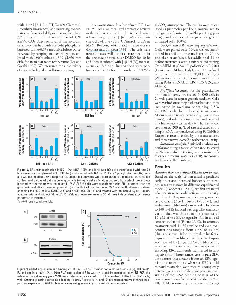
Figure 2. ERα transactivation in BG-1 (A), MCF-7 (B), and Ishikawa (C) cells transfected with the ERluciferase reporter plasmid XETL (ERE-luc) and treated with 100 nmol/L E2 or 1 μmol/L atrazine (Atr), withand without 10 μmol/L ER antagonist ICI. Luciferase activities were normalized to the internal transfectioncontrol, and values of cells receiving vehicle (–) were set as 1-fold induction, from which the activityinduced by treatments was calculated. (D–F) SkBr3 cells were transfected with ER luciferase reportergene XETL and ERα expression plasmid (D) and with Gal4 reporter gene (GK1) and the Gal4 fusion proteinsencoding the HBD of ERα (GalERα; E) and or ERβ (GalERβ; F) and treated with 100 nmol/L E2 or 1 μmol/Latrazine, with and without 10 μmol/L ICI. Values shown are mean ± SD of three independent experimentsperformed in triplicate. *p < 0.05 compared with vehicle.
40
30
20
10
1
– E2
Atr ICI E2 +
ICI
E2 +
Atr
ERE-luc
Fire
fly/
Re
nil
la l
uc
ife
rase
ac
tivi
ty (
fold
in
du
cti
on
) 40
30
20
10
1
– E2
Atr ICI E2 +
ICI
E2 +
AtrERE-luc
Fire
fly/
Re
nil
la l
uc
ife
rase
ac
tivi
ty (
fold
in
du
cti
on
) 40
30
20
10
1
– E2
Atr ICI E2 +
ICI
E2 +
Atr
ERE-luc
Fire
fly/
Re
nil
la l
uc
ife
rase
ac
tivi
ty (
fold
in
du
cti
on
)
– E2
Atr ICI E2 +
ICI
E2 +
Atr
ERE-luc + ERα
40
30
20
10
1
Fire
fly/
Re
nil
la l
uc
ife
rase
ac
tivi
ty (
fold
in
du
cti
on
) 40
30
20
10
1
Fire
fly/
Re
nil
la l
uc
ife
rase
ac
tivi
ty (
fold
in
du
cti
on
)
– E2
Atr ICI E2 +
ICI
E2 +
Atr
GK1 + GalERα
40
30
20
10
1
Fire
fly/
Re
nil
la l
uc
ife
rase
ac
tivi
ty (
fold
in
du
cti
on
)
– E2
Atr ICI E2 +
ICI
E2 +
Atr
GK1 + GalERβ
A B C
D E F
BG-1 MCF-7 ISHIKAWA
SkBr3 SkBr3 SkBr3
*
**
*
* *
*
**
*
* *
*
**
*
**
Figure 3. mRNA expression and binding of ERα in BG-1 cells treated for 24 hr with vehicle (–), 100 nmol/LE2, or 1 μmol/L atrazine (Atr). (A) mRNA expression of ERα was evaluated by semiquantitative RT-PCR; thevalues of housekeeping gene 36B4 were determined as a control. (B) Immunoblot of ERα from BG-1 cells,with 100 nmol β-actin serving as a loading control. Results in (A) and (B) are representative of three inde-pendent experiments. (C) ERα binding assay using increasing concentrations of atrazine.
100
75
50
25
Sp
ec
ific
bin
din
g (
%)
Competitor (M)–10 –9 –8 –7 –6 –5
– E2
Atr
ERα
36B4
– E2
Atr
β-Actin
ERα
A
B
E2
Atr
C
Protein
mRNA
cells were strongly activated by E2 but notupon atrazine treatment (Figure 2E,F), furthercorroborating the aforementioned results.
Atrazine neither regulates ERα expressionnor competes with estrogen binding to ERα.Considering that the down-regulation of ERαinduced by an agonist has been considered anadditional hallmark of receptor activation(Santagati et al. 1997), we further investigatedwhether atrazine could modulate ERα expres-sion in BG-1 cells, which lack ERβ (data notshown), and express a receptor expression pat-tern similar to that found in primary ovariantumors (Bardin et al. 2004; Geisinger et al.1989). As shown in Figure 3A,B, 100 nM E2down-regulated ERα at both mRNA andprotein levels, whereas 1 µM atrazine did notproduce any modulatory effect. In agreementwith these results and those obtained in trans-fection experiments, atrazine showed no bind-ing capacity for ERα (Figure 3C), as previouslyreported (Cooper et al. 2007). Altogether, ourfindings rule out that the estrogen action ofatrazine occurs through binding and directactivation of ERα.
Aromatase activity is not induced byatrazine. Given that atrazine is able to up-regulate aromatase expression and function indifferent cell contexts (Cooper et al. 2007;Fan et al. 2007a, 2007b; Roberge et al. 2004;Sanderson et al. 2000, 2001), we then deter-mined aromatase activity by tritiated water
release assays in BG-1 cells. As shown inFigure 4, 1 µM atrazine did not stimulate aro-matase activity, which in contrast wasstrongly induced in human H295R adreno-corticocarcinoma cells previously used as amodel system to assess aromatase catalyticactivity (Heneweer et al. 2004; Sandersonet al. 2001). In addition, the low aromataseprotein expression detected in BG-1 cells didnot increase upon exposure to 1 µM atrazine(data not shown). Hence, atrazine is neitheran ERα activator nor an aromatase regulatorin estrogen-sensitive ovarian cancer cells.
ERK phosphorylation is stimulated byatrazine. In recent years, numerous reportshave demonstrated that estrogens and xeno-estrogens can generate rapid signaling via sec-ond messenger systems such as Ca2+, cAMP,nitric oxide, and G-proteins, which in turnleads to activation of different downstreamkinases (Bulayeva and Watson 2004; Watsonet al. 2007).
To evaluate whether the potential estro-genic activity of atrazine is exerted through arapid cellular response, we investigated itsability to produce ERK phosphorylation inBG-1 cells. Interestingly, atrazine stimulated
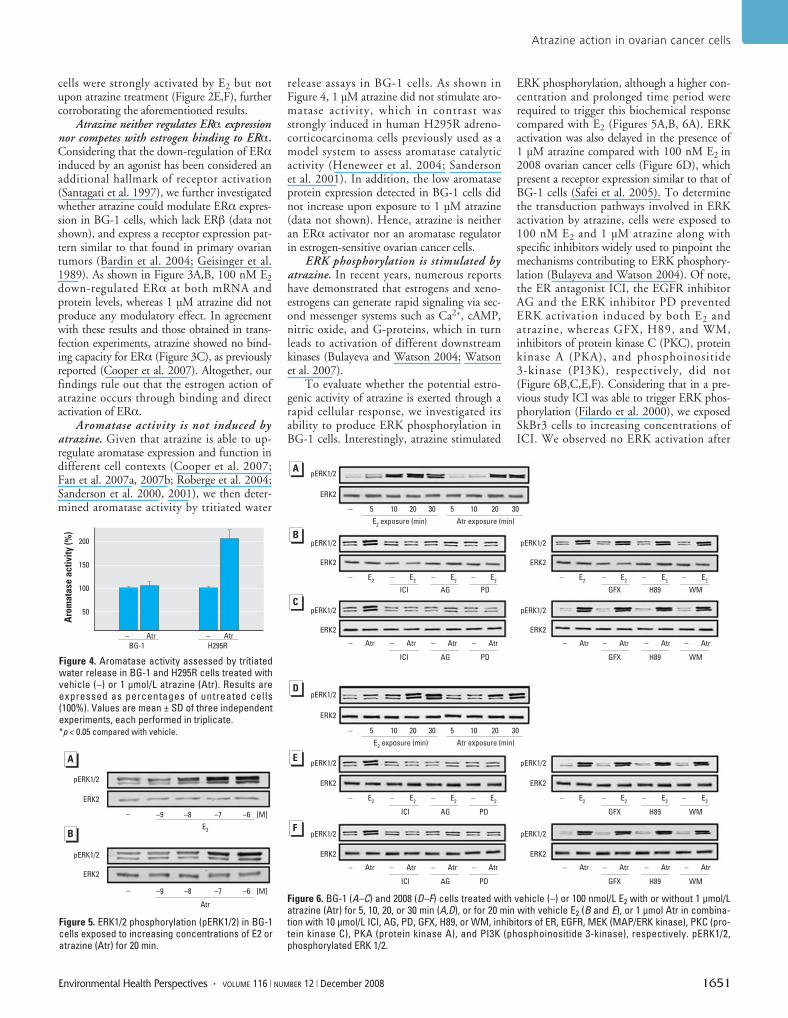
ERK phosphorylation, although a higher con-centration and prolonged time period wererequired to trigger this biochemical responsecompared with E2 (Figures 5A,B, 6A). ERKactivation was also delayed in the presence of1 µM atrazine compared with 100 nM E2 in2008 ovarian cancer cells (Figure 6D), whichpresent a receptor expression similar to that ofBG-1 cells (Safei et al. 2005). To determinethe transduction pathways involved in ERKactivation by atrazine, cells were exposed to100 nM E2 and 1 µM atrazine along withspecific inhibitors widely used to pinpoint themechanisms contributing to ERK phosphory-lation (Bulayeva and Watson 2004). Of note,the ER antagonist ICI, the EGFR inhibitorAG and the ERK inhibitor PD preventedERK activation induced by both E2 andatrazine, whereas GFX, H89, and WM,inhibitors of protein kinase C (PKC), proteinkinase A (PKA), and phosphoinositide3-kinase (PI3K), respectively, did not(Figure 6B,C,E,F). Considering that in a pre-vious study ICI was able to trigger ERK phos-phorylation (Filardo et al. 2000), we exposedSkBr3 cells to increasing concentrations ofICI. We observed no ERK activation after
Atrazine action in ovarian cancer cells
Environmental Health Perspectives • VOLUME 116 | NUMBER 12 | December 2008 1651
Figure 4. Aromatase activity assessed by tritiatedwater release in BG-1 and H295R cells treated withvehicle (–) or 1 μmol/L atrazine (Atr). Results areexpressed as percentages of untreated cells(100%). Values are mean ± SD of three independentexperiments, each performed in triplicate. *p < 0.05 compared with vehicle.
200
150
100
50
– –Atr Atr
BG-1 H295R
Aro
ma
tase
ac
tivi
ty (
%)
Figure 5. ERK1/2 phosphorylation (pERK1/2) in BG-1cells exposed to increasing concentrations of E2 oratrazine (Atr) for 20 min.
– –9 –8 –7 –6 [M]
– –9 –8 –7 –6 [M]
E2
Atr
pERK1/2
ERK2
pERK1/2
ERK2
A
B
Figure 6. BG-1 (A–C) and 2008 (D–F) cells treated with vehicle (–) or 100 nmol/L E2 with or without 1 μmol/Latrazine (Atr) for 5, 10, 20, or 30 min (A,D), or for 20 min with vehicle E2 (B and E), or 1 μmol Atr in combina-tion with 10 μmol/L ICI, AG, PD, GFX, H89, or WM, inhibitors of ER, EGFR, MEK (MAP/ERK kinase), PKC (pro-tein kinase C), PKA (protein kinase A), and PI3K (phosphoinositide 3-kinase), respectively. pERK1/2,phosphorylated ERK 1/2.
–
Atr exposure (min)
pERK1/2
ERK2
A
B
pERK1/2
5 10 20 30 5 10 20 30
E2 exposure (min)
ERK2
pERK1/2
ERK2
pERK1/2
ERK2
pERK1/2
ERK2
ERK2
pERK1/2
– E2
– E2
– E2
– E2
ICI AG PD
– E2
– E2
– E2
– E2
GFX H89 WM
– Atr – Atr – Atr – Atr – Atr – Atr – Atr – Atr
ICI AG PD GFX H89 WM
–
Atr exposure (min)
5 10 20 30 5 10 20 30
E2 exposure (min)
pERK1/2
ERK2
pERK1/2
ERK2
– E2
– E2
– E2
– E2
ICI AG PD
– E2
– E2
– E2
– E2
GFX H89 WM
pERK1/2
ERK2
pERK1/2
ERK2
– Atr – Atr – Atr – Atr – Atr – Atr – Atr – Atr
ICI AG PD GFX H89 WM
C
D
E
F
either 5 min (data not shown) or 20 min oftreatment (Figure 7). Hence, in our experi-mental conditions, ICI showed only ERKinhibitor activity.
Atrazine up-regulates the mRNA expressionof estrogen target genes. Having determined thatatrazine signals through a rapid ERK activation,we evaluated in BG-1 cells its ability to regulatethe expression of c-fos, an early gene thatresponds to a variety of extracellular stimuli,including estrogens (Maggiolini et al. 2004;Nephew et al. 1993; Singleton et al. 2003;Vivacqua et al. 2006ab), along with other estro-gen target genes. To this end, we performedsemiquantitative RT-PCR experiments com-paring mRNA levels after standardization witha housekeeping gene encoding the ribosomalprotein 36B4. A short treatment (1 hr) with1 µM atrazine enhanced c-fos and cyclin A lev-els, although to a lesser extent than 100 nM E2,which also stimulated PR, pS2, and cyclin D1expression (Table 1). After a 24-hr treatment,atrazine increased PR, pS2, and cyclin A levels,whereas E2 additionally induced the expressionof c-fos, cathepsin D, cyclin D1, and cyclin E(Table 1). We obtained results similar to thosedescribed above in 2008 cells (data not shown).Hence, atrazine is able to stimulate the expres-sion of diverse estrogen target genes without anapparent activation of ERα.
Transduction pathways involved byatrazine in the up-regulation of c-fos proteinlevels. Using c-fos expression as a molecular sen-sor of atrazine action at the genomic level, wesought to determine whether c-fos protein levelsare also regulated by atrazine in a rapid mannerand the transduction pathways involved in thisresponse (Figure 8). Interestingly, the up-regulation of c-fos observed in BG-1 and 2008
cells after a short treatment (2 hr) was abolishedby the ER antagonist ICI, the EGFR inhibitorAG, or the ERK inhibitor PD (Figure 8). Onthe contrary, GFX, H89, and WM, inhibitorsof PKC, PKA, and PI3K, respectively, did notinterfere with c-fos stimulation (Figure 8).Thus, in ovarian cancer cells, atrazine involvesERα and the EGFR/MAPK pathway to triggerc-fos protein increase.
On the basis of these and our previousresults showing that c-fos stimulation by E2occurs through GPR30 and requires ERα andEGFR-mediated signaling in cancer cellsexpressing both receptors (Albanito et al. 2007;Maggiolini et al. 2004; Vivacqua et al. 2006a,2006b), we examined whether atrazine couldact in a similar manner. Interestingly, both E2and atrazine were no longer able to induce c-fos
Albanito et al.
1652 VOLUME 116 | NUMBER 12 | December 2008 • Environmental Health Perspectives
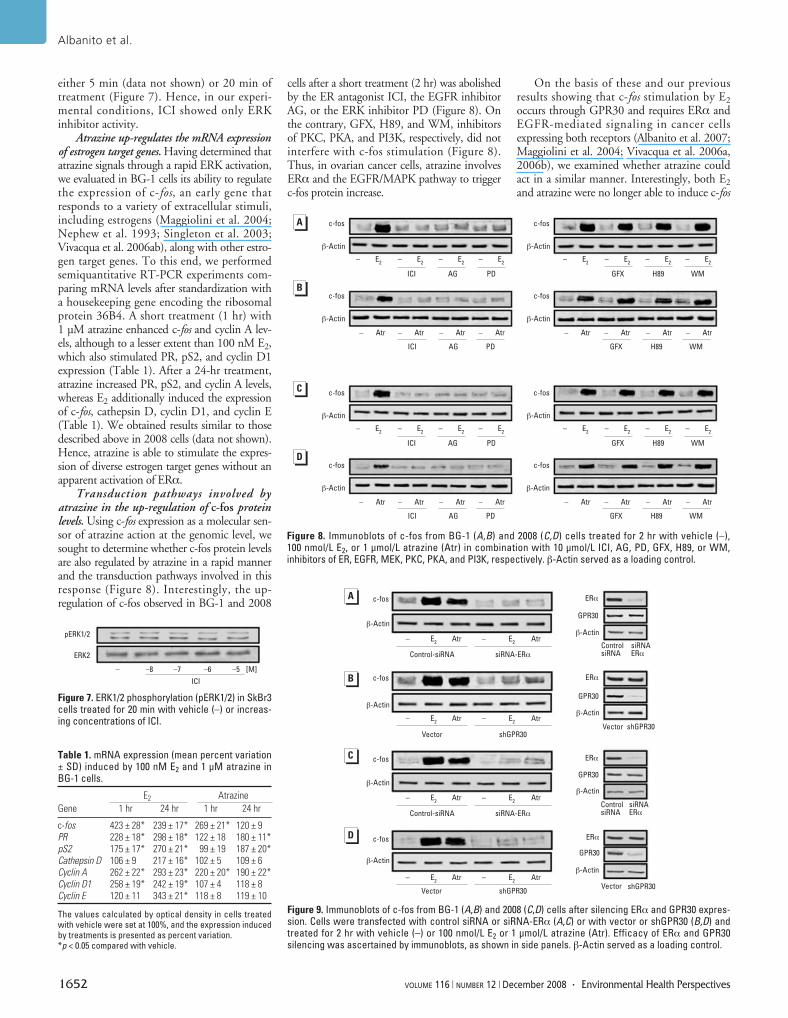
Figure 9. Immunoblots of c-fos from BG-1 (A,B) and 2008 (C,D) cells after silencing ERα and GPR30 expres-sion. Cells were transfected with control siRNA or siRNA-ERα (A,C) or with vector or shGPR30 (B,D) andtreated for 2 hr with vehicle (–) or 100 nmol/L E2 or 1 μmol/L atrazine (Atr). Efficacy of ERα and GPR30silencing was ascertained by immunoblots, as shown in side panels. β-Actin served as a loading control.
Figure 8. Immunoblots of c-fos from BG-1 (A,B) and 2008 (C,D) cells treated for 2 hr with vehicle (–),100 nmol/L E2, or 1 μmol/L atrazine (Atr) in combination with 10 μmol/L ICI, AG, PD, GFX, H89, or WM,inhibitors of ER, EGFR, MEK, PKC, PKA, and PI3K, respectively. β-Actin served as a loading control.
c-fos
β-Actin
– E2
– E2
– E2
– E2
GFX H89 WM
– Atr – Atr – Atr – Atr – Atr – Atr – Atr – Atr
ICI AG PD GFX H89 WM
C
c-fos
β-Actin
– E2
– E2
– E2
– E2
ICI AG PD
c-fos
β-Actin
c-fos
β-Actin
c-fos
β-Actin
– E2
– E2
– E2
– E2
GFX H89 WM
– Atr – Atr – Atr – Atr – Atr – Atr – Atr – Atr
ICI AG PD GFX H89 WM
c-fos
β-Actin
– E2
– E2
– E2
– E2
ICI AG PD
c-fos
β-Actin
c-fos
β-Actin
D
B
A
Figure 7. ERK1/2 phosphorylation (pERK1/2) in SkBr3cells treated for 20 min with vehicle (–) or increas-ing concentrations of ICI.
– –8 –7 –6 –5 [M]
ICI
pERK1/2
ERK2
Table 1. mRNA expression (mean percent variation± SD) induced by 100 nM E2 and 1 μM atrazine inBG-1 cells.
E2 Atrazine
Gene 1 hr 24 hr 1 hr 24 hr
c-fos 423 ± 28* 239 ± 17* 269 ± 21* 120 ± 9PR 228 ± 18* 298 ± 18* 122 ± 18 180 ± 11*pS2 175 ± 17* 270 ± 21* 99 ± 19 187 ± 20*Cathepsin D 106 ± 9 217 ± 16* 102 ± 5 109 ± 6Cyclin A 262 ± 22* 293 ± 23* 220 ± 20* 190 ± 22*Cyclin D1 258 ± 19* 242 ± 19* 107 ± 4 118 ± 8Cyclin E 120 ± 11 343 ± 21* 118 ± 8 119 ± 10
The values calculated by optical density in cells treatedwith vehicle were set at 100%, and the expression inducedby treatments is presented as percent variation. *p < 0.05 compared with vehicle.
c-fos
β-Actin
Atr– E2
B
A
c-fos
β-Actin
c-fos
β-Actin
c-fos
β-Actin
Atr– E2
Control-siRNA siRNA-ERα
Vector shGPR30
Atr– E2
Atr– E2
Atr– E2
Atr– E2
Control-siRNA siRNA-ERα
Vector shGPR30
Atr– E2
Atr– E2
ERα
GPR30
β-Actin
ERα
GPR30
β-Actin
ERα
GPR30
β-Actin
ERα
GPR30
β-Actin
siRNAERα
ControlsiRNA
shGPR30Vector
siRNAERα
ControlsiRNA
shGPR30Vector
C
D
expression after silencing either ERα orGPR30 in BG-1 and 2008 cells (Figure 9). Toevaluate whether atrazine could induce a rapidresponse in a cell context expressing GPR30alone, we turned to ER-negative SkBr3 breastcancer cells. Notably, both ERK phosphoryla-tion and c-fos induction stimulated by atrazinewere abolished after silencing GPR30(Figure 10), indicating that the response toatrazine is differentially regulated according tocancer cell type.
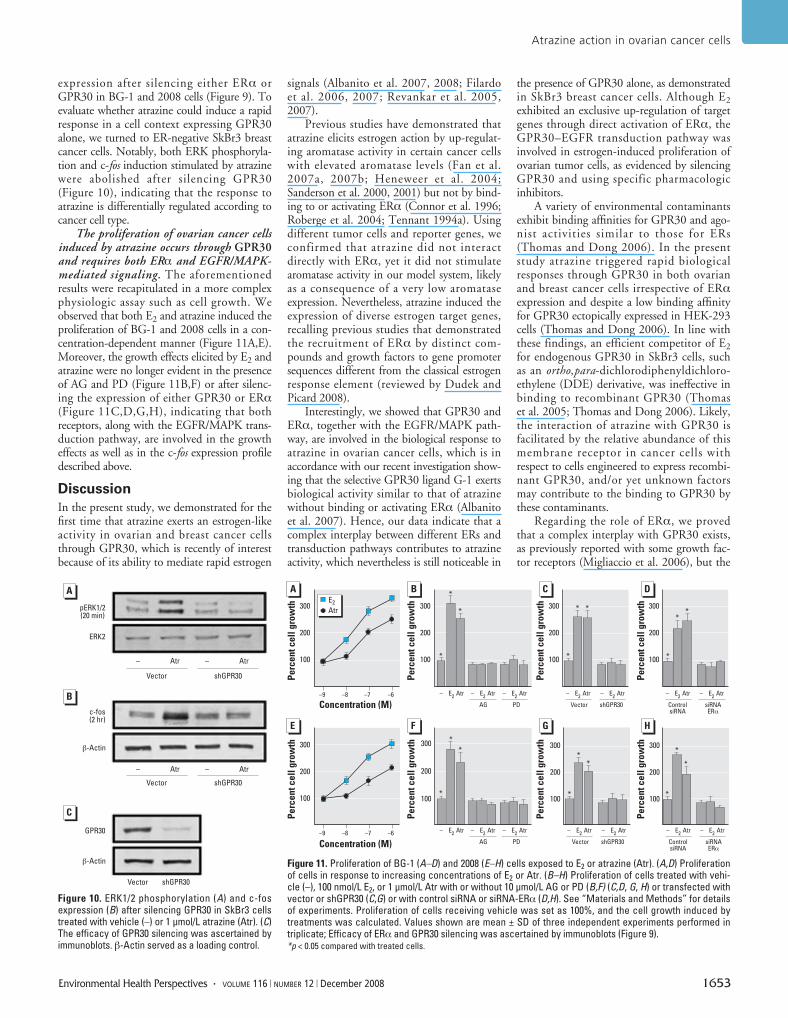
The proliferation of ovarian cancer cellsinduced by atrazine occurs through GPR30and requires both ERα and EGFR/MAPK-mediated signaling. The aforementionedresults were recapitulated in a more complexphysiologic assay such as cell growth. Weobserved that both E2 and atrazine induced theproliferation of BG-1 and 2008 cells in a con-centration-dependent manner (Figure 11A,E).Moreover, the growth effects elicited by E2 andatrazine were no longer evident in the presenceof AG and PD (Figure 11B,F) or after silenc-ing the expression of either GPR30 or ERα(Figure 11C,D,G,H), indicating that bothreceptors, along with the EGFR/MAPK trans-duction pathway, are involved in the growtheffects as well as in the c-fos expression profiledescribed above.
Discussion
In the present study, we demonstrated for thefirst time that atrazine exerts an estrogen-likeactivity in ovarian and breast cancer cellsthrough GPR30, which is recently of interestbecause of its ability to mediate rapid estrogen
signals (Albanito et al. 2007, 2008; Filardoet al. 2006, 2007; Revankar et al. 2005,2007).
Previous studies have demonstrated thatatrazine elicits estrogen action by up-regulat-ing aromatase activity in certain cancer cellswith elevated aromatase levels (Fan et al.2007a, 2007b; Heneweer et al. 2004;Sanderson et al. 2000, 2001) but not by bind-ing to or activating ERα (Connor et al. 1996;Roberge et al. 2004; Tennant 1994a). Usingdifferent tumor cells and reporter genes, weconfirmed that atrazine did not interactdirectly with ERα, yet it did not stimulatearomatase activity in our model system, likelyas a consequence of a very low aromataseexpression. Nevertheless, atrazine induced theexpression of diverse estrogen target genes,recalling previous studies that demonstratedthe recruitment of ERα by distinct com-pounds and growth factors to gene promotersequences different from the classical estrogenresponse element (reviewed by Dudek andPicard 2008).
Interestingly, we showed that GPR30 andERα, together with the EGFR/MAPK path-way, are involved in the biological response toatrazine in ovarian cancer cells, which is inaccordance with our recent investigation show-ing that the selective GPR30 ligand G-1 exertsbiological activity similar to that of atrazinewithout binding or activating ERα (Albanitoet al. 2007). Hence, our data indicate that acomplex interplay between different ERs andtransduction pathways contributes to atrazineactivity, which nevertheless is still noticeable in
the presence of GPR30 alone, as demonstratedin SkBr3 breast cancer cells. Although E2exhibited an exclusive up-regulation of targetgenes through direct activation of ERα, theGPR30–EGFR transduction pathway wasinvolved in estrogen-induced proliferation ofovarian tumor cells, as evidenced by silencingGPR30 and using specific pharmacologicinhibitors.
A variety of environmental contaminantsexhibit binding affinities for GPR30 and ago-nist activities similar to those for ERs(Thomas and Dong 2006). In the presentstudy atrazine triggered rapid biologicalresponses through GPR30 in both ovarianand breast cancer cells irrespective of ERαexpression and despite a low binding affinityfor GPR30 ectopically expressed in HEK-293cells (Thomas and Dong 2006). In line withthese findings, an efficient competitor of E2for endogenous GPR30 in SkBr3 cells, suchas an ortho,para-dichlorodiphenyldichloro-ethylene (DDE) derivative, was ineffective inbinding to recombinant GPR30 (Thomaset al. 2005; Thomas and Dong 2006). Likely,the interaction of atrazine with GPR30 isfacilitated by the relative abundance of thismembrane receptor in cancer cells withrespect to cells engineered to express recombi-nant GPR30, and/or yet unknown factorsmay contribute to the binding to GPR30 bythese contaminants.
Regarding the role of ERα, we provedthat a complex interplay with GPR30 exists,as previously reported with some growth fac-tor receptors (Migliaccio et al. 2006), but the
Atrazine action in ovarian cancer cells
Environmental Health Perspectives • VOLUME 116 | NUMBER 12 | December 2008 1653
Figure 10. ERK1/2 phosphorylation (A) and c-fosexpression (B) after silencing GPR30 in SkBr3 cellstreated with vehicle (–) or 1 μmol/L atrazine (Atr). (C)The efficacy of GPR30 silencing was ascertained byimmunoblots. β-Actin served as a loading control.
pERK1/2(20 min)
ERK2
Vector
GPR30
β-Actin
c-fos(2 hr)
β-Actin
shGPR30
Vector shGPR30
Vector shGPR30
– Atr – Atr
– Atr – Atr
A
B
C
Figure 11. Proliferation of BG-1 (A–D) and 2008 (E–H) cells exposed to E2 or atrazine (Atr). (A,D) Proliferationof cells in response to increasing concentrations of E2 or Atr. (B–H) Proliferation of cells treated with vehi-cle (–), 100 nmol/L E2, or 1 μmol/L Atr with or without 10 μmol/L AG or PD (B,F) (C,D, G, H) or transfected withvector or shGPR30 (C,G) or with control siRNA or siRNA-ERα (D,H). See “Materials and Methods” for detailsof experiments. Proliferation of cells receiving vehicle was set as 100%, and the cell growth induced bytreatments was calculated. Values shown are mean ± SD of three independent experiments performed intriplicate; Efficacy of ERα and GPR30 silencing was ascertained by immunoblots (Figure 9). *p < 0.05 compared with treated cells.
Concentration (M)
Concentration (M)
A B C
300
200
100
–9 –8 –7 –6
300
200
100
– E2
Atr – E2
Atr – E2
Atr
300
200
100
– E2
Atr – E2
Atr
AG PD Vector shGPR30
300
200
100
– E2
Atr – E2
Atr
ControlsiRNA
siRNAERα
Pe
rce
nt
ce
ll g
row
th
300
200
100
Pe
rce
nt
ce
ll g
row
th
–9 –8 –7 –6
300
200
100
300
200
100
300
200
100
– E2
Atr – E2
Atr – E2
Atr – E2
Atr – E2
Atr
AG PD Vector shGPR30
– E2
Atr – E2
Atr
ControlsiRNA
siRNAERα
E2
Atr
Pe
rce
nt
ce
ll g
row
thP
erc
en
t c
ell
gro
wth
Pe
rce
nt
ce
ll g
row
thP
erc
en
t c
ell
gro
wth
Pe
rce
nt
ce
ll g
row
thP
erc
en
t c
ell
gro
wth
D
E F G H
*
*
*
*
* *
*
**
*
*
*
**
*
*
*
*
molecular mechanisms involved remain to beelucidated. Our study and previous investiga-tions indicate that environmental estrogensexert pleiotropic actions by directly bindingto ERα as well as through GPR30–EGFRsignaling, which can engage ERα dependingon the receptor expression pattern present indifferent cell types. This mode of action ofxenoestrogens fits well with the resultsobtained after silencing GPR30 or ERαexpression in ovarian cancer cells, becausesilencing each gene prevented the growthresponse to atrazine.
Our data recall the results of previousstudies showing that xenoestrogens mimicrapid estrogen action in several animal and cellmodels (Bulayeva and Watson 2004; Loomisand Thomas 2000; Nadal et al. 2000;Ruehlmann et al. 1988; Watson et al. 1999,2007). Particularly, in GH3/B6/F10 pituitarytumor cells, diverse xenoestrogens inducedERK phosphorylation with a temporally dis-tinct activation pattern compared with E2(Bulayeva and Watson 2004). In the latterstudy, on the basis of the inhibitory activityexerted by ICI, the authors hypothesized thatan ER localized to the plasma membranecould mediate the ERK phosphorylationresponse by xenoestrogens, depending on theirdifferent ER binding affinities. Moreover, theauthors suggested that the signaling cascadesleading to ERK activation may involve thenature of membrane ERs and their ability tointeract with various signaling partners(Bulayeva and Watson 2004). Interestingly,our findings have provided evidence that ERαmay be involved by xenoestrogens without adirect binding activity and produce relevantresponses such as ERK phosphorylation, geneexpression, and cell growth.
A subset of estrogen-sensitive cell tumorscan proliferate independently from ER expres-sion (i.e., ER-negative cells). In this condition,well represented by SkBr3 breast cancer cells,GPR30–EGFR signaling may still allow forenvironmental estrogen activity as we haveshown in the present study as well as in a pre-vious study (Maggiolini et al. 2004). Hence,multiple transduction pathways triggeredsimultaneously at the membrane level, as wellas within each cell type, may contribute to thenature and magnitude of biological responsesto distinct estrogenic compounds. These con-sequently should be examined individually fortheir complex mechanistic and functional out-comes that result from interaction with a dif-ferent repertoire of receptor proteins.
Atrazine, a potent endocrine disruptor, isthe most common pesticide contaminant ofgroundwater and surface water. Here, we haveprovided novel insight regarding the potentialrole of GPR30 in mediating the action ofatrazine in endocrine-related diseases, such asestrogen-sensitive tumors.
REFERENCES
Albanito L, Madeo A, Lappano R, Vivacqua A, Rago V,
Carpino A, et al. 2007. G protein-coupled receptor 30
(GPR30) mediates gene expression changes and growth
response to 17beta-estradiol and selective GPR30 ligand
G-1 in ovarian cancer cells. Cancer Res 67:1859–1866.
Albanito L, Sisci D, Aquila S, Brunelli E, Vivacqua A, Madeo A,
et al. 2008. Epidermal growth factor induces G protein-cou-
pled receptor expression in estrogen receptor-negative
breast cancer cells. Endocrinology 149:3799–3808.
Babic-Gojmerac T, Kniewald Z, Kniewald J. 1989. Testosterone
metabolism in neuroendocrine organs in male rats under
atrazine and deethylatrazine influence. J Steroid Biochem
33:141–146.
Bardin A, Hoffman P, Boulle N, Katsaros D, Vignon F, Pujol P,
et al. 2004. Involvement of estrogen receptor beta in ovarian
carcinogenesis. Cancer Res 64:5861–5869.
Bologa CG, Revankar CM, Young SM, Edwards BS, Arterburn JB,
Kiselyov AS, et al. 2006. Virtual and biomolecular screening
converge on a selective agonist for GPR30. Nat Chem Biol
4:207–212.
Brawer JR, Sonnenschein C. 1975. Cytopathological effects of
estradiol on the arcuate nucleus of the female rat. A possible
mechanism for pituitary tumorigenesis. Am J Anat 144:57–88.
Bulayeva NN, Watson CS. 2004. Xenoestrogen-induced ERK-1
and ERK-2 activation via multiple membrane-initiated sig-
naling pathways. Environ Health Perspect 15:1481–1487.
Bunone G, Briand PA, Miksicek RJ, Picard D. 1996. Activation
of the unliganded estrogen receptor by EGF involves the
MAP kinase pathway and direct phosphorylation. EMBO J
15:2174–2183.
Connor K, Howell J, Chen I, Liu H, Berhane K, Sciarretta C,
et al. 1996. Failure of chloro-S-triazine-derived compounds
to induce estrogen receptor-mediated responses in vivo
and in vitro. Fundam Appl Toxicol 30:93–101.
Cooper RL, Laws SC, Das PC, Narotsky MG, Goldman JM,
Tyrey EL, et al. 2007. Atrazine and reproductive function:
mode and mechanism of action studies. Birth Defects Res
80:98–112.
Cooper RL, Stoker TE, McElroy WK. 1999. Atrazine (ATR) disrupts
hypothalamic catecholamines and pituitary function.
Toxicologist 42:60–66.
Cooper RL, Stoker TE, Tyrey L, Goldman JM, McElroy WK. 2000.
Atrazine disrupts the hypothalamic control of pituitary
ovarian function. Toxicol Sci 53:297–307.
Crain D, Guillette LJ, Rooney AA, Pickford D. 1997. Alterations
in steroidogenesis in alligators (Alligator mississippiensis)
exposed naturally and experimentally to environmental
contaminants. Environ Health Perspect 105:528–533.
Cummings A, Rhodes B, Cooper R. 2000. Effect of atrazine on
implantation and early pregnancy in 4 strains of rats. Toxicol
Sci 58:135–143.
Donna A, Crosignani P, Robutti F, Betta PG, Bocca R, Mariani N,
et al. 1989. Triazine herbicides and ovarian epithelial neo-
plasms. Scand J Work Environ Health 15:47–53.
Dudek P, Picard D. 2008. Genomics of signaling crosstalk of
estrogen receptor α in breast cancer cells. PLoS ONE
3:1–11.
Falkenstein EHC, Tillmann HC, Christ M, Feuring M, Wehling M.
2000. Multiple actions of steroid hormones a focus on
rapid, nongenomic effects. Pharmacol Rev 52:513–556.
Fan W, Yanase T, Morinaga H, Gondo S, Okabe T, Nomura M,
et al. 2007a. Atrazine-induced aromatase expression is SF-1
dependent: implications for endocrine disruption in wildlife
and reproductive cancers in humans. Environ Health
Perspect 115:720–727.
Fan W, Yanase T, Morinaga H, Gondo S, Okabe T, Nomura M,
et al. 2007b. Herbicide atrazine activates SF-1 by direct
affinity and concomitant co-activators recruitments to
induce aromatase expression via promoter II. Biochem
Biophys Res Commun 355:1012–1018.
Fenelon J, Moore R. 1998. Transport of agrochemicals to
ground and surface waters in a small central Indiana
watershed. J Environ Qual 27:884–894.
Filardo EJ, Graeber CT, Quinn JA, Resnick MB, Giri D, DeLellis
RA, et al. 2006. Distribution of GPR30, a seven membrane-
spanning estrogen receptor, in primary breast cancer and
its association with clinicopathologic determinants of tumor
progression. Clin Cancer Res 12:6359–6366.
Filardo EJ, Quinn JA, Bland KI, Frackelton AR. 2000. Estrogen-
induced activation of Erk-1 and Erk-2 requires the G protein-
coupled receptor homolog, GPR30, and occurs via
trans-activation of the epidermal growth factor receptor
through release of HB-EGF. Mol Endocrinol 14:1649–1660.
Filardo EJ, Quinn JA, Frackelton AR, Bland KI. 2002. Estrogen
action via the G protein-coupled receptor, GPR30: stimula-
tion of adenylyl cyclase and cAMP-mediated attenuation of
the epidermal growth factor receptor-to-MAPK signaling
axis. Mol Endocrinol 16:70–84.
Filardo EJ, Quinn JA, Pang C, Graeber CT, Shaw S, Dong J, et al.
2007. Activation of the novel estrogen receptor G protein
coupled receptor 30 (GPR30) at the plasma membrane.
Endocrinology 148:3236–3245.
Friedmann A. 2002. Atrazine inhibition of testosterone produc-
tion in rat males following peripubertal exposure. Reprod
Toxicol 16:275–279.
Gammon DW, Aldous CN, Carr WC Jr, Sanborn JR, Pfeifer KF.
2005. A risk assessment of atrazine use in California: human
health and ecological aspects. Pest Manag Sci 61:331–355.
Geisinger KR, Kute TE, Pettenati MJ, Welander CE, Dennard Y,
Collins LA, et al. 1989. Characterization of a human ovarian
carcinoma cell line with estrogen and progesterone
receptors. Cancer 63:280–288.
Heneweer M, van den Berg M, Sanderson J. 2004. A comparison
of human H295R and rat R2C cell lines as in vitro screening
tools for effects on aromatase. Toxicol Lett 146:183–194.
Keller J, McClellan-Green P. 2004. Effects of organochlorine
compounds on cytochrome P450 aromatase activity in an
immortal sea turtle cell line. Mar Environ Res 58:347–351.
Kettles MK, Browning SR, Prince TS, Horstman SW. 1997.
Triazine herbicide exposure and breast cancer incidence:
an ecologic study of Kentucky counties. Environ Health
Perspect 105:1222–1227.
Kniewald J, Mildner P, Kniewald Z. 1979. Effects ofs-triazine
herbicides on 5α-dihydrotestosterone receptor complex
formation, 5α-reductase and 3α-hydroxysteroid dehydro-
genase activity at the anterior pituitary level. J Steroid
Biochem 11:833–838.
Kniewald J, Mildner P, Kniewald Z. 1980. Effects of s-triazine
herbicides on 5α-dihydrotestosterone receptor complex
formation in the hypothalamus and ventral prostate. In:
Pharmacological Modulation of Steroid Action (Genazzani E,
DiCarlo F, Mainwaring WIP, eds). New York:Raven Press,
159–169.
Kniewald J, Osredecki V, Gojmerac T, Zechner V, Kniewald Z.
1995. Effect of s-triazine compounds on testosterone
metabolism in the rat prostate. J Appl Toxicol 15:215–218.
Kolpin D, Barbash J, Gilliom R. 1998. Occurence of pesticides in
shallow groundwater of the United States: initial results
from the National Water-Quality Assessment Program.
Environ Sci Technol 32:558–566.
Lee YJ, Gorski J. 1996. Estrogen-induced transcription of the prog-
esterone receptor gene does not parallel estrogen receptor
occupancy. Proc Natl Acad Sci USA 93:15180–15184.
Lehmann TP, Biernacka-Lukanty JM, Saraco N, Langlois D,
Li JY, Trzeciak WH. 2005. Temporal pattern of the induc-
tion of SF-1 gene expression by the signal transduction
pathway involving 3´,5´-cyclic adenosine monophosphate.
Acta Biochim Pol 52:485–491.
Lephart ED, Simpson ER. 1991. Assay of aromatase activity.
Methods Enzymol 206:477–483.
Lode O, Eklo O, Holen B, Svensen A, Johnsen A. 1995. Pesticides
in precipitation in Norway. Sci Total Environ 160:421–431.
Loomis AK, Thomas P. 2000. Effects of estrogens and xeno-
estrogens on androgen production by Atlantic croaker
testes in vitro: evidence for a nongenomic action mediated
by an estrogen membrane receptor. Biol Reprod
62:995–1004.
Maggiolini M, Donzé O, Picard D. 1999. A non-radioactive
method for inexpensive quantitative RT-PCR. Biol Chem
380:695–700.
Maggiolini M, Vivacqua A, Fasanella G, Recchia AG, Sisci D,
Pezzi V, et al. 2004. The G protein-coupled receptor GPR30
mediates c-fos up-regulation by 17-beta estradiol and
phytoestrogens in breast cancer cells. J Biol Chem
279:27008–27016.
McElroy JA, Gangnon RE, Newcomb PA, Kanarek MS,
Anderson HA, Brook JV, et al. 2007. Risk of breast cancer
for women living in rural areas from adult exposure to
atrazine from well water in Wisconsin. J Expo Sci Environ
Epidemiol 17:207–214.
Migliaccio A, Castoria G, Di Domenico M, Ciociola A,
Lombardi M, De Falco A, et al. 2006. Crosstalk between
EGFR and extranuclear steroid receptors. Ann NY Acad Sci
1089:194–200.
Miller S, Sweet C, Depinto J, Hornbuckle K. 2000. Atrazine and
Albanito et al.
1654 VOLUME 116 | NUMBER 12 | December 2008 • Environmental Health Perspectives
nutrients in precipitation: results from the Lake Michigan
mass balance study. Environ Sci Technol 34:55–61.
Morinaga H, Yanase T, Nomura M, Okabe T, Goto K, Harada N,
et al. 2004. A benzimidazole fungicide, benomyl, and its
metabolite, carbendazim, induce aromatase activity in a
human ovarian granulose-like tumor cell line (KGN).
Endocrinology 145:1860–1869.
Müller S, Berg M, Ulrich M, Schwarzenbach RP. 1997. Atrazine
and its primary metabolites in Swiss lakes: input charac-
teristics and long-term behavior in the water column.
Environ Sci Technol 31:2104–2113.
Nadal A, Ropero AB, Laribi O, Maillet M, Fuentes E, Soria B.
2000. Nongenomic actions of estrogens and xenoestrogens
by binding at a plasma membrane receptor unrelated to
estrogen receptor alpha and estrogen receptor beta. Proc
Natl Acad Sci USA 97:11603–11608.
Narotsky M, Best DS, Guidici DL, Cooper RL. 2001. Strain com-
parisons of atrazine-induced pregnancy loss in the rat.
Reprod Toxicol 15:61–69.
National Center for Biotechnology Information. 2008. Searching
GenBank. Available: http://www.ncbi.nlm.nih.gov/Genbank/
GenbankSearch.html [accessed 21 October 2008].
Nephew KP, Polek TC, Akcali KC, Khan SA. 1993. The anti-
estrogen tamoxifen induces c-fos and c-jun, but not c-jun
or Jun-D, protooncogenes in the rat uterus. Endocrinology
133:419–422.
Norman AW, Mizwicki MT, Norman DP. 2004. Steroid-hormone
rapid actions, membrane receptors and a conformational
ensemble model. Nat Rev Drug Discov 3:27–41.
Pintér A, Török G, Börzsönyi M, Surján A, Csík M, Kelecsényi Z,
et al. 1990. Long-term carcinogenicity bioassay of the herbi-
cide atrazine in F344 rats. Neoplasma 37:533–544.
Revankar CM, Cimino DF, Sklar LA, Arterburn JB, Prossnitz ER.
2005. A transmembrane intracellular estrogen receptor
mediates rapid cell signalling. Science 307:1625–1630.
Revankar CM, Mitchell HD, Field AS, Burai R, Corona C,
Ramesh C, et al. 2007. Synthetic estrogen derivatives
demonstrate the functionally of intracellular GPR30. ACS
Chem Biol 2:536–544.
Revelli A, Massobrio M, Tesarik J. 1998. Nongenomic actions
of steroid hormones in reproductive tissues. Endocr Rev
19:3–17.
Roberge M, Hakk H, Larsen G. 2004. Atrazine is a competitive
inhibitor of phosphodiesterase but does not affect the
estrogen receptor. Toxicol Lett 154:61–68.
Ruehlmann DO, Steinert JR, Valverde MA, Jacob R, Mann GE.
1988. Environmental estrogenic pollutants induce acute
vascular relaxation by inhibiting C-type Ca2+ channels in
smooth muscle cells. FASEB J 12:613–619.
Rusiecki JA, De Roos A, Lee WJ, Dosemeci M, Lubin JH,
Hoppin JA, et al. 2004. Cancer incidence among pesticide
applicators exposed to atrazine in the Agricultural Health
Study. J Natl Cancer Inst 96:1375–1382.
Safei R, Katano K, Larson BJ, Samimi G, Holzer AK,
Naerdemann W, et al. 2005. Intracellular localization and
trafficking of fluorescein-labeled cisplatin in human ovar-
ian carcinoma cells. Clin Cancer Res 11:756–767.
Sanderson JT, Boerma J, Lansbergen G, Van den Berg M. 2002.
Induction and inhibition of aromatase (CYP19) activity by
various classes of pesticides in H295R human adrenocorti-
cal carcinoma cells. Toxicol Appl Pharmacol 182:44–54.
Sanderson JT, Letcher RJ, Heneweer M, Giesy JP, Van den
Berg M. 2001. Effects of chloro-s-triazine herbicides and
metabolites on aromatase activity in various human cell
lines and on vitellogenin production in male carp hepato-
cytes. Environ Health Perspect 109:1027–1031.
Sanderson JT, Seinen W, Giesy JP, van den Berg M. 2000.
2-Chloro-triazine herbicides induce aromatase (CYP19)
activity in H295R human adrenocortical carcinoma cells: a
novel mechanism for estrogenicity? Toxicol Sci 54:121–127.
Santagati S, Gianazza E, Agrati P, Vegeto E, Patrone C, Pollio G,
et al. 1997. Oligonucleotide squelching reveals the mecha-
nism of estrogen receptor autologous down-regulation.
Mol Endocrinol 11:938–949.
Sass JB, Colangelo A. 2006. European Union bans atrazine,
while the United States negotiates continued use. Int J
Occup Environ Health 12:260–267.
Seipel K, Georgiev O, Schaffner W. 1992. Different activation
domains stimulate transcription from remote (‘enhancer’)
and proximal (‘promoter’) positions. EMBO J 11:4961–4968.
Shafer TJ, Ward TR, Meacham CA, Cooper RL. 1999. Effects of
the cholorotriazine herbicide, cyanizine on GABAA recep-
tors in cortical tissue from rat brain. Toxicology 142:57–68.
Simic B, Kniewald Z, Davies J, Kniewald J. 1991. Reversibility
of inhibitory effect of atrazine and lindane on 5-dihydro-
testosterone receptor complex formation in rat prostate.
Bull Environ Contam Toxicol 46:92–99.
Singleton DW, Feng Y, Burd CJ, Khan SA. 2003. Nongenomic
activity and subsequent c-fos induction by estrogen
receptor ligands are not sufficient to promote deoxy-
ribonucleic acid synthesis in human endometrial adeno-
carcinoma cells. Endocrinology 144:121–128.
Solomon K, Baker D, Richards R, Dixon K, Klaine S, LaPoint T,
et al. 1996. Ecological risk assessment of atrazine in North
American surface waters. Environ Toxicol Chem 15:31–76.
Spano L, Tyler C, van Aerle R, Devos P, Mandiki S, Silvestre F,
et al. 2004. Effects of atrazine on sex steroid dynamics,
plasma vitellogenin concentration and gonad development
in adult goldfish (Carassius auratus). Aquat Toxicol
66:369–379.
Stoker TE, Laws S, Guidici D, Cooper R. 2000. The effect of
atrazine on puberty in male Wistar rats: an evaluation in
the protocol for the assessment of pubertal development
and thyroid function. Toxicol Sci 58:50–59.
Stoker TE, Robinette CL, Cooper RL. 1999. Maternal exposure to
atrazine during lactation suppresses suckling-induced pro-
lactin release and results in prostatitis in the adult offspring.
Toxicol Sci 52:68–79.
Tennant MK, Hill DS, Eldridge JC, Wetzel LT, Breckenridge CB,
Stevens JT. 1994a. Chloro-s-triazine antagonism of estro-
gen action: limited interaction with estrogen receptor
binding. J Toxicol Environ Health 43:197–211.
Tennant MK, Hill DS, Eldridge JC, Wetzel LT, Breckenridge CB,
Stevens JT. 1994b. Possible antiestrogenic properties of
chloro-s-triazines in rat uterus. J Toxicol Environ Health
43:183–196.
Thomas P. 2000. Chemical interference with the genomic and
nongenomic actions of steroids in fishes: role of receptor
binding. Mar Environ Res 50:127–134.
Thomas P, Dong J. 2006. Binding and activation of the seven-
transmembrane estrogen receptor GPR30 by environmen-
tal estrogens: a potential novel mechanism of endocrine
disruption. J Steroid Biochem Mol Biol 102:175–179.
Thomas P, Pang Y, Filardo EJ, Dong J. 2005. Identity of an
estrogen membrane receptor coupled to a G-protein in
human breast cancer cells. Endocrinology 146:624–632.
Thurman E, Cromwell A. 2000. Atmospheric transport, deposi-
tion, and fate of triazine herbicides and their metabolites
in pristine areas at Isle Royale National Park. Environ Sci
Technol 34:3079–3085.
Vivacqua A, Bonofiglio D, Albanito L, Madeo A, Rago V,
Carpino A, et al. 2006a. 17-beta-Estradiol, genistein, and 4-
hydroxytamoxifen induce the proliferation of thyroid can-
cer cells through the G protein coupled-receptor GPR30.
Mol Pharmacol 70:1414–1423.
Vivacqua A, Bonofiglio D, Recchia AG, Musti AM, Picard D,
Andò S, et al. 2006b. The G protein-coupled receptor
GPR30 mediates the proliferative effects induced by
17beta-estradiol and hydroxytamoxifen in endometrial
cancer cells. Mol Endocrinol 20:631–646.
Watson CS, Alyea RA, Jeng YJ, Kochukov MY. 2007.
Nongenomic actions of low concentration estrogens and
xenoestrogens on multiple tissues. Mol Cell Endocrinol
274:1–7.
Watson CS, Campbell CH, Gametchu B. 1999. Membrane
estrogen receptors on rat pituitary tumor cells: immuno-
identification and response to oestradiol and xeno-
estrogens. Exp Physiol 84:1013–1018.
Webb P, Nguyen P, Shinsako J, Anderson C, Feng W,
Nguyen MP, et al. 1998. Estrogen receptor activation func-
tion 1 works by binding p160 coactivator proteins. Mol
Endocrinol 12:1605–1618.
Wetzel LT, Luempert LG, Breckenridge CB, Tisdel MO,
Stevens JT, Thakur AK, et al. 1994. Chronic effects of
atrazine on estrus and mammary tumor formation in
female Sprague-Dawley and Fischer 344 rats. J Toxicol
Environ Health 43:169–182.
Young HA, Mills PK, Riordan DG, Cress RD. 2005. Triazine herbi-
cides and epithelial ovarian cancer risk in central California.
J Occup Environ Med 47:1148–1156.
Atrazine action in ovarian cancer cells
Environmental Health Perspectives • VOLUME 116 | NUMBER 12 | December 2008 1655