Nonproliferative and Proliferative Lesions of the Rat and ...
Upload
khangminh22Category
view
3download
0
Louisiana State UniversityLSU Digital Commons
LSU Doctoral Dissertations Graduate School
2007
Lymphohistiocytic Proliferative Syndrome ofAlligators (Alligator mississippiensis): a cutaneousmanifestation of West Nile virusJavier G. NevarezLouisiana State University and Agricultural and Mechanical College, [email protected]
Follow this and additional works at: https://digitalcommons.lsu.edu/gradschool_dissertations
Part of the Veterinary Medicine Commons
This Dissertation is brought to you for free and open access by the Graduate School at LSU Digital Commons. It has been accepted for inclusion inLSU Doctoral Dissertations by an authorized graduate school editor of LSU Digital Commons. For more information, please [email protected].
Recommended CitationNevarez, Javier G., "Lymphohistiocytic Proliferative Syndrome of Alligators (Alligator mississippiensis): a cutaneous manifestation ofWest Nile virus" (2007). LSU Doctoral Dissertations. 3521.https://digitalcommons.lsu.edu/gradschool_dissertations/3521

LYMPHOHISTIOCYTIC PROLIFERATIVE SYNDROME OF ALLIGATORS (ALLIGATORMISSISSIPPIENSIS): A CUTANEOUS MANIFESTATION OF WEST NILE VIRUS
A Dissertation
Submitted to the Graduate Faculty of theLouisiana State University and
Agricultural and Mechanical Collegein fulfillment of the
requirements for the degree ofDoctor of Philosophy
in
The Interdepartmental Program inVeterinary Medical Sciences through
the Department of Veterinary Clinical Sciences
byJavier G. Nevarez
B.S., Louisiana State University, 1998D.V.M., Louisiana State University, 2001
May 2007

ii
To the gators.

iii
ACKNOWLEDGEMENTS
This project would not have been possible without the efforts of multiple individuals and
organizations that supported me either intellectually, spiritually, or financially. The end product
is a prime example of what can be accomplished when people from different backgrounds work
together towards a common goal.
I would like to thank my mentor and friend for over 11 years, Dr. Mark A Mitchell, who
has guided my professional and personal growth. This project was just one of many amazing
opportunities I had to work with a unique reptile species alongside Dr. Mitchell. I will always be
indebted to him for his guidance, patience and understanding. I also have to thank Dr. Thomas
N. Tully for his friendship and mentorship. Dr. Tully has also contributed to my professional
and personal growth by opening a world of opportunities for me within the exotic animal service
at the LSU School of Veterinary Medicine. I will always consider both of them the two most
influential people in my veterinary career. In addition, I would like to thank the other members
of my committee, Drs. John Hawke, Gary Sod, and Lane Foil. Their diverse background and
expertise was instrumental in making this project a successful one and making me consider all
angles of a research project. Thank you for your mentorship, and know that I am a better person
and a better veterinarian thanks to your teachings.
Other major contributors to this project include Drs. Timothy Morgan, Michael Garner,
April Johnson, Dr. Peter Jowett and Alma Roy. Dr. Roy and her staff at the Louisiana
Veterinary Medicine Diagnostic Laboratory, Rob Poston, Tarra Harden, Heather Lampinen, Dr.
Alejandra Baudena, Dr. Kim Bowles, and Durriya Sarkar, were one of the most professional
groups of people I have had the opportunity to work with during the project. Their hard work,
commitment, and proficiency are acknowledged and appreciated. An additional group that
revealed the professionalism that is present at the university was the histology laboratory

iv
personnel, Mr. Hal Holloway, Cheryl Crowder, and Julie Millard. Their input and guidance was
also instrumental in ensuring a sound research project. I would also like to thank the
administration and faculty from the department of Veterinary Clinical Sciences, and more
specifically Dr. David Senior, Mrs. Jackie Bourgeois, Mrs. Jackie Murray for their hard work
and support. A special thanks to Shelly Lyles, Randy Nehlig, and Marlana Roundtree for their
assistance during certain phases of the project.
I must express a special gratitude to Dr. April Johnson who in the middle of her own
dissertation project graciously performed the WNV ELISA tests so needed for this project.
This project would not have been possible without the financial support of the Louisiana
Department of Wildlife and Fisheries, Fur and Refuge Division. Mr. Noel Kinler and Mrs. Ruth
Elsey provided knowledge and support that was indispensable for making this project a reality.
The Louisiana Alligator Council, the Louisiana Alligator Association, and all the Louisiana
Alligator producers also contributed significantly to this project. I would like to thank Mr. Jeff
Donald, Dane Ledet, and Jerry Savoie for their support. It was Mr. Ledet who originally
identified an association between WNV and LPSA in his facility. Without his keen sense of
observation it would have taken a longer time to complete the study.
Friends and family are always a big part of the support net that one must rely upon to be
successful in life. In my case I was lucky to have such a network to guide me through the
completion of the project. To my friends Jason Blackburn and Mary-Claire Blackburn, thank
you for the meals and lodging during the times of field work. To my family, thank you for
always believing in me and supporting my endless educational endeavors. Finally, and most
importantly, thank you to my wife Emily for her help during the project and her patience when I
was writing and had little patience with those around me. I must also acknowledge all the living
creatures in our house, Sophie, Eddie, Soleil, Sid, Daisy, and the Skinks for accepting me

v
regardless of my mood and making me laugh when I needed it the most. The success of this
work is shared with all of you.

vi
TABLE OF CONTENTS
DEDICATION . . . . . . . . . . . . . . . . . . . . . . . . . . . . . . . . . . . . . . . . . . . . . . . . . . . . . . . . . . . . . . . . ii
ACKNOWLEDGEMENTS . . . . . . . . . . . . . . . . . . . . . . . . . . . . . . . . . . . . . . . . . . . . . . . . . . . . . .iii
ABSTRACT . . . . . . . . . . . . . . . . . . . . . . . . . . . . . . . . . . . . . . . . . . . . . . . . . . . . . . . . . . . . . . . . viii
INTRODUCTION . . . . . . . . . . . . . . . . . . . . . . . . . . . . . . . . . . . . . . . . . . . . . . . . . . . . . . . . . . . . . 1
LITERATURE REVIEW . . . . . . . . . . . . . . . . . . . . . . . . . . . . . . . . . . . . . . . . . . . . . . . . . . . . . . . 4Louisiana Alligator Industry . . . . . . . . . . . . . . . . . . . . . . . . . . . . . . . . . . . . . . . . . . . . . . . .4Taxonomy . . . . . . . . . . . . . . . . . . . . . . . . . . . . . . . . . . . . . . . . . . . . . . . . . . . . . . . . . . . . . .6Anatomy and Physiology . . . . . . . . . . . . . . . . . . . . . . . . . . . . . . . . . . . . . . . . . . . . . . . . . . 6Husbandry . . . . . . . . . . . . . . . . . . . . . . . . . . . . . . . . . . . . . . . . . . . . . . . . . . . . . . . . . . . . .12Nutrition . . . . . . . . . . . . . . . . . . . . . . . . . . . . . . . . . . . . . . . . . . . . . . . . . . . . . . . . . . . . . . 15Preventive Medicine . . . . . . . . . . . . . . . . . . . . . . . . . . . . . . . . . . . . . . . . . . . . . . . . . . . . .16Handling and Restraint . . . . . . . . . . . . . . . . . . . . . . . . . . . . . . . . . . . . . . . . . . . . . . . . . . .18History and Physical Examination . . . . . . . . . . . . . . . . . . . . . . . . . . . . . . . . . . . . . . . . . . 19Diagnostic Testing . . . . . . . . . . . . . . . . . . . . . . . . . . . . . . . . . . . . . . . . . . . . . . . . . . . . . . 21Stress and Immunosuppression . . . . . . . . . . . . . . . . . . . . . . . . . . . . . . . . . . . . . . . . . . . . .24Bacterial Diseases . . . . . . . . . . . . . . . . . . . . . . . . . . . . . . . . . . . . . . . . . . . . . . . . . . . . . . .26Viral Diseases . . . . . . . . . . . . . . . . . . . . . . . . . . . . . . . . . . . . . . . . . . . . . . . . . . . . . . . . . .31Fungal Diseases . . . . . . . . . . . . . . . . . . . . . . . . . . . . . . . . . . . . . . . . . . . . . . . . . . . . . . . . 35Parasitic Diseases . . . . . . . . . . . . . . . . . . . . . . . . . . . . . . . . . . . . . . . . . . . . . . . . . . . . . . . 36Nutritional Diseases . . . . . . . . . . . . . . . . . . . . . . . . . . . . . . . . . . . . . . . . . . . . . . . . . . . . . 38Respiratory Diseases . . . . . . . . . . . . . . . . . . . . . . . . . . . . . . . . . . . . . . . . . . . . . . . . . . . . 40Neurologic Diseases . . . . . . . . . . . . . . . . . . . . . . . . . . . . . . . . . . . . . . . . . . . . . . . . . . . . .40Musculoskeletal Diseases . . . . . . . . . . . . . . . . . . . . . . . . . . . . . . . . . . . . . . . . . . . . . . . . 41Gastrointestinal Diseases . . . . . . . . . . . . . . . . . . . . . . . . . . . . . . . . . . . . . . . . . . . . . . . . . 42Integumentary Diseases . . . . . . . . . . . . . . . . . . . . . . . . . . . . . . . . . . . . . . . . . . . . . . . . . . 43Toxicities . . . . . . . . . . . . . . . . . . . . . . . . . . . . . . . . . . . . . . . . . . . . . . . . . . . . . . . . . . . . . 45Runting . . . . . . . . . . . . . . . . . . . . . . . . . . . . . . . . . . . . . . . . . . . . . . . . . . . . . . . . . . . . . . . 46West Nile Virus History . . . . . . . . . . . . . . . . . . . . . . . . . . . . . . . . . . . . . . . . . . . . . . . . . . 46
CHAPTER 1. LYMPHOHISTIOCYTIC PROLIFERATIVE SYNDROME OF ALLIGATORS:PRELIMINARY INVESTIGATION . . . . . . . . . . . . . . . . . . . . . . . . . . . . . . . . . . . . . . . . . . . . . .55
Introduction . . . . . . . . . . . . . . . . . . . . . . . . . . . . . . . . . . . . . . . . . . . . . . . . . . . . . . . . . . . 55Materials and Methods . . . . . . . . . . . . . . . . . . . . . . . . . . . . . . . . . . . . . . . . . . . . . . . . . . 56Results . . . . . . . . . . . . . . . . . . . . . . . . . . . . . . . . . . . . . . . . . . . . . . . . . . . . . . . . . . . . . . . 60Discussion . . . . . . . . . . . . . . . . . . . . . . . . . . . . . . . . . . . . . . . . . . . . . . . . . . . . . . . . . . . . .68
CHAPTER 2. WEST NILE VIRUS IN ALLIGATOR, ALLIGATOR MISSISSIPPIENSIS,RANCHES FROM LOUISIANA. . . . . . . . . . . . . . . . . . . . . . . . . . . . . . . . . . . . . . . . . . . . . . . . . 72
Introduction . . . . . . . . . . . . . . . . . . . . . . . . . . . . . . . . . . . . . . . . . . . . . . . . . . . . . . . . . . . 72Materials and Methods . . . . . . . . . . . . . . . . . . . . . . . . . . . . . . . . . . . . . . . . . . . . . . . . . . 73Results . . . . . . . . . . . . . . . . . . . . . . . . . . . . . . . . . . . . . . . . . . . . . . . . . . . . . . . . . . . . . . . 79Discussion . . . . . . . . . . . . . . . . . . . . . . . . . . . . . . . . . . . . . . . . . . . . . . . . . . . . . . . . . . . . .81

vii
CHAPTER 3. ESTABLISHING AN ASSOCIATION BETWEEN WEST NILE VIRUSSEROPOSITIVITY AND THE DEVELOPMENT OF LYMPHOHISTIOCYTICPROLIFERATIVE SYNDROME OF ALLIGATORS . . . . . . . . . . . . . . . . . . . . . . . . . . . . . . . . 85
Introduction . . . . . . . . . . . . . . . . . . . . . . . . . . . . . . . . . . . . . . . . . . . . . . . . . . . . . . . . . . . 85Materials and Methods . . . . . . . . . . . . . . . . . . . . . . . . . . . . . . . . . . . . . . . . . . . . . . . . . . 86Results . . . . . . . . . . . . . . . . . . . . . . . . . . . . . . . . . . . . . . . . . . . . . . . . . . . . . . . . . . . . . . . 88Discussion . . . . . . . . . . . . . . . . . . . . . . . . . . . . . . . . . . . . . . . . . . . . . . . . . . . . . . . . . . . . .88
CHAPTER 4: LYMPHOHISTIOCYTIC PROLIFERATIVE SYNDROME OF ALLIGATORS:A CUTANEOUS MANIFESTATION OF WEST NILE VIRUS . . . . . . . . . . . . . . . . . . . . . . . . 90
Introduction . . . . . . . . . . . . . . . . . . . . . . . . . . . . . . . . . . . . . . . . . . . . . . . . . . . . . . . . . . . 90Materials and Methods . . . . . . . . . . . . . . . . . . . . . . . . . . . . . . . . . . . . . . . . . . . . . . . . . . 91Results . . . . . . . . . . . . . . . . . . . . . . . . . . . . . . . . . . . . . . . . . . . . . . . . . . . . . . . . . . . . . . . .93Discussion . . . . . . . . . . . . . . . . . . . . . . . . . . . . . . . . . . . . . . . . . . . . . . . . . . . . . . . . . . . . .95
CHAPTER: 5 PREVENTION, SURVEILLANCE, AND CONTROL METHODS FOR WNVIN ALLIGATOR RANCHES. . . . . . . . . . . . . . . . . . . . . . . . . . . . . . . . . . . . . . . . . . . . . . . . . . . .98
Introduction . . . . . . . . . . . . . . . . . . . . . . . . . . . . . . . . . . . . . . . . . . . . . . . . . . . . . . . . . . .98Prevention . . . . . . . . . . . . . . . . . . . . . . . . . . . . . . . . . . . . . . . . . . . . . . . . . . . . . . . . . . . .98Surveillance. . . . . . . . . . . . . . . . . . . . . . . . . . . . . . . . . . . . . . . . . . . . . . . . . . . . . . . . . . . 101Control . . . . . . . . . . . . . . . . . . . . . . . . . . . . . . . . . . . . . . . . . . . . . . . . . . . . . . . . . . . . . . 105Discussion . . . . . . . . . . . . . . . . . . . . . . . . . . . . . . . . . . . . . . . . . . . . . . . . . . . . . . . . . . . .109
CONCLUSIONS. . . . . . . . . . . . . . . . . . . . . . . . . . . . . . . . . . . . . . . . . . . . . . . . . . . . . . . . . . . . 111
REFERENCES. . . . . . . . . . . . . . . . . . . . . . . . . . . . . . . . . . . . . . . . . . . . . . . . . . . . . . . . . . . . . . 118
APPENDIX: LETTER OF PERMISSION FROM THE JOURNAL OF HERPETOLOGICALMEDICINE AND SURGERY . . . . . . . . . . . . . . . . . . . . . . . . . . . . . . . . . . . . . . . . . . . . . . . . . . 129
VITA. . . . . . . . . . . . . . . . . . . . . . . . . . . . . . . . . . . . . . . . . . . . . . . . . . . . . . . . . . . . . . . . . . . . . . 130

viii
ABSTRACT
In 1999, there were reports of a new type of lesion in the hides of captive reared alligators
from Florida. Similar lesions were first reported from alligator hides in Louisiana in 2001;
however, it wasn’t until 2002 that small epizootics became apparent. In 2002, the Louisiana
Department of Wildlife and Fisheries began a collaborative effort with the Louisiana State
University School of Veterinary Medicine (LSU SVM) to help elucidate the etiology of “PIX”
disease, later renamed Lymphohistiocytic Proliferative Syndrome of Alligators (LPSA).
Preliminary work concluded that LPSA was a systemic disease affecting multiple
tissues. Based on the results of this preliminary study, particularly the histopathologic evaluation
of LPSA tissues, a viral etiology was established as the top differential for LPSA. Further work
revealed that LPSA positive alligators were 476 (95% CI: 79.6, 2845.2) times more likely to be
seropositive for WNV than LPSA negative alligators. At that point it was also becoming clear
that the occurrence of LPSA matched the occurrence of WNV in alligator farms. Another
project was performed to further elucidate the association between WNV and LPSA based on
results of WNV serology, WNV RT-PCR, and histopathologic evaluation of animals with
(treatment) and without (control) LPSA lesions. Results from this study revealed that in the
treatment group, 97.5% (95%CI: 92.7-102.3 %) of the LPSA skin lesions (TxA) were positive
for WNV via RT-PCR. Of the skins within the treatment group that had no LPSA lesions (TxB),
8% (95%CI: 0-16.9%) were positive for WNV. In the control group, all of the skin samples
(CxS) were negative for WNV. All alligators in TxA were significantly (p=0.07-20) more likely
to have RT-PCR WNV positive skin than those in CxS, and TxB (p=0.08-16). There was no
significant difference in the recovery of WNV from the skins of alligators from TxB and CxS
(p=0.24).

ix
The results of this work support the theory that LPSA is a cutaneous manifestation of
chronic WNV exposure/infection in captive reared alligators. Therefore the epidemiology of
LPSA follows the epidemiology of WNV and prevention, surveillance and control methods used
for WNV should effectively decrease the occurrence of LPSA.

1
INTRODUCTION
Modern day crocodilians date back over 65 million years to the Mesozoic era, yet they
have been able to survive with relatively small evolutionary changes. Their biology, physiology,
and anatomy are very different from other reptiles. Unfortunately, these evolutionary marvels
are now starting to suffer from anthropogenic influences on their environment, and many are
now threatened or near extinction. Of the 23 species of crocodilians found worldwide, 13 can be
found on the red list of endangered species (IUCN 2004). Fortunately, there are now
international conservation programs dedicated to the preservation of these species.
In the United States, the American alligator (Alligator mississippiensis) is the most
common and best-known crocodilian. The only other species of native crocodilian found in the
United States, the American crocodile (Crocodylus acutus), is a highly vulnerable species that
has a limited range in southern Florida. The American alligator was also at critical levels during
the 1960’s, but a federal conservation program designed to protect wild populations through
farming and ranching has brought their population numbers back to sustainability. In Louisiana
alone, it is estimated that there are over one million animals. The first type of operations
consisted of farms. Breeding pairs of alligators were kept in outdoor enclosed areas where they
could mate and nest (Figure 1). The eggs were then collected and incubated. The farming
operations later developed into a ranching operation in which eggs were harvested from the wild
and incubated in private facilities (Figure 2). The majority of the hatchlings in this system are
raised for their hide and meat; however, to help maintain the wild populations, 14% of the
hatchlings are eventually returned to the wild. Today, Louisiana is the world’s primary producer
of alligators; however, captive rearing operations can also be found in Florida, Texas, Georgia,
and other southern states alligators naturally inhabit. In Louisiana, the majority of the operations
are ranches.

2
In 1999, there were reports of a new type of lesions in the hides of captive reared
alligators from Florida. These skin lesions were described as 1mm pit scars on tanned alligator
hides (Dickson 2001, Cardeilhac 2001). These scars were termed “PIX” disease because the
lesions, according to the tanners, appeared as if made with an ice pick. Original work performed
by Cardeilhac et al. (Cardeilhac et al. 2001) described the lesions as lymphocytic infiltrations and
granulomas with a weakened cornified layer. They also observed fungal hyphae in some of the
Figure 1. Outdoors enclosure used for farming Morelet’s crocodiles in Mexico.
Figure 2. Buildings used for alligator ranching in Louisiana.
lesions and were able to obtain fungal growth from one sample. The fungus was identified as
Hortaea werneckii, and it was believed to be at least in part the presumptive agent for “PIX”
disease (Cardeilhac 2001).
3
Similar histologic lesions were first reported from alligator hides in Louisiana in 2001;
however, it wasn’t until 2002 that small epizootics became apparent. In 2002, the Louisiana
Department of Wildlife and Fisheries (LDWF) began a collaborative effort with the Louisiana
State University School of Veterinary Medicine (LSU SVM) to help elucidate the etiology of
“PIX” disease. In addition to performing research dealing with “PIX” disease, the LSU SVM
also provided clinical veterinary services to the Louisiana Alligator Industry (LAI). Early on
during these collaborative efforts the authors re-classified “PIX” disease as Lymphohistiocytic
Proliferative Syndrome of Alligators (LPSA). This classification was used because it provided a
histopathologic description of the original skin lesions. The work presented here describes the
research and clinical work performed between 2002 and 2007 to define the epidemiology of
LPSA in alligators from Louisiana alligator ranches.
Biological Hypotheses
1. LPSA is an internal/systemic disease.
2. The etiology of LPSA is infectious because affected animals are generally found in the
same pen or building.
3. The histopathologic lesions associated with LPSA are consistent with viral disease.
4. LPSA is a clinical sign associated with West Nile virus (WNV) infection in alligators.
5. The appearance of LPSA in farmed alligators in Louisiana coincided with the appearance
of WNV in Louisiana.

4
LITERATURE REVIEW
Because of their relative age, crocodilians represent an important group of vertebrates.
These animals have survived for millions of years utilizing many unique evolutionary features.
Most of our knowledge regarding these animals has come through research; however, the
majority of the research is related to evolutionary biology, physiology, taxonomy and behavior.
To date, there have been few examples of research evaluating the health of these animals. The
purpose of this literature review is to provide a concise review of our current knowledge
regarding these animals.
Louisiana Alligator Industry
The LAI is regulated by the LDWF, and has its roots in the Rockefeller Refuge in Grand
Chenier, Louisiana. During the 1960s the population of wild alligators in Louisiana was
estimated at less than 100,000 (LFAC 2007). Because of the decreasing population size, a
moratorium on alligator hunting and regulation of alligator harvest were implemented in an
attempt to re-establish the alligator populations. As part of those efforts, an alligator farming and
ranching program was established in order to promote alligators as a natural renewable resource
in the state. This program would help conserve alligator habitat as well as promote the
preservation of wild alligators. An alligator ranching program was established in 1986. Under
this program, the LDWF granted egg harvest permits to licensed alligator ranchers in the state.
There are currently 59 alligator ranches in the state and collectively they produce approximately
390,000 alligators every year. This market has an annual value of approximately 30 million
dollars. The alligator ranching system works via the sale of egg harvest permits by LDWF for
collection of alligator eggs from private and public lands. Egg collection usually takes place
between June and August. Eggs numbers can range from 20 to 60 per nest. Once the eggs are
collected, they are taken to a private facility where the eggs are incubated in designated buildings

5
or incubators. The eggs collected from the wild are at different stages of incubation, so once at
the alligator ranch it may take anywhere from a few weeks to months for the eggs to hatch
depending on the time of collection. The total incubation period ranges from 40 to 90 days.
Incubation temperature will determine the sex ratio of the alligators between the 7th and 35th day
of incubation. Temperatures of 30oC (86oF) or below will yield a higher proportion of females
while temperatures >34oC (93oF) will yield a higher proportion of males (Grenard 1991). The
majority of alligator ranchers do not attempt to select for a specific sex. Once the alligators are
hatched, they are placed in indoor buildings where they are raised until the time of slaughter.
Alligators are usually raised in 18 to 20 inches of water. The water depth may be adjusted
depending on the size of the alligators and the condition of their skin. A wooden table is
sometimes placed in the pens and used as a feeding surface, while others place the food directly
on the water. Dry commercial feeds ranging in protein content from 45 to 56% are readily
available. The majority of the facilities will feed a commercial diet exclusively, while others
mix in fish, chicken, or nutria meat. Within 10 to 12 months, alligators can be raised to a market
size of 36 inches. At that point the ranchers begin the slaughter of the alligators in order to sell
the hide and meat. The alligator hides are sold to dealers and tanners for further processing. The
LAI is the largest supplier of alligator hides in the world. Louisiana alligator hides eventually
end up in high end retail products such as watch bands, shoes, purses, and other fashion items.
These products have the benefit of coming from a natural renewable resource that is
environmentally friendly and helps contribute to the wild populations of alligators. In addition to
the hide, alligator meat is sold to processing plants and distributed to restaurants, supermarkets,
and specialty stores.
The conservation aspect of the LAI comes from the 14% of alligators at each facility
(based on the yearly egg harvest) that are destined for release back into the wild. The percent of

6
alligators released is based on an expected survival of 10 to 20% of the hatchlings under natural
conditions, and amounts to approximately 55,000 alligators being released back to the wild every
year (LFAC 2007). Alligators being released to the wild are identified using toe tags and a tail
notch. This program has been extremely successful, and has served as an important model for
other crocodilian species around the world. Today, the population of wild alligators is estimated
at well over 1 million animals, a 90% increase from the 1960s estimates.
Taxonomy
The taxonomic identification of crocodilians is variable, with some authors arguing over
what should be classified as a species or a sub-species. Tables 1 to 3 list the generally accepted
23 crocodilian species by their common name, scientific name, and geographic distribution.
These lists are not meant to be absolute, but rather a general guideline. Countries such as
Australia, India, Mexico, Papua New Guinea, South Africa, and the United States maintain
intensive production operations for various species.
Anatomy and Physiology
One of the most commonly asked questions’ regarding a crocodilian is how to distinguish
a crocodile from an alligator. The first major difference is that they belong to two different
families. There are currently three distinct families of crocodilians: Alligatoridae (Table 1)
(alligators and caimans), Gavialidae (Table 2) (gharials or gavials), and Crocodylidae (Table 3)
(the crocodiles). Geographical location can also help in the identification of some species.
Alligators are thought to tolerate colder temperatures and live at higher latitudes, while
crocodiles and caimans are less cold resistant and live in warmer areas (Huchzermeyer 2003).
However, there are some anatomical features that will be most useful in differentiating alligators
from crocodiles. Alligators and caimans have a broad u-shaped snout, while crocodiles have a
more narrow v-shaped snout. This comparison can be made when looking at the dorsal aspect of

7
Table 1. Taxonomy of the Family Alligatoridae
Common Name Scientific Name Geographical Distribution
American alligator Alligator mississippiensis Southeast United States
Chinese alligator Alligator sinensis Eastern China
Spectacled/Common caiman Caiman crocodilus Central and South America
Broad-snouted caiman Caiman latirostris South America
Jacare caiman Caiman yacare South America
Black caiman Melanosuchus niger South America
Cuvier's dwarf caiman Paleosuchus palpebrosus South America
Table 2. Taxonomy of the Family Gavialidae.
Common Name Scientific Name Geographical Distribution
Indian gharial/True gharial Gavialis gangeticus Indian subcontinent
the head (Figure 3). A more obvious distinction can be made when looking at their mouth from
the side. Alligators and caimans have notches in the maxilla that fit the mandibular teeth.
Therefore, they have no mandibular teeth visible if observed from the side with their mouth
closed. On the other hand, the fourth mandibular tooth of crocodiles is readily apparent when
looking at the profile of the animal (Figure 4). On close inspection of the integument, one
can also look for the presence of integumentary sensing organs (ISO’s) or dome pressure
receptors (DPR’s). These receptors are clear to gray pits that are present on the skin of
crocodilians, but their location varies with species. Their function is not completely understood,
but they may play a role as mechanoreceptors in prey detection or even as chemoreceptors aiding

8
Table 3. Taxonomy of the Family Crocodylidae
Common Name Scientific Name Geographical Distribution
American crocodile Crocodylus acutus North, Central, and South America
Slender-snouted crocodile Crocodylus cataphractus Africa
Orinoco crocodile Crocodylus intermedius South America
Fresh water crocodile/Johnston's crocodile Crocodylus johnstoni Australia
Philippine crocodile Crocodylus mindorensis Philippines
Morelet's crocodile Crocodylus moreletii Central America
Nile crocodile Crocodylus niloticus Africa and Madagascar
New Guinea crocodile Crocodylus novaeguineae Papua New Guinea and Irian Java
Mugger crocodile/Swamp crocodile Crocodylus palustris Indian subcontinent
Saltwater/Estuarine crocodile Crocodylus porosus Australia and Southeast Asia
Cuban crocodile Crocodylus rhombifer Cuba
Siamese crocodile Crocodylus siamensis South East Asia
African dwarf crocodile Osteolaemus tetraspis Africa
False gharial Tomistoma schlegelii South East Asia
in detection of salinity levels (Soares 2002, Britton 2005). Alligators and caimans only have
ISO’s on the lateral aspect of the mandible (Fig. 5). Crocodiles and gharials have ISO’s
throughout the body, most noticeably on the ventral scales (Fig. 6). Their presence can be used
to differentiate the skins from the two main groups in the leather market. An additional feature
that could be used for differentiation is the presence or absence of salt glands, which are absent
in the tongue of alligators and caimans but well developed in crocodiles and gharials.

9
Figure 3. Dorsal view comparison of caiman and alligator (A) versus crocodile (C).
Figure 4. Lateral view comparison of caiman and alligator (A) versus crocodile (C).
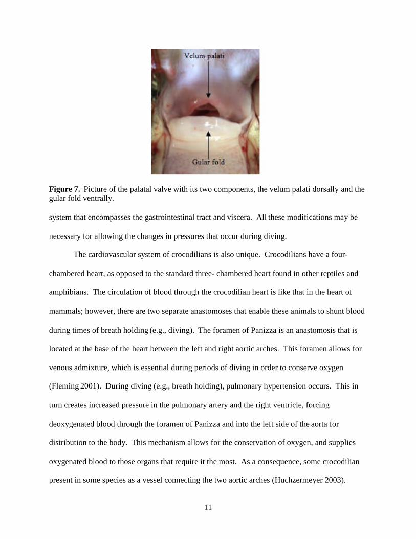
One of the most interesting anatomical features of crocodilians is the palatal or gular valve.
There is some discrepancy as to the name of this structure and its two components, but I will
attempt to describe them based on their anatomical location. Crocodilians have a true hard palate
that ends in a soft palate caudally. This soft palate has a ventral flap that is referred to as the
velum palati. The velum palati is the dorsal component of the palatal valve. Its second and
ventral component is the gular fold. This structure projects in a craniodorsal direction from the
A C
A C

10
Figure 5. Integumentary sensing organs (gray dots) on the lateral mandible and maxilla of analligator.
Figure 6. Integumentary sensing organs (arrows) on the ventral skin of a crocodile.
base of the tongue and has a cartilaginous base to it that is part of the larynx. Together, the
velum palati and the gular fold form what is known as the palatal or gular valve (Fig. 7). The
function of this valve is to seal the pharyngeal cavity while under water to prevent aspiration,
allowing these animals to catch prey under water. Crocodilians also have control of their nares,
and are able to open and close them as needed to prevent water influx.
The respiratory system of crocodilians is comprised of a pair of well- developed lungs.
Respiration in these animals is an active process, and is aided by the intercostal muscles and the
septum post hepaticum. The septum post hepaticum is analogous to a mammalian diaphragm,
and creates a partial separation of the thoracic and abdominal viscera. There are also a number
of membranous connections that separate the lungs and the liver and an intricate mesentery
11
Figure 7. Picture of the palatal valve with its two components, the velum palati dorsally and thegular fold ventrally.
system that encompasses the gastrointestinal tract and viscera. All these modifications may be
necessary for allowing the changes in pressures that occur during diving.
The cardiovascular system of crocodilians is also unique. Crocodilians have a four-
chambered heart, as opposed to the standard three- chambered heart found in other reptiles and
amphibians. The circulation of blood through the crocodilian heart is like that in the heart of
mammals; however, there are two separate anastomoses that enable these animals to shunt blood
during times of breath holding (e.g., diving). The foramen of Panizza is an anastomosis that is
located at the base of the heart between the left and right aortic arches. This foramen allows for
venous admixture, which is essential during periods of diving in order to conserve oxygen
(Fleming 2001). During diving (e.g., breath holding), pulmonary hypertension occurs. This in
turn creates increased pressure in the pulmonary artery and the right ventricle, forcing
deoxygenated blood through the foramen of Panizza and into the left side of the aorta for
distribution to the body. This mechanism allows for the conservation of oxygen, and supplies
oxygenated blood to those organs that require it the most. As a consequence, some crocodilian
present in some species as a vessel connecting the two aortic arches (Huchzermeyer 2003).

12
Other anatomical features include the presence of submandibular and paracloacal glands.
The hard dorsal scales are comprised of osteoderms, which are bony plates located in the dermis.
They also have a smaller gastric compartment distal to the stomach that is a gizzard-like
structure in which rocks and other materials may be found. It, however, does not appear as
developed as the ventriculus in a bird. Their intestines have a thick wall and can have well
developed diffuse aggregates of lymphoid tissues analogous to Peyer’s patches. Crocodilians
have no urinary bladder, but can hold large amounts of urine and water in their colon. A gall
bladder is present. Sexing can be performed by palpation. Males have a phallus, which can be
palpated and extracted from the cloaca. Females have a well- developed clitoris that can be
confused with a phallus. Internally, they have paired gonads that are in close association with
the ventral surface of the kidneys.
The body temperature, heart rate and respiration will vary with species and
environmental factors. Environmental temperature, season of the year, and age represent some
of the factors that can influence physiologic data.
Husbandry
Environmental Considerations
The environmental considerations for crocodilians will vary with the scenario in which
you are working. In captive rearing operations, facilities are aimed at producing large number of
animals in the most efficient manner possible. In a zoo or other educational institution, the
facilities are aimed at exhibiting the animals in the most accurate artificial environment possible.
Regardless, both scenarios should concentrate on having clean water, appropriate diet, and
enough space to accommodate the growth of the animals. As with most exotic animals, the
challenge is to mimic their natural environment as closely as possible in captivity.

13
Enclosure Size
There are no specific recommendations for enclosure size for crocodilians in captivity.
The size of the enclosure will largely depend on the species of crocodilian and their purpose in
captivity. You should have a general understanding of their biology and natural behavior in
order to design an appropriate enclosure for display (e.g., zoo or aquarium). If in doubt, you
should contact an institution housing the species for advice on what has worked or not worked
for them. There are, however, some general guidelines for the commercial production of
American alligators (A. mississippiensis). The recommendations for maintaining alligators in a
commercial operation are as follows: One square foot per alligator up to 24 inches in length
(snout to tip of tail), three square feet per alligator for those between 25 and 48 inches in length,
and after that add an additional square foot of space for every 6 inches in body length beyond 48
inches (Elsey 2004). These are considered to be recommendations for the maximum stocking
rate for alligators in commercial operations.
For a zoo or educational facility, the recommendation should be made to make the
exhibit as big as possible. First, consider the species of interest. Some species grow larger than
others and may be more territorial, which would require a larger enclosure. You must also
consider how many animals will be kept in the enclosure and the gender ratio of animals in the
enclosure. Males can become quite aggressive during the reproductive season, and keeping them
separated should be a consideration if enough space is not available. Exhibits can be maintained
outdoors, indoors, or using a combination of both. Outdoor exhibits will likely mimic the natural
environment more closely, but also present more challenges for maintaining hygiene of the water
and environment and exposure to diseases. Geographical location will also play a role in the
creation of outdoor exhibits, as not all species of crocodilians can tolerate cold weather. Finally,
some species can generate large burrows, and may escape an exhibit that is not secured.

14
Temperature and Humidity
The temperature and humidity requirements for crocodilians in captivity vary with
species. Once again, it is important to have an understanding of their biology and natural history
to be able to duplicate their natural environment. The provision of both circadian variations in
the light cycle and environmental temperature to mimic the natural environment should be made.
Unfortunately, this is not the case in many commercial operations, where they are maintained at
a fairly constant temperature and humidity in order to obtain faster growth. From a health
standpoint, this may allow infectious organisms that typically affect mammals to also affect these
reptiles. As the animals are maintained at higher temperatures, approaching those of mammals,
new diseases may adapt to infecting reptile hosts. In an enclosure, the water temperature can be
maintained via heating elements contained within the concrete slab or in line water heaters if
using a re-circulating system. The water temperature must also be maintained during the
refilling of the pen or enclosure to avoid a drastic temperature change.
Lighting
Lighting requirements for reptiles are still a controversial subject. As a general rule, full
spectrum lighting possessing adequate ultraviolet B radiation (UVB) is recommended for
herbivorous and omnivorous reptiles. Ultraviolet light is essential for the synthesis of vitamin
D3, specifically its active form 1, 25 dihydroxyvitamin D, which is essential for the metabolism
of calcium and phosphorus. A deficiency of vitamin D3 can lead to inappropriate calcium
absorption, which in turn creates a metabolic imbalance (e.g., metabolic bone disease).
Metabolic bone disease, and more specifically secondary nutritional hyperparathyroidism, is
recognized in many reptile species that are not provided UVB and/or calcium. Carnivorous
reptiles may also benefit from UVB, but are thought to obtain enough vitamin D3 and calcium
from their prey. The UVB requirement for crocodilians is unknown, but as true carnivores it

15
might be expected that they can thrive with minimal exposure to UVB. It is a common practice
at alligator ranches to raise these animals in darkness with no source of UVB or a normal light
cycle. Most animals will grow well under these conditions, and some have even been grown to
adulthood without signs of metabolic diseases. However, the author has also observed evidence
of metabolic bone disease in a subset of captive American alligators being fed a commercial diet
and with no exposure to ultraviolet light. In these cases, we must also consider the possibility
that the commercial diet may be deficient in calcium. Anecdotal stories from alligator ranches
claim that weak, anorectic animals appear to improve after being exposed to sunlight over a
period of time. Further research is needed to determine the UVB requirements of crocodilians,
and the potential benefits of exposure to UVB. Natural unfiltered sunlight is the best source of
UVB, but various artificial sources are also available.
Substrate
The two main substrates used in a crocodilian exhibit are water and soil/sand. The
species, age, and feeding habits must be taken into account in order to avoid selecting a substrate
that may be accidentally ingested. It is also important to prevent the public from throwing coins
and trash into exhibits, as this may represent a source of toxicity and foreign body impactions.
In commercial operations, a smooth covering is applied to the concrete in order to preserve the
quality of the hide. Either an epoxy coating or plastic liners are routinely used as substrate.
Nutrition
Crocodilians are true carnivores, as evidenced by their short intestinal length and cutting/
tearing teeth. As such, they will require a high protein diet that is low in fiber. Their feeding
habits in the wild will vary with age and food availability. The diet of juvenile animals often is
comprised of small invertebrates, amphibians, and other reptiles. As they grow, they will pursue
larger sized prey of the same type and add fish and birds to their diet. As adults, they will start

16
preying on small mammals too. There has been some study into the nutritional requirements of
alligators (Staton et al. 1990) (Coulson et al. 1987). Different feeding schemes were also
evaluated in the early 1990s (Coulson et al. 1992). In captivity, there are various commercial
feeds available for alligators. These are extruded pellet diets that aim to meet the complete
nutritional requirements of an alligator. These diets are generally comprised of 45%, 47%, or
56% protein, less than 11% fat, and fiber content less than 3% (Burris 2005). Over time these
diets have been refined. They are widely used in commercial ranching operations, but may
prove too expensive and/or inappropriate for the long term feeding of alligators. A variety of
whole prey feeds, such as chicken, nutria and fish, may also be recommended. If using nutria,
you must be sure that it was not killed using lead shot, as this can lead to lead toxicosis in
alligators (Camus et al. 1998). If feeding fish that has been frozen, you must also supplement the
diet with thiamine to prevent thiamine deficiency.
Preventive Medicine
Quarantine
All newly acquired crocodilians should be quarantined before being introduced into a
facility. A detailed history from the facility providing the animals should also be obtained in
order to gather information related to previous disease history. It is important to always work
with reputable individuals or institutions. Wild caught animals rarely come with much history.
Crocodilians should be quarantined for a minimum of 60 to 90 days. Quarantine should always
be done in a building that is separate from the main facility. During this time, the animals can be
examined for any sign of illness, and diagnostic tests (e.g., complete blood count, chemistry,
West Nile virus (WNV) antibodies) performed to assess the overall health status of the animals.
If they originated from an area where WNV is prevalent, it may be advisable to test for
antibodies to determine exposure to WNV.

17
Unfortunately, a true quarantine process can not be done in many cases because of
limited facilities, time or money. Quarantine can also be a real challenge for commercial
operations where you may be dealing with tens of thousands of animals. Nonetheless, zoological
institutions should always attempt to quarantine crocodilians.
Routine Exams
Routine physical exams should be performed on crocodilians at zoological institutions as
part of their preventive health program. As animals get larger in size, this activity may become
more hazardous. Chemical immobilization can be used, if needed, in order to perform necessary
examinations. In commercial operations, the skin of a subset of animals will be examined at
some interval before the time of slaughter. These instances present an opportunity for
veterinarians to examine the animals in this type of operations. Other than these, routine
physical exams are not commonly performed in crocodilians in commercial operations.
Biosecurity
Biosecurity is an essential part of disease prevention in any animal facility, and
crocodilians are no exception. A sanitation station should be placed at the entrance of each
building. These stations should contain a footbath and a hand washing station. The presence of
these stations should remind individuals of the importance of biosecurity, and minimize the
transfer of diseases between exhibits or buildings. A brush should be provided at each footbath
to clean the boots/shoes thoroughly. The solution for the bath can be made of sodium
hypochlorite or other commercial disinfectant, preferably with virucidal activity. Footbaths
should be changed daily or as they are dirtied since organic material can deactivate disinfectants.
The footbath should always be used before and after entering the building. A hand washing
station should also be provided. If a water source is available, then a sink and hand soap should
be provided. Alternatively, you can provide a waterless hand sanitizer product or exam gloves.

18
In addition to the sanitation station, separate tools for working in each building should be
provided. This will also prevent the transfer of diseases via nets, brooms, rakes, or other
cleaning supplies. The buildings themselves must also be maintained free of pests and cleaned
thoroughly when possible. In commercial operations, it is common practice to empty and
disinfect the buildings after the slaughter period and before introducing new animals. Finally,
the water the animals are housed in should be cleaned regularly. Poor water quality often leads
to the development of health problems in these animals. Regular complete changes of the water
and/or the addition of filtration can be used to maintain the quality of the water.
Handling and Restraint
Manual restraint of crocodilians is required for examining, medicating, and
transporting/relocating crocodilians. The size and species of crocodilian will determine the most
appropriate restraint method to use. Always work with someone who is familiar with the
behavior and restraint of a particular species. Alligators and caimans are usually thought of as
being less aggressive than crocodiles, but this may not always be true. All sizes and species
should be handled carefully, considering both the safety of the individuals restraining the animal
and the animal itself. Crocodilians less than 3 feet in length (snout to tip of tail) may be handled
by one or two individuals. Those between 3 and 6 feet in length should be handled by at least
two or three individuals. Those longer than 6 feet in length will require at least four to five
individuals for safe restraint. There are a number of restraint tools, such as pole snares, nets,
squeeze cages and traps, which can be used to restrain crocodilians. The head, tail, and limbs
must be immobilized and kept under control to effectively immobilize a crocodilian. Once the
animal is captured, the mouth should be secured with strong tape or a rope. Albino and leucistic
animals can have a more sensitive skin than those that are normally pigmented, and should
therefore be handled more carefully. Restraining a crocodilian is a stressful procedure, so it is
19
important to minimize the amount of contact time spent wit the animal. Chemical restraint
should be considered for aggressive or overly fractious animals.
History and Physical Examination
Signs of illness in captive crocodilians usually begin as non-specific in nature. Anorexia,
lethargy and death may be the first indication that something is wrong in a collection of animals
or commercial operation. A change in the behavior of the animals may also be observed. A visit
to the facility should be performed during feeding time to avoid additional stress to the animals.
A thorough review of the husbandry practices should be evaluated. A thorough history should
include questions about the number of animals, source, age, most recent introduction, quarantine
practices, diet, general husbandry practices, frequency of feeding, water quality parameters,
clinical signs, time since first signs were observed, recent changes in management techniques,
and whether any treatments have been provided. If working in a commercial operation, a subset
of animals should be collected for diagnostics and necropsy. In addition to obtaining routine
samples, tissues should be frozen for possible bacterial, fungal, or viral cultures. If working in a
zoological institution, you may not be able to sacrifice live animals, but you should obtain
diagnostic samples from those with and without clinical signs. Necropsies should be performed
in all dead animals. Live animals should be examined closely.
Once the animal is properly restrained, you can perform a through physical exam. You
must, however, be aware of the location of the head and tail at all times for your own safety. The
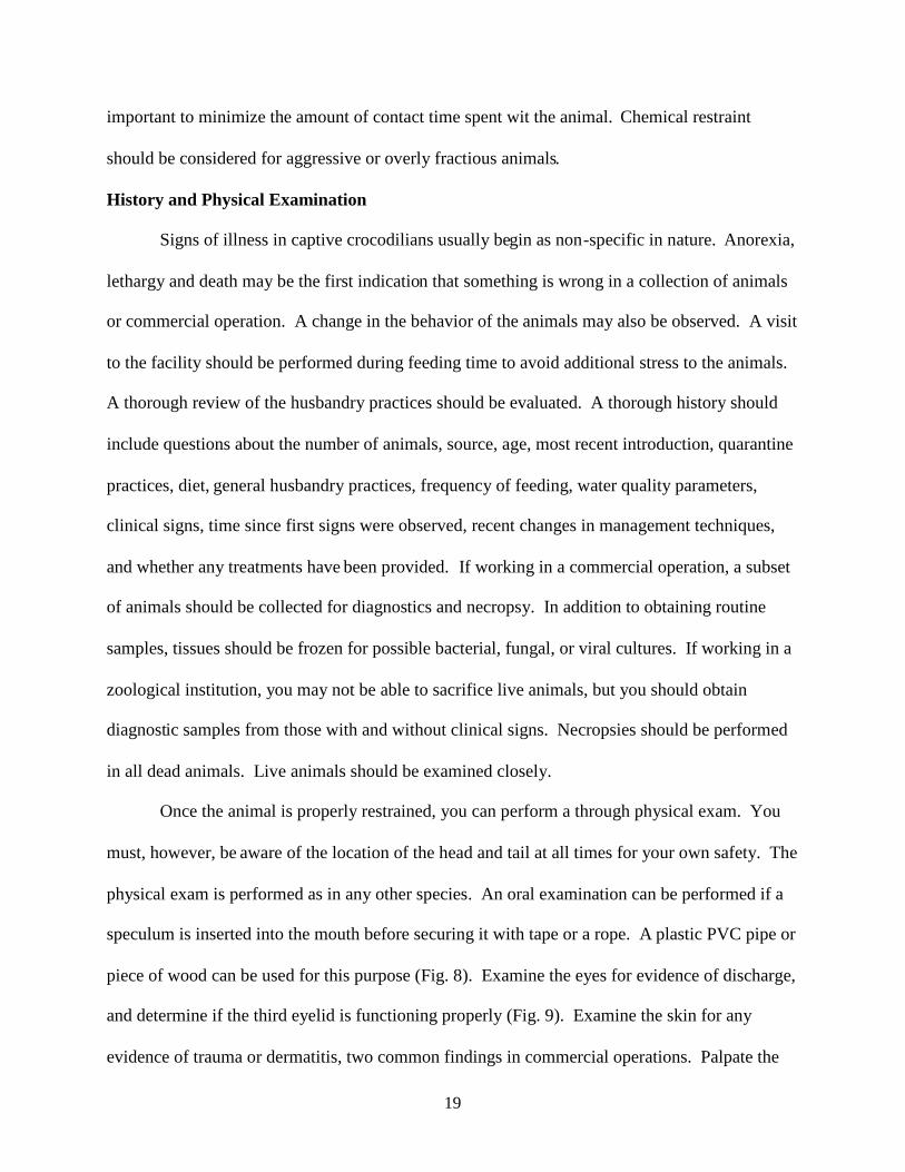
physical exam is performed as in any other species. An oral examination can be performed if a
speculum is inserted into the mouth before securing it with tape or a rope. A plastic PVC pipe or
piece of wood can be used for this purpose (Fig. 8). Examine the eyes for evidence of discharge,
and determine if the third eyelid is functioning properly (Fig. 9). Examine the skin for any
evidence of trauma or dermatitis, two common findings in commercial operations. Palpate the

20
extremities and joints for any evidence of swelling. Joint swelling is a common sequellae to
mycoplasmosis or trauma. Examine the musculature of neck, pelvic region and tail. Poor body
condition may be reflected in these areas. In commercial operations, it is important to examine
the skin on the ventrum. Many disease manifestations will be noted on the ventral skin. It is
Figure 8. A speculum inserted in the mouth of an alligator allows for visualization of theoropharyangeal cavity. This may also be used as a protective guard while performing endoscopyor retrieving foreign bodies from the intestinal tract.
Figure 9. Conjunctivitis with involvement of the third eyelid in a Morelet’s crocodile.
also common to find tooth marks, scratches, and lacerations. Finally, examine the vent and
cloaca for any abnormalities. If working in a commercial operation, examine multiple animals to
determine if an observation is associated with disease in multiple animals. If working in a
zoological institution and any findings are suspected to be infectious in origin, you should
examine other animals in the exhibit. As with any species you need to be aware of what is
normal in order to recognize clinical signs of disease.

21
Diagnostic Testing
Diagnostic testing in crocodilians should follow the same standard protocols advocated
with other species. One of the limitations associated with some of the diagnostic tests available
for crocodilians is that they are not validated or are based on a loose set of reference ranges. It is
important to always inquire about the specifics of a test in order to make an accurate
interpretation of the results. Published literature and the experience of other colleagues is an
invaluable part of interpreting diagnostics in crocodilians.
Venipuncture
There are various sites for venipuncture in crocodilians. The ventral coccygeal vein can
be accessed from either the ventral or lateral aspect of the tail (Fig 10). This vessel lays ventral
to the vertebral processes and on midline with the vertebrae. A second alternative is the
Figure 10. Venipuncture being performed from the ventral coccygeal vein using a lateralapproach.
supravertebral sinus located on midline at the junction of the head and the neck (Fig. 11). You
must be careful not to insert the needle past the spine, as it may be possible to damage the spinal
cord. A third site for venipuncture is the lateral occipital sinus, located lateral to the
supravertebral sinus (Wilhite and Nevarez 2004). This site can be approached from the dorsal
aspect of the neck, and is surrounded by muscles, decreasing the risk of contact with the spinal

22
Figure 11. Venipuncture being performed from the supravertebral sinus in an alligator.
Figure 12. Venipuncture being performed from the lateral occipital sinus in an alligator. Noticethat it is farther away from midline, eliminating the risk of damaging the spinal cord.
cord (Fig. 12). A 3cc syringe and a 22 gauge needle can be used for blood collection. Lithium
heparin and EDTA tubes can be used for chemistries and complete blood counts, respectively. If
collecting serum, the tubes may have to sit for at least 45 to 60 minutes before centrifugation to
allow proper separation of the serum from the red blood cells.
Clinical Pathology
As with other species, complete blood counts (CBC’s) and chemistry panels are a
fundamental part of diagnostics in crocodilians. These tests can help us assess the overall health
status of an animal. A CBC is generally used to measure whether an animal has a leukocytosis
(e.g., stress or inflammation) or leukopenia. Plasma chemistry panels can provide insight into
the physiologic status of an animal. Measurement of the packed cell volume (PCV) and total

23
solids/ total proteins of an animal are also important to provide insight into the erythron,
hydration status, or possible inflammatory response (e.g., hyperglobulinemia) of a patient. Fine
needle aspirates, impression smears and fluid analysis are useful diagnostic tools in crocodilian
medicine. Urinalysis is not a practical test in crocodilians due to the absence of a urinary bladder
and functional loop of Henle, and the fact that they will likely release the urine product in the
water.
A CBC can be done using a hemacytometer with the Unopette Eosinophil Determination
for Manual Methods stain (Becton Dickinson and Company, Franklin Lakes, NJ 07417-1885). A
blood smear stained with diff quick (Quik-dip stain, Mercedes Medical Physician and Laboratory
Products, 7490 Commerce Ct. Sarasota, FL 34342) is also needed to complete your total
estimated white blood cell count and obtain the cell differential count. Interpretation of CBC’s
from crocodilians can be challenging at first, if you are not familiar with the morphology of their
white blood cells. In order to familiarize yourself with these cells, spend some time scanning the
blood smear to become familiar with all the different types of cells and their appearances.
Alternatively, the samples can be submitted to a commercial laboratory that is comfortable
interpreting crocodilian CBC’s.
Imaging
Most imaging modalities (e.g., radiographs, Computed Topography scan, Magnetic
Resonance Imaging, and ultrasound) can be used for crocodilian imaging, as long as there is a
general understanding of their anatomy. This will be crucial for the interpretation of diagnostic
images. Knowledge of the anatomy will also help locate organs during ultrasound examination.
Radiographs can be used to locate foreign bodies as well as diagnose fractures. Contrast studies
can also be performed. Barium sulfate or iohexol can be administered at 5-20 ml/kg PO to
improve the quality of the images. Iohexol can be diluted with water at a 1:1 ratio (Carpenter et

24
al. 2001). Depending on the size of the animal and the site of interest, some imaging procedures
may be performed with the animal under manual restraint only. However chemical restraint may
be required in some instances.
Stress and Immunosuppression
Stress has been defined as “a physiological answer to a perceived threat that includes, but
is not restricted to, increased adrenal secretion” (Rooney and Guillette 2001). Stress can also be
any event that challenges homeostasis. The response of the body to that event is complex and
involves more than an adrenal response. The autonomic nervous system, the hypothalamic
adrenal axis, neuropeptides, neurotransmitters, and neuroimmunological mediators all play a role
in the response of the immune system to stress (Dohms and Metz 1991). Measuring stress and
immunosuppression is a challenge in veterinary medicine. There are no specific tests available
to provide a clinical measure of stress. Because of this, we generally concentrate on identifying
a combination of physiologic changes that give us an idea of what is involved in a stress
response. The stress response in crocodilians has been examined in relation to restraint, long
term corticosterone implants, cold shock, and stocking densities (Rooney and Guillette 2001,
Lance and Elsey 1999, Morici et al. 1997, Lance and Elsey 1999a, Elsey et al. 1990). Lance et
al. provides an overview of the physiology and endocrinology of stress in crocodilians (Lance et
al. 2001). Catecholamines, glucocorticoids, glucose, and lactate have been implicated in the
stress response of crocodilians. Changes in the white blood cells have also been implicated with
immunosuppression and the stress response (Lance and Elsey 1999, Morici et al. 1997, Lance
and Elsey 1999a, Lance et al. 2001). There is enough evidence to suggest that stress plays an
important role in the physiology of crocodilians, and it may indeed predispose them to illness.
Overcrowding, handling, excessive noise, diet changes, and temperature irregularities should all

25
be considered as predisposing or confounding factors of disease. All these factors must be
considered in the history of a clinical case and at the moment of making recommendations.
An example of how stress can play a role in disease susceptibility can be observed in a
case seen by the author. A commercial alligator facility had a chronic history of dermatitis. The
alligators with the most severe lesions were consistently located on one end of the building while
those at the other end were unaffected or only mildly affected. The location of the severely
affected animals was consistent in all buildings. One building had no affected animals. After
obtaining a thorough history and visiting the operation, a possible explanation for the occurrence
of disease was found. All the buildings with affected animals had PVC pipes on the inside walls
that delivered water to each individual pen. A strong water stream fell from the pipes down into
the water and the strength of the stream decreased from one entrance of the building (inflow) to
the other entrance (outflow). The strength of the water stream was considerable near the inflow.
The building with no affected animals did not have any source of falling water into the pens.
Water quality, temperature, and diet were the same in affected and non-affected buildings. The
most affected animals happened to be in the pens at the inflow side of the building with the
number of affected animals decreasing as you approached the outflow side of the building. This
constant flow of water was creating a constant movement of the water surface and consequently
stimulating the alligators via the ISO’s. Once this watering system was changed, the cases of
dermatitis decreased and no new cases were reported. Although other factors such as water
quality may have contributed to the dermatitis, the change in the watering system decreased the
progression and occurrence of the disease. Other stressors for captive reared alligators include
construction and drastic changes in water temperature. This reiterates the importance of
addressing the environment as well as the animals when working in commercial operations.
26
Bacterial Diseases
Most bacterial infections in captive crocodilians are probably opportunistic in nature.
Poor water quality, trauma, and stress are some of the factors that contribute to bacterial
infections in crocodilians. Crocodilians in their natural environment seem to cope well with
most bacterial infections. This may be in part to reports of antibacterial properties in serum and
tissues of crocodilians (Merchant et al. 2003, Shaharabany et al. 1999). This remains a
controversial topic and some are investigating it further. It may also mislead people into
thinking that crocodilians are not susceptible to bacterial infections. It is true that they appear to
tolerate trauma and other lesions that would be fatal in many species, but they are still capable of
succumbing to an array of microorganisms, including bacteria. In fact, a number of bacterial
infections have been reported in crocodilians and septicemias are thought to be a frequent
finding. Septicemias are definitively diagnosed post-mortem and can be associated with a wide
number of bacteria, many of them generally associated with the intestinal microflora. There is
an abundance of information about bacteria recovered from American alligators (Gorden et al.
1979, Shotts et al. 1972, Millichamp et al. 1983, Jacobson 1984, Flandry et al. 1989, Novak and
Seigel 1986, Wallace et al. 1966, Mainster et al. 1972, Brown et al. 2001, Jacobson et al. 1984,
Clippinger et al. 2000, Barnett and Cardeilhac 1995, Newton 1992, Bounds and Normand 1991,
Russell and Herman 1970, Brown et al. 2001a) (Table 4) and African dwarf crocodiles
(Osteolaemus tetraspis) (Huchzermeyer et al. 2000 ). Some bacteria (e.g., Aeromonas sp.) are
commonly isolated from crocodilians, while others (e.g., Erysipelothrix sp. and Clostridium sp.)
are not as common.
Salmonellosis
The importance of Salmonella sp. arises more from its zoonotic potential rather than its
ability to cause disease in crocodilians, although it has been associated with mortalities in Nile
27
crocodile (C. niloticus) hatchlings (Huchzermeyer et al. 1994). In most commercial operations,
the meat is sold as a by-product of hide production. Various species of Salmonella have been
isolated from the meat of crocodiles from commercial operations, and may represent a potential
source of infection for humans (Barnett and Cardeilhac 1995, Russell and Herman 1970, Millan
et al. 1997, Madsen 1996, Madsen 1993, Manolis et al. 1991, Rickard et al. 1995). Petting zoos
at zoological institutions that include crocodilians may also serve as a potential zoonotic source
of infection for humans. Although the zoonotic potential for this organism does exist, there are
no well- documented cases of human infections originating from crocodilians.
Chlamydiosis
Chlamydiosis has also been reported in crocodilians. There is a report of an isolate
closely resembling Chlamydophila psittaci that was obtained from the livers of Nile crocodiles in
Zimbabwe (Huchzermeyer et al. 1994). This infection is thought to have an acute course, and
was characterized by hepatitis and heavy mortalities in hatchlings. A chronic form
characterized by conjunctivitis has also been reported and may be more common. There are also
other reports of Chlamydia sp. isolates in cases of mycoplasmosis and adenoviral infections
(Huchzermeyer 2003).
Dermatophilosis (“Brown Spot Diseases”)
“Brown spot disease” has been attributed to Dermatophilus sp., with most of the cultures
resembling Dermatophilus congolensis. It has been reported in both crocodiles and alligators
(Newton 1992, Bounds and Normand 1991, Buenviaje et al. 1997, Buenviaje et al. 1998, Barnett
and Cardeilhac 1998). Affected animals present with brown to red lesions on the skin that are
usually located at the junction of the ventral abdominal scales. Over time, these lesions may
become ulcerated. This disease does not respond well to antibiotic therapy, but can be prevented
by practicing intensive hygiene methods.

28
Table 4. Bacteria isolated from A. mississippiensis with and without clinical signs of disease.
Isolate TissueClinical
Signs/Lesions Reference
Aeromonas hydrophila Blood Yes Brown et al. 2001
Lungs, heart, liver,kidneys,
Yes, No Gorden et al. 1979
intestines, oral cavity
Lungs, blood Yes Shotts et al. 1972
Eye Yes Millichamp et al. 1983,Jacobson et al. 1984
Oral cavity, water No Flandry et al. 1989
Aeromonas sp. Lungs Yes Clippinger et al. 2000
Acitenobacter calcoaceticus Oral cavity No Flandry et al. 1989
Aerobacter radiobacter Oral cavity No Flandry et al. 1989
Bacillus sp. Not specified Yes Barnett and Cardeilhac 1995
Bacteroides asaccharolyticus Oral cavity No Flandry et al. 1989
Bacteroides bivius Oral cavity, water No Flandry et al. 1989
Bacteroides loescheii/denticola Oral cavity, water No Flandry et al. 1989
Bacteroides oralis Oral cavity No Flandry et al. 1989
Bacteroides sordellii Oral cavity No Flandry et al. 1989
Bacteroides thetaiotamicron Oral cavity No Flandry et al. 1989
Bacteroides vulgatus Oral cavity No Flandry et al. 1989
Bacteroides sp. Water No Flandry et al. 1989
Citrobacter freundii Blood Yes Novak and Seigel 1986
Oral cavity No Flandry et al. 1989
Clostridium bifermentans Oral cavity, water No Flandry et al. 1989
Lungs Yes Clippinger et al. 2000
Clostridium clostriidoforme Oral cavity No Flandry et al. 1989
Clostridium innoculum Water No Flandry et al. 1989
Clostridium limosum Oral cavity No Flandry et al. 1989
Clostridium sordellii Oral cavity, water No Flandry et al. 1989
Clostridium sporogenes Blood Yes Clippinger et al. 2000
Clostridium tetani Oral cavity No Flandry et al. 1989

29
(Table 4. Continued)Clostridium sp. Blood Yes Clippinger et al. 2000
Corynebacterium sp. Tail abscess Yes Shotts et al. 1972
Dermatophilus sp. Skin Yes Newton 1992, Bounds andNormand 1991
Diphtheroid sp. Oral cavity No Flandry et al. 1989
Edwardsiella tarda Kidney, feces Yes Wallace et al. 1966
Fat body, pericardial fluid Yes Clippinger et al. 2000
Enterobacter agglomerans Blood Yes Novak and Seigel 1986
Enterobacter cloacae Oral cavity, water No Flandry et al. 1989
Enterobacillus sp. Lungs Yes Shotts et al. 1972
Escherichia coli Systemic Yes Russell and Herman 1970
Fusobacterium nucleatum Oral cavity No Flandry et al. 1989
Fusobacterium varium Oral cavity No Flandry et al. 1989
Klebsiella oxytoca Skin Yes Novak and Seigel 1986
Oral cavity No Flandry et al. 1989
Klebsiella sp. Lungs Yes Nevarez 2002-03
Micrococcus kristinae Blood Yes Brown et al. 2001
Moraxella sp. Oral cavity, water No Flandry et al. 1989
Morganella morganii Blood Yes Novak and Seigel 1986
Oral cavity No Flandry et al. 1989
Lung Yes Clippinger et al. 2000
Mycoplasma alligatoris Multiple tissues Yes Brown et al. 2001 , 39
Pasteurella haemolytica Oral cavity No Flandry et al. 1989
Pasteurella multocida Lungs Yes Mainster et al. 1972
Pasteurella sp. Oral cavity, water No Flandry et al. 1989
Peptococcus magnus Oral cavity No Flandry et al. 1989
Peptococcus prevotii Oral cavity No Flandry et al. 1989
Proteus mirabilis Blood Yes Brown et al. 2001
Proteus vulgaris Oral cavity No Flandry et al. 1989
Oviduct Yes Wallace et al. 1966
Blood Yes Brown et al. 2001
Lung Yes Clippinger et al. 2000

30
(Table 4. Continued)
Proteus sp. Blood Yes Novak and Seigel 1986
Providencia rettgeri Lung, brain, liver, spleen Yes Camus and Hawke 2002
Pseudomonas cepacia Oral cavity No Flandry et al. 1989
Pseudomonas diminuta Water No Flandry et al. 1989
Pseudomonas fluorescens Water No Flandry et al. 1989
Pseudomonas pickettii Oral cavity No Flandry et al. 1989
Pseudomonas vesicularis Water No Flandry et al. 1989
Pseudomonas sp. Lungs, Pharynx Yes Shotts et al. 1972
Water No Flandry et al. 1989
Salmonella typhimurium Gastrointestinal tract Yes Shotts et al. 1972
Salmonella braenderup, anatum, Cloaca No Shotts et al. 1972
Arizona spp.
Serratia marcescens Skin Yes Novak and Seigel 1986
Serratia odorifera Oral cavity No Flandry et al. 1989
Staphylococcus aureus Lungs Yes Mainster et al. 1972
Staphylococcus cohnii Blood Yes Brown et al. 2001
Streptococcus sp.β-hemolytic Lungs Yes Clippinger et al. 2000
Vibrio parahemolyticus Blood Yes Brown et al. 2001
Mycoplasmosis
Mycoplasma alligatoris is a recognized respiratory pathogen of crocodilians. It has been
documented in American alligators and the broad-nosed caiman (Caiman latirostris) (Brown et
al. 2001, Brown et al. 2001a, Pye et al. 2001). Other crocodilian species closely related to
alligators may also be susceptible. Clinical signs are non-specific and include lethargy,
weakness, anorexia, white ocular discharge, paresis, and edema (facial, periocular, cervical,
limbs) (Clippinger et al. 2000). Necropsy often reveals evidence of pneumonia, pericarditis, and
polyarthritis. Helmick et al. reported antimicrobial susceptibility for M. alligatoris with
doxycycline, oxytetracycline, enrofloxacin, sarafloxacin, tilmicosin, and tylosin (Helmick et al.
2002). A second mycoplasma species, M. crocodyli, was also described in Nile crocodiles and

31
the lesions were similar to those observed with M. alligatoris (Mohan et al. 1995). Some studies
have examined the use of an autogenous vaccine for M. crocodyli, but its efficacy is yet to be
determined (Mohan et al. 1997, Mohan et al. 2001).
Mycobacteriosis
Cases of pulmonary and enteric mycobacterial infections are mentioned by
Youngprapakorn et al. and Huchzermeyer (Huchzermeyer 2003, Youngprapakorn et al. 1995).
These reports include M. marinum and M. fortuitum from different spectacled caimans (C.
crocodilus), M. avium from an unspecified species of crocodile and from Nile crocodiles, and M.
ulcerans from Johnston’s crocodiles (C. johnstoni) (Huchzermeyer 2003). Many other reported
cases are based on gross findings and histopathology with acid- fast positive organisms identified
microscopically. The author has observed some clinical cases of suspected, yet unconfirmed,
pulmonary mycobacteriosis based on necropsy and histopathologic evaluation (Nevarez 2002-
03). Difficulties of growing Mycobacterium sp. make its definitive diagnosis a challenge. Other
diagnostic tools such as PCR may also be beneficial in identification of the bacteria.
Viral Diseases
The identification and diagnosis of viruses in reptiles has been slow as compared to other
species. This is in great part due to difficulties in developing diagnostic tests, as well as a lack of
knowledge about what viruses affect reptiles and under what conditions. Many clinical
presentations that go undiagnosed could be attributed to viruses. These may be new viruses or
just unknown to occur in a particular species. In crocodilians there are only a handful of
recognized viruses that have been documented over the years. Poxvirus and West Nile virus are
recognized pathogens in crocodilians. Jacobson et al. described an adenovirus-like infection in
captive Nile crocodiles characterized by nonspecific clinical signs, lethargy and anorexia.
Conjunctivitis and blepharitis was also observed in one of two crocodiles (Huchzermeyer 2003,

32
Jacobson et al. 1984). Intranuclear inclusions are usually found in the liver but may also occur in
the intestines, pancreas, and lung. Both horizontal and vertical transmissions have been
postulated (Huchzermeyer 2003). Diagnosis is obtained postmortem and no treatment regimes
have been established.
Coronavirus, influenza C virus, and paramyxovirus have been identified by transmission
electron microscope in the feces of crocodilians (Huchzermeyer 2003). Herpes virus-like
particles were identified by electron microscopy in the skin of a saltwater crocodile (C. porosus)
(McCowan et al. 2004). There has been a second report of herpes virus identified from the
cloaca of an American alligator via PCR (Johnson 2005). The clinical significance of these
findings is unknown. Seroconversion to paramyxovirus and eastern equine encephalitis virus has
also been reported (Huchzermeyer 2003).
Pox Virus
Parapoxvirus or pox-like viruses have been identified in five different crocodilian
species: spectacled caiman (Caiman crocodilus fuscus) (Jacobson et al. 1979, Penrith et al. 1991)
Brazilian caiman (Caiman crocodilus acre) (Ramos et al. 2002), Nile crocodile (Horner 1990,
Pandey et al. 1990), saltwater crocodile (Buenviaje et al. 1992), and freshwater crocodile
(Crocodylus johnstoni) (Buenviaje et al. 1992). Pox lesions in caimans will present as 1-3 mm
diameter, gray to white, coalescing to macular. Their location on the body includes the head,
palpebra, maxilla, mandible, limbs, palate, tongue, and gingiva (Jacobson et al. 1979, Penrith et
al. 1991, Ramos et al. 2002). Palpebral and generalized edema can also be present. Resolution of
clinical signs has been observed with and without changes in husbandry (Penrith et al. 1991,
Ramos et al. 2002). In crocodiles the lesions are described as 2-8 mm in diameter, yellow to
brown, wart-like, sometimes firm, and unraised to raised nodules with occasional shallow ulcers.
Their location on the body includes the head, palpebra, nostrils, sides of the mouth, oral cavity,

33
limbs, ventral neck and coelom, and at the root of the tail (Horner 1990, Pandey et al. 1990).
Resolution of lesions was reported to occur as early as three to four weeks (Horner 1990).
Histopathologic findings include epithelial hyperplasia, acanthosis, hyperkeratosis, and necrosis.
Borrel and Bollinger’s bodies may also be visible in some cases (Jacobson et al. 1979, Penrith et
al. 1991, Ramos et al. 2002, Horner 1990, Pandey et al. 1990). Secondary bacterial and fungal
infections may occur concurrently. At this time there are no specific treatment
recommendations. The use of an autogenous vaccine to treat poxvirus in Nile crocodiles has
been reported with some success (Horner 1990). Mosquito control and following good hygiene
practices are essential in preventing and controlling poxvirus outbreaks.
West Nile Virus
West Nile virus (WNV) has been reported from various crocodilian species, including the
American alligator (Miller et al. 2003), the Nile crocodile (Steinman et al. 2003), and the
Morelet’s crocodile (Crocodylus moreletii) (Rubio 2003). Crocodilians likely become infected
via a mosquito bite, as occurs in birds and mammals. There is also the possibility of infection
after ingestion of an animal with a high viral load of WNV as demonstrated by Klenk et al.
(Klenk et al. 2004). This last scenario is more likely to occur when housed outdoors and not in
enclosed buildings as in most alligator ranching operations. It has been demonstrated that
alligators can serve as amplifiers of WNV (Klenk et al. 2004). Although there is a lot to be
learned about WNV in crocodilians, we believe that once infected they can develop high
viremias and shed the virus in the feces. Fecal shedding leads to horizontal transmission of the
virus. The author has observed clinical evidence for this in commercial operations. Fecal
shedding and high viremias also raise the concern of zoonosis, especially in commercial
operations where animals are being slaughtered and people come in contact with blood and
tissues. A strict building quarantine and hygiene strategies should be implemented to prevent

34
spread to other animals in the facility as well as to the personnel. In the state of Louisiana,
alligator producers were provided with biosecurity recommendations to help them deal with
episodes of WNV and prevent spread of the disease.
Affected animals in captive operations have ranged in age from one month to over
twelve months old. In younger animals, infection is usually severe and acute with as much as
60% mortality. The pattern of deaths is peracute, usually seen as a sudden onset of mortalities
followed by a peak and subsequent decline in the number of deaths. However, sporadic
mortalities may also be seen, especially in older animals. Clinical signs of WNV in alligators
include swimming in circles, head tilt, muscle tremors, weakness, lethargy, and anorexia.
Bloating and difficulties swimming have also been observed (Nevarez et al. 2005b).
Gross findings are nonspecific. Light microscopy of tissues from infected animals may
reveal diffuse severe heterophilic, histiocytic, and necrotizing enterocolitis, heterophilic
meningoencephalitis, necrotizing and heterophilic hepatitis, heterophilic and histiocytic splenitis,
generalized heterophilic and histiocytic lymphoid folliculitis, and necrotizing and heterophilic
pancreatitis. You may also observe a mild multifocal heterophilic and lymphohistiocytic
interstitial nephritis, gastritis, and mild pulmonary congestion and edema. Strong
immunopositivity has been observed in the brain, liver, spleen, pancreas, kidney, and
gastrointestinal tract after immunohistochemistry testing for WNV. Proliferative enteritis has
been diagnosed in a group of alligators positive for WNV. These animals had colon lesions
tested positive for WNV via Reverse Transcriptase Polymerase Chain Reaction (RT-PCR),
culture and immunohistochemistry (Nevarez 2002-03, Nevarez et al. 2005b). The affected
alligators were bloated and unable to submerge under water. In the author’s opinion, this was
due to a blockage caused by the fibrinous membrane in the colon leading to bloating. This same
presentation has not been observed in any other cases of WNV.
35
There are no proposed treatments for WNV in crocodilians. Mosquito control, strict
quarantine and biosecurity are essential for the prevention of WNV. Once WNV is present in
any operation, it is critical to maintain affected animals and any in contact with them in strict
isolation. These must not be moved to other areas or buildings were WNV has not been
observed. Failure to isolate them may result in the spread of the virus. In addition, there should
be strict biosecurity measures, especially in the affected areas or buildings. Tools, such as nets,
feeding utensils, and boots, should be left in the affected area and not introduced to other areas or
buildings. Once the infection runs its course, the surviving animals will continue to thrive;
however, the duration of shedding in these cases is unknown. The author has observed antibody
titers of 1:640 and 1:320 in two alligators 14 months after exposure to WNV. These results were
obtained via a Plaque Reduction Neutralization test (PRNT). Definitive diagnosis of WNV
infection should be based on history, clinical signs and the results of diagnostic tests.
Postmortem diagnosis can be performed via RT-PCR and/or viral culture of brain, spinal cord, or
liver. Immunohistochemistry also provides supportive evidence of WNV infection. This last
technique may prove useful as an ante-mortem test when applied to biopsies of the colon
mucosa.
Fungal Diseases
Fungal infections also occur with some frequency in captive crocodilians. Most fungi are
opportunistic invaders of the integument, respiratory system, and gastrointestinal tract. Poor
water quality, trauma, stress, and extreme temperatures can contribute to the occurrence of
fungal disease. It is thought that most fungal infections in crocodilians are of enteric origin and
occur in other tissues secondary to a stressor (Huchzermeyer 2003). It is also important to
remember that fungi occur as ubiquitous organisms in nature. Therefore, it is not uncommon to
isolate fungi from tissues that are in contact with water and soil, such as the skin and intestinal

36
tract. The presence of these fungi will vary with geographic location and management
techniques that provide different environments for their growth. There are a number of fungi
that have been isolated from crocodilians (Huchzermeyer 2003, Huchzermeyer et al. 2000,
Buenviaje et al. 1998, Nevarez 2003, Buenviaje et al. 1994, Fromtling et al. 1979, Trevino 1972,
Frelier 1985, Silberman et al. 1985, Thomas et al. 2002, Maslen et al. 1988) (Table 5). Some of
these were associated with disease, while others were incidental findings. Diagnosis of fungal
disease requires the identification of a fungal organism via culture or special stains of the
affected tissues with concomitant histopathologic identification. Positive identification to genus
and species may be difficult. Treatment is expensive and may only be affordable in a small
number of animals. It also requires a long administration periods of at least two or three months.
For these reasons, many fungal infections go untreated and recommendations are made for
prevention of disease.
Parasitic Diseases
A number of parasites are known to affect crocodilians. Protozoa, nematodes,
trematodes, and pentastomes have all been reported in crocodilians. Cestode larvae have been
found in some species but no adult tapeworms are reported from crocodilians. External parasites
such as leeches, flies, mosquitoes, ticks and mites can also affect crocodilians (Huchzermeyer
2003). The presence of scales has created a misconception about the possibilities for external
parasitism in crocodilians; however there are a number of areas in a crocodilian’s body that have
soft skin and allow an external parasite to attach and/or obtain a meal. The recent cases of WNV
in the American alligator have reminded us that arthropods can play an important role in disease
transmission. Internal parasites are not a common problem in ranching operations where animals
are maintained in concrete pools without organic substrate. Endoparasites can be a concern in
wild crocodilians or those kept outdoors on organic substrates. Ectoparasites can be a problem in

37
both captive and wild populations. If a parasitism problem is encountered, one must assess the
environment of the animals as well as the life cycle of the parasite in order to determine the best
prevention and control methods.
Table 5. Fungi isolated from crocodilians around the world. This table presents isolates frommultiple tissues regardless of whether a disease process was attributed to the fungus.
Fungus Sample/Tissue Species ReferenceAcremonium sp. intestinal contents Osteolaemus tetraspis Huchzermeyer et al. 2000
Alternaria alternata skin Alligator mississippiensis Nevarez 2003Arthrinium sp. intestinal contents Osteolaemus tetraspis Huchzermeyer et al. 2000
Aspergillus clavatus intestinal contents Osteolaemus tetraspis Huchzermeyer et al. 2000Aspergillus flavus not specified Crocodylus porosus Huchzermeyer 2003
skin Crocodylus porosus Buenviaje et al. 1998intestinal contents Osteolaemus tetraspis Huchzermeyer et al. 2000
Aspergillus fumigatus skin Caimans (species not specified) Huchzermeyer 2003lungs Alligator mississippiensis Huchzermeyer 2003
Aspergillus niger not specified Crocodylus porosus Buenviaje et al. 1994skin Crocodylus porosus Buenviaje et al. 1998
intestinal contents Osteolaemus tetraspis Huchzermeyer et al. 2000skin Alligator mississippiensis Nevarez 2003
Aspergillus ustus lungs Alligator mississippiensis Huchzermeyer 2003Aspergillus versicolor not specified Crocodylus porosus Huchzermeyer 2003Beauveria bassiana lungs Alligator mississippiensis Fromtling et al. 1979
lungs Crocodylus niloticus Huchzermeyer 2003Beauveria sp. intestinal contents Osteolaemus tetraspis Huchzermeyer et al. 2000
Candida albicans oral cavity Caimans (species not specified) Huchzermeyer 2003Candida guillermondii intestinal contents Osteolaemus tetraspis Huchzermeyer et al. 2000
Candida krusei intestinal contents Osteolaemus tetraspis Huchzermeyer et al. 2000Candida parasilosis skin Crocodylus porosus Buenviaje et al. 1998
Candida sp. skin Crocodylus porosus Buenviaje et al. 1998Cephalosporium sp. muscle Caiman crocodilus Huchzermeyer 2003
lungs Caiman sclerops Trevino 1972Chaetomium globosum skin Alligator mississippiensis Nevarez 2003
Chaetomium sp skin Alligator mississippiensis Nevarez 2003Chrysosporium sp. intestinal contents Osteolaemus tetraspis Huchzermeyer et al. 2000
Cladosporium cladosporioides skin Alligator mississippiensis Nevarez 2003Cladosporium oxysporum skin Alligator mississippiensis Nevarez 2003
Cladosporium sp. skin Caimans (species not specified) Huchzermeyer 2003Cryptococcus lipolytica intestinal contents Osteolaemus tetraspis Huchzermeyer et al. 2000Cryptococcus luteolus intestinal contents Osteolaemus tetraspis Huchzermeyer et al. 2000
Curvularia lunata skin Alligator mississippiensis Nevarez 2003Curvularia lunata varaeria skin Crocodylus porosus Buenviaje et al. 1994
Curvularia sp. intestinal contents Osteolaemus tetraspis Huchzermeyer et al. 2000Epicoccum purpurascens skin Alligator mississippiensis Nevarez 2003
Eurotium chevalieri skin Alligator mississippiensis Nevarez 2003Fusarium moniliforme lungs Alligator mississippiensis Frelier et al. 1985
Fusarium solani internal organs Crocodylus porosus Huchzermeyer 2003,Buenviaje et al. 1994
38
(Table 5. Continued)Fusarium sp. not specified Crocodylus porosus Huchzermeyer 2003
gingiva Caiman crocodilus fuscus Huchzermeyer 2003skin Crocodylus porosus Buenviaje et al. 1998
intestinal contents Osteolaemus tetraspis Huchzermeyer et al. 2000Geotrichum candidum not specified Crocodylus porosus Huchzermeyer 2003
intestinal contents Osteolaemus tetraspis Huchzermeyer et al. 2000Geotrichum sp. not specified Crocodylus porosus Huchzermeyer 2003
Metarhizium anisopliae lungs Crocodile (species not specified) Huchzermeyer 2003lungs Osteolaemus tetraspis Huchzermeyer 2003lungs Alligator mississippiensis Huchzermeyer 2003
Monascus ruber skin Alligator mississippiensis Nevarez 2003Mucor circinelloides skin Caimans (species not specified) Huchzermeyer 2003
gastric ulcers Osteolaemus tetraspis Huchzermeyer 2003skin Alligator mississippiensis Nevarez 2003
Mucor sp. lungs Crocodile (species not specified) Silberman et al. 1977Nannizziopsis vriesii skin Crocodylus porosus Thomas et al. 2002
Paecilomyces lilacinus liver Crocodylus porosus Maslen et al. 1988Paecilomyces farinosus lungs Alligator mississippiensis Huchzermeyer 2003Paecilomyces variotii skin Alligator mississippiensis Nevarez 2003
Paecilomyces sp. not specified Crocodylus porosus Huchzermeyer 2003intestinal contents Osteolaemus tetraspis Huchzermeyer et al. 2000
Penicillium citrinum skin Alligator mississippiensis Nevarez 2003
Penicillium lilacinum lungs Alligators and Crocodiles (speciesnot specified) Huchzermeyer 2003
Penicillium oxalicum skin Crocodylus porosus Buenviaje et al. 1994Penicillium sp. not specified Crocodylus porosus Huchzermeyer 2003
intestinal contents Osteolaemus tetraspis Huchzermeyer et al. 2000Phoma sp. intestinal contents Osteolaemus tetraspis Huchzermeyer et al. 2000
Syncephalastrum racemosum skin Alligator mississippiensis Nevarez 2003Syncephalastrum sp. skin Crocodylus porosus Buenviaje et al. 1998
Trichodermasp. skin Alligator mississippiensis Huchzermeyer 2003intestinal contents Osteolaemus tetraspis Huchzermeyer et al. 2000
Trichophyton sp. skin Alligator mississippiensis Huchzermeyer 2003Trichosporon beigelii intestinal contents Osteolaemus tetraspis Huchzermeyer et al. 2000
Trichosporon capitatum intestinal contents Osteolaemus tetraspis Huchzermeyer et al. 2000Trichosporon cutaneum skin Crocodylus porosus Buenviaje et al. 1998
Trichosporon sp. tongue, gingivae Crocodylus niloticus, C. fuscus Huchzermeyer 2003
Nutritional Disease
Nutritional diseases are probably not observed as frequently now as they were in the past.
Increased understanding of the biology, natural history, and dietary requirements of the different
crocodilian species has allowed better feeding schemes. Over time there has been a push to
develop commercial diets that will allow for high feed to weight gain ratios. These are available
for feeding American alligators, and although not perfect, do allow producers to grow captive
hatched alligators up to 36 inches in length in 12 months or less. In zoological institutions, the
goal is not rapid growth but rather steady and healthy development. In these cases, there is

39
common use of fresh/frozen prey (e.g., poultry, swine, beef, fish, nutria, native prey) with bones
as a source of calcium. This is also a common practice in crocodile farms. It is essential to use a
reputable source and ensure that no lead shots have been used to kill the prey item in order to
avoid Lead toxicity. Despite better feeding schemes, there are still some presentations that can
be associated with nutritional deficiencies.
A common nutritional disease of reptiles is metabolic bone disease (MBD). There are
various manifestations and pathogenesis for MBD but it is usually associated with a
calcium/phosphorus imbalance that leads to increased bone resorption. Calcium and vitamin D3
deficiencies are the two main etiologies for MBD. Vitamin D3 deficiencies may be associated
with a lack of exposure to UVB light. There are various light products in the market that can be
used to provide reptiles with UVB lighting. Clinical signs of MBD include weakness, lethargy,
kyphosis, scoliosis, osteodystrophy, pathologic fractures, paresis, and tooth decalcification. The
development of MBD is most often reported in herbivorous or omnivorous reptiles. MBD is not
common in commercial crocodilian operations, even in those were animals are raised in the dark
with no source of UVB. One explanation for this may come from the carnivorous nature of
crocodilians, which may allow them to obtain vitamin D3 from the diet without a true
requirement for UVB. However, if an appropriate calcium source is not offered, these animals
will likely develop MBD. The author has observed adult alligators grown in enclosed buildings
with no light source and a commercial diet with some meat supplementation to develop
normally. However, anecdotal comments from various ranchers indicate that the animals appear
to thrive better if exposed to sunlight. To date, metabolic bone disease continues to be a rare
occurrence in alligator ranches. Nonetheless, this is an area where more research is needed to
determine the UVB and calcium requirements of crocodilians.

40
Gout has been reported in crocodilians. It can occur as either an articular or visceral
form. Some predisposing factors for gout include high protein diets, dehydration, and stress.
Limb paresis/paralysis and joint enlargement may be observed with chronic gout; otherwise the
clinical signs are non- specific. Ariel et al. reported a case of gout with concurrent
hypovitaminosis A in crocodile hatchlings (Ariel et al. 1997).
Deficiencies of vitamins A, B, C, or E can also lead to a variety of musculoskeletal
disorders. These are thought to be less common when commercial diets are fed in addition to
meat products.
Respiratory Disease
Respiratory disease is one of the most common presentations of captive crocodilians in
commercial operations, second only to integumentary disease. Gross pathologic lesions in the
lungs are a common finding during necropsies. While many of these findings are incidental,
some will show clinical signs before death. Clinical signs in affected animals can be non-
specific anorexia, lethargy, and weakness, or be more system related and include dyspnea,
tachypnea, nasal secretion, excessive basking, respiratory stridor, and abnormal swimming
(either in circles or on one side of the body). In some cases, the animals may appear neurologic
as they become weak and ataxic. A clear distinction must be established between respiratory
disease and neurological disease. On the other hand, respiratory disease may be secondary to
neurologic disease as a consequence to the weakness that leads to aspiration. Most respiratory
infections are either bacterial or fungal in origin. Upper respiratory diseases, including rhinitis
and pharyngitis, have also been reported in crocodilians (Huchzermeyer 2003).
Neurologic Disease
Neurologic diseases are not common in crocodilians; however, the etiologies implicated
with this presentation deserve special attention. The most common neurologic presentation will

41
relate to abnormal swimming behavior (e.g., swimming in circles, swimming on one side of the
body). Once outside the water, these animals may show signs of lethargy, ataxia, head tilt, and
muscle tremors. Anorexia will also be observed and is perhaps an early sign observed by
personnel working with the animals. Any infectious agent affecting the nervous system can lead
to this presentation. The most recent etiology of neurologic signs in crocodilian species is WNV.
This should be included as a rule out for any crocodilian that presents with neurologic signs and
is housed in an area with prior history of WNV. Hypoglycemia has also been reported as a cause
of neurologic signs in alligators (Wallach et al. 1967). Another cause of neurologic signs may be
associated with increased ammonia/oxygen depletion in enclosed buildings, especially during the
summer. The author has observed neurologic signs and deaths of American alligators from a
commercial operation where a building was not cleaned on schedule. The water in the building
had not been changed in 72 hours and there was evidence of food left over from previous
feedings. The owner found both dead alligators and some with neurologic signs. Necropsy and
histopathologic evaluation pointed towards toxicity as the cause of death. Both dead and live
animals tested negative for WNV. All other buildings were cleaned on schedule and had no
evidence of disease. Mortalities decreased after cleaning of the affected building. Based on the
history, clinical observations, and histopathologic findings, we believe these animals showed
neurologic signs as a result of either increased ammonia level in the water, oxygen depletion in
the air, or both.
Musculoskeletal Disease
Musculoskeletal disease can occur due to changes in the incubation temperature or
humidity, trauma from fighting, transport, or restraint and from infectious diseases (Figure 13).
Deformities of the head, limbs, and tails are common malformations influenced by incubation
rather than true genetics (Figure 14). Fractures and limb amputations can occur due to trauma

42
and fighting. Many of these heal without major complications. This can be seen in captive as
well as wild crocodilians. In severe cases, trauma can lead to nerve or muscle damage and
paresis or paralysis. The most common infectious disease known to have effects on the
musculoskeletal system is mycoplasmosis. Both M. alligatoris and M. crocodyli can cause
polyarthritis in affected animals.
Gastrointestinal Disease
Ingestion of foreign bodies, gastric ulcers, enteritis, and trauma to the oral cavity are
some of the most common gastrointestinal diseases reported in crocodilians. Anorexia usually
accompanies gastrointestinal disease. Ingestion of foreign bodies occurs in both wild and captive
crocodilians. Construction, malfunction of water pumps and filters, forgotten objects in the
enclosure, and tossed objects by the public are some of the sources of foreign bodies in captive
scenarios. Sharp objects, such as nails, can cause severe problems, while other objects may pass
Figure 13. Mandibular trauma in a wild alligator. This type of trauma is usually observed inmales during the breeding season.

43
Figure 14. Maxillary deviation in a captive hatched alligator. These abnormalities are believedto be caused by irregularities in temperature and or humidity during the incubation period.
without difficulties. Infectious enteritis is usually diagnosed post- mortem based on gross and
histopathologic observations. Crocodilians appear to have an aggressive response to insult of the
gastrointestinal tract. Grossly, you may observe accumulation of fibrinous or fibrous material
and/or necrosis of the mucosa. (Figure 15) In some instances, this can even lead to obstruction
due to accumulation of fibrinous/fibrous material. Fecal impactions and torsions are probably
rare but can also occur in crocodilians. Gastric ulcerations may be associated with stress and
diet. The intestinal tract of crocodilians appears to contain a significant amount of gut associated
lymphoid tissue similar to Peyer’s patches. In the author’s opinion, there is opportunity for an
aggressive inflammatory response to infectious agents in the intestinal tract.
Integumentary Disease
Integumentary disease is likely the most important disease process affecting captive
crocodilians. Water quality, temperature, and stressors contribute to the occurrence of
integumentary diseases. The economic impact in commercial operations can be devastating.
Secondary bacterial and fungal dermatitis are a major problem in these operations. Lacerations,

44
Figure 15. Fibrin deposition and necrosis of intestinal mucosa in an alligator.
abscesses, and draining tracts can serve as a nidus for microorganisms, especially fungi and
bacteria. An additional factor in captive operations is the accumulation of fat in the upper water
column. This fat creates a slime layer on the walls and water surface, which is then transferred
to the skin of the animals creating an ideal environment for fungal and bacterial growth. This is
more of a problem when meat is the sole source of food or is provided in addition to a
commercial diet. A surfactant disinfectant can be used to break down the fat accumulation in the
water as well as on the walls of the enclosure. In most instances, both bacteria and fungi will be
present in these skin lesions. It is important to recognize the problem early on to institute
appropriate cleaning measures and possible therapy. Culture and sensitivity testing can be
frustrating in these cases due to the mixed flora found in the lesions. Antimicrobial therapy is
indicated if you suspect bacterial involvement. The use of systemic antifungal medications is
cost prohibitive in commercial operations, but may be used if only a small number of animals are
affected. Improved hygiene, water quality, stable temperatures, and decreased stress are
essential for managing these cases. In most instances those animals that are already affected may
not recover but you can prevent the occurrence of disease in new animals by improving the
husbandry methods.
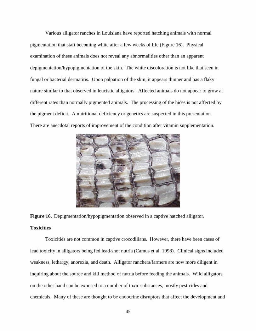
45
Various alligator ranches in Louisiana have reported hatching animals with normal
pigmentation that start becoming white after a few weeks of life (Figure 16). Physical
examination of these animals does not reveal any abnormalities other than an apparent
depigmentation/hypopigmentation of the skin. The white discoloration is not like that seen in
fungal or bacterial dermatitis. Upon palpation of the skin, it appears thinner and has a flaky
nature similar to that observed in leucistic alligators. Affected animals do not appear to grow at
different rates than normally pigmented animals. The processing of the hides is not affected by
the pigment deficit. A nutritional deficiency or genetics are suspected in this presentation.
There are anecdotal reports of improvement of the condition after vitamin supplementation.
Figure 16. Depigmentation/hypopigmentation observed in a captive hatched alligator.
Toxicities
Toxicities are not common in captive crocodilians. However, there have been cases of
lead toxicity in alligators being fed lead-shot nutria (Camus et al. 1998). Clinical signs included
weakness, lethargy, anorexia, and death. Alligator ranchers/farmers are now more diligent in
inquiring about the source and kill method of nutria before feeding the animals. Wild alligators
on the other hand can be exposed to a number of toxic substances, mostly pesticides and
chemicals. Many of these are thought to be endocrine disruptors that affect the development and

46
function of the reproductive organs. This in turn can lead to developmental defects in young
crocodilians (Vonier et al. 1996, Lind et al. 2004, Stoker et al. 2003, Guillette et al. 2002,
Rauschenberger et al. 2004). There is an abundance of information to support the impact of
environmental contamination on crocodilians as well as other species. This reiterates the critical
state of the natural habitat of many species around the world. Crocodilians have served as
sentinels for anthropogenic changes that ultimately may come back to affect human health.
Runting
Runting is a phenomenon observed in captive crocodilian operations. It describes a
general state of unthriftiness, lack of growth and weakness. There is a visible size difference in
same age animals between the runts and the otherwise healthy ones. It is not unusual to find
some buildings in commercial operations that contain the runt animals for that year. It is also in
this group that disease problems may be observed with higher frequency. Dominance by other
animals, environment, and even incubation factors contribute to the presence of runts (Allstead
and Lang 1995).
West Nile virus History
West Nile virus is a Flavivirus found within the Japanese encephalitides virus group,
which also includes Cacipacore, Koutango, Alfuy, Kunjin, Japanese encephalitis, Murray Valley
encephalitis, St. Louis encephalitis, Usutu, and Yaounde viruses (Mackenzie et al. 2002). Most
flaviviruses are positive sense single stranded RNA viruses that measure between 40 and 60nm
in diameter and have an enveloped icosahedral nucleocapsid (CDC 2003). WNV virus was first
identified in 1937 during an epidemiologic study of yellow fever in Africa (Smithburn et al.
1940). During this investigation, serum from affected individuals was inoculated intracerebrally
into mice and a subinoculation then performed from mice that died after the original inoculation
(Smithburn et al. 1940). Smithburn et al. (1940) reported the isolation of various transmissible

47
agents, one of which they named WNV. This newly identified virus, WNV, was isolated from a
woman in the West Nile province of Uganda who was suffering from a mild case of febrile
illness (Smithburn et al. 1940). At the time, the febrile illness was thought to be associated with
yellow fever, and no additional history to relate the clinical signs to the newly discovered virus
was obtained (Smithburn et al. 1940). Since this discovery of WNV, sporadic cases were
reported around the Middle East, Africa, Europe, and Asia; however, it wasn’t until 1951 that the
first documented epidemic of WNV was reported in Israel (Mackenzie et al. 2002). This led to
continued interest in the virus and various research projects to determine its origins, cycle, and
pathogenicity. From this original research, it was determined that the epidemiology of WNV
included mosquitoes and birds, and that human and horses were incidental hosts (Mackenzie et
al. 2002). A serological survey at the time demonstrated greater than 60% exposure in humans
along the Egyptian Nile (Mackenzie et al. 2002). By 1950, Aedes albopictus, Culex pipiens, and
Culex tritaeniorhynchus, three species of mosquitoes, had been shown to experimentally transmit
WNV (Philip and Smadel 1943, Kitaoka 1950). Additional mosquito species from which WNV
was isolated during the 1950s included Culex antennatus and Culex univittatus, which some
considered to be the primary vector in many areas of Egypt (Hayes 2001). During the 1960s,
Culex modestus and Culex vishnui were implicated as vectors of WNV in France and India,
respectively (Hayes 2001). Today C. pipiens pipiens is considered to be the most important
mosquito vector in North America and parts of Europe (Hayes 2001). As of 2005, WNV had
been isolated from over 60 different species of mosquitoes (CDC 2005).
The pathogenicity of WNV in humans was first described in 1954 after experimental
inoculation of humans with the Egypt 101, 21, and 19 WNV isolates. The original plan of the
authors was to evaluate the possible antineoplastic properties of the virus (Southam and Moore
1954). The clinical signs reported in the human patients during the study ranged from being

48
asymptomatic to fever and encephalitis (Southam and Moore 1954). This early study of WNV
infection in humans led to the conclusion that humans were likely incidental hosts and did not
play a significant role in the cycle of WNV (Southam and Moore 1954). Southam and Moore
also showed that viremia in humans could be detected from 24 hours to 12 days after inoculation
with WNV, which is consistent with the 3 to 15 day incubation period that is generally accepted
today. In the original experimental studies, the authors were also able to establish a correlation
between the persistence of a viremia and the severity of clinical disease (Southam and Moore
1954, Mackenzie et al. 2002). The histopathologic descriptions of WNV infections in deceased
patients were not very clear, and were confounded by the neoplastic disease that the patients
initially had before being infected with WNV.
The epidemiology of WNV was also of great interest at the time. It was believed that the
increasing density of human populations, coupled with increasing land cultivation in rural areas
and increased prevalence of mosquitoes and birds, all played a role in the cycle and
epidemiology of the disease (Mackenzie et al. 2002). The presence of mosquitoes, and
consequently WNV in birds and humans was a link identified very early on during the study of
the WNV epidemiology. Taylor et al. (1956) observed a progressive increase in WNV
seropositivity with age amongst children in the Sindbis district of Egypt in 1950 (Taylor et al.
1956). It was then proposed that WNV must be endemic in areas where such findings are
observed, and that the virus must over-winter in order for that endemicity to be maintained
(Taylor et al. 1956). Taylor et al. (1956) also observed that while migratory birds may play a
role in the epidemiology of WNV, they were unlikely to be the source of yearly outbreaks since
the areas more frequented by migratory birds along Egypt’s coast also had the lowest antibody
rates. They further proposed three over-wintering methods for WNV: hibernating mosquitoes,
other arthropods such as ticks, and small numbers of mosquitoes that remain active during the

49
winter (Taylor et al. 1956). In their study, they determined that there was no evidence for
hibernation of the main Culex species in Egypt. They were also unable to isolate WNV from
over 26,000 ticks from 8 different taxa (Taylor et al. 1956). They concluded that mosquitoes
continued to survive during the winter in Egypt and presented evidence of Culex collections
during winter time from which WNV was presumably identified but not confirmed. An
additional piece of evidence that suggested this cycle was that the children seroconverted for
WNV via complement fixation during April and May, and thus must have been exposed to WNV
during the preceding winter months (Taylor et al. 1956). While some of the evidence could not
be confirmed, the study presented some of the earliest evidence for the same over-wintering
theories that are now being proposed in an effort to explain the endemicity of WNV in certain
areas of the United States. Taylor et al. (1956) went on to present a hypothetical life cycle for
WNV that said mosquitoes were the vectors for the disease, birds the primary hosts, and humans
a secondary host unable to maintain the cycle without the presence of birds. Domestic
quadrupeds were also mentioned as being frequently infected based on the presence of
antibodies, but not reaching high enough viremias to be of significance in the propagation of the
virus (Taylor et al. 1956). The Taylor et al. (1956) study concluded that in the endemic areas of
the Nile Delta, WNV is “a childhood disease with yearly peaks of transmission during mid-
summer” with the impression of WNV being a “self-limited, non-fatal febrile disease rarely
associated with definite manifestations of encephalitis” (Taylor et al. 1956). In addition, they
state that “on both experimental and ecological grounds the main cycle of WNV is probably
through mosquitoes and birds in which man may become involved and domestic quadrupeds are
probably tangential or dead-end infections” (Taylor et al. 1956). Finally, they reported that there
is “evidence for believing that the virus over-winters through a process of retarded transmission
by the mosquitoes that remain active throughout the colder months” and that “non-endemicity in

50
the northern Delta may be accounted for by less dense human and bird populations and the
scarcity of what appears to be the most important vector, C. univittatus” (Taylor et al. 1956).
This study was perhaps one of the most important of its time for identifying some of the same
concepts that today are thought to play a role in the ecology and epidemiology of WNV.
Since the original epidemic in Israel, additional outbreaks in both animals and humans
have been reported in Israel, Egypt, France, and South Africa. Continued reports of sporadic
cases were also reported throughout Africa, Europe, Asia, and the Middle East well into the late
1990s and the early part of the 21st century (Smithburn et al. 1940). It wasn’t until 1999 that the
first cases of WNV were reported in the United States from the area of northern Queens in New
York City (Nash et al. 2001, Ludwig et al. 2002, MMWR 1999a, MMWR 1999b). During
August of 1999, arboviral encephalitis, characterized by meningoencephalitis with muscle
weakness, was reported to the New York City Department of Health (Nash et al. 2001, MMWR
1999a). The original report of these cases led to identification of a cluster of six patients with
encephalitis (MMWR 1999a). Five of the six had profound muscle weakness based on axonal
neuropathy by electromyelogram. Four of these patients also required respiratory support
(MMWR 1999a). Initial testing revealed antibodies to St. Louis Encephalitis virus (SLE) via an
IgM- capture enzyme linked immunosorbent assay (ELISA), and led to active surveillance for
arboviruses as well as treatment for mosquitoes around the four square mile area from which the
cases originated (MMWR 1999a). A case definition was also established: “presumptive
diagnosis of viral encephalitis with or without muscle weakness or acute flaccid paralysis,
Guillain-Barre syndrome, aseptic meningitis, or presence of the clinical syndrome characterizing
the initial cluster of cases in a patient presenting after August 1” (MMWR 1999a). In addition,
increased mortalities of birds and other wildlife were observed before and after the report of
human cases (MMWR 1999a, Ludwig et al. 2002). Testing of both animal and human samples
51
by polymerase reaction (PCR) and consequent genomic analysis revealed the presence of a West
Nile-like virus (MMWR 1999a). By October 5, 1999, the human cases increased to 50, with five
deaths, and crows were being recognized as sentinels for the geographic distribution of the virus
(MMWR 1999b). Mosquito collection was also initiated early on as part of the surveillance
methods.
The response by the city of New York to this outbreak was characterized by an
immediate active surveillance plan that included testing of mosquitoes and wild animals as well
as a public outreach program. Telephone hotlines were established to address public concerns
and DEET (N, N-diethyl-meta-toluamide) mosquito repellant was distributed along with
educational material about how people could protect themselves against mosquito bites (MMWR
1999a). In addition, active spraying for mosquitoes was initiated in the affected areas.
Of particular interest in the history of WNV is the fact that outbreaks typically occurred
in rural areas and that disease was characterized as a mild febrile illness. This changed during
1996-1999, when three major epidemics occurred in urban areas of Romania, Russia, and the
United States. These outbreaks were also characterized by the presence of severe neurological
disease and increased mortality in case patients, and an increased mortality in birds (Hayes
2001). In all three outbreaks, C. pipiens was determined to be the primary vector for WNV
transmission (Hayes 2001).
Today, a lot more is known about WNV and its epidemiology in humans. As opposed to
earlier studies which reported WNV as being a childhood disease with signs consisting of mild
febrile illness (Taylor et al. 1956), today WNV is recognized as a disease that can have higher
mortalities and morbidity amongst the adult and elderly populations in urban areas (Nash et al.
2001, Weinberger et al. 2001). Nash et al. (2001) also proposed that the more severe morbidity
and mortality observed during 1999 in New York City may have been in part due to the fact that

52
the population of individuals in the area had naïve immune systems, and that more severe disease
could be expected in non-endemic areas. These observations have changed the way that WNV is
perceived around the world and led local, state, and federal agencies to work aggressively
towards the prevention and control of WNV.
With an increase in morbidity and mortality has come a better understanding of the
pathology of WNV in both humans and animals. Clinical signs of WNV infection can range
from being absent to fever, myalgia, headache, conjunctivitis, lymphadenopathy, anorexia,
vomiting, photophobia, arthralgia and skin eruptions (Anderson et al. 2004, Asnis et al. 2001,
Hayes and Gubler 2006). The skin eruptions are of particular importance to our study because
they reveal a dermatological component to WNV infection similar to what we have observed in
alligators. Other less common clinical manifestations include hepatitis, pancreatitis,
rhabdomyolysis, myocarditis, cardiac dys-rhythmias, orchitis, uveitis, vitritis, optic neuritis, and
chorioretinitis (Hayes and Gubler 2006). The primary histopathologic finding associated with
WNV infection in humans is encephalitis or meningoencephalitis (Asnis et al. 2001). Specific
cellular changes include neuronal loss, perivascular inflammation, microglial nodules,
neuronophagia, and necrosis (Hayes and Gubler 2006, Hayes et al. 2005). These changes
predominate in the brainstem, deep gray nuclei, and anterior horns of the spinal cells (Hayes and
Gubler 2006). Muscle necrosis and perivascular inflammation without major inflammation has
also been reported in patients suffering from paralysis associated with WNV (Hayes and Gubler
2006). In humans, the histopathologic changes appear milder than infections with other
flaviviruses and infections in animals (Sampson et al. 2000). The pathology of WNV in animals
is quite varied. Some of the microscopic changes reported in birds infected with WNV include
lymphoplasmacytic, histiocytic encephalitis and myocarditis, lymphoplasmacytic chorioiditis,
heterophilic and lymphoplasmacytic thyroiditis and adrenalitis, hepatocellular necrosis, and

53
perivascular cuffing amongst others (Wünschmann et al. 2005). In horses the lesions are
predominantly in the central nervous system (CNS), and are characterized by perivascular
cuffing with lymphohistiocytic inflammation and hemorrhage of the CNS (Castillo-Olivares and
Wood 2004). In alligators, WNV has systemic effects and acute lesions are primarily
heterophilic in nature (Nevarez et al. 2005b).
A diagnosis of WNV infection in a human patient is mainly done by serologic testing,
more specifically IgM via a WNV ELISA. This ELISA can cross-react with antibodies to other
flaviviruses, so a positive results should be confirmed by a WNV plaque reduction neutralization
assay (PRNT), ideally on paired sera (Hayes and Gubler 2006). In cases showing neurologic
signs, CSF can also be tested using the ELISA. Polymerase chain reaction technology can also
be used to test for the virus in serum or CSF. Postmortem diagnosis may be performed by RT-
PCR, immunohistochemistry or viral culture coupled with histopathologic findings. Currently
there is no specific treatment for WNV infection. In the presence of infection, supportive
therapy must be initiated in both humans and animals. In cases of mild febrile illness, recovery
will usually occur within several days while those suffering from severe illness without
neurologic signs usually recover within weeks to months (Hayes and Gubler 2006). Those with
neurologic signs but without focal deficits usually recover completely, but once encephalitis and
flaccid paralysis ensue, the prognosis is poor (Hayes and Gubler 2006).
Prevention and control of WNV is primarily aimed at mosquito control. Active mosquito
surveillance and control programs are in place throughout the United States in order to control
mosquito levels and the exposure to WNV in the human and animal population. These programs
also rely on the public being conscientious about decreasing mosquito breeding habitats and
protecting themselves by using repellents and avoiding outdoor activities during peak hours for
mosquito activity. Ultimately, WNV will remain an endemic disease in the United States, and

54
the morbidity and mortality associated with infection should subside if it follows the same trend
observed in other countries. There are WNV vaccines approved for horses but none for humans
or other animal species. These same vaccines have been studied in animal species with varying
degrees of efficacy. So far there has been no vaccine developed for use in humans although this
may change in the future.
An additional method of preventing the dissemination of WNV is through active
surveillance to identify the virus early on before it spreads through an area. Sentinel chickens
have been used as a method to monitor for WNV activity. Birds are placed in areas where WNV
may appear and serially tested for the presence of seroconversion. Mosquito surveillance has
become one of the most important tools in preventing and controlling WNV. This method tests
pools of mosquitoes that have been trapped in areas where WNV may be suspected to occur.
The tests performed on the mosquito pools detect the antigen itself and therefore serve as a more
accurate tool to identify the active virus in a region as opposed to exposure. Results of mosquito
surveillance for WNV are used to direct the efforts of mosquito control agencies. The
information will provide these agencies with a priority of areas that should be sprayed for
mosquitoes first.
Besides virus surveillance and mosquito control, an educational campaign in affected
states has been an equally important part of WNV prevention. Individuals in a community must
understand the risks of infection and come together to minimize conditions that promote
mosquito breeding. In addition, individuals must protect themselves against infection via the use
of mosquito repellents and by minimizing outdoors activities during peak mosquito hours in their
area. All these aspect working together will play a key role in decreasing the morbidity and
mortality of WNV during future outbreaks.

55
CHAPTER 1: LYMPHOHISTIOCYTIC PROLIFERATIVE SYNDROME OFALLIGATORS: PRELIMINARY INVESTIGATION
Introduction
The Louisiana alligator industry is an important resource for the state. The industry has
been used to establish a successful conservation program for the American alligator (Alligator
mississippiensis) in Louisiana, and a financial resource for individuals in economically depressed
regions of the state. Since the inception of the alligator aquaculture industry in Louisiana,
producers have experienced significant fluctuations in the monetary value of alligator hides. The
introduction of disease or any factor that could affect the production of high quality hides could
be financially devastating.
“PIX” is a generic term used to describe a small gross lesion in alligator hides that
appears as if someone has struck the skin with an ice pick (Cardeilhac 2001). These lesions
reduce the overall value of the hide, and in some cases completely devalue the hide. Because of
the investment placed into producing market size alligators, losses associated with this “disease”
can be significant and devastating to the ranchers.
According to previous work generated by the LA Department of Wildlife and Fisheries in
collaboration with Dr. Paul Cardeilhac (University of Florida College of Veterinary Medicine),
the first reports of “PIX” in captive alligators occurred in Florida in 1999. These reports were
limited to a single farm. Although there was concern that this “disease” might be introduced into
Louisiana, there were no reports made through 2001. The first report of “PIX” in Louisiana was
made in 2002. Initially, the reports were sporadic, and there were no preliminary estimates of
the prevalence of “PIX” in the state. Alligators with gross “PIX” lesions were presented in early
2002 to the Louisiana State University School of Veterinary Medicine (LSU SVM) for
examination. Histopathology of the gross lesions revealed a consistent lymphohistiocytic
proliferative lesion. Consequently, “PIX” was re-named as lymphohistiocytic proliferative

56
syndrome of alligators (LPSA). Initial differentials for this syndrome included: infectious
disease, immune-mediated disease, allergens, nutritional deficiencies, toxins, and neoplasia. The
current study was initiated as a first attempt to characterize the epidemiology of LPSA in
Louisiana alligator farms.
Materials and Methods
A cross-sectional study was performed to estimate the prevalence of LPSA in Louisiana
alligator farms. The prevalence of LPSA was determined based on the presence of both gross
and microscopic lesions. Alligator farms were selected based on a previous history of LPSA
being described at the farm. Six farms were evaluated in this study. The farms were from
geographically distinct areas within the state. The production size of the alligator farms were
variable, and ranged from 5,000 – 50,000 alligators produced annually. All the farms raised their
alligators indoors. Four farms used rectangular buildings and two farms used round buildings.
The alligators were maintained at temperatures between 29 - 32oC (85 - 90oF). Stocking
densities for the alligators varied within farms based on size and time of year and were in
accordance to recommendations from the Department of Wildlife and Fisheries. All of the farms
used open water systems, in which the water within the pens was removed and replaced with
fresh water. The three water sources for alligator farms were city water, well water, and bayou
water, or a combination of these. Water was pre-heated prior to being provided to the alligators
at all farms. All of the farms fed their alligators a commercial diet, either Burris (4/6) or Lone
Star (2/6). Both diets are similar in composition but vary in the size of the pellets and the length
of time the pellets will remain afloat in water. At 4 farms the diet was supplemented with
chicken, fish, or beef liver; however, this could vary through the year depending on prices and
availability. All farms report adding bleach to the water on a subjective manner. The subjective
use of salt and/or antibiotics was also reported by four farms.

57
A complete and thorough diagnostic evaluation was planned for 50 alligators, 25 with
gross lesions (cases) and 25 without gross lesions (controls). The diagnostic tests performed for
this study included: a complete blood count, plasma biochemistry analysis, protein
electrophoresis, heavy metal screening, bacterial and fungal cultures of specific lesions, and
histopathology. Electron microscopy and herpes virus PCR were also performed on selected
tissues. An additional 32 alligators, 16 cases and 16 controls, were also sacrificed for necropsy
only.
Histopathology
A complete necropsy was performed on 82 alligators, and the following tissues collected:
brain, spinal cord, heart, liver, gall bladder, lung, esophagus, stomach, small intestine, pancreas,
large intestine, mesentery, kidney, ureter, adrenal gland, gonad, skin, bone marrow, spleen,
tonsil, conjunctiva, thyroid, eye, muscle, and nerve. All of the tissue samples were placed in
10% neutral buffered formalin. The samples were shipped to Northwest Zoo Path (654 W. Main
St. Monroe, WA 98272) for processing and interpretation.
Electron Microscopy
Electron microscopy (EM) is a diagnostic tool that provides a microscopic view of tissues
that is far beyond the capacity of a light microscope. Specifically, EM provides the opportunity
to directly examine individual cellular components and small organisms such as viruses and
bacteria that may otherwise be missed with light microscopy. The use of EM in the study would
aid in the identification of possible etiologic agent(s) of LPSA. Selected tissue samples with
obvious lesions (N=5) were also analyzed by Northwest Zoo Path.
Fungal Isolation
Skin samples with gross lesions consistent with LPSA were collected at necropsy and
cultured for fungi at the LSU SVM. The exterior and interior surfaces of the skin sample were

58
sterilized by immersion in alcohol followed by flaming. Each sample was cut into smaller pieces
and mixed with a maximum of 0.5ml sterile saline before plating. Saboraud-dextrose, mycocel,
and potato flake agars were used to isolate potential fungi. Plates were maintained at room
temperature in the dark for a minimum of 14 days. The cultures were observed daily. Isolates
were sub-cultured a second time to obtain pure cultures. Samples were shipped to the Fungus
Testing Laboratory at the University of Texas (7703 Floyd Curl Drive San Antonio, TX 78229-
3900) for identification.
Bacterial Isolation
Bacterial cultures were performed at the LSU SVM using routine methods. The skin
samples used for the fungal cultures were also used for the bacterial cultures, although no saline
was added to the bacterial samples. Samples were placed on TSA with 5% Sheep blood agar and
MacConkey culture media and incubated at 37oC for 24-48 hours under aerobic conditions.
Bacterial isolates were characterized by colony morphology, Gram-stain characteristics, and
biochemical processes.
Plasma Biochemistries
Plasma biochemistries can provide important physiologic information regarding the
general health status of an animal. Blood was obtained from the supravertebral sinus of 46
alligators. Each sample was placed into a lithium heparin vacutainer tube and centrifuged on
site. The plasma was removed, placed into sterile cryovials, and transported on wet ice to the
LSU SVM for analysis.
Protein Electrophoresis
Protein electrophoresis is a diagnostic test used to separate total plasma protein into its
various components. This test is not widely performed in reptiles, but could provide insight
towards diagnosing a specific etiology or group of etiologies and provide information about the

59
chronicity of a disease process. We elected to perform this test after detecting total protein,
albumin, and globulin alterations in preliminary plasma biochemistries performed in a pilot
study. Plasma samples were shipped to the University of Miami for testing.
Complete Blood Counts
Complete blood counts (CBC) are used to evaluate the erythron and white blood cells
patterns of a patient. This diagnostic test is invaluable in characterizing inflammatory responses
in an animal. Complete blood counts were performed at the LSU SVM on 45 alligators using
standard methods. In short, an eosinophil unopette system (Becton Dickinson and Company,
Franklin lakes, NJ 07417) was used to estimate the heterophil and eosinophil counts of the
animals, and a differential estimated from a Wright-Giemsa stained blood smear.
Heavy Metals and Trace Element Screening
Heavy metals can cause a severe inflammatory response when they are disseminated into
tissues. The type of response elicited by heavy metals may be similar to that described in LPSA.
Therefore, blood and tissue samples were collected from alligators and evaluated for the
presence of various heavy metals. The blood samples were collected prior to the alligators being
euthanized, while the tissue samples were collected at necropsy. The samples were processed at
the Louisiana Veterinary Medical Diagnostic Laboratory.
Water Analysis
Water samples were obtained from each of the pens that housed the study subjects.
Approximately 100mls of water was collected in sterile plastic containers that could be sealed
tightly. The samples were then transported on wet ice to the LSU Agricultural Chemistry
Laboratory for processing. The following parameters were measured: pH, ammonia, bromide,
fluoride, nitrate, phosphate, sulphate, total solids, suspended solids, and dissolved solids.

60
Herpes Virus PCR
During sampling for this project, a subset of tissue samples was sent to the University of
Florida to test for herpes and iridoviruses. These preliminary tests revealed the possible presence
of a herpes virus in the tissues. Follow up work was performed at the LSU SVM. A consensus
primer PCR was used based on a protocol described by van Devanter et al. (van Devanter et al.,
1996). This protocol was the same one used at the University of Florida and has the advantage
of being able to detect and identify herpes viruses without prior DNA information.
Statistical Methods
Descriptive statistics were generated for each parameter. 95% binomial confidence
intervals (CI) were calculated for proportion data. A Shapiro-Wilk test, and the kurtosis and
skewness of the data, were used to evaluate the distribution of continuous data. Data that was
normally distributed was reported as a mean, while those not normally distributed as a median.
A Kruskal-Wallis ANOVA was used to compare the various data between farms. A p<0.05 was
considered statistically significant. SPSS 11.0 (SPSS Inc., Chicago, IL) was used to analyze the
data.
Results
Histopathology
The majority of the alligators collected from the farms had lesions consistent with LPSA in
multiple organs (Table 6). Additionally, rhabdomyolysis and vacuolar changes in the liver and
adrenal glands were observed in a number of the alligators necropsied for this study (Table 7).
Based on the 95% CI, the organs that were most often affected were the intestine, lungs, and,
stomach. The pathologist reported that the lesions identified in these alligators were similar, and
were characterized by an infiltration of lymphocytes and histiocytes. Initially, the findings from
the histopathologic examination suggest that our control alligators also had lesions consistent

61
with LPSA; however, we know understand that lymphoid aggregates are a normal finding in the
intestines, lungs, and stomach of healthy alligators. If we omit the histopathologic findings in
these tissues, as they are normal findings, we can see that the 44% prevalence of skin lesions are
from the group of animals with gross lesions. This provides a more clear assessment and shows
that two distinct groups of animals, one with gross skin lesions and one without gross skin
lesions were indeed collected.
The combination of the histopathologic findings being strongly suggestive of an
infectious agent, and a report from Florida suggesting that a fungal agent may be responsible for
LPSA, led us to specifically evaluate these lesions for infectious agents. There was no evidence
of fungal disease in the samples. However, acid fast stains were positive in the lungs of two
alligators (2.4%, 95%CI: 0-5.7%), suggesting the possible presence of Mycobacterium sp. This
bacterium is fastidious, requiring specific culture methods that were not implemented in this
study. Because these bacteria were not found consistently, it was not considered to be unrelated
to LPSA. However, further study to the role of these bacteria should be performed if more
consistent lesions are found. No viral disease was identified using light microscopy; however,
perivascular cuffing, a common pathologic finding with infectious processes, was evident in
some of these samples.
Of interest was the fact that all alligators appeared to have lymphoid lesions in some
organ. Alligators in both the control and treatment group had lymphoid aggregates in the lungs,
stomach and intestines, similar to the lesions observed in the skin. In addition, 38% (10/26; 95%
CI: 20-58) of those alligators that had apparent skin lesions could not be corroborated on
histopathologic exam. This may have been the result of sample processing or suggests that gross
examination of the skin may be an inaccurate method of characterizing the LPSA status of
alligators.

62
Table 6. Prevalence of lymphonodular lesions observed in tissues of 82 alligators.
Tissue Number of alligators with lesions Prevalence 95% Confidence IntervalIntestine 76 93% 87.5 – 98.5%Lung 64 78% 69.1 – 86.9%Stomach 64 78% 69.1 – 86.9%Skin* 36 44% 33.2 – 54.8%Kidney 27 33% 22.8 – 43.2%Mesentery 20 24% 14.8 – 33.2%Esophagus 17 21% 12.2 – 29.8%Eye 14 17% 8.9 – 25.1%Tonsil 12 15% 7.3 – 22.7%Brain 10 12% 5.0 – 19.0%Thyroid 10 12% 5.0 – 19.0%Ovary 9 11% 4.2 – 17.8%Conjunctiva 8 10% 3.5 – 16.5%Heart 7 9% 2.8 – 15.2%Testicle 7 9% 2.8 – 15.2%Pancreas 6 7% 1.5 – 12.5%Gall Bladder 4 5% 0.3 – 9.7%Bone Marrow 3 4% 0.2 – 8.2%Salivary gland 1 1% 0 – 3.2%Spinal cord 1 1% 0 – 3.2%Spleen 1 1% 0 – 3.2%Ureter 1 1% 0 – 3.2%
*Based on 81 skin samples
Table 7. Additional histopathologic changes recorded in 82 alligators
Tissue Lesion description
Number ofanimals with
lesionsPrevalence 95%Confidence
Interval
Adrenal gland Vacuolar changes/stress response 36 44% 33.3-54.7%Liver Vacuolar changes/unknown significance 78 95% 90.3-99.7%
Muscle Rhabdomyolysis/stress response 23 28% 18.3-37.7%
The histopathologic findings in the lungs and the intestinal tract (stomach and intestines)
were novel. Reptiles do not have lymph nodes, and instead use aggregates of lymphoid tissues
(e.g., Peyer’s patches) to process antigens. One of our concerns was that some of these visceral
sites might represent normal aggregates of lymphoid tissue and therefore contributed to the
perceived lack of a control group. Additional tissue samples collected from four hatchling
alligators showed similar lymphonodular responses in the stomach (3/4 hatchlings = 75%) and
mesentery (1/4 hatchlings = 25%). The findings of the lymphoid infiltrates may represent areas

63
responsible for processing foreign antigens. When an antigen such as an allergen, virus, bacteria,
or fungus enters the body, the intestines or lungs are likely to be the first sites of antigenic
stimulation. The processing of these antigens, especially in the face of chronic stimulation, can
lead to a significant cell-mediated and/or humoral immune response. The inflammatory response
characterized in these alligators was consistent with those described in higher vertebrates.
Although the findings in the intestine and lungs may be associated with expected lymphoid
aggregates, similar lesions in other organs not generally considered to be site specific for antigen
processing suggest that the histopathologic findings are abnormal.
The histopathologic findings reported in this study suggest that a systemic disease process is
occurring. More specifically, the findings were highly suggestive of an infectious disease
process.
Electron Microscopy
Results of electron microscopy revealed flagellated protozoa (Figure 17) and an
intracellular bacterium that lacked a cell wall and was consistent with Mycoplasma sp. (Figure
18). The protozoa could not be characterized any further. These results could not be duplicated.
Fungal Isolation
Fourteen different fungi were isolated from the skin of alligators during this project
(Figure 19). These findings included multiple isolates from multiple animals as well as
individual animals. Most of these fungi are ubiquitous in nature and are found in soil and water.
Some of these fungi have been previously reported as causing secondary disease in reptiles
(Tappe et al.1984, Jacobson 1979b, Cheatwood 2000).
Bacterial Isolation
Bacterial cultures revealed a mixed population of Gram-positive and Gram-negative
organisms. No pure isolates were obtained. Histopathologic exam revealed acid-fast positive

64
bacilli, which were consistent with Mycobacterium species. However, this type of bacteria
requires more specialized culture methods than were employed in this project.
Figure 17. Transmission electron microscopic examination of a skin lesion from an alligatorswith mild LPSA revealed the above protozoa.
Figure 18. Transmission electron microscopic examination of a skin lesion from an alligatorwith mild LPSA revealed these bacteria. The bacteria identified in the EM did not have a cellwall.
Alternaria alternateAspergillus nigerCladosporium cladosporioides, Cladosporium oxysporumChaetomium globosum, Chaetomium spp.Curvularia lunataEpicoccum purpurascensEurotium chevalieriMonascus rubberMucor circinelloides (group)Paecilomyces variotiiPenicillium citrinumSyncephalastrum racemosum
Figure 19. Fungi isolated from the skin of captive American alligators.

65
Plasma Biochemistries
The results of the chemistry analytes are presented in table 8. The mean/median values
for creatine kinase (CK) and aspartate aminotransferase (AST) were elevated, when compared to
other reptile species. Both of these enzymes originate in the muscle and are likely an indicator of
muscle necrosis, which can occur as a result of capture, restraint and venipuncture. A review of
the other analytes suggests that there is no obvious physiologic disturbance to the plasma
biochemistries, except for CK and AST, associated with the clinical and histopathologic findings
of LPSA.
Protein Electrophoresis
When comparing our results with those reported in avian species, there are apparent
changes in the protein electrophoresis results that may be of importance. Phylogenetically, birds
are close relative to alligators and may prove valuable as a comparative species. The alligators
in this study had decreased albumin-globulin ratios (A/G) and albumin levels, while the alpha- 2
proteins were increased (Tables 9 and10). The alpha – 1 and gamma protein fractions were on
the high end of values observed in other reptile species. The changes in the protein
electrophoresis observed in these alligators may be indicative of acute inflammatory disease.
Complete Blood Counts
Results of the CBC’s are presented on table 11. Overall, the white blood cell count and
cell differentials are consistent with a stress response and possibly an underlying inflammatory
process. There are several outliers that had extremely elevated white blood cell and monocyte
counts, but these might be expected in a population of animals that are held under intensive
production operations. There is no evidence of a severe inflammatory response associated with
LPSA.

66
Table 8. Plasma biochemistry values for 46 captive reared alligators
Analyte Mean/Median1 SD2 Minimum Maximum 95% CI3
Glucose MG/DL 110 - 83 244 -
Aspartate Aminotransferase U/L 261 - 162 589 -
Creatine kinase U/L 599 - 134 4337 -
Tot. Protein G/Dla 5 1.36 4 7 5.2 – 5.6
Albumin G/DL 2 - 1 2 -
Globulin G/Dla 4 1.14 3 5 3.6 – 4
Calcium MG/Dla 12 1.3 10 13 11.5 – 11.9
Phosphorus MG/Dla 5 1.84 4 8 5 – 6
Sodium mmol/L 151 - 89 162 -
Potassium mmol/La 4 1.46 2 6 3.9 – 4.3
Chloride mmol/L 113 - 66 124 -
TCO2 mmol/La 14 11.66 3 26 12 – 15
Anion Gap mmol/La 28 18.04 12 51 25 – 30
Uric Acid MG/DL 1 - 0 4 -1Mean reported for normally distributed values, and median for non-normally distributed values, 2SD = two standard deviations,3 Confidence Interval (CI), a Values with normal distribution.
Table 9. Results of Protein Electrophoresis from 45 captive alligators
Analyte Mean/Median1 SD2 Minimum Maximum 95%CI3
Total Protein 5.3 - 3.4 7.6 -
Albumin/Globulin 0.2 - 0.2 0.2 -
Albumin 0.9 - 0.5 1.4 -
Alpha 1a 0.6 0.6 0.2 2.2 0.5 - 0.6
Alpha 2 2 - 0.8 3 -
Beta 1.1 - 0.7 1.6 -
Gamma 0.7 - 0.3 1.4 -1 Mean reported for normally distributed values, and median for non-normally distributed values. 2 SD = two standarddeviations, 3 Confidence Interval (CI), a Values with normal distribution
Table 10. Protein Electrophoresis Relative % Fractions from 50 Alligator mississippiensis.
Fraction Mean/Median1 SD2 Minimum Maximum 95% CI2
Albumin 17.78 - 13.49 22.39 -
Alpha 1a 10 0.6 4.07 13.8 8.71 - 10.0
Alpha 2 38.01 - 34.67 42.66 -
Beta 20.42 - 15.85 25.12 -
Gammaa 13.49 0.5 5.01 22.91 12.59 - 14.131 Mean reported for normally distributed values, and median for non-normally distributed values. 2SD = two standard deviations,3 Confidence Interval (CI), a Values with normal distribution

67
Table 11. Complete Blood Counts from 45 alligators
Value Median Min Max
Total White Cell Count 7771 4098 27291
Heterophils 4118 1821 20741
Lymphocytes 818 0 2558
Monocytes 1324 366 3493
Basophils 828 0 2863
Eosinophils 609 100 1945
Water Analysis
Results of the water analysis are reported in table 12. There was no significant difference
in the water chemistry parameters among farms (Table 12).
Table 12. Water analysis values from alligator farms (N=11 samples from 6 farms)
Analyte Median Minimum Maximum p Value1
pH 7.57 6.86 8.33 0.2Ammonia (ppm) 30.86 0.46 171 0.71
Bromide (ppm) 0.4 0.2 1.2 0.08Fluoride (ppm) 1.83 0.53 9.67 0.44Nitrate (ppm) 1.59 0.3 6.49 0.7Phosphate (ppm) 22.12 1 97.3 0.7
Sulphate (ppm) 36.59 1.26 138 0.27Total solids (ppm) 1094.72 446 2242 0.68Suspended solids (ppm) 259.31 20 881 0.22Dissolved solids (ppm) 837.23 427 1593 0.23
1 p value for comparison of each parameter between farms
Heavy Metal and Trace Elements Screening
Blood and tissue concentrations for copper, iron, lead, mercury, selenium, and zinc are
reported in table 13. These values approximate levels previously reported from crocodilians
(Burger et al. 2000, Jeffree et al. 2001).
Herpes Virus PCR
We were unable to identify any herpes viruses from the tissues of alligators. A herpes
simplex one virus was identified but determined to be a contaminant since the laboratory where
the work was performed routinely works with herpes simplex. Attempts to minimize
contamination, and re-evaluation of reagents and procedures were performed in order to identify
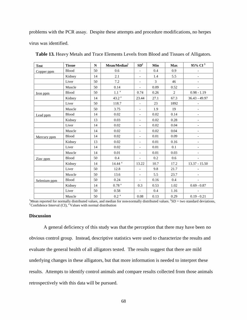
68
problems with the PCR assay. Despite these attempts and procedure modifications, no herpes
virus was identified.
Table 13. Heavy Metals and Trace Elements Levels from Blood and Tissues of Alligators.
Test Tissue N Mean/Median1 SD2 Min Max 95% CI 3
Copper ppm Blood 50 0.6 - 0.4 0.9 -
Kidney 14 2.1 - 1.4 5.5 -Liver 50 7.2 - 3 46 -
Muscle 50 0.14 - 0.09 0.52 -
Iron ppm Blood 50 1.1 a 0.74 0.26 2 0.98 - 1.19
Kidney 14 43.2 a 23.44 27.1 67.3 36.43 - 49.97Liver 50 118.7 - 23 1892 -
Muscle 50 3.75 - 1.9 19 -
Lead ppm Blood 14 0.02 - 0.02 0.14 -
Kidney 13 0.03 - 0.02 0.28 -Liver 14 0.02 - 0.02 0.04 -
Muscle 14 0.02 - 0.02 0.04 -
Mercury ppm Blood 14 0.02 - 0.01 0.09 -
Kidney 13 0.02 - 0.01 0.16 -Liver 14 0.02 - 0.01 0.1 -
Muscle 14 0.01 - 0.01 0.03 -
Zinc ppm Blood 50 0.4 - 0.2 0.6 -
Kidney 14 14.44 a 13.22 10.7 17.2 13.37 - 15.50Liver 50 12.8 - 9.8 21.7 -
Muscle 50 13.6 - 5.5 23.7 -
Selenium ppm Blood 50 0.24 - 0.16 0.4 -
Kidney 14 0.78 a 0.3 0.53 1.02 0.69 - 0.87Liver 50 0.58 - 0.4 1.16 -
Muscle 50 0.2 a 0.08 0.13 0.29 0.19 - 0.211Mean reported for normally distributed values, and median for non-normally distributed values. 2SD = two standard deviations,3 Confidence Interval (CI), a Values with normal distribution
Discussion
A general deficiency of this study was that the perception that there may have been no
obvious control group. Instead, descriptive statistics were used to characterize the results and
evaluate the general health of all alligators tested. The results suggest that there are mild
underlying changes in these alligators, but that more information is needed to interpret these
results. Attempts to identify control animals and compare results collected from those animals
retrospectively with this data will be pursued.

69
From these descriptive results, it is apparent that these alligators are producing a systemic
inflammatory response. Although LPSA is generally considered a disease of the skin, it is
apparently more than that. Attempts to characterize the degree of normality associated with the
lymphohistiocytic proliferative syndrome in the intestines and lungs must be done. This will be
further evaluated when the control alligators are found and processed. Because of the economic
importance of LPSA, it is imperative that attempts to classify a specific etiology are pursued.
The histopathologic findings were crucial to help determine which disease etiology (-ies)
may be more likely associated with LPSA. The findings of the electron microscopy were not
duplicable and were ultimately interpreted to be incidental findings with no association to the
pathology observed in the tissues. Besides the incidental finding on EM, parasitic organisms
were not observed in any of the LPSA lesions. Of great importance is the lack of histologic
evidence of a fungal infection in LPSA tissues. For this reason we believe that there is no
evidence to indicate that fungi are the causative agents of LPSA in Louisiana. The results of the
fungal culture were not unexpected, as a significant amount of fungal organisms are ubiquitous
and can normally be identified using culture techniques. What was interesting was the wide
variety of fungi that was isolated. These data can provide a reference of fungal organisms that
can be found in alligator farms in Louisiana. If primary fungal infections in captive alligators
were to be diagnosed, one of these fungi may be responsible for such infections. Finally, we
must look back at the cell types present within the LPSA lesions. Lymphohistiocytic
inflammation is more commonly associated with a viral diseases or an immune mediated disease.
In light of the information presented and the lack of evidence to support a bacterial or fungal
etiology, a virus remains as the most likely cause of LPSA.
An interesting finding was the identification of acid fast positive organisms consistent
with Mycobacterium spp.. However, histopathologic evaluation of these tissues was not

70
consistent with expected changes for a mycobacterium infection making it a likely incidental
finding. There is also the possibility of those organisms being a different acid fast bacteria other
than Mycobacterium sp., such as Nocardia sp.. There was no histologic association observed
between LPSA lesions and the acid fast organisms. Furthermore no bacterial organisms of any
type were observed within LPSA lesions, making a bacterial etiology less likely for LPSA.
The findings of the CBC suggest a lack of a significant inflammatory response despite the
LPSA lesions. A possible explanation for this finding is that alligators with LPSA in the early or
late stages may not have mobilized significant cell numbers or may be past the major cell
migration. The observation that many of the lesions in the tissues were mild may support an
early state of disease. The combination of the histopathologic lesions, protein electrophoresis
and CBC results suggest that there is evidence of a low-grade inflammatory response underlying
the stress response. To further characterize the suspected inflammatory response, control
alligators or serial samples from the same affected alligator would be required.
Heavy metal values were consistent with values previously reported in crocodilians
(Burger et al. 2000, Jeffree et al. 2001). Because these values were similar to those previously
reported, it is unlikely that these compounds play an important role in the development of LPSA.
However, toxins can play a secondary role in disease processes, and should never be ruled out
based only on a comparative reference. The results for the various water chemistries reveal
higher levels of ammonia and solids than what would be acceptable for fish aquaculture (Brune
and Tomasso 1991, Noga 2000). These higher values are not surprising due to the management
techniques employed in alligator facilities, the high protein content of the alligator diet, and the
large amount of fecal matter produced. The lack of difference between farms and the fact that
these levels are typical of what is observed in facilities without LPSA, make water quality an
unlikely contributor to the development of LPSA.

71
During this study it became evident that LPSA was more widespread than previously
imagined. Although this syndrome appears to have only been identified in 2002, it is possible
that the disease was present before that time and went unnoticed. One would suspect that it
would be identified at the tanner, but if the lesions were less common, they may have been
missed. Attempts to evaluate leather processed prior to 2002 for LPSA should be done. One
interesting observation about the occurrence of LPSA is that it appeared to match the occurrence
of WNV which was first observed in 2001 in Louisiana. Based on the results of this study,
particularly the histopathologic evaluation of LPSA tissues, a viral etiology remains as the top
differential for LPSA.

72
CHAPTER 2: WEST NILE VIRUS IN ALLIGATOR, ALLIGATOR MISSISSIPPIENSIS,RANCHES FROM LOUISIANA*
Introduction
West Nile virus (WNV) has been reported to affect various crocodilian species
including the American alligator, Alligator mississippiensis, (Miller et al. 2003), the Nile
crocodile, Crocodylus niloticus, (Steinman et al. 2003), and the Morelet’s crocodile,
Crocodylus moreletii, (Rubio 2004). The role of crocodilians and other reptiles in the
epidemiology of WNV infection is largely unknown. However there is evidence of high
viremias and viral shedding in crocodilian species, specifically the American alligator,
based on published articles and our clinical experience (Miller et al. 2003). If crocodilians
are able to maintain high viral loads, they can serve as an amplifying host. This creates a
potential public health problem in captive operations where humans may be exposed to
WNV by direct contact with the animal’s feces and tissues. There have been three cases
of suspected WNV infection in humans working in an alligator ranch in Louisiana. In
addition there was one confirmed case of WNV exposure in Idaho based on antibody titers
(Tengelsen 2004). This individual had handled tissues from WNV positive alligators but
did not report clinical signs.
Florida, Georgia, Louisiana, and Idaho have all experienced cases of WNV in
alligators. This case report presents our clinical experience with diagnosis of WNV in four
alligator ranches from Louisiana. The first suspect cases in Louisiana occurred in
October of 2003. All cases were observed between October and December of 2003. Of
the ranches in Louisiana, two stocked imported hatchlings from Florida, one from Texas,
and the fourth stocked locally hatched alligators.
* Reprinted with permission from the Journal of Herpetological Medicine and Surgery.

73
Materials and Methods
All hatchlings were housed indoors with water temperatures between 29 – 32°C (85
– 90°F). Water sources varied between ranches and included city water, well water, or
bayou water. Water changes were performed either daily or every other day. Some farms
added bleach at an unknown concentration with each water change. Stocking densities
varied but were in accordance with the recommendations from the Louisiana Department
of Wildlife and Fisheries. These recommendations are as follows: one square foot per
animal for alligators smaller than twenty four inches in length, three square feet per animal
for alligators measuring twenty five to forty eight inches, for every six inches in length
beyond forty eight inches you should add one square foot of space (Elsey 2004). Feed
consisted of a commercial pelleted alligator ration. There was no exposure to meat
sources such as beef, horse meat, pork, or nutria. One ranch occasionally supplemented
with chicken or fish. There was no exposure to wild birds inside the buildings.
However, mosquitoes were reported to be present inside the building on a regular basis.
The initial clinical signs were observed approximately seven to ten days after the
imported animals arrived at the respective facilities. These animals were imported at
approximately 20 to 30 d old. The locally harvested alligators ranged in age from two
months to fourteen months. Clinical signs varied from peracute death to a variety of
neurological manifestations. Swimming in circles, ataxia, head and muscle tremors, and
head tilt were observed in the affected alligators (Figure 20). Stomachs were void of
ingesta, but were full of water at necropsy, suggesting that these animals were anorectic.
The large amount of water in the stomach may also suggest that the basihyoid and palatine
valves were not working properly, perhaps due to weakness associated with the disease. A
less common sign was bloating, as evidenced by the alligator’s inability to submerge

74
properly under the water. The caudal aspect of the body remained afloat, sometimes
sideways, while the cranial aspect of the body was observed underwater. Necropsy of
these animals revealed a colitis characterized by formation of a fibrinous membrane on the
mucosa (Figure 21).
Figure 20. American Alligator with a head tilt as consequence of WNV infection.
This last presentation was primarily observed in the yearling alligators (12 – 14
months old). Mortalities ranged from 40 – 60% of alligators within an affected pen or
building with an estimate of over 5,000 deaths in the whole state. In most instances,
mortalities were swift in one to two month old animals with deaths occurring suddenly
within a week of onset of clinical signs. This was followed by an equally fast decline in
the number of deaths. On the other hand, mortalities in yearling animals followed a more
sporadic course and spread over weeks or even months.
For example, a building housing 5 wk old alligators may have experienced 50 or 60
mortalities a day, while in yearling alligators the deaths rarely surpassed 15 – 20 animals

75
per day. Mortalities were primarily confined to a single pen or building. Once a peak in
mortalities occurred, the disease appeared to dissipate and remaining animals continued to
Figure 21. Fibrinous membrane on the colon mucosa of a WNV positive alligator. WNVwas identified from this lesion via RTPCR, viral isolation, and immunohistochemistry.
thrive without evidence of illness. However, in one particular case, an alligator rancher
reported mortalities of alligators spreading through the facility. After obtaining a detailed
history of events preceding the deaths, it was evident that the mortalities followed a pattern
of animal movement between buildings. The owner had been instructed not to move alli-
gators from affected buildings; however, because of size constraints, animals were moved
from affected buildings into non-affected buildings. The mortalities in the new buildings
followed in chronology the addition of new animals previously housed in buildings with
WNV positive alligators. The first deaths were observed approximately seven to ten days
after the introduction of affected alligators. By the time we confirmed WNV infection,
mortalities were identified in all of the buildings into which the previously exposed animals
were introduced. Although this spread of WNV was only identified in one ranch, it rein-
forces the importance of increased biosecurity in order to minimize the spread of WNV
between captive alligators.

76
The clinical approach for these cases consisted of a visit to the facilities in order to
observe and collect affected animals. Necropsies were performed using sterile techniques.
All tissues were collected from fresh specimens. Brain was collected from all animals and
submitted for either real time RT-PCR, viral isolation, or both. Thirteen brain samples
were submitted for real time RT-PCR and viral isolation. An additional nine samples
were submitted for viral isolation alone. A sample of colon from an alligator displaying
clinical signs of bloating with fibrinous colitis was also submitted for real time RT-PCR
and viral isolation. A second colon sample was submitted for viral isolation only. One
fecal sample was submitted for viral isolation. Histopathologic examination was per-
formed on fourteen alligators.
RNA Extraction and Real Time RT-PCR
The RNA was extracted from tissue using the Qiagen Rneasy kit (Qiagen, 27220
Turnberry Lane Suite 200, Valencia, CA) following the manufacturer’s protocol.
Approximately 20 mg of tissue was homogenized in 350 µl of Rneasy lysis buffer in a
Retsch Laboratory Vibration Mill Type MM 300 with a copper BB at 25 cycles per second
for 5 min (Retsch Inc., 74 Walker Lane, Newtown, PA). The homogenized samples were
subjected to Rneasy extraction and eluted in 30 µl of Rnase-free water. A primer-probe set
targeting the WNV envelope gene was used for the real time RT-PCR (Lanciotti et al.
2000). The probe contained a 5’ reporter 6-carboxyfluorescein (FAM) and a BHQ- 1
quencher as reported by Lanciotti (Lanciotti et al. 2000). The assay was performed on an
ABI 7900 Taqman Sequence Detector (Applied Biosystems, 850 Lincoln Centre Dr,
Foster City, CA) with Quantitec Probe RT-PCR master mixture (Qiagen, 27220 Turnberry
Lane Suite 200, Valencia, CA). The reaction mixture contained 50 picomoles of each
primer, 10 picomoles of the probe and 5 µl of RNA elute in a total volume of 50 µl. The

77
reaction conditions were: 30 min at 48°C (118°F) for RT, 15 min at 95°C (203°F) to
activate the Taq, and then 45 cycles of 15 sec at 95°C (203°F) and one minute at 60°C
(140°F).
Viral Isolation
For viral culture attempts, a 10 – 15% tissue suspension was made using a
stomacher 80 homogenizer (Seward LTD., London, UK) in an isotonic solution containing
0.22 M sucrose, buffered with 0.01M potassium phosphate, and containing 0.1 mg/ml
gentamicin (Sigma Laboratories, St. Louis, MO), 0.5 mg/ml kanamycin (Sigma
Laboratories, St. Louis, MO), and 0.1 mg/ml vancomycin (Sigma Laboratories, St. Louis,
MO) and 0.01 mg/ml amphotericin B (Sigma Laboratories, St. Louis, MO). The
suspensions were refrigerated overnight, and then clarified by centrifugation at 2000 x g for
30 min at 10°C (50°F). The cell culture growth medium consisted of Modified Eagle’s
Medium (MEM) with 2.2 g/L of bicarbonate and 2.0 mM L-glutamine (Hyclone, Logan,
UT), 25 mM HEPES buffer (Sigma Laboratories, St. Louis, MO), and 5% fetal bovine
serum (Hyclone, Logan, UT) and was removed from one day old sub-confluent Vero cell
line grown on cover slips in 1 dram, 15 x 45 mm, shell vials (03-339-26B, Fisher Scientific,
Pittsburg, PA) (Lennette et al. 1999). Each clarified tissue suspension was inoculated into
a separate set of shell vials in volumes of 0.2 ml per vial. The inoculated shell vials were
incubated at 36°C (96.8°F) for 90 min. The inocula were removed from the shell vials,
which were then rinsed and replenished with MEM as in the cell culture growth medium
composition mentioned before but containing 1% fetal bovine serum, 200 U/mL of
penicillin (Sigma Laboratories, St. Louis, MO), and 0.2 mg/mL of streptomycin (Sigma
Laboratories, St. Louis, MO). The vials were incubated at 36°C (96.8°F) and examined
daily for evidence of cytopathic effect (CPE). At seven days post-inoculation (PI), material

78
was scraped from the bottom of one vial of each inoculated set and sub-cultured onto a
fresh set of Vero shell vials. If there was no CPE evident by three days PI on the sub-
culture (ten days PI from the original inoculation), then the cover slips were stained with
the Giemsa method and examined at 100X for microscopic evidence of cytopathic effects
(CPE). Negative cultures were completed at this point, and suspicious cultures were sub-
cultured again. If CPE appeared during the course of incubation, material was sub-
cultured onto chamber slides (Lab-Tek Chamber Slides, Nalge Nunc International,
Naperville, IL) containing sub-confluent Vero cell line as described above. The slides were
incubated for two to three days to allow CPE to develop, and then were rinsed, acetone-
fixed, and stained with a two-step, indirect immunofluorescent method using
commercially-available anti arboviral monoclonal antibodies (Chemicon International, Inc,
Temecula, CA) and FITC conjugated anti-mouse antibody (KPL, Gaithersburg, MD).
These were reactive to West Nile virus, St. Louis encephalitis virus, and eastern equine
encephalitis virus, as well as western equine encephalitis virus.
Histopathology
Tissue specimens were fixed in 10% neutral buffered neutral formalin overnight,
trimmed, processed, sectioned, and stained with hematoxylin and eosin (H&E) for
microscopic examination. For immunohistochemistry, a two-step, non-avidin-biotin
method (EnVision®+, Dako Cytomation California Inc., 6392 Via Real, Carpinteria, CA)
was performed. Formalin-fixed, paraffin-embedded tissues were sectioned by 4 µm,
placed on positive-charged slides, and pretreated with proteinase K (Dako Cytomation
California Inc.) for ten minutes. The staining procedures were carried out according to the
manufacturer’s recommendation. Mouse anti-WNV monoclonal antibody (ATCC,
Manassas, VA) was used as primary antibody. Normal mouse serum replaced the primary

79
antibody for negative control. Visualization was achieved by using Nova Red® (Vector
Laboratories, Burlingame, CA). H&E stain was used for counter-staining.
Results
Real time RT-PCR was a reliable test for diagnosing WNV from the brain tissue of
alligators (Table 14). All of the samples submitted for real time RT-PCR (13/13; 100%)
were positive for WNV. Of these thirteen samples, ten (77%, 95% CI: 54-100%) were also
positive via viral isolation. Seven out of nine additional samples (78%, 95% CI: 51-100%)
submitted for viral isolation only also tested positive for WNV (Table 14). These results
add up to a total of seventeen positive cases diagnosed by viral isolation (77%, 95% CI: 59-
95%). For the colon samples; one was positive via both real time RT-PCR and viral
isolation, while a second sample submitted only for viral isolation also tested positive.
Positive WNV cultures typically exhibited viral CPE before the end of the sub-
culture period. Certain isolates displayed CPE earlier in the course of culture (three to five
days PI during the primary culture period) presumably because the original tissue from
these alligators contained a relatively high amount of infectious virus. All WNV positive
cultures displayed immunofluorescence when stained with WNV and flavivirus-specific
monoclonals (Figure 22). Cultures and controls were negative when stained with
monoclonals against St Louis Encephalitis (SLE), Eastern Equine Encephalitis (EEE), and
Western Equine Encephalitis (WEE) viruses. Negative samples and controls failed to
develop viral CPE. Selected CPE-negative cultures failed to reveal fluorescence when
stained with monoclonals as described above. RNA from culture material of one positive
WNV alligator isolate was extracted, analyzed and verified as WNV by RT-PCR. A
nucleotide segment of approximately 250 base pairs in length was produced using a nested

80
Table 14. Results of WNV RT-PCR and viral isolation from 22 American alligators inLouisiana.
Ranch N Alligator Tissue RT-PCR ViralIsolation
A 5 1 Brain positive
2 Brain positive
3 Brain positive4 Brain negative
5 Brain positiveB 4 1 Brain positive positive
2 Brain positive positive3 Brain positive positive
4 Brain positive positive
C 4 1 Brain positive positive2 Brain positive
3 Brain positive4 Brain positive positive
D 9 1 Brain positive positive
2 Brain positive negative3 Brain negative
Colon positive
Feces negative4 Brain positive
Colon positive positive5 Brain positive negative
6 Brain positive negative7 Brain positive positive
8 Brain positive positive
9 Brain positive positive
Figure 22. Image of WNV infected Vero cells with stained fluorescent antibodies againstflavivirus envelope glycoprotein shown as green (100X).

81
RT-PCR procedure as for diagnostic testing (Johnson et al. 2001), which targeted a
conserved region partially coding for the WNV envelope glycoprotein. The segment was
sequenced to demonstrate homology (approximately 95%) with a published sequence of the
corresponding portion of the WNV genome (GenBank accession number AF196835).
Microscopically, all alligators had similar lesions but differed in severity (Table 15).
The common lesions were diffuse severe heterophilic, histiocytic, and necrotizing
enterocolitis, heterophilic meningoencephalitis, necrotizing and heterophilic hepatitis,
heterophilic and histiocytic splenitis, generalized heterophilic and histiocytic lymphoid
folliculitis, and necrotizing and heterophilic pancreatitis. Less common were mild
multifocal heterophilic and lymphohistiocytic interstitial nephritis, gastritis, and mild
pulmonary congestion and edema. In addition, immunohistochemistry for WNV revealed
strong immunopositivity in the brain, liver, spleen, pancreas, kidney, and gastrointestinal
tract of all animals that tested positive for WNV via RT-PCR and/or viral isolation
(Figure 23).
Discussion
The early recognition of clinical signs is essential in the diagnosis of WNV in
alligators. Because affected animals developed a consistent array of neurological signs,
WNV should be high in a differential list for alligators with neurological diseases. All
suspected cases in which viral tests were performed were positively diagnosed by RT-
PCR. Two cases in which viral isolation was attempted were negative.
Immunohistochemistry may also prove to be a useful test with potential for ante-mortem
diagnosis.
The early appearance of CPE in many, but not all, of the alligator isolates, relative
to our experiences with avian and equine isolates, suggests that either alligator field

82
Table 15. Histopathologic Findings in 14 alligators diagnosed with WNV in Louisiana.
Tissue Lesion Prevalence 95% CI1
Brain Heterophilic meningoencephalitis 100% -
Intestine Heterophilic enterocolitis 35.7% 10.7-60.7%
Liver Heterophilic /histiocytic hepatitis 35.7% 10.7-60.7%
Lungs Heterophilic inflammation 35.7% 10.7-60.7%
Stomach Heterophilic gastritis 28.6% 4.9-52.3%
Pancreas Heterophilic pancreatitis 21.4% 0.1-42.7%
Coagulative necrosis 14.3% 0-32.5%
Spleen Heterophilic splenitis 21.4% 0.1-42.7%
Kidney Heterophilic nephritis 14.3% 0-32.5%
Thymus Heterophilic inflammation 14.3% 0-32.5%
Heart Heterophilic inflammation 14.3% 0-32.5%
Ovaries Heterophilic inflammation 7.1% 0-20.5%195% Confidence Interval
Figure 23. Colon of an alligator showing positive immunohistochemistry for WNV. Blackarrows show immunopositive stained cells (100X).
strains are able to adapt and grow more readily than other WNV strains in the Vero cell
line, or that the original tissue samples contained an inordinately high amount of
infectious virus, relative to tissues from other naturally-infected species. So far we have
no evidence that WNV strains in alligators differ from those in other species in
Louisiana. The early CPE was judged to be dose dependent, rather than due to
adaptability, because all alligator isolates failed to exhibit uniformly early CPE. WNV

83
may be able to grow in the tissues of immunologically naive alligators with greater
efficiency than in tissues of other species. This possibility, along with observations that
most alligators within affected habitats displayed evidence of infection, and that WNV
was recovered by culture from nearly all alligators sampled in affected ranches, suggests
that WNV may be spread in alligators by additional, more aggressive modes of
transmission, rather than vector transmission alone. Parenteral and oral experimental
infection of alligators with WNV has been recently described (Klenk et al.2004.)
We believe that horizontal transmission plays a key role in the spread of WNV in
alligators. This is based on our clinical observations and the positive identification of
WNV from the intestines. Because only one of the affected facilities supplemented with
chicken or fish, the possibility of infection occurring via the feeding of meat products,
such as horse meat, is not likely in our scenario. Although the pelleted diets were not tested
for WNV, not all facilities used the same brand of feed making this an unlikely source.
Therefore, a bite by an infected mosquito remains the original route by which these
alligators must have been infected. Afterwards, the rest of the animals in the pen or
building may become infected by being in close contact with the WNV positive animals.
The question still remains as to whether the imported hatchlings became infected in
Louisiana or were they already infected at their original location in Texas or Florida. We
do not have enough evidence to answer this. However we know that the alligator
hatchlings imported from Florida were obtained from the same Florida facility that
exported hatchlings to an alligator ranch in Idaho that same year. The hatchlings in Idaho
were also diagnosed WNV positive. At that time Idaho had no reported cases of WNV.
Therefore there appears to be a link between ranches in Louisiana and the ranch in Idaho as
they obtained hatchlings diagnosed WNV positive from the same source in Florida.

84
As previously mentioned, one ranch continued to have mortalities associated with
WNV after movement of infected animals. The mortalities in the buildings to which the
animals were moved surpassed the amount of affected animals that were introduced. The
mortalities were usually observed at seven to ten days after the animals had been moved.
At the time of animal’s movements, the rancher had already instituted an aggressive
mosquito control pro-gram in conjunction with the local mosquito control and abatement
unit. The numbers of mosquitoes were significantly less when the animals were moved.
Interestingly, this facility is also the one in which the three suspect human cases of WNV
were found. Two of the three individuals lived and worked in the ranch. The third
individual worked in the ranch. All of them were active in the slaughter and processing of
alligators. Although this is only one site, it provided us with an abundance of information
into the way that WNV can behave in these facilities. It also provided evidence of the
zoonotic risk of handling alligators infected with WNV and/or working in the affected
facilities.

85
CHAPTER 3: ESTABLISHING AN ASSOCIATION BETWEEN WEST NILE VIRUSSEROPOSITIVITY AND THE DEVELOPMENT OF LYMPHOHISTIOCYTIC
PROLIFERATIVE SYNDROME OF ALLIGATORS*
Introduction
Lymphohistiocytic Proliferative Syndrome of Alligators (LPSA) was first described in
captive reared American alligators (Alligator mississippiensis) from Florida and Louisiana in
1999 and 2002, respectively (Dickson et al. 2001, Cardeilhac et al. 2001, Nevarez et al. 2003a).
This syndrome is of primary interest to the alligator industry because it affects the quality of the
alligator hide, resulting in significant financial losses for the alligator producer. Gross findings
are generally characterized by the presence of 1-2mm, gray to red foci on the ventral scales of
the abdomen, tail, and mandible (Figure 24). Lesions have not been identified on the dorsal
scales, but this is likely due to the dark pigmentation of the skin in this area. Although the skin is
the most common site for identifying gross lesions, microscopic evaluation of affected animals
consistently reveals lymphocytic-histiocytic infiltrates in the intestines, lungs, stomach, and skin
(Nevarez et al 2003b). The lymphocytic-histiocytic infiltrates have also been identified in other
Figure 24. LPSA lesions (black arrows).
* Reprinted with permission from the Journal of Herpetological Medicine and Surgery.

86
tissues although with less frequency (Nevarez et al 2003b). In alligators, an increased number of
infiltrates in the intestines and lungs may in part be due to normal aggregates of lymphoid tissue
that are similar to the gut associated lymphoid tissue of mammals. However, the presence of
these infiltrates in other non-gastrointestinal and respiratory tissues (e.g., skin, eye, brain) was
not expected and suggests the findings represent a systemic response to a foreign material.
Since originally being described in Louisiana in 2002, regional LPSA epizootics continue
to be reported in alligator farms in the state. The diagnosis of LPSA is generally made by the
farmers at the time of slaughter or by individuals grading the hides at the leather processing
plants. Discussions with alligator farmers from Louisiana have produced an interesting
association between the development of LPSA lesions in their animals and the diagnosis of West
Nile virus (WNV) on their farms. Alligators removed from a pen or building where WNV was
diagnosed were, anecdotally, more likely to have LPSA at the time of slaughter, whereas animals
from buildings where WNV was not diagnosed were not likely to have skin lesions. In the past,
characterizing an association between the development of LPSA and WNV exposure was
difficult because of limited diagnostic test availability. However, an indirect enzyme-linked
immunoassay (ELISA) to detect WNV exposure in alligators is now available (Jacobson et al
2005). The purpose of this study was to determine if there is an association between the
development of LPSA lesions in alligators and WNV exposure. The specific hypothesis being
tested in this study was that LPSA positive alligators would be more likely to be seropositive for
WNV than LPSA negative alligators.
Materials and Methods
Three Louisiana alligator farms with a history of LPSA-positive alligators were enrolled
in the study between December, 2004 and March, 2006. Farm A was located in south Louisiana
and raised approximately 50,000 alligators annually; farm B was located in central Louisiana and

87
raised approximately 13,000 alligators annually; farm C was located in northern Louisiana and
raised approximately 30,000 alligators annually. WNV was diagnosed on farms A and B in the
fall of 2004 and on farm C in the fall of 2005. The alligator farmers were asked to identify a
building on their property with a history of animals with LPSA and another building in which
LPSA animals had not been observed. Ten alligators destined for slaughter were collected from
each building on each farm. The presence or absence of LPSA lesions on the skin was
confirmed by both direct visualization of the integument on the live animal as well as
observation of the hide on a light table after euthanasia. A single individual (JGN) with
extensive experience diagnosing LPSA evaluated all of the alligator hides. A 3-ml blood sample
was collected from the supravertebral sinus of each alligator ante-mortem. The blood samples
were stored on frozen gel packs and transported to the Louisiana State University School of
Veterinary Medicine for processing. The samples were centrifuged at 3,400 rpm for 10 minutes.
The serum was removed and stored at -70oC until transported on frozen gel packs to the
University of Florida College of Veterinary Medicine (Gainesville, FL 32608 USA) for serologic
testing. An indirect ELISA validated for the detection of antibodies to WNV in American
alligators was used to determine WNV exposure (Jacobson et al. 2005). A single-blind study
was performed to minimize the potential for bias in interpreting the results of the ELISA.
A crude odds ratio (OR) was calculated to determine if there was an association between
LPSA status and WNV serostatus of the alligators for all three farms. A Mantel-Haenszel (MH)
adjusted OR was calculated by stratifying on farm to determine if there was a farm effect. If the
difference between the crude and adjusted OR was >30%, then the adjusted OR was accepted. A
MH χ2 test was calculated to test the original hypothesis. A p <0.05 was used to determine
significance.

88
Results
All of the alligators from farms A, B and C with LPSA skin lesions were seropositive for
WNV (30/30), while all the LPSA negative alligators were seronegative (30/30). After adjusting
by farm, the MH adjusted OR revealed that LPSA positive alligators were 476 (95% CI: 79.6,
2845.2) times more likely to be seropositive for WNV than LPSA negative alligators. Because
the difference between the crude and adjusted OR’s was >30%, the adjusted OR is accepted.
Alligators with LPSA lesions were significantly more likely to be WNV seropositive than LPSA
negative alligators (MH χ2 : 45.67, p<0.005).
Discussion
LPSA is an inflammatory disease that is characterized by the presence of lymphocytes
and histiocytes (lymphohistiocytic). These cells serve many different roles in vertebrates,
including the induction and expression of the cellular and humoral immune response,
phagocytosis and antigen presentation. This type of inflammatory response is primarily
associated with viral infection, and has been observed in WNV cases with geese (Swayne et al.
2001), fox squirrels (Kiupel et al. 2003), and reindeer (Palmer et al. 2004). Prior work with
LPSA has ruled out a bacterial or fungal etiology (Nevarez et al. 2003b). Herpes viruses are also
known for creating lymphohistiocytic inflammation, but we were unable to find evidence of
herpes virus via PCR (Nevarez et al. 2005a) or electron microscopy in animals with LPSA
(Nevarez et al. 2003b). The results of this study suggest that there is a very strong association
between LPSA and WNV serostatus.
In all three farms, the LPSA skin lesions appeared after WNV infection was diagnosed.
The findings of this study suggest that seropositive alligators are 476 times more likely to
develop LPSA skin lesions than those with no seroconversion. The lymphohistiocytic lesions
found in LPSA animals may prove to be a chronic manifestation of WNV in alligators, as

89
compared to the heterophilic inflammation that is observed in acute cases. WNV has been
actively diagnosed in the state of Louisiana since the fall of 2003 (Nevarez et al. 2005b). Before
this date WNV was not considered an important disease of alligators and the absence of related
diagnostic tests made it difficult to identify exposure. In 2005, there were three confirmed cases
of WNV in Louisiana alligator farms, and all three farms reported LPSA skin lesions as early as
30 days post diagnosis of WNV. In the author’s experience, the epidemiology of WNV in
Louisiana has matched the occurrence of LPSA in alligator farms. In each confirmed case of
WNV on a farm, the infection was isolated to one or two buildings, and the dissemination of the
disease was minimized or prevented by strict quarantine. This was also found for LPSA
outbreaks.
The findings of this study raise important questions regarding the epidemiology of WNV
in the United States. PIX disease, a skin disease first described in Florida in 1999, is
histologically similar to LPSA (Cardeilhac et al. 2001, Nevarez et al. 2003a, Nevarez et al.
2003b). If LPSA (and PIX disease) are the chronic manifestation of WNV in alligators, then
WNV was potentially circulating in Florida in 1999, the same time it was first diagnosed in New
York City, New York (CDC 1999). This suggests that WNV was not introduced into the United
States in 1999, but at least one year earlier. This would be consistent with the epidemiology of
other arboviruses, with the disease not being diagnosed until sufficient spillover occurred. It
might be expected that WNV could be disseminated between New York City and Florida via
bird migration. Based on these assumptions, it is also possible that WNV was first introduced in
Florida, and that the 1999 outbreak in New York only occurred after sufficient viral levels were
achieved. In addition we should consider the potential for other arboviruses causing similar
lesions in alligators. Further work is required to elucidate this theory.

90
CHAPTER 4: LYMPHOHISTIOCYTIC PROLIFERATIVE SYNDROME OFALLIGATORS: A CUTANEOUS MANIFESTATION OF WEST NILE VIRUS
Introduction
Lymphohistiocytic proliferative syndrome of alligators (LPSA) and West Nile virus
(WNV) have been previously described as diseases affecting captive reared American alligators
(Alligator mississippiensis) (Nevarez et al. 2003, 2005b, Jacobson 2005a). Of these two
diseases, WNV presents a public health concern. It has been shown that alligators can amplify
WNV and serve as a reservoir for the virus (Klenk 2004). It is also known that personnel
working in alligator farms at the time of a WNV outbreak have been exposed to the disease, and
there is one confirmed case of direct exposure to WNV by handling tissues from an infected
alligator (Tengelsen 2004). Until recently the etiology of LPSA was unknown. Past studies
have ruled out a bacterial and fungal etiology, but were unable to conclude whether the disease
was of viral origin (Nevarez et al. 2003b). Herpes viruses were initially suspected based on the
histopathologic findings; however, polymerase chain reaction assays (PCR) were inconclusive
(Nevarez 2005a). An association between exposure to WNV and the development of LPSA was
described by Nevarez et al. (2006) following reports from Louisiana alligator ranchers about a
possible link between WNV and the appearance of LPSA lesions in alligators that had survived
WNV outbreaks.
The most challenging aspect of studying LPSA is the difficulty in correctly identifying
the skin lesions ante-mortem. There are a number of gross lesions on the skin of alligators that
are routinely misidentified as LPSA lesions. Despite this challenge, one of the authors (JGN)
has extensive experience in identifying LPSA lesions based on gross observations combined with
histopathologic identification. In addition, it had also become clear that LPSA animals would
only be found in ranches with prior WNV outbreaks. Alligator ranches with a history of WNV
would also have a history of LPSA and vice versa.

91
The objective of this study was to characterize the association of WNV with LPSA
lesions in captive reared alligators. The specific hypotheses for this study were that animals with
LPSA lesions would be significantly more likely to have WNV exposure based on antibody
testing and WNV positive lesions based on RT-PCR tests.
Materials and Methods
This study was approved by the Louisiana State University Institutional Animal Care and
Use Committee (IACUC). A total of 81, 6 to 9 month old, captive hatched and reared American
alligators were used for the study. All alligators were obtained from a private rancher in
Louisiana between March and June of 2006. The facility had a previous history of WNV and
LPSA. The most recent WNV outbreak was diagnosed in October of 2005. Alligators were
housed in rectangular buildings with appropriate stocking densities, water temperatures between
29 – 32oC (85o-90oF), and fed a commercial alligator diet that was occasionally mixed with
whole ground chicken. Husbandry practices at the facility were comparable to other facilities in
the state. One building contained animals with LPSA lesions, while another contained animals
without LPSA lesions. Within each building alligators were selected at random. Animals were
selected based on the presence (treatment group) or absence (control group) of LPSA skin
lesions as identified grossly by one of the authors (JGN). The forty alligators selected for the
treatment group (LPSA positive) originated from the only building in the facility in which LPSA
had been observed. This also happened to be the same, and only building where, the WNV
outbreak occurred in October of 2005. The additional 41 alligators representing the control
group (LPSA negative) were obtained from a separate building in which no WNV or LPSA had
been reported or diagnosed. All alligators from the treatment and control groups were collected
and processed on different occasions to minimize the chance of cross-contamination. Alligators

92
were euthanized with Beuthanasia-D Special (Schering Plough Animal Health, Union NJ 07083)
at a dose of 1ml/2.5kg administered in the supravertebral sinus.
WNV Antibody Testing
Each alligator was bled from either the supravertebral sinus or the lateral occipital sinus.
Blood was placed in blood tubes with no preservative. The tubes were centrifuged at 5,000 RPM
for 5 minutes before removing the serum and placing it in sterile vials. The serum was then
stored at -70oC until being shipped with ice packs to the University of Florida for WNV antibody
testing. The WNV antibody tests were performed using an indirect enzyme-linked
immunosorbent assay for detection of antibodies to WNV in American alligators as reported by
Jacobson et al. (Jacobson et al. 2005b).
WNV RT-PCR
A real time reverse transcriptase polymerase chain reaction test (RT-PCR) was performed
on the skin and a liver/brain tissue pool from all animals. A 4mm punch biopsy was used to
obtain the skin samples after determination that this biopsy size provided the amount of tissue
needed for testing. An appropriate amount of liver and brain tissue was also obtained for WNV
RT-PCR testing. In the treatment group, two skin biopsies were obtained. One biopsy was
obtained directly over a scale with an LPSA lesion (TxA) in order to ensure that the lesions itself
was being tested. A second skin biopsy was obtained from a scale section over which there was
no LPSA skin lesion (TxB) present. This second biopsy was obtained as an additional intra-
biopsy control. One potential pitfall of collecting the second skin sample in the treatment
animals was that an LPSA lesion that was not grossly visible could have been obtained with the
biopsy. In the control group a skin biopsy (CxS) was obtained from a scale after corroboration
that there were no LPSA lesions in the whole ventral skin surface. Multiple biopsies were
obtained from each animal in order to have additional tissues for banking. Brain and liver

93
samples were collected from each group during necropsy. All tissue samples (skin, liver/brain)
were placed in a sterile plastic vial containing RNAlater® (Ambion Inc., 2130 Woodward
St.Austin, TX 78744-1832USA) at a 5: 1 ratio of solution to tissue and stored at -20oC until
testing was performed. The RT-PCR procedure was performed according to a previously
published protocol (Nevarez et al. 2005b).
Pathology
A gross examination was performed on each animal to document the presence or absence
of LPSA skin lesions. Following euthanasia, a complete necropsy was performed on each
alligator and tissues samples were collected and saved in 10% neutral buffered formalin for
histopathologic examination. Preserved tissues were embedded in paraffin, sectioned to 5
micrometer thickness, adhered to a glass slide, and stained with hematoxylin and eosin using
standard histologic procedures. Additional 5 micrometer sections of paraffin embedded tissues
were tested for the presence of WNV using standard immunohistochemical staining.
Statistical Methods
The 95% binomial confidence intervals were calculated for each proportion. For these
cases where the prevalence was 0, the technique described by van Belle was used (van Belle
2002). A Fisher’s exact test was used to compare the treatment group and control group for each
diagnostic (RT-PCR, serology, and histopathology). A p ≤0.05 was considered statistically
significant.
Results
WNV Antibody Testing
All 40 animals in the treatment group tested seropositive for WNV antibodies, while all
41 animals in the control group were seronegative for WNV. These results are consistent with a
previous study that revealed an association between WNV seropositivity and the presence of
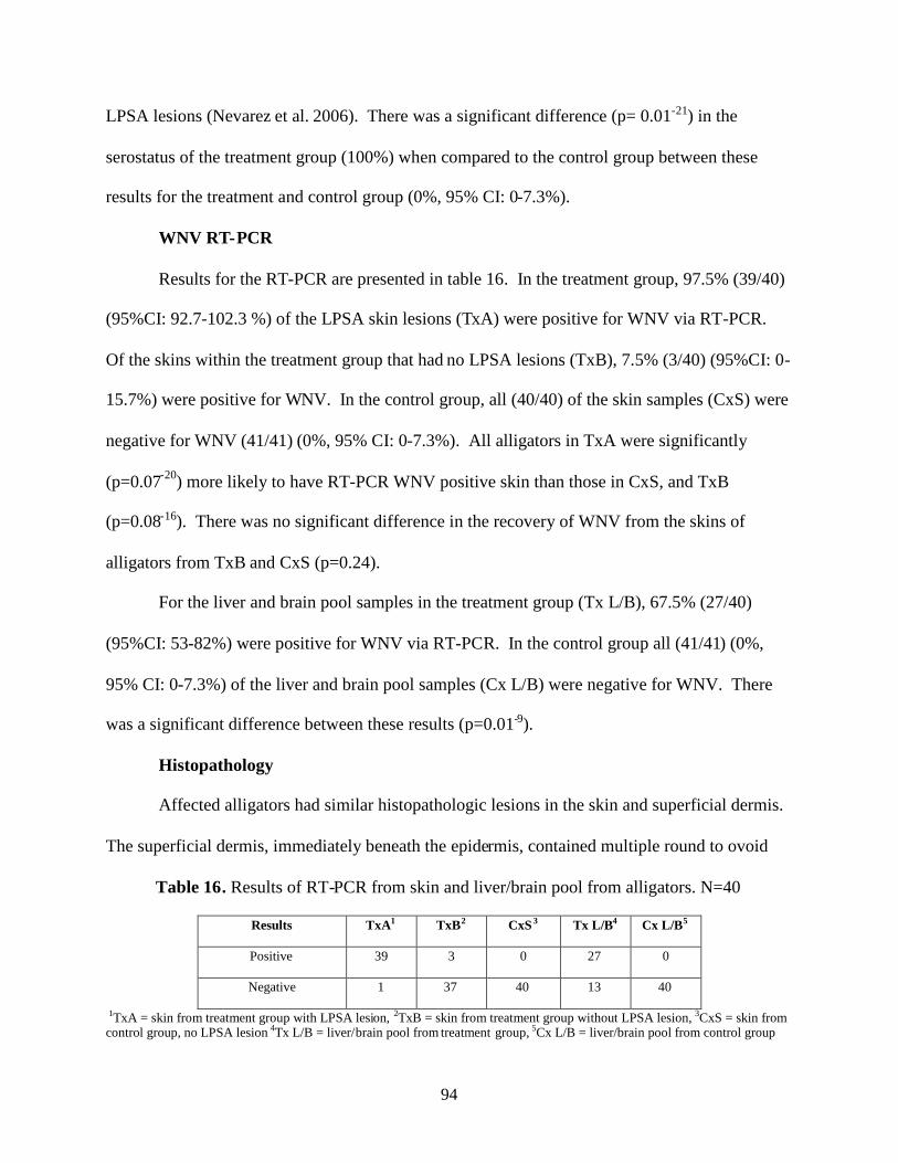
94
LPSA lesions (Nevarez et al. 2006). There was a significant difference (p= 0.01-21) in the
serostatus of the treatment group (100%) when compared to the control group between these
results for the treatment and control group (0%, 95% CI: 0-7.3%).
WNV RT-PCR
Results for the RT-PCR are presented in table 16. In the treatment group, 97.5% (39/40)
(95%CI: 92.7-102.3 %) of the LPSA skin lesions (TxA) were positive for WNV via RT-PCR.
Of the skins within the treatment group that had no LPSA lesions (TxB), 7.5% (3/40) (95%CI: 0-
15.7%) were positive for WNV. In the control group, all (40/40) of the skin samples (CxS) were
negative for WNV (41/41) (0%, 95% CI: 0-7.3%). All alligators in TxA were significantly
(p=0.07-20) more likely to have RT-PCR WNV positive skin than those in CxS, and TxB
(p=0.08-16). There was no significant difference in the recovery of WNV from the skins of
alligators from TxB and CxS (p=0.24).
For the liver and brain pool samples in the treatment group (Tx L/B), 67.5% (27/40)
(95%CI: 53-82%) were positive for WNV via RT-PCR. In the control group all (41/41) (0%,
95% CI: 0-7.3%) of the liver and brain pool samples (Cx L/B) were negative for WNV. There
was a significant difference between these results (p=0.01-9).
Histopathology
Affected alligators had similar histopathologic lesions in the skin and superficial dermis.
The superficial dermis, immediately beneath the epidermis, contained multiple round to ovoid
Table 16. Results of RT-PCR from skin and liver/brain pool from alligators. N=40
Results TxA1 TxB2 CxS 3 Tx L/B4 Cx L/B5
Positive 39 3 0 27 0
Negative 1 37 40 13 40
1TxA = skin from treatment group with LPSA lesion, 2TxB = skin from treatment group without LPSA lesion, 3CxS = skin fromcontrol group, no LPSA lesion 4Tx L/B = liver/brain pool from treatment group, 5Cx L/B = liver/brain pool from control group
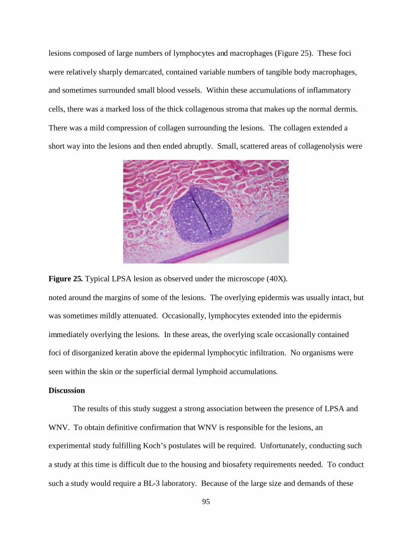
95
lesions composed of large numbers of lymphocytes and macrophages (Figure 25). These foci
were relatively sharply demarcated, contained variable numbers of tangible body macrophages,
and sometimes surrounded small blood vessels. Within these accumulations of inflammatory
cells, there was a marked loss of the thick collagenous stroma that makes up the normal dermis.
There was a mild compression of collagen surrounding the lesions. The collagen extended a
short way into the lesions and then ended abruptly. Small, scattered areas of collagenolysis were
Figure 25. Typical LPSA lesion as observed under the microscope (40X).
noted around the margins of some of the lesions. The overlying epidermis was usually intact, but
was sometimes mildly attenuated. Occasionally, lymphocytes extended into the epidermis
immediately overlying the lesions. In these areas, the overlying scale occasionally contained
foci of disorganized keratin above the epidermal lymphocytic infiltration. No organisms were
seen within the skin or the superficial dermal lymphoid accumulations.
Discussion
The results of this study suggest a strong association between the presence of LPSA and
WNV. To obtain definitive confirmation that WNV is responsible for the lesions, an
experimental study fulfilling Koch’s postulates will be required. Unfortunately, conducting such
a study at this time is difficult due to the housing and biosafety requirements needed. To conduct
such a study would require a BL-3 laboratory. Because of the large size and demands of these

96
animals, this is not possible at this time; however, plans to perform such a study are in progress.
Nonetheless, without the privilege of having performed an experimental infection, the data
clearly shows a very strong association and strongly suggest that WNV is highly correlated to the
presence of LPSA lesions.
A disadvantage of the RT-PCR procedure is its inability to differentiate between viable
(live) and dead virus. We attempted to culture WNV from five of the skin lesions but were
unable to do so. This may have been explained by the chronicity of the disease leading to a
lower amount of virus present in the tissue or the virus not being viable any more. One
interesting finding that helps provide some insight into this matter is the fact that only 67.5% of
the liver/brain pools tested positive in the treatment group as compared to 97.5% of the skin
samples. This finding is consistent with an expected reduction in tissue levels with chronicity of
the disease. Perhaps earlier sampling after the initial WNV outbreak would yield different
results, having a higher proportion of tissues test positive for WNV and being able to culture the
virus from the skin. The few positive results in TxB may be explained by the fact that LPSA
lesions are sometimes difficult to visualize and in a positive skin, any given section has the
possibility of housing a lesion.
Skin manifestations associated with WNV have been described in humans, although
histopathologic evaluation of the lesions has been limited. Nonetheless, there are reports of
exanthema and rash lesions associated with WNV infection dating back to 1956 (Marberg et
al.1956, Cernescu et al. 2000, Asnis et al. 2000, Nash et al. 2001, Klein et al. 2002, Estival et al.
2001, Seivar et al. 2003, Pealer et al. 2002, Ferguson et al. 2005, Anderson et al. 2004). There is
also a report of WNV identification in the skin of two Goshawks (Accipiter gentilis) via
immunohistochemistry (Wünschmann et al.2005). These reports support our findings or LPSA
as a skin manifestation of WNV. We believe this type of inflammation in the skin of alligators

97
may be prompted by an aggressive immune response secondary to chronic exposure to WNV but
further studies are required to understand the immune response to the virus.

98
CHAPTER 5: PREVENTION, SURVEILLANCE, AND CONTROL METHODS FORWNV IN ALLIGATOR RANCHES
Introduction
It appears that WNV will remain an important enzootic disease in the United States, and
that we should continue to focus on designing effective surveillance, prevention and control
methods that will decrease morbidity and mortality in both human and animal populations. For
alligator ranches, this goal is no different in light of a lack of effective treatment for animals and
humans infected with WNV. The economic impact of WNV in alligator ranches in Louisiana
between 2003 and 2006 was estimated at over 8 million dollars, with a total loss of over 50,000
alligators. This represented losses from dead alligators only, and does not take into account
those that survived WNV infection but later developed LPSA. The true economic impact of
LPSA is currently unknown. Because these financial losses seriously impacted the Louisiana
alligator industry, local ranchers mobilized to seriously consider and implement prevention and
control plans for WNV in their facilities. The key to decreasing and possibly eliminating WNV
outbreaks in alligator ranches lays in the effective implementation of surveillance, prevention,
and control methods. The three are closely associated, and together should form part of an
overall program for WNV control in alligator ranches.
Prevention
The first step in designing a prevention program is to understand the biology and ecology
of the disease in question. One must be able to identify the host and vector relationships, as well
as the environmental conditions that stimulate or inhibit the spread of the disease. In the case of
WNV, its life cycle primarily consists of birds (mostly passeriformes) as reservoir hosts and
mosquitoes as primary transmission vectors. Humans and animals are incidental hosts, some
with the ability to amplify the virus (e.g., alligators) and others being dead end hosts (e.g., horses
and humans). Of the two key players in the cycle of WNV, birds and mosquitoes, prevention

99
methods can be more easily applied to the transmission vector. For any control plan to be
effective, it is important to break the life cycle of the organism. One major challenge in the
control of WNV is the improbability of eliminating all the mosquitoes within a specific
geographic area. In addition, mosquito control does not eliminate the reservoir host. Once these
challenges are recognized, it becomes important to effectively spend the money and resources
available for controlling the disease.
The mainstay of WNV prevention is eliminating, or at least decreasing the number of
vectors. This can be done through aggressive mosquito control, with the intent to minimize the
amount of mosquitoes that can potentially carry the virus and infect alligators. Both non-
chemical and chemical mosquito control methods can be used. The control methods are aimed at
reducing both larval densities, as well as the adults. The most basic element of mosquito control
involves the elimination of habitats that promote breeding. Emptying containers that can hold
stagnant water (e.g., tires, feed troughs, plastic bins), keeping the grass cut, agitating the water in
ponds, and even painting the buildings in light colors that are less attractant to mosquitoes, are all
non-chemical methods that can be used to help control mosquitoes in an alligator facility. In
addition, it is also important to minimize the likelihood of mosquito entry into alligator
buildings. Buildings should be inspected for cracks or other damage that allows mosquito entry,
and any building defects should be repaired immediately. One major entry way for mosquitoes
into alligator buildings is via gaps in the doors or during times of feeding and cleaning when the
doors remain open. These problems can be alleviated by adding appropriate weather stripping to
the doors to ensure a tight seal when closed. Screen doors can also be added to the doorways so
that while feeding and cleaning, the main doors can remain open but the screen doors prevent
mosquitoes from entering the building.

100
Unfortunately, non-chemical methods alone are insufficient to control mosquito
populations. Active spraying for mosquitoes and treating against larvae is a critical aspect of the
prevention strategy. These treatments may be performed by local agencies or by the owners of
the facilities themselves. While some mosquito control agencies have provided guidance and
assistance to alligator ranchers, ultimately the owners of the facility have to take on the primary
responsibility of spraying chemicals to decrease mosquito populations. Many ranch owners have
purchased the necessary equipment and chemicals, but should consult with mosquito control
agents or the companies that manufacture the products to ensure their success. Type of
application, frequency, time of day, and environmental conditions are some of the factors to
consider when using these chemicals to ensure their effectiveness as well as limit their impact on
the environment.
An additional prevention strategy in alligator ranches deals with the proper disposal of
carcasses from animals that died of WNV infection. Because alligators can serve as an
amplifying host, proper disposal of infected carcasses is essential. Current recommendations are
to immediately burn carcasses or freeze them until the time of burning. In the past, most
ranchers disposed of their carcasses above ground in fields within close proximity to their
facility, creating a possible source of live virus for vectors. Fomites also represent a source of
disease transfer between alligator buildings. The use of foot baths and hand washing stations
outside every building would minimize the potential of disease transfers within a facility. Each
building should also have a separate set of tools such as feeding buckets, nets, and any other
object that are routinely used within the building. Ideally each building would also have a
designated pair of rubber boots for each worker. These tools should also be disinfected after
each use. A critical part of these preventive methods is to ensure that the disinfectants being
used (e.g., bleach, clorhexidine) are changed daily in order to maintain their effectiveness.

101
Since the initial WNV outbreaks in alligator ranches during 2003, there has been an
increase in the mosquito control efforts at various alligator facilities in the state of Louisiana.
The owners of the facilities have invested in equipment to spray for mosquitoes, altered their
buildings and even altered management techniques to prevent WNV infection in their facility.
Improved weather stripping of doors has been observed in some facilities in an attempt to
prevent mosquito entry. Others have added screen doors so that while the main doors remain
open during cleaning, the screen doors prevent mosquito entry. One facility has implemented an
automatic feeding system that minimizes entry into the buildings and consequently decreases the
likelihood of mosquito entry. More alligator ranchers are also conscious about the risks of
stagnant water and perform routine inspections of their facilities to empty any containers holding
water. The long term effects of these programs will continue to be assessed in the future.
Surveillance
The term surveillance has been assigned multiple definitions by different authors over the
years, and there is no stand alone definition that is universally accepted (Thrusfield 2005). The
types of surveillance are also varied, and range from sentinel surveillance to serologic
surveillance and active and passive surveillance (Thrusfield 2005, Moore et al. 1993). Perhaps
the most important distinction between the types of surveillance is whether it is passive or active.
Passive surveillance relies on examination of clinically affected animals only and therefore
occurs once the disease is present within the population. Active surveillance involves the
examination of clinically normal animals, and therefore has the advantage of being able to detect
the presence of subclinical and/or carrier animals within the population (Thrusfield 2005).
Ideally, a disease surveillance program would incorporate both passive and active methods of
surveillance. The type of disease, diagnostic tests available, funding, and personnel are some of
the factors that influence which method is employed. For our purposes, we will define WNV

102
surveillance as the organized gathering and analysis of virus activity as it pertains to its presence
in vectors, as well as exposure and infection in animal hosts, in order to appropriately allocate
and implement available resources for control and prevention of disease. The ultimate goal of
surveillance programs in alligator ranches would be early identification of virus activity within a
facility before alligators are exposed or infected. This in turn would lead to rapid and continued
implementation and follow up of vector control and other prevention and control methods.
Surveillance would also allow for evaluation of current methods of controlling vectors and
determining the effectiveness of those methods. Implementation of an active surveillance
method to parallel applied passive surveillance methods would provide the best approach to
control WNV in alligator facilities.
WNV surveillance can be performed using sentinel birds and/or trapping mosquitoes.
Sentinel birds should be placed in a location where WNV is suspected to be a problem based on
past experiences or models that predict that an area is a suitable habitat for reservoir and vector.
The ideal sentinel bird should be susceptible to infection, resistant to disease, and mount a rapid
immune response that can be detected with available antibody tests such as an ELISA (Komar
2001). It is also important for these birds to undergo seroconversion before the onset of the
disease outbreak within the geographical location. Finally, these birds should be easy to
maintain, present minimal health risks to the handlers, and must not contribute to the
transmission cycle of the virus (Komar 2001). The sentinel birds are tested for the presence of
antibodies to WNV on a regular interval (weekly or biweekly). If antibodies are detected at any
time, it would be an indication that WNV is present in the area and prevention methods should
be heightened.
One major limitation of using sentinel birds, such as chickens, is a historical lack of
success with these programs around the nation (Komar 2001). It is reported that in many states,

103
the positive sentinel chickens were identified after the onset of human cases (Komar 2001).
Some possible explanations for the limited success of these programs may lay in the design of
the sentinel program itself. The cage design and location, bird species, height above ground,
flock size, and proximity to susceptible populations are all factors that will influence the success
of sentinel programs (Komar 2001). In alligator ranches, there is the advantage of having a well
defined geographical area defined by the boundaries of the property. Once these boundaries are
defined, further breakdown of possible sites for locating sentinel chickens can be defined based
on the known location of mosquito larvae as well as adult mosquitoes. Because of limited land
space in most facilities, these areas of larvae and adult mosquito concentration also happens to be
in close proximity to the susceptible population of alligators. Together these two elements
facilitate the placement of the sentinel birds and increase the chances of success of the program.
One major limitation would be the attractiveness of the sentinel birds versus the alligators
themselves. There can be between two and five thousand alligators within a building, which
may prove to be more attractive for mosquitoes than a few sentinel birds in a cage. The inside of
the alligator buildings will also have a higher temperature, humidity, CO2, and organic odor than
the outside environment. These factors may greatly hinder the effectiveness of sentinel programs
in alligator facilities because it will simply be too difficult to compete against the environment
within an alligator building when it comes to attracting mosquitoes.
An alternative to captive sentinel birds is utilizing free ranging birds within the property.
Free ranging birds will cover more ground and would not be limited to a single location.
Sentinel free range birds could include domesticated birds within the property as well as wild
birds captured and bled for purposes of testing. The capture of resident wild bird species would
require a permit, while non-native species, such as the house sparrow (Passer domesticus),
European starling (Sturnus vulgaris), and the domestic pigeon (Columba livia), can be captured

104
and tested without a permit. The use of free ranging sentinels also has its limitations as they
would have the ability to move into areas with low mosquito concentrations and would also be
competing with the alligators for attracting mosquitoes. There is also a degree of difficulty in
trapping the birds, and determining when exposure to WNV occurred. From this information it
is evident that sentinel programs have certain limitations that should be weighed against their
cost and effectiveness. Perhaps it is important that the alligator facilities be evaluated further by
personnel familiar with sentinel programs, such as entomologists, in order to determine the
potential success of these programs. One major issue that must be addressed is the attractiveness
of the alligator buildings themselves when compared to other hosts in the property. Ultimately
sentinel surveillance is aimed at providing information about the risk level in an enzootic region.
A second surveillance system that can be employed is vector (mosquito) surveillance.
This can be further divided into larval and adult surveillance. Identification of potential breeding
grounds and larval habitat is critical for estimating future vector populations as well as treating
the larva (Moore et al. 1993). Surveillance of adult mosquito populations will provide
information about species, density, seasonality, viral infection, and minimum infection ratios
(MIR) (White 2001). In addition, adult vector surveillance can be used to monitor the
effectiveness of prevention programs. While larval surveillance is important, adult mosquito
surveillance is critical in determining the presence of WNV in the vector within a geographical
region. Various methods are available for trapping mosquitoes for surveillance purposes. The
alligator industry should seek help from individuals trained in mosquito surveillance to design
appropriate plans for their facilities. The owners of the alligator facilities could also have trained
personnel within their operations to set the traps and collect the mosquitoes, which then could be
shipped for WNV testing. This would limit their dependency on local agencies. Mosquito
surveillance plans also have some basic requirements for their success. Utilizing the appropriate

105
equipment and supplies, time and placement of the traps, species identification, and appropriate
handling of samples are all critical for the success of a mosquito surveillance plan. In addition.
there must be effective and diligent communication between the laboratory testing the mosquito
pools for WNV and the alligator ranchers/farmers so that the information obtained can be
directly applied to the prevention and surveillance methods.
Control
Despite the implementation of prevention and surveillance practices, WNV may still
make its way into an alligator building and infect the animals within. In these instances, it is
important to have a control plan in place to prevent further spread of the virus and decrease the
morbidity and mortality levels. The first and most essential step is strict isolation of any
building(s) housing the infected/exposed animals. This strict isolation of a building would
restrict any movements of animals in and out of that building, and so far has proven to be an
effective way of preventing the spread of WNV within a facility. Part of this strict quarantine
includes enforcing the use of foot baths and hand washing stations, as well as the disinfection of
tools that must only be used in the affected building. Feeding and cleaning must be performed
last in the affected building, as an added measure to minimize the spread of disease. Ideally
there would only be one individual designated to work in the affected building, and/or it would
be required that any persons working in that building take a shower and change clothes
immediately after their work is competed. Due to the zoonotic nature of WNV, the individual
working in the affected building must not be immunocompromised. While not common, human
infection amongst workers during a WNV outbreak in an alligator facility has occurred. Some of
these prevention methods have already been successfully implemented during some of the WNV
outbreaks in Louisiana.

106
Other methods of control have been explored, but their effectiveness has yet to be
determined. One promising technique is the active control of water temperatures in alligator
buildings according to time of the year and WNV exposure. While little is known about the
pathogenesis of WNV, temperature is believed to play a role in viral replication. Temperature
associations have been documented for the replication of WNV in Culex pipiens (Dohm and
Turell 2001). Dohm and Turell experimentally infected mosquitoes by allowing them to feed on
chickens inoculated with 104 pfu/ml of WNV (Dohm and Turell 2001). This study showed that
engorged mosquitoes held at 26oC (79oF) had detectable virus while engorged mosquitoes held at
18oC (65oF) and 10oC (50oF) had no detectable virus (Dohm and Turell 2001). Furthermore, if
the mosquitoes held at 18oC (65oF) and 10oC (50oF) where then kept at 26oC (79oF) for as little
as 1 day, WNV was detected (Dohm and Turell 2001). Mosquitoes held at 26oC (79oF) had
100% infection rate with increased time of incubation (Dohm and Turell 2001). A similar
temperature association has been observed for infection of WNV in alligators. The author has
observed three instances of a sudden decrease in the identification of WNV via RT-PCR from
liver and brain of captive reared alligators during a WNV outbreak. In all instances, the sudden
decrease in the identification of WNV occurred after the water temperatures in the alligator
buildings dropped below 26.6oC (80oF) due to a failure in the water heaters or artesian well
pump. This clinical evidence adds support to the possibility of there being an internal body
temperature threshold that is necessary for WNV to replicate in a host’s cells. It is also
interesting to note the differences observed in the pathogenicity of WNV between captive reared
and free-ranging populations of alligators. In a study by Jacobson et al., only 1.5% (10/669) of
free-ranging alligators tested positive for WNV antibodies (Jacobson et al. 2005). There is also
no indication that WNV is responsible for mortalities of free-ranging alligators. These data
differ from what is usually observed in captive reared alligators in which mortalities from WNV

107
can reach up to 60% (Nevarez et al. 2005). Captive reared alligators are maintained at constant
water temperatures between 29oC (85oF) and 32oC (90oF), which in turn allows the alligators to
maintain body temperatures closer to what may be expected in mammals. In contrast, free-
ranging alligators in a natural environment are subjected to a circadian fluctuation in
environmental temperatures during a 24 hour period. In the wild, the temperature fluctuations
could possibly vary as much as 15 – 20 degrees. These environmental fluctuations will also lead
to internal body temperature fluctuations in free-ranging alligators. This is especially true at
night when free-ranging alligators may have lower body temperatures that would lead to a
hypothermic state in mammals.
These differences between captive and free ranging alligators reveal the possibility of a
strong temperature effect on the susceptibility of captive reared alligators to WNV infection.
While it could be argued that stocking densities may also contribute to increased number of
exposure within captive populations, there are many areas in Louisiana that have high
concentrations of free-ranging alligators and yet fail to show any impact from WNV infection
based on a lack of increased mortality reported from these areas. It is also interesting to know
that no other species of reptiles has been show to be affected by WNV to the extent that captive
reared alligators are affected. Once again, a major difference in the husbandry of other reptile
species raised in commercial operations is that alligators are constantly housed at warm water
temperatures, while these other reptiles are not. This begs the question of whether preemptive
control of water temperatures by allowing circadian fluctuations in a captive environment,
similar to a natural environment, may decrease the occurrence of WNV infection in captivity.
While there is much left to learn about this association, there are some possible
applications that could be pursued in the meantime. One possibility would be to determine the
seasonality of WNV, and the most likely time of the year for infection in captive alligators. With

108
that information, the alligator producers could then change the water temperature scheme to
match a more natural variation during the months of the year with a higher risk of WNV
infection. For example, between the months of August to December, when most WNV
outbreaks in alligators occur, water temperatures within an alligator building could be made to
fluctuate between 24oC (75oF) and 29oC (85oF) in a 24 hour period. If the temperature
association is true, one might expect to see a drastic decrease in the occurrence of WNV in
alligator ranches. The trade off of these temperature fluctuations would be a slower growth rate,
which may lead to an extended period in captivity before reaching market size. An alternative
would be to rely on surveillance and prevention methods and only decrease the water
temperatures if a WNV outbreak occurs. At this point, the specific temperature and period of
time for which this temperature change should be maintained are unknown. However, based on
the clinical cases previously mentioned, a drop in water temperature to 24oC (75oF) for 14 days
may drastically reduce WNV infection within a captive population. The state of these alligators
once the temperatures are raised back again is unknown. One major question is whether
susceptibility would increase again and whether these animals would still be capable of shedding
the virus.
An additional control method may rely on reducing the amount of WNV exposure by
treating the water. It is known that alligators can amplify WNV and shed it in the feces. Fecal
oral contamination is probably the primary route of WNV spread within a building after initial
exposure of some animals via a mosquito. At this point it is unknown whether the virus can
survive in the water, for how long, and at what levels. In the future, the use of disinfectants (e.g.,
bleach, clorhexidine, Baquacil (Arch Chemicals Inc., Norwalk Connecticut)) in the water may be
an important part of the control strategies for WNV.

109
Antibiotic therapy may also be warranted in some case when secondary bacterial
infections occur in animals infected/exposed to WNV. If alligators infected with WNV are also
determined to have a secondary bacterial infection, such as pneumonia, a culture and sensitivity
should be obtained during necropsy in order to identify the offending bacteria and ensure that the
selected antimicrobial is appropriate for the infection. Antimicrobial treatment of remaining
animals may improve their chances of survival.
Discussion
The available information about WNV infection in captive alligators has increased over
the past 5 years, but there are still many unanswered questions. What is clear is the fact that
captive alligators are susceptible to WNV, and that there must be a plan in place to minimize
exposure. This plan must rely on surveillance for WNV in the environment, preventive methods
to minimize exposure, and control methods to decrease the speared of the virus if infection
occurs. The integration of all three areas is necessary in order to be successful in decreasing
morbidity and mortality. It is also important to recognize that WNV outbreaks in alligator
facilities have both an acute and a chronic effect leading to monetary losses. This makes WNV
an even more devastating disease for the alligator producers. It now appears that once WNV
occurs in a facility, LPSA will also occur. With this in mind we must realize that primary efforts
should concentrate on the surveillance and prevention of WNV infection. Once an outbreak
occurs the goal is to minimize losses realizing that an economic impact will be felt.
The alligator industry must look beyond their operations and continue to recruit
experienced individuals from the veterinary, entomology, ornithology, and virology fields that
can help them design cost effective plans for the surveillance and prevention of WNV.
Management changes are also essential in these facilities. Some have already started
implementing changes, such as maintaining tightly sealed buildings, adding screen doors, and

110
utilizing foot baths. These methods must become part of the daily management of these facilities
and not allowed to become lax. We are hopeful that theses measures will lead to a decrease in
the amount of WNV cases in the future.

111
CONCLUSIONS
After three years of investigation, we have gathered sufficient evidence to suggest that
the presence of LPSA is significantly associated with the serologic and molecular manifestations
of WNV infection in captive reared alligators. Furthermore, we have been able to characterize
two disease stages of WNV in alligators, an acute and a chronic stage. The acute stage of WNV
in alligators has been previously characterized by a primary heterophilic inflammatory response
in the tissues (Nevarez et al. 2005b), while the chronic stage of WNV is characterized by the
presence of lymphohistiocytic infiltrates. Gross examination, histopathology, RT-PCR, and
WNV serology have all been used to provide a link between the presence of LPSA and WNV.
The most striking results are presented in chapter four, which revealed a statistically significant
difference between TxA (skin from treatment group with LPSA lesion) and both CxS (skin from
control group, no LPSA lesion) and TxB (skin from treatment group without LPSA lesion)
(Table 16) that indicates that WNV is present within the LPSA lesions themselves. A similar
skin manifestation associated with WNV infection has also been reported in humans and birds
(Anderson et al. 2004, Wünschmann et al.2005). The results of this study have shed new light
into the epidemiology of WNV in a species of reptile, and also revealed that the pathologic
responses observed in these animals are similar to those described for other vertebrates (e.g.,
birds, mammals).
It is interesting that this disease appears to be primarily associated with captive alligators.
To date, there has been a single study evaluating WNV exposure in alligators from the wild
(Jacobson et al. 2005b), and the prevalence of disease appears to be low. On the surface, the
disease noted in the captive animals may appear to be associated with the high stocking densities
of animals; however, the physiologic conditions of the captive animals are also different, and
should be considered. Captive alligators are maintained at temperatures that are constant (29.oC-

112
32.oC; 85oF – 90oF), allowing them to maintain body temperatures that are more similar to
mammals. The shift to a warmer body temperature may be sufficient to allow the virus to
replicate in the alligators. In contrast, wild alligators are exposed to circadian changes in
temperature, and may experience body temperatures well below that which the virus can
replicate at. There remains more to be learned about the immune response of alligators to
infectious agents. Cellular immunity in such a primitive species is likely to play a more
significant role than what is currently recognized. While the humoral response will also have a
significant contribution, it is unclear how both of these responses work together in alligators.
One theory in this respect is that the cellular response has the primary domain in alligators and
that LPSA lesions are the result of a severe response to an antigen, in this case WNV. This
cellular response to the chronic presence of an antigen is so drastic that it eventually leads to
focal obliteration of normal tissue (e.g., connective tissue in the dermis) in order to eliminate the
offending antigen. Further studies are required to better understand the immune response in
alligators and its effect on different types of antigens.
As we learn more about the epidemiology of WNV, it has become more apparent that the
life cycle may incorporate more than just birds and mosquitoes. As it has been shown, alligators
have the potential of playing an important role in the cycle of WNV (Nevarez et al. 2005b)
(Figure 26). Based on this research, the epidemiology of LPSA appears to mimic the
epidemiology of WNV. The virus is maintained in the environment by birds serving as
reservoir hosts. Mosquitoes will bite these birds and serve as a vector for the virus.
Mosquitoes living in the alligator ranches are attracted to the alligator buildings housing
thousands of animals exhaling CO2 and producing a large amount of organic waste that
creates a warm, humid environment even during the coldest time of the year. Mosquitoes
will bite alligators on the oral mucous membranes, between the scales, or around the

113
Figure 26. Role of Alligators in the cycle of WNV
eyes, where the skin is softer and easier to penetrate. Once infected, the internal body
temperature of the captive alligators provides an appropriate environment within its cells
for WNV to replicate and be amplified. The virus is then shed via the feces. High
viremias are also likely maintained for an unknown period of time. Viral spread will be
possible via fecal oral contamination and/or by mosquitoes feeding within the buildings.
This will lead to a rapid spread of the virus within a building that can reach mortalities of
60% (Nevarez et al. 2005b). Alligators that survive the WNV outbreak will continue to
thrive, but frequently develop skin lesions within 4 weeks of exposure to WNV. These
skin lesions appear to be the manifestation of a chronic WNV infection/exposure.
The findings of this study raise important questions regarding the epidemiology of WNV
in the United States. “PIX” disease was first described in Florida in 1999, and is histologically
MosquitoHumans
Alligator Alligator

114
similar to LPSA (Cardeilhac et al. 2001, Nevarez et al. 2003a, Nevarez et al. 2003b). If LPSA
and PIX disease represent the same disease process, then WNV was potentially circulating in
Florida in 1999; the same time it was first diagnosed in New York City, New York (CDC 1999).
This would suggest that WNV was not introduced into the United States in 1999, but at least one
year earlier. Based on our general knowledge of the epidemiology of other arboviruses, this
would not be unexpected as disease is not generally diagnosed until there is sufficient viral
spillover to humans or domestic species. It might be expected that WNV could be disseminated
between New York City and Florida via bird migration. Based on these assumptions, it is
possible that WNV was first introduced into Florida, and that the 1999 outbreak in New York
only occurred after sufficient viral levels were achieved. At this time we have been unable to
obtain “PIX” affected tissues from alligators (1999 samples) in order to perform WNV testing
and determine if those lesions were indeed the similar to the Louisiana LPSA lesions. It is
interesting to note that of the three cases of WNV in Louisiana alligator farms in 2003, two
occurred after importation of Florida alligators infected with WNV. The United States
Department of Agriculture, Wildlife and Fisheries, and veterinarians alike should work
together to develop requirements for the import/export of alligators that include
surveillance of WNV in alligator facilities. West Nile virus is just one example of how
diseases may be crossing state lines and ultimately creating the potential for devastating
effects both in the captive and wild populations of alligators. This surveillance would be
important even between WNV endemic states, since we can not ensure that alligator
producers in other states are implementing the same WNV prevention methods as in
Louisiana. If alligators continue to be imported from Florida, WNV may still make its
way into the state despite the best surveillance, prevention and control programs.

115
Fortunately, the alligator production setting in Louisiana is primarily an all-in and
all-out operation, which allows us to break the cycle of the virus and take preventive mea-
sures before introducing new animals. Surveillance, prevention, and control methods for
WNV will continue to be an integral part of the daily management of alligator ranches
across Louisiana and other alligator producing states. We will continue to monitor WNV
activity in and around alligator facilities and observe the effectiveness of the
recommendations for WNV surveillance, prevention and control in the years to come. If
these recommendations are implemented, the incidence of WNV in alligator ranches
should decrease drastically. As of the spring of 2007, there has been less concern from
alligator ranchers about the occurrence of LPSA. In 2006, there was only one case of
WNV in an alligator facility. This reveals what is likely the beginning of a decline in the
occurrence of WNV and LPSA in alligator ranches. Once the effects of temperature on the
cycle of WNV are elucidated, temperature changes in the buildings may also be used as a control
tool for WNV.
This project has revealed an interesting connection between husbandry practices in
alligator facilities and the occurrence of WNV. As mentioned previously, the temperatures
under which these alligators are raised likely plays a key role in allowing WNV to adapt in these
animals. This should create awareness in the reptile community about the fact that reptiles are
just as susceptible to what were previously thought to be mammalian diseases, if maintained at
the right environmental conditions. Finally, we must also consider the possibility that other
arboviruses may be able to cause the same or similar tissue reactions in alligators. They would
remain important because of their economic impact. While we did not test for any other
arboviruses, diagnostics should be considered when similar lesions arise in the face of WNV
negative tests. However, with the long history of eastern equine encephalitis and St. Louis

116
encephalitis in Louisiana, and the absence of clinical disease prior to the arrival of WNV, this
point may be moot.
There are still future studies that need to be performed in order to completely define the
relationship between the presence of LPSA lesions and WNV positivity. A WNV infectivity
study would be ideal in order to meet Koch’s postulates and obtain a definitive answer. On the
other hand, the type of lesion observed with LPSA is a nonspecific type of lesions that is usually
associated with a viral infection. Therefore, as mentioned previously, there is a possibility that
this reaction may not be restricted to WNV alone but could also be elicited by other flaviviruses.
An infectivity study would also allow us to follow the progression of LPSA and determine the
time occurrence from the time of infection. It is also important to be able to monitor LPSA
positive animals and determine if the lesions will egress at some point. So far we have no
evidence to suggest that the lesions will heal or egress and the amount of pathology in the tissues
make a complete healing unlikely.
In addition to the infectivity study, we need to examine more carefully the shedding and
transmission cycles of the virus in alligator facilities. We believe that primary transmission
occurs within an affected building via fecal-oral contamination, after the initial animals are
infected via a mosquito bite. However, the amount of fecal shedding and viremia that occurs in
these animals is unknown at this time. Knowledge of the survivability of WNV in water,
especially water saturated with organic material, should also be pursued. If the virus was
determined to survive in the water, then studies could be performed to determine the efficacy of
various compounds (e.g. salt, chlorine, clorhexidine) at eliminating the virus from the water.
The pursuit of this information is required to better define the epidemiology of WNV
within alligator facilities, and ensure that the surveillance, prevention, and control methods are
adjusted accordingly. Even without this knowledge, it is becoming evident that our efforts and

117
recommendations have had a positive impact on the alligator industry and the occurrence of
WNV and LPSA has begun to decline.

118
REFERENCES
Allsteadt J, Lang JW. 1995. Incubation temperature affects body size and energy reserves ofhatchling American alligators (Alligator mississippiensis). Physiological Zoology 68(1):76-97.
Anderson RC, Horn KB, Hoang MP, et al. 2004. Punctate exanthem of West Nile Virusinfection: Report of 3 cases. J Am Acad Dermatol 51:820-823.
Ariel E, Ladds PW, Buenviaje GN. 1997. Concurrent gout and suspected hypovitaminosis A incrocodile hatchlings. Aust Vet J 75(4):247-249.
Asnis DS, Conetta R, Teixeira AA, et al. 2000. The West Nile Virus outbreak of 1999 in NewYork: the Flushing Hospital experience. Clin Infect Dis 30:413-418.
Asnis DS, Conetta R, Waldman G, et al. 2001. The West Nile virus encephalitis outbreak in theUnited States (1999-2000). Ann NY Acad Sci 951:161-171.
Barnett JD, Cardeilhac PT. 1995. Clinical values associated with opportunistic bacterial diseasesin farm raised alligators. IAAAM Proceedings 26:22.
Barnett JD, Cardeilhac PT. 1998. Brown spot disease in the American alligator (Alligatormississippiensis). IAAAM Proceedings 29:124-125.
Bounds HC, Normand RA. 1991. Brown spot disease of commercially-raised alligators: apreliminary report. Louisiana Academy of Sciences 54:62.
Britton A. 2005. http://crocodilian.com. Accessed January 05.
Brown DR, Farley JM, Zacher LA, et al. 2001a. Mycoplasma alligatoris sp. nov., from Americanalligators. Int J Syst Evol Microbiol 51:419-424.
Brown DR, Nogueira MF, Schoeb TR, et al. 2001. Pathology of experimental mycoplasmosis inAmerican alligators. J Wildl Dis 37(4):671-679.
Brune DE, Tomasso JR. 1991. Proceedings of the first international symposium on water qualityand aquaculture. Los Angeles, Feb 14-16, 1989.
Buenviaje GN, Hirst RG, Ladds PW, et al. 1997. Isolation of Dermatophilus sp from skin lesionsin farmed saltwater crocodiles (Crocodylus porosus). Aust Vet J 75(5):365-367.
Buenviaje GN, Ladds PW, Martin Y. 1998. Pathology of skin diseases in crocodiles. Aust Vet J76(5):357-363.
Buenviaje GN, Ladds PW, Melville L, et al. 1994. Disease-husbandry associations in farmedcrocodiles in Queensland and the Northern Territory. Aus Vet J. June 71(6):165-173.Buenviaje GN, Ladds PW, Melville L. 1992. Poxvirus infection in two crocodiles. Aust Vet J69(1):15-16.

119
Burger J, Gochfeld M, Rooney AA, et al. 2000. Metals and metalloids in tissues of Americanalligators in three Florida lakes. Arch Environ Contam Toxicol 38:501-508.
Burris Alligator Feeds. 2005. http://www.burrismill.com/Screens/Alligator.aspx. AccessedJanuary 2005.
Camus AC, Hawke JO. 2002. Providencia rettgeri-associated septicemia andmeningoencephalitis in juvenile farmed American alligators Alligator mississippiensis. J AquatAnim Health 14:149-153.
Camus AC, Mitchell MM, Williams JF, et al. 1998. Elevated lead levels in farmed Americanalligators (Alligator mississippiensis) consuming nutria (Myocastor coypus) meat contaminatedby lead bullets. J World Aquaculture Society 29:370-376.
Cardeilhac PT, Ashley DJ, Froleich E, et al. 2001. Report to Louisiana Department of Wildlifeand Fisheries Fur and Refuge Division, New Iberia, Louisiana.
Carpenter JW, Mashima TY, Rupiper DJ. 2001. Exotic Animal Formulary, ed. 2, Philadelphia,WB Saunders.
Castillo-Olivares J, Wood J. 2004. West Nile virus infection in horses. Vet Res 35:467-483.
Centers for Disease Control and Prevention. 1999. Update: West Nile virus encephalitis--NewYork. MMWR Morbidity and Mortality Weekly Report 48:944-946, 955.
Centers for Disease Control and Prevention. 2003. Virology: Classification of West Nile Virus.http://www.cdc.gov/ncidod/dvbid/westnile/virus.htm (Accessed January 13, 2006).
Centers for Disease Control and Prevention. 2005. Entomology.http://www.cdc.gov/ncidod/dvbid/westnile/mosquitoSpecies.htm (Accessed January 15, 2007).
Cernescu C, Nedelcu N-I, Tarei G, et al. 2000. Continued transmission of West Nile Virus tohumans in Southeastern Romania, 1997-1998. J Infect Dis 181:710-712.
Cheatwood JL. 2000. An outbreak of fungal dermatitis and stomatitis in a wild population ofpigmy rattlesnakes, Sistrurus miliarius barbouri, in Florida: description, factors, cyclicity, andprevention. Master Thesis, University of Florida.http://etd.fcla.edu/UF/ane5876/Cheatwood_Thesis.PDF, accessed March 8, 2007.
Clippinger TL, Bennet RA, Johnson CM, et al. 2000. Morbidity and mortality associated with anew mycoplasma species from captive American alligators (Alligator mississippiensis). J ZooWildl Med 31(3):303-314.
Coulson RA, Coulson TD, Herbert JD, et al. 1987. Protein nutrition in the alligator. CompBiochem Physiol 87A (2):449-459.

120
Coulson RA, Coulson TD, Herbert JD. 1992. Alligator feed evaluations. Proceedings of theLouisiana Aquaculture Conference, Baton Rouge, LA:48-51.
Dickson HM, Cardeilhac PT. 2001. PIX: a new skin disease of alligators. 53rd Annual Meetingof Animal Disease Research Workers of Southern States. Abstract 10.
Dohm DJ, Turell MJ. 2001. Effect of incubation at over wintering temperatures on thereplication of West Nile virus in New York Culex pipiens (Diptera: Culicidae). J Med Entomol38(3):462-464.
Dohms JE, Metz A. 1991. Stress-mechanisms of immunosuppression. Vet ImmunolImmunopathol 30:89-109.
Elsey R. 2004. Stocking rates of ranched American alligators (Alligator mississippiensis),personal communication.
Elsey RM, Joanen T, McNease L, et al. 1990. Growth rate and plasma corticosterone levels injuvenile alligators maintained at different stocking densities. J Exp Zool 255:30-36.
Elsey RM. Personal Communication. 2004. Louisiana Department of Wildlife andFisheries, Rockefeller Wildlife Refuge, Grand Chenier, LA 70643, USA.
Estival JL, Skowron F, Dupin M, et al. 2001.-infectiona virus West-Nile (French). Ann DermatolVenereol 128:656-658.
Ferguson DD, Gershman K, LeBailly A, et al. 2005. Characteristics of the Rash Associated withWest Nile Virus Fever. Clin Infect Dis 41:1204–1207.
Flandry F, Lisecki EJ, Domingue GJ, et al. 1989. Initial antibiotic therapy for alligator bites:characterization of the oral flora of Alligator mississippiensis. South Med J 82(2): 262-266.
Fleming GJ. 2001. Crocodilian Anesthesia. Vet Clin North Am Exot Anim Pract 4(1):19-145.
Frelier PF, Sigler L, Nelson PE. 1985. Mycotic pneumonia caused by Fusarium moniliforme inan alligator. Sabouraudia 23(6):399-402.
Fromtling RA, Jensen JM, Robinson, et al.1979. Fatal mycotic pulmonary disease of captiveAmerican alligators. Vet Pathol 16:428-431.
Gorden RW, Hazen TC, Esch GW, et al. 1979. Isolation of Aeromonas hydrophila from theAmerican alligator, Alligator mississippiensis. J Wildl Dis 15(2):239-243.
Grenard S. 1991. Handbook of alligators and crocodiles, ed. 1. Krieger Publishing Company,Malabar, Florida.
Guillette LJ Jr, Vonier PM, McLachlan JA. 2002. Affinity of the alligator estrogen receptor forserum pesticide contaminants. Toxicology 181-182:151-154.

121
Harvey RW, Price TH. 1983. Salmonella isolation from reptilian faeces: a discussion ofappropriate cultural techniques. J Hyg 91(1):25-32.
Hayes CG. 2001. West Nile virus: Uganda, 1937, to New York City, 1999. Ann NY Acad Sci951:25-37.
Hayes EB, Gubler DJ. 2006. West Nile virus: epidemiology and clinical features of an emergingepidemic in the United States. Annu Rev Med 57:181-194.
Hayes EB, Sejvar JJ, Zaki SR, et al. 2005. Virology, pathology, and clinical manifestations ofWest Nile virus disease. Emerg Inf Dis 11(8):1174-1179.
Helmick KE, Brown DR, Jacobson ER, et al. 2002. In Vitro Susceptibility Pattern ofMycoplasma alligatoris from Symptomatic American Alligators (Alligator mississippiensis). JZoo Wildl Med 33(2):108-111.
Horner RF. 1990. Poxvirus in farmed Nile crocodiles. Vet Rec 122(19):459-462.
Huchzermeyer FW, Gerdes GH, Foggin CM, et al. 1994. Hepatitis in farmed hatchling Nilecrocodiles (Crocodylus niloticus) due to chlamydial infection. J S Afr Vet Assoc 65(1):20-22.
Huchzermeyer FW, Henton MM, Riley J, et al. 2000. Aerobic intestinal flora of wild-caughtAfrican dwarf crocodiles Osteolaemus tetraspis. Onderstepoort J Vet Res 67(3):201-204.
Huchzermeyer FW. 2003. Crocodiles: Biology, Husbandry and Diseases, ed 1, Wallingford,CABI Publishing.
IUCN Red List of Threatened Species. 2004. http://www.iucnredlist.org. Accessed January2005.
Jacobson ER, Gardiner CH, Foggin CM. 1984. Adenovirus-like infection in two Nile crocodiles.J Am Vet Med Assoc 185(11):1421-1422.
Jacobson ER, Gaskin JM, Shields RP, et al. 1979b. Mycotic pneumonia in mariculture-rearedgreen sea turtles. J Am Vet Med Assoc 175(9)929-933.
Jacobson ER, Ginn PE, Troutman JM, et al. 2005a. West Nile virus infection in farmedAmerican alligators (Alligator mississippiensis) in Florida. J Wildl Dis 41(1):96-106.
Jacobson ER, Johnson AJ, Hernandez JA, et al. 2005b. Validation and use of an indirect enzyme-linked immunosorbent assay for detection of antibodies to West Nile virus in American alligators(Alligator mississippiensis) in Florida. J Wildl Dis 41(1):107-114.
Jacobson ER, Popp JA, Shields RP, et al. 1979. Poxlike skin lesions in captive caimans. J AmVet Med Assoc 175(9):937-940.

122
Jacobson ER. 1984. Immobilization, blood sampling, necropsy techniques and diseases ofcrocodilians: A review. J Zoo An Med 15:38-45.
Jeffree RA, Markich SJ, Twining JR. 2001. Element concentrations in the flesh and osteodermsof estuarine crocodiles (Crocodylus porosus) from the Alligator Rivers region, northernAustralia: biotic and geographic effects. Arch Environ Contam Toxicol 40:236-245.
Johnson A. 2005. Personal communication. March.
Johnson DJ, Ostlund EN, Pedersen DD, et al. 2001. Detection of North American West Nilevirus in animal tissue by a reverse transcription-nested polymerase chain reaction assay.Emerg Infect Dis 7(4):739-741.
Kitaoka M. 1950. Experimental transmission of the West Nile virus by the mosquito. Jap Med J3:77-81.
Kiupel M, Simmons HA, Fitzgerald SD, et al. 2003. West Nile virus infection in Eastern foxsquirrels (Sciurus niger).Vet Path 40(6):703-707.
Klein C, Kimiagar I, Pollak L, et al. 2002. Neurological features of West Nile Virus infectionduring the 2000 outbreak in a regional hospital in Israel. J Neurol Sci 200:63-66.
Klenk K, Snow J, Morgan K, et al. Alligators as West Nile virus amplifiers. Emerg InfectDis [serial on the Internet]. 2004 Dec [cited 1 -31-05]. Available fromhttp://www.cdc.gov/ncidod/EID/vol10no12/04-0264.htm
Klenk K, Snow J, Morgan K, et al. 2004. Alligators as West Nile virus amplifiers. EmergingInfectious Diseases 10(12):2150-2154.
Komar N. 2001. West Nile virus surveillance using sentinel birds. In West Nile virus: detection,surveillance, and control. Annals of the New York Academy of Sciences 951:58–73.Lance VA, Elsey RM. 1999. Plasma catecholamines and plasma corticosterone followingrestraint stress in juvenile alligators. J Exp Zool 283(6):559-565.
Lance VA, Elsey RM. 1999a. Hormonal and metabolic response of juvenile alligators to coldshock. J Exp Zool 283:566-572.
Lance VA, Morici LA, Elsey RM. 2001. Physiology and endocrinology of stress in crocodilians.In Grigg GC, Seebacher F, Franklin CE, editors: Crocodilian Biology and Evolution, ed 1,Chipping Norton, Surrey Beatty and Sons.
Lanciotti R, Kerst A, Nasci R, et al. 2000. Rapid detection of West Nile virus from humanclinical specimens, field-collected mosquitoes, and avian samples by a TaqMan reversetranscriptase-PCR assay. J. Clin Micro, 38(11):4066-4071.
Lane TJ. 1996. Crocodilians. In Mader DR, editor: Reptile Medicine and Surgery, Philadelphia,WB Saunders.

123
Lennette EH, Smith TF. 1999. Laboratory diagnosis of viral infections, 3r ed., MarcelDekker, Inc., New York chapter 1, clinical virology, concepts and perspectives; Section II,laboratory approaches in virus isolation; paragraph B, Virus Isolation in Shell Vial Cultureswith Centrifugation Enhanced Inoculation, 6-7.
LFAC (Louisiana Fur and Alligator Council). http://alligatorfur.com/index.htm. Accessed March1, 2007.
Lind PM, Milnes MR, Lundberg R, et al. 2004. Abnormal bone composition in female juvenileAmerican alligators from a pesticide-polluted lake (Lake Apopka, Florida). Environ HealthPerspect 112(3):359-362.
Ludwig GV, Calle PP, Mangiafico JA, et al. 2002. An outbreak of West Nile virus in a NewYork city captive wildlife population. Am J Trop Med Hyg 67(1): 67-75.
Mackenzie JS, Barrett ADT, Deubel V. 2002. Japanese Encephalitis and West Nile viruses.Springer-Verlag.
Madsen M, Hangartner P, West K, et al. 1998. Recovery rates, serotypes, and antimicrobialsusceptibility patterns of salmonellae isolated from cloacal swabs of wild Nile crocodiles(Crocodylus niloticus) in Zimbabwe. J Zoo Wildl Med 29(1):31-34.
Madsen M. 1993. Microbial flora of frozen tail meat from captive Nile crocodiles (Crocodylusniloticus). Int J Food Microbiol 18(1):71-76.
Madsen M. 1996. Prevalence and serovar distribution of Salmonella in fresh and frozen meatfrom captive Nile crocodiles (Crocodylus niloticus). Int J Food Microbiol 29(1):111-118.
Mainster ME, Lynd FT, Cragg PC, et al. 1972. Treatment of multiple cases of Pasteurellamultocida and Staphylococcal pneumonia in Alligator mississippiensis on a herd basis.Proceedings American Association Zoo Veterinarians:33-36.
Manolis SC, Webb GJ, Pinch D, et al. 1991. Salmonella in captive crocodiles (Crocodylusjohnstoni and C. porosus). Aust Vet J 68(3):102-105.
Marberg K, Goldblum N, Sterk VV, Jasinkska-Klingberg W, Klingberg MA. 1956. The naturalhistory of West Nile Fever. Clinical observations during an epidemic in Israel. Am J Hyg.64:259-69.
Maslen M, Whitehead J, Forsyth WM, et al. 1988. Systemic mycotic disease of captive crocodilehatchling (Crocodylus porosus) caused by Paecilomyces lilacinus. J Med Vet Mycol 26(4):219-225.
McCowan C, Shepherdley C, Slocombe RF. 2004. Herpes virus-like particles in the skin of asaltwater crocodile (Crocodylus porosus). Aust Vet J 82(6):375-377.

124
Merchant ME, Roche C, Elsey RM, et al. 2003. Antibacterial properties of serum from theAmerican alligator (Alligator mississippiensis). Comp Biochem Physiol B Biochem Mol Biol136(3):505-513.
Millan JM, Purdie JL, Melville LF. 1997. Public health risks of the flesh of farmed crocodiles.Rev Sci Tech 16(2):605-681.
Miller DL, Mauel MJ, Baldwin C, et al. 2003. West Nile virus in farmed alligators. EmergInfect Dis 9(7):794-799.
Miller DM, Mauel MJ, Baldwin C, et al. 2003. West Nile virus in farmed alligators. EmergInfect Dis Jul 9(7):794-799.
Millichamp NJ, Jacobson ER, Wolf ED. 1983. Diseases of the eye and ocular adnexae in reptiles.J Am Vet Med Assoc 183(11):1205-1212.
MMWR. 1999a. Outbreak of West Nile-like viral encephalitis – New York, 1999. Morb MortWkly Rep 48(38):845-849.
MMWR. 1999b. Update: West Nile-like viral encephalitis – New York, 1999. Morb Mort WklyRep 48(39):890-892.
Mohan K, Foggin CM, Dziva F, et al. 2001. Vaccination to control an outbreak of Mycoplasmacrocodyli infection. Onderstepoort J Vet Res 68(2):149-150.
Mohan K, Foggin CM, Muvavarirwa P, et al. 1995. Mycoplasma-associated polyarthritis infarmed crocodiles (Crocodylus niloticus) in Zimbabwe. Onderstepoort J Vet Res 62(1):45-49.
Mohan K, Foggin CM, Muvavarirwa P, et al. 1997. Vaccination of farmed crocodiles(Crocodylus niloticus) against Mycoplasma crocodyli infection. Vet Rec 141(18): 476.
Moore CG., McLean RG., Mitchell CJ., Nasci RS., Tsai TF, et al. 1993. Guidelines for arbovirussurveillance programs in the United States. Fort Collins, CO:USDHHS/PHS/CDC/NCID/DVBID.
Morici LA, Elsey RM, Lance VA. 1997. Effects of long-term corticosterone implants on growthand immune function in juvenile alligators (Alligator mississippiensis). J Exp Zool 279(2):156-162.
Nash D., Mostashari F., Fine A., Miller J., et al. 2001. The outbreak of West Nile virus infectionin the New York city area in 1999. N Engl J Med 344: 1807-1814.
Nevarez JG, Mitchell MA, Foster T. Screening for herpes viruses in American alligators(Alligator mississippiensis) with Lymphohistiocytic Proliferative Syndrome of Alligators(LPSA). Unpublished data.

125
Nevarez JG, Mitchell MA, Garner MM. 2003b. Characterizing Lymphohistiocytic ProliferativeSyndrome (“PIX”) in Captive-Reared American Alligators (Alligator mississippiensis) fromLouisiana. Final report to the Louisiana Department of Wildlife and Fisheries Fur and RefugeDivision, New Iberia, Louisiana.
Nevarez JG, Mitchell MA, Johnson AJ, et al. 2006. Establishing an Association between WestNile Virus Seropositivity and the Development of Lymphohistiocytic Proliferative Syndrome ofAlligators. Journal of Herpetological Medicine and Surgery. Submitted for Publication.
Nevarez JG, Mitchell MA, Kim DY, et al. 2003a. Lymphohistiocytic Proliferative Syndrome ofCaptive Reared American Alligators (Alligator mississippiensis) from Louisiana. InProceedings: ARAV Minneapolis, MN:73-75.
Nevarez JG, Mitchell MA, Kim DY, et al. 2005b. West Nile Virus in Alligator, Alligatormississippiensis, Ranches from Louisiana. J Herp Med Surg 5(3):4-9.
Nevarez JG. 2002-03. Unpublished data from clinical and research cases. Louisiana.
Nevarez JG. 2003. Unpublished data. Fungal isolates from the skin of American alligators(Alligator mississippiensis).
Newton J. 1992. Brown spot disease in the Louisiana alligator industry: what we know about thedisease and possible control protocols. Proceedings Louisiana Aquaculture Conference, BatonRouge, LA:46-47.
Noga EJ. 2000. Fish disease: diagnosis and treatment, ed. 2. Iowa State University Press, Ames,Iowa.
Novak SS, Seigel RA. 1986. Gram-negative septicemia in American alligators (Alligatormississippiensis). J Wildl Dis 22(4):484-487.
Palmer MV, Stoffregen WC, Rogers DG, et al. 2004. West Nile virus infection in reindeer(Rangifer tarandus). J Vet Diagn Invest 16(3):219-222.
Pandey GS, Inoue N, Ohshima K, et al. 1990. Poxvirus infection in Nile crocodiles (Crocodylusniloticus). Res Vet Sci 49(2):171-176.
Pealer LN, Marfin AA, Petersen LR, et al. 2003. Transmission of West Nile Virus through bloodtransfusion in the United States in 2002. N Engl J Med 349:1236-1245.
Penrith ML, Nesbit JW, Huchzermeyer FW. 1991. Pox virus infection in captive juvenilecaimans (Caiman crocodilus fuscus) in South Africa. J S Afr Vet Assoc 62(3):137-139.
Philip CB, Smadel JE. 1943. Transmission of West Nile virus by infected Aedes albopictus.Proc Soc Exper Biol and Med 77: 661-665.

126
Pye GW, Brown DR, Nogueira MF, et al. 2001. Experimental Inoculation of broad-nosedcaimans (Caiman latirostris) and Siamese crocodiles (Crocodylus siamensis) with Mycoplasmaalligatoris. J Zoo Wildl Med 32(2):196-201.
Ramos MC, Coutinho SD, Matushima ER, et al. 2002. Poxvirus dermatitis outbreak in farmedBrazilian caimans (Caiman crocodilus yacare). Aust Vet J 80(6):371-372.
Rauschenberger RH, Wiebe JJ, et al. 2004. Achieving environmentally relevant organochlorinepesticide concentrations in eggs through maternal exposure in Alligator mississippiensis. MarEnviron Res 58(2-5):851-856.
Rickard MW, Thomas AD, Bradley S, et al. 1995. Microbiological evaluation of dressingprocedures for crocodile carcasses in Queensland. Aust Vet J 72(5):172-176.
Rooney AA, Guillette LJJ. 2001. Biotic and abiotic factors in crocodilian stress: the challenge ofa modern environment. In Grigg GC, Seebacher F, Franklin CE, editors: Crocodilian Biologyand Evolution, ed 1, Chipping Norton, Surrey Beatty and Sons.
Rubio A. 2003. Personal Communication, Mexico.
Russell WC, Herman KL. 1970. Colibacillosis in captive wild animals. Journal of Zoo AnimalMedicine 1:17-21.
Sampson BA, Ambrosi C, Charlot A, et al. 2000. The pathology of human West Nile virusinfection. Human Pathology 31(5):527-531.
Sejvar JJ, Jaddad MB, Tierney BC, et al. 2003. Neurologic manifestations and outcome of WestNile Virus infection. JAMA 290:511-515.
Shaharabany M, Gollop N, Ravin S, et al. 1999. Naturally occurring antibacterial activities ofavian and crocodile tissues. J Antimicrob Chemother 44(3):416-418.
Shotts EB, Gaines JL, Martin L, et al. 1972. Aeromonas-induced deaths among fish and reptilesin an eutrophic inland lake. J Am Vet Med Assoc 161(6):603-607.
Silberman MS, Blue J, Mahaffey E. 1977. Phycomycoses resulting in death of crocodiles in acommon pool. Proceedings of the American Association of Zoo Veterinarians:100-101.
Smithburn KC, Hughes TP, Burke AW, et al. 1940. A neurotropic virus isolated from the bloodof a native of Uganda. Am J Trop Med 20:471-492.
Soares D. 2002. An ancient sensory organ in crocodilians. Nature 417:241–242.
Southam CM., Moore AE. 1954. Induced virus infection in man by the Egypt isolates of WestNile virus. Am J Trop Med Hyg 3:19-50.

127
Staton MA, Edwards HMJr, Brisbin ILJr., et al. 1990. Essential fatty acid nutrition of theAmerican alligator (Alligator mississippiensis). J Nutr 120(7):674-685.
Steinman A, Banet-Noach C, Tal S, et al. 2003. West Nile virus infection in crocodiles.Emerg Infect Dis 9(7):887-889.
Stoker C, Rey F, Rodriguez H, et al. 2003. Sex reversal effects on Caiman latirostris exposed toenvironmentally relevant doses of the xenoestrogen bisphenol A. Gen Comp Endocrinol133(3):287-296.
Swayne DE, Beck JR, Smith CS, et al. 2001. Fatal encephalitis and myocarditis in youngdomestic geese (Anser anser domesticus) caused by West Nile virus. Emerg Infect Dis 7(4):751-753.
Tappe JP, Chandler FW, Liu SK, et al. 1984. Aspergillosis in two San Esteban chuckwallas. JAm Vet Med Assoc 185(11):1425-1428.
Taylor RM, Work TH, Hurlbut HS, et al. 1956. A study of the ecology of West Nile virus inEgypt. Am J Trop Med Hyg 5: 579-620.
Tengelsen L. 2004. Personal Communication.. Idaho State Department of Health andWelfare, Boise, Idaho
Thomas AD, Sigler L, Peucker S, et al. 2002. Chrysosporium anamorph of Nannizziopsis vriesiiassociated with fatal cutaneous mycoses in the salt-water crocodile (Crocodylus porosus). MedMycol 40(2):143-151.
Thrusfield M. 2005. Veterinary Epidemiology. 3rd ed. 2005. Blackwell Science Ltd., Ames,Iowa.
Thrusfield M. 2005. Surveillance: Veterinary Epidemiology, 168- 187. Great Britain: BlackwellPublishing.
Trevino GS. 1972. Cephalosporiosis in three caimans. J Wildl Dis 8:385-388.
Van Belle G. 2002. Statistical Rules of Thumb. John Wiley & Sons. New York, NY.
van der Walt ML, Huchzermeyer FW, Steyn HC. 1997. Salmonella isolated from crocodiles andother reptiles during the period 1985-1994 in South Africa. Onderstepoort J Vet Res 64(4):277-283.
vanDevanter DR, Warrener P, Bennett L, et al. 1996. Detection and analysis of diverseherpesviral species by consensus primer PCR. Journal of Clinical Microbiology 34(7):1666-1671.

128
Vonier PM, Crain DA, McLachlan JA, et al. 1996. Interaction of environmental chemicals withthe estrogen and progesterone receptors from the oviduct of the American alligator. EnvironHealth Perspect. (12):1318-1322.
Wallace LJ, White FH, Gore HL. 1966. Isolation of Edwardsiella tarda from a sea lion and twoalligators. J Am Vet Med Assoc 149(7):881-883.
Wallach JD, Hoessle C, Bennett J. 1967. Hypoglycemic shock in captive alligators. J Am VetMed Assoc 51(7):893-896.
Weinberger M, Pitlik SD, Gandacu D, et al. 2001. West Nile fever outbreak, Israel, 2000:epidemiologic aspects. Emerg Inf Dis 7(4): 686-691.White DJ. 2001. Vector surveillance for West Nile virus. In West Nile virus: detection,surveillance, and control. Annals of the New York Academy of Sciences 951:74-83.
Wilhite DR, Nevarez JG. 2004. Unpublished description of a new site for venipuncture in theAmerican alligator (A. mississippiensis).
Wünschmann A, Shivers J, Bender J, et al. 2005. Pathologic and immunohistochemical findingsin Goshawks (Accipiter gentiles) and Great Horned Owls (Bubo virginianus) naturally infectedwith West Nile virus. Avian Diseases 49:252-259.
Youngprapakorn P, Ousavaplangchai L, Kanchanapangka S. 1995. A Color Atlas of Diseases ofthe Crocodile, ed 1, Thailand, Style Creative House Co., Ltd.

129
APPENDIX: LETTER OF PERMISSION FROM THE JOURNAL OFHERPETOLOGICAL MEDICINE AND SURGERY

130
VITA
Javier G. Nevarez was born in San Juan, Puerto Rico, on September 20, 1976. He graduated
from the high school El Colegio San Ignacio de Loyola in May of 1994. That same fall, Javier
began undergraduate studies at the Louisiana State University in the animal sciences three plus
one program. In 1997, Javier was admitted to the Louisiana State University School of
Veterinary Medicine. Javier obtained his Bachelor of Science degree with a concentration in
animal science in May of 1998, and his Doctor of Veterinary Medicine degree in May of 2001.
Following graduation from veterinary school, Javier completed a one year internship in
zoological medicine at the Louisiana State University School of Veterinary Medicine in June of
2002. After completion of the internship, Dr. Nevarez enrolled in a Doctor of Philosophy
program at the Department of Veterinary Clinical Sciences in the Louisiana State University
School of Veterinary Medicine. In 2003, Dr. Nevarez was hired as an instructor of zoological
medicine in the Veterinary Clinical Sciences department. Since that time, Dr. Nevarez has
continued to work towards the fulfillment of his Doctor of Philosophy degree requirements as
well as carrying out teaching and research responsibilities within the Louisiana State University
School of Veterinary Medicine.
Copyright © 2022 FDOKUMEN