Lipoprotein alterations, abdominal fat distribution and breast cancer
Upload
independentCategory
view
4download
0
EXPERIMENTAL and MOLECULAR MEDICINE, Vol. 40, No. 1, 98-108, February 2008
Alterations of proliferative and differentiation potentials of
human embryonic stem cells during long-term culture
Yong Bin Park1,2
, Yoon Young Kim2,
Sun Kyung Oh2,3
, Sun Gan Chung1,
Seung-Yup Ku2,3
, Seok Hyun Kim2,3
,Young Min Choi
2,3,4 and Shin Yong Moon
2,3
1Central Research Institute
Sam Jin Pharm. Co. Ltd.
Hwasung 445-746, Korea2Institute of Reproductive Medicine and Population
Medical Research Center3Department of Obstetrics and Gynecology
Seoul National University College of Medicine
Seoul 110-799, Korea4Corresponding author: Tel, 82-2-2072-2385;
Fax, 82-2-744-8945; E-mail, [email protected]
Accepted 26 December 2007
Abbreviations: bFGF, basic fibroblast growth factor; EB, embryoid
body; hESCs, human embryonic stem cells; HNF, hepatocyte
nuclear factor; ICM, inner cell mass; SSEA, stage specific em-
bryonic antigen
Abstract
Human embryonic stem cells (hESCs) are considered to be able to stably maintain their characteristics in vi-tro for prolonged periods, but we had previously en-countered changes in proliferative ability and differ-entiation potential during extended culture of hESCs. Therefore, we investigated the proliferative ability and differentiation potential of hESCs during long-term culture. The hESCs, SNUhES3, were used to analyze population-doubling time, proliferation rate and differ-entiation potential. We classified hESCs into three groups according to culture period. Ten colonies of hESCs for each group were daily measured colony area and population-doubling time was assessed by the changes of colony area. Proliferation rate of hESCs was measured by 5-bromo-2'-deoxyuridine (BrdU) as-say and telomerase activity. To evaluate differentiation potentials for hESCs, expression levels of undiffer-entiated and/or differentiated hESCs markers were ex-amined by FACS, RT-PCR and immunostaining. Population-doubling time of early passage hESCs was longer than those of middle or late passage. Prolifer-ative ability of hESCs was accelerated depending on
culture periods. Cellular morphologies and the ex-pression level of each three germ layer markers were obviously different from each passage of reattached embryoid bodies (EBs) after spontaneous differentia-tion. Differentiated cells of late passage expressed higher levels of undifferentiated markers such as Oct4 and SSEA4 than those of early and middle passage. But differentiated cells of early and middle passage ex-pressed higher level of differentiated state markers, Nestin (ectoderm), Brachyury (mesoderm), HNF3β (endoderm). From these results, it can be inferred that hESCs show higher proliferative abilities and reduced differentiation potentials as the passage number increased. Therefore, we conclude that early passage hESCs could be more suitable than middle and late pas-sage hESCs in differentiation studies.
Keywords: cell differentiation; cell proliferation; em-bryonic stem cells; humans
Introduction
Embryonic stem cells (ESCs), derived from inner cell mass (ICM) of preimplantation embryos, can proliferate indefinitely in undifferentiated state and maintain a normal karyotype in vitro (Evans and Kaufman, 1981). Also, ESCs have ability to dif-ferentiate into any cell type under proper culture conditions (Dushnik-Levinson and Benvensity, 1995). Since the first report on derivation of hESCs was issued by Thomson et al. (1998), many studies have been performed to identify genes and factors related to early human development and differentiation in order to use hESCs as a cell source for cell-based therapy. However, the use of hESCs for cell-based therapy requires detailed protocols for their differentiation into given cell types and subsequent isolation. Also, issues such as immune rejection and the transmission of unknown pathogens from mouse feeder cell must be solved before human transplantation is undertaken. The stabilities of hESCs cultured in vitro seem to depend on the culture environment (Draper et al., 2004). When undifferentiated hESCs were cultured for prolonged period, some population of undif-ferentiated hESCs might be altered. These cells are morphologically undistinguishable under the microscope regardless of the expansion method

Proliferative and differentiation potentials of hESCs 99
(Oh et al., 2005b), although they show enhanced growth and proliferation. In addition, these altered cells may become the major population in long- term culture of hESCs in vitro (Smith, 2001). In mouse ESCs, alterations in pluripotency can occur because of chromosomal abnormalities during long-term culture (Longo et al., 1997). In hESCs, Draper et al. (2004) reported chromosomal abnor-malities with an addition in chromosome 17q and 12 in long-term culture of undifferentiated hESCs. It is well known that undifferentiated hESCs can maintain their pluripotent characteristics stably in spite of an extended period of in vitro cultivation. However, we have been encountered changes in proliferative ability and differentiation potential dur-ing extended culture of hESCs. We observed that proliferative ability of hESCs was accelerated and differentiation potential was reduced as the number of passage increased. In addition, patterns of hu-man embryoid body (EB) formation were differed from each group. As the number of passage in-creased, the ratio of simple EB formation was in-creased whereas cystic EB formation decreased. To analyze the alterations in stability of hESCs, we examined population-doubling time, prolifera-tion rate and change of expression level of specific markers related to undifferentiated and differen-tiated states according to extended culture periods.
Materials and Methods
Materials
A human embryonic stem cell line, SNUhES3, was used in this study. SNUhES3 has a normal karyotype (46, XY) and expresses undifferentiated hESC specific markers such as alkaline phos-phatase, Oct4, SSEA3, SSEA4, Tra-1-60 and Tra-1-81 (Oh et al., 2005a). We defined passages up to 40 as early passage, up to 90 as middle passage, and up to 130 as late passage.
Culture and spontaneous differentiation of hESCs
Undifferentiated hESCs were maintained on mitomycin C (Sigma, St. Louis, MO) treated STO (ATCC, Manassas, VA) feeder layer. Undiffer-entiated hESC colonies were dissected using a glass pipette and replated onto a fresh feeder layer every 7 days. DMEM/F12 (Invitrogen, Carlsbad, CA), supplemented with 20% knockout serum re-placement (Invitrogen), 1% nonessential amino acids (Invitrogen), 0.1 mM β -mercaptoethanol (Sigma), 0.4 ng/ml bFGF (Invitrogen), 50 U/ml penicillin (Invitrogen) and 50 µg/ml streptomycin (Invitrogen), was used as hESC culture medium.
For spontaneous differentiation, undifferentiated hESC colonies were cultured for 6 days and then treated with 2 mg/ml collagenase type IV (Invi-trogen) for 30 min at 37oC to detach the colonies from the dish bottom. Detached hESC colonies were transferred to bacterial dishes and cultured for 2 weeks in suspension for the formation and spontaneous differentiation of EBs. hESC culture medium without bFGF was used as EB culture medium. After 2 weeks, EBs were reattached onto 0.1% gelatin-coated culture dish and cultured for 7 days for further differentiation.
Measurement of population-doubling time
We defined the population-doubling time as the time required for a colony area to expand two-fold. Population-doubling time was measured daily from 2 days after plating of undifferentiated hESCs to 6 days and the approximate area of hESC colonies was calculated by using the area equation of ellipse [S = π × a (horizontal diameter) × b (long-itudinal diameter) / 4] (Reubinoff et al., 2001). The area of ten colonies was measured in early, middle and late passage, respectively. Population-doubl-ing time was analyzed by regression curve an-alysis using an exponential series function.
Karyotyping
For mitotic preparations, hESCs were cultured in hESC medium supplemented with 0.1 mg/ml colcemid (Invitrogen) for up to 4 h. The cells were harvested and fixed according to the GTG banding method for standard chromosome analysis (Seol et al., 2007). More than twenty of metaphases were karyotyped in the hESCs using chromosome imag-ing analyzer system (Chips, GenDix, Korea).
BrdU incorporation assay
Undifferentiated hESCs of early, middle and late passage were labeled with 10 µM 5-bromo-2- deoxyuridine (BrdU, Roche Diagnostics, Germany) for 24 h to assess mitotic activity. After washing with PBS, hESC colonies were detached from the dish bottom using 0.5 mg/ml dispase (Invitrogen). Detached colonies were then incubated with 0.25% trypsin-EDTA (Invitrogen) for 7 min and dissociated into single cells by repetitive pipetting. Single cells were fixed with 70% EtOH for 30 min at room temperature (RT). After washing with PBS, cells were denatured with 4 M HCl for 10 min at RT. Then cells were repeatedly washed with PBS until the pH reached 6.5 or above, and incubated with blocking solution for 30 min at RT to prevent

100 Exp. Mol. Med. Vol. 40(1), 98-108, 2008
PCR GeneSequences Product
size (bp)Annealingtemp (oC)
No. ofcyclesForward Reverse
RT-PCR GAPDH AGCCACATCGCTCAGACACC GTACTCAGCGGCCAGCATCG 302 60 28Oct4 GAGAACAATGAGAACCTTCAGGA TTCTGGCGCCGGTTACAGAACCA 219 60 27Nanog TGCCTCACACGGAGACTGTC TGCTATTCTTCGGCCAGTTG 380 60 28Nestin CAGCTGGCGCACCTCAAGATG AGGGAAGTTGGGCTCAGGACTGG 209 60 28Brachyury ATCGTGGACAGCCAGTACGA GCCAACTGCATCATCTCCAC 339 55 38HNF3β CTACGCCAACATGAACTCCA GAGGTCCATGATCCACTGGT 199 58 38
Quantitative GAPDH GGCGTTCTCTTTGGAAAGGTGTTC GTACTCAGCGGCCAGCATCG real-time CyclinD1 GATCAAGTGTGACCCGGACT TCCTCCTCCTCTTCCTCCTC PCR CyclinD2 GTCTCAAAGCTTGCCAGGAG ATATCCCGCACGTCTGTAGG
Table 1. Primer sequences and conditions used in this study.
nonspecific reactions. Mouse anti-BrdU antibody (Chemicon, Temecula, CA) was added and incu-bated for 45 min at 37oC. After washing three times with PBS, Alexa Fluor 488-labeled donkey anti- mouse IgG antibody (Molecular Probes, Eugene, OR) was added and incubated for 45 min at 37oC and then cells were washed three times with PBS. Propidium Iodide (PI; Sigma) was added and incu-bated for 30 min at RT to distinguish live cells from dead cells. Cells were analyzed using a BD FACS CaliburTM (BD Sciences, San Jose, CA).
Quantitative real-time PCR
Total RNA was extracted using RNeasy mini kit (Qiagen, Valencia, CA) according to manufac-turer’s instructions. cDNA was synthesized from 1 µg of total RNA using the Superscript II first-strand synthesis system (Invitrogen). Quantitative PCR was carried out in Rotor-Gene 3000 (Corbett Life Science, Sydney, Australia) using the QuantiTect SYBR green PCR kit (Qiagen). Primers used for the reactions are listed in Table 1. The amplifi-cation program included an initial step at 95oC for 15 min, followed by 45 cycles of denaturation at 95oC for 15 s, annealing at 58oC for 20 s and extension at 72oC for 30 s. All reactions were run in triplicate. The CT was defined as the cycle at which a statistically significant increase in the magnitude of the signal generated by the PCR reaction was first detected. CT was calculated under default settings for the Rotor-gene 6 program (Corbett Life Science). Relative gene expression was nor-malized to GAPDH expression.
TRAP assay
Telomerase activity was detected using TRAPEZE telomerase detection kit (Chemicon) according to manufacturer’s instruction. Briefly, purified protein
concentrations were determined by Bradford assay kit (Bio Rad, Hercules, CA) using BSA as a standard. To make heat-inactivated control, pro-teins were heat-inactivated at 85°C for 10 min. PCR was performed as 2-step PCR at 94°C for 30 s and at 59°C for 30 s for 30 cycles. PCR products were analyzed on a 12.5% nondenaturing poly-acrylamide gel and stained with SYBR green I (Sigma).
FACS analysis
Undifferentiated hESC colonies were detached using 2 mg/ml collagenase type IV. After washing three times with PBS, 0.25% trypsin-EDTA was treated to dissociate them into single cells. After washing twice with PBS, blocking solution include-ing 0.05% Triton × 100 (Sigma) was treated. Rabbit anti-human Oct4 (Santa Cruz Biotechn-ology, Santa Cruz, CA) and mouse anti-human SSEA4 antibodies (Chemicon) were used as pri-mary antibodies. Secondary antibodies, Alexa Fluor 488-labeled donkey anti-mouse IgG and 594- labeled donkey anti-rabbit IgG (Molecular probes), were treated for 30 min and then washed three times with PBS. Cells were analyzed using BD FACS CaliburTM (BD Sciences).
RT-PCR
Total RNA was extracted using RNeasy mini kit (Qiagen) according to manufacturer’s instructions. cDNAs were synthesized from 0.5 µg of RNA using first strand cDNA synthesis kit for RT-PCR (Roche Diagnostics). PCR was performed with HiPi PCR PreMix (Elpis Biotech, Daejeon, Korea) with each primer. Each primer sequence, product size, an-nealing temperature and cycle numbers used for the reaction are listed in Table 1. PCR products were analyzed in 2% agarose gel.

Proliferative and differentiation potentials of hESCs 101
Figure 1. Population-doubling time and karyotype of human embryonic stem cells (hESCs). (A) The area ratio was calculated by the area of day2hESC colonies. (B) Karyotype of hESCs was analyzed by GTG banding method for standard chromosome analysis.
Immunostaining
hESCs were washed with PBS and fixed se-quentially with 70%, 80%, 90% and absolute EtOH for 5 min at RT. Perforation solution was treated for 10 min at RT and washed with PBS. To block nonspecific reaction, cells were incubated with 3% BSA solution for overnight at 4oC. After washing with PBS, rabbit anti-human Oct4 (Santa Cruz), mouse anti-human SSEA4 (Chemicon), mouse anti-human Nestin (R&D Systems, Minneapolis, MN), rabbit anti-human Brachyury (Santa Cruz) and goat anti-human HNF3β (Santa Cruz) anti-bodies were treated for 1 h at RT. After washing with PBS for three times, Alexa Fluor 594-labeled donkey anti-goat IgG, Alexa Fluor 594-labeled donkey anti-rabbit IgG and Alexa Fluor 488-labeled donkey anti-mouse IgG antibodies were treated and then washed with PBS for three times. Cells were analyzed using a confocal microscope (Bio Rad).
Results
Changes in proliferation rate of hESCsaccording to culture period
To evaluate changes in proliferation rate according to culture period, we calculated population-doubl-ing time indirectly by measuring colony area. Ten colonies from each group were evaluated from day 2 to 6 after subculturing. We measured the length of the major and minor axis of the hESC colonies, calculated hESC colony area using these para-meters and determined population-doubling time by regression curve analysis. In the early passage, population-doubling time was 37.6 h, whereas population-doubling time of middle or late passage was about 30 h. These results demonstrate that the population-doubling time of hESC colonies differed according to culture period and reduced as passage number was increased (Figure 1A). To examine whether the altered characteristics of
102 Exp. Mol. Med. Vol. 40(1), 98-108, 2008
hESCs caused by chromosomal abnormalities, we performed cytogenetic analysis in each passage group. All hESCs maintained normal karyotype (46, XY) independent of culture period (Figure 1B).
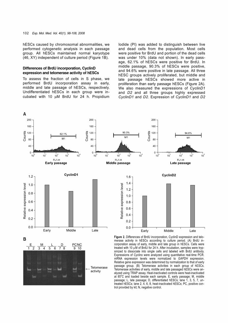
Differences of BrdU incorporation, CyclinD expression and telomerase activity of hESCs
To assess the fraction of cells in S phase, we performed BrdU incorporation assay in early, middle and late passage of hESCs, respectively. Undifferentiated hESCs in each group were in-cubated with 10 µM BrdU for 24 h. Propidium
Iodide (PI) was added to distinguish between live and dead cells from the population. Most cells were positive for BrdU and portion of the dead cells was under 10% (data not shown). In early pass-age, 62.1% of hESCs were positive for BrdU. In middle passage, 90.3% of hESCs were positive, and 94.6% were positive in late passage. All three hESC groups actively proliferated, but middle and late passage hESCs showed more active in proliferation than early passage hESCs (Figure 2A). We also measured the expressions of CyclinD1 and D2 and all three groups highly expressed CyclinD1 and D2. Expression of CyclinD1 and D2
Figure 2. Differences of BrdU incorporation, CyclinD expression and telo-merase activity in hESCs according to culture period. (A) BrdU in-corporation assay of early, middle and late group in hESCs. Cells were treated with 10 µM of BrdU for 24 h. After incubation, samples were tryp-sinized to dissociate into single cells and labeled with BrdU antibody. Expressions of Cyclins were analyzed using quantitative real-time PCR. mRNA expression levels were normalized to GAPDH expression. Relative gene expression was determined by normalization to that of early passage group. (B) Telomerase activities in each group of hESCs. Telomerase activities of early, middle and late passaged hESCs were an-alyzed using TRAP assay. Heat-inactivated controls were heat-inactivated at 85oC and loaded beside each sample. E, early passage; M, middle passage; L, late passage; D, differentiated hESCs; lane 1, 3, 5, 7, un-treated hESCs; lane 2, 4, 6, 8, heat-inactivated hESCs; PC, positive con-trol provided by kit; N, negative control.

Proliferative and differentiation potentials of hESCs 103
Figure 3. FACS analysis of undifferentiated markers, Oct4 and SSEA4, in the early, middle and late passage of undifferentiated hESCs.
was slightly increased in late passage compared to early passage, however, the expression level of CyclinD2 in middle and late passage hESCs was not significantly different. These results demon-strate that proliferation rate of hESCs were ac-celerated as passage number increased. However, telomerase activities were high both in three groups of hESCs and differentiated hESCs (Figure 2B).
Changes in differentiation potential of hESCs
To compare differentiation potential according to extended culture period, we firstly evaluated the expression of undifferentiated state markers in each group using FACS, RT-PCR and immuno-staining. FACS analysis results indicated that a high proportion of undifferentiated hESCs in all three groups commonly expressed undifferentiated state markers, Oct4 and SSEA4. All three groups expressed high levels of undifferentiated state markers, but differences in positive population bet-ween them were not apparent. Expression of Oct4 and SSEA4 were 98.52% and 98.76% in the early passage group, 88.79% and 98.44% in the middle passage group, 90.05% and 99.54% in the late passage group, respectively (Figure 3). Secondly, to evaluate the changes of differentiation potential
during differentiation, each group of hESCs was spontaneously differentiated as follows: EBs were made from each passage of hESCs and reattached to induce further differentiation. EBs classified into simple and cystic forms by morphological ap-pearance. All three groups of hESCs generated EB efficiently, although more cystic EBs were pro-duced from early and middle passage hESCs than late passage, and the majority of EBs from late hESCs were simple EBs (Figure 4A-C). The ex-pression of undifferentiated state markers Oct4 and Nanog was analyzed by RT-PCR. No sig-nificant differences in the expressions of Oct4 and Nanog were observed in all three groups of undifferentiated hESCs. In EBs and reattached cells, the expression levels of Oct4 and Nanog were higher in late passage than in the early and middle passage (Figure 5A). However, Oct4 and Nanog were all highly expressed in simple EBs, but their expression reduced in cystic EBs (Figure 5B). These mean that cystic EBs possessed more differentiated proportion of cells than simple EBs. Also, we demonstrate that Oct4 and Nanog are highly expressed in long-term cultured hESCs though the cells were differentiated spontaneously. These findings suggest that Oct4 and Nanog expression are maintained by a low incidence of

104 Exp. Mol. Med. Vol. 40(1), 98-108, 2008
Figure 4. Morphologies of EBs andreattached cells of EBs from early(A, D), middle (B, E), late (C, F)passage groups. Upper panel(A-C) showed EBs formed fromeach group and lower panel (D-F)showed reattached cells of EBsfrom each group.
Figure 5. RT-PCR for the expressions of undifferentiated state markersfrom each group of hESCs and EBs. (A) Expression levels of Oct4 andNanog in human embryonic stem cells (ES), embryoid bodies (EB)and reattached embryoid bodies (EB-AD). (B) Expression levels ofOct4 and Nanog in simple and cystic EBs from each passage group.E, early passage; M, middle passage; L, late passage; -RT, PCR with-out addition of reverse transcriptase.
spontaneous differentiation and that hESCs have a tendency to proliferate rather than differentiate on prolonged culture. As the spontaneous differentiation progressed, in early group, reattached cells from EBs were dif-ferentiated into various cell types including poly-gonal cells (Figure 4D). Reattached cells from middle group contained a small proportion of undif-ferentiated cells (Figure 4E). However, reattached cells from the late group showed a higher propor-tion of undifferentiated cells than those from the early and middle groups (Figure 4F). To confirm these findings, we evaluated the expression of
Oct4 and SSEA4 as undifferentiated state markers by immunostaining. Reattached cells from the early, middle and late group of EBs still expressed Oct4 and SSEA4 in spontaneously differentiated cells, but their expression was significantly reduced in the early group, not in the late group (Figure 6). In addition, we evaluated the expressions of Nestin, Brachyury, HNF3β as differentiated hESCs markers by immunostaining, which represent ecto-derm, mesoderm, endoderm, respectively. All three germ layer markers were expressed in all passage groups. But the expressions were significantly re-duced in late group (Figure 7A). Also RT-PCR analysis showed similar expression pattern to that of immunostaining (Figure 7B).
Discussion
Since the derivation of hESCs (Thomson et al., 1998; Reubinoff et al., 2000; Oh et al., 2005a), successful differentiation studies of hESCs into various cell types were reported (Rambhatla et al., 2003; Snir et al., 2003; Segev et al., 2004; Kang et al., 2006). Many studies continue on subjects as diverse as developmental regulation and determi-nation of the characteristics and development of cell lines containing genetic modifications that are suitable for cell-based therapy (Gropp et al., 2003; Ma et al., 2003). hESCs generally maintain their novel charac-teristics, such as expression of undifferentiated cell surface antigens, transcription factors and telo-merase activity after continuous long-term culture or even after cryopreservation (Ha et al., 2005), but some alterations in characteristics, including pro-liferation rate and differentiation potential, are

Proliferative and differentiation potentials of hESCs 105
Figure 6. Immunostaining of un-differentiated state markers in re-attached EBs from each group ofhESCs. Expressions of Oct4 andSSEA4 in reattached EBs were ob-served by confocal microscope.
Figure 7. Immunostaining and RT-PCR of three germ layer markers inreattached EBs from each group ofhESCs. (A) Expressions of Nestin,Brachyury, HNF3β in reattachedEBs were observed by confocalmicroscope. (B) Expressions ofNestin, Brachyury, HNF3β in re-attached EBs were analyzed byRT-PCR. Expression level of threegerm layer markers were detectedin reattached EBs from each groupof hESCs. E, early passage; M,middle passage; L, late passage;-RT, PCR without addition of re-verse transcriptase.

106 Exp. Mol. Med. Vol. 40(1), 98-108, 2008
appeared. In previous reports, researchers have demonstrated that maintaining pluripotent state in long-term culture may lead to alterations of stability of hESCs. Rosler et al. (2004) reported that a small proportion of long-term cultured cells could un-dergo genetic mutation and the mutated cells pro-bably possessed enhanced growth rate or proli-ferative ability. Moreover, adaptation to culture conditions shortens population-doubling time of pluripotent stem cells and these cells also respond more flexibly to environmental stimuli or stress than less adapted cells. These kinds of changes may be lead into epigenetic alterations in the cells. Several studies are ongoing to evaluate the epigenetic status of various hESC lines maintained under different culture conditions (Maitra et al., 2005; Rugg-Gunn et al., 2005). Gene profiling analysis is commonly used to analyze the variety of established hESCs. Maitra et al. (2005) reported an alteration in the cellular DNA of late passage hESCs. Their findings suggest that hESCs may be sensitive to culture conditions for maintaining pluripotency and that hESCs are epigenetically regulated by environmental factors. Also, Draper et al. (2004) reported chromosomal abnormality, namely additions to chromosome 17q and 12 in long-term cultured hESCs. Chromosome 17q is known to contain genes related to the regulation of apoptosis and differentiation (Burdon et al., 2002; Chiou et al., 2003) and chromosome 12 contains genes related to the regulation of pluripotency in human embryonic carcinoma cells (Mostert et al., 1998). Based on these reports, they concluded that the addition of chromosome 17q and 12 could be explained the adaptation, i.e. increased prolifera-tive ability of hESCs according to culture en-vironment (Draper et al., 2004). Recently, Baker et al. (2007) reported that genetic change of hESCs increased the capacity of cells to proliferate. They concluded that changes in hESCs in culture reflect tumorigenic events that occur in vivo, particulary in testicular germ cell tumors. In our laboratory, we observed changes of char-acteristics in long-term cultured hESCs. These alterations included changes in population-doubl-ing time, lower ratio of cystic EB formation, and reduced differentiation potential. To analyze the changes in the characteristics of long-term cultured hESCs, we evaluated different passage of cells and found that characteristics of hESCs were dependent on in vitro culture period. In the present study, we evaluated the changes in population- doubling time and the expression patterns of undifferentiated state markers in hESCs, EBs and differentiated cells according to in vitro culture period. Also, expressions of three germ layer
markers were evaluated with respect to differen-tiation potential. We measured population-doubling time and found that the mean value was 37.6 h in the early group and 30 h in the late group, which was about 8 h shorter than in the early group. This result in-dicates that characteristics of hESCs were possibly changed depending on culture period and that late passage hESCs appeared to be grown faster and adapted stably to culture environment than the early group. It was reported that pluripotent cells including mESCs (Savatier et al., 1994; Stead et al., 2002), and murine embryonal germ cells (Resnick et al., 1992) have a cell cycle structure lacking fully formed G1 and G2 gap phases. Recently, Becker et al. (2006) reported that hESCs showed unique cell cycle structure, which was shortened G1 cell cycle phase and most of cells were in S phase. In this study, we found that most of hESCs in each group were positive for BrdU and these results indicated that most of hESCs were in S phase. In early passage, 62.1% of hESCs were positive. In middle and late passage, 90.3% and 94.6% were positive. We also evaluated expression of CyclinD1 and D2 in each passage group. D type cyclins are known to promote G1 phase cell cycle regulation (Wianny et al., 1998, White and Dalton, 2005) and highly expressed in hESCs (Becker et al., 2006). CyclinDs were highly expressed in all three pass-age groups. From these results, long-term cultured cells have more proliferative abilities. Telomerase is a ribonucleoprotein that adds telo-mere repeats to chromosome ends and is re-sponsible for maintaining telomere length, which plays an important role in the replicative life span of cells (Harley, 1991). hESCs generally highly express human telomere reverse transcriptase (hTERT) activity (Thomson et al., 1998; Oh et al., 2005a). In the present study, all of three groups of hESCs expressed high levels of telomerase ac-tivity, and this activity was not reduced in dif-ferentiated cells. These results explain that all three groups have different proliferative activity, however, their telomerase activity was not affected by increasing passage number. All from early, middle and late passage of hESCs showed typical morphology of undifferentiated hESC colonies regardless of culture period. FACS analysis showed that the specific transcription factor Oct4 and the cell surface antigen SSEA4 were highly expressed in all three groups. In addition, different characteristics were observed in the EBs formed from the three groups. Conley et al. (2004) reported that mouse ESCs and hESCs spontaneously differentiated into sphere termed

Proliferative and differentiation potentials of hESCs 107
EBs when cultured in suspension. EBs are classi-fied into two types. Simple EBs is generally formed after 2-3 days and endoderm is formed outer layer of EBs. Cystic EBs are more differentiated struc-tures bearing central cavity and double layered structure with an inner ectodermal layer and outer layer of endoderm enclosing cavity (Keller, 1995). In this study, the early and middle passage groups formed simple and cystic EBs after sus-pension culture, whereas cystic EBs were rarely observed in the late passage group. RT-PCR results demonstrated that the expressions of Oct4 and Nanog in cystic EBs were low and slightly higher in those of middle passage group than in early passage group. However, the expressions of Oct4 and Nanog in simple EBs were equivalent in all three groups. This indicates that as passage number increased, the differentiation potential of hESCs was decreased. Immunostaining results showed that Oct4 and SSEA4 expression was significantly higher in the differentiated cells (i.e. EB reattached cells) produced from the hESCs of late passage group than those of early and middle passage group, although three germ layer mar-kers, Nestin (ectoderm), Brachyury (mesoderm) and HNF3β (endoderm), were expressed in all three groups. However, the expression levels of three germ layer markers were significantly re-duced in late passage. Ko et al. (2007) also reported that the degree of hESC differentiation varied depending on the passage number of undifferentiated hESCs in HSF-6 hES cell line. These results indicated that although long-term cultured hESCs retained somewhat their pluripo-tency, they also possessed a tendency to maintain an undifferentiated state rather than to differentiate. In conclusion, the present study demonstrates that long-term culture in vitro alters the proliferative ability and differentiation potential of hESCs and analyzed the alterations in hESCs of early, middle and late passage. It was found that hESCs have a tendency to a higher proliferative ability and a reduced differentiation potential on increasing the passage number. These results show that hESCs are sensitive to culture environment and charac-teristics may be altered by extended culture. Therefore, we suggest that short-term cultured hESCs are more appropriate for various re-searches of hESCs.
Acknowledgement
This research was supported by grants (SC 2040 and 1020) from Stem Cell Research Center of the 21st Century Frontier Research Program funded by the Ministry of Science and Technology, Korea.
References
Baker DE, Harrison NJ, Maltby E, Smith K, Moore HD, Shaw PJ, Heath PR, Holden H, Andrews PW. Adaptation to culture of human embryonic stem cells and oncogenesis in vivo. Nat Biotechnol 2007;25:207-15Becker KA, Ghule PN, Therrien JA, Lian JB, Stein KL, van Wijnen AJ, Stein GS. Self-renewal of human embryonic stem cells is supported by a shortened G1 cell cycle phase. J Cell Physiol 2006;209:883-93Burdon T, Smith A, Savatier P. Signaling, cell cycle and pluripotency in embryonic stem cells. Trends Cell Biol 2002;12:432-8 Chiou SK, Jones MK, Tarnawski AS. Surviving an anti- apoptosis protein: its biological roles and implications for cancer and beyond. Med Sci Monit 2003;9:PI25-9Conley BJ, Young JC, Trounson AO, Mollard R. Dervation, propagation and differentiation of human embryonic stem cells. Int J Biochem Cell Biol 2004;36:555-67Draper JS, Smith K, Gokhale P, Moore HD, Maltby E, Johnson J, Meisner L, Zwaka TP, Thomson J, Andrews PW. Recurrent gain of chromosomes 17q and 12 in cultured human embryonic stem cells. Nat Biotechnol 2004;22:53-4Dushnik-Levinson M, Benvenisty N. Embryogenesis in vitro: study of differentiation of embryonic stem cells. Biol Neonate 1995;67:77-83Evans MJ, Kaufman MH. Establishment in culture of pluripotential cells from mouse embryos. Nature 1981;292: 154-6Gropp M, Itsykson P, Singer O, Ben-Hur T, Reinhartz E, Galun E, Reubinoff BE. Stable genetic modification of human embryonic stem cells by lentiviral vectors. Mol Ther 2003; 7:281-7Ha SY, Jee BC, Suh CS, Kim HS, Oh SK, Kim SH, Moon SY. Cryopreservation of human embryonic stem cells without the use of a programmable freezer. Human Reproduction 2005;20:1779-85Harley CB. Telomere loss: mitotic clock or genetic time bomb? Mutat Res 1991;256:271-82Kang SM, Cho MS, Seo H, Yoon CJ, Oh SK, Choi YM, Kim DW. Efficient induction of oligodendrocytes from human embryonic stem cells. Stem Cells 2006;52:419-24Keller GM. In vitro differentiation of embryonic stem cells. Curr Opin Cell Biol 1995;7:862-9Ko JY, Park CH, Koh HC, Cho YH, Kyhm JH, Lim YS, Lee SH. Human embryonic stem cell-derived neural precursors as a continuous, stable, and on-demand source of human dopamine neurons. J Neurochem 2007;103:1417-29Longo L, Bygrave A, Grosveld FG, Pandolfi PP. The chromosome make-up of mouse embryonic stem cells is predictive of somatic and germ cell chimaerism. Transgenic Res 1997;6:321-8Ma Y, Ramezani A, Lewis R, Hawley RG, Thomson JA. High-level sustained transgene expression in human embryonic stem cells using lentiviral vectors. Stem Cells

108 Exp. Mol. Med. Vol. 40(1), 98-108, 2008
2003;21:111-7Maitra A, Arking DE, Shivapurkar N, Ikeda M, Stastny V, Kassauei K, Sui G, Cutler DJ, Liu Y, Brimble SN, Noaksson K, Hyllner J, Schulz TC, Zeng X, Freed WJ, Crook J, Abraham S, Colman A, Sartipy P, Matsui S, Carpenter M, Gazdar AF, Rao M, Chakravarti A. Genomic alterations in cultured human embryonic stem cells. Nature Genetics 2005;37: 1099-103Mostert MC, Verkerk AJ, van de Pol M, Heighway J, Marynen P, Rosenberg C, van Kessel AG, van Echten J, de Jong B, Oosterhuis JW, Looijenga LH. Identification of the critical region of 12p over-representation in testicular germ cell tumors of adolescents and adults. Oncogene 1998;16: 2617-27Oh SK, Kim HS, Ahn HJ, Seol HW, Kim YY, Park YB, Yoon CJ, Kim DW, Kim SH, Moon SY. Derivation and charac-terization of new human embryonic stem cell lines, SNUhES1, SNUhES2, and SNUhES3. Stem Cells 2005a; 23:211-9Oh SK, Kim HS, Park YB, Seol HW, Kim YY, Cho MS, Ku SY, Choi YM, Kim DW, Moon SY. Methods for expansion of human embryonic stem cells. Stem Cells 2005b;23:605-9Rambhatla L, Chiu CP, Kundu P, Peng Y, Carpenter MK. Generation of hepatocyte-like cells from human embryonic stem cells. Cell Transplant 2003;12:1-11Resnick JL, Bixler LS, Cheng L, Donovan PJ. Long-term proliferation of mouse primordial germ cells in culture. Nature 1992;10:87-95 Reubinoff BE, Pera MF, Fong CY, Trounson A, Bongso A. Embryonic stem cell lines from human blastocysts: somatic differentiation in vitro. Nat Biotechnol 2000;18:399-404Reubinoff BE, Pera MF, Vajta G, Trounson AO. Effective cryopreservation of human embryonic stem cells by the open pulled straw vitrification method. Human Reproduction 2001;16:2187-94Rosler ES, Fisk G, Ares X, Irving J, Miura T, Rao MS, Carpenter MK. Long-term culture of human embryonic stem cells in feeder-free conditions. Developmental Dynamics
2004;229:259-74Rugg-Gunn PJ, Ferguson-Smith AC, Pedersen RA. Epi-genetic status of human embryonic stem cells. Nat Genet 2005;37:585-7Savatier P, Huang S, Szekely L, Wiman KG, Samarut J. Contrasting patterns of retinoblastoma protein expression in mouse embryonic stem cells and embryonic fibroblasts. Oncogene 1994;9:809-18Segev H, Fishman B, Ziskind A, Shulman M, Itskovitz-Eldor J. Differentiation of human embryonic stem cells into insulin-producing clusters. Stem Cells 2004;22:265-74Seol HW, Oh SK, Kim HS, Yim MY, Baek JA, Suh J, Moon SY, Choi YM. A study of optimal methods for chromosome analysis of human embryonic stem cells. J Reproductive Medicine and Population 2007;20:86-93Smith AG. Embryo-derived stem cells: of mice and men. Annu Rev Cell Dev Biol 2001;17:435-62Snir M, Kehat I, Gepstein A, Coleman R, Itskovitz-Eldor J, Livne E, Gepstein L. Assessment of the ultrastructural and proliferative properties of human embryonic stem cell- derived cardiomyocytes. Am J Physiol Heart Circ Physiol 2003;285:H2355-63Stead E, White J, Faast R, Conn S, Goldstone S, Rathjen J, DhingraU, Rathjen P, Walker D, Dalton S. Pluripotent cell division cycles are driven by ectopic Cdk2, cyclin A/E and E2F activities. Oncogene 2002;21:8320-33Thomson JA, Itskovitz-Eldor J, Shapiro SS, Waknitz MA, Swiergiel JJ, Marshall VS, Jones JM. Embryonic stem cell lines derived from human blastocysts. Science 1998;282: 1145-7White J, Dalton S. Cell cycle control of embryonic stem cells. Stem Cell Reviews 2005;1:131-8 Wianny F, Real FX, Mummery CL, Van Rooijen M, Lahti J, Samarut J, Savatier P. G1-phase regulators, cyclin D1, cyclin D2, and cyclin D3: up-regulation at gastrulation and dynamic expression during neurulation. Dev Dyn 1998;212:49-62
Copyright © 2022 FDOKUMEN