Disulfide stress: a novel type of oxidative stress in acute pancreatitis
Upload
independentCategory
view
0download
0
Review ArticleTheScientificWorldJOURNAL (2011) 11, 1749–1761ISSN 1537-744X; doi:10.1100/2011/289182
Protein Disulfide Isomerase and Host-PathogenInteraction
Beatriz S. Stolf,1 Ioannis Smyrnias,2 Lucia R. Lopes,3 Alcione Vendramin,4 Hiro Goto,5
Francisco R. M. Laurindo,6 Ajay M. Shah,2 and Celio X. C. Santos2
1Department of Parasitology, University of São Paulo, São Paulo, SP, Brazil2Cardiovascular Division, British Heart Foundation Centre, King’s College London,125 Coldharbour Lane, London SE5 9NU, UK
3Department of Pharmacology, Institute of Biomedical Sciences, University ofSão Paulo, São Paulo, SP, Brazil
4Department of Parasitology, School of Medicine of Jundiaí, São Paulo, SP, Brazil5School of Medicine and Tropical Medicine Institute, University of São Paulo,São Paulo, SP, Brazil
6Vascular Biology Laboratory, Heart Institute (InCor), São Paulo, Brazil
Received 3 June 2011; Accepted 7 September 2011
Academic Editor: Mauro Perretti
Reactive oxygen species (ROS) production by immunological cells is known to cause damageto pathogens. Increasing evidence accumulated in the last decade has shown, however, thatROS (and redox signals) functionally regulate different cellular pathways in the host-pathogeninteraction. These especially affect (i) pathogen entry through protein redox switches andredox modification (i.e., intra- and interdisulfide and cysteine oxidation) and (ii) phagocytic ROSproduction via Nox family NADPH oxidase enzyme and the control of phagolysosome functionwith key implications for antigen processing. The protein disulfide isomerase (PDI) family of redoxchaperones is closely involved in both processes and is also implicated in protein unfolding andtrafficking across the endoplasmic reticulum (ER) and towards the cytosol, a thiol-based redoxlocus for antigen processing. Here, we summarise examples of the cellular association of hostPDI with different pathogens and explore the possible roles of pathogen PDIs in infection. A betterunderstanding of these complex regulatory steps will provide insightful information on the redoxrole and coevolutional biological process, and assist the development of more specific therapeuticstrategies in pathogen-mediated infections.
KEYWORDS: Host, pathogen, redox, endoplasmic reticulum, parasites, PDI, Nox
Correspondence should be addressed to Celio X. C. Santos, [email protected] © 2011 Beatriz S. Stolf et al. This is an open access article distributed under the Creative Commons Attribution License,which permits unrestricted use, distribution, and reproduction in any medium, provided the original work is properly cited.Published by TheScientificWorldJOURNAL; http://www.tswj.com/
TheScientificWorldJOURNAL (2011) 11, 1749–1761
1. INTRODUCTION
Host cells have the ability to cope with the progression and severity of infection in response to differenttypes of pathogen. On the other hand, numerous mechanisms have evolved that support the use of thehost cell machinery to facilitate pathogen survival and multiplication. Such co-evolutionary processesare directly affected by different physicochemical factors within different cell compartments, both in thehost and in pathogen. For instance, pH critically affects antigen stability of the influenza virus whichmodulates endosome acidity that attenuates its own infection [1]. ROS (and Reactive Nitrogen Species)production and the redox state of different cell compartments are also critically involved in cellular host-parasite interaction. Among the many redox sensitive proteins that are altered during the course of differentinfections, protein disulfide isomerase (PDI-) mediated redox switches have been associated with pathogenattachment-internalization, antigen processing in the ER/phagosome, and the regulation of ROS productionby Nox family enzymes. Thus, PDI emerges as a ubiquitous redox protein that regulates different steps ofdiverse infection processes. Several pathogens also have their own PDI that act as an important virulencefactor (Table 1). Other redox modifications directly mediated by ROS and especially via nitric oxide (NO)generated by inducible nitric oxide synthase (iNOS), which is abundant in phagocytic cells, have beenreviewed elsewhere [2, 3] and are not considered in this article. Below, the main cellular redox aspects ofhost and pathogen PDI will be discussed.
2. GENERAL ASPECTS OF PDI AND ITS ROLE IN HOST AND PATHOGEN
The ancient PDI is a ubiquitous redox chaperone belonging to the thioredoxin oxireductase super familyand can reduce (reaction 1), oxidize (reaction 2), and catalyse dithiol-disulfide exchange reactions (i.e.,isomerase activities, reaction 3, Figure 1). Such broad range of activities overlaps with the chaperone roleof PDI that overall performs a housekeeping function in helping to maintain proteins in a more stableconformation. There are around 20 PDI homologues, and the detailed structure and function of eukaryoticPDIs have been covered in recent excellent reviews [4, 5]. The classic mammalian PDI (55 kDa) has severaldomains ordered as a-b-b′-a′-c with 2 thioredoxin-like motifs (Trp-Cys-Gly-His-Cys) displayed in the aand a′ domain [4–6] (Figure 1). PDI is abundant in the ER (∼ 0.5mM) where the relatively oxidizingconditions at basal level (i.e., GSH/GSSG ratios ∼ 2-3 : 1) favours PDI isomerase/oxidase activity, whichis primarily involved in client protein redox folding (reaction 2-3, Figure 1). The oxidizing equivalents forthis process are driven mainly by the ER thiol-containing oxidase, Ero1 (endoplasmic reticulum oxidase-1), which binds FAD and is in turn re-oxidized via electron transfer to oxygen, generating H2O2 in theprocess [7–10]. The H2O2 destiny is elusive, but it can oxidize ER-located peroxiredoxin IV (PrxIV) thatis further reduced by PDI that is oxidized in the process [11]. This redox circuit is thought to increasetotal protein folding and thiol oxidation via Ero1 [11]. However, even in the absence of Ero1, proteinfolding still occurs, and it is suggested that other oxidases may compensate for redox demand in the ERin some circumstances [12, 13]. Nevertheless, the PDI-Ero1-dependent oxidative activity is balanced tocytosolic glutathione levels suggesting a functional redox interplay between these compartments [12]. PDIreductase activity has been primarily associated to more reducing compartments (i.e., GSH/GSSG ratios∼ 30–100 : 1), such as those in the vicinity of the plasma membrane [6, 10]. PDI redox versatility is mainlygoverned by the low pKa of the proximal cysteine on the active N-terminal a domain. Indeed, the lowerpKa of 4.5 renders PDI a much better oxidase than thioredoxin, which has a pKa of 7.1 and is mainly areductase in most neutral pH cell compartments. It should also be noted that PDI functions as a chaperoneindependently of its redox-active domains as especially required for its ATPase and Ca2+ activity, althoughPDI redox motifs still stabilize binding interaction [4, 5, 10]. In the ER, PDI is tightly associated withprolyl-4 hydroxylase (the rate-limiting enzyme for collagen biosynthesis), the Sec61 translocon, and theMHC class I complex (see later). It can be also found as a heterodimer with microsomal triglyceride transferprotein [6, 10]. PDI is a soluble homodimer and does not have a transmembrane domain and, similarly toother ER chaperones, carries the KDEL C-terminal sequence which binds to respective receptors in the COP
1750
TheScientificWorldJOURNAL (2011) 11, 1749–1761
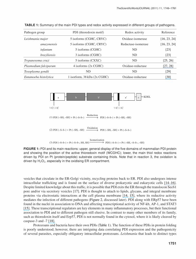
TABLE 1: Summary of the main PDI types and redox activity expressed in different groups of pathogens.
Pathogen group PDI (thioredoxin motif) Redox activity Reference
Leishmania major 5 isoforms (CGHC, CRYC) Oxidase-isomerase [16, 23, 24]
amazonensis 5 isoforms (CGHC, CRYC) Reductase-isomerase [16, 23, 24]
infantum 5 isoforms (CGHC) ND [23]
braziliensis 3 isoforms (CGHC) ND [23]
Trypanosoma cruzi 5 isoforms (CXXC) ND [25, 26]
Plasmodium falciparum 4 isoforms (2x CGHC) Oxidase-reductase [27, 28]
Toxoplasma gondii ND ND [29]
Entamoeba histolytica 1 isorform, 38 kDa (2x CGHC) Oxidase-reductase [30]
a b KDEL
(1)
(2)
(3)
Reduction
Oxidation
Isomerization
cab
PDI (–S–S–)
(–S–S–)
+ Pt (–SH, –SH)
PDI (–S–S–) + Pt (–SH, –SH) (–SH, –SH)
PDI (–S–S–) + Pt (–S–S–, SH, SH)
PDI + Pt
(–S–S–)(–SH, –SH)PDI + Pt
PDI (–S–S–) + Pt ( , –S–S–, –SH–SH )
FIGURE 1: PDI and its main reactions: upper, general display of the five domains of mammalian PDI proteinand showing the position of the active thioredoxin motif (WCGHC); lower, the main thiol redox reactionsdriven by PDI on Pt (protein/peptide) substrate containing thiols. Note that in reaction 3, the oxidation isdriven by H2O2, especially in the oxidising ER compartment.
vesicles that circulate in the ER-Golgi vicinity, recycling proteins back to ER. PDI also undergoes intenseintracellular trafficking and is found on the surface of diverse prokaryotic and eukaryotic cells [14–16].Despite limited knowledge about this traffic, it is possible that PDI exits the ER through the translocon Sec61pore and/or via secretory vesicles [17]. PDI is thought to attach to lipids, glycans, and integral membraneproteins via electrostatic interactions at the cell plasma membrane [14, 15], where its reductive activitymediates the infection of different pathogens (Figure 2, discussed later). PDI along with ERp57 have beenfound in the nuclei in association to DNA and affecting transcriptional activity of NF-kb, AP-1, and STAT3[15]. These transcriptional regulators are key elements in many inflammatory processes, but their functionalassociation to PDI and to different pathogen still elusive. In contrast to many other members of its family,such as thioredoxin itself and Erp57, PDI is not normally found in the cytosol, where it is likely cleaved bycaspase-3 and -7 [18].
Protozoans and bacteria have their own PDIs (Table 1). The function of these PDIs in protein foldingis poorly understood; however, there are intriguing data correlating PDI expression and the pathogenicityof several parasites, especially obligatory intracellular protozoans. Leishmania that leads to distinct types
1751

TheScientificWorldJOURNAL (2011) 11, 1749–1761
Nox2
TAP
Nucleus
Mitochondria
Endocyticvesicles
Lysosome
Phago-lysosome
Proteasome
ER
ab
ab
Plasma membrane
PDI
PDI
PDI
PDI
Class IIMHC
Class IMHC
Nox2
Class IMHC
Nox2
FIGURE 2: Main cellular routes involved in host mammalian PDI and pathogens.
of leishmaniases is a well-known example. The disease affects millions of individuals worldwide, andmore recently this parasite emerged as an important opportunistic infection among patients with HIV [19].There is a large variety of Leishmania species that results in different infection manifestation, generallyclassified as cutaneous (L. major, L. mexicana, L. amazonensis, L. braziliensis, etc.), diffuse cutaneous (L.amazonensis), mucocutaneous (L. braziliensis), and visceral (L. donovani, L. chagasi, and/or L. infantum)[20]. If L. chagasi is a subgroup of L. infantum brought to America by European colonist or other speciein its own still a matter of controversy (see discussion in [21]). The infection cycle in the vertebrate hostand is initiated when Leishmania promastigote is injected into the skin by the insect vector. In the host,the promastigote is phagocytised especially by macrophages, and further it is converted into intracellularamastigote. Amastigote replicates inside the phagosome within the cell and is liberated after the cell lyses,subsequently infecting other cells resulting in the progression to disease [21, 22]. L. amazonensis has at leastfour PDIs, and the use of specific PDI inhibitors substantially affected parasite growth [23]. In L. major,the increased levels of Leishmania PDI (LmPDI) expression and secretion at the parasite surface reflectsoptimal protein folding balanced to parasite multiplication. Importantly, this is correlated to high virulenceof the parasite strains [16]. More recently, the use of LmPDI antigens to generate a vaccine for L. majorpartially protected BALB/c animals and accelerated the cure of different strains of mice [24]. Similarlyto Leishmania species, other parasites of the trypanosomatid group such as Trypanosoma contain severalgenes predicted to encode for PDIs, that can execute N-glycosylation and protein folding in the ER [25, 26].Although PDI was considered essential for T. brucei survival, PDI activity was not essential for the growth oftrypanosomes in vitro [26]. PDI is also expressed in different species of Plasmodium protozoans (Table 1),the parasites that cause malaria [27, 28]. P. falciparum expresses at least nine different PDIs and the PfPDI-8 has great similarity to the prototype PDI and is expressed during all stages of parasite life cycle. This PDIfacilitates the disulfide-dependent conformational folding of EBA-175 protein, an emerging candidate forthe development of malaria vaccines [28]. This is intriguing given that malaria parasites express proteinswith high content of cysteine, which are associated to parasite invasion and sequestration in the vertebratehost and transmission into mosquito host [28]. Finally, Toxoplasma gondii PDI was identified in host tears,suggesting an extracellular location and adhesion to host cells during the initial phase of infection [29].
1752

TheScientificWorldJOURNAL (2011) 11, 1749–1761
3. PDI AND HOST-PATHOGEN INTERACTION
Antigen presentation occur through two distinct pathways. Antigen presenting cells (APCs; especiallymacrophages and dendritic cells; DCs) are long-lived cells that capture antigens and subsequently processand present them at the cell surface, where they are recognized by T-lymphocytes. This process provides along-term adaptive immune response to fungi, bacteria, and parasite. After internalization by the APC,antigens pass through phagosome/lysosome vesicles, where they form complexes with MHC class II(Figure 2), which are recognized by helper CD4+ T lymphocytes (exogenous pathway). In contrast, selfcell antigens and virus synthesized within cells (mostly non-APCs) are degraded by the proteasome inthe cytosol and nucleus. In successive steps, the antigen is processed, folded, and incorporated into theMHC class I (Figure 2) and the complex exposed on the cell surface, and recognized by cytotoxic CD8+ Tlymphocytes (endogenous pathway). These two pathways overlap and some antigens are presented by bothMHC class I and II, in a process called cross-presentation. This has been described in DCs respondingto viral infection, transplant rejection, and some autoimmune diseases and cancer. Moreover, a widerange of pathogens passing or living in the phagosome such as Mycobacterium tuberculosis, Salmonellatyphimurium, Toxoplasma gondii, and especially Leishmania spp and Trypanosoma cruzi are all cross-presented in association to high levels of CD8+ T cells [31]. PDI as part of the ER protein folding machinerydirectly regulates antigen processing of the MHC class I complex [32–35]. Antigens that are degraded bypeptidases and proteasome to shorter peptides in the cytosol and nucleus can be further transported to theER through the TAP system, a transmembrane ER type of ATP-binding cassette (ABC) peptide transporterfamily [36]. ER-located PDI interacts with the peptide-loading complex (PCL) that efficiently promotespeptide assembly with MHC class I molecules and supporting the exit of the peptide-antigen complexfrom the ER [32–34]. Other PCL components include calreticulin, tapasin, ERp57 (another PDI familymember), and the TAP transporter itself. Cells lacking PDI present much less peptide loading to MHCclass I and the disulfide bridge between the peptide and MHC groove remains in a reduced redox state[32]. Normally, this interaction is affected by the redox exchange between PDI (predominantly oxidized)and ERp57 (predominantly reduced) [32], a condition in which PDI favours the release of peptide-MHCclass I from the PCL and the antigen-MHCI complex is exited from ER [34]. In fact, PDI-bound peptidefacilitates the disassembly of the tapasin-ERp57 complex while the PDI unbound to the complex is unableto interact with tapasin-ERp57, retaining MHC I molecules in the ER [34]. Overall, PDI redox activitymodulates the stability of the antigen peptide-MHC class I complex and further determines the transportof the complex to the plasma membrane [32–35]. These redox effects may vary according to the typeof antigen and some pathogens interfere with this pathway to escape antigen process and evading CD8+T-cells recognition. This is the case for US3 protein from human cytomegalovirus, which enhances PDIdegradation via the proteasome [32]. PDI participation in immune response, however, goes beyond its rolein the ER protein folding machinery and it acts at other cellular steps of host-pathogen interaction. PDI inthe ER is also thought to play a role in parasite phagocytosis, and the PDI displayed on the cell surface canmediate the entry of some viral, bacterial, and protozoan. PDI is also implicated in protein unfolding andtrafficking of some pathogenic antigens across the endoplasmic reticulum and towards the cytosol by theendoplasmic reticulum-associated degradation system (ERAD). This is the main pathway where proteinsare retrotranslocated from the ER to cytosol and further degradated by the proteasome. Next, we discusssome examples of the cellular association between host PDI and different pathogens.
3.1. PDI and the Phagocytic Process
Phagocytosis is the main gate for large microbes to enter into APCs. After binding and attaching tothe pathogen, these cells can internalize organisms and large particles even bigger then their own size,which are then phagocytosed in an active process that involves intense membrane remodelling [37].Proteomics studies accumulated over the last decade revealed the presence of ER chaperones in the isolatedphagosome, uncovering a process called ER-mediated phagocytosis [38–45]. ER chaperones were detected
1753
TheScientificWorldJOURNAL (2011) 11, 1749–1761
in phagosomes of macrophages exposed to different particulate material and pathogens, including latex-beads opsonised or not with immunoglobulin G (IgG) or mouse serum (to facilitate entry through the FcR orcomplement receptors), IgG-opsonized erythrocytes, promastigotes of Leishmania donovani derived fromwild-type cells or cell-surface LPG knockout, among other parasites [38]. A mix of ER and endocyticvesicles in the formation of the parasitophorous vacuoles (PVs) during the uptake of different Leishmaniaspp. was recently shown in macrophages overexpressing ER-tagged green fluorescent protein [41]. Thepresence of the ER proteins Sec61, Bip/Grp78, and PDI in the phagosome of APCs [38, 41] support theidea that the ER provides the necessary machinery for antigen translocation from the phagosome to thecytoplasm and thus, possibly converges MHC class I and class II antigen cross-presentation [42] (Figure 2).There are several other complementary hypotheses on how peptides cross from phagosomes to cytoplasmduring the cross-presentation process [43, 44]. Neutrophils are short-lived cells (half-life of 4–8 h in thehuman circulation) and very active in the phagocytosis of large microbes such as bacteria, parasites, andfungus. Contrary to APCs, neutrophils only contain restricted amounts of ER machinery and are thought tolack the ER-mediated phagocytosis process [38].
Whether ER proteins functionally operate on phagocytosis-mediated infection has not been wellcharacterised yet. An important work has shown that Dictyostelium lacking both ER calreticulin andcalnexin present altered phagocytic cup formation and substantial decline in phagocytosis [45]. These twoproteins utilise Ca, and their disruption per se affects actin filaments and plasma membrane remodellingduring phagocytosis [45]. We recently showed that PDI is critically involved in Leishmania parasiteinfection in vitro [22]. We showed that phagocytosis of promastigotes (but not amastigotes) of Leishmaniachagasi was significantly inhibited by macrophage incubation with the thiol/PDI inhibitors DTNB,bacitracin, phenylarsine oxide, and neutralizing PDI antibody in a parasite redox-dependent way [22]. Thephenylarsine response is of particular interest, since this arsenic compound may act similarly to antimonials,widely used in leishmaniasis chemotherapy [46, 47]. PDI preferentially affects parasite internalizationand the phagocytosis of the promastigote forms is increased when wild-type PDI is overexpressed inmacrophages, an effect opposed by PDI knockdown. At later stages of infection (i.e., after 4 h), PDI frompromastigote-infected J774 macrophages was immunoprecipitated and subsequently blotted with an anti-Leishmania antibody revealing a parasite band at ∼ 94KDa [16, Figure 10(b), lane 5]. Subsequent removaland analysis of this band by mass fingerprint spectrometry showed a 58% match with elongation factor2 (EF2) of L. major (Q4Q259; data not shown). The incubation of purified bovine PDI (Sigma, P3818)and parasites did not yield any detectable protein complexes, suggesting that the macrophage milieu maybe important to sustain PDI-EF2 association [22]. Interestingly, Leishmania EF2 has important virulentfeatures and acts as a soluble antigen in lymphocyte stimulation in vitro [48] and in vivo [49]. Moreover,proteomics studies revealed that EF2 is secreted during promastigote differentiation into the amastigotestage with potential immunomodulatory proprieties in animal models [50]. Leishmania EF2 is thereforeof particular interest for Leishmania therapeutic interventions such as vaccines. Although our studies didnot address the role of the ER in mediating phagocytosis, these data provide compelling evidence fora functional role of ER-PDI in a host-parasite interaction. Other mechanisms underlining PDI-mediatedL. chagasi promastigote phagocytosis involves its association to ROS production by phagocyte NADPHoxidase and this is discussed next.
3.2. PDI and NADPH Oxidase Regulation
The NADPH oxidase (Nox) family of enzymes uses NADPH as an electron donor to convert oxygen tosuperoxide anion (O2
•−), a precursor of H2O2 and other powerful oxidants such as hydroxyl radical andperoxynitrite (in the presence of nitric oxide), collectively called ROS [3, 51, 52]. Each of the seven oxidasefamily members is characterized by a distinct catalytic subunit (i.e., Nox1-5 and Duox1-2), and has differingrequirements for additional protein subunits [51, 52]. The prototypic member of the Nox family, Nox2oxidase (or gp91phox oxidase), is best known for its role in neutrophil and macrophage phagocytosis. Geneticdefects in the enzyme are related to chronic granulomatous disease, a condition in which affected children
1754

TheScientificWorldJOURNAL (2011) 11, 1749–1761
suffer from recurrent severe fungal and bacterial infections due to defective phagocyte function [51]. EachNox isoform forms heterodimers with a lower molecular weight p22phox subunit and is predicted to bemembrane-bound. Nox2 is normally quiescent and acutely activated by agonists such as PMA, LPG, andcytokines in a tightly regulated process in which cytosolic subunits (p47phox, p67phox, p40phox, and Rac1in the case of macrophages and dendritic cells, or Rac2 in neutrophil) associate with the Nox2-p22phox
heterodimer to initiate enzyme activity [51, 52]. Nox2 also has electrogenic features [53] and in APC cellsis linked to the regulation of phagosome/lysosome pH and antigen processing [54, 55]. Usually, phagosomeacidity is maintained by a vacuolar ATPase (V-ATPase) that transports protons from the cytosol into thephagosome lumen, therefore regulating the function of lysosome proteases in the fused phagolysosomes.Savina et al. [56–58] have shown that Nox2-derived superoxide in the phagosomal vesicle promptlyconsumes protons maintaining a higher pH ambient in dendritic cells during particle internalization, whichfavours antigen processing and presentation [56–58]. Opposite results were found in macrophages [56–58].The selective role of Nox2 in different phagocytic cells remains to be defined. The jury is out on whether theresults shown in macrophages (association of Nox2 to Rab27a; a member of Rab family of GTPases) arerelated to vesicle traffic molecule assembly and quality, or rather associated to degradation processes [59].
Nox complex protein expression and function is greatly affected by redox compounds, and it isespecially regulated by PDI with implications for cell signalling [60]. The association of PDI to p22phox andother Nox isoforms in different cell types, especially in vascular cells, has been previously described [60–62]. A functional and spatial/physical interaction between PDI and the p22phox oxidase subunit was shownin macrophages [22] and more recently between PDI and p47phox in neutrophils [62]. In macrophages,PDI-Nox association was correlated to Leishmania infection in vitro [22]. It is well known that duringphagocytosis of Leishmania, Nox2 is activated and parasite uptake is inhibited by antioxidants such ascatalase [22]. Intriguingly, in the course of promastigote infection, some parasites evade that stressfulcondition and convert themselves into intracellular amastigotes, multiplying and resulting in progression toa disease process. Overall, our studies support the view that parasite phagocytosis/infection by macrophagesis a redox process mediated by PDI in at least two ways. Initially, PDI-NADPH oxidase increases ROSproduction generating an oxidizing milieu, which seems to favour promastigote infection. The downstreamrole of ROS generated by PDI-NADPH oxidase remains unknown but can be related to the unfolded proteinresponse signalling [63] or, similar to PDI-Ero1, to protein folding in the macrophage ER compartmentwith key implications for antigen processing. Nevertheless, at later stages of infection, macrophage PDIphysically associates with Leishmania elongation factor-2 (as discussed earlier).
3.3. PDI Role on Cell Surface and Pathogen Attachment
Some viruses envelop their genetic material within a protein-coated capsid in a further lipid membranelayout, for example, influenza virus, baculovirus, hepatitis-C, HIV, and Herpes virus. These envelopedparticles require successive steps to successfully entry and infect host cells. They usually first attach ontohost receptors (and attachment factors), and their membranes fuse to interact with endosome vesicles thattraffic the virus toward the endoplasmic reticulum, where it is uncoated. The proteins are finally transportedto the cytosol and nucleus [64] (Figure 2). There is convincing evidence showing that most viral infectionsare strongly influenced by changes in the redox environment and that host PDI mediates infection ofenveloped viruses [65–70].
In the course of HIV infection, the virus first binds to attachment factors, for example, mannosebinding C-type lectin receptor and intracellular adhesion molecule (ICAM-3) on the surface of host CD4+T cells. The glycoprotein 120 (gp120) subunit of the virus envelope binds to immunoglobulin G ofCD4+ and undergoes conformational changes, allowing the virus to interact with its coreceptors, CXCR4or CCR5. These interactions favour downstream conversions of gp41 envelope subunit to a competentfusion conformation. Initial studies showed that membrane-impermeable PDI inhibitors and monoclonalantibodies against PDI prevent HIV-1 infection [65]. It was then revealed that the domain D2 of the CD4 hasredox-active disulfide bonds and is regulated by thioredoxin [66]. Using membrane-impermeable reducing
1755
TheScientificWorldJOURNAL (2011) 11, 1749–1761
agents (especially arsenical-derived compounds) and labelling thiol reagents, it was demonstrated thatCD4+ reactive thiols critically drive HIV entry into cells [66]. Work from another group also revealed thatPDI, on the surface of HIV-1 target cells, reduces disulfide bonds of the recombinant envelope glycoproteingp120 (reaction 1, Figure 1), a reaction prevented by the usual PDI inhibitors [67]. Intriguingly, PDIsilencing in U373 and HeLa cells had little impact on HIV infection itself as compared to the effect mediatedby general thiol inhibitors [68]. The reasons for this discrepancy remain to be elucidated and raise thequestion whether the reductive effect of PDI is coupled to other redox proteins (e.g., thioredoxin or Nox’s)that could amplify virus-CD4 redox association in some cells. It is noteworthy that in these later studies, PDIknockdown on the cell surface was not evident as compared to massive loss of most PDIs within the ER;an observation that supports the idea that PDI in the ER has little impact in HIV-mediated infection [68].Thiol inhibitors also affect viral fusion as that mediated by the fusion (F) protein from the ParamyxovirusNewcastle disease virus [69]. The overexpression of PDI family members PFDI and ERdj5 has also beenshown to significantly catalyze the reduction of thiols in F protein, facilitating membrane fusion [70]. Thereis evidence suggesting a possible association between PDI and infection mediated by some members of theof Herpesviridae viruses family [71].
PDI is also implicated in the attachment of some bacteria from different species of Chlamydia [72–74]. Chlamydia is an obligatory intracellular pathogen that causes diverse diseases in humans. The mostcommon species are Chlamydia trachomatis, which is sexually transmitted and can cause blindness andinfertility, and C. pneumoniae, which affects the respiratory tract. CHO cells have impaired endogenous PDIexpression due to a defect in truncated mRNA processing, thus providing a valuable model to understandthe effect of PDI-mediated cell-cell interaction and infection. These cells are very resistant to Chlamydiainfection showing impaired attachment, an effect restored by ectopic expression of PDI [73]. Similar to HIVinfection, the molecular mechanisms most likely include the reductive activity of PDI (reaction 1, Figure 1)on the surface of CHO cells [72].
3.4. PDI and Antigen Translocation from ER to Cytosol
Crossing the endoplasmic reticulum (ER) membrane is an irreversible process for most proteins. In somecases, however, this flow is reversed and misfolded proteins retained in the ER are retrotranslocatedto the cytosol via ERAD to be degraded by the proteasome. This pathway is also exploited by smallpathogens, especially non-enveloped viruses and some bacterial toxins, to gain access to the cytosol. Inthese cases, antigenic particles that reach the ER by different means suffer molecular redox rearrangementsand binding to PDI allowing them to be transported back to the cytosol or nucleus. Well-known examplesare infections mediated by Polyomaviruses (Py) and Simian virus 40 (SV40) extensively studied in thefield of carcinogenesis. After SV40 interaction with the GM1 receptor on the cell surface, the particleenters the host cell through endocytosis and traffics via the caveosome (a particular caveolin containingendosome with neutral pH) towards the ER compartment [75, 76]. SV40-coated pentamers are linked toeach other by disulfide bonds between cysteine 104 (C104). Further isomerisation in the ER is crucialfor the viral uncoating process. In vitro cell screening shows that among all ER-resident proteins, PDI andERp57 more specifically regulate SV40 infection [75]. PDI silencing substantially decreases SV40 infectionthat is also dependent on some redox sensitive cysteines on the viral particle [75]. PDI cooperation with ER-associated ERAD proteins Derlin-1 and Sel1L is Ca dependent and facilitates SV40 traffic through ERAD[75]. A similar pathway is used by some nonobligatory intracellular bacteria that exert their effect throughproduction of potent endotoxins, such as diphtheria toxin (DT) and cholera toxin (CT). These proteinsfunction similarly to some plant toxins, such as ricin and abrin. Conversion into toxic proteins involvescleavage of their interchain disulfide bond, allowing them to traffic into the endocytic pathway within thehost cell [77, 78]. In humans, CT is derived from the Bacterium Vibrio cholerae that causes cholera diseaseand has 2 subunits (A1 and A2). The protein first attaches to the host cell surface via GM1 and the subunitA2, which contains a KDEL sequence, and is transported back to the ER (see earlier discussion). There,PDI reduces and unfolds A2 and A1 that exit the ER via the Sec61 channel into the cytosol. PDI in the
1756
TheScientificWorldJOURNAL (2011) 11, 1749–1761
reduced state (reaction 1, Figure 1) binds to the toxin and subsequent oxidation of PDI, probably via Ero1α,enables the release of CT toxin [79, 80]. The active polypeptide A1 efficiently modifies a heterotrimeric Gprotein in the cytosol that leads to massive loss of chlorine and water secretion by intestinal epithelial cellsin mammals, resulting in severe diarrhoea.
4. CONCLUSION
In this article we have reviewed the main cellular aspects of PDI-mediated host pathogen interactions and thepathways that are involved in viral, bacterial (including bacterial toxins), and parasitic infections. A numberof cellular mechanisms through which PDI modulates some specific cellular pathways in immune cellshave been described, such as redox-sensitive attachment, antigen presentation in the ER and exit from it,and association to phagosome and ROS production by NADPH oxidase (Figure 2). Many of these responsesare antigen-specific and the precise mechanisms of action remain to be fully elucidated, especially in thecontext of redox changes in cross-presentation phenomena. Moreover, little is known about the role ofPDI in infection per se, as well as how PDI signals to a more integrated cellular response to stress [63]. PDIglobal knockout mice are only viable until birth, but partial gene-modified mice and also modified pathogenswill help to reveal the significant redox role of PDI and its redox partners. Overall, PDI is a key regulatorthat may propagate or limit the severity of the infection processes, depending on the infectious organisminvolved. A better understanding of these complex regulatory steps will provide insightful information onthe redox role and coevolutional biological process, and assist the development of more specific therapeuticstrategies in pathogen-mediated infections.
ABBREVIATIONS
ER: Endoplasmic reticulumMHC: Major histocompatibility complexH2O2: Hydrogen peroxideERp57: A member of PDI family also know as glucose-regulated protein or 58-kD (GRP58)NF-kB: Factor nuclear kappa BAP-1: Activator protein 1STAT-3: Signal transducer and activator of transcription 3.
ACKNOWLEDGMENTS
The authors thank Daniel C. Pimenta for mass spectrometry experiments (Centre for Applied ToxinologyCAT/CEPID, Butantan Institute). They also thank Dr. Joao Wosniak Jr. from Vascular Biology Laboratory,Heart Institute (InCor) for helpful discussion. This paper was supported by Auxılio Fundacao de Amparoa Pesquisa do Estdado de Sao Paulo (FAPESP) and Conselho Nacional de Pesquisa (CNPq), Instituto doMilenio Redoxoma; Fundacao EJ Zerbini (InCor). A. M. Shah, S. Ioannis and C. X. C. Santos are also sup-ported by the British Heart Foundation and a Leducq Fondation Transatlantic Network of Excellence award.
REFERENCES
[1] E. Fenouillet, R. Barbouche, and I. M. Jones, “Cell entry by enveloped viruses: redox considerations for HIV andSARS-coronavirus,” Antioxidants & Redox Signaling, vol. 9, no. 8, pp. 1009–1034, 2007.
[2] C. F. Nathan and J. B. Hibbs, “Role of nitric oxide synthesis in macrophage antimicrobial activity,” CurrentOpinion in Immunology, vol. 3, no. 1, pp. 65–70, 1991.
[3] O. Augusto, M. G. Bonini, A. M. Amanso, E. Linares, C. C. X. Santos, and S. L. De Menezes, “Nitrogen dioxideand carbonate radical anion: two emerging radicals in biology,” Free Radical Biology and Medicine, vol. 32, no.9, pp. 841–859, 2002.
1757
TheScientificWorldJOURNAL (2011) 11, 1749–1761
[4] C. Appenzeller-Herzog and L. Ellgaard, “The human PDI family: versatility packed into a single fold,”Biochimica et Biophysica Acta, vol. 1783, no. 4, pp. 535–548, 2008.
[5] F. Hatahet and L. W. Ruddock, “Protein disulfide isomerase: a critical evaluation of its function in disulfide bondformation,” Antioxidants & Redox Signaling, vol. 11, no. 11, pp. 2807–2850, 2009.
[6] R. Noiva, “Protein disulfide isomerase: the multifunctional redox chaperone of the endoplasmic reticulum,”Seminars in Cell and Developmental Biology, vol. 10, no. 5, pp. 481–493, 1999.
[7] E. Gross, C. S. Sevier, N. Heldman et al., “Generating disulfides enzymatically: reaction products and electronacceptors of the endoplasmic reticulum thiol oxidase Ero1p,” Proceedings of the National Academy of Sciencesof the United States of America, vol. 103, no. 2, pp. 299–304, 2006.
[8] B. P. Tu and J. S. Weissman, “The FAD- and O2-dependent reaction cycle of Ero1-mediated oxidative proteinfolding in the endoplasmic reticulum,” Molecular Cell, vol. 10, no. 5, pp. 983–994, 2002.
[9] R. Xiao, B. Wilkinson, A. Solovyov et al., “The contributions of protein bisulfide isomerase and its homologuesto oxidative protein folding in the yeast endoplasmic reticulum,” Journal of Biological Chemistry, vol. 279, no.48, pp. 49780–49786, 2004.
[10] B. Wilkinson and H. F. Gilbert, “Protein disulfide isomerase,” Biochimica et Biophysica Acta, vol. 1699, no. 1-2,pp. 35–44, 2004.
[11] T. J. Tavender, J. J. Springate, and N. J. Bulleid, “Recycling of peroxiredoxin IV provides a novel pathway fordisulphide formation in the endoplasmic reticulum,” The EMBO Journal, vol. 29, no. 24, pp. 4185–4197, 2010.
[12] E. Zito, E. P. Melo, Y. Yang, A. Wahlander, T. A. Neubert, and D. Ron, “Oxidative protein folding by anendoplasmic reticulum-localized peroxiredoxin,” Molecular Cell, vol. 40, no. 5, pp. 787–797, 2010.
[13] E. Margittai and G. Banhegyi, “Oxidative folding in the endoplasmic reticulum: towards a multiple oxidanthypothesis?” FEBS Letters, vol. 584, no. 14, pp. 2995–2998, 2010.
[14] K. Terada, P. Manchikalapudi, R. Noiva, H. O. Jauregui, R. J. Stockert, and M. L. Schilsky, “Secretion, surfacelocalization, turnover, and steady state expression of protein disulfide isomerase in rat hepatocytes,” Journal ofBiological Chemistry, vol. 270, no. 35, pp. 20410–20416, 1995.
[15] C. Turano, S. Coppari, F. Altieri, and A. Ferraro, “Proteins of the PDI family: unpredicted non-ER locations andfunctions,” Journal of Cellular Physiology, vol. 193, no. 2, pp. 154–163, 2002.
[16] Y. Ben Achour, M. Chenik, H. Louzir, and K. Dellagi, “Identification of a disulfide isomerase protein ofLeishmania major as a putative virulence factor,” Infection and Immunity, vol. 70, no. 7, pp. 3576–3585, 2002.
[17] S. N. Molteni, A. Fassio, M. R. Ciriolo et al., “Glutathione limits Ero1-dependent oxidation in the endoplasmicreticulum,” Journal of Biological Chemistry, vol. 279, no. 31, pp. 32667–32673, 2004.
[18] K. S. Na, B. C. Park, M. Jang et al., “Protein disulfide isomerase is cleaved by caspase-3 and -7 during apoptosis,”Molecules and Cells, vol. 24, no. 2, pp. 261–267, 2007.
[19] M. P. Posada-Vergara, J. A. L. Lindoso, J. E. Tolezano, V. L. Pereira-Chioccola, M. V. Silva, and H. Goto,“Tegumentary leishmaniasis as a manifestation of immune reconstitution inflammatory syndrome in 2 patientswith AIDS,” Journal of Infectious Diseases, vol. 192, no. 10, pp. 1819–1822, 2005.
[20] M. J. McConville and E. Handman, “The molecular basis of Leishmania pathogenesis,” International Journal forParasitology, vol. 37, no. 10, pp. 1047–1051, 2007.
[21] J. J. Shaw, “Further thoughts on the use of the name Leishmania (Leishmania) infantum chagasi for theaetiological agent of American visceral leishmaniasis,” Memorias do Instituto Oswaldo Cruz, vol. 101, no. 5,pp. 577–579, 2006.
[22] C. X. C. Santos, B. S. Stolf, P. V. A. Takemoto et al., “Protein disulfide isomerase (PDI) associates with NADPHoxidase and is required for phagocytosis of Leishmania chagasi promastigotes by macrophages,” Journal ofLeukocyte Biology, vol. 86, no. 4, pp. 989–998, 2009.
[23] B. X. Hong and L. Soong, “Identification and enzymatic activities of four protein disulfide isomerase (PDI)isoforms of Leishmania amazonensis,” Parasitology Research, vol. 102, no. 3, pp. 437–446, 2008.
[24] F. Benhnini, M. Chenik, D. Laouini, H. Louzir, P. A. Cazenave, and K. Dellagi, “Comparative evaluation of twovaccine candidates against experimental leishmaniasis due to Leishmania major infection in four inbred mousestrains,” Clinical and Vaccine Immunology, vol. 16, no. 11, pp. 1529–1537, 2009.
[25] M. P. Hsu, M. L. Munich, and J. C. Boothroyd, “A developmentally regulated gene of trypanosomes encodes ahomologue of rat protein-disulfide isomerase and phosphoinositol-phospholipase C,” Biochemistry, vol. 28, no.15, pp. 6440–6446, 1989.
1758
TheScientificWorldJOURNAL (2011) 11, 1749–1761
[26] J. Rubotham, K. Woods, J. A. Garcia-Salcedo, E. Pays, and D. P. Nolan, “Characterization of two protein disulfideisomerases from the endocytic pathway of bloodstream forms of Trypanosoma brucei,” Journal of BiologicalChemistry, vol. 280, no. 11, pp. 10410–10418, 2005.
[27] I. Florenta, E. Mouray, F. Dali Ali et al., “Cloning of Plasmodium falciparum protein disulfideisomerase homologue by affinity purification using the antiplasmodial inhibitor 1,4-bis{3-[N-(cyclohexylmethyl)amino]propyl}piperazine,” FEBS Letters, vol. 484, no. 3, pp. 246–252, 2000.
[28] B. Mahajan, R. Noiva, A. Yadava et al., “Protein disulfide isomerase assisted protein folding in malaria parasites,”International Journal for Parasitology, vol. 36, no. 9, pp. 1037–1048, 2006.
[29] B. Meek, J. W. Back, V. N. A. Klaren, D. Speijer, and R. Peek, “Protein disulfide isomerase of Toxoplasma gondiiis targeted by mucosal IgA antibodies in humans,” FEBS Letters, vol. 522, no. 1–3, pp. 104–108, 2002.
[30] R. E. Mares, P. D. Magana, S. G. Melendez-Lopez, A. F. Licea, J. M. Cornejo-Bravo, and M. A. Ramos,“Oxidative folding and reductive activities of EhPDI, a protein disulfide isomerase from Entamoeba histolytica,”Parasitology International, vol. 58, no. 3, pp. 311–313, 2009.
[31] S. Bertholet, R. Goldszmid, A. Morrot et al., “Leishmania antigens are presented to CD8+ T cells by a transporterassociated with antigen processing-independent pathway in vitro and in vivo,” Journal of Immunology, vol. 177,no. 6, pp. 3525–3533, 2006.
[32] B. Park, S. Lee, E. Kim et al., “Redox regulation facilitates optimal peptide selection by MHC class I duringantigen processing,” Cell, vol. 127, no. 2, pp. 369–382, 2006.
[33] Y. Kim, K. Kang, I. Kim et al., “Molecular mechanisms of MHC class I-antigen processing: redoxconsiderations,” Antioxidants & Redox Signaling, vol. 11, no. 4, pp. 907–936, 2009.
[34] S. Lee, B. Park, K. Kang, and K. Ahn, “Redox-regulated export of the major histocompatibility complex classI-peptide complexes from the endoplasmic reticulum,” Molecular Biology of the Cell, vol. 20, no. 14, pp. 3285–3294, 2009.
[35] K. Kang, B. Park, C. Oh, K. Cho, and K. Ahn, “A role for protein disulfide isomerase in the early folding andassembly of MHC class I molecules,” Antioxidants & Redox Signaling, vol. 11, no. 10, pp. 2553–2561, 2009.
[36] R. Abele and R. Tampe, “Peptide trafficking and translocation across membranes in cellular signaling and self-defense strategies,” Current Opinion in Cell Biology, vol. 21, no. 4, pp. 508–515, 2009.
[37] M. Rabinovitch, “Professional and non-professional phagocytes: an introduction,” Trends in Cell Biology, vol. 5,no. 3, pp. 85–87, 1995.
[38] J. Garin, R. Diez, S. Kieffer et al., “The phagosome proteome: insight into phagosome functions,” Journal of CellBiology, vol. 152, no. 1, pp. 165–180, 2001.
[39] M. Desjardins, “ER-mediated phagocytosis: a new membrane for new functions,” Nature Reviews Immunology,vol. 3, no. 4, pp. 280–291, 2003.
[40] E. Gagnon, S. Duclos, C. Rondeau et al., “Endoplasmic reticulum-mediated phagocytosis is a mechanism of entryinto macrophages,” Cell, vol. 110, no. 1, pp. 119–131, 2002.
[41] B. Ndjamen, B. H. Kang, K. Hatsuzawa, and P. E. Kima, “Leishmania parasitophorous vacuoles interactcontinuously with the host cell’s endoplasmic reticulum; parasitophorous vacuoles are hybrid compartments,”Cellular Microbiology, vol. 12, no. 10, pp. 1480–1494, 2010.
[42] M. Houde, S. Bertholet, E. Gagnon et al., “Phagosomes are competent organelles for antigen cross-presentation,”Nature, vol. 425, no. 6956, pp. 402–406, 2003.
[43] J. M. Vyas, A. G. van der Veen, and H. L. Ploegh, “The known unknowns of antigen processing and presentation,”Nature Reviews Immunology, vol. 8, no. 8, pp. 607–618, 2008.
[44] N. Touret, P. Paroutis, M. Terebiznik et al., “Quantitative and dynamic assessment of the contribution of the ERto phagosome formation,” Cell, vol. 123, no. 1, pp. 157–170, 2005.
[45] A. Muller-Taubenberger, A. N. Lupas, H. Li, M. Ecke, E. Simmeth, and G. Gerisch, “Calreticulin and calnexin inthe endoplasmic reticulum are important for phagocytosis,” The EMBO Journal, vol. 20, no. 23, pp. 6772–6782,2001.
[46] Ashutosh, S. Sundar, and N. Goyal, “Molecular mechanisms of antimony resistance in Leishmania,” Journal ofMedical Microbiology, vol. 56, no. 2, pp. 143–153, 2007.
[47] S. Wyllie, M. L. Cunningham, and A. H. Fairlamb, “Dual action of antimonial drugs on thiol redox metabolism inthe human pathogen Leishmania donovani,” Journal of Biological Chemistry, vol. 279, no. 38, pp. 39925–39932,2004.
1759
TheScientificWorldJOURNAL (2011) 11, 1749–1761
[48] S. K. Gupta, B. S. Sisodia, S. Sinha et al., “Proteomic approach for identification and characterization of novelimmunostimulatory proteins from soluble antigens of Leishmania donovani promastigotes,” Proteomics, vol. 7,no. 5, pp. 816–823, 2007.
[49] P. Probst, E. Stromberg, H. W. Ghalib et al., “Identification and characterization of T cell-stimulating antigensfrom Leishmania by CD4 T cell expression cloning,” Journal of Immunology, vol. 166, no. 1, pp. 498–505, 2001.
[50] M. Chenik, S. Lakhal, N. Ben Khalef, L. Zribi, H. Louzir, and K. Dellagi, “Approaches for the identification ofpotential excreted/secreted proteins of Leishmania major parasites,” Parasitology, vol. 132, no. 4, pp. 493–509,2006.
[51] A. van der Vliet, “NADPH oxidases in lung biology and pathology: host defense enzymes, and more,” FreeRadical Biology and Medicine, vol. 44, no. 6, pp. 938–955, 2008.
[52] J. D. Lambeth, “NOX enzymes and the biology of reactive oxygen,” Nature Reviews Immunology, vol. 4, no. 3,pp. 181–189, 2004.
[53] L. M. Henderson, J. B. Chappell, and O. T. G. Jones, “The superoxide-generating NADPH oxidase of humanneutrophils is electrogenic and associated with an H+ channel,” Biochemical Journal, vol. 246, no. 2, pp. 325–329, 1987.
[54] A. R. Cross and A. W. Segal, “The NADPH oxidase of professional phagocytes—prototype of the NOX electrontransport chain systems,” Biochimica et Biophysica Acta, vol. 1657, no. 1, pp. 1–22, 2004.
[55] A. Savina and S. Amigorena, “Phagocytosis and antigen presentation in dendritic cells,” Immunological Reviews,vol. 219, no. 1, pp. 143–156, 2007.
[56] A. Savina, C. Jancic, S. Hugues et al., “NOX2 controls phagosomal pH to regulate antigen processing duringcrosspresentation by dendritic cells,” Cell, vol. 126, no. 1, pp. 205–218, 2006.
[57] C. Jancic, A. Savina, C. Wasmeier et al., “Rab27a regulates phagosomal pH and NADPH oxidase recruitment todendritic cell phagosomes,” Nature Cell Biology, vol. 9, no. 4, pp. 367–378, 2007.
[58] A. R. Mantegazza, A. Savina, M. Vermeulen et al., “NADPH oxidase controls phagosomal pH and antigen cross-presentation in human dendritic cells,” Blood, vol. 112, no. 12, pp. 4712–4722, 2008.
[59] J. Huang, V. Canadien, G. Y. Lam et al., “Activation of antibacterial autophagy by NADPH oxidases,”Proceedings of the National Academy of Sciences of the United States of America, vol. 106, no. 15, pp. 6226–6231, 2009.
[60] F. R. M. Laurindo, D. C. Fernandes, A. M. Amanso, L. R. Lopes, and C. X. C. Santos, “Novel role of proteindisulfide isomerase in the regulation of NADPH oxidase activity: pathophysiological implications in vasculardiseases,” Antioxidants & Redox Signaling, vol. 10, no. 6, pp. 1101–1113, 2008.
[61] M. Janiszewski, L. R. Lopes, A. O. Carmo et al., “Regulation of NAD(P)H oxidase by associated protein disulfideisomerase in vascular smooth muscle cells,” Journal of Biological Chemistry, vol. 280, no. 49, pp. 40813–40819,2005.
[62] A. M. de A. Paes, S. Verıssimo-Filho, L. L. Guimaraes et al., “Protein disulfide isomerase redox-dependentassociation with p47phox: evidence for an organizer role in leukocyte NADPH oxidase activation,” Journal ofLeukocyte Biology, vol. 90, no. 4, pp. 799–810, 2011.
[63] C. X. Santos, L. Y. Tanaka, J. Wosniak, and F. R. Laurindo, “Mechanisms and implications of reactive oxygenspecies generation during the unfolded protein response: roles of endoplasmic reticulum oxidoreductases,mitochondrial electron transport, and NADPH oxidase,” Antioxidants & Redox Signaling, vol. 11, no. 10, pp.2409–2427, 2009.
[64] A. E. Smith and A. Helenius, “How viruses enter animal cells,” Science, vol. 304, no. 5668, pp. 237–242, 2004.[65] H. J. P. Ryser, E. M. Levy, R. Mandel, and G. J. DiSciullo, “Inhibition of human immunodeficiency virus
infection by agents that interfere with thiol-disulfide interchange upon virus-receptor interaction,” Proceedingsof the National Academy of Sciences of the United States of America, vol. 91, no. 10, pp. 4559–4563, 1994.
[66] L. J. Matthias, P. T. W. Yam, X. M. Jiang et al., “Disulfide exchange in domain 2 of CD4 is required for entry ofHIV-I,” Nature Immunology, vol. 3, no. 8, pp. 727–732, 2002.
[67] A. Gallina, T. M. Hanley, R. Mandel et al., “Inhibitors of protein-disulfide isomerase prevent cleavage of disulfidebonds in receptor-bound glycoprotein 120 and prevent HIV-1 entry,” Journal of Biological Chemistry, vol. 277,no. 52, pp. 50579–50588, 2002.
[68] W. Ou and J. Silver, “Role of protein disulfide isomerase and other thiol-reactive proteins in HIV-1 envelopeprotein-mediated fusion,” Virology, vol. 350, no. 2, pp. 406–417, 2006.
1760
TheScientificWorldJOURNAL (2011) 11, 1749–1761
[69] S. Jain, L. W. McGinnes, and T. G. Morrison, “Thiol/disulfide exchange is required for membrane fusion directedby the Newcastle disease virus fusion protein,” Journal of Virology, vol. 81, no. 5, pp. 2328–2339, 2007.
[70] S. Jain, L. W. McGinnes, and T. G. Morrison, “Overexpression of thiol/disulfide isomerases enhances membranefusion directed by the newcastle disease virus fusion protein,” Journal of Virology, vol. 82, no. 24, pp. 12039–12048, 2008.
[71] H. O. Al-Barazi and A. M. Colberg-Poley, “The human cytomegalovirus UL37 immediate-early regulatoryprotein is an integral membrane N-glycoprotein which traffics through the endoplasmic reticulum and Golgiapparatus,” Journal of Virology, vol. 70, no. 10, pp. 7198–7208, 1996.
[72] S. Abromaitis and R. S. Stephens, “Attachment and entry of chlamydia have distinct requirements for host proteindisulfide isomerase,” PLoS Pathogens, vol. 5, no. 4, Article ID e1000357, 2009.
[73] C. G. Conant and R. S. Stephens, “Chlamydia attachment to mammalian cells requires protein disulfideisomerase,” Cellular Microbiology, vol. 9, no. 1, pp. 222–232, 2007.
[74] C. H. Davis, J. E. Raulston, and P. B. Wyrick, “Protein disulfide isomerase, a component of the estrogen receptorcomplex, is associated with Chlamydia trachomatis serovar E attached to human endometrial epithelial cells,”Infection and Immunity, vol. 70, no. 7, pp. 3413–3418, 2002.
[75] M. Schelhaas, J. Malmstrom, L. Pelkmans et al., “Simian virus 40 depends on ER protein folding and qualitycontrol factors for entry into host cells,” Cell, vol. 131, no. 3, pp. 516–529, 2007.
[76] J. Gilbert, W. Ou, J. Silver, and T. Benjamin, “Downregulation of protein disulfide isomerase inhibits infectionby the mouse polyomavirus,” Journal of Virology, vol. 80, no. 21, pp. 10868–10870, 2006.
[77] E. P. Feener, W. C. Shen, and H. J. P. Ryser, “Cleavage of disulfide bonds in endocytosed macromolecules. Aprocessing not associated with lysosomes or endosomes,” Journal of Biological Chemistry, vol. 265, no. 31, pp.18780–18785, 1990.
[78] H. J. P. Ryser, R. Mandel, and F. Ghani, “Cell surface sulfhydryls are required for the cytotoxicity of diphtheriatoxin but not of ricin in Chinese hamster ovary cells,” Journal of Biological Chemistry, vol. 266, no. 28, pp.18439–18442, 1991.
[79] B. Tsai and T. A. Rapoport, “Unfolded cholera toxin is transferred to the ER membrane and released from proteindisulfide isomerase upon oxidation by Ero1,” Journal of Cell Biology, vol. 159, no. 2, pp. 207–215, 2002.
[80] P. Moore, K. M. Bernardi, and B. Tsai, “The ero1α-PDI redox cycle regulates retro-translocation of choleratoxin,” Molecular Biology of the Cell, vol. 21, no. 7, pp. 1305–1313, 2010.
This article should be cited as follows:
Beatriz S. Stolf, Ioannis Smyrnias, Lucia R. Lopes, Alcione Vendramin, Hiro Goto, Francisco R. M.Laurindo, Ajay M. Shah, and Celio X. C. Santos, “Protein Disulfide Isomerase and Host-PathogenInteraction,” TheScientificWorldJOURNAL, vol. 11, pp. 1749–1761, 2011.
1761
Copyright © 2022 FDOKUMEN