Pressure-dependent optical and vibrational properties of monolayer molybdenum disulfide
Upload
khangminh22Category
view
0download
0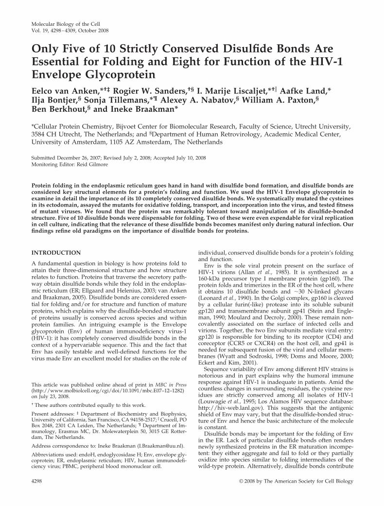
Molecular Biology of the CellVol. 19, 4298–4309, October 2008
Only Five of 10 Strictly Conserved Disulfide Bonds AreEssential for Folding and Eight for Function of the HIV-1Envelope GlycoproteinEelco van Anken,*†‡ Rogier W. Sanders,†§ I. Marije Liscaljet,*†� Aafke Land,*Ilja Bontjer,§ Sonja Tillemans,*¶ Alexey A. Nabatov,§ William A. Paxton,§Ben Berkhout,§ and Ineke Braakman*
*Cellular Protein Chemistry, Bijvoet Center for Biomolecular Research, Faculty of Science, Utrecht University,3584 CH Utrecht, The Netherlands; and §Department of Human Retrovirology, Academic Medical Center,University of Amsterdam, 1105 AZ Amsterdam, The Netherlands
Submitted December 26, 2007; Revised July 2, 2008; Accepted July 10, 2008Monitoring Editor: Reid Gilmore
Protein folding in the endoplasmic reticulum goes hand in hand with disulfide bond formation, and disulfide bonds areconsidered key structural elements for a protein’s folding and function. We used the HIV-1 Envelope glycoprotein toexamine in detail the importance of its 10 completely conserved disulfide bonds. We systematically mutated the cysteinesin its ectodomain, assayed the mutants for oxidative folding, transport, and incorporation into the virus, and tested fitnessof mutant viruses. We found that the protein was remarkably tolerant toward manipulation of its disulfide-bondedstructure. Five of 10 disulfide bonds were dispensable for folding. Two of these were even expendable for viral replicationin cell culture, indicating that the relevance of these disulfide bonds becomes manifest only during natural infection. Ourfindings refine old paradigms on the importance of disulfide bonds for proteins.
INTRODUCTION
A fundamental question in biology is how proteins fold toattain their three-dimensional structure and how structurerelates to function. Proteins that traverse the secretory path-way obtain disulfide bonds while they fold in the endoplas-mic reticulum (ER; Ellgaard and Helenius, 2003; van Ankenand Braakman, 2005). Disulfide bonds are considered essen-tial for folding and/or for structure and function of matureproteins, which explains why the disulfide-bonded structureof proteins usually is conserved across species and withinprotein families. An intriguing example is the Envelopeglycoprotein (Env) of human immunodeficiency virus-1(HIV-1): it has completely conserved disulfide bonds in thecontext of a hypervariable sequence. This and the fact thatEnv has easily testable and well-defined functions for thevirus made Env an excellent model for studies on the role of
individual, conserved disulfide bonds for a protein’s foldingand function.
Env is the sole viral protein present on the surface ofHIV-1 virions (Allan et al., 1985). It is synthesized as a160-kDa precursor type I membrane protein (gp160). Theprotein folds and trimerizes in the ER of the host cell, whereit obtains 10 disulfide bonds and �30 N-linked glycans(Leonard et al., 1990). In the Golgi complex, gp160 is cleavedby a cellular furin(-like) protease into its soluble subunitgp120 and transmembrane subunit gp41 (Stein and Engle-man, 1990; Moulard and Decroly, 2000). These remain non-covalently associated on the surface of infected cells andvirions. Together, the two Env subunits mediate viral entry:gp120 is responsible for binding to its receptor (CD4) andcoreceptor (CCR5 or CXCR4) on the host cell, and gp41 isneeded for subsequent fusion of the viral and cellular mem-branes (Wyatt and Sodroski, 1998; Doms and Moore, 2000;Eckert and Kim, 2001).
Sequence variability of Env among different HIV strains isnotorious and in part explains why the humoral immuneresponse against HIV-1 is inadequate in patients. Amid thecountless changes in surrounding residues, the cysteine res-idues are strictly conserved among all isolates of HIV-1(Louwagie et al., 1995; Los Alamos HIV sequence database:http://hiv-web.lanl.gov). This suggests that the antigenicshield of Env may vary, but that the disulfide-bonded struc-ture of Env and hence the basic architecture of the moleculeis constant.
Disulfide bonds may be important for the folding of Envin the ER. Lack of particular disulfide bonds often rendersnewly synthesized proteins in the ER maturation incompe-tent: they either aggregate and fail to fold or they partiallyoxidize into species similar to folding intermediates of thewild-type protein. Alternatively, disulfide bonds contribute
This article was published online ahead of print in MBC in Press(http://www.molbiolcell.org/cgi/doi/10.1091/mbc.E07–12–1282)on July 23, 2008.† These authors contributed equally to this work.
Present addresses: ‡ Department of Biochemistry and Biophysics,University of California, San Francisco, CA 94158-2517; � Crucell, POBox 2048, 2301 CA Leiden, The Netherlands; ¶ Department of Im-munology, Erasmus MC, Dr. Molewaterplein 50, 3015 GE Rotter-dam, The Netherlands.
Address correspondence to: Ineke Braakman ([email protected]).
Abbreviations used: endoH, endoglycosidase H; Env, envelope gly-coprotein; ER, endoplasmic reticulum; HIV, human immunodefi-ciency virus; PBMC, peripheral blood mononuclear cell.
4298 © 2008 by The American Society for Cell Biology
to the structural integrity of the mature protein. In the caseof Env, disulfide bonds seem to be important also for theconformational changes during receptor binding and subse-quent fusion of the viral and host cell membranes (Bar-bouche et al., 2003; Matthias and Hogg, 2003; Markovic et al.,2004).
Cysteine mutants of ER client proteins are excellent toolsto provide insight into the relevance of individual disulfidebonds, but so far no systematic cysteine mutation studies ofhighly disulfide-linked ER clients have been published.Some Env cysteine mutants have been analyzed in differentstrains (Hemming et al., 1989; Tschachler et al., 1990; Bolm-stedt et al., 1991; Freed and Risser, 1991; Syu et al., 1991;Dedera et al., 1992; Lekutis et al., 1992), but together they donot give a consistent notion of the relative importance of the10 disulfide bonds of Env for folding and function.
To systematically assess the role of each individual disul-fide bond in Env folding and function, we analyzed a com-plete series of mutants, where all cysteines were replaced byalanines, both individually and pair-wise. In spite of thestrict conservation of all 10 disulfide bonds, only five turnedout to be essential for folding of Env in the ER, whereas twodisulfide bonds were expendable not only for folding buteven for function of Env.
MATERIALS AND METHODS
Site-directed Mutagenesis and Molecular CloningBoth single mutants and double mutants of cysteine pairs were generated toeliminate corresponding disulfide bonds in Env by site-directed mutagenesis,either by the pALTER system (Promega, Madison, WI), or by the Quick-change system (Invitrogen, Carlsbad, CA) according to the manufacturers’instructions, using mutagenic oligos where the TGT and TGC cysteine-en-coding codons were changed into GCT, respectively, into GCC alanine en-coding codons with flanking sequences of 12–14 nucleotides on either side.An extra round of site-directed mutagenesis of one of the correspondingsingle mutants generated double mutants. Mutations were confirmed bydideoxynucleotide sequencing and mutant Env open reading frames (ORFs)were cloned into pcDNA3 (Invitrogen), yielding pcDNA3-gp160 constructs. Astop codon was introduced at the cleavage site between the gp120 and gp41sequences by PCR, yielding corresponding pcDNA3-gp120 constructs. Forfunctional assays, mutant Env ORFs were cloned into a full-length molecularclone pLAI (Peden et al., 1991) of the HIV-1LAI isolate. Although we studiedEnv of the LAI isolate, we followed the canonical HXB2 residue numbering,which relates to the LAI numbering as follows: because of an insertion of fiveresidues in the V1 loop of LAI Env, all cysteine residues beyond this loop havea number 5 residues lower in HXB2 than in LAI: until Cys131, numbering isidentical, but Cys162 in LAI becomes 157 in HXB2, etc.
CellsHeLa cells were cultured in MEM (Life Technologies, Rockville, MD) supple-mented with 10% FCS (Life Technologies), penicillin and streptomycin (100U/ml), 2 mM glutamax (Life Technologies), and nonessential amino acids(Life Technologies). C33A cervix carcinoma cells were maintained in DMEM(Invitrogen), supplemented with 10% FCS, penicillin, and streptomycin.SupT1 T-cells were cultured in RPMI medium supplemented with 10% FCS,penicillin, and streptomycin. Peripheral blood mononuclear cells (PBMCs)were isolated from fresh buffy coats (Central Laboratory Blood Bank, Am-sterdam) by standard Ficoll-Hypaque density centrifugation. PBMCs werefrozen in multiple vials at a high concentration and, when required, werethawed and activated with 5 �g/ml phytohemagglutinin (Sigma, St. Louis,MO) and cultured in RPMI medium (Life Technologies) containing 10% FCS,penicillin and streptomycin (100 U/ml) with recombinant interleukin-2 (rIL-2;100 U/ml). On day 4 of culture, PBMCs underwent CD4� enrichment byincubating them with CD8 immunomagnetic beads (Dynal, Lake Success, NY)and separating out the CD8� lymphocytes.
Folding AssaysSubconfluent HeLa cells were infected with recombinant vaccinia virus ex-pressing T7 polymerase (Fuerst et al., 1986) to drive expression of Env mutantsunder control of the T7 promoter. Thirty minutes after infection, cells weretransfected with a mixture of 4 �g of mutant or wild-type pcDNA3-gp120 orpcDNA3-gp160, and 10 �l lipofectin (Invitrogen), according to the manufac-turer’s instructions. Pulse-chase experiments were performed essentially asdescribed (Braakman et al., 1991; Land et al., 2003). Five hours after infection,
cells were depleted of cysteine and methionine for 15–30 min before they werepulse-labeled with 50 �Ci of Redivue promix l-[35S] in vitro labeling mix(Amersham Biosciences, Piscataway, NJ). Cells were chased for various in-tervals in medium containing an excess of unlabeled cysteine and methionine.Chase samples were stopped by aspirating the medium and adding ice-coldHBSS (Invitrogen-BRL) containing 20 mM iodoacetamide (IAM) to block freesulfhydryl groups. Cells were lysed in ice-cold MNT (20 mM MES, 100 mMNaCl, 30 mM Tris-HCl, pH 7.5) containing 0.5% (vol/vol) Triton X-100, 20mM IAM, and protease inhibitor cocktail (10 �g/ml each of chymostatin,leupeptin, antipain, and pepstatin, 1 mM PMSF, and 1 mM EDTA). Celllysates were spun for 10 min at 15,000 � g to pellet nuclei and postnuclearlysates were immunoprecipitated with a polyclonal antibody that recognizesall forms of HIV-1 Env. In addition, secreted or shed gp120 molecules wereimmunoprecipitated from the culture media at later chase times. Washedimmunoprecipitates were resuspended in 0.2% SDS in 100 mM sodium ace-tate, pH 5.4, and incubated at 95°C for 5 min. An equal volume of 100 mMsodium acetate, pH 5.4, was added, which contained 2% Triton X-100 in MNT,protease inhibitor cocktail, and 0.0025 U endoglycosidase H (endoH; Roche,Rotkreuz, Switzerland). Samples were incubated for 2 h at 37°C. After incu-bation, SDS-PAGE sample buffer was added, and samples were incubated at95°C for 5 min. Samples were analyzed by reducing or nonreducing 7.5%SDS-PAGE. Gels were dried and signals were detected on Biomax MR films(Eastman Kodak, Rochester, NY).
Viruses and InfectionsVirus stocks were produced as follows: C33A cells were transfected with 10�g wild-type or mutant pLAI constructs by calcium phosphate precipitation(Das et al., 1999). Three days after transfection, virus-containing culture su-pernatants were harvested, filtered, and stored at �80°C. Virus concentra-tions were quantitated by capsid CA-p24 ELISA (Jeeninga et al., 2000). Thesevalues were used to normalize the amount of virus for infection experiments.SupT1 cells (50 � 103) were infected with the equivalent of 500 pg CA-p24 ofC33A-produced HIV-1LAI per well in a 96-well plate, and virus spread wasmeasured for 14 d using CA-p24 ELISA. The 50% tissue culture infectiousdose (TCID50) in SupT1 cells was determined after 14 d by endpoint dilution.
Ultracentrifugation of VirionsC33A cells were transfected with 40 �g pLAI per T75 flask. Medium wasrefreshed at day 1 after transfection. At 3 d after transfection, culture mediawere harvested, centrifuged, and passed through a 0.45-�m filter to removeresidual cells and debris. Virus particles were pelleted from filtered culturemedia by ultracentrifugation (100,000 � g for 45 min at 4°C) and resuspendedin 0.5 ml lysis buffer (50 mM Tris-HCl, pH 7.4, 10 mM EDTA, 100 mM NaCl,1% SDS).
Quantitation of gp120 by ELISAConcentrations of gp120 in cell, virion, and supernatant fractions were mea-sured as described before (Moore and Ho, 1993; Sanders et al., 2002), withsome modifications. ELISA plates were coated overnight with sheep antibodyD7324 (10 �g/ml; Aalto Bio Reagents, Ratharnham, Dublin, Ireland), directedto the gp120 C5 region, in 0.1 M NaHCO3. After blocking by 2% milk inTris-buffered saline (TBS) for 30 min, gp120 was captured by incubation for 2hat room temperature. Recombinant HIV-1LAI gp120 (Progenics Pharmaceuti-cals, Tarrytown, NY) was used as a reference. Unbound gp120 was washedaway with TBS and purified HIV-1� serum Ig (HIVIg) was added for 1.5 h in2% milk, 20% sheep serum (SS), 0.5% Tween. HIVIg binding was detectedwith alkaline phosphatase–conjugated goat anti-human Fc (1:10,000, JacksonImmunoResearch, West Grove, PA) in 2% milk, 20% SS, and 0.5% Tween.Detection of alkaline phosphatase activity was performed using AMPAKreagents (DAKO, Carpinteria, CA). The measured gp120 contents in viruswere normalized for CA-p24.
HIV-1 Neutralization Assay Using Primary CD4�
LymphocytesViruses were tested for their relative inhibition sensitivity against increasingconcentrations of either sCD4 (BD ImmunoDiagnostics, Orangeburg, NY) orthe CXCR4 binding compound AMD3100 (a generous gift from Dr. D. Schols,Katholieke Universiteit, Leuven, Belgium). TCID50 values were determinedon purified CD4� lymphocytes isolated from an individual who did not carrythe �32CCR5 allele (CCR5�/�) screened for by standard PCR. CD4�-enrichedlymphocytes were plated at 2 � 105 cells/well in 96-well plates with fivefoldserial dilutions of the virus. Cells were fed on day 7 with fresh media andscored on day 14 for CA-p24 levels, with the number of positive wells beingused to identify the TCID50 value for each virus. Each virus (100 TCID50 in 50�l of the culture RPMI medium) was mixed with an equal volume of seriallydiluted compound for 1 h at 37°C in flat-bottomed 96-well plates, and allneutralization reactions were performed in triplicate. After 1 h of incubation2.0 � 105 positively selected CD4� lymphocytes were added to each well in0.1 ml of culture media containing rIL-2 (100 U/ml). For each neutralizationexperiment a positive control, virus incubated with cells in the absence ofinhibitory compound, and a negative control, virus in the absence of cells, was
Importance of HIV Env Disulfide Bonds
Vol. 19, October 2008 4299
included. The negative control CA-p24 concentration was subtracted from alltest results. Supernatant was taken on days 7, 10, and 14 and assayed forCA-p24 with fresh media added to the cells. Neutralization responses weredetermined for each virus at the day CA-p24 levels peaked. We calculated theCA-p24 production in the presence of inhibitory compound as a percentage ofthat in the cultures with virus only.
RESULTS
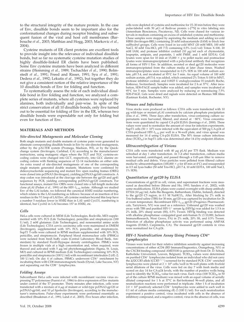
Disulfide Bonds and Cysteine Mutants of EnvWe set out to assess the role in folding and function ofindividual disulfide bonds in the Envelope glycoprotein ofthe HIV-1LAI isolate. The gp120 part of Env contains ninedisulfide bonds, whereas gp41 harbors a single disulfidebond (Figure 1A). The gp120 soluble subunit has a core thatis composed of conserved regions C1 to C5. The variableregions V1 to V5 are present on protein loops that protrudefrom this core. The gp120 core largely overlaps with theavailable crystal structures of gp120 (Kwong et al., 1998;Chen et al., 2005; Figure 1B). Six disulfide bonds locate to thegp120 core. The C1 region contains a single disulfide bond,54–74, whereas the C2 region contains two disulfide bonds:218–247 and 228–239. The 378–445 disulfide bond co-valently links the C3 and C4 regions. Two bonds are foundat the base of variable loops: 296–331 (V3) and 385–418 (V4).The two most prominent loops, which encompass the V1and V2 regions, have three disulfide bonds at their stem(s):119–205, 126–196, and 131–157. The disulfide bond in gp41closes off a very small loop: 598–604. We systematically
replaced all cysteines in the ectodomain of Env by alaninesand refer to mutants as follows: C54A and C74A are thesingle mutants, and C54/74A is the double mutant of disul-fide bond 54–74.
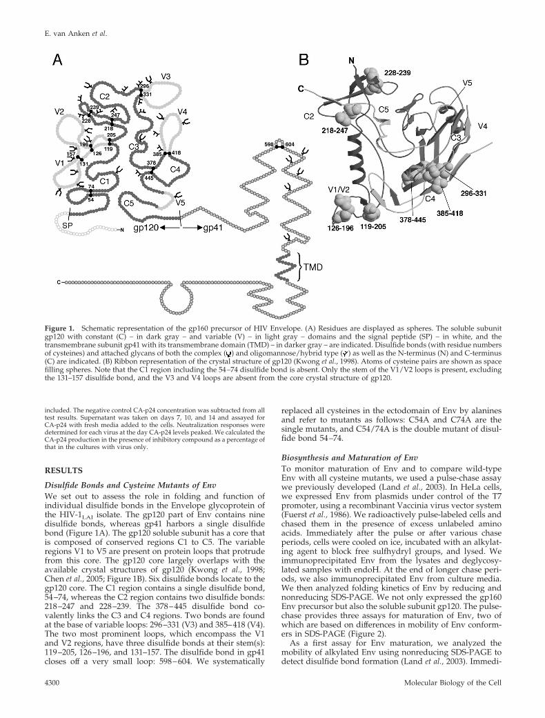
Biosynthesis and Maturation of EnvTo monitor maturation of Env and to compare wild-typeEnv with all cysteine mutants, we used a pulse-chase assaywe previously developed (Land et al., 2003). In HeLa cells,we expressed Env from plasmids under control of the T7promoter, using a recombinant Vaccinia virus vector system(Fuerst et al., 1986). We radioactively pulse-labeled cells andchased them in the presence of excess unlabeled aminoacids. Immediately after the pulse or after various chaseperiods, cells were cooled on ice, incubated with an alkylat-ing agent to block free sulfhydryl groups, and lysed. Weimmunoprecipitated Env from the lysates and deglycosy-lated samples with endoH. At the end of longer chase peri-ods, we also immunoprecipitated Env from culture media.We then analyzed folding kinetics of Env by reducing andnonreducing SDS-PAGE. We not only expressed the gp160Env precursor but also the soluble subunit gp120. The pulse-chase provides three assays for maturation of Env, two ofwhich are based on differences in mobility of Env conform-ers in SDS-PAGE (Figure 2).
As a first assay for Env maturation, we analyzed themobility of alkylated Env using nonreducing SDS-PAGE todetect disulfide bond formation (Land et al., 2003). Immedi-
Figure 1. Schematic representation of the gp160 precursor of HIV Envelope. (A) Residues are displayed as spheres. The soluble subunitgp120 with constant (C) – in dark gray – and variable (V) – in light gray – domains and the signal peptide (SP) – in white, and thetransmembrane subunit gp41 with its transmembrane domain (TMD) – in darker gray – are indicated. Disulfide bonds (with residue numbersof cysteines) and attached glycans of both the complex ( ) and oligomannose/hybrid type ( ) as well as the N-terminus (N) and C-terminus(C) are indicated. (B) Ribbon representation of the crystal structure of gp120 (Kwong et al., 1998). Atoms of cysteine pairs are shown as spacefilling spheres. Note that the C1 region including the 54–74 disulfide bond is absent. Only the stem of the V1/V2 loops is present, excludingthe 131–157 disulfide bond, and the V3 and V4 loops are absent from the core crystal structure of gp120.
E. van Anken et al.
Molecular Biology of the Cell4300
ately after the pulse, wild-type gp160 and gp120 appeared asa fuzzy band in nonreducing gels (Figure 2, 0 h, nonreduc-ing) with mobility close to that of the corresponding reducedprotein. The reduced samples appeared as a discrete bandhowever (Figure 2, 0 h, reducing), implying that some di-sulfide bonds had formed already before the end of thepulse. With time, “smears” of faster running protein ap-peared in the nonreduced samples, corresponding to vari-ous species of partially oxidized folding intermediates (ITs).After 4 h of chase, most of the wild-type gp160 and gp120proteins appeared as a sharp band in nonreducing gels(Figure 2, 4 h, nonreducing), indicating that they reached thefully oxidized native state (NT).
Proteins that fold in the ER are targeted to enter thiscompartment by a signal peptide (Walter and Johnson,1994), which in soluble or type I proteins is cleaved offalready during synthesis. A striking exception however isthe signal peptide of HIV-1 Env, which is removed long aftersynthesis has been completed (Li et al., 1994). The signalpeptide of Env can be removed only after the protein hasgone through some initial folding steps, making this cleav-age another indicator of Env maturation (Land et al., 2003).By analyzing the chase samples via reducing SDS-PAGE, wehence used signal peptide cleavage as a second assay for Envmaturation. Immediately after the pulse only a single bandwas visible (Figure 2, 0 h, reducing), which corresponds tothe reduced protein with its signal peptide still attached(Ru � reduced uncleaved). With time, a second band ap-peared below Ru, which corresponds to the reduced proteinfrom which the signal peptide had been removed (Rc �reduced cleaved). After 4 h of chase, the signal peptide hadbeen removed from the majority of wild-type gp160 andgp120 molecules. Except for some faint band at the top of thegel, which likely represents aggregated Env, we detected noother bands or smears in the reducing gel. This implies thatintracellular pools of Env were fully deglycosylated by en-doH, as we previously reported (Land et al., 2003). Env didundergo heterogeneous glycan modifications in the Golgi asis evident from the “smeary” character of the shed andsecreted gp120.
This gp120 harvested from culture media was analyzed byreducing SDS-PAGE as a third assay for Env maturation.Correctly folded Env exits the ER and travels along thesecretory pathway. Env expressed as soluble subunit gp120is secreted, whereas gp160 undergoes proteolytic cleavageinto gp41 and gp120 subunits. Because the gp120 subunitsare no longer covalently attached to the remainder of theEnv spikes, they quantitatively shed into the culture me-dium. As anticipated, substantial amounts of both shed andsecreted (endoH resistant) wild-type gp120 were present inthe culture medium after 4 h of chase (Figure 2, 4 h, secr/shed). The pool of endoH-resistant intracellular Env is toosmall to detect because the average time it takes Env totravel via the Golgi to the cell surface and be cleaved andshed or secreted is very short compared with the time theprotein resides in the ER.
Altogether, we concluded that wild-type Env matured inthe vaccinia virus T7 heterologous expression system similaras in SupT1 cells infected with HIV-1 (Das et al., 1999) and inHeLa or CHO cells infected with gp120 or gp160 expressingrecombinant vaccinia viruses (Kieny et al., 1986; Land et al.,2003). As shown before (Land et al., 2003), oxidative foldingof the gp120 subunit was similar to that of the gp160 pre-cursor, except that gp120 folded slightly faster and thatdifferences in mobility in SDS-PAGE of Env species are moreevident for gp120 because of its smaller size. Maturation ofEnv is slow, but after 4 h the majority of wild-type Envreaches the native state (Earl et al., 1991; Otteken et al., 1996;Land et al., 2003). We therefore compared maturation of Envmutants after 4 h of chase (Figure 3A) and quantified theresults.
Five Disulfide Bonds Are Dispensable for Folding of EnvThe 0-h chase samples of all mutants migrated similarly tothat of wild type in both reducing and nonreducing gels,indicating that the starting point of folding was indistin-guishable for mutants and wild-type proteins (Figure 3A,
Figure 2. Folding of wild-type gp160 and gp120. Wild-type Env, as precursor gp160 or as the soluble subunit gp120 alone, was expressedin HeLa cells using the Vaccinia T7 heterologous expression system. Cells were pulse-labeled for 2 min (gp120) or 5 min (gp160) and chasedfor various times (h) as indicated. Cells were lysed and Env proteins were immunoprecipitated from lysates and from the chase medium.Immunoprecipitates from lysates were fully deglycosylated with endoH (see Materials and Methods) and analyzed by nonreducing (NR) orreducing (R) 7.5% SDS-PAGE. Immunoprecipitates from the chase media of secreted or shed (S) gp120 were analyzed directly on reducing7.5% SDS-PAGE. Aggregates (agg), folding intermediates (ITs), the native form (NT), and the reduced protein from which the signal peptidewas cleaved off (Rc) or not (Ru), as well as the shed or secreted gp120 (S) are indicated.
Importance of HIV Env Disulfide Bonds
Vol. 19, October 2008 4301

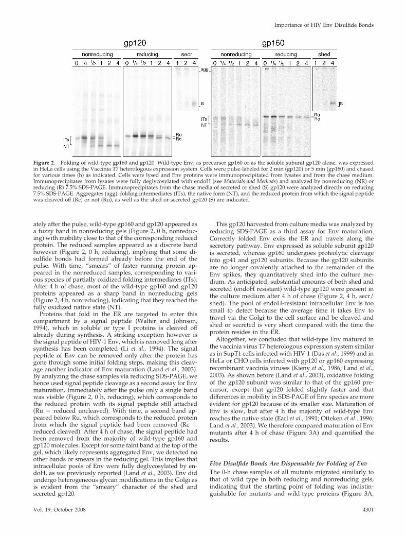
Figure 3. Folding “endpoint” analysis of Env cysteine mutants. (A) Wild-type or cysteine mutants of gp160 or gp120 were expressed inHeLa cells using the Vaccinia T7 heterologous expression system. Cells were pulse-labeled for 10 min and chased for 0 or 4 h. Cells were lysedand Env proteins were immunoprecipitated from lysates and chase medium. Samples were analyzed as in Figure 2. (B) Data were quantifiedwith ImageQuant (Molecular Dynamics, Sunnyvale, CA), and percentage aggregation, NT formation, Rc formation, and shedding/secretionare depicted in histograms.
E. van Anken et al.
Molecular Biology of the Cell4302
0-NR/0-R). After 4 h of chase, however, the Env mutantsdisplayed many differences in maturation (Figure 3A,4-NR/4-R/4-S).
Based on the three assays, mutants of five disulfide bondswere maturation competent: mutants of the three disulfidebonds in the V1/V2 loops (119–205, 126–196, and 131–157),of the disulfide bond that bridges C3 and C4 (378–445) andof the single disulfide bond in gp41 (598–604). These mu-tants reached a native state (Figure 3A, 4-NR), and signalpeptide removal was substantial (Figure 3A, 4-R). Accord-ingly, they could exit the ER as was evident from secretionor shedding of gp120 (Figure 3A, 4-S).
The folding patterns of maturation competent mutantswere not necessarily identical to those of wild type. In caseof the 378–445 mutants, fewer molecules than wild type hadreached the native state and had lost their signal peptide(Figure 3A, 4-NR/4-R). Shedding from the 598–604 mutantswas poorer than from wild type (Figure 3A, gp160, 4-S), andmutants of the V1/V2 loops accumulated a folding interme-diate that was almost undetectable for wild type (Figure 3A,4-NR). Results were similar for gp160 and gp120. We there-fore concluded that five disulfide bonds (119–205, 126–196,131–157, 378–445, and 598–604) were not strictly essentialfor Env to reach a folded state similar to wild type, butrather improved folding yields and rates.
Mutants of the remaining five disulfide bonds displayedsevere folding defects: of the single disulfide bond in the C1region (54–74), of the two disulfide bonds in the C2 region(218–247 and 228–239), and of the single disulfide bonds atthe stem of the V3 loop (296–331) and the V4 loop (385–418).Compared with wild type, signal peptide cleavage of thesemutants was poor (Figure 3A, 4-R). Disulfide bond forma-
tion varied: the persistence of the reduced-like band thatwas already present at the end of the pulse, in mutants of the296–331 and 385–418 disulfide bonds, indicated that few ifany disulfide bonds were formed in the majority of theseEnv species (Figure 3A, 4-NR). In contrast, mutants of the54–74 disulfide bond and, to a lesser extent, of the 228–239and 218–247 disulfide bonds did reach a native-like band(Figure 3A, 4-NR). These mutants nevertheless failed to shedgp120 (Figure 3A, gp160, 4-S). Conversely, mutants of the385–418 disulfide bond shed marginal amounts of gp120into the chase medium (Figure 3A, gp160, S; Figure 3B,gp160, shedding).
None of the gp120 mutants formed aggregates, whetherfolding deficient or not (Figure 3A, gp120, 4-NR). In case ofthe gp160 mutants, high-molecular-weight species werepresent after 4 h of chase, which represent aggregates oftrimeric and larger size (Figure 3A, gp160, 4-NR/R). Aggre-gation was modest (never exceeding 13% of the signal; Fig-ure 3B), but was more apparent for mutants that were mat-uration incompetent (54–74, 218–247, 228–239, 296–331, and385–418) or displayed slower maturation (378–445). Thesingle mutants C598A and C604A produced more of thesehigh-molecular-weight species than the corresponding dou-ble mutant because of their odd number of cysteines, whichleaves one free for intermolecular bonding.
Aggregates were fully reduced in the reducing gels (Fig-ure 3A, gp160, 4-R), revealing that Env from lysates wasfully endoH sensitive after 4 h. On the one hand this impliesthat the travel of the folding competent mutants through theGolgi and subsequent surface expression and shedding weresimilarly as fast as for wild type, because endoH resistancedevelops only as a result of glycan modifications in the
Figure 3. (cont).
Importance of HIV Env Disulfide Bonds
Vol. 19, October 2008 4303
Golgi. On the other hand, the lack of shedding (Figure 3A,gp160, 4-S) and of endoH resistance of the mutants withfolding problems (Figure 3A, gp160, 4-R) implied that theywhere strictly retained in the ER, as we anticipated. Still, forsome of these mutants gp120 was released from cells moreby secretion than by shedding from gp41, in particular forthe gp120s from the 54–74 mutants, for C247A, and forC296/331A (Figure 3A, gp120, 4-S). Retention in the ERhence was less stringent for gp120 than for gp160, possiblybecause gp120 is a monomeric soluble protein whereasgp160 must trimerize and is a membrane protein (Singh etal., 1990). We concluded that elimination of any of the 54–74,218–247, 228–239, 296–331, or 385–418 disulfide bonds ledto severe folding deficiencies in the ER. We categorized themas class 1 disulfide bonds, as opposed to class 2 disulfidebonds, which were expendable for productive folding.
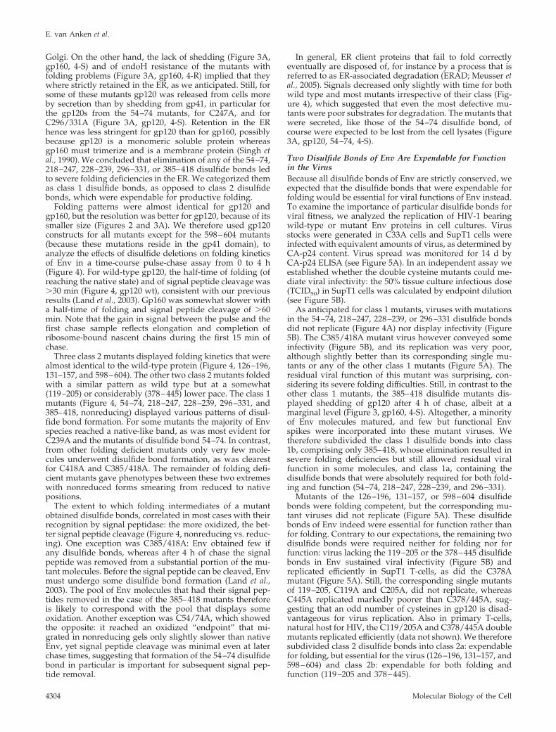
Folding patterns were almost identical for gp120 andgp160, but the resolution was better for gp120, because of itssmaller size (Figures 2 and 3A). We therefore used gp120constructs for all mutants except for the 598–604 mutants(because these mutations reside in the gp41 domain), toanalyze the effects of disulfide deletions on folding kineticsof Env in a time-course pulse-chase assay from 0 to 4 h(Figure 4). For wild-type gp120, the half-time of folding (ofreaching the native state) and of signal peptide cleavage was�30 min (Figure 4, gp120 wt), consistent with our previousresults (Land et al., 2003). Gp160 was somewhat slower witha half-time of folding and signal peptide cleavage of �60min. Note that the gain in signal between the pulse and thefirst chase sample reflects elongation and completion ofribosome-bound nascent chains during the first 15 min ofchase.
Three class 2 mutants displayed folding kinetics that werealmost identical to the wild-type protein (Figure 4, 126–196,131–157, and 598–604). The other two class 2 mutants foldedwith a similar pattern as wild type but at a somewhat(119–205) or considerably (378–445) lower pace. The class 1mutants (Figure 4, 54–74, 218–247, 228–239, 296–331, and385–418, nonreducing) displayed various patterns of disul-fide bond formation. For some mutants the majority of Envspecies reached a native-like band, as was most evident forC239A and the mutants of disulfide bond 54–74. In contrast,from other folding deficient mutants only very few mole-cules underwent disulfide bond formation, as was clearestfor C418A and C385/418A. The remainder of folding defi-cient mutants gave phenotypes between these two extremeswith nonreduced forms smearing from reduced to nativepositions.
The extent to which folding intermediates of a mutantobtained disulfide bonds, correlated in most cases with theirrecognition by signal peptidase: the more oxidized, the bet-ter signal peptide cleavage (Figure 4, nonreducing vs. reduc-ing). One exception was C385/418A: Env obtained few ifany disulfide bonds, whereas after 4 h of chase the signalpeptide was removed from a substantial portion of the mu-tant molecules. Before the signal peptide can be cleaved, Envmust undergo some disulfide bond formation (Land et al.,2003). The pool of Env molecules that had their signal pep-tides removed in the case of the 385–418 mutants thereforeis likely to correspond with the pool that displays someoxidation. Another exception was C54/74A, which showedthe opposite: it reached an oxidized “endpoint” that mi-grated in nonreducing gels only slightly slower than nativeEnv, yet signal peptide cleavage was minimal even at laterchase times, suggesting that formation of the 54–74 disulfidebond in particular is important for subsequent signal pep-tide removal.
In general, ER client proteins that fail to fold correctlyeventually are disposed of, for instance by a process that isreferred to as ER-associated degradation (ERAD; Meusser etal., 2005). Signals decreased only slightly with time for bothwild type and most mutants irrespective of their class (Fig-ure 4), which suggested that even the most defective mu-tants were poor substrates for degradation. The mutants thatwere secreted, like those of the 54–74 disulfide bond, ofcourse were expected to be lost from the cell lysates (Figure3A, gp120, 54–74, 4-S).
Two Disulfide Bonds of Env Are Expendable for Functionin the VirusBecause all disulfide bonds of Env are strictly conserved, weexpected that the disulfide bonds that were expendable forfolding would be essential for viral functions of Env instead.To examine the importance of particular disulfide bonds forviral fitness, we analyzed the replication of HIV-1 bearingwild-type or mutant Env proteins in cell cultures. Virusstocks were generated in C33A cells and SupT1 cells wereinfected with equivalent amounts of virus, as determined byCA-p24 content. Virus spread was monitored for 14 d byCA-p24 ELISA (see Figure 5A). In an independent assay weestablished whether the double cysteine mutants could me-diate viral infectivity: the 50% tissue culture infectious dose(TCID50) in SupT1 cells was calculated by endpoint dilution(see Figure 5B).
As anticipated for class 1 mutants, viruses with mutationsin the 54–74, 218–247, 228–239, or 296–331 disulfide bondsdid not replicate (Figure 4A) nor display infectivity (Figure5B). The C385/418A mutant virus however conveyed someinfectivity (Figure 5B), and its replication was very poor,although slightly better than its corresponding single mu-tants or any of the other class 1 mutants (Figure 5A). Theresidual viral function of this mutant was surprising, con-sidering its severe folding difficulties. Still, in contrast to theother class 1 mutants, the 385–418 disulfide mutants dis-played shedding of gp120 after 4 h of chase, albeit at amarginal level (Figure 3, gp160, 4-S). Altogether, a minorityof Env molecules matured, and few but functional Envspikes were incorporated into these mutant viruses. Wetherefore subdivided the class 1 disulfide bonds into class1b, comprising only 385–418, whose elimination resulted insevere folding deficiencies but still allowed residual viralfunction in some molecules, and class 1a, containing thedisulfide bonds that were absolutely required for both fold-ing and function (54–74, 218–247, 228–239, and 296–331).
Mutants of the 126–196, 131–157, or 598–604 disulfidebonds were folding competent, but the corresponding mu-tant viruses did not replicate (Figure 5A). These disulfidebonds of Env indeed were essential for function rather thanfor folding. Contrary to our expectations, the remaining twodisulfide bonds were required neither for folding nor forfunction: virus lacking the 119–205 or the 378–445 disulfidebonds in Env sustained viral infectivity (Figure 5B) andreplicated efficiently in SupT1 T-cells, as did the C378Amutant (Figure 5A). Still, the corresponding single mutantsof 119–205, C119A and C205A, did not replicate, whereasC445A replicated markedly poorer than C378/445A, sug-gesting that an odd number of cysteines in gp120 is disad-vantageous for virus replication. Also in primary T-cells,natural host for HIV, the C119/205A and C378/445A doublemutants replicated efficiently (data not shown). We thereforesubdivided class 2 disulfide bonds into class 2a: expendablefor folding, but essential for the virus (126–196, 131–157, and598–604) and class 2b: expendable for both folding andfunction (119–205 and 378–445).
E. van Anken et al.
Molecular Biology of the Cell4304
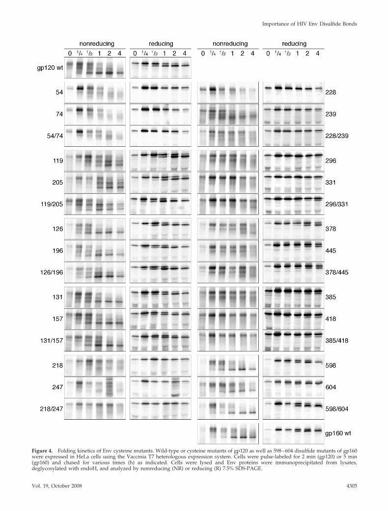
Figure 4. Folding kinetics of Env cysteine mutants. Wild-type or cysteine mutants of gp120 as well as 598–604 disulfide mutants of gp160were expressed in HeLa cells using the Vaccinia T7 heterologous expression system. Cells were pulse-labeled for 2 min (gp120) or 5 min(gp160) and chased for various times (h) as indicated. Cells were lysed and Env proteins were immunoprecipitated from lysates,deglycosylated with endoH, and analyzed by nonreducing (NR) or reducing (R) 7.5% SDS-PAGE.
Importance of HIV Env Disulfide Bonds
Vol. 19, October 2008 4305
Lack of infectivity and replication of class 2a mutant vi-ruses could result from dysfunctional Env on virions or fromtheir absence in viral particles. We therefore measured in-corporation of Env in virus particles by gp120 ELISA onpurified virus fractions and found that the molar Gag-to-Env ratio in the wild-type virus sample was �100:1 (data notshown). This corresponds to an average of 4–8 trimeric Envspikes per virus particle, which agrees with previously pub-lished results (O’Brien et al., 1994; Chertova et al., 2002). Asexpected, considerable amounts of C119/205A Env spikeswere incorporated onto virions (�80% of wild-type levels;
Figure 5C). Incorporation of C378/445A spikes was lower(�45% of wild-type levels; Figure 5C), which correlated withthe reduced folding rate (Figures 3 and 4), delayed replica-tion (Figure 5A), and slightly reduced viral infectivity (Fig-ure 5B) compared with wild type. We did not anticipate todetect any incorporation of those mutants that could not foldand exit the ER. The residual incorporation we found for theclass 1a mutants (Figure 5C) therefore likely representsbackground. Incorporation of the class 2a mutants that didfold but failed to replicate, C126/196A, C131/157A, andC598/604A, was in that same range. We concluded that the
Figure 5. Effects of Env cysteine deletions on viral replication, infectivity, and spike incorporation into the virus. (A) SupT1 cells (50 � 103)were infected with 500 pg CA-p24 equivalents of HIV-1LAI, yielded from supernatants of C33A cells that were transfected with genomic pLAIbearing the wild-type or mutant Env gene. Virus spread was measured for 14 d using CA-p24 ELISA. (B) The infectivities (TCID50) in SupT1cells of virus stocks as in A were measured by endpoint dilution. (C) Virus particles were harvested by ultracentrifugation, and gp120 andCA-p24 contents of virus particles were measured by ELISA. The gp120 amounts were standardized for CA-p24 input and the gp120 contentsof mutants are given as percentages of wild type.
E. van Anken et al.
Molecular Biology of the Cell4306
lack of viral replication of the class 2a mutants was due toabsence or low abundance of functional Env spikes on themutant virions.
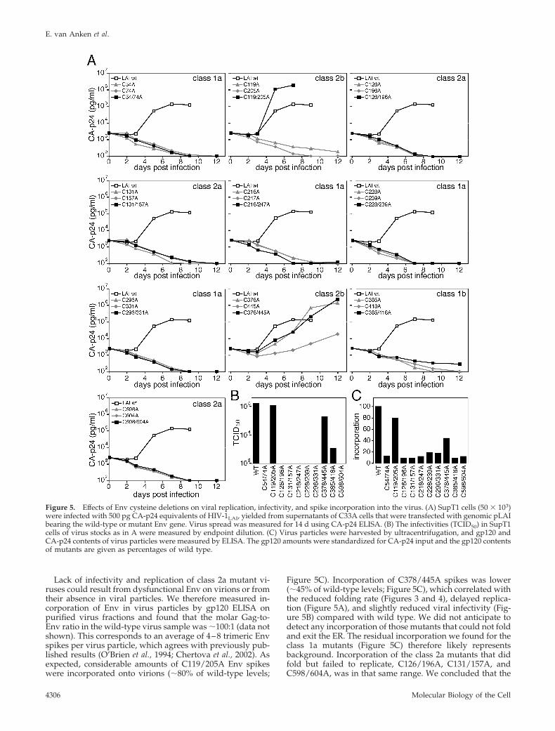
Functional Env Mutants Are Similar But Not Identical toWild TypeTo ascertain whether the class 2b mutant viruses had differ-ent Env properties than the parental strain, we tested usageof the receptor (CD4) and coreceptor (CXCR4 for the LAIisolate) for C119/205A and C378/445A. CD4�-enrichedlymphocytes were inoculated with equivalent amounts ofmutant and wild-type virus, but viral entry, and hence prop-agation, was inhibited with limiting dilutions of either sol-uble CD4 (sCD4) or the CXCR4-binding compoundAMD3100. Compared with the LAI parental strain and theC378/445A virus, less sCD4 was required to inhibit viralreplication of the C119/205A mutant virus (Figure 6, top),which indicates that receptor usage was different for thismutant. The C378/445A mutant on the other hand dis-played alterations in CXCR4 coreceptor usage, with lowerconcentrations of AMD3100 required to inhibit virus repli-cation than for either the wild-type or C119/205A viruses(Figure 6, bottom).
The changes in specific molecular usage of receptor (C119/205A) or coreceptor (C378/445) could be attributed to, forinstance, increased binding affinity or increased ability to un-dergo the conformational changes that accompany (co-)recep-
tor binding. In any case, these differences indicated that thelack of either disulfide bond changed the architecture or flex-ibility of the Env spikes, be it that these changes did notprevent the virus from propagating in tissue culture cells.
DISCUSSION
Elimination of disulfide bonds from proteins often leads tomisfolding, aggregation, and loss of function. In contrast, wefound that Env was remarkably tolerant toward manipula-tion of its disulfide bonds even though they are strictlyconserved among all HIV sequences. Two of the 10 disulfidebonds were completely expendable for function (receptorbinding and/or fusion). Three other disulfide bonds wereessential for the virus, but dispensable for oxidative foldingof Env in the ER and subsequent exit from the compartment.
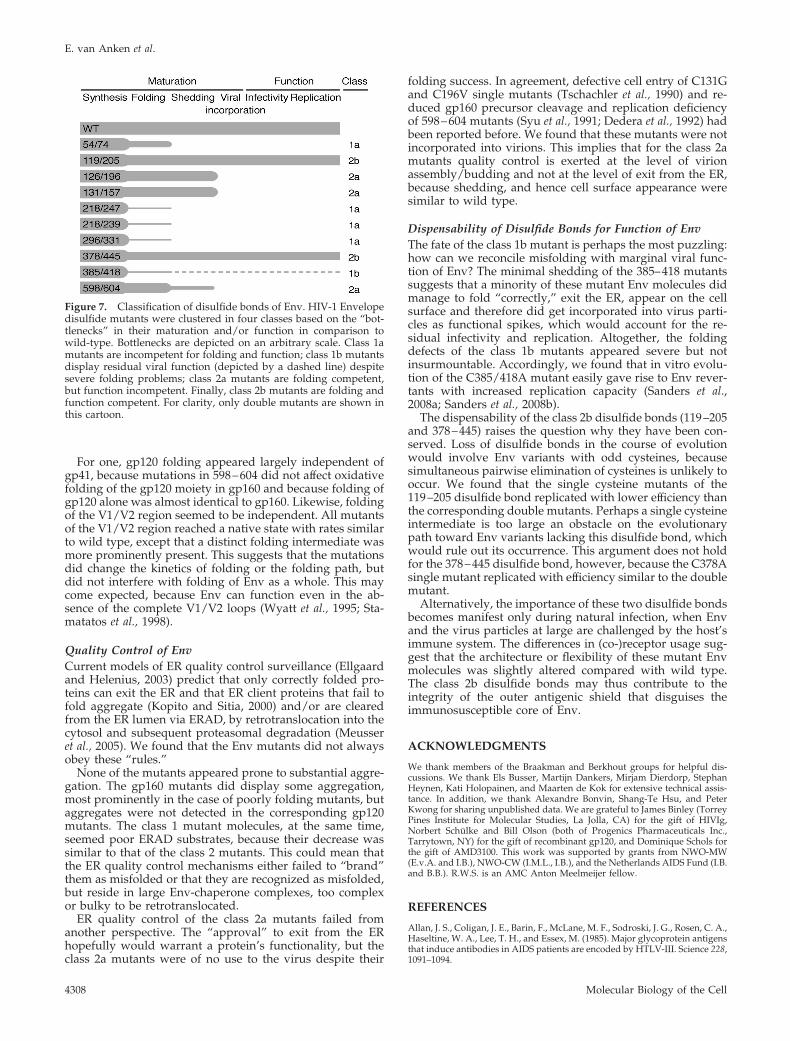
Classification of the 10 Disulfide Bonds of EnvBecause all disulfide bonds in Env are strictly conserved, weanticipated that most if not all would be crucial for Envfolding, let alone for its function. In the case of influenzavirus hemagglutinin (HA), for instance, all disulfide bondsare necessary for the protein to reach the native state, be itthat one displayed a conditional phenotype (Braakman et al.,unpublished data). To our surprise, we found that disrup-tion of several disulfide bonds was compatible with Envfolding and/or function. We categorized the 10 disulfidebonds of Env, based on the consequences of their elimina-tion for folding and function: class 1 comprises disulfidebonds whose elimination led to severe folding deficiencies:54–74, 218–247, 228–239, and, as reported earlier, 296–331(Tschachler et al., 1990; Freed and Risser, 1991; Travis et al.,1992) and 385–418 (Hemming et al., 1989; Tschachler et al.,1990; Bolmstedt et al., 1991; Lekutis et al., 1992). Becausedeletion of the latter disulfide bond was compatible withsome residual viral function in spite of folding failure, wefurther distinguished class 1 into class 1b, which only con-tained 385–418, with residual viral function despite foldingdeficiency, and class 1a, which contained the other disulfidebonds in class 1. Likewise, we divided the disulfide bondswhose elimination still allowed proper folding into class 2a,containing the disulfide bonds that were essential for viralfunction despite their dispensability for folding, 126–196,131–157, and 598–604, and class 2b, containing the disulfidebonds that were expendable for both folding and function,119–205 and 378–445 (Figure 7).
The Folding Pathway of EnvAll Env mutants underwent oxidative folding, meaning thatat 4 h of chase they either reached a fully oxidized state orthey displayed a smear of partially oxidized species. Mostsingle cysteine mutants displayed folding kinetics similar tothat of the corresponding double mutants, which signifiesthat orphan cysteines did not promiscuously team up withnonnative cysteine partners. This suggests that nonnativedisulfide bonds are highly transient in nature and are easilybroken again. In support of such a scenario, we have wit-nessed waves of oxidation and reduction in gp120 in thepresence of low concentrations of reductant (Land et al.,2003). The smears therefore may represent heterogeneousbut “genuine” folding intermediates. Altogether, we cannotpinpoint a single obligatory “initiation” disulfide bond thatmust form before any of the others can. It was thereforeimpossible to delineate a straightforward folding pathwaywhereby formation of one disulfide bond was essential forformation of the next. Instead, we found evidence that dif-ferent modules of Env fold independently of each other.
Figure 6. Effect of inhibition of (co-)receptor binding on replicatingmutant viruses. CD4� lymphocytes (2.0 � 105/well in 96-wellplates) were inoculated with equal amounts (100 TCID50) of theparental HIV-1LAI or mutant virus containing Env lacking either the119–205 or the 378–445 disulfide bond. CD4 receptor binding wasinhibited by limiting dilutions of sCD4 (top) and CXCR4 coreceptorbinding with AMD3100 (bottom). As a measure for inhibition, thepercentage reduction in CA-p24 production at various concentra-tions of either compound as compared with the CA-p24 productionin the absence of compound was calculated. The experiment wasrepeated several times, and results were reproducible.
Importance of HIV Env Disulfide Bonds
Vol. 19, October 2008 4307
For one, gp120 folding appeared largely independent ofgp41, because mutations in 598–604 did not affect oxidativefolding of the gp120 moiety in gp160 and because folding ofgp120 alone was almost identical to gp160. Likewise, foldingof the V1/V2 region seemed to be independent. All mutantsof the V1/V2 region reached a native state with rates similarto wild type, except that a distinct folding intermediate wasmore prominently present. This suggests that the mutationsdid change the kinetics of folding or the folding path, butdid not interfere with folding of Env as a whole. This maycome expected, because Env can function even in the ab-sence of the complete V1/V2 loops (Wyatt et al., 1995; Sta-matatos et al., 1998).
Quality Control of EnvCurrent models of ER quality control surveillance (Ellgaardand Helenius, 2003) predict that only correctly folded pro-teins can exit the ER and that ER client proteins that fail tofold aggregate (Kopito and Sitia, 2000) and/or are clearedfrom the ER lumen via ERAD, by retrotranslocation into thecytosol and subsequent proteasomal degradation (Meusseret al., 2005). We found that the Env mutants did not alwaysobey these “rules.”
None of the mutants appeared prone to substantial aggre-gation. The gp160 mutants did display some aggregation,most prominently in the case of poorly folding mutants, butaggregates were not detected in the corresponding gp120mutants. The class 1 mutant molecules, at the same time,seemed poor ERAD substrates, because their decrease wassimilar to that of the class 2 mutants. This could mean thatthe ER quality control mechanisms either failed to “brand”them as misfolded or that they are recognized as misfolded,but reside in large Env-chaperone complexes, too complexor bulky to be retrotranslocated.
ER quality control of the class 2a mutants failed fromanother perspective. The “approval” to exit from the ERhopefully would warrant a protein’s functionality, but theclass 2a mutants were of no use to the virus despite their
folding success. In agreement, defective cell entry of C131Gand C196V single mutants (Tschachler et al., 1990) and re-duced gp160 precursor cleavage and replication deficiencyof 598–604 mutants (Syu et al., 1991; Dedera et al., 1992) hadbeen reported before. We found that these mutants were notincorporated into virions. This implies that for the class 2amutants quality control is exerted at the level of virionassembly/budding and not at the level of exit from the ER,because shedding, and hence cell surface appearance weresimilar to wild type.
Dispensability of Disulfide Bonds for Function of EnvThe fate of the class 1b mutant is perhaps the most puzzling:how can we reconcile misfolding with marginal viral func-tion of Env? The minimal shedding of the 385–418 mutantssuggests that a minority of these mutant Env molecules didmanage to fold “correctly,” exit the ER, appear on the cellsurface and therefore did get incorporated into virus parti-cles as functional spikes, which would account for the re-sidual infectivity and replication. Altogether, the foldingdefects of the class 1b mutants appeared severe but notinsurmountable. Accordingly, we found that in vitro evolu-tion of the C385/418A mutant easily gave rise to Env rever-tants with increased replication capacity (Sanders et al.,2008a; Sanders et al., 2008b).
The dispensability of the class 2b disulfide bonds (119–205and 378–445) raises the question why they have been con-served. Loss of disulfide bonds in the course of evolutionwould involve Env variants with odd cysteines, becausesimultaneous pairwise elimination of cysteines is unlikely tooccur. We found that the single cysteine mutants of the119–205 disulfide bond replicated with lower efficiency thanthe corresponding double mutants. Perhaps a single cysteineintermediate is too large an obstacle on the evolutionarypath toward Env variants lacking this disulfide bond, whichwould rule out its occurrence. This argument does not holdfor the 378–445 disulfide bond, however, because the C378Asingle mutant replicated with efficiency similar to the doublemutant.
Alternatively, the importance of these two disulfide bondsbecomes manifest only during natural infection, when Envand the virus particles at large are challenged by the host’simmune system. The differences in (co-)receptor usage sug-gest that the architecture or flexibility of these mutant Envmolecules was slightly altered compared with wild type.The class 2b disulfide bonds may thus contribute to theintegrity of the outer antigenic shield that disguises theimmunosusceptible core of Env.
ACKNOWLEDGMENTS
We thank members of the Braakman and Berkhout groups for helpful dis-cussions. We thank Els Busser, Martijn Dankers, Mirjam Dierdorp, StephanHeynen, Kati Holopainen, and Maarten de Kok for extensive technical assis-tance. In addition, we thank Alexandre Bonvin, Shang-Te Hsu, and PeterKwong for sharing unpublished data. We are grateful to James Binley (TorreyPines Institute for Molecular Studies, La Jolla, CA) for the gift of HIVIg,Norbert Schulke and Bill Olson (both of Progenics Pharmaceuticals Inc.,Tarrytown, NY) for the gift of recombinant gp120, and Dominique Schols forthe gift of AMD3100. This work was supported by grants from NWO-MW(E.v.A. and I.B.), NWO-CW (I.M.L., I.B.), and the Netherlands AIDS Fund (I.B.and B.B.). R.W.S. is an AMC Anton Meelmeijer fellow.
REFERENCES
Allan, J. S., Coligan, J. E., Barin, F., McLane, M. F., Sodroski, J. G., Rosen, C. A.,Haseltine, W. A., Lee, T. H., and Essex, M. (1985). Major glycoprotein antigensthat induce antibodies in AIDS patients are encoded by HTLV-III. Science 228,1091–1094.
Figure 7. Classification of disulfide bonds of Env. HIV-1 Envelopedisulfide mutants were clustered in four classes based on the “bot-tlenecks” in their maturation and/or function in comparison towild-type. Bottlenecks are depicted on an arbitrary scale. Class 1amutants are incompetent for folding and function; class 1b mutantsdisplay residual viral function (depicted by a dashed line) despitesevere folding problems; class 2a mutants are folding competent,but function incompetent. Finally, class 2b mutants are folding andfunction competent. For clarity, only double mutants are shown inthis cartoon.
E. van Anken et al.
Molecular Biology of the Cell4308

Barbouche, R., Miquelis, R., Jones, I. M., and Fenouillet, E. (2003). Protein-disulfide isomerase-mediated reduction of two disulfide bonds of HIV enve-lope glycoprotein 120 occurs post-CXCR4 binding and is required for fusion.J. Biol. Chem. 278, 3131–3136.
Bolmstedt A. et al. (1991). Effects of mutations in glycosylation sites anddisulphide bonds on processing, CD4-binding and fusion activity of humanimmunodeficiency virus envelope glycoproteins. J. Gen. Virol. 72, 1269–1277.
Braakman, I., Hoover-Litty, H., Wagner, K. R., and Helenius, A. (1991).Folding of influenza hemagglutinin in the endoplasmic reticulum. J. Cell Biol.114, 401–411.
Chen, B., Vogan, E. M., Gong, H., Skehel, J. J., Wiley, D. C., and Harrison, S. C.(2005). Structure of an unliganded simian immunodeficiency virus gp120core. Nature 433, 834–841.
Chertova E. et al. (2002). Envelope glycoprotein incorporation, not sheddingof surface envelope glycoprotein (gp120/SU), Is the primary determinant ofSU content of purified human immunodeficiency virus type 1 and simianimmunodeficiency virus. J. Virol. 76, 5315–5325.
Das, A. T., Land, A., Braakman, I., Klaver, B., and Berkhout, B. (1999). HIV-1evolves into a nonsyncytium-inducing virus upon prolonged culture in vitro.Virology 263, 55–69.
Dedera, D., Gu, R. L., and Ratner, L. (1992). Conserved cysteine residues in thehuman immunodeficiency virus type 1 transmembrane envelope protein areessential for precursor envelope cleavage. J. Virol. 66, 1207–1209.
Doms, R. W., and Moore, J. P. (2000). HIV-1 membrane fusion: targets ofopportunity. J. Cell Biol. 151, F9–F14.
Earl, P. L., Moss, B., and Doms, R. W. (1991). Folding, interaction withGRP78-BiP, assembly, and transport of the human immunodeficiency virustype 1 envelope protein. J. Virol. 65, 2047–2055.
Eckert, D. M., and Kim, P. S. (2001). Mechanisms of viral membrane fusionand its inhibition. Annu. Rev. Biochem. 70, 777–810.
Ellgaard, L., and Helenius, A. (2003). Quality control in the endoplasmicreticulum. Nat. Rev. Mol. Cell Biol. 4, 181–191.
Freed, E. O., and Risser, R. (1991). Identification of conserved residues in thehuman immunodeficiency virus type 1 principal neutralizing determinantthat are involved in fusion. AIDS Res. Hum. Retroviruses 7, 807–811.
Fuerst, T. R., Niles, E. G., Studier, F. W., and Moss, B. (1986). Eukaryotictransient-expression system based on recombinant vaccinia virus that synthe-sizes bacteriophage T7 RNA polymerase. Proc. Natl. Acad. Sci. USA 83,8122–8126.
Hemming, A., Bolmstedt, A., Flodby, P., Lundberg, L., Gidlund, M., Wigzell,H., and Olofsson, S. (1989). Cystein 402 of HIV gp 120 is essential forCD4-binding and resistance of gp 120 to intracellular degradation. Arch.Virol. 109, 269–276.
Jeeninga, R. E., Hoogenkamp, M., Armand-Ugon, M., de Baar, M., Verhoef,K., and Berkhout, B. (2000). Functional differences between the long terminalrepeat transcriptional promoters of human immunodeficiency virus type 1subtypes A through G. J. Virol. 74, 3740–3751.
Kieny, M. P. et al. (1986). AIDS virus ENV protein expressed from a recom-binant Vaccinia virus. Bio/Technology 4, 790–795.
Kopito, R. R., and Sitia, R. (2000). Aggresomes and Russell bodies. Symptomsof cellular indigestion? EMBO Rep 1, 225–231.
Kwong, P. D., Wyatt, R., Robinson, J., Sweet, R. W., Sodroski, J., andHendrickson, W. A. (1998). Structure of an HIV gp120 envelope glycoproteinin complex with the CD4 receptor and a neutralizing human antibody. Nature393, 648–659.
Land, A., Zonneveld, D., and Braakman, I. (2003). Folding of HIV-1 envelopeglycoprotein involves extensive isomerization of disulfide bonds and confor-mation-dependent leader peptide cleavage. FASEB J. 17, 1058–1067.
Lekutis, C., Olshevsky, U., Furman, C., Thali, M., and Sodroski, J. (1992).Contribution of disulfide bonds in the carboxyl terminus of the humanimmunodeficiency virus type I gp120 glycoprotein to CD4 binding. J. Acquir.Immune Defic. Syndr. 5, 78–81.
Leonard, C. K., Spellman, M. W., Riddle, L., Harris, R. J., Thomas, J. N., andGregory, T. J. (1990). Assignment of intrachain disulfide bonds and charac-terization of potential glycosylation sites of the type 1 recombinant humanimmunodeficiency virus envelope glycoprotein (gp120) expressed in Chinesehamster ovary cells. J. Biol. Chem. 265, 10373–10382.
Li, Y., Luo, L., Thomas, D. Y., and Kang, C. Y. (1994). Control of expression,glycosylation, and secretion of HIV-1 gp120 by homologous and heterologoussignal sequences. Virology 204, 266–278.
Louwagie, J., Janssens, W., Mascola, J., Heyndrickx, L., Hegerich, P., van derGroen, G., McCutchan, F. E., and Burke, D. S. (1995). Genetic diversity of theenvelope glycoprotein from human immunodeficiency virus type 1 isolates ofAfrican origin. J. Virol. 69, 263–271.
Markovic, I., Stantchev, T. S., Fields, K. H., Tiffany, L. J., Tomic, M., Weiss,C. D., Broder, C. C., Strebel, K., and Clouse, K. A. (2004). Thiol/disulfideexchange is a prerequisite for CXCR4-tropic HIV-1 envelope-mediated T-cellfusion during viral entry. Blood 103, 1586–1594.
Matthias, L. J., Hogg, P. J. (2003). Redox control on the cell surface: implica-tions for HIV-1 entry. Antioxid. Redox Signal. 5, 133–138.
Meusser, B., Hirsch, C., Jarosch, E., and Sommer, T. (2005). ERAD: the longroad to destruction. Nat. Cell Biol. 7, 766–772.
Moore, J. P., and Ho, D. D. (1993). Antibodies to discontinuous or conforma-tionally sensitive epitopes on the gp120 glycoprotein of human immunode-ficiency virus type 1 are highly prevalent in sera of infected humans. J. Virol.67, 863–875.
Moulard, M., and Decroly, E. (2000). Maturation of HIV envelope glycopro-tein precursors by cellular endoproteases. Biochim. Biophys. Acta 1469, 121–132.
O’Brien, W. A., Mao, S. H., Cao, Y., Moore, J. P. (1994). Macrophage-tropic andT-cell line-adapted chimeric strains of human immunodeficiency virus type 1differ in their susceptibilities to neutralization by soluble CD4 at differenttemperatures. J. Virol. 68, 5264–5269.
Otteken, A., Earl, P. L., and Moss, B. (1996). Folding, assembly, and intracel-lular trafficking of the human immunodeficiency virus type 1 envelope gly-coprotein analyzed with monoclonal antibodies recognizing maturationalintermediates. J. Virol. 70, 3407–3415.
Peden, K., Emerman, M., and Montagnier, L. (1991). Changes in growthproperties on passage in tissue culture of viruses derived from infectiousmolecular clones of HIV-1LAI, HIV-1MAL, and HIV-1ELI. Virology 185,661–672.
Sanders, R. W., Venturi, M., Schiffner, L., Kalyanaraman, R., Katinger, H.,Lloyd, K. O., Kwong, P. D., and Moore, J. P. (2002). The mannose-dependentepitope for neutralizing antibody 2G12 on human immunodeficiency virustype 1 glycoprotein gp120. J. Virol. 76, 7293–7305.
Sanders, R. W. et al. (2008a). The carbohydrate at asparagine 386 on HIV-1gp120 is not essential for protein folding and function but is involved inimmune evasion. Retrovirology 5, 10.
Sanders, et al. (2008b). Evolution rescues Folding of HIV-1 envelope glycop-rotein gp120 lacking a conserved disulfide bond. Mol. Biol. cell (in press).
Singh, I., Doms, R. W., Wagner, K. R., and Helenius, A. (1990). Intracellulartransport of soluble and membrane-bound glycoproteins: folding, assemblyand secretion of anchor-free influenza hemagglutinin. EMBO J. 9, 631–639.
Stamatatos, L., Wiskerchen, M., and Cheng-Mayer, C. (1998). Effect of majordeletions in the V1 and V2 loops of a macrophage-tropic HIV type 1 isolate onviral envelope structure, cell entry, and replication. AIDS Res. Hum. Retro-viruses 14, 1129–1139.
Stein, B. S., and Engleman, E. G. (1990). Intracellular processing of thegp160 HIV-1 envelope precursor. Endoproteolytic cleavage occurs in a cisor medial compartment of the Golgi complex. J. Biol. Chem. 265, 2640 –2649.
Syu, W. J., Lee, W. R., Du, B., Yu, Q. C., Essex, M., and Lee, T. H. (1991). Roleof conserved gp41 cysteine residues in the processing of human immunode-ficiency virus envelope precursor and viral infectivity. J. Virol. 65, 6349–6352.
Travis, B. M., Dykers, T. I., Hewgill, D., Ledbetter, J., Tsu, T. T., Hu, S. L., andLewis, J. B. (1992). Functional roles of the V3 hypervariable region of HIV-1gp160 in the processing of gp160 and in the formation of syncytia in CD4�cells. Virology 186, 313–317.
Tschachler, E., Buchow, H., Gallo, R. C., and Reitz, M.S.J. (1990). Func-tional contribution of cysteine residues to the human immunodeficiencyvirus type 1 envelope. J. Virol. 64, 2250 –2259.
van Anken, E., and Braakman, I. (2005). Versatility of the endoplasmic retic-ulum protein folding factory. Crit. Rev. Biochem. Mol. Biol. 40, 191–228.
Walter, P., and Johnson, A. E. (1994). Signal sequence recognition and proteintargeting to the endoplasmic reticulum membrane. Annu. Rev. Cell Biol. 10,87–119.
Wyatt, R., Moore, J., Accola, M., Desjardin, E., Robinson, J., and Sodroski, J.(1995). Involvement of the V1/V2 variable loop structure in the exposure ofhuman immunodeficiency virus type 1 gp120 epitopes induced by receptorbinding. J. Virol. 69, 5723–5733.
Wyatt, R., and Sodroski, J. (1998). The HIV-1 envelope glycoproteins: fuso-gens, antigens, and immunogens. Science 280, 1884–1888.
Importance of HIV Env Disulfide Bonds
Vol. 19, October 2008 4309
Copyright © 2022 FDOKUMEN