Network assembly of gold nanoparticles linked through fluorenyl dithiol bridges
Upload
independentCategory
view
1download
0
ARTICLE IN PRESS
Journal of Luminescence 128 (2008) 1969– 1974
Contents lists available at ScienceDirect
Journal of Luminescence
0022-23
doi:10.1
� Corr
E-m
scbhatta
journal homepage: www.elsevier.com/locate/jlumin
Size-dependent interaction of gold nanoparticles with transport protein:A spectroscopic study
Smritimoy Pramanik, Paltu Banerjee, Arindam Sarkar, Subhash Chandra Bhattacharya �
Department of Chemistry, Jadavpur University, Kolkata 700032, India
a r t i c l e i n f o
Article history:
Received 25 September 2007
Received in revised form
9 April 2008
Accepted 10 June 2008Available online 21 June 2008
Keywords:
Fluorescence
Quenching
Nanoparticles
BSA
Adsorption
13/$ - see front matter & 2008 Elsevier B.V. A
016/j.jlumin.2008.06.008
esponding author. Tel.: +9133 2414 6223; fax
ail addresses: [email protected],
[email protected] (S.C. Bhattacha
a b s t r a c t
Gold nanoparticles of different sizes have been synthesized using sodium citrate as a reducing agent for
tetrachloroauric (III) acid. The formed gold nanoparticles have been characterized by the UV–visible and
transmission electron microscopy (TEM) measurements. The different sized gold nanoparticles have
been used to study the interaction with model transport protein, bovine serum albumin (BSA).
Experimental results reveal that BSA molecules adsorbed on the metallic surfaces, suffer strong
quenching of their fluorescence and the rate of quenching efficiency is different for different particle
size. The analysis of the quenching results has been performed in terms of the Stern–Volmer equation.
The mechanism of quenching of fluorescence has been explained. The extent of adsorption of BSA on the
gold nanoparticles has been estimated.
& 2008 Elsevier B.V. All rights reserved.
1. Introduction
Nanoparticles have been the focus of intensive research, notjust because of their size-dependent optical property but also dueto their dimensional similarities with biological molecules [1–7].Development of new materials at the nanometric scale, especiallyin the case of metals and semiconductors has been the object ofintense research for the last few decades [8–13]. The physical andchemical properties of the surface of these nanoscale materialsare also attracting increasing attention in catalytic chemistry[14,15]. The use of nanostructured metals in different fields suchas medicine is well known and has been studied vastly fromdifferent point of views [16–18].
As the dimensions of the particle shrink into the nanometerrange, there are significant role of the surface in controlling theoverall energy of the particles. Solid clusters in the nanometer sizeshow interesting size-dependent chemical properties. Thesechemical properties help the chemists for the functionalizationof different sized nanoparticle. Conjugates of chromophores andgold nanoparticles can be used as multi labels in simultaneousoptical and electron microscopy and as energy-transfer assays forthe detection of DNA, [19–23] or proteins, [24] or in optoelec-tronics [25]. Molecular chromophores situated in the vicinity of
ll rights reserved.
: +9133 24146584.
rya).
isolated colloidal metal particles in suspension usually experiencequenching of their fluorescence [26–34].
Quantification of protein adsorption on colloids or nanoparti-cles is a challenging problem as small changes in the surfacestructure can profoundly affect the stability of the colloidalsuspension. Thus, the quantitative study of protein interactionwith surface is difficult. The quantitative analysis of protein andnucleic acids is quite essential in biochemistry. Fluorescentorganic dyes have been employed for this purpose [10,11]. Bovineserum albumin (BSA) is the principal carrier of fatty acids that areinsoluble in circulatory plasma. BSA has a great affinity for fattyacids, hematin, bilirubin, etc. It can form covalent adducts withvarious metals such as Cu (II), Ni (II), Ag (II), Au (III) [12]. Kineticsof adsorption of serum albumins (human and bovine) to ahydrated metal oxide surfaces had been successfully investigatedby Kurrat et al. [35,36]. Interactions of aqueous solutions ofaluminum polyoxocations and aluminum hydroxide suspensionsof varying particle sizes with BSA, have been investigated byDeschaume et al. [37]. Mikhaylova et al. [38] have introduced twodifferent double-step immobilization approaches to study theimmobilization of BSA on surface-modified superparamagneticiron oxide nanoparticles. Fluorescence and absorption spectro-scopy are the most important technique to study the interactionsof the metals with proteins because of its high sensitivity andrelative ease of use. Interactions of different fluorophores withBSA have been studied by different authors [39–42].
In this paper we have exploited the fluorescence technique tostudy the interaction of BSA with gold nanoparticles of different sizes.

ARTICLE IN PRESS
S. Pramanik et al. / Journal of Luminescence 128 (2008) 1969–19741970
The present investigation reflects that there is a marked change inthe fluorescence property of BSA upon binding with gold nanopar-ticles of different sizes.
2. Experimental
2.1. Reagents
Chloroauric acid (HAuCl4 � xH2O) was purchased from Aldrichand BSA (fraction V) was procured from SRL (India). HEPES buffer(C8H18N2O4S) and tri sodium citrate (Na3C6H5O7 �2H20) wereobtained from E-Merck (Germany). Except BSA all other reagentswere AR grade products and were used as received. Double-distilled water was used through out the course of investigationfor the preparation of solutions.
2.2. Preparation of gold nanoparticles and interaction with BSA
For the verification of the size effect of gold particles innanometer regime, we have prepared gold nanoparticles employ-ing Frens’ method [43]. Gold nanoparticles having different sizeswithin the size range of 8–70 nm were prepared by varyingreactant concentrations, i.e. by varying [Au3+]/[citrate] ratio [4]. Tocontrol the size of gold nanoparticles, different ratio of [Au (III)]and [citrate] were maintained, which has been summarized in thefollowing Table 1.
Concentration of the stock HAuCl4 solution was 0.25 mM and10 ml 1% tri-sodium citrate solution was prepared by taking 0.1 gcitrate in 10 ml of double-distilled water. The different mixture ofchloroauric acid and citrate was taken in a flask on a stirring hotplate and a magnetic stirrer bar was added and the solution wasbrought to boiling condition. The gold nanoparticles graduallyform as the citrate reduces the gold (III). After the addition ofcitrate in the gold solution (aqueous) the whole solution turnsfaint blue when the boiling starts. Then approximately after the70–75 s, blue colour suddenly changed into a brilliant red one,indicating the formation of the gold nanoparticles. The suspensionwas allowed to stand for 24 h before their use for further studies.The absorption spectra of the gold sol of different sizes weremeasured from 450 to 650 nm.
Fifty millimolar HEPES buffer solution was prepared in waterand the pH was adjusted to 7.0, and used in all studies ofnanoparticles protein interactions. The albumin protein concen-trations were fixed (25mM) in all solutions and the concentrationsof the nanoparticles were varied in the range 0.05–0.25 nM. Theparticle concentrations were measured by UV–visible spectro-scopy using the molar extinction coefficient values at thewavelength of the maximum absorption of gold sol [44]. HEPESbuffer solution has no interaction with gold nanoparticles.
Table 1Synthetic and characterization parameters of gold sol
Solution
number
Volume of 0.25 mM
HAuCl4 (taken in ml)
Volume of 1% tri-sodium
citrate added in (ml)
Size
(diameter)
(nm)
labsmax
(nm)
1 0.625 2.000 8 518
2 0.625 1.500 10 519
3 0.625 1.300 16 521
4 0.625 1.000 25 523
5 0.625 0.875 34 525
6 0.625 0.750 41 527
7 0.625 0.500 47 530
8 0.625 0.400 55 532
9 0.625 0.300 70 540
2.3. Instrumentation
UV–visible spectra were recorded in a Shimadzu UV-1700spectrophotometer, Japan utilizing quartz cuvettes of path length1 cm. The emission spectra were recorded in a Spex Fluorolog IIAspectrofluorimeter with a slit width of 1.25 mm. pH of thesolutions were measured using a digital pH meter (BasicCyberscan Bench pH meter, Eutech). Fluorescence lifetimes weredetermined from time-resolved intensity decay by the method oftime-correlated single-photon counting using a nanosecond diodelaser at 295 nm (IBH, N-295) as a light source. The typicalresponse time of this laser system was 1.0 ns. The decays wereanalyzed using IBH DAS-6 decay analysis software. For all thelifetime measurements the fluorescence decay curves wereanalyzed by biexponential iterative fitting program provided byIBH. Mean (average) fluorescence lifetime (/tS) for biexponentialiterative fitting were calculated from the decay times (s1, s2) andthe preexponential factors (a1, a2) using the following relation:/sS ¼ a1s1+a2s2. The size of the nanoparticles were determinedby transmission electron microscope (TEM). Transmission elec-tron microscopy was carried out on a model Hitachi H-7000,operating at an accelerating voltage of 100 kV. Samples for TEMwere prepared by directly droping the dispersion onto the carbon-coated copper grid followed by air-drying.
3. Results and discussion
The different sizes of gold nanoparticles in aqueous mediumwere prepared and the sizes of the nanoparticles were measuredusing TEM. It has been found that the gold nanoparticles formedwere spherical with average diameters of 8–70 nm. Fig. 1 depictsthe TEM images of gold nanoparticles. The sizes of the nanopar-ticles produced, have been given in Table 1. Our results agree withthe result obtained by Ghosh et al. [4].
3.1. Absorption study
From the UV–visible spectroscopic measurement it is foundthat the labs
max of plasmon resonance peak of gold varies from 518 to540 nm due to variation of sizes from 8 to 70 nm (Table 1). Fig. 2shows normalized absorption spectra of four different size goldnanoparticles. labs
max bears a linear relationship with the size of thenanoparticles. The shape of the peak is symmetric and the peakwidth is very narrow which indicated that the prepared goldnanoparticles had good monodispersity and spherical shape[45,46]. BSA has its absorbance at around 280 nm and it givesthe tryptophan fluorescence at 350 nm. The absorption spectra ofBSA in HEPES buffer at pH 7.0 shows a broad and unsaturatedlowest energy band with a maximum at around 280 nm. Additionof gold nanoparticles of different sizes to the aqueous buffersolution of protein does not cause a significant change in theabsorption spectra.
3.2. Fluorescence study
In 50 mM HEPES buffer medium (pH 7.02), BSA possesses well-defined emission band at 350 nm (due to tryptophan moiety ofthe protein) as depicted in Fig. 3. Addition of gold nanoparticles tothe aqueous buffer solution of BSA the fluorescence spectra showa decrement in the fluorescence intensity. As shown in Fig. 3 thefluorescence was quenched with increasing concentration of goldnanoparticles. Under the experimental condition it is important tonote that excitation wavelength (290 nm) of BSA does not coincidewith the plasmon resonance peak of gold nanoparticles. Then the

ARTICLE IN PRESS
Fig. 1. (a) TEM micrograph of 8 nm size gold nanoparticles. (b) TEM micrograph of 10 nm size gold nanoparticles and (c) TEM micrograph of 70 nm size gold nanoparticles.
S. Pramanik et al. / Journal of Luminescence 128 (2008) 1969–1974 1971
quenching of BSA indicates that the nanoparticles are responsiblefor quenching. The Stern–Volmer quenching constant (KSV) can beevaluated using Stern–Volmer equation
F0
F¼ 1þ KSV½Q � (1)
where F0 and F are the fluorescence intensities of BSA in absenceand presence of quencher Q, i.e. gold nonoparticles.
The plot of F0/F versus [Au] at five different sizes of goldnanoparticles has been shown in Fig. 4. The linearity of the plotindicates that only one type of quenching occurs in the system[47]. The Stern–Volmer constants were evaluated for differentsizes of gold nanoparticles and the values are given in Table 2. KSV
increases with decrease in the particle size. The variationof Ksv values with particle surface area has been depicted inFig. 5. Below a certain size (o30 nm) the slope is higher compared
to that in the region of higher particle size. The change inquenching efficiency (F0�F/F0), where F0 and F are the fluores-cence intensities of BSA molecules in the absence and presence ofgold nanoparticles, respectively, as a function of particle surfacearea indicates that the smaller particles of gold quench, thefluorescence of BSA more while the larger particles to a lesserextent. The bimolecular quenching rate constant (kq) can beevaluated using the equation KSV ¼ kqt0, where t0 is the lifetimeof the fluorescencent probe in absence of the quencher. Thelifetime (t0) has been measured from the fluorescence decay andthe value is 4.9�10�9 s. The rate constant kq would be of the orderof 1016 M�1 s�1. The maximum value of diffusion collision rateconstant of various biopolymers with various quenchers is2.0�1010 dm3 mol�1 s�1 [46]. Since the rate constant of quenchingis greater than 2.0�1010 dm3 mol�1 s�1, the quenching processmay be static in nature.

ARTICLE IN PRESS
400 500 600 700 800 9000.0
0.2
0.4
0.6
0.8
1.0
Nor
mal
ised
abs
orba
nce
(a.u
)
Wavelength (nm)
Fig. 2. Normalized absorption spectra of citrated-coated gold nanoparticles of different sizes. (———) 8 nm, (– – – –) 16 nm, ( � � � � � � ) 41 nm and (– � – – � –) 70 nm.
300 325 350 375 400 425 450
6
1
Fluo
resc
ence
Inte
nsity
(a.u
)
Wavelength (nm)
Fig. 3. Fluorescence spectra of BSA in presence of gold nanoparticles having size
25 nm. [Gold]/nM: (1) 0.0, (2) 0.05, (3) 0.10, (4) 0.15, (5) 0.20, (6) 0.25.
[BSA] ¼ 25mM.
1.00
1.04
1.08
1.12
1.16
F 0 /
F
[Au] (nM)
1 2 3 4 5 6 7 8 9
0.00 0.05 0.10 0.15 0.20 0.25
Fig. 4. Plot of F0/F versus [Au] for different gold nanoparticles having sizes (1) 8 (2)
10 (3) 16 (4) 25 (5) 34 (6) 41 (7) 47 (8) 55 and (9) 70 nm.
Table 2Stern–Volmer quenching constant (Ksv) of BSA by gold nanoparticles of different
sizes
Size (diameter) (nm) Surface area (nm2) Ksv�10�8 (dm3 mol�1)
8 201 5.95
10 314 5.79
16 804 5.13
25 1963 4.37
34 3631 3.60
41 5280 2.90
47 6939 2.63
55 9501 2.39
70 15,394 2.09
0 4000 8000 12000 16000
2
3
4
5
6
KS
V X
10-8
(dm
3 m
ol-1
)
Particle surface area (nm2)
Fig. 5. KSV as a function of surface area of gold nanoparticles.
S. Pramanik et al. / Journal of Luminescence 128 (2008) 1969–19741972
It is important to note here that no such quenching of BSAemission occurs in the presence of citrate ions. This indicates thatnanosize gold particles are responsible for this effect. The sizeregime dependence of the gold particles on the fluorescencequenching can be explained by the correlation of the size of the
ARTICLE IN PRESS
0 4000 8000 12000 160000.06
0.08
0.10
0.12
0.14
0.16
0.18
0.20
[QT]
(nM
)
S. Pramanik et al. / Journal of Luminescence 128 (2008) 1969–1974 1973
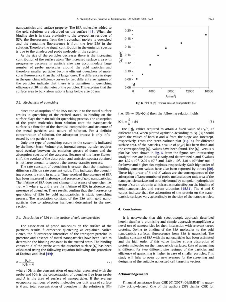
nanoparticles and surface property. The BSA molecules added tothe gold solutions are adsorbed on the surface [48]. When thebinding site is in close proximity to the tryptophan residues ofBSA, the fluorescence from the tryptophan moiety is quenchedand the remaining fluorescence is from the free BSA in thesolution. Therefore the signal contribution in the emission spectrais due to the unadsorbed probe molecule in the system.
As the size of the particles decreases there is the increasingcontribution of the surface atom. The increased surface area withprogressive decrease in particle size can accommodate largenumber of probe molecules around the gold particles andtherefore smaller particles become efficient quenchers of mole-cular fluorescence than that of larger ones. The difference in slopein the quenching efficiency curves for two different size regimes ofthe particles indicate that there is a transition in quenchingefficiency at 30 nm diameter of the particles. This explains that thesurface area to bulk atom ratio is large below size 30 nm.
A (nm2)
Fig. 6. Plot of [Q]T versus area of nanoparticles (A).
3.3. Mechanism of quenchingSince the adsorption of the BSA molecule to the metal surfaceresults in quenching of the excited states, so binding on thesurface plays the main role for quenching process. The adsorptionof the probe molecules from solution onto the nanoparticlesurface is a function of the chemical composition and structure ofthe metal particles and nature of solution. For a definiteconcentration of solution, the adsorption process is only influ-enced by the particle size.
Only one type of quenching occurs in the system is indicatedby the linear Stern–Volmer plot. Internal energy transfer requiresgood overlap between the emission spectra of donor and theabsorption spectra of the acceptor. Because of the large Stokes-shift, the overlap of the absorption and emission spectra obtainedis not large enough to support the energy-transfer process.
The rate constant of quenching is greater than the maximumdiffusion collision rate constant value. This indicates the quench-ing process is static in nature. Time-resolved fluorescence of BSAhas been measured in absence and presence of gold nanoparticles.The lifetime of BSA does not change in presence of quencher, i.e.t0/t ¼ 1 where t0 and t are the lifetime of BSA in absence andpresence of quencher. These results confirm that the fluorescencequenching of BSA by gold nanoparticles is static quenchingprocess. The association constant of the BSA with gold nano-particles due to adsorption has been determined in the nextsection.
3.4. Association of BSA on the surface of gold nanoparticles
The association of probe molecules on the surface of theparticles results fluorescence quenching as explained earlier.Hence, the fluorescence intensities of the transport proteins inpresence and absence of metal nanoparticles have been used todetermine the binding constant in the excited state. The bindingconstant, K of the probe with the quencher surface (Q) has beencalculated using the following equation following the procedureof Encinas and Lissi [49]:
K ¼½Q �P½Q �F � A
(2)
where [Q]P is the concentration of quencher associated with theprobe and [Q]F is the concentration of quencher free from probeand A is the area of surface of nanoparticles. If the averageoccupancy numbers of probe molecules per unit area of surfaceis n̄ and total concentration of quencher in the solution is [Q]T
(i.e. [Q]T ¼ [Q]P+[Q]F) then the following relation holds:
½Q �T ¼n̄
Kþ n̄A (3)
The [Q]T values required to attain a fixed value of (F0/F) atdifferent area, when plotted against A according to Eq. (3) shouldyield the values of both n̄ and K from the slope and intercept,respectively. From the Stern–Volmer plot (Fig. 4) for differentsurface area, of the particles, a value of (F0/F) has been fixed andthe corresponding [Q]T values have been found. The [Q]T versus A
plot has been shown in Fig. 6. From the figure, two intersectingstraight lines are indicated clearly and determined n̄ and K valuesare 1.37�104, 2.07�1014 and 3.89�103, 3.01�1013 dm3 mol�1
for lower and higher size regimes, respectively. Such high order ofbinding constant values have also been reported by others [50].These high order of n̄ and K values are the consequences of theadsorption of large number of probe molecules per unit area of thenanoparticle surface and strongly bound by nonpolar hydrophobicgroup of serum albumin which act as main effect on the binding ofgold nanoparticles and serum albumins [45,51]. The n̄ and K
values indicate that the adsorption of the protein on the nano-particle surfaces vary accordingly to the size of the nanoparticles.
4. Conclusions
It is noteworthy that this spectroscopic approach describedherein signifies a promising and simple approach exemplifying anovel use of nanoparicles for their interaction with relevant targetproteins. Owing to binding of the BSA molecules to the goldnanoparticle surfaces, fluorescence from BSA is quenched. Thebinding constant of BSA with the nanoparticles has been estimatedand the high order of this value implies strong adsorption ofprotein molecules on the nanoparticle surfaces. Rate of quenchingis different for two different size regimes of the particles andefficiency of quenching is higher in case of smaller particles. Thisstudy will help to open up new avenues for the screening anddesigning of the suitable nanosized cell targeting vectors.
Acknowledgements
Financial assistance from CSIR (01(2057)/06/EMR-II) is grate-fully acknowledged. One of the authors (SP) thanks CSIR for

ARTICLE IN PRESS
S. Pramanik et al. / Journal of Luminescence 128 (2008) 1969–19741974
proving SRF and A.S thanks UGC for providing JRF. Thanks to thereviewers for their valuable comments.
References
[1] Y.Y. Yu, S.S. Chang, C.L. Lee, C.R.C. Wang, J. Phys. Chem. B 101 (1997) 6661.[2] J. Zhu, W. Yongchang, L. Yimin, Colloid Surf. A 232 (2004) 155.[3] A.A. Lazarides, K.L. Kelly, T.R. Jensen, G.C. Schatz, J. Mol. Struct. 529 (2000) 59.[4] S.K. Ghosh, A. Pal, S. Kundu, S. Nath, T. Pal, Chem. Phys. Lett. 395 (2004) 366.[5] L. Pasquato, P. Pengo, P. Scrimin, J. Mater. Chem. 14 (2004) 3481.[6] A. Filankemho, M.P. Pileni, J. Phys. Chem. B 104 (2000) 5865.[7] C. Burde, X. Chen, R. Narayanan, M.A. El-Sayed, Chem. Rev. 105 (2005) 1025.[8] S. Pramanik, S.C. Bhattacharya, T. Imae, J. Lumin. 126 (2006) 155.[9] S. Pramanik, D. Das, K. Das, S.C. Bhattacharya, J. Nanosci. Nanotechnol. 7
(2007) 663.[10] J. Zhang, B. Han, D. Liu, J. Chen, Z. Liu, T. Mu, R. Zhang, G. Yang, Phys. Chem.
Chem. Phys. 6 (2004) 2391.[11] N.R. Jana, L. Gearheart, C.J. Murphy, J. Phys. Chem. B 105 (2001) 4065.[12] H. Fan, E.W. Leve, C. Scullin, J. Gabaldon, D. Tallant, S. Bunge, T. Boyle, M.C.
Wilson, C.J. Brinker, Nano Lett. 5 (2005) 645.[13] A. Ghezelbash, B. Koo, B.A. Korgel, Nano Lett. 6 (2006) 1832.[14] N.L. Rosi, C.A. Mirkin, Chem. Rev. 105 (2005) 1547.[15] N. Wu, L. Fu, M. Su, M. Aslam, K.C. Wong, V.P. Dravid, Nano Lett. 4 (2004) 383.[16] P. Apostoli, J. Chromatogr. B: Anal. Technol. Biomed. Life Sci. 778 (2002) 63.[17] D.E. Reichert, M.J. Welch, Chem. Rev. 212 (2001) 111.[18] A.V. Singh, B.M. Bandgar, M. Kasture, B.L.V. Prasad, M. Sastry, J. Mater. Chem.
15 (2005) 5115.[19] R. Elghanian, J.J. Storhoff, R.C. Mucic, R.L. Letsinger, C.A. Mirkin, Science 277
(1997) 1078.[20] B. Dubertret, M. Calame, A.J. Libchaber, J. Nat. Biotechnol. 19 (2001) 365.[21] D.J. Maxwell, J.R. Taylor, S. Nie, J. Am. Chem. Soc. 124 (2002) 9606.[22] C.K. Kim, R.R. Kalluru, J.P. Singh, A. Fortner, J. Griffin, G.K. Darbha, P.C. Ray,
Nanotechnology 17 (2006) 3085.[23] P.C. Ray, A. Fortner, G.K. Darbha, J. Phys. Chem. B 110 (2006) 20745.[24] K. Aslan, V.H. Pe0rez-Luna, J. Fluorescence 14 (2004) 401.[25] H. Imahori, S. Fukuzumi, Adv. Mater. 13 (2001) 1197.[26] A. Aguila, R.W. Murray, Langmuir 16 (2000) 5949.
[27] J. Hu, J. Zhang, F. Liu, K. Kittredge, J.K. Whitesell, M.A. Fox, J. Am. Chem. Soc.123 (2001) 1464.
[28] B.I. Ipe, K.G. Thomas, S. Barazzouk, S. Hotchandani, P.V. Kamat, J. Phys. Chem.B 106 (2002) 18.
[29] T. Huang, R.W. Murray, Langmuir 18 (2002) 7077.[30] T. Gu, J.K. Whitesell, M.A. Fox, Chem. Mater. 15 (2003) 1358.[31] M. Montalti, L. Prodi, N. Zaccheroni, G. Battistini, Langmuir 20 (2004)
7884.[32] M.H.V. Werts, H. Zaim, M. Blanchard-Desce, Photochem. Photobiol. Sci. 3
(2004) 29.[33] Z. Gueroui, A. Libchaber, Phys. Rev. Lett. 93 (2004) 166108(1).[34] E. Dulkeith, M. Ringler, T.A. Klar, J. Feldmann, A.M. Javier, W.J. Parak, Nano
Lett. 5 (2005) 585.[35] R. Kurrat, J.J. Ramsden, J.E. Prenosil, J. Chem. Soc. Faraday Trans. 90 (1994)
587.[36] R. Kurrat, J.E. Prenosil, J.J. Ramsden, J. Colloid Interface Sci. 185 (1997) 1.[37] O. Deschaume, K.L. Shafran, C.C. Perry, Langmuir 22 (2006) 10078.[38] M. Mikhaylova, D.K. Kim, C.C. Berry, A. Zagorodni, M. Toprak, A.S.G. Curtis, M.
Muhammed, Chem. Mater. 16 (2004) 2344.[39] B. Haldar, A. Mallick, N. Chattopadhyay, J. Photochem. Photobiol. B: Biol. 80
(2005) 217.[40] S.K. Ghosh, Sk.U. Hossain, S. Bhattacharya, S.C. Bhattacharya, J. Photochem.
Photobiol. B: Biol. 81 (2005) 121.[41] A. Mallick, S.C. Bera, S. Maiti, N. Chattopadhyay, Biophys. Chem. 112 (2004) 9.[42] A. Mallick, B. Haldar, N. Chattopadhyay, J. Phys. Chem. B 109 (2005)
14683.[43] G. Frens, Nat. Phys. Sci. 241 (1973) 20.[44] J.-S. Lee, S.I. Stoeva, C.A. Mirkin, J. Am. Chem. Soc. 128 (2006) 8899.[45] H.H. Yu, D.S. Jiang, Specrosc. Spectral Anal. (China) 22 (2002) 511.[46] D. Gao, Y. Tian, S. Bi, Y. Chen, A. Yu, H. Zhang, Spectrochim. Acta Part A 62
(2005) 1203.[47] J.R. Lakowicz, Principles of Fluorescence Spectroscopy, Plenum Press, New
York, 1999, p. 267 (Chapter 9).[48] S.H. Brewer, W.R. Glomm, M.C. Johnson, M.K. Knag, S. Franzen, Langmuir 21
(2005) 9303.[49] M.V. Encinas, E.A. Lissi, Chem. Phys. Lett. 91 (1982) 55.[50] L.-M. Ao, F. Gao, B.-F. Pan, D.-X. Cui, H.-C. Gu, Chin. J. Chem. 24 (2006) 253.[51] P.R. Selvakannan, S. Mandal, S. Phadtare, A. Gole, R. Pasricha, S.D. Adyanthaya,
M. Sastry, J. Colloid Interface Sci. 269 (2004) 97.
Copyright © 2022 FDOKUMEN