Green Synthesis of Gold Nanoparticles: An Eco-Friendly ...
25
Citation: Budime Santhosh, P.; Genova, J.; Chamati, H. Green Synthesis of Gold Nanoparticles: An Eco-Friendly Approach. Chemistry 2022, 4, 345–369. https://doi.org/ 10.3390/chemistry4020026 Academic Editor: Katharina M. Fromm Received: 16 March 2022 Accepted: 23 April 2022 Published: 25 April 2022 Publisher’s Note: MDPI stays neutral with regard to jurisdictional claims in published maps and institutional affil- iations. Copyright: © 2022 by the authors. Licensee MDPI, Basel, Switzerland. This article is an open access article distributed under the terms and conditions of the Creative Commons Attribution (CC BY) license (https:// creativecommons.org/licenses/by/ 4.0/). Review Green Synthesis of Gold Nanoparticles: An Eco-Friendly Approach Poornima Budime Santhosh * , Julia Genova * and Hassan Chamati * Institute of Solid State Physics, Bulgarian Academy of Sciences, Tzarigradsko Chaussée 72, 1784 Sofia, Bulgaria * Correspondence: [email protected] (P.B.S.); [email protected] (J.G.); [email protected] (H.C.) Abstract: By virtue of their unique physicochemical properties, gold nanoparticles (AuNPs) have gained significant interest in a broad range of biomedical applications such as sensors, diagnosis, and therapy. AuNPs are generally synthesized via different conventional physical and chemical methods, which often use harmful chemicals that induce health hazards and pollute the environment. To overcome these issues, green synthesis techniques have evolved as alternative and eco-friendly approaches to the synthesis of environmentally safe and less-expensive nanoparticles using naturally available metabolites from plants and microorganisms such as bacteria, fungi, and algae. This review provides an overview of the advances in the synthesis of AuNPs using different biological resources with examples, and their profound applications in biomedicine. A special focus on the biosynthesis of AuNPs using different medicinal plants and their multifunctional applications in antibacterial, anti-inflammatory, and immune responses are featured. Additionally, the applications of AuNPs in cancer theranostics, including contrast imaging, drug delivery, hyperthermia, and cancer therapeutics, are comprehensively discussed. Moreover, this review will shed light on the importance of the green synthesis approach, and discuss the advantages, challenges, and prospects in this field. Keywords: gold nanoparticles; green materials; biosynthesis; plant extracts; microorganisms; biomedical applications 1. Introduction Nanotechnology is a promising field that integrates the various disciplines of science, engineering, and technology. The rapid scientific advances in this field have led to the development of different types of functional nanoparticles (NPs), with at least one di- mension in the typical size range of 1 to 100 nm. Among the various metallic NPs, gold nanoparticles (AuNPs) have attracted huge attention due to their unique surface plasmon resonance properties, facile synthesis, tunable sizes, and multifunctional abilities with well- characterized properties [1,2]. They are versatile materials, relatively inert, biocompatible, and generally stable. Due to their well-defined surface chemistry, AuNPs can be easily conjugated with different molecules such as proteins, dyes, drugs, antibodies, enzymes, and nucleic acids [3–5]. AuNPs functionalized with different targeting moieties have enormous scope in various biomedical applications such as diagnosis, targeting, drug/nucleic acid de- livery, imaging, and therapy (Figure 1). Furthermore, by employing the surface-enhanced Raman scattering technique, AuNPs are used as sensitive probes in Raman scattering and imaging applications [6]. The potential of AuNPs in biomedical fields has been tremen- dously increased by virtue of their applications in photothermal therapy, radiation therapy, computed tomography, biosensors, etc. [7]. Due to their intrinsic electrical and optical properties, as well as their ability to conjugate with different biomolecules, AuNPs-based biosensors with high sensitivity and selectivity are being developed [8,9]. In the last decade, AuNPs-based biosensors have attracted great attention in the diagnosis of various types of diseases. Recently, Antonio et al. [10] highlighted the various AuNPs-based biological assays for the detection and quantification of analytes in urinary samples, with a focus on protein analysis. Such assays using AuNPs are useful in the diagnosis of several illnesses such as kidney disorders, cancer, and heart diseases [4,11]. Chemistry 2022, 4, 345–369. https://doi.org/10.3390/chemistry4020026 https://www.mdpi.com/journal/chemistry
-
Upload
khangminh22 -
Category
Documents
-
view
1 -
download
0
Transcript of Green Synthesis of Gold Nanoparticles: An Eco-Friendly ...

Citation: Budime Santhosh, P.;
Genova, J.; Chamati, H. Green
Synthesis of Gold Nanoparticles: An
Eco-Friendly Approach. Chemistry
2022, 4, 345–369. https://doi.org/
10.3390/chemistry4020026
Academic Editor: Katharina M.
Fromm
Received: 16 March 2022
Accepted: 23 April 2022
Published: 25 April 2022
Publisher’s Note: MDPI stays neutral
with regard to jurisdictional claims in
published maps and institutional affil-
iations.
Copyright: © 2022 by the authors.
Licensee MDPI, Basel, Switzerland.
This article is an open access article
distributed under the terms and
conditions of the Creative Commons
Attribution (CC BY) license (https://
creativecommons.org/licenses/by/
4.0/).
Review
Green Synthesis of Gold Nanoparticles: An Eco-Friendly ApproachPoornima Budime Santhosh * , Julia Genova * and Hassan Chamati *
Institute of Solid State Physics, Bulgarian Academy of Sciences, Tzarigradsko Chaussée 72, 1784 Sofia, Bulgaria* Correspondence: [email protected] (P.B.S.); [email protected] (J.G.); [email protected] (H.C.)
Abstract: By virtue of their unique physicochemical properties, gold nanoparticles (AuNPs) havegained significant interest in a broad range of biomedical applications such as sensors, diagnosis,and therapy. AuNPs are generally synthesized via different conventional physical and chemicalmethods, which often use harmful chemicals that induce health hazards and pollute the environment.To overcome these issues, green synthesis techniques have evolved as alternative and eco-friendlyapproaches to the synthesis of environmentally safe and less-expensive nanoparticles using naturallyavailable metabolites from plants and microorganisms such as bacteria, fungi, and algae. This reviewprovides an overview of the advances in the synthesis of AuNPs using different biological resourceswith examples, and their profound applications in biomedicine. A special focus on the biosynthesisof AuNPs using different medicinal plants and their multifunctional applications in antibacterial,anti-inflammatory, and immune responses are featured. Additionally, the applications of AuNPs incancer theranostics, including contrast imaging, drug delivery, hyperthermia, and cancer therapeutics,are comprehensively discussed. Moreover, this review will shed light on the importance of the greensynthesis approach, and discuss the advantages, challenges, and prospects in this field.
Keywords: gold nanoparticles; green materials; biosynthesis; plant extracts; microorganisms;biomedical applications
1. Introduction
Nanotechnology is a promising field that integrates the various disciplines of science,engineering, and technology. The rapid scientific advances in this field have led to thedevelopment of different types of functional nanoparticles (NPs), with at least one di-mension in the typical size range of 1 to 100 nm. Among the various metallic NPs, goldnanoparticles (AuNPs) have attracted huge attention due to their unique surface plasmonresonance properties, facile synthesis, tunable sizes, and multifunctional abilities with well-characterized properties [1,2]. They are versatile materials, relatively inert, biocompatible,and generally stable. Due to their well-defined surface chemistry, AuNPs can be easilyconjugated with different molecules such as proteins, dyes, drugs, antibodies, enzymes, andnucleic acids [3–5]. AuNPs functionalized with different targeting moieties have enormousscope in various biomedical applications such as diagnosis, targeting, drug/nucleic acid de-livery, imaging, and therapy (Figure 1). Furthermore, by employing the surface-enhancedRaman scattering technique, AuNPs are used as sensitive probes in Raman scattering andimaging applications [6]. The potential of AuNPs in biomedical fields has been tremen-dously increased by virtue of their applications in photothermal therapy, radiation therapy,computed tomography, biosensors, etc. [7]. Due to their intrinsic electrical and opticalproperties, as well as their ability to conjugate with different biomolecules, AuNPs-basedbiosensors with high sensitivity and selectivity are being developed [8,9]. In the last decade,AuNPs-based biosensors have attracted great attention in the diagnosis of various typesof diseases. Recently, Antonio et al. [10] highlighted the various AuNPs-based biologicalassays for the detection and quantification of analytes in urinary samples, with a focus onprotein analysis. Such assays using AuNPs are useful in the diagnosis of several illnessessuch as kidney disorders, cancer, and heart diseases [4,11].
Chemistry 2022, 4, 345–369. https://doi.org/10.3390/chemistry4020026 https://www.mdpi.com/journal/chemistry

Chemistry 2022, 4 346
Figure 1. Multifunctional applications of gold nanoparticles.
Gold nanomaterials can be synthesized via different techniques in various inter-nal structure, sizes, and shapes structures, including nanospheres, nanorods, nanocubes,nanoshells, nanowires, nanocages, nanoflowers, etc. [12]. They exhibit exceptional proper-ties such as fluorescence, attenuation of X-rays, etc., and act as excellent contrast agents foroptical, fluorescence, X-ray, and photoacoustic imaging [13,14]. Their intrinsic features (op-tics, electronics, and physicochemical characteristics) can be altered by adjusting their sizeand shape. To improve their compatibility and stability in a biological environment, AuNPsare coated with different biomolecules such as phospholipids, proteins, and polymers suchas polyethylene glycol [15,16]. By fine-tuning the aspect ratio (length/width of the particle),gold nanorods can be manipulated to absorb light very strongly in the near-infrared region,convert it into heat energy, and transmit it to the surrounding environment. This process,called photohyperthermia, is widely used to attenuate cancer cells, where gold nanorodsare administered near the tumor region to destroy the cancer cells without causing muchdamage to the healthy neighboring cells [17,18]. These properties have made AuNPs awidely used nanomaterial for global academic research and in the production of variousindustrial products and medical devices.
2. General Methods for Synthesis of AuNPs2.1. Physicochemical Methods
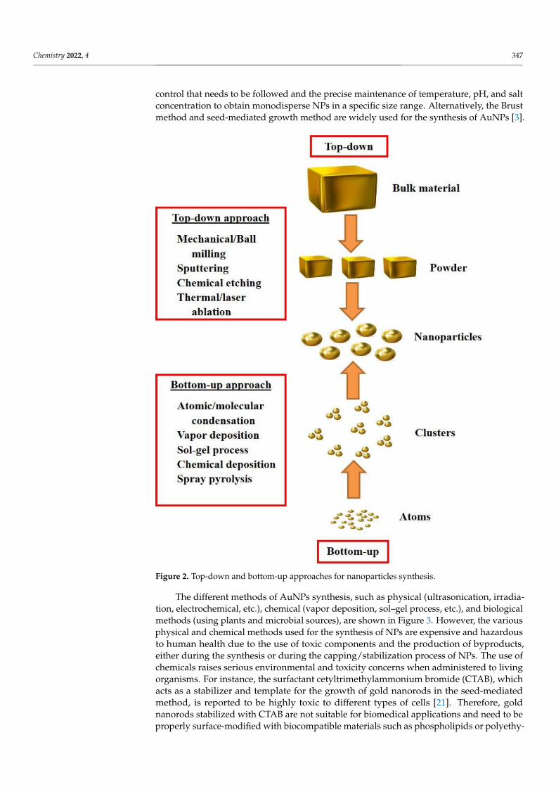
NPs are typically synthesized via two basic methods: “top-down” or “bottom-up”(Figure 2). In the top-down approach, the constituent bulk materials are initially brokendown to powder form and subsequently reduced to fine nanoparticles using varioustechniques such as etching, grinding, sputtering, thermal/laser ablation, etc. On the otherhand, the bottom-up method involves the self-assembly of atoms to form nuclei, whichthen transform into particles of nanoscale range. The bottom-up method is widely used toobtain NPs with uniform morphology and chemical composition. The Turkevich methodis a conventional chemical synthesis method commonly used to produce spherical smallAuNPs around 10 to 30 nm in diameter [19]. However, it was observed that for the synthesisof AuNPs above 30 nm size, the results were less reproducible and resulted in a broadersize distribution of particles [20]. The major limitation of this method is the strict process
Chemistry 2022, 4 347
control that needs to be followed and the precise maintenance of temperature, pH, and saltconcentration to obtain monodisperse NPs in a specific size range. Alternatively, the Brustmethod and seed-mediated growth method are widely used for the synthesis of AuNPs [3].
Figure 2. Top-down and bottom-up approaches for nanoparticles synthesis.
The different methods of AuNPs synthesis, such as physical (ultrasonication, irradia-tion, electrochemical, etc.), chemical (vapor deposition, sol–gel process, etc.), and biologicalmethods (using plants and microbial sources), are shown in Figure 3. However, the variousphysical and chemical methods used for the synthesis of NPs are expensive and hazardousto human health due to the use of toxic components and the production of byproducts,either during the synthesis or during the capping/stabilization process of NPs. The use ofchemicals raises serious environmental and toxicity concerns when administered to livingorganisms. For instance, the surfactant cetyltrimethylammonium bromide (CTAB), whichacts as a stabilizer and template for the growth of gold nanorods in the seed-mediatedmethod, is reported to be highly toxic to different types of cells [21]. Therefore, goldnanorods stabilized with CTAB are not suitable for biomedical applications and need to beproperly surface-modified with biocompatible materials such as phospholipids or polyethy-

Chemistry 2022, 4 348
lene glycol [22]. Hence, there is a growing requirement to search for reliable, non-expensive,biocompatible, and environmentally friendly methods for the synthesis of NPs.
Figure 3. Different methods for synthesis of gold nanoparticles.
2.2. Biosynthetic Mechanism of AuNPs
The mechanism of AuNPs biosynthesis is a simple two-step process and does notrequire a dramatic increase in temperature and pressure. In the first step, the biologicalextract (e.g., plant, bacterial, or fungal extract) is mixed with the HAuCl4 salt solution,which causes the reduction of gold (Au3+) ions to gold atoms (Au0). In the second step,growth and stabilization result in the AuNPs formation (Figure 4). Finally, the color changeof the resulting solution indicates the formation of AuNPs [23,24]. The chemical reactionsinvolved in the reduction of Au3+ to Au0 in the presence of H2O molecules are expressedin the below reactions:
Dissociation: HAuCl4(Chloroauric acid)
H2O−−→ H+ + Au3+ + 4Cl−,
Oxidation: 4Cl− −→ 2Cl2 + 4e−,Reduction: Au3+ + 4e− −→ Au0 + e−.A variety of biocompounds (enzymes, phenols, sugars, etc.) can participate both in the
reduction and stabilization of different types of particles, including AuNPs [25]. Figure 5a,bshow the biosynthesis mechanism of bacterial microorganisms, which can act as a “factory”for the production of AuNPs. The biosynthesis mechanism of the microorganisms canbe either extracellular or intracellular based on the location of AuNPs production [26].Extracellular biosynthesis occurs outside the bacterial cell by trapping and reducing metalions in the presence of enzymes. On the contrary, in the intracellular method, metal ionsare transported into the microbial cell to form NPs in the presence of enzymes [27].

Chemistry 2022, 4 349
Figure 4. Schematic representation of AuNPs biosynthesis mechanism.
Figure 5. Schematic mechanism of (a) extracellular and (b) intracellular AuNPs biosynthesis.
3. Green Synthesis of AuNPs3.1. Importance of Green Synthesis of AuNPs
As the applications of AuNPs are increasing day by day, the demand for their synthesisalso increases simultaneously. It has aroused interest among researchers worldwide todevelop novel interdisciplinary routes for the synthesis of highly stable, monodisperse, and

Chemistry 2022, 4 350
safe AuNPs for various applications. To overcome the challenges in the conventional chem-ical synthesis method, an alternative approach to synthesize biocompatible NPs, termed“green synthesis”, has evolved. It is an emerging branch of nanotechnology and has at-tracted huge attention among researchers and industries, as well as people concerned aboutenvironmental pollution and health hazards. Green synthesis techniques are important asthey are an eco-friendly approach that involves the use of natural bioresources and avoidstoxic chemicals to synthesize different types of NPs [28]. For instance, green synthesisof cobalt ferrite nanoparticles using extracts of grape peel, pulp, and honey-mediatedsynthesis of cobalt-zinc ferrite NPs were reported earlier [29,30].
A variety of plants, microorganisms, and biomolecules derived from them are used asa source for the synthesis of various types of NPs. Extracts from different parts of the plantsuch as leaves, roots, seeds, flowers, fruits, bark, etc., and microbes, including bacteria,fungi, and algae, are widely used to synthesize NPs with varying sizes and shapes usinginterdisciplinary routes [19,31]. The precursor gold salt solution is treated either with themicrobial culture or plant extracts, which are then bioreduced to form AuNPs. Differentmetabolites and biomolecules such as sugars, fatty acids, proteins, enzymes, and phenolsplay a key role in the synthesis of the AuNPs [32,33]. Further, this biological approachinvolves the use of (i) an environmentally acceptable solvent medium for NPs synthesis,(ii) natural reducing agents, and (iii) nontoxic capping agents, mostly polyphenols andother secondary metabolites. Green synthesis of AuNPs using different bioagents and theirapplications is shown in Table 1.
Table 1. Green synthesis of AuNPs using different bioagents and their applications.
Bioagent Size (nm) Shape Application Ref.
Areca catechu 13.7 Spherical Anticancer, Antibacterial, Antioxidant,Catalyst [34]
Mangifera indica Linn 6–18 Spherical Drug delivery [35]Olive leaves 50–100 Spherical, Triangular, Hexagonal Antioxidant [36]
Citrus limon 15–80 Spherical, Triangular Anticancer, Antimicrobial,Anti-inflammatory [37]
Coreopsis lanceolata 20–30 Spherical Detections of aflatoxins [38]Musa paradisiaca <50 Spherical, Triangular Anticancer [39]Zingiber officinale 5–20 Spherical, Triangular, Hexagonal Antibacterial [40]Cocoa extract 150–200 Spherical, prismatic, Rod Photothermal Therapy, contrast agents [41]Capsicum annuum var.grossum 6–37 Quasi-spherical, Triangular, Hexagonal Catalyst [42]
Citrus maxima 25 Spherical Catalyst [43]Trianthema decandra 17.9–79.9 Spherical, Hexagonal, Cubical Antimicrobial [44]Mammea suriga 22–50 Spherical, Square Antibacterial [45]Abelmoschus esculentus 45–75 Spherical Antifungal [46]Shewanella oneidensis 2–50 Spherical Antibacterial [47]Streptomyces sp. 90 Cubical Antifungal [48]Gordonia amara 15–40 Spherical, Polycrystalline Biosensor [49]Bacillus stearothermophilus 5–30 Spherical, Triangular and other Biosensor [50]Pseudomonas fluorescens 5–50 Spherical Bactericidal [51]Stenotrophomonas malophilia 40 Spherical Bioremediation [52]Sporosarcina koreensis DC4 30–50 Spherical Catalyst [53]Rhizopus oryzae 10 Nanocrystalline Pesticides [54]Fusarium semitectum 18–50 Spherical Optoelectronics [55]Candida albicans 20–80 Spherical, Non-spherical Detection of liver cancer [56]Volvariella volvacea 20–150 Spherical,Triangular, Hexagonal Therapeutic [57]Helminthosporum solani 2–70 Polydispersed Anticancer drug [58]Penicillium brevicompactum 10–50 Spherical Anticancer [59]Verticillium sp. 20 Spherical Biomedical [60]Alternaria alternata 12 ± 5 Spherical, Triangular Biomedical [61]Verticillium luteoalbum <10 Spherical,Triangular, Hexagonal Optics and Sensor [62]Fusarium oxysporum 46–70 Spherical, Triangular Biomedical [63]Aureobasidium pullulans 29 ± 6 Spherical Biomedical [64]Pichia jadinii 10–100 Spherical, Triangular, Hexagonal Optics and Sensor [65]Yarrowia lipolytica 7.5–27 Spherical, Triangular, Hexagonal Biomedical [66]Gracilaria corticata 5 Spherical Antibacterial, antioxidant [67]Shewanella algae 9.6–200 Spherical, Nanoplates Biomedical [68]

Chemistry 2022, 4 351
3.2. Advantages of Green Synthesis of AuNPs
The green synthesis method offers the following advantages over the chemical meth-ods: (i) safety: this method avoids the exposure of chemicals or their toxic byproducts,either during the NPs synthesis step or during their stabilization process; (ii) cost-effective:no external stabilizing agent is required. On the contrary, chemical methods use expensiveand hazardous chemicals and stabilizing agents; (iii) simplicity: biosynthesis of NPs fromplant extract is a simple process; (iv) renewable feedstock: e.g., algal biomass; (v) easyavailability of source materials; (vi) biocompatibility: since natural sources are used tosynthesize and stabilize the particles, AuNPs synthesized via green synthesis methods arebiocompatible to different cell types; (vii) the whole green synthesis process is dynamic,reproducible, and energy-efficient; (viii) suitable for large scale production of NPs forcommercial applications; (ix) AuNPs synthesized via green synthesis method are alsoreported to exhibit antibacterial, antifungal, anticancer, and anti-inflammatory properties,and antioxidant and catalytic activity due to the presence of phytochemicals from the bioex-tract [69,70]. All these factors have rendered the green synthesis approach more rewardingthan conventional methods.
4. Biosynthesis of AuNPs from Plant Sources
Biosynthesis of AuNPs from plant sources is facile and involves a single-step processin the one-pot method. To synthesize AuNPs from plant sources, different parts of the plant(leaves, fruits, bark, flower, peels, seed, rhizome root, etc.) are washed with distilled water,dried, ground into powder or chopped into small pieces, and boiled in distilled water toa specific temperature to obtain the extract. Then, filtration or centrifugation techniquesare used to purify the extract, which is then simply mixed with various concentrations ofgold salt solution (based on the plant parts and their species). The gold salt solution isreduced into AuNPs and the reaction completes in minutes to a few hours. The reactionmixture is further incubated to reduce the gold salt completely, and is visually monitoredby color change. Finally, the synthesized AuNPs are purified by centrifugation and washedthoroughly in water for further use. The whole process is simple, eco-friendly, and can bescaled up easily.
Plants are rich in alkaloids, flavonoids, saponins, steroids, tannins, and other naturalcompounds [71]. The plant extract contains various secondary metabolites, which act asboth reducing and stabilizing agents for the biogenesis of NPs [72]. Numerous reportshave shown the successful synthesis of different types of NPs, such as silver, copper, gold,cobalt, palladium, magnetite, and zinc oxide [73,74]. Although various parts of plants havebeen reported in the biosynthesis of AuNPs, leaves are widely used. Variations in the levelof metabolites content from different plant parts, and even variation among plants, play acrucial role in shaping the morphology of NPs.
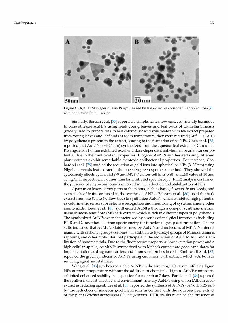
Islam et al. [75] reported a reproducible green synthetic method to produce highlystable AuNPs, using the leaves extract of the plant Salix alba L. (syn: white willow), whichbelongs to the family Salicaceae. The leaves and bark of this plant are rich in phenoliccontents such as salicin, which acts as a precursor in the development of aspirin. Hence, theyare traditionally used for musculoskeletal pain relief and treatment of different ailments,owing to their antipyretic and anti-inflammatory properties. When the aqueous gold ionswere treated with Salix alba L. leaves extract, they were reduced, leading to the synthesisof AuNPs. The UV–Vis absorption spectra data revealed that the synthesized AuNPswere found to be colloidally stable at different pH and salt concentrations. The AuNPsfunctionalized with the phytochemicals of leaf extracts exhibited good antifungal activity,pain-relieving, and muscle relaxant effect, which enhances their potential for variousbiomedical and pharmaceutical applications. Narayan et al. [76] reported the extracellularsynthesis of AuNPs using coriander leaf extract as the reducing agent. Transmissionelectron microscopy (TEM) images have shown the formation of stable AuNPs in the sizerange of 6.75–57.91 nm (Figure 6) with varying shapes, such as spherical, triangle, truncatedtriangles, and decahedral morphologies.
Chemistry 2022, 4 352
Figure 6. (A,B) TEM images of AuNPs synthesized by leaf extract of coriander. Reprinted from [76]with permission from Elsevier.
Similarly, Boruah et al. [77] reported a simple, faster, low-cost, eco-friendly techniqueto biosynthesize AuNPs using fresh young leaves and leaf buds of Camellia Sinensis(widely used to prepare tea). When chloroauric acid was treated with tea extract preparedfrom young leaves and leaf buds at room temperature, they were reduced (Au3+ → Au0)by polyphenols present in the extract, leading to the formation of AuNPs. Chen et al. [78]reported that AuNPs (∼8–25 nm) synthesized from the aqueous leaf extract of CurcumaeKwangsiensis Folium exhibited excellent, dose-dependent anti-human ovarian cancer po-tential due to their antioxidant properties. Biogenic AuNPs synthesized using differentplant extracts exhibit remarkable cytotoxic antibacterial properties. For instance, Cha-hardoli et al. [79] studied the reduction of gold ions into spherical AuNPs (3–37 nm) usingNigella arvensis leaf extract in the one-step green synthesis method. They showed thecytotoxicity effects against H1299 and MCF-7 cancer cell lines with an IC50 value of 10 and25 µg/mL, respectively. Fourier transform infrared spectroscopy (FTIR) analysis confirmedthe presence of phytocompounds involved in the reduction and stabilization of NPs.
Apart from leaves, other parts of the plants, such as barks, flowers, fruits, seeds, andeven peels of fruits, are used in the synthesis of NPs. Bahram et al. [80] used the barkextract from the S. alba (willow tree) to synthesize AuNPs which exhibited high potentialas colorimetric sensors for selective recognition and monitoring of cysteine, among otheramino acids. Leon et al. [81] synthesized AuNPs through a one-pot synthesis methodusing Mimosa tenuiflora (Mt) bark extract, which is rich in different types of polyphenols.The synthesized AuNPs were characterized by a series of analytical techniques includingFTIR and X-ray photoelectron spectrometry for functional group determination. The re-sults indicated that AuMt (colloids formed by AuNPs and molecules of Mt) NPs interactmainly with carbonyl groups (ketones), in addition to hydroxyl groups of Mimosa tannins,saponins, and other molecules that participate in the reduction of Au3+ to Au0 and stabi-lization of nanomaterials. Due to the fluorescence property at low excitation power and ahigh cellular uptake, AuMtNPs synthesized with Mt bark extracts are good candidates forimplementation as drug nanocarriers and fluorescent probes in cells. Elmiitwalli et al. [82]reported the green synthesis of AuNPs using cinnamon bark extract, which acts both asreducing agent and stabilizer.
Wang et al. [83] synthesized stable AuNPs in the size range 10–30 nm, utilizing ligninNPs at room temperature without the addition of chemicals. Lignin–AuNP compositesexhibited enhanced stability in suspension for more than 7 days. Parida et al. [84] reportedthe synthesis of cost-effective and environment-friendly AuNPs using onion (Allium cepa)extract as reducing agent. Lee et al. [85] reported the synthesis of AuNPs (32.96 ± 5.25 nm)by the reduction of aqueous gold metal ions in contact with the aqueous peel extractof the plant Garcinia mangostana (G. mangostana). FTIR results revealed the presence of
Chemistry 2022, 4 353
phenols, flavonoids, benzophenones, and anthocyanins, which suggests that they may actas reducing agent.
Reports [86,87] have revealed that the temperature and pH of the reaction mixture playan important role in determining the final size and shape of the synthesized AuNPs. Forexample, Bogireddy et al. [86] described the formation of size-tunable crystalline AuNPsusing sundried Coffea arabica seed (CAS) extract at room temperature. The influenceof pH on the size of AuNPs was investigated by manipulating the pH of the reactionmixture (pH 5, 7, 9, and 11). The size, shape, and crystallinity of the NPs were analyzedusing different techniques, including TEM and X-ray diffraction (XRD). The results showedthe formation of larger NPs (∼69 nm) at lower pH value (∼5), which was probably dueto the limited availability of capping agents (OH− functional groups), whereas smaller,quasi-spherical NPs (∼13 nm) were formed at higher pH values (>10). Thus, the obtainedresults stipulate the possibility to manipulate the size and shape anisotropy of NPs bycontrolling the pH of the reaction mixture. FTIR results revealed that the phenolic groupspresent in the CAS extract helped to reduce Au3+ to Au0 and stabilize the synthesizedAuNPs. Similarly, Oueslati et al. [87] reported the synthesis of ultra-small and large AuNPsusing polyphenol extracted from the Salvia officinalis plant. In both alkaline (pH∼11) andacidic media (pH∼5), polyphenols induced rapid reduction of the Au (III) salt and led tothe formation of highly monodisperse, ultra-small (∼6 nm), and larger (∼27 nm) sphericalAuNPs, respectively. FTIR results revealed that different polyphenols were capped ontothe surface of NPs favoring high colloidal stability.
Anbu et al. [88] synthesized spherical-shaped AuNPs with an average size of 15 nmusing Platycodon grandiflorum (balloon flower plant) extracts and evaluated their an-tibacterial potential against Escherichia coli and Bacillus subtilis. The synthesized AuNPssignificantly inhibited bacterial growth and demonstrated their antibacterial applications.Sett et al. [89] reported a novel method of AuNPs synthesis using aqueous fruit extract ofDillenia indica. The high phenolic content of the aqueous core extract of D. indica with astrong antioxidant property helped in the reduction of gold ions to AuNPs. The phytochem-icals present in the fruit extract act as an effective reducing and capping agent to synthesizeAuNPs. TEM images of AuNPs revealed an average size range of 5–50 nm, which is verypromising for most biological applications. The synthesized AuNPs did not show any formof cytotoxicity in the normal fibroblast cell line L929, thus proving their compatibility.
Elia et al. [90] compared the biocompatibility and stability of AuNPs synthesizedusing the extracts of the following four different plants: Salvia officinalis, Lippia citri-odora, Pelargonium graveolens, and Punica granatum. When chloroauric ions were treatedwith the extract of different plants, the gold ions were reduced to gold atoms, which thenaggregated to form AuNPs. TEM images have shown the formation of smaller spheri-cal/triangular NPs starting from about 10 nm in size, whereas larger particles (∼150 nm)were also formed with different geometrical shapes, such as triangles, pentagons, andhexagons. The cytotoxicity studies of all the synthesized AuNPs on L-cells (a murinefibroblast cell line) did not show deleterious effects and expressed biocompatibility as wellas high stability for over 3 weeks. Therefore, the synthesized NPs have the potential to beapplied in biomedical applications. Literature data have shown the formation of AuNPs ofvarying geometrical shapes when extracts from different plants were used [91,92].
Rao et al. [93] reported the green synthesis of AuNPs (20–30 nm) by reducing chloroau-ric acid with flower and leaf extracts of Ocimum tenuiflorum, leaves of Azadirachta indicaand Mentha spicata, and peel of Citrus sinensis plants. The synthesized AuNPs weretested on pathogenic Staphylococcus aureus (Gram-positive) and Pseudomonas aeruginosa(Gram-negative) bacteria, which are dangerous to humans and other living organisms.The phytochemicals from the plant extract formed in situ capping on the NPs surfaceand exhibited antibacterial properties up to 99%. The toxicity study inferred that thephytochemicals-capped AuNPs ruptured the bacterial cell wall and affected the normalmetabolic process of pathogenic bacteria. Interestingly, the phytochemicals-capped AuNPsproduced via green synthesis exhibited higher antibacterial activities than the other metallic
Chemistry 2022, 4 354
NPs produced by chemical methods. This proves the benefits of NPs synthesized throughgreen routes compared to chemical methods and their superior features for biomedicalapplications.
5. Biosynthesis of AuNPs Using Microorganisms
Green synthesis of AuNPs using different types of microorganisms such as bacteria,fungi, algae, actinomycetes, etc., has triggered great interest in industrial microbiologyowing to the numerous benefits they offer. Easy handling and processing, low-cost mediumfor their growth, and ability to adsorb and reduce various metal ions into NPs are someof the attractive reasons [19,94,95]. Large-scale cultivation of microbes in bulk fermenterswill enable the surplus extraction of enzymes and various secondary metabolites in a lesseconomical way. Various fungal strains can be cultivated on different substrates suchas cellulosic wastes, coir-pith, and agricultural wastes, thereby enabling the usage ofless-expensive raw materials for their growth, helping in waste recycling and reducingenvironmental pollution [96].
Microorganisms such as bacteria, filamentous fungi, yeast, algae, and actinomyceteshave huge scope in the bioremediation process and have the potential to degrade con-taminants, such as heavy metals, dyes, and toxic chemicals, that pose environmental andhuman risks [97,98]. In other words, microbes can be effectively utilized to biologicallydegrade harmful pollutants into nontoxic substances.
5.1. Fungi and Algae
Fungi are excellent candidates for large-scale production of NPs because of the simplic-ity, high scalability, downstream processing, easy handling, and cost-efficiency of fungalgrowth on both the laboratory and the industrial scale. The filamentous fungi are wellknown for their high metal tolerance and bioaccumulation properties. Fungal cell wallspossess different functional groups such as amine, carboxyl, sulfhydryl, hydroxyl, andphosphate groups, that act as ligands and help to chelate metal ions [99]. Further, theysecrete a wide array of proteins and enzymes such as ATPase, 3-glucanase, hemicellulose,glyceraldehyde-3-phosphate dehydrogenase, cell wall lytic enzyme β-1, etc., which playan important role in the feasible, large-scale synthesis of metallic NPs. Fungi such asPenicillium chrysogenum, Fusarium oxysporum, and Verticillium sp. are reported in thebiosynthesis of metallic NPs such as platinum, silver, silicon, and titanium [100].
Numerous reports have elaborated the biogenesis of AuNPs using unicellular andmulticellular fungi [56,101]. Extracellular or intracellular extracts of different fungi such asCandida albicans, Aspergillus niger, Aspergillus clavatus, and Penicillium sp. are widelyused for the synthesis of AuNPs [102,103]. Priyadarshini et al. [104] reported an ecofriendly,ambient temperature protocol for size-controlled synthesis of AuNPs, using the fungusAspergillus terreus IF0. AuNPs were formed immediately by adding chloroauric acid tothe aqueous fungal culture extract. TEM results have revealed that the particles were foundto be in the size range of 10–19 nm. FTIR analysis has indicated the presence of carboxyl,amino, and thiol functional groups from the fungal extracts, which were responsible forboth bioreduction and stabilization of NPs. The synthesized AuNPs demonstrated excellentantibacterial activity against the Gram-negative bacteria, Escherichia coli, and have excitingscope in clinical applications.
Quite recently, Nguyen et al. [105] demonstrated the green-synthesis of silver andgold NPs using Ganoderma lucidum, mushroom extract, as reducing and capping agents.The synthesized NPs showed excellent catalytic, antibacterial activity, and colorimetricdetection of Fe3+ ions in real water systems and exhibited their outstanding propertiesin environmental and biotechnological applications. Sastry et al. [106] observed the in-tracellular and extracellular production of AuNPs using two different genera of fungi,Verticillium sp. and Fusarium oxysporum. When the aqueous gold and silver ions wereexposed to Verticillium sp., the metal ions were reduced intracellularly to form gold andsilver NPs in the size range 2–20 nm. On the other hand, the same aqueous gold and silver
Chemistry 2022, 4 355
ions were reduced extracellularly in the case of F. oxysporum, leading to the formation of goldand silver NPs around 2–50 nm in size. The active biomolecules produced by the fungi,the concentration of precursor gold salt solution, and optimization of the experimentalconditions play an important role in controlling the size distribution, shape, and biochem-ical composition of the synthesized NPs. For instance, Dhanasekar et al. [107] explaineda simple and eco-friendly approach to synthesize AuNPs of different sizes (7–93 nm) andshapes by exposing the cell-free filtrate of filamentous fungus Alternaria sp. to three differ-ent concentrations (0.3, 0.5, and 1 mM) of chloroauric solution. In all cases, the Au3+ ionswere reduced to Au0, leading to the formation of stable AuNPs. TEM analysis has revealedthe presence of spherical, square, rod, pentagonal, and hexagonal morphologies for 1 mMchloroauric solution and quasi-spherical and spherical NPs for lower concentrations (0.3and 0.5 mM) of chloroauric solution. FTIR analysis has revealed the presence of aromaticprimary amines, amino acids such as tryptophan/tyrosine, or phenylalanine as the cappingand stabilizing agents on the surface of AuNPs.
Different approaches have been used for the biosynthesis of AuNPs from fungalextracts. However, there is no clear knowledge about the limitations of all these methods.To gain better understanding, Molnar et al. [108] investigated 29 different thermophilicfilamentous fungal strains to compare the AuNPs formed using either the extracellularfraction, the autolysate of fungi, or the intracellular fraction of fungi. The results haveshown that AuNPs of varying sizes (6–40 nm) with high standard deviations rangingbetween 30% and 70% were formed based on the difference in the fungal strain andenvironmental conditions. Mishra et al. [59] reported the fungus-mediated synthesisof AuNPs using an industrially important fungus Penicillium rugulosum. TEM resultsrevealed that the size of synthesized NPs was in the range of 20–80 nm. The AuNPs werethen conjugated with isolated genomic DNA of bacteria Escherichia coli and Staphylococcusaureus. Stability analysis results have shown that DNA-conjugated AuNPs were highlystable and monodispersed, which infers that the presence of genomic DNA on the surface ofNPs prevents them from aggregation due to their negatively charged phosphate backbone.Such surface modification of AuNPs will improve their shelf life during in vivo applicationsand enhance their scope in biomedicine.
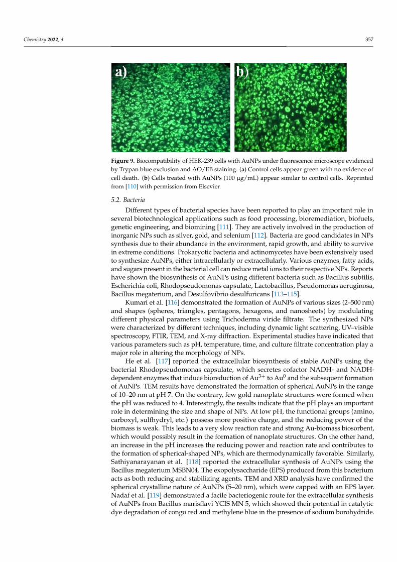
A variety of algae, such as Turbinaria conoides, Spirulina platensis, Galaxaura elongate,and Shewanella algae, are used as bionanofactories for the synthesis of AuNPs [67,68].Singh et al. [109] reported the synthesis of AuNPs using aqueous extract of Dunaliellasalina, a unicellular, halotolerant microalga. The synthesis, characterization, and in vitroanticancer activity of the biosynthesized AuNPs is shown in Figure 7. The anticancerpotential of AuNPs was tested against the breast cancer cell line (MCF7) and normalbreast epithelial cell line (MCF 10A), and commercial anticancer drug cisplatin was usedas a positive control. The cell viability results (Figure 8A–D) have indicated that AuNPssynthesized using D. salina selectively attenuated cancer cells and were not detrimentalto the normal cell line, whereas cisplatin affected normal cells as well at 48 h exposure.Chellapandian et al. [110] demonstrated a facile one-pot synthesis of AuNPs using anaqueous solution of the marine red seaweed, Gracilaria verrucosa. The biocompatibility ofthe synthesized AuNPs was assessed using human embryonic kidney (HEK-293) cells. Thefluorescence microscopy images using Trypan blue exclusion and AO/EB staining haveshown (Figure 9) that the cells treated with biosynthesized AuNPs (100 µg/mL) appearsimilar to control cells, indicating the cell viability.

Chemistry 2022, 4 356
Figure 7. Green synthesis of gold nanoparticles from Dunaliella salina, UV–Vis characterizationspectra, and in vitro anticancer activity on breast cancer cell line. Reprinted from [109] with permissionfrom Elsevier.
Figure 8. Cell viability using MTT assay after treatment with AuNPs and cisplatin on MCF 10A(A,B) and MCF 7 cell lines (C,D). Reprinted from [109] with permission from Elsevier.
Chemistry 2022, 4 357
Figure 9. Biocompatibility of HEK-239 cells with AuNPs under fluorescence microscope evidencedby Trypan blue exclusion and AO/EB staining. (a) Control cells appear green with no evidence ofcell death. (b) Cells treated with AuNPs (100 µg/mL) appear similar to control cells. Reprintedfrom [110] with permission from Elsevier.
5.2. Bacteria
Different types of bacterial species have been reported to play an important role inseveral biotechnological applications such as food processing, bioremediation, biofuels,genetic engineering, and biomining [111]. They are actively involved in the production ofinorganic NPs such as silver, gold, and selenium [112]. Bacteria are good candidates in NPssynthesis due to their abundance in the environment, rapid growth, and ability to survivein extreme conditions. Prokaryotic bacteria and actinomycetes have been extensively usedto synthesize AuNPs, either intracellularly or extracellularly. Various enzymes, fatty acids,and sugars present in the bacterial cell can reduce metal ions to their respective NPs. Reportshave shown the biosynthesis of AuNPs using different bacteria such as Bacillus subtilis,Escherichia coli, Rhodopseudomonas capsulate, Lactobacillus, Pseudomonas aeruginosa,Bacillus megaterium, and Desulfovibrio desulfuricans [113–115].
Kumari et al. [116] demonstrated the formation of AuNPs of various sizes (2–500 nm)and shapes (spheres, triangles, pentagons, hexagons, and nanosheets) by modulatingdifferent physical parameters using Trichoderma viride filtrate. The synthesized NPswere characterized by different techniques, including dynamic light scattering, UV–visiblespectroscopy, FTIR, TEM, and X-ray diffraction. Experimental studies have indicated thatvarious parameters such as pH, temperature, time, and culture filtrate concentration play amajor role in altering the morphology of NPs.
He et al. [117] reported the extracellular biosynthesis of stable AuNPs using thebacterial Rhodopseudomonas capsulate, which secretes cofactor NADH- and NADH-dependent enzymes that induce bioreduction of Au3+ to Au0 and the subsequent formationof AuNPs. TEM results have demonstrated the formation of spherical AuNPs in the rangeof 10–20 nm at pH 7. On the contrary, few gold nanoplate structures were formed whenthe pH was reduced to 4. Interestingly, the results indicate that the pH plays an importantrole in determining the size and shape of NPs. At low pH, the functional groups (amino,carboxyl, sulfhydryl, etc.) possess more positive charge, and the reducing power of thebiomass is weak. This leads to a very slow reaction rate and strong Au-biomass biosorbent,which would possibly result in the formation of nanoplate structures. On the other hand,an increase in the pH increases the reducing power and reaction rate and contributes tothe formation of spherical-shaped NPs, which are thermodynamically favorable. Similarly,Sathiyanarayanan et al. [118] reported the extracellular synthesis of AuNPs using theBacillus megaterium MSBN04. The exopolysaccharide (EPS) produced from this bacteriumacts as both reducing and stabilizing agents. TEM and XRD analysis have confirmed thespherical crystalline nature of AuNPs (5–20 nm), which were capped with an EPS layer.Nadaf et al. [119] demonstrated a facile bacteriogenic route for the extracellular synthesisof AuNPs from Bacillus marisflavi YCIS MN 5, which showed their potential in catalyticdye degradation of congo red and methylene blue in the presence of sodium borohydride.
Chemistry 2022, 4 358
Few bacterial species can survive at toxic metal ion concentrations and extreme tem-peratures. They have unique defense mechanisms to tolerate such high stress and toxicityof metal ions. Ahmad et al. [120] reported the intracellular synthesis of AuNPs using anovel alkalotolerant actinomycete (Rhodococcus species). When the cells were exposed tochloroauric acid, gold ions were rapidly reduced. TEM analysis of thin sections of actino-mycete cells has shown that highly monodispersed NPs in the size range of 5–15 nm wereformed on the cytoplasmic membrane. The synthesized AuNPs were not toxic to the cellsand the cells continued to grow even after the biogenesis of AuNPs. Llanten et al. [121]reported the biosynthesis of AuNPs using a thermophilic bacterium belonging to the genusGeobacillus, strain ID17, isolated from Antarctica. When exposed to Au3+ ions, the bac-terial cells turned from colorless into a dark purple color. This bioconversion process isenzymatically mediated by the reductase enzyme and NADH as cofactors. TEM resultshave shown intracellular accumulation of quasi-hexagonal-shaped AuNPs with sizes rang-ing from 5–50 nm. Sharma et al. [94] exploited a novel strain of Marinobacter pelagius,which belongs to marine bacteria and can tolerate high salt concentration and can evadetoxicity of different metal ions for the production of AuNPs. TEM images have shown theformation of stable and monodisperse AuNPs around 10 nm size upon exposure of thechloroauric acid solution to whole cells. The result indicated that this bacterial strain cansynthesize stable, quick, and monodisperse AuNPs around 2–6 nm in size.
Several bacterial strains have the potential of adsorbing/binding metal ions and re-ducing them into NPs by enzymes produced during metabolic processes in cells, andthis property helps to enhance their applications in bioremediation and bioleaching.Nangia et al. [52] identified a new bacterial strain, Stenotrophomonas malophilia (Au-Red02), and isolated it from gold-enriched soil samples, which can synthesize well-dispersedAuNPs of size about 40 nm. FTIR results showed that the synthesized AuNPs were cappedwith negatively charged phosphate groups, which improves their stability in the aqueousmedium. Kunoh et al. [122] reported that the cells of Leptothrix (iron-oxidizing bacteria)released extracellular RNA which has the ability to reduce Au (III) to form spherical AuNPswhen treated with an aqueous chloroauric acid solution under ambient conditions.
There are some drawbacks in the usage of bacteria for the synthesis of AuNPs. First,maintaining the bacterial culture is a tedious process. Second, safety measures have tobe strictly followed in a clean environment to prevent them from mass contamination.Third, the reduction process is slow and takes time from hours to days. Hence, they arenot a preferable choice for the commercial synthesis of AuNPs. Nevertheless, few recentreports have highlighted that the AuNPs synthesized from various bacterial strains exhib-ited superior properties when compared to the NPs prepared by chemical methods. Forinstance, Li et al. [123] reported that the AuNPs synthesized from Deinococcus radioduransshowed significant antibacterial activity against both Gram-positive and Gram-negativebacteria. This opens up additional scope for the NPs as an antibacterial agent. Similarly,Shabani et al. [124] reported the enzymatic synthesis of AuNPs (∼10 nm) using Escherichiacoli. The synthesized NPs exhibited strong antifungal properties against various humanpathogenic fungi and nontoxicity for Vero and Hep-2 cell lines in vitro at concentrationsranging from 0.31 to 10%.
6. Green Synthesis of Different Types of Nanoparticles
Apart from AuNPs, the green synthesis technique is commonly used to synthesizedifferent types of nanoparticles. For instance, Vinodhini et al. [125] demonstrated the greensynthesis of silver nanoparticles (AgNPs) in the size range of 40–57 nm from the leaf extractsof medicinal herbs such as Tabernaemontana divaricate, Basella alba, and Allium fistulosum. Thebiosynthesised AgNPs were nontoxic and exhibited antibacterial, antifungal, antioxidant,and antidiabetic properties due to the presence of phenolics and other active componentspresent in these medicinal plants. Similarly, Tyagi et al. [126] reported the biosynthesisof magnetic iron oxide nanoparticles (FeNPs) from spinacia oleracea (spinach) and musaacuminata (banana). Iqbal et al. [127] demonstrated the production of zinc oxide nanoparti-
Chemistry 2022, 4 359
cles (ZnONPs) using Elaeagnus angustifolia leaf extracts, and Gulbagca et al. [128] reportedthe green synthesis of palladium nanoparticles (PdNPs) using Urtica plant extracts. Amongthe various types of nanoparticles, AuNPs are highly preferred in biomedical applicationsas they exhibit unique properties such as surface plasmon resonance, nontoxicity, and highbiocompatibility.
7. Biosynthesized AuNPs in Cancer Theranostics: Imaging, Drug Delivery,and Treatment
Cancer is a disease characterized by abnormal and unrestricted cell growth withpotential to spread to other parts of the body. The World Health Organization (WHO)reported nearly 10 million deaths in 2020 and this is expected to increase to an estimated12 million deaths by 2030 [129]. Among the different types of nanomaterials used to treatcancer, biosynthesized AuNPs functionalized with targeting ligands and anticancer drugsare considered as promising candidates in diagnosis and cancer therapy [24,130]. Kimet al. [131] explored this concept to prove the efficacy of AuNPs in treating glioblastomamultiforme (GBM), the most common primary grade 4 brain tumor. They demonstratedthat the oral delivery of AuNPs conjugated with milk protein lactoferrin and polyethyleneglycol (PEG), a biocompatible polymer, was able to cross the blood–brain barrier andbind with lactoferrin receptors that are highly expressed in the brain tumor cells in micemodels. Further, when irradiated with laser light, the administered AuNPs increased thetemperature in GBM due to photothermiaproperties and induced a significant reductionin the tumor volume. A schematic illustration for oral absorption of lactoferrin (Lf)–PEG-conjugated AuNPs and the mechanism of action of Lf–PEG–AuNP targeting Glioblastomamultiforme (GBM) through lactoferrin receptor pathway of the small intestine, the blood–brain barrier, and GBM cells is given in Ref. [131].
AuNPs, either individually or in combination with other treatment modalities suchas radio/chemotherapy, have the ability to induce hyperthermia or deliver the drug inthe targeted region or cell to produce a synergetic effect and thus help to facilitate cancertreatment. For instance, Rezaeian et al. [132] used a green chemistry approach to synthe-size curcumin-coated AuNPs and performed in vitro study to the compare nanoparticle-mediated photothermal therapy and radiofrequency electric field hyperthermia on mousecolorectal cancer (CT26) cell lines. The results have shown that the NPs induced apoptosiscell death considerably using both photothermal therapy and radiofrequency electric fieldhyperthermia treatments. Another study used the green chemistry method to synthesizestable AuNPs coupled with 5-Fluorouracil, a chemotherapeutic drug that is widely usedfor the treatment of liver cancer [133]. This study focused on in vivo toxicity induced byAuNPs in the zebrafish embryo model, in vitro drug release behavior, and efficacy of theNPs in human liver cancer (HepG2) cell lines. In vivo biodistribution analysis indicatedthat a higher amount of AuNPs accumulated in the liver induced significant cytotoxicityin HepG2 cell lines, which signifies that the AuNPs could be used as a tool for both imag-ing and targeted drug delivery with minimal side effects of liver cancer. Various reportsspecifying anticancer activity of AuNPs biosynthesized from different plant sources arementioned in Table 2.
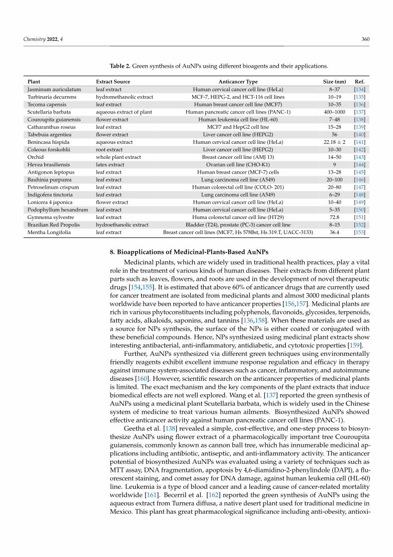
Chemistry 2022, 4 360
Table 2. Green synthesis of AuNPs using different bioagents and their applications.
Plant Extract Source Anticancer Type Size (nm) Ref.Jasminum auriculatum leaf extract Human cervical cancer cell line (HeLa) 8–37 [134]Turbinaria decurrens hydromethanolic extract MCF-7, HEPG-2, and HCT-116 cell lines 10–19 [135]Tecoma capensis leaf extract Human breast cancer cell line (MCF7) 10–35 [136]Scutellaria barbata aqueous extract of plant Human pancreatic cancer cell lines (PANC-1) 400–1000 [137]Couroupita guianensis flower extract Human leukemia cell line (HL-60) 7–48 [138]Catharanthus roseus leaf extract MCF7 and HepG2 cell line 15–28 [139]Tabebuia argentiea flower extract Liver cancer cell line (HEPG2) 56 [140]Benincasa hispida aqueous extract Human cervical cancer cell line (HeLa) 22.18 ± 2 [141]Coleous forskohlii root extract Liver cancer cell line (HEPG2) 10–30 [142]Orchid whole plant extract Breast cancer cell line (AMJ 13) 14–50 [143]Hevea brasiliensis latex extract Ovarian cell line (CHO-K1) 9 [144]Antigonon leptopus leaf extract Human breast cancer (MCF-7) cells 13–28 [145]Bauhinia purpurea leaf extract Lung carcinoma cell line (A549) 20–100 [146]Petroselinum crispum leaf extract Human colorectal cell line (COLO- 201) 20–80 [147]Indigofera tinctoria leaf extract Lung carcinoma cell line (A549) 6–29 [148]Lonicera 4 japonica flower extract Human cervical cancer cell line (HeLa) 10–40 [149]Podophyllum hexandrum leaf extract Human cervical cancer cell line (HeLa) 5–35 [150]Gymnema sylvestre leaf extract Huma colorectal cancer cell line (HT29) 72.8 [151]Brazilian Red Propolis hydroethanolic extract Bladder (T24), prostate (PC-3) cancer cell line 8–15 [152]Mentha Longifolia leaf extract Breast cancer cell lines (MCF7, Hs 578Bst, Hs 319.T, UACC-3133) 36.4 [153]
8. Bioapplications of Medicinal-Plants-Based AuNPs
Medicinal plants, which are widely used in traditional health practices, play a vitalrole in the treatment of various kinds of human diseases. Their extracts from different plantparts such as leaves, flowers, and roots are used in the development of novel therapeuticdrugs [154,155]. It is estimated that above 60% of anticancer drugs that are currently usedfor cancer treatment are isolated from medicinal plants and almost 3000 medicinal plantsworldwide have been reported to have anticancer properties [156,157]. Medicinal plants arerich in various phytoconstituents including polyphenols, flavonoids, glycosides, terpenoids,fatty acids, alkaloids, saponins, and tannins [136,158]. When these materials are used asa source for NPs synthesis, the surface of the NPs is either coated or conjugated withthese beneficial compounds. Hence, NPs synthesized using medicinal plant extracts showinteresting antibacterial, anti-inflammatory, antidiabetic, and cytotoxic properties [159].
Further, AuNPs synthesized via different green techniques using environmentallyfriendly reagents exhibit excellent immune response regulation and efficacy in therapyagainst immune system-associated diseases such as cancer, inflammatory, and autoimmunediseases [160]. However, scientific research on the anticancer properties of medicinal plantsis limited. The exact mechanism and the key components of the plant extracts that inducebiomedical effects are not well explored. Wang et al. [137] reported the green synthesis ofAuNPs using a medicinal plant Scutellaria barbata, which is widely used in the Chinesesystem of medicine to treat various human ailments. Biosynthesized AuNPs showedeffective anticancer activity against human pancreatic cancer cell lines (PANC-1).
Geetha et al. [138] revealed a simple, cost-effective, and one-step process to biosyn-thesize AuNPs using flower extract of a pharmacologically important tree Couroupitaguianensis, commonly known as cannon ball tree, which has innumerable medicinal ap-plications including antibiotic, antiseptic, and anti-inflammatory activity. The anticancerpotential of biosynthesized AuNPs was evaluated using a variety of techniques such asMTT assay, DNA fragmentation, apoptosis by 4,6-diamidino-2-phenylindole (DAPI), a flu-orescent staining, and comet assay for DNA damage, against human leukemia cell (HL-60)line. Leukemia is a type of blood cancer and a leading cause of cancer-related mortalityworldwide [161]. Becerril et al. [162] reported the green synthesis of AuNPs using theaqueous extract from Turnera diffusa, a native desert plant used for traditional medicine inMexico. This plant has great pharmacological significance including anti-obesity, antioxi-

Chemistry 2022, 4 361
dant, antibacterial, anti-inflammatory, antidiabetic, antimycotic, and cytotoxic activities.The cytotoxicity and immunomodulatory effects of AuNPs synthesized using this plantextract were investigated on the leukocytes of longfin yellowtail Seriola rivoliana, a marinefish, and antibacterial activity against Vibrio parahaemolyticus and Aeromonas hydrophila,Gram-negative bacteria. The results indicated that the AuNPs increased the phagocytosisactivity, attenuated the reactive oxygen species in leukocytes production, and increased thecellular antibacterial mechanism mediated by nitric oxide production.
Yasmin et al. [163] employed green techniques for rapid synthesis of spherical AuNPsin the size range of 16–30 nm using Hibiscus rosasinensis, a medicinal plant that has a lotof beneficial applications such as anti-infectious, anthelmintic, anti-inflammatory, diuretic,and antipyretic properties. The synthesized particles were found to be stable for up to afew months and have the potential to be used for medical and biosensor applications. Arecent study reported the synthesis of AuNPs using the aqueous Mentha longifolia leafextract. The major constituents of this plant include polyphenols, alkaloids, organic acids,terpenoids, etc., and this plant has been used as an antihypertensive and antitussive drug intraditional medicine. The biosynthesized nanoparticles were found to be effective againstvarious breast cancer cell lines, such as breast adenocarcinoma (MCF7), breast carcinoma(Hs 578Bst), breast infiltrating ductal cell carcinoma (Hs 319.T), and breast infiltratinglobular carcinoma (UACC-3133) cell lines, without causing cytotoxicity against a normal cellline (HUVEC). Therefore, AuNPs synthesized using Mentha longifolia leaf aqueous extractcan be tested as an anti-breast cancer drug in humans in the near future [153]. Anotherreport described the biogenic synthesis of AuNPs using the Jasminum auriculatum leafextract. The leaves of this plant have numerous medicinal applications such as antilithiatic,wound healing activity, and diuretic activity. They are used in the treatment of leprosy, skindiseases, ulcers, and wounds. The MTT assay performed using these NPs against humancervical cancer (HeLa) cells revealed significant anticancer activity without the requirementof doping additional molecules [134].
Hasan et al. [135] biosynthesized AuNPs from Turbinaria decurrens, which is anEgyptian marine brown macroalga, which has a diverse group of phytochemicals withunique bioactivities and is widely used as food and medicine. The authors compared thechemical composition and antioxidant and anticancer activities of both the hydromethano-lic extract (HME) of this plant and the HME–AuNPs on three different cancer cell lines(MCF-7, HEPG-2, and HCT-116) using MTT assay. The results showed the strong anticanceractivity of AuNPs against all the three studied cell lines. Their findings indicated that thebiosynthesized AuNPs could be used as a source for the discovery of novel therapeuticagents in the biomedical field to treat oxidative stress-related diseases, particularly can-cer. Hosny et al. [136] explained a phytofabrication technique to synthesize AuNPs thatremained stable for up to three months using the aqueous leaf extract of Tecoma capensis, aflowering plant commonly found in tropical and subtropical areas of Africa. The anticancerefficacy of T. capensis–AuNPs was tested against human breast cancer cell line (MCF7)using MTT assay and the results revealed the excellent potency of AuNPs in preventing thedevelopment and proliferation of MCF7 cells. All these studies explain that the synthesis ofAuNPs based on medicinal plants is beneficial to express their health and medical benefitsalong with their multifunctional potential in treating different diseases, including cancer.
9. Challenges and Future Prospects
In the recent decade, the green synthesis approach has been successfully used tosynthesize a variety of NPs, including AuNPs, with varying morphology and properties.Numerous research articles have been published worldwide on the interdisciplinary routesin the synthesis of NPs from different plants and microorganisms. However, there areseveral limitations and drawbacks in the green synthesis method which limit their large-scale production for commercial purposes and diminish their subsequent applications inbiotechnology and nanomedicine. The current challenges are summarized below:
Chemistry 2022, 4 362
• Lack of sufficient data to control the size and shape of NPs: Many reports have demon-strated the formation of AuNPs with different morphologies during the synthesisprocess. The formation of NPs with uniform morphology and narrow size distributionis essential for pharmacological and biological applications. Proper knowledge tooptimize the reaction mixture, experimental time, pH, temperature, rotational speed,concentrations of chloroauric acid concentrations, etc., have to be taken into care tosolve this problem.
• Lack of standardized protocols to reproduce NPs with the same characteristics: NPsfrom different parts even of the same plant have variations in their structure andproperties, as, e.g., AuNPs synthesized from different plant parts exhibit differentlevels of cytotoxicity due to the difference in the antioxidant/metabolite contents.
• Lack of clear knowledge on the mechanism of NPs synthesis is a major drawback,and identification of key components present in different metabolites from plants andmicrobial sources that play an active role in the synthesis of NPs are challenging.
• Separation and purification of NPs from the complex reaction mixture is anotherimportant aspect that still remains a hurdle.
• Scalability of synthesized NPs from a laboratory approach to meet the huge demandsin the industrial and pharmaceutical scale is a major concern.
• A detailed toxicological study of the biosynthesized NPs is crucial to enhance theirscope in diverse fields.
• Technical barriers and regulatory policies for the commercial synthesis of NPs limitthe scale-up process, which needs to be overcome.
• Stability and functionalization: More research on green synthesis is required to surface-modify the synthesized NPs to improve their stability in biological media and func-tionalize them with specific antibodies or peptides to improve their applications indrug delivery and cancer therapy.
Even though the green synthesis approach is very popular, overcoming the abovechallenges could improve the global acceptability and adaptability of the commercialsynthesis of AuNPs. Future research and development in this sector should be directedtowards overcoming the present hurdles and coming up with novel standardized protocols,designing smart and safe AuNPs functionalized with different biomolecules, and targetingmoieties for multifunctional applications.
10. Conclusions
Due to their unique characteristics, AuNPs have enormous applications in variousfields such as electronics, catalysis, optics, sensors, and biology. Though different physic-ochemical methods are used in the synthesis of NPs, the green synthesis method is apromising approach to produce different types of NPs, including gold nanomaterials, in asimple, eco-friendly, and cost-effective manner. This method has several advantages overthe conventional physical and chemical methods used for NPs synthesis, such as safety,and does not involve the use of hazardous chemicals or the addition of external harmfulsubstances during the synthesis/durability of NPs. Different parts of plants and a varietyof microbes, including bacteria, fungi, algae, and yeast, are used as a natural source inthe biosynthesis process. Further, NPs synthesized using biological extracts have severalbeneficial properties, such as high anticancer, antimicrobial, anti-inflammatory, antioxidant,and catalytic activity, etc., which find exciting applications in nanomedicine.
This review article focuses on the “state-of-the-art” research on the “green synthesis” ofAuNPs, different sources of green materials, with special emphasis on biosynthesis of NPsfrom different parts of the plant, intracellular and extracellular synthesis from microbes, andtheir overall applications. Further, the advantage of the green synthesis method over theconventional chemical synthesis methods, current challenges in this field, and the prospectsare discussed in detail. Green synthesis is an emerging field and current research on thebiogenesis of AuNPs, characterization, and functionalization of the synthesized particlesare still in the developing phase. A thorough understanding of the basic principles of green
Chemistry 2022, 4 363
chemistry and more research work is required to gain sufficient knowledge in this field.Addressing the current challenges in this field and overcoming them with standardizedprotocols and innovative techniques can revolutionize the synthesis of AuNPs on bothlaboratory and commercial scales.
Author Contributions: P.B.S.—writing, conceived the study design, data collection, and prepared anoriginal draft; J.G.—review and editing manuscript; H.C.—general guidance, project directing, andmanuscript revisions. All authors have read and agreed to the published version of the manuscript.
Funding: We gratefully acknowledge the financial support from the project KP-06-DB-8/01.12.2020with the Bulgarian National Science Fund under the National Scientific Program P. Beron 2020 andGrant No. D01-229/27.10.2021 from the Ministry of Education and Science of Bulgaria.
Institutional Review Board Statement: Not applicable.
Informed Consent Statement: Not applicable.
Data Availability Statement: The data generated within this research is included in the paper.
Conflicts of Interest: The authors declare no conflict of interest.
References1. Sarfraz, N.; Khan, I. Plasmonic Gold Nanoparticles (AuNPs): Properties, Synthesis and their Advanced Energy, Environmental
and Biomedical Applications. Chem. Asian J. 2021, 16, 720–742. doi:10.1002/asia.202001202.2. Hammami, I.; Alabdallah, N.M.; jomaa, A.A.; kamoun, M. Gold nanoparticles: Synthesis properties and applications. J. King
Saud-Univ.—Sci. 2021, 33, 101560. doi:10.1016/j.jksus.2021.101560.3. Amina, S.J.; Guo, B. Review on the Synthesis and Functionalization of Gold Nanoparticles as a Drug Delivery Vehicle. Int. J.
Nanomed. 2020, 15, 9823–9857. doi:10.2147/IJN.S279094.4. Hu, X.; Zhang, Y.; Ding, T.; Liu, J.; Zhao, H. Multifunctional Gold Nanoparticles: A Novel Nanomaterial for Various Medical
Applications and Biological Activities. Front. Bioeng. Biotechnol. 2020, 8, 990. doi:10.3389/fbioe.2020.00990.5. Menon, S.; Rajeshkumar, S.; Kumar, V. A review on biogenic synthesis of gold nanoparticles, characterization, and its applications.
Resour.-Effic. Technol. 2017, 3, 516–527. doi:10.1016/j.reffit.2017.08.002.6. Tian, F.; Bonnier, F.; Casey, A.; Shanahan, A.E.; Byrne, H.J. Surface enhanced Raman scattering with gold nanoparticles: Effect of
particle shape. Anal. Methods 2014, 6, 9116–9123. doi:10.1039/C4AY02112F.7. Bansal, S.A.; Kumar, V.; Karimi, J.; Singh, A.P.; Kumar, S. Role of gold nanoparticles in advanced biomedical applications.
Nanoscale Adv. 2020, 2, 3764–3787. doi:10.1039/D0NA00472C.8. Aldewachi, H.; Chalati, T.; Woodroofe, M.N.; Bricklebank, N.; Sharrack, B.; Gardiner, P. Gold nanoparticle-based colorimetric
biosensors. Nanoscale 2018, 10, 18–33. doi:10.1039/C7NR06367A.9. Kulabhusan, P.K.; Tripathi, A.; Kant, K. Gold Nanoparticles and Plant Pathogens: An Overview and Prospective for Biosensing in
Forestry. Sensors 2022, 22, 1259. doi:10.3390/s22031259.10. António, M.; Vitorino, R.; Daniel-da Silva, A.L. Gold nanoparticles-based assays for biodetection in urine. Talanta 2021, 230, 122345.
doi:10.1016/j.talanta.2021.122345.11. Sibuyi, N.R.S.; Moabelo, K.L.; Fadaka, A.O.; Meyer, S.; Onani, M.O.; Madiehe, A.M.; Meyer, M. Multifunctional Gold Nanoparti-
cles for Improved Diagnostic and Therapeutic Applications: A Review. Nanoscale Res. Lett. 2021, 16, 174. doi:10.1186/s11671-021-03632-w.
12. Kato, Y.; Kikuchi, F.; Imura, Y.; Yoshimura, E.; Suzuki, M. Various Shapes of Gold Nanoparticles Synthesized by GlycolipidsExtracted from Lactobacillus casei. In Biomineralization; Endo, K., Kogure, T., Nagasawa, H., Eds.; Springer: Singapore, 2018;pp. 259–265. doi:10.1007/978-981-13-1002-7_27.
13. Luo, D.; Wang, X.; Burda, C.; Basilion, J.P. Recent Development of Gold Nanoparticles as Contrast Agents for Cancer Diagnosis.Cancers 2021, 13, 1825. doi:10.3390/cancers13081825.
14. Ko, W.C.; Wang, S.J.; Hsiao, C.Y.; Hung, C.T.; Hsu, Y.J.; Chang, D.C.; Hung, C.F. Pharmacological Role of Functionalized GoldNanoparticles in Disease Applications. Molecules 2022, 27, 1551. doi:10.3390/molecules27051551.
15. Santhosh, P.B.; Thomas, N.; Sudhakar, S.; Chadha, A.; Mani, E. Phospholipid stabilized gold nanorods: Towards improvedcolloidal stability and biocompatibility. Phys. Chem. Chem. Phys. 2017, 19, 18494–18504. doi:10.1039/C7CP03403B.
16. Zhang, L.; Han, F. Protein coated gold nanoparticles as template for the directed synthesis of highly fluorescent gold nanoclusters.Nanotechnology 2018, 29, 165702. doi:10.1088/1361-6528/aaae47.
17. Vines, J.B.; Yoon, J.H.; Ryu, N.E.; Lim, D.J.; Park, H. Gold Nanoparticles for Photothermal Cancer Therapy. Front. Chem. 2019,7, 167.
18. Mandhata, C.P.; Sahoo, C.R.; Padhy, R.N. Biomedical Applications of Biosynthesized Gold Nanoparticles from Cyanobacteria:An Overview. Biol. Trace Elem. Res. 2022. doi:10.1007/s12011-021-03078-2.
Chemistry 2022, 4 364
19. Lee, K.X.; Shameli, K.; Yew, Y.P.; Teow, S.Y.; Jahangirian, H.; Rafiee-Moghaddam, R.; Webster, T.J. Recent Developments in theFacile Bio-Synthesis of Gold Nanoparticles (AuNPs) and Their Biomedical Applications. Int. J. Nanomed. 2020, 15, 275–300.doi:10.2147/IJN.S233789.
20. Dong, J.; Carpinone, P.L.; Pyrgiotakis, G.; Demokritou, P.; Moudgil, B.M. Synthesis of Precision Gold Nanoparticles UsingTurkevich Method. KONA Powder Part. J. 2020, 37, 224–232. doi:10.14356/kona.2020011.
21. Jia, Y.P.; Shi, K.; Liao, J.F.; Peng, J.R.; Hao, Y.; Qu, Y.; Chen, L.J.; Liu, L.; Yuan, X.; Qian, Z.Y.; et al. Effects of Cetyltrimethylammo-nium Bromide on the Toxicity of Gold Nanorods Both In Vitro and In Vivo: Molecular Origin of Cytotoxicity and Inflammation.Small Methods 2020, 4, 1900799. doi:10.1002/smtd.201900799.
22. Locatelli, E.; Monaco, I.; Franchini, M.C. Surface modifications of gold nanorods for applications in nanomedicine. RSC Adv.2015, 5, 21681–21699. doi:10.1039/C4RA16473C.
23. Javed, R.; Zia, M.; Naz, S.; Aisida, S.O.; Ain, N.u.; Ao, Q. Role of capping agents in the application of nanoparticles in biomedicineand environmental remediation: Recent trends and future prospects. J. Nanobiotechnol. 2020, 18, 172. doi:10.1186/s12951-020-00704-4.
24. Mikhailova, E.O. Gold Nanoparticles: Biosynthesis and Potential of Biomedical Application. J. Funct. Biomater. 2021, 12, 70.doi:10.3390/jfb12040070.
25. Gu, X.; Xu, Z.; Gu, L.; Xu, H.; Han, F.; Chen, B.; Pan, X. Preparation and antibacterial properties of gold nanoparticles: A review.Environ. Chem. Lett. 2021, 19, 167–187. doi:10.1007/s10311-020-01071-0.
26. Chowdhury, N.K.; Choudhury, R.; Gogoi, B.; Chang, C.M.; Pandey, R.P. Microbial synthesis of gold nanoparticles and theirapplication. Curr. Drug Targets 2022, 23. doi:10.2174/1389450123666220128152408.
27. Ghosh, S.; Ahmad, R.; Zeyaullah, M.; Khare, S.K. Microbial Nano-Factories: Synthesis and Biomedical Applications. Front. Chem.2021, 9, 626834.
28. Soltys, L.; Olkhovyy, O.; Tatarchuk, T.; Naushad, M. Green Synthesis of Metal and Metal Oxide Nanoparticles: Principles ofGreen Chemistry and Raw Materials. Magnetochemistry 2021, 7, 145. doi:10.3390/magnetochemistry7110145.
29. Tatarchuk, T.; Danyliuk, N.; Shyichuk, A.; Kotsyubynsky, V.; Lapchuk, I.; Mandzyuk, V. Green synthesis of cobalt ferrite usinggrape extract: The impact of cation distribution and inversion degree on the catalytic activity in the decomposition of hydrogenperoxide. Emergent Mater. 2021, 5, 89–103. doi:10.1007/s42247-021-00323-1.
30. Tatarchuk, T.; Shyichuk, A.; Sojka, Z.; Grybos, J.; Naushad, M.; Kotsyubynsky, V.; Kowalska, M.; Kwiatkowska-Marks, S.; Danyliuk,N. Green synthesis, structure, cations distribution and bonding characteristics of superparamagnetic cobalt-zinc ferrites nanoparti-cles for Pb(II) adsorption and magnetic hyperthermia applications. J. Mol. Liq. 2021, 328, 115375. doi:10.1016/j.molliq.2021.115375.
31. Rattan, R.; Shukla, S.; Sharma, B.; Bhat, M. A Mini-Review on Lichen-Based Nanoparticles and Their Applications as AntimicrobialAgents. Front. Microbiol. 2021, 12, 336.
32. Can, M. Green gold nanoparticles from plant-derived materials: An overview of the reaction synthesis types, conditions, andapplications. Rev. Chem. Eng. 2020, 36, 859–877. doi:10.1515/revce-2018-0051.
33. Teimuri-mofrad, R.; Hadi, R.; Tahmasebi, B.; Farhoudian, S.; Mehravar, M.; Nasiri, R. Green synthesis of gold nanoparticles usingplant extract: Mini-review. Nanochem. Res. 2017, 2, 8–19. doi:10.22036/ncr.2017.01.002.
34. Rajan, A.; Vilas, V.; Philip, D. Studies on catalytic, antioxidant, antibacterial and anticancer activities of biogenic gold nanoparticles.J. Mol. Liq. 2015, 212, 331–339. doi:10.1016/j.molliq.2015.09.013.
35. Yang, N.; WeiHong, L.; Hao, L. Biosynthesis of Au nanoparticles using agricultural waste mango peel extract and its in vitrocytotoxic effect on two normal cells. Mater. Lett. 2014, 134, 67–70. doi:10.1016/j.matlet.2014.07.025.
36. Khalil, M.M.H.; Ismail, E.H.; El-Magdoub, F. Biosynthesis of Au nanoparticles using olive leaf extract: 1st Nano Updates. Arab. J.Chem. 2012, 5, 431–437. doi:10.1016/j.arabjc.2010.11.011.
37. Sujitha, M.V.; Kannan, S. Green synthesis of gold nanoparticles using Citrus fruits (Citrus limon, Citrus reticulata and Cit-rus sinensis) aqueous extract and its characterization. Spectrochim. Acta A Part Mol. Biomol. Spectrosc. 2013, 102, 15–23.doi:10.1016/j.saa.2012.09.042.
38. Abhijith, K.S.; Thakur, M.S. Application of green synthesis of gold nanoparticles for sensitive detection of aflatoxin B1 based onmetal enhanced fluorescence. Anal. Methods 2012, 4, 4250–4256. doi:10.1039/C2AY25979F.
39. Vijayakumar, S.; Vaseeharan, B.; Malaikozhundan, B.; Gopi, N.; Ekambaram, P.; Pachaiappan, R.; Velusamy, P.; Murugan, K.;Benelli, G.; Suresh Kumar, R.; et al. Therapeutic effects of gold nanoparticles synthesized using Musa paradisiaca peel extractagainst multiple antibiotic resistant Enterococcus faecalis biofilms and human lung cancer cells (A549). Microb. Pathog. 2017,102, 173–183. doi:10.1016/j.micpath.2016.11.029.
40. Velmurugan, P.; Anbalagan, K.; Manosathyadevan, M.; Lee, K.J.; Cho, M.; Lee, S.M.; Park, J.H.; Oh, S.G.; Bang, K.S.; Oh, B.T. Greensynthesis of silver and gold nanoparticles using Zingiber officinale root extract and antibacterial activity of silver nanoparticlesagainst food pathogens. Bioprocess Biosyst. Eng. 2014, 37, 1935–1943. doi:10.1007/s00449-014-1169-6.
41. Fazal, S.; Jayasree, A.; Sasidharan, S.; Koyakutty, M.; Nair, S.V.; Menon, D. Green Synthesis of Anisotropic Gold Nanoparticles forPhotothermal Therapy of Cancer. ACS Appl. Mater. Interfaces 2014, 6, 8080–8089. doi:10.1021/am500302t.
42. Yuan, C.G.; Huo, C.; Yu, S.; Gui, B. Biosynthesis of gold nanoparticles using Capsicum annuum var. grossum pulp extract and itscatalytic activity. Phys. E Low-Dimens. Syst. Nanostructures 2017, 85, 19–26. doi:10.1016/j.physe.2016.08.010.
43. Yu, J.; Xu, D.; Guan, H.N.; Wang, C.; Huang, L.K.; Chi, D.F. Facile one-step green synthesis of gold nanoparticles using Citrusmaxima aqueous extracts and its catalytic activity. Mater. Lett. 2016, 166, 110–112. doi:10.1016/j.matlet.2015.12.031.
Chemistry 2022, 4 365
44. Geethalakshmi, R.; Sarada, D. Characterization and antimicrobial activity of gold and silver nanoparticles synthesized usingsaponin isolated from Trianthema decandra L. Ind. Crop. Prod. 2013, 51, 107–115. doi:10.1016/j.indcrop.2013.08.055.
45. Poojary, M.M.; Passamonti, P.; Adhikari, A.V. Green Synthesis of Silver and Gold Nanoparticles Using Root Bark Extract ofMammea suriga: Characterization, Process Optimization, and Their Antibacterial Activity. BioNanoScience 2016, 6, 110–120.doi:10.1007/s12668-016-0199-8.
46. Jayaseelan, C.; Ramkumar, R.; Rahuman, A.A.; Perumal, P. Green synthesis of gold nanoparticles using seed aqueous extract ofAbelmoschus esculentus and its antifungal activity. Ind. Crop. Prod. 2013, 45, 423–429. doi:10.1016/j.indcrop.2012.12.019.
47. Suresh, A.K.; Pelletier, D.A.; Wang, W.; Broich, M.L.; Moon, J.W.; Gu, B.; Allison, D.P.; Joy, D.C.; Phelps, T.J.; Doktycz, M.J.Biofabrication of discrete spherical gold nanoparticles using the metal-reducing bacterium Shewanella oneidensis. Acta Biomater.2011, 7, 2148–2152. doi:10.1016/j.actbio.2011.01.023.
48. Vinay Gopal, J.; Thenmozhi, M.; Kannabiran, K.; Rajakumar, G.; Velayutham, K.; Rahuman, A.A. Actinobacteria mediatedsynthesis of gold nanoparticles using Streptomyces sp. VITDDK3 and its antifungal activity. Mater. Lett. 2013, 93, 360–362.doi:10.1016/j.matlet.2012.11.125.
49. Bennur, T.; Khan, Z.; Kshirsagar, R.; Javdekar, V.; Zinjarde, S. Biogenic gold nanoparticles from the Actinomycete Gordoniaamarae: Application in rapid sensing of copper ions. Sensors Actuators B Chem. 2016, 233, 684–690. doi:10.1016/j.snb.2016.04.022.
50. Luo, P.; Liu, Y.; Xia, Y.; Xu, H.; Xie, G. Aptamer biosensor for sensitive detection of toxin A of Clostridium difficile using goldnanoparticles synthesized by Bacillus stearothermophilus. Biosens. Bioelectron. 2014, 54, 217–221. doi:10.1016/j.bios.2013.11.013.
51. Syed, B.; Prasad, N.M.N.; Satisha, S. Endogenic mediated synthesis of gold nanoparticles bearing bactericidal activity. J. Microsc.Ultrastruct. 2016, 4, 162. doi:10.1016/j.jmau.2016.01.004.
52. Nangia, Y.; Wangoo, N.; Sharma, S.; Wu, J.S.; Dravid, V.; Shekhawat, G.S.; Raman Suri, C. Facile biosynthesis of phosphate cappedgold nanoparticles by a bacterial isolate Stenotrophomonas maltophilia. Appl. Phys. Lett. 2009, 94, 233901. doi:10.1063/1.3141519.
53. Singh, P.; Singh, H.; Kim, Y.J.; Mathiyalagan, R.; Wang, C.; Yang, D.C. Extracellular synthesis of silver and goldnanoparticles by Sporosarcina koreensis DC4 and their biological applications. Enzym. Microb. Technol. 2016, 86, 75–83. doi:10.1016/j.enzmictec.2016.02.005.
54. Das, S.K.; Das, A.R.; Guha, A.K. Gold Nanoparticles: Microbial Synthesis and Application in Water Hygiene Management.Langmuir 2009, 25, 8192–8199. doi:10.1021/la900585p.
55. Dasaratrao Sawle, B.; Salimath, B.; Deshpande, R.; Dhondojirao Bedre, M.; Krishnamurthy Prabhakar, B.; Venkataraman, A.Biosynthesis and stabilization of Au and Au–Ag alloy nanoparticles by fungus, Fusarium semitectum. Sci. Technol. Adv. Mater.2008, 9, 035012. doi:10.1088/1468-6996/9/3/035012.
56. Chauhan, A.; Zubair, S.; Tufail, S.; Sherwani, A.; Sajid, M.; Raman, S.C.; Azam, A.; Owais, M. Fungus-mediated biologicalsynthesis of gold nanoparticles: Potential in detection of liver cancer. Int. J. Nanomed. 2011, 6, 2305–2319. doi:10.2147/IJN.S23195.
57. Philip, D. Biosynthesis of Au, Ag and Au–Ag nanoparticles using edible mushroom extract. Spectrochim. Acta Part A Mol. Biomol.Spectrosc. 2009, 73, 374–381. doi:10.1016/j.saa.2009.02.037.
58. Kumar, S.A.; Peter, Y.A.; Nadeau, J.L. Facile biosynthesis, separation and conjugation of gold nanoparticles to doxorubicin.Nanotechnology 2008, 19, 495101. doi:10.1088/0957-4484/19/49/495101.
59. Mishra, A.; Tripathy, S.K.; Wahab, R.; Jeong, S.H.; Hwang, I.; Yang, Y.B.; Kim, Y.S.; Shin, H.S.; Yun, S.I. Microbial synthesis of goldnanoparticles using the fungus Penicillium brevicompactum and their cytotoxic effects against mouse mayo blast cancer C2C12cells. Appl. Microbiol. Biotechnol. 2011, 92, 617–630. doi:10.1007/s00253-011-3556-0.
60. Mukherjee, P.; Ahmad, A.; Mandal, D.; Senapati, S.; Sainkar, S.R.; Khan, M.I.; Ramani, R.; Parischa, R.; Ajayakumar, P.V.; Alam,M.; et al. Bioreduction of AuCl4-Ions by the Fungus, Verticillium sp. and Surface Trapping of the Gold Nanoparticles Formed.Angew. Chem. Int. Ed. 2001, 40, 3585–3588. doi:10.1002/1521-3773(20011001)40:19<3585::AID-ANIE3585>3.0.CO;2-K.
61. Sarkar, J.; Ray, S.; Chattopadhyay, D.; Laskar, A.; Acharya, K. Mycogenesis of gold nanoparticles using a phytopathogenAlternaria alternata. Bioprocess Biosyst. Eng. 2012, 35, 637–643. doi:10.1007/s00449-011-0646-4.
62. Gericke, M.; Pinches, A. Microbial production of gold nanoparticles. Gold Bull. 2006, 39, 22–28. doi:10.1007/BF03215529.63. Shankar, S.S.; Ahmad, A.; Pasricha, R.; Khan, M.I.; Kumar, R.; Sastry, M. Immobilization of biogenic gold nanoparticles in
thermally evaporated fatty acid and amine thin films. J. Colloid Interface Sci. 2004, 274, 69–75. doi:10.1016/j.jcis.2003.12.011.64. Zhang, X.; He, X.; Wang, K.; Yang, X. Different Active Biomolecules Involved in Biosynthesis of Gold Nanoparticles by Three
Fungus Species. J. Biomed. Nanotechnol. 2011, 7, 245–254. doi:10.1166/jbn.2011.1285.65. Gericke, M.; Pinches, A. Biological synthesis of metal nanoparticles. Hydrometallurgy 2006, 83, 132.66. Pimprikar, P.S.; Joshi, S.S.; Kumar, A.R.; Zinjarde, S.S.; Kulkarni, S.K. Influence of biomass and gold salt concentration on
nanoparticle synthesis by the tropical marine yeast Yarrowia lipolytica NCIM 3589. Colloids Surfaces B Biointerfaces 2009,74, 309–316. doi:10.1016/j.colsurfb.2009.07.040.
67. Naveena, B.; Prakash, S. Biological Synthesis of Gold Nanoparticles Using Marine Algae Gracilaria Corticata and its Applicationas a Potent Antimicrobial and Antioxidant Agent. Asian J. Pharm. Clin. Res. 2013, 6, 179.
68. Ogi, T.; Saitoh, N.; Nomura, T.; Konishi, Y. Room-temperature synthesis of gold nanoparticles and nanoplates using Shewanellaalgae cell extract. J. Nanopart. Res. 2010, 12, 2531–2539. doi:10.1007/s11051-009-9822-8.
69. Usman, A.I.; Aziz, A.A.; Noqta, O.A. Application of Green Synthesis Of Gold Nanoparticles: A Review. J. Teknol. 2019,81, 171–182. doi:10.11113/jt.v81.11409.

Chemistry 2022, 4 366
70. Nadeem, M.; Abbasi, B.H.; Younas, M.; Ahmad, W.; Khan, T. A review of the green syntheses and anti-microbial applications ofgold nanoparticles. Green Chem. Lett. Rev. 2017, 10, 216–227. doi:10.1080/17518253.2017.1349192.
71. Kuppusamy, P.; Yusoff, M.M.; Maniam, G.P.; Govindan, N. Biosynthesis of metallic nanoparticles using plant deriva-tives and their new avenues in pharmacological applications—An updated report. Saudi Pharm. J. 2016, 24, 473–484.doi:10.1016/j.jsps.2014.11.013.
72. Ahmed, S.; Annu.; Ikram, S.; Yudha S., S. Biosynthesis of gold nanoparticles: A green approach. J. Photochem. Photobiol. B Biol.2016, 161, 141–153. doi:10.1016/j.jphotobiol.2016.04.034.
73. Singh, J.; Dutta, T.; Kim, K.H.; Rawat, M.; Samddar, P.; Kumar, P. ‘Green’ synthesis of metals and their oxide nanoparticles:applications for environmental remediation. J. Nanobiotechnol. 2018, 16, 84. doi:10.1186/s12951-018-0408-4.
74. Drummer, S.; Madzimbamuto, T.; Chowdhury, M. Green Synthesis of Transition-Metal Nanoparticles and Their Oxides: AReview. Materials 2021, 14, 2700. doi:10.3390/ma14112700.
75. Islam, N.U.; Jalil, K.; Shahid, M.; Rauf, A.; Muhammad, N.; Khan, A.; Shah, M.R.; Khan, M.A. Green synthesis and biologicalactivities of gold nanoparticles functionalized with Salix alba. Arab. J. Chem. 2019, 12, 2914–2925. doi:10.1016/j.arabjc.2015.06.025.
76. Narayanan, K.B.; Sakthivel, N. Coriander leaf mediated biosynthesis of gold nanoparticles. Mater. Lett. 2008, 62, 4588–4590.doi:10.1016/j.matlet.2008.08.044.
77. Konwar Boruah, S.; Medhi.; Okhil Kumar Medhi, C.; Kumar Boruah, P.; Sarma, P. Green Synthesis Of Gold Nanoparticles UsingCamellia Sinensis And Kinetics Of The Reaction. Adv. Mater. Lett. 2012, 3, 481–486. doi:10.5185/amlett.2012.icnano.103.
78. Chen, J.; Li, Y.; Fang, G.; Cao, Z.; Shang, Y.; Alfarraj, S.; Ali Alharbi, S.; Li, J.; Yang, S.; Duan, X. Green synthesis, characterization,cytotoxicity, antioxidant, and anti-human ovarian cancer activities of Curcumae kwangsiensis leaf aqueous extract green-synthesized gold nanoparticles. Arab. J. Chem. 2021, 14, 103000. doi:10.1016/j.arabjc.2021.103000.
79. Chahardoli, A.; Karimi, N.; Sadeghi, F.; Fattahi, A. Green approach for synthesis of gold nanoparticles from Nigella arvensis leafextract and evaluation of their antibacterial, antioxidant, cytotoxicity and catalytic activities. Artif. Cells Nanomed. Biotechnol.2018, 46, 579–588. doi:10.1080/21691401.2017.1332634.
80. Bahram, M.; Mohammadzadeh, E. Green synthesis of gold nanoparticles with willow tree bark extract: a sensitive colourimetricsensor for cysteine detection. Anal. Methods 2014, 6, 6916–6924. doi:10.1039/C4AY01362J.
81. Rodríguez-León, E.; Rodríguez-Vázquez, B.E.; Martínez-Higuera, A.; Rodríguez-Beas, C.; Larios-Rodríguez, E.; Navarro, R.E.;López-Esparza, R.; Iñiguez-Palomares, R.A. Synthesis of Gold Nanoparticles Using Mimosa tenuiflora Extract, Assessments ofCytotoxicity, Cellular Uptake, and Catalysis. Nanoscale Res. Lett. 2019, 14, 334. doi:10.1186/s11671-019-3158-9.
82. ElMitwalli, O.S.; Barakat, O.A.; Daoud, R.M.; Akhtar, S.; Henari, F.Z. Green synthesis of gold nanoparticles using cinnamon barkextract, characterization, and fluorescence activity in Au/eosin Y assemblies. J. Nanopart. Res. 2020, 22, 309. doi:10.1007/s11051-020-04983-8.
83. Wang, B.; Yang, G.; Chen, J.; Fang, G. Green Synthesis and Characterization of Gold Nanoparticles Using Lignin Nanoparticles.Nanomaterials 2020, 10, 1869. doi:10.3390/nano10091869.
84. Parida, U.K.; Bindhani, B.K.; Nayak, P. Green Synthesis and Characterization of Gold Nanoparticles Using Onion (Allium cepa)Extract. World J. Nano Sci. Eng. 2011, 1, 93–98. doi:10.4236/wjnse.2011.14015.
85. Xin Lee, K.; Shameli, K.; Miyake, M.; Kuwano, N.; Bt Ahmad Khairudin, N.B.; Bt Mohamad, S.E.; Yew, Y.P. Green Synthe-sis of Gold Nanoparticles Using Aqueous Extract of Garcinia mangostana Fruit Peels. J. Nanomater. 2016, 2016, e8489094.doi:10.1155/2016/8489094.
86. Bogireddy, N.K.R.; Pal, U.; Gomez, L.M.; Agarwal, V. Size controlled green synthesis of gold nanoparticles using Coffea arabicaseed extract and their catalytic performance in 4-nitrophenol reduction. RSC Adv. 2018, 8, 24819–24826. doi:10.1039/C8RA04332A.
87. Oueslati, M.H.; Ben Tahar, L.; Harrath, A.H. Synthesis of ultra-small gold nanoparticles by polyphenol extracted from Salviaofficinalis and efficiency for catalytic reduction of p-nitrophenol and methylene blue. Green Chem. Lett. Rev. 2020, 13, 18–26.doi:10.1080/17518253.2019.1711202.
88. Anbu, P.; Gopinath, S.C.; Jayanthi, S. Synthesis of gold nanoparticles using Platycodon grandiflorum extract and its antipathogenicactivity under optimal conditions. Nanomater. Nanotechnol. 2020, 10, 1–9. doi:10.1177/1847980420961697.
89. Sett, A.; Gadewar, M.; Sharma, P.; Deka, M.; Bora, U. Green synthesis of gold nanoparticles using aqueous extract of Dilleniaindica. Adv. Nat. Sci: Nanosci. Nanotechnol. 2016, 7, 025005. doi:10.1088/2043-6262/7/2/025005.
90. Elia, P.; Zach, R.; Hazan, S.; Kolusheva, S.; Porat, Z.; Zeiri, Y. Green synthesis of gold nanoparticles using plant extracts asreducing agents. Int. J. Nanomed. 2014, 9, 4007–4021. doi:10.2147/IJN.S57343.
91. Chen, R.; Wu, J.; Li, H.; Cheng, G.; Lu, Z.; Che, C.M. Fabrication of gold nanoparticles with different morphologies in HEPESbuffer. Rare Met. 2010, 29, 180–186. doi:10.1007/s12598-010-0031-5.
92. Tsuji, M.; Miyamae, N.; Hashimoto, M.; Nishio, M.; Hikino, S.; Ishigami, N.; Tanaka, I. Shape and size controlled synthesis of goldnanocrystals using oxidative etching by AuCl4− and Cl− anions in microwave-polyol process. Colloids Surfaces A Physicochem.Eng. Asp. 2007, 302, 587–598. doi:10.1016/j.colsurfa.2007.03.044.
93. Rao, Y.; Inwati, G.K.; Singh, M. Green synthesis of capped gold nanoparticles and their effect on Gram-positive and Gram-negativebacteria. Future Sci. OA 2017, 3, FSO239. doi:10.4155/fsoa-2017-0062.
94. Sharma, N.; Pinnaka, A.K.; Raje, M.; FNU, A.; Bhattacharyya, M.S.; Choudhury, A.R. Exploitation of marine bacteria forproduction of gold nanoparticles. Microb. Cell Factories 2012, 11, 86. doi:10.1186/1475-2859-11-86.

Chemistry 2022, 4 367
95. Akintelu, S.A.; Yao, B.; Folorunso, A.S. Bioremediation and pharmacological applications of gold nanoparticles synthesized fromplant materials. Heliyon 2021, 7, e06591. doi:10.1016/j.heliyon.2021.e06591.
96. Siddiqi, K.S.; Husen, A. Fabrication of Metal Nanoparticles from Fungi and Metal Salts: Scope and Application. Nanoscale Res.Lett. 2016, 11, 98. doi:10.1186/s11671-016-1311-2.
97. Francis, S.; Koshy, E.P.; Mathew, B. Microwave aided and plant reduced gold nanoparticles as talented dye degradation catalysts.Sci. Iran. 2019, 26, 1944–1950. doi:10.24200/sci.2018.50357.1656.
98. Rautray, S.; Rajananthini, A.U. Therapeutic Potential of Green, Synthesized Gold Nanoparticles. BioPharm Int. 2020, 33, 30–38.99. Vaishaly, A.; Mathew, B.; Krishnamurthy, N.; Krishnamurthy, T. Bioaccumulation of heavy metals by fungi. Int. J. Environ. Chem.
Chromatogr. 2015, 1, 15–21.100. Dikshit, P.K.; Kumar, J.; Das, A.K.; Sadhu, S.; Sharma, S.; Singh, S.; Gupta, P.K.; Kim, B.S. Green Synthesis of Metallic Nanoparticles:
Applications and Limitations. Catalysts 2021, 11, 902. doi:10.3390/catal11080902.101. Soni, N.; Prakash, S. Synthesis of gold nanoparticles by the fungus Aspergillus niger and its efficacy against mosquito larvae. RIP
2012, 2, 1–7. doi:10.2147/RIP.S29033.102. Du, L.; Xian, L.; Feng, J.X. Rapid extra-/intracellular biosynthesis of gold nanoparticles by the fungus Penicillium sp. J. Nanopart.
Res. 2011, 13, 921–930. doi:10.1007/s11051-010-0165-2.103. Verma, V.C.; Kharwar, R.N.; Singh, S.K.; Solanki, R.; Prakash, S. Erratum to: Biofabrication of anisotropic gold nanotriangles
using extract of endophytic Aspergillus clavatus as a dual functional reductant and stabilizer. Nanoscale Res. Lett. 2011, 6, 261.doi:10.1186/1556-276X-6-261.
104. Priyadarshini, E.; Pradhan, N.; Sukla, L.B.; Panda, P.K. Controlled Synthesis of Gold Nanoparticles Using Aspergillus ter-reus IF0 and Its Antibacterial Potential against Gram Negative Pathogenic Bacteria. J. Nanotechnol. 2014, 2014, e653198.doi:10.1155/2014/653198.
105. Nguyen, T.H.A.; Nguyen, V.C.; Phan, T.N.H.; Le, V.T.; Vasseghian, Y.; Trubitsyn, M.A.; Nguyen, A.T.; Chau, T.P.; Doan, V.D. Novelbiogenic silver and gold nanoparticles for multifunctional applications: Green synthesis, catalytic and antibacterial activity, andcolorimetric detection of Fe(III) ions. Chemosphere 2022, 287, 132271. doi:10.1016/j.chemosphere.2021.132271.
106. Sastry, M.; Ahmad, A.; Khan, M.; Kumar, R. Biosynthesis of metal nanoparticles using fungi and actinomycete. Curr. Sci. 2003, 85,162–170.
107. Dhanasekar, N.N.; Rahul, G.R.; Narayanan, K.B.; Sakthivel, G.R.a.N. Green Chemistry Approach for the Synthesis of GoldNanoparticles Using the Fungus Alternaria sp. J. Microbiol. Biotechnol. 2015, 25, 1129–1135. doi:10.4014/jmb.1410.10036.
108. Molnár, Z.; Bódai, V.; Szakacs, G.; Erdélyi, B.; Fogarassy, Z.; Sáfrán, G.; Varga, T.; Kónya, Z.; Tóth-Szeles, E.; Szucs, R.; et al. Greensynthesis of gold nanoparticles by thermophilic filamentous fungi. Sci. Rep. 2018, 8, 3943. doi:10.1038/s41598-018-22112-3.
109. Singh, A.K.; Tiwari, R.; Singh, V.K.; Singh, P.; Khadim, S.R.; Singh, U.; Laxmi.; Srivastava, V.; Hasan, S.H.; Asthana, R.K. Greensynthesis of gold nanoparticles from Dunaliella salina, its characterization and in vitro anticancer activity on breast cancer cellline. J. Drug Deliv. Sci. Technol. 2019, 51, 164–176. doi:10.1016/j.jddst.2019.02.023.
110. Chellapandian, C.; Ramkumar, B.; Puja, P.; Shanmuganathan, R.; Pugazhendhi, A.; Kumar, P. Gold nanoparticles using redseaweed Gracilaria verrucosa: Green synthesis, characterization and biocompatibility studies. Process Biochem. 2019, 80, 58–63.doi:10.1016/j.procbio.2019.02.009.
111. Jorquera, M.; Martínez, O.; Maruyama, F.; Marschner, P.; Mora, M.d.l.L. Current and Future Biotechnological Applications ofBacterial Phytases and Phytase-Producing Bacteria. Microbes Environ. 2008, 23, 182–191. doi:10.1264/jsme2.23.182.
112. Grasso, G.; Zane, D.; Dragone, R. Microbial Nanotechnology: Challenges and Prospects for Green Biocatalytic Synthesis ofNanoscale Materials for Sensoristic and Biomedical Applications. Nanomaterials 2020, 10, 11. doi:10.3390/nano10010011.
113. Brown, S.; Sarikaya, M.; Johnson, E. A genetic analysis of crystal growth11Edited by M. Gottesman. J. Mol. Biol. 2000, 299, 725–735.doi:10.1006/jmbi.2000.3682.
114. Nair, B.; Pradeep, T. Coalescence of Nanoclusters and Formation of Submicron Crystallites Assisted by Lactobacillus Strains.Cryst. Growth Des. 2002, 2, 293–298. doi:10.1021/cg0255164.
115. Husseiny, M.I.; El-Aziz, M.A.; Badr, Y.; Mahmoud, M.A. Biosynthesis of gold nanoparticles using Pseudomonas aeruginosa.Spectrochim. Acta Part A Mol. Biomol. Spectrosc. 2007, 67, 1003–1006. doi:10.1016/j.saa.2006.09.028.
116. Kumari, M.; Mishra, A.; Pandey, S.; Singh, S.P.; Chaudhry, V.; Mudiam, M.K.R.; Shukla, S.; Kakkar, P.; Nautiyal, C.S. Physico-Chemical Condition Optimization during Biosynthesis lead to development of Improved and Catalytically Efficient Gold NanoParticles. Sci. Rep. 2016, 6, 27575. doi:10.1038/srep27575.
117. He, S.; Guo, Z.; Zhang, Y.; Zhang, S.; Wang, J.; Gu, N. Biosynthesis of gold nanoparticles using the bacteria Rhodopseudomonascapsulata. Mater. Lett. 2007, 61, 3984–3987. doi:10.1016/j.matlet.2007.01.018.
118. Sathiyanarayanan, G.; Kiran, G.S.; Selvin, J.; Saibaba, G. Optimization of polyhydroxybutyrate production by marine Bacillusmegaterium MSBN04 under solid state culture. Int. J. Biol. Macromol. 2013, 60, 253–261. doi:10.1016/j.ijbiomac.2013.05.031.
119. Nadaf, N.Y.; Kanase, S.S. Biosynthesis of gold nanoparticles by Bacillus marisflavi and its potential in catalytic dye degradation.Arab. J. Chem. 2019, 12, 4806–4814. doi:10.1016/j.arabjc.2016.09.020.
120. Ahmad, A.; Senapati, S.; Khan, M.I.; Kumar, R.; Ramani, R.; Srinivas, V.; Sastry, M. Intracellular synthesis of gold nanoparticles bya novel alkalotolerant actinomycete,Rhodococcusspecies. Nanotechnology 2003, 14, 824–828. doi:10.1088/0957-4484/14/7/323.
Chemistry 2022, 4 368
121. Correa-Llantén, D.N.; Muñoz-Ibacache, S.A.; Castro, M.E.; Muñoz, P.A.; Blamey, J.M. Gold nanoparticles synthesized byGeobacillus sp. strain ID17 a thermophilic bacterium isolated from Deception Island, Antarctica. Microb. Cell Factories 2013, 12, 75.doi:10.1186/1475-2859-12-75.
122. Kunoh, T.; Takeda, M.; Matsumoto, S.; Suzuki, I.; Takano, M.; Kunoh, H.; Takada, J. Green Synthesis of Gold NanoparticlesCoupled with Nucleic Acid Oxidation. ACS Sustain. Chem. Eng. 2018, 6, 364–373. doi:10.1021/acssuschemeng.7b02610.
123. Li, J.; Li, Q.; Ma, X.; Tian, B.; Li, T.; Yu, J.; Dai, S.; Weng, Y.; Hua, Y. Biosynthesis of gold nanoparticles by the extremebacterium Deinococcus radiodurans and an evaluation of their antibacterial properties. Int. J. Nanomed. 2016, 11, 5931–5944.doi:10.2147/IJN.S119618.
124. Gholami-Shabani, M.; Shams-Ghahfarokhi, M.; Gholami-Shabani, Z.; Akbarzadeh, A.; Riazi, G.; Ajdari, S.; Amani, A.; Razzaghi-Abyaneh, M. Enzymatic synthesis of gold nanoparticles using sulfite reductase purified from Escherichia coli: A green eco-friendlyapproach. Process Biochem. 2015, 50, 1076–1085. doi:10.1016/j.procbio.2015.04.004.
125. Vinodhini, S.; Vithiya, B.S.M.; Prasad, T.A.A. Green synthesis of silver nanoparticles by employing the Allium fistulosum,Tabernaemontana divaricate and Basella alba leaf extracts for antimicrobial applications. J. King Saud Univ.—Sci. 2022, 34, 101939.doi:10.1016/j.jksus.2022.101939.
126. Tyagi, P.K.; Gupta, S.; Tyagi, S.; Kumar, M.; Pandiselvam, R.; Dastan, S.D.; Sharifi-Rad, J.; Gola, D.; Arya, A. Green Synthesis ofIron Nanoparticles from Spinach Leaf and Banana Peel Aqueous Extracts and Evaluation of Antibacterial Potential. J. Nanomater.2021, 2021, 4871453. doi:10.1155/2021/4871453.
127. Iqbal, J.; Abbasi, B.A.; Yaseen, T.; Zahra, S.A.; Shahbaz, A.; Shah, S.A.; Uddin, S.; Ma, X.; Raouf, B.; Kanwal, S.; et al. Greensynthesis of zinc oxide nanoparticles using Elaeagnus angustifolia L. leaf extracts and their multiple in vitro biological applications.Sci. Rep. 2021, 11, 20988. doi:10.1038/s41598-021-99839-z.
128. Gulbagca, F.; Aygün, A.; Gülcan, M.; Ozdemir, S.; Gonca, S.; Sen, F. Green synthesis of palladium nanoparticles: Preparation,characterization, and investigation of antioxidant, antimicrobial, anticancer, and DNA cleavage activities. Appl. Organomet. Chem.2021, 35, e6272. doi:10.1002/aoc.6272.
129. Ferlay, J.; Colombet, M.; Soerjomataram, I.; Parkin, D.M.; Piñeros, M.; Znaor, A.; Bray, F. Cancer statistics for the year 2020: Anoverview. Int. J. Cancer 2021, 149, 778–789. doi:10.1002/ijc.33588.
130. Bharadwaj, K.K.; Rabha, B.; Pati, S.; Sarkar, T.; Choudhury, B.K.; Barman, A.; Bhattacharjya, D.; Srivastava, A.; Baishya, D.; Edinur,H.A.; et al. Green Synthesis of Gold Nanoparticles Using Plant Extracts as Beneficial Prospect for Cancer Theranostics. Molecules2021, 26, 6389. doi:10.3390/molecules26216389.
131. Kim, H.S.; Lee, S.J.; Lee, D.Y. Milk protein-shelled gold nanoparticles with gastrointestinally active absorption for aurotherapy tobrain tumor. Bioact. Mater. 2022, 8, 35–48. doi:10.1016/j.bioactmat.2021.06.026.
132. Rezaeian, A.; Amini, S.M.; Najafabadi, M.R.H.; Farsangi, Z.J.; Samadian, H. Plasmonic hyperthermia or radiofrequency electricfield hyperthermia of cancerous cells through green-synthesized curcumin-coated gold nanoparticles. Lasers Med. Sci. 2021, 37,1333–1341. doi:10.1007/s10103-021-03399-7.
133. Ganeshkumar, M.; Ponrasu, T.; Raja, M.D.; Subamekala, M.K.; Suguna, L. Green synthesis of pullulan stabilized gold nanoparticlesfor cancer targeted drug delivery. Spectrochim. Acta Part A Mol. Biomol. Spectrosc. 2014, 130, 64–71. doi:10.1016/j.saa.2014.03.097.
134. Balasubramanian, S.; Kala, S.M.J.; Pushparaj, T.L. Biogenic synthesis of gold nanoparticles using Jasminum auricula-tum leaf extract and their catalytic, antimicrobial and anticancer activities. J. Drug Deliv. Sci. Technol. 2020, 57, 101620.doi:10.1016/j.jddst.2020.101620.
135. Hasan, E.A.; El-Hashash, M.A.; Zahran, M.K.; El-Rafie, H.M. Comparative study of chemical composition, antioxidant andanticancer activities of both Turbinaria decurrens Bory methanol extract and its biosynthesized gold nanoparticles. J. Drug Deliv.Sci. Technol. 2022, 67, 103005. doi:10.1016/j.jddst.2021.103005.
136. Hosny, M.; Fawzy, M.; El-Badry, Y.A.; Hussein, E.E.; Eltaweil, A.S. Plant-assisted synthesis of gold nanoparticles for photocatalytic,anticancer, and antioxidant applications. J. Saudi Chem. Soc. 2022, 26, 101419. doi:10.1016/j.jscs.2022.101419.
137. Wang, L.; Xu, J.; Yan, Y.; Liu, H.; Karunakaran, T.; Li, F. Green synthesis of gold nanoparticles from Scutellaria barbataand its anticancer activity in pancreatic cancer cell (PANC-1). Artif. Cells Nanomed. Biotechnol. 2019, 47, 1617–1627.doi:10.1080/21691401.2019.1594862.
138. Geetha, R.; Ashokkumar, T.; Tamilselvan, S.; Govindaraju, K.; Sadiq, M.; Singaravelu, G. Green synthesis of gold nanoparticlesand their anticancer activity. Cancer Nano 2013, 4, 91–98. doi:10.1007/s12645-013-0040-9.
139. Muthukumar, T.; Sudhakumari.; Sambandam, B.; Aravinthan, A.; Sastry, T.P.; Kim, J.H. Green synthesis of gold nanoparticles andtheir enhanced synergistic antitumor activity using HepG2 and MCF7 cells and its antibacterial effects. Process Biochem. 2016,51, 384–391. doi:10.1016/j.procbio.2015.12.017.
140. M. Reddy. Synthesis of gold nanoparticles from the flower extracts of tabebuia argentiea and their anticancer activity. Int. J.Pharm. Biol. Sci. 2017, 8, 379–383.
141. Al Saqr, A.; Khafagy, E.S.; Alalaiwe, A.; Aldawsari, M.F.; Alshahrani, S.M.; Anwer, M.K.; Khan, S.; Lila, A.S.A.; Arab, H.H.; Hegazy,W.A.H. Synthesis of Gold Nanoparticles by Using Green Machinery: Characterization and In Vitro Toxicity. Nanomaterials 2021,11, 808. doi:10.3390/nano11030808.
142. Dhayalan, M.; Denison, M.I.J.; Ayyar, M.; Gandhi, N.N.; Krishnan, K.; Abdulhadi, B. Biogenic synthesis, characterization of goldand silver nanoparticles from Coleus forskohlii and their clinical importance. J. Photochem. Photobiol. B Biol. 2018, 183, 251–257.doi:10.1016/j.jphotobiol.2018.04.042.

Chemistry 2022, 4 369
143. Sun, B.; Hu, N.; Han, L.; Pi, Y.; Gao, Y.; Chen, K. Anticancer activity of green synthesised gold nanoparticles from Marsdeniatenacissima inhibits A549 cell proliferation through the apoptotic pathway. Artif. Cells Nanomed. Biotechnol. 2019, 47, 4012–4019.doi:10.1080/21691401.2019.1575844.
144. Santos, N.M.; Gomes, A.S.; Cavalcante, D.G.; Santos, L.F.; Teixeira, S.R.; Cabrera, F.C.; Job, A.E. Green synthesis of colloidal goldnanoparticles using latex from Hevea brasiliensis and evaluation of their in vitro cytotoxicity and genotoxicity. IET Nanobiotechnol.2019, 13, 307–315. doi:10.1049/iet-nbt.2018.5225.
145. Balasubramani, G.; Ramkumar, R.; Krishnaveni, N.; Pazhanimuthu, A.; Natarajan, T.; Sowmiya, R.; Perumal, P. Structuralcharacterization, antioxidant and anticancer properties of gold nanoparticles synthesized from leaf extract (decoction) of Antigononleptopus Hook. & Arn. J. Trace Elem. Med. Biol. 2015, 30, 83–89. doi:10.1016/j.jtemb.2014.11.001.
146. Vijayan, R.; Joseph, S.; Mathew, B. Anticancer, antimicrobial, antioxidant, and catalytic activities of green-synthesized silver andgold nanoparticles using Bauhinia purpurea leaf extract. Bioprocess Biosyst. Eng. 2019, 42, 305–319. doi:10.1007/s00449-018-2035-8.
147. El-Borady, O.M.; Ayat, M.S.; Shabrawy, M.A.; Millet, P. Green synthesis of gold nanoparticles using Parsley leaves extractand their applications as an alternative catalytic, antioxidant, anticancer, and antibacterial agents. Adv. Powder Technol. 2020,31, 4390–4400. doi:10.1016/j.apt.2020.09.017.
148. Vijayan, R.; Joseph, S.; Mathew, B. Indigofera tinctoria leaf extract mediated green synthesis of silver and gold nanoparticlesand assessment of their anticancer, antimicrobial, antioxidant and catalytic properties. Artif. Cells Nanomed. Biotechnol. 2018,46, 861–871. doi:10.1080/21691401.2017.1345930.
149. Patil, M.P.; Bayaraa, E.; Subedi, P.; Piad, L.L.A.; Tarte, N.H.; Kim, G.D. Biogenic synthesis, characterization of gold nanopar-ticles using Lonicera japonica and their anticancer activity on HeLa cells. J. Drug Deliv. Sci. Technol. 2019, 51, 83–90.doi:10.1016/j.jddst.2019.02.021.
150. Jeyaraj, M.; Arun, R.; Sathishkumar, G.; MubarakAli, D.; Rajesh, M.; Sivanandhan, G.; Kapildev, G.; Manickavasagam, M.; Thajud-din, N.; Ganapathi, A. An evidence on G2/M arrest, DNA damage and caspase mediated apoptotic effect of biosynthesized goldnanoparticles on human cervical carcinoma cells (HeLa). Mater. Res. Bull. 2014, 52, 15–24. doi:10.1016/j.materresbull.2013.12.060.
151. Arunachalam, P.K.; Arun, L.; Annamalai, D.S.K.; Arunachalam, A. Biofunctionalized Gold Nanoparticles Synthesis from Gymnemasylvestre And Its Preliminary Anticancer Activity. Int. J. Pharm. Pharm. Sci. 2014, 6, 423–430.
152. Botteon, C.E.A.; Silva, L.B.; Ccana-Ccapatinta, G.V.; Silva, T.S.; Ambrosio, S.R.; Veneziani, R.C.S.; Bastos, J.K.; Marcato, P.D.Biosynthesis and characterization of gold nanoparticles using Brazilian red propolis and evaluation of its antimicrobial andanticancer activities. Sci. Rep. 2021, 11, 1974. doi:10.1038/s41598-021-81281-w.
153. Li, S.; Al-Misned, F.A.; El-Serehy, H.A.; Yang, L. Green synthesis of gold nanoparticles using aqueous extract of Mentha Longifolialeaf and investigation of its anti-human breast carcinoma properties in the in vitro condition. Arab. J. Chem. 2021, 14, 102931.doi:10.1016/j.arabjc.2020.102931.
154. Chen, X.; Martin, C.; Chen, W.; Chen, X.; Martin, C.; Chen, W. Medicinal Plant Biology: A new era for medicinal plant research.Med. Plant Biol. 2022, 1, 1. doi:10.48130/MPB-2022-0001.
155. Achour, S.; Chebaibi, M.; Essabouni, H.; Bourhia, M.; Ouahmane, L.; Mohammad Salamatullah, A.; A M Aboul-Soud, M.;Giesy, J.P. Ethnobotanical Study of Medicinal Plants Used as Therapeutic Agents to Manage Diseases of Humans. Evid.-BasedComplement. Altern. Med. 2022, 2022, e4104772. doi:10.1155/2022/4104772.
156. Muddapur, U.M.; Alshehri, S.; Ghoneim, M.M.; Mahnashi, M.H.; Alshahrani, M.A.; Khan, A.A.; Iqubal, S.M.S.; Bahafi, A.; More,S.S.; Shaikh, I.A.; et al. Plant-Based Synthesis of Gold Nanoparticles and Theranostic Applications: A Review. Molecules 2022,27, 1391. doi:10.3390/molecules27041391.
157. Tilaoui, M.; Ait Mouse, H.; Zyad, A. Update and New Insights on Future Cancer Drug Candidates From Plant-Based Alkaloids.Front. Pharmacol. 2021, 12, 719694.
158. Hiba, H.; Thoppil, J.E. Medicinal herbs as a panacea for biogenic silver nanoparticles. Bull. Natl. Res. Cent. 2022, 46, 9.doi:10.1186/s42269-021-00692-x.
159. Oyenihi, O.R.; Oyenihi, A.B.; Erhabor, J.O.; Matsabisa, M.G.; Oguntibeju, O.O. Unravelling the Anticancer Mechanisms ofTraditional Herbal Medicines with Metabolomics. Molecules 2021, 26, 6541. doi:10.3390/molecules26216541.
160. Cai, F.; Li, S.; Huang, H.; Iqbal, J.; Wang, C.; Jiang, X. Green synthesis of gold nanoparticles for immune response regulation:Mechanisms, applications, and perspectives. J. Biomed. Mater. Res. Part A 2022, 110, 424–442. doi:10.1002/jbm.a.37281.
161. Lin, X.; Wang, J.; Huang, X.; Wang, H.; Li, F.; Ye, W.; Huang, S.; Pan, J.; Ling, Q.; Wei, W.; et al. Global, regional, andnational burdens of leukemia from 1990 to 2017: a systematic analysis of the global burden of disease 2017 study. Aging 2021,13, 10468–10489. doi:10.18632/aging.202809.
162. Reyes-Becerril, M.; Ruvalcaba, F.; Sanchez, V.; López, M.G.; Silva-Jara, J.; Hernandez-Adame, L.; Angulo, C. Green synthesis ofgold nanoparticles using Turnera diffusa Willd enhanced antimicrobial properties and immune response in Longfin yellowtailleukocytes. Aquac. Res. 2021, 52, 3391–3402. doi:10.1111/are.15184.
163. Yasmin, A.; Ramesh, K.; Rajeshkumar, S. Optimization and stabilization of gold nanoparticles by using herbal plant extract withmicrowave heating. Nano Converg. 2014, 1, 12. doi:10.1186/s40580-014-0012-8.