Monodispersed and size-controlled multibranched gold nanoparticles with nanoscale tuning of surface...
6
Monodispersed and size-controlled multibranched gold nanoparticles with nanoscale tuning of surface morphology† Gabriele Maiorano, a Loris Rizzello, a Maria Ada Malvindi, a Sangaru Shiv Shankar, a Luigi Martiradonna, a Andrea Falqui, b Roberto Cingolani ab and Pier Paolo Pompa * a Received 26th January 2011, Accepted 2nd March 2011 DOI: 10.1039/c1nr10107b A novel seed-mediated synthetic route to produce multibranched gold nanoparticles is reported, in which it is possible to precisely tune both their size and nanostructuration, while maintaining an accurate level of monodispersion. The nanoscale control of surface nanoroughness/branching, ranging from small bud-like features to elongated spikes, allows to obtain fine tuning of the nanoparticle optical properties, up to the red and near-IR region of the spectrum. Such anisotropic nanostructures were demonstrated to be excellent candidates for SERS applications, showing significantly higher signals with respect to the standard spherical nanoparticles. Introduction The increasing understanding of the size- and shape-dependent properties of nanomaterials results in new ideas and novel applications. 1 As such, there is an increasing demand for protocols delivering nanomaterials of good quality and high control of the final product. Among various nanomaterials, gold nanoparticles (AuNPs) are of particular interest because of their multiple biological applications and surface functionalization possibilities in combination with their size-, shape- and surface- dependent properties. 2–4 To date, different shapes of AuNPs have been synthesized, namely nanorods, 5 nanotriangles, 6 nanocubes/ nanocages, 7 and nanoshells. 8 Recently, multibranched gold nanoparticles are attracting interest, due to their catalytic activity, 3 molecular detection 9,10 and biological applications, such as in biosensing, 11 immunoassays 12 and dark field imaging of cells. 13 These nanoparticles have also been alternatively named by different groups as nanoflowers, 14 nanostars 15 or nano- urchins. 16 Similar to other anisotropic nanoparticles discussed above, they exhibit interesting optical properties, depending on their structural features. 14–19 The multibranched nanoparticles are, thus, promising candidates to meet the increasing demands of stable, multifunctional nanoparticles for biodiagnostics and nanomedicine. Additionally, these anisotropic nanostructures exhibit strong electromagnetic field enhancement upon irradia- tion and, for this reason, they are important for Surface Enhanced Raman Scattering (SERS) spectroscopy and imaging. 9,20 Most reported protocols to synthesize branched gold nano- particles employ different surfactants, polymers or biomolecules as shape-directing agents in seed mediated 15,21 or direct synthesis. 22 However, all these interesting approaches have some drawbacks, such as the long reaction times or the large size and shape dispersion. Moreover, the shape-directing agents (cetyl- trimethylammonium bromide, CTAB; polyvinylpyrrolidone, PVP; etc.) can strongly adsorb onto the nanoparticles surface, severely restricting their application fields, as they are difficult to remove or modify. In some selected reports, alternative approaches that do not use such strong surface adsorbing molecules have been demonstrated. 23,24 All the above mentioned strategies were able to produce multibranched AuNPs and partially control, in certain cases, their branching. However, to the best of our knowledge, the control of both size and surface nanoroughness/branching of these particles using a single synthesis approach has never been accomplished and still remains a challenge. In this paper we demonstrate a synthetic route toward multi- branched gold nanoparticles (Au MBNPs) fabrication that allows to finely control both surface nanoroughness/branching and final size in a reproducible way. Our synthesis procedure is surfactantless and high-yield, and it is based on the enlargement of colloidal AuNPs (seeds) by using the Au surface-catalyzed reduction of Au 3+ by hydroxylamine (NH 2 OH). 25,26 This process, usually exploited for size control of spherical nanoparticles, was coupled with the ability of 2-[4-(2-hydroxyethyl)-1-piperazinyl] ethane-sulfonic acid (HEPES) to act as a precise shape-directing agent. HEPES is a popular pH buffer, used extensively in chemistry and biochemistry laboratories and in tissue culturing. 27 Recently, HEPES was employed for a single step synthesis of branched colloidal gold nanoflowers, in which it acted as both a Italian institute of Technology, Center for Biomolecular Nanotechnology (CBN), Via Barsanti 1, 73010 Arnesano, LE, Italy. E-mail: pierpaolo. [email protected]; Fax: +39-0832-295708; Tel: +39-0832-295714 b Italian Institute of Technology, Central Research Laboratories, Via Morego, 30-16136 Genova, Italy † Electronic supplementary information (ESI) available. See DOI: 10.1039/c1nr10107b This journal is ª The Royal Society of Chemistry 2011 Nanoscale, 2011, 3, 2227–2232 | 2227 Dynamic Article Links C < Nanoscale Cite this: Nanoscale, 2011, 3, 2227 www.rsc.org/nanoscale PAPER Downloaded by Pennsylvania State University on 31 October 2011 Published on 04 April 2011 on http://pubs.rsc.org | doi:10.1039/C1NR10107B View Online / Journal Homepage / Table of Contents for this issue
Transcript of Monodispersed and size-controlled multibranched gold nanoparticles with nanoscale tuning of surface...

Dynamic Article LinksC<Nanoscale
Cite this: Nanoscale, 2011, 3, 2227
www.rsc.org/nanoscale PAPER
Dow
nloa
ded
by P
enns
ylva
nia
Stat
e U
nive
rsity
on
31 O
ctob
er 2
011
Publ
ishe
d on
04
Apr
il 20
11 o
n ht
tp://
pubs
.rsc
.org
| do
i:10.
1039
/C1N
R10
107B
View Online / Journal Homepage / Table of Contents for this issue
Monodispersed and size-controlled multibranched gold nanoparticles withnanoscale tuning of surface morphology†
Gabriele Maiorano,a Loris Rizzello,a Maria Ada Malvindi,a Sangaru Shiv Shankar,a Luigi Martiradonna,a
Andrea Falqui,b Roberto Cingolaniab and Pier Paolo Pompa*a
Received 26th January 2011, Accepted 2nd March 2011
DOI: 10.1039/c1nr10107b
A novel seed-mediated synthetic route to produce multibranched gold nanoparticles is reported, in
which it is possible to precisely tune both their size and nanostructuration, while maintaining an
accurate level of monodispersion. The nanoscale control of surface nanoroughness/branching, ranging
from small bud-like features to elongated spikes, allows to obtain fine tuning of the nanoparticle optical
properties, up to the red and near-IR region of the spectrum. Such anisotropic nanostructures were
demonstrated to be excellent candidates for SERS applications, showing significantly higher signals
with respect to the standard spherical nanoparticles.
Introduction
The increasing understanding of the size- and shape-dependent
properties of nanomaterials results in new ideas and novel
applications.1 As such, there is an increasing demand for
protocols delivering nanomaterials of good quality and high
control of the final product. Among various nanomaterials, gold
nanoparticles (AuNPs) are of particular interest because of their
multiple biological applications and surface functionalization
possibilities in combination with their size-, shape- and surface-
dependent properties.2–4 To date, different shapes of AuNPs have
been synthesized, namely nanorods,5 nanotriangles,6 nanocubes/
nanocages,7 and nanoshells.8 Recently, multibranched gold
nanoparticles are attracting interest, due to their catalytic
activity,3 molecular detection9,10 and biological applications,
such as in biosensing,11 immunoassays12 and dark field imaging
of cells.13 These nanoparticles have also been alternatively named
by different groups as nanoflowers,14 nanostars15 or nano-
urchins.16 Similar to other anisotropic nanoparticles discussed
above, they exhibit interesting optical properties, depending on
their structural features.14–19 The multibranched nanoparticles
are, thus, promising candidates to meet the increasing demands
of stable, multifunctional nanoparticles for biodiagnostics and
nanomedicine. Additionally, these anisotropic nanostructures
exhibit strong electromagnetic field enhancement upon irradia-
tion and, for this reason, they are important for Surface
aItalian institute of Technology, Center for Biomolecular Nanotechnology(CBN), Via Barsanti 1, 73010 Arnesano, LE, Italy. E-mail: [email protected]; Fax: +39-0832-295708; Tel: +39-0832-295714bItalian Institute of Technology, Central Research Laboratories, ViaMorego, 30-16136 Genova, Italy
† Electronic supplementary information (ESI) available. See DOI:10.1039/c1nr10107b
This journal is ª The Royal Society of Chemistry 2011
Enhanced Raman Scattering (SERS) spectroscopy and
imaging.9,20
Most reported protocols to synthesize branched gold nano-
particles employ different surfactants, polymers or biomolecules
as shape-directing agents in seed mediated15,21 or direct
synthesis.22 However, all these interesting approaches have some
drawbacks, such as the long reaction times or the large size and
shape dispersion. Moreover, the shape-directing agents (cetyl-
trimethylammonium bromide, CTAB; polyvinylpyrrolidone,
PVP; etc.) can strongly adsorb onto the nanoparticles surface,
severely restricting their application fields, as they are difficult to
remove or modify. In some selected reports, alternative
approaches that do not use such strong surface adsorbing
molecules have been demonstrated.23,24 All the above mentioned
strategies were able to produce multibranched AuNPs and
partially control, in certain cases, their branching. However, to
the best of our knowledge, the control of both size and surface
nanoroughness/branching of these particles using a single
synthesis approach has never been accomplished and still
remains a challenge.
In this paper we demonstrate a synthetic route toward multi-
branched gold nanoparticles (Au MBNPs) fabrication that
allows to finely control both surface nanoroughness/branching
and final size in a reproducible way. Our synthesis procedure is
surfactantless and high-yield, and it is based on the enlargement
of colloidal AuNPs (seeds) by using the Au surface-catalyzed
reduction of Au3+ by hydroxylamine (NH2OH).25,26 This process,
usually exploited for size control of spherical nanoparticles, was
coupled with the ability of 2-[4-(2-hydroxyethyl)-1-piperazinyl]
ethane-sulfonic acid (HEPES) to act as a precise shape-directing
agent. HEPES is a popular pH buffer, used extensively in
chemistry and biochemistry laboratories and in tissue culturing.27
Recently, HEPES was employed for a single step synthesis of
branched colloidal gold nanoflowers, in which it acted as both
Nanoscale, 2011, 3, 2227–2232 | 2227

Dow
nloa
ded
by P
enns
ylva
nia
Stat
e U
nive
rsity
on
31 O
ctob
er 2
011
Publ
ishe
d on
04
Apr
il 20
11 o
n ht
tp://
pubs
.rsc
.org
| do
i:10.
1039
/C1N
R10
107B
View Online
a weakly reducing and a shape-directing agent.14,28 However, fine
tuning of the optical properties, by controlling their size or the
uniform level of branching, was not demonstrated. In our seed
mediated synthesis, HEPES is predominantly employed as
a shape-directing agent, and it is exploited efficiently to control
the branching and size of the gold nanorough particles, whereas
hydroxylamine operates as the reducing agent. This technique
yields high quality nanoparticles without any purification step,
and the branching on a spherical core can be precisely controlled
at nanometre level, from small bud-like features to elongated
spikes. Such a remarkable control gives the opportunity to
conveniently tune their optical properties over a wide range, up
to the red and IR region of the spectrum. Furthermore, these
nanoparticles were demonstrated to act as optimal nano-
structures for SERS applications if compared to the classical
spherical NPs, providing both significantly higher Raman signals
and the possibility to use a very low concentration of the reporter
molecules.
Fig. 1 TEM images of 40 and 60 nm Au MBNPs synthesized in
increasing concentration of HEPES ranging from 0 mM HEPES
(a and b) to 25 mM HEPES (m and n).
Table 1 Mean size and standard deviation (SD) for 40 and 60 nm AuMBNPs (taken from at least 100 NPs)
[HEPES]/mM Mean diameter/nm SD/nm
40 nm Au MBNPs 0 40.6 2.30.2 40.4 3.01 40.3 2.82.5 40.0 2.65 40.5 3.425 40.4 3.2
60 nm Au MBNPs 0 59.8 3.50.2 59.9 4.11 60.0 3.62.5 60.5 3.65 60.3 3.125 60.1 5.0
Results and discussion
Monodisperse gold nanoparticles of controlled size can be
synthesized by the method reported by Brown and Natan26 and
used for the growth of different multibranched nanoparticles.
Here, we exploited, as seeds, 18 and 40 nm spherical, citrate-
capped AuNPs (see Experimental section and Fig. S1† in the
ESI). In order to modulate the surface structure at the nanoscale
with different levels of branching, the Au seeds were grown in the
presence of increasing amount of HEPES, ranging from 0 to 0.2,
1, 2.5, 5 up to 25 mM concentration in the final solution (see
Experimental section). The amount of gold ions used in all
reactions was equivalent to that required for the growth of
a continuous gold layer of�10 nm thickness around the seeds, to
achieve a final size of �40 and �60 nm (starting from seeds of 18
and 40 nm, respectively). The growth reaction occurred instan-
taneously during the dropwise addition of chloroauric acid to
a seed solution containing hydroxylamine as a reducing agent.
In Fig. 1, we report representative TEM images of the gold
nanoparticles that were formed under different conditions. At
first glance, very good control of the final sizes is accomplished,
leading to highly monodisperse samples in all cases. Further-
more, a very precise control of the nanostructuration of the gold
nanoparticles in each set of samples is also obtained. In partic-
ular, a clear modulation of the surface nanostructuration can be
noticed, ranging from very low levels of nanoroughness with
bud-like features of �4 nm (Fig. 1c and d), to highly rough Au
MBNPs with branches up to�20 nm (Fig. 1m and n). The extent
of surface nanostructuration roughly follows the same trend for
both 40 nm (left column of Fig. 1) and 60 nm Au MBNPs (right
column of Fig. 1). At the same time, the size distribution data of
the MBNPs, listed in Table 1 (see also Fig. S2 and S3†), revealed
that we were able to efficiently control also their final size. The
low magnification SEM images (Fig. 2) further confirmed the
excellent size dispersion of the nanoparticles, around 6 � 2% for
both sizes of MBNPs (see also Fig. S2 and S3†). In addition,
Fig. 1 shows that the branching is very similar for the two sizes,
while the number of branches is higher in the case of bigger
nanoparticles, owing to the larger surface area of the seed
nanoparticles. Finally, we remark that, in the absence of HEPES,
2228 | Nanoscale, 2011, 3, 2227–2232
the growth of seeds only leads to enlarged spherical nanoparticles
without surface structuration. This indicates the importance of
HEPES as the shape-directing agent in the present synthesis
procedure.
The precise control of the nanostructuration of the AuNPs can
also be appreciated by comparing their optical properties. Fig. 3a
and b show the UV-Vis spectra of the 18 and 40 nm gold
nanoparticle seeds grown in the presence of different concen-
trations of HEPES, as mentioned above. An increasing red-shift
of the surface plasmon resonance (SPR) peak occurs as a func-
tion of the increasing nanostructuration of the MBNPs up to the
NIR region (peaking at ca. 720 nm for the 40 nmMBNPs and at
This journal is ª The Royal Society of Chemistry 2011

Fig. 2 Representative SEM images of 60 nmAuMBNPs obtained at different HEPES concentrations, namely 0 mM (a), 0.2 mM (b), 1 mM (c), 2.5 mM
(d), 5 mM (e) and 25 mM (f), highlighting the high monodispersion of such NPs. Scale bars are 500 nm.
Dow
nloa
ded
by P
enns
ylva
nia
Stat
e U
nive
rsity
on
31 O
ctob
er 2
011
Publ
ishe
d on
04
Apr
il 20
11 o
n ht
tp://
pubs
.rsc
.org
| do
i:10.
1039
/C1N
R10
107B
View Online
ca. 708 nm for the 60 nm MBNPs). In the case of nanoparticles
grown in the presence of a low concentration of HEPES, the SPR
bands merely show a small red-shift in the peak value. On the
other hand, nanoparticles with higher nanostructuration exhibit
also the presence of a new absorption band. By analyzing these
data in combination with the TEM images (Fig. 1), it can be
inferred that the less rough MBNPs with small bud-like surface
features are characterized by an absorption spectrum similar to
a spherical nanoparticle, with a red shift proportional to the
increased dimension.4 In the presence of a higher concentration
of HEPES, when these bud-like structures outgrow into
Fig. 3 (a and b) UV-Vis spectra of the 40 and 60 nm AuMBNPs grown in the
shift is also well observable by a representative photograph of freshly synthe
This journal is ª The Royal Society of Chemistry 2011
branch-like structures with higher aspect ratios, an additional
band resembling the longitudinal plasmon peak of gold nano-
rods appears at longer wavelengths.29 With increasing the aspect
ratio of the branches, the longitudinal component of the plasmon
band becomes more and more intense and red-shifts. Concur-
rently, above 2.5 mM HEPES concentration, the transversal
component of the plasmon band begins to appear as a shoulder
blue shifted with respect to the single plasmon peaks observed for
the Au MBNPs with lower branching. These features are similar
to the optical characteristics that have been demonstrated
experimentally and predicted theoretically for gold nanorods.29
presence of different concentrations of HEPES. The increasing SPR red-
sized Au MBNPs (c and d).
Nanoscale, 2011, 3, 2227–2232 | 2229

Fig. 4 HRTEM images of Au MBNPs grown in the presence of 5 mM
HEPES (a and b) and 0.2 mMHEPES (c). The well grown branches in (a)
and (b) are single crystalline with their crystallographic orientations
indicated, and the lattice spacing matching with the standard fcc gold
values. The black arrows in (b) and (c) indicate the twin boundaries of the
nanoparticles.
Dow
nloa
ded
by P
enns
ylva
nia
Stat
e U
nive
rsity
on
31 O
ctob
er 2
011
Publ
ishe
d on
04
Apr
il 20
11 o
n ht
tp://
pubs
.rsc
.org
| do
i:10.
1039
/C1N
R10
107B
View Online
Such optical behavior can be clearly noticed even with the naked
eye (Fig. 3c and d) by the changing color of the sample solutions
(from red to blue-grey). The small difference observed between
the 40 and 60 nm MBNPs (maximum red shift of 720 nm and
708 nm, respectively) is attributed to the relatively smaller size of
40 nm MBNPs core with respect to the branches, leading to
a more distinct longitudinal plasmon peak with a larger red-
shift.15,30 These results demonstrate that our new synthetic route,
combining the ability of hydroxylamine sulfate to act as
a reducing agent with the capability of HEPES to operate as
a shape-directing mediator, allows us to obtain highly controlled
AuMBNPs, in terms of both size and surface nanostructuration,
thus presenting an efficient way to finely tune their optical
properties.
In order to validate that, in this synthetic route, HEPES acts
predominantly as the shape directing agent for Au MBNPs, with
a negligible role as reducing agent, we carried out also synthesis
in the absence of hydroxylamine sulfate in the reaction mixture
(negative control). The time dependent evolution of such reac-
tions, monitored by UV-Vis measurements, elucidated that the
growth of Au MBNPs in the absence of hydroxylamine takes
a relatively longer time scale, and spectra are not comparable to
those observed for the growth with NH2OH (Fig. S4†). TEM
analyses of these AuNPs revealed the formation of polydisperse
NPs with various shapes, due to disparate and uncontrollable
phenomena of gold salt reduction (Fig. S5†). These latter images
show, in fact, the coexistence of both small and bigger multi-
shaped particles, demonstrating the presence of seed-mediated
and independently nucleated AuNPs. Concerning our synthetic
route, it is important to consider three arguments: (i) HEPES is
a slow reducing agent;28 (ii) unlike HEPES, hydroxylamine
strongly reduces Au3+ ions only in the presence of gold
surfaces;25,26 (iii) in the direct reaction of HEPES with Au3+ ions,
initially, individual nanocrystals are formed, and then they
agglomerate and grow anisotropically to form the resultant Au
nanoflowers.14 Considering such characteristics, we believe that,
in our case, the growth of Au MBNPs does not proceed through
the aggregation and growth mechanism, but selectively through
directional growth on the seed particle surface, facilitated by
hydroxylamine. As hydroxylamine plays the crucial role of
surface catalyzed reduction of Au3+ ions, it consumes all the ions
prior to the possible formation of independent nanoparticles by
the shape directing HEPES molecules. Thus, hydroxylamine, by
altering the mechanism of growth, leads to the formation of
better monodisperse goldMBNPs. Furthermore, as the growth is
now not dependent on the aggregation of individual spherical
nanocrystals, the variation of the MBNPs spectral features is not
restricted to a range moderately greater than that for spherical
particles.14,29 As shown above, directed growth on the seed
particles allowed us to grow branches of longer dimension, thus
tuning their optical properties over a wider range.
To assess the internal structure of the Au MBNPs, we per-
formed high resolution transmission electron microscopy
(HRTEM) analyses. From HRTEM measurements, reported in
Fig. 4, it is clear that branches are defect free single crystalline.
No twin boundaries along the branches could be detected
(Fig. 4a), as it was shown in a few other reports.31 Therefore, it
appears that, in the present case, the branches on the gold
nanoparticles do not grow by the addition of gold ions over the
2230 | Nanoscale, 2011, 3, 2227–2232
{111} faces on either side of the twin boundaries of the seed
nanocrystal. Such a mechanism leads to a growth of the branch
normal to the twin boundary, away from the surface, resulting in
a twin plane running through the central axis of the branch.31 On
a careful observation of the HRTEM images, it can be noticed
that, though there is no twin plane running through the branch,
there appears to be a twin plane at the point of attachment of the
branch to the core nanoparticle (Fig. 4b). It can be concluded,
therefore, that the branches are not growing along the twin
boundaries, but they are growing over the adjacent facets bound
by the twin boundaries on the seed gold nanoparticles.32 We
ascribe such differences in the growth mechanism to the presence
of HEPES as the surface binding agent. In fact, in the absence of
any strong surface capping agent, the reduced metal ions tend to
deposit around the twin boundaries because of their higher
interfacial energy that makes them more active. However, when
HEPESmolecules are added to the solution, they preferably bind
more strongly at these active twin boundary regions. Hence, in
the presence of appropriate rate of influx of reduced gold atoms,
growth of branches appears to preferentially occur over the
facets enclosed within the twin boundaries (Fig. 4c). In Fig. 4c,
a nascent Au MBNP synthesized at low concentrations of
HEPES is shown. Clearly, the nanoparticles are highly twinned
and the raised features within the twin bound areas (marked by
black arrows) strongly suggest the preferred growth direction in
appropriate conditions.
The highly controlled morphology of our Au MBNPs allowed
us to investigate the role that surface nanoroughness, apart from
size, exerts over SERS efficiency. SERS spectroscopy is attract-
ing increasing interest for chemical and bioanalytical sensing and
imaging applications,33–36 and anisotropic metallic NPs were
recently exploited as promising SERS substrates; in fact, several
studies suggested that these kinds of nanostructures exhibit
strong SERS enhancing activity.14,20,37–39 The as synthesized 60
nm Au MBNPs were investigated to characterize their ability to
enhance the Raman signal of Crystal Violet (CV) molecules. We
focused our attention on 60 nm Au MBNPs because Krug and
co-workers demonstrated that spherical gold nanoparticles with
a diameter between 60 and 80 nm are particularly efficient for
SERS, with excitations in the region of red (630–650 nm) and
near-infrared (785 nm).38 Three aspects were carefully taken into
account in our experiments: (i) working with nanoparticles at the
same concentration and surface chemistry; (ii) avoid particles
aggregation, which leads to localized formation of extremely
high electromagnetic field enhancements, usually called
This journal is ª The Royal Society of Chemistry 2011
Dow
nloa
ded
by P
enns
ylva
nia
Stat
e U
nive
rsity
on
31 O
ctob
er 2
011
Publ
ishe
d on
04
Apr
il 20
11 o
n ht
tp://
pubs
.rsc
.org
| do
i:10.
1039
/C1N
R10
107B
View Online
hotspots;40 (iii) checking for the optimal concentration of the
Raman reporter in order to acquire the maximum Raman
enhancement. Regarding such latter point, it must be taken into
account that the signal intensity is typically weak at low reporter
concentrations, it increases reaching a maximum at a proper
concentration, while further reporter addition decreases the
SERS signal. This behavior is due to the NPs surface coverage by
the analyte; above the saturation point, a multilayer of reporter
molecules occurs, resulting in a decrease of the SERS signal
intensity.9
The 60 nm Au MBNPs series was negatively charged
uniformly by HEPES capping after synthesis (see Experimental
section), as confirmed by Z-potential analysis, which shows
values of ca. �30 mV (data not shown). Subsequently, MBNPs
(44 pM final concentration) were incubated with the positively
charged CV Raman reporter in a broad range of concentration,
spanning from 8.27 � 10�13 M to 1.13 � 10�5 M, i.e., largely
below and above the theoretical saturation point of total surface
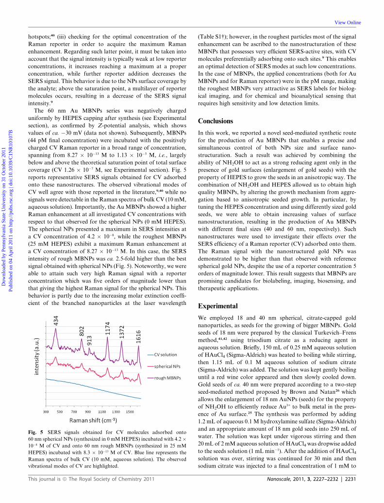
coverage (CV 1.26 � 10�7 M, see Experimental section). Fig. 5
reports representative SERS signals obtained for CV adsorbed
onto these nanostructures. The observed vibrational modes of
CV well agree with those reported in the literature,9,40 while no
signals were detectable in the Raman spectra of bulk CV (10mM,
aqueous solution). Importantly, the AuMBNPs showed a higher
Raman enhancement at all investigated CV concentrations with
respect to that observed for the spherical NPs (0 mM HEPES).
The spherical NPs presented a maximum in SERS intensities at
a CV concentration of 4.2 � 10�8, while the roughest MBNPs
(25 mM HEPES) exhibit a maximum Raman enhancement at
a CV concentration of 8.27 � 10�13 M. In this case, the SERS
intensity of rough MBNPs was ca. 2.5-fold higher than the best
signal obtained with spherical NPs (Fig. 5). Noteworthy, we were
able to attain such very high Raman signal with a reporter
concentration which was five orders of magnitude lower than
that giving the highest Raman signal for the spherical NPs. This
behavior is partly due to the increasing molar extinction coeffi-
cient of the branched nanoparticles at the laser wavelength
Fig. 5 SERS signals obtained for CV molecules adsorbed onto
60 nm spherical NPs (synthesized in 0 mMHEPES) incubated with 4.2 �10�8 M of CV and onto 60 nm rough MBNPs (synthesized in 25 mM
HEPES) incubated with 8.3 � 10�13 M of CV. Blue line represents the
Raman spectra of bulk CV (10 mM, aqueous solution). The observed
vibrational modes of CV are highlighted.
This journal is ª The Royal Society of Chemistry 2011
(Table S1†); however, in the roughest particles most of the signal
enhancement can be ascribed to the nanostructuration of these
MBNPs that possesses very efficient SERS-active sites, with CV
molecules preferentially adsorbing onto such sites.9 This enables
an optimal detection of SERS modes at such low concentrations.
In the case of MBNPs, the applied concentrations (both for Au
MBNPs and for Raman reporter) were in the pM range, making
the roughest MBNPs very attractive as SERS labels for biolog-
ical imaging, and for chemical and bioanalytical sensing that
requires high sensitivity and low detection limits.
Conclusions
In this work, we reported a novel seed-mediated synthetic route
for the production of Au MBNPs that enables a precise and
simultaneous control of both NPs size and surface nano-
structuration. Such a result was achieved by combining the
ability of NH2OH to act as a strong reducing agent only in the
presence of gold surfaces (enlargement of gold seeds) with the
property of HEPES to grow the seeds in an anisotropic way. The
combination of NH2OH and HEPES allowed us to obtain high
quality MBNPs, by altering the growth mechanism from aggre-
gation based to anisotropic seeded growth. In particular, by
tuning the HEPES concentration and using differently sized gold
seeds, we were able to obtain increasing values of surface
nanostructuration, resulting in the production of Au MBNPs
with different final sizes (40 and 60 nm, respectively). Such
nanostructures were used to investigate their effects over the
SERS efficiency of a Raman reporter (CV) adsorbed onto them.
The Raman signal with the nanostructured gold NPs was
demonstrated to be higher than that observed with reference
spherical gold NPs, despite the use of a reporter concentration 5
orders of magnitude lower. This result suggests that MBNPs are
promising candidates for biolabeling, imaging, biosensing, and
therapeutic applications.
Experimental
We employed 18 and 40 nm spherical, citrate-capped gold
nanoparticles, as seeds for the growing of bigger MBNPs. Gold
seeds of 18 nm were prepared by the classical Turkevich–Frens
method,41,42 using trisodium citrate as a reducing agent in
aqueous solution. Briefly, 150 mL of 0.25 mM aqueous solution
of HAuCl4 (Sigma-Aldrich) was heated to boiling while stirring,
then 1.15 mL of 0.1 M aqueous solution of sodium citrate
(Sigma-Aldrich) was added. The solution was kept gently boiling
until a red wine color appeared and then slowly cooled down.
Gold seeds of ca. 40 nm were prepared according to a two-step
seed-mediated method proposed by Brown and Natan26 which
allows the enlargement of 18 nm AuNPs (seeds) for the property
of NH2OH to efficiently reduce Au3+ to bulk metal in the pres-
ence of Au surface.25 The synthesis was performed by adding
1.2 mL of aqueous 0.1 M hydroxylamine sulfate (Sigma-Aldrich)
and an appropriate amount of 18 nm gold seeds into 250 mL of
water. The solution was kept under vigorous stirring and then
20 mL of 2mM aqueous solution of HAuCl4 was dropwise added
to the seeds solution (1 mL min�1). After the addition of HAuCl4solution was over, stirring was continued for 30 min and then
sodium citrate was injected to a final concentration of 1 mM to
Nanoscale, 2011, 3, 2227–2232 | 2231

Dow
nloa
ded
by P
enns
ylva
nia
Stat
e U
nive
rsity
on
31 O
ctob
er 2
011
Publ
ishe
d on
04
Apr
il 20
11 o
n ht
tp://
pubs
.rsc
.org
| do
i:10.
1039
/C1N
R10
107B
View Online
stabilize the colloidal solution. The quality of the so-prepared 18
and 40 nm seeds was verified by using TEM, UV-Vis spectros-
copy, Dynamic Light Scattering (DLS) and Z-potential analysis
(see Fig. S1†).
The synthesis of increasing nanorough 40 nm Au MBNPs was
carried out in 25 mL aqueous solution presenting growing
concentrations of HEPES (Sigma-Aldrich) adjusted to pH 7.0
with NaOH, namely 0.2, 1, 2.5, 5, and 25 mM. Then, the
appropriate amount of 18 nm AuNPs (seeds) and 0.12 mL of
100 mM NH2OH was added in the reaction mixture. The
enlargement of 18 nm Au seeds was achieved by dropwise adding
5 mL of 0.8 mM aqueous solution of HAuCl4 (0.5 mL min�1).
The same enlargement procedure was employed to obtain 60 nm
Au MBNPs, with the same trend of surface nanostructuration,
employing the 40 nm AuNPs as seeds. These reactions were all
conducted at 4 �C. The quality of the MBNPs was investigated
by TEM, HR-TEM, SEM, and UV-Vis spectroscopy.
For the SERS experiments, in a freshly prepared 60 nm Au
MBNPs series, HEPES solution (pH 7.0) was added to a final
concentration of 25 mM to ensure the same surface chemistry by
HEPES capping. After stirring for 1 hour, nanoparticles were
subjected to three washing steps by centrifugation and resus-
pension in ultrapure, sterile water. The nanoparticles series was
investigated by DLS and Z-potential analysis to ensure good
quality. The concentration of such Au MBNPs was determined
as follows: the freshly synthesized Au MBNPs possess the same
number of nanoparticles since we employed the same amount of
Au seeds and chloroauric acid for all the synthesis regarding
a specific final size. For this reason, the spectra shown in Fig. 3a
and (b) represent the absorbance of solutions presenting the same
number of nanoparticles, with increasing surface roughness. The
molar extinction coefficient for spherical AuNPs is known43 and
so, from the spectra obtained in Fig. 3, we extrapolated the
theoretical molar extinction coefficient for the rough nano-
particles (from 0.2 mM HEPES to 25 mMHEPES). By applying
absorption spectroscopy, we diluted the negatively charged
MBNPs to a final concentration of 0.044 nM. After, a reporter
solution of Crystal Violet (CV) chloride (Sigma-Aldrich) was
added to the colloidal solutions to a final concentration ranging
from 8.27 � 10�13 M to 1.13 � 10�5 M. The CV concentration
needed to fully cover 0.044 nM of 60 nm spherical gold NPs is
calculated to be about 4.2 � 10�8 M, considering that the surface
area of a CV molecule is about 4 nm2 (assuming a parallel
orientation of the adsorbed molecule at the NP surface).9 Prior to
performing SERS experiments, the solutions containing SERS
active nanostructures and Raman reporter, electrostatically
interacting, were monitored by DLS in order to verify the
absence of colloidal aggregations. SERS experiments on Au
MBNPs were carried out by using a Jobin Yvon Fluorog 3
spectrofluorometer equipped with double monochromators and
a cooled photomultiplier, and a 632.8 nm, 30.0 mW HeNe laser
as the excitation source.
References
1 C. Burda, X. Chen, R. Narayanan and M. A. El-Sayed, Chem. Rev.,2005, 105, 1025.
2 P. K. Jain, X. Huang, I. H. El-Sayed and M. A. El-Sayed, Acc. Chem.Res., 2008, 41, 1578.
3 B. K. Jena and C. R. Raj, Langmuir, 2007, 23, 4064.
2232 | Nanoscale, 2011, 3, 2227–2232
4 K. Yu, K. L. Kelly, N. Sakai and T. Tatsuma, Langmuir, 2008, 24,5849.
5 T. R. Kuo, V. A. Hovhannisyan, Y. C. Chao, S. L. Chao, S. J. Chiang,S. J. Lin, C. Y. Dong and C. C. Chen, J. Am. Chem. Soc., 2010, 132,14163.
6 J. E. Millstone, D. G. Georganopoulou, X. Xu, W. Wei, S. Li andC. A. Mirkin, Small, 2008, 4, 2176.
7 J. Chen, C. Glaus, R. Laforest, Q. Zhang, M. Yang, M. Gidding,M. J. Welch and Y. Xia, Small, 2010, 6, 811.
8 R. Huschka, O. Neumann, A. Barhoumi and N. J. Halas, Nano Lett.,2010, 10, 4117.
9 E. N. Esenturk and A. R. H. Walker, J. Raman Spectrosc., 2009, 40,86.
10 J. Aaron, E. de la Rosa, K. Travis, N. Harrison, J. Burt,M. J. Yacaman and K. Sokolov, Opt. Express, 2008, 16, 2153.
11 S. K. Dondapati, T. K. Sau, C. Hrelescu, T. A. Klar, F. D. Stefani andJ. Feldmann, ACS Nano, 2010, 4, 6318.
12 S. Wang, Z. Wu, F. Qu, S. Zhang, G. Shen and R. Yu, Biosens.Bioelectron., 2008, 24, 1020.
13 Z. Wang, J. Zhang, J. M. Ekman, P. J. A. Kenis and Y. Lu, NanoLett., 2010, 10, 1886.
14 J. Xie, Z. Q. Zhang, J. Y. Lee and D. I. C. Wang, ACS Nano, 2008, 2,2473.
15 P. S. Kumar, I. Pastoriza-Santos, B. Rodrı̀guez-Gonz�alez,F. J. Garcı̀a de Abajo and L. M. Liz-Marz�an, Nanotechnology,2008, 19, 015606.
16 O. M. Bakr, B. H. Wunsch and F. Stellacci, Chem. Mater., 2006, 18,3297.
17 C. L. Nehl, H. Liao and J. H. Hafner, Nano Lett., 2006, 6, 683.18 E. C. Hao, R. C. Bailey, G. G. Schatz, J. T. Hupp and S. Y. Li, Nano
Lett., 2004, 4, 327.19 J. R. Fernandez, A. M. Funston, J. P. Juste, R. A. A. Puebla,
L. M. Liz-Marzan and P. Mulvaney, Phys. Chem. Chem. Phys.,2009, 11, 5909.
20 C. G. Khoury and T. J. Vo-Dinh, J. Phys. Chem. C, 2008, 112,18849.
21 T. K. Sau and C. J. Murphy, J. Am. Chem. Soc., 2004, 126,8648.
22 W. Wang, X. Yang and H. Cui, J. Phys. Chem. C, 2008, 112,16348.
23 X. Q. Zou, E. B. Ying and S. J. Dong,Nanotechnology, 2006, 17, 4758.24 G. Palui, S. Ray and A. Banerjee, J. Mater. Chem., 2009, 19, 3457.25 G. Stremsdoerfer, H. Perrot, J. R. Martin and P. J. Clechet, J.
Electrochem. Soc., 1988, 135, 2881.26 K. R. Brown and M. J. Natan, Langmuir, 2008, 14, 726.27 N. E. Good, G. D. Winget, W. Winter, T. N. Connolly, K. Izana and
R. M. M. Singh, Biochemistry, 1966, 5, 467.28 J. Xie, J. Y. Lee and D. I. C. Wang, Chem. Mater., 2007, 19,
2823.29 S. Link and M. A. El-Sayed, J. Phys. Chem. B, 1999, 103, 8410.30 F. Hao, C. L. Nehl, J. H. Hafner and P. Nordlander, Nano Lett.,
2007, 7, 729.31 C. H. Kuo and M. H. Huang, Langmuir, 2005, 21, 2012.32 J. L. Burt, J. L. Elechiguerra, J. Reyes-Gasga, J. M. Montejano-
Carrizales and M. Jose-Yacaman, J. Cryst. Growth, 2005, 285, 681.33 T. Vo-Dinh, H. N. Wang and J. Scaffidi, J. Biophotonics, 2010, 3, 89.34 L. Fabris, M. Schierhorn, M. Moskovits and G. C. Bazan, Small,
2010, 19, 1550.35 X. M. Qian and S. M. Nie, Chem. Soc. Rev., 2008, 37, 912.36 X. M. Qian, X. H. Peng, D. O. Ansari, Q. Yin-Goen, G. Z. Chen,
D. M. Shin, L. Yang, A. N. Young, M. D. Wang and S. M. Nie,Nat. Biotechnol., 2008, 26, 83.
37 L. Rodr�ıguez-Lorenzo, R. A. Alvarez-Puebla, I. Pastoriza-Santos,S. Mazzucco, O. Stephan, M. Kociak, L. M. Liz-Marzan andF. J. J. Garc�ıa de Abajo, J. Am. Chem. Soc., 2009, 131, 4616.
38 J. T. Krug, G. D. Wang, S. R. Emory and S. M. Nie, J. Am. Chem.Soc., 1999, 121, 9208.
39 M. Moskovits and D. H. Jeong, Chem. Phys. Lett., 2004, 397, 91.40 D. Cialla, H. Uwe, H. Schneidewind, R. Moller and J. Popp,
ChemPhysChem, 2008, 9, 758.41 J. Turkevich, P. C. Stevenson and J. A. Hillier,Discuss. Faraday Soc.,
1951, 11, 55.42 G. Frens, Nature (London), Phys. Sci., 1973, 241, 20.43 J. S. Lee, S. I. Stoeva and C. A. Mirkin, J. Am. Chem. Soc., 2006, 128,
8899.
This journal is ª The Royal Society of Chemistry 2011