Synthesis and Characterization of Gold Nanoparticles by Tryptophane
12
Int. J. Nano and Biomaterials, Vol. 5, Nos. 2/3, 2014 181 Copyright © 2014 Inderscience Enterprises Ltd. Synthesis and characterisation of gold nanoparticles using Mentha piperita leaf extract: a green, non-toxic and rapid method Germán Ayala Valencia* Faculty of Animal Science and Food Engineering, Food Science Department, University of São Paulo, Pirassununga, São Paulo, Brazil and Faculty of Animal Science and Food Engineering, Basic Sciences Department ZAB/FZEA, University of São Paulo, São Paulo, Pirassununga, Brazil Email: [email protected] *Corresponding author Luci Cristina de Oliveira Vercik, Leticcia Gavião Ferreira, Jaiber Humberto Rodríguez Llanos and Andrés Vercik Faculty of Animal Science and Food Engineering, Basic Sciences Department ZAB/FZEA, University of São Paulo, São Paulo, Pirassununga, Brazil Email: [email protected] Email: [email protected] Email: [email protected] Email: [email protected] Abstract: Eco-friendly synthesis of gold nanoparticles (AuNPs) using Mentha piperita leaf extract was addressed in this work. Different extract concentrations (0.5, 1.0 and 1.5% w/v) were used for the synthesis of AuNPs. Reaction rate increased with temperature and leaf extract concentration; however, prolonged heating might affect negatively the AuNPs synthesis. AuNPs in colloid state were synthesised in acid pH (between 2.5 and 3.1), with potentials for charged surface between –21.8 and –24.1 mV, these colloids were highly stable for a storage time of four weeks. Bioreduction has generated AuNPs with spherical and triangular shape and size range between 3–26 nm for all concentrations of leaf extract, indicating a weak dependence of the nanoparticle size on extract concentrations. The present work, thus, offers a simple, rapid and non-toxic process for the synthesis of AuNPs that would potentially be used in different areas such as foods, cosmetic, medicine, electronics and polymer science.
-
Upload
independent -
Category
Documents
-
view
2 -
download
0
Transcript of Synthesis and Characterization of Gold Nanoparticles by Tryptophane
Int. J. Nano and Biomaterials, Vol. 5, Nos. 2/3, 2014 181
Copyright © 2014 Inderscience Enterprises Ltd.
Synthesis and characterisation of gold nanoparticles using Mentha piperita leaf extract: a green, non-toxic and rapid method
Germán Ayala Valencia* Faculty of Animal Science and Food Engineering, Food Science Department, University of São Paulo, Pirassununga, São Paulo, Brazil and Faculty of Animal Science and Food Engineering, Basic Sciences Department ZAB/FZEA, University of São Paulo, São Paulo, Pirassununga, Brazil Email: [email protected] *Corresponding author
Luci Cristina de Oliveira Vercik, Leticcia Gavião Ferreira, Jaiber Humberto Rodríguez Llanos and Andrés Vercik Faculty of Animal Science and Food Engineering, Basic Sciences Department ZAB/FZEA, University of São Paulo, São Paulo, Pirassununga, Brazil Email: [email protected] Email: [email protected] Email: [email protected] Email: [email protected]
Abstract: Eco-friendly synthesis of gold nanoparticles (AuNPs) using Mentha piperita leaf extract was addressed in this work. Different extract concentrations (0.5, 1.0 and 1.5% w/v) were used for the synthesis of AuNPs. Reaction rate increased with temperature and leaf extract concentration; however, prolonged heating might affect negatively the AuNPs synthesis. AuNPs in colloid state were synthesised in acid pH (between 2.5 and 3.1), with potentials for charged surface between –21.8 and –24.1 mV, these colloids were highly stable for a storage time of four weeks. Bioreduction has generated AuNPs with spherical and triangular shape and size range between 3–26 nm for all concentrations of leaf extract, indicating a weak dependence of the nanoparticle size on extract concentrations. The present work, thus, offers a simple, rapid and non-toxic process for the synthesis of AuNPs that would potentially be used in different areas such as foods, cosmetic, medicine, electronics and polymer science.
182 G.A. Valencia et al.
Keywords: bioreduction; nanomaterials; green synthesis; gold nanoparticles; Mentha piperita.
Reference to this paper should be made as follows: Valencia, G.A., de Oliveira Vercik, L.C., Ferreira, L.G., Llanos, J.H.R. and Vercik, A. (2014) ‘Synthesis and characterisation of gold nanoparticles using Mentha piperita leaf extract: a green, non-toxic and rapid method’, Int. J. Nano and Biomaterials, Vol. 5, Nos. 2/3, pp.181–192.
Biographical notes: Germán Ayala Valencia received his degree of Agroindustrial Engineering and Master of Sciences in Food Engineering from the Univerdad Nacional de Colombia (2011) and the University of São Paulo (2013) respectively. Actually, he is a PhD student in Sciences of Food Engineering at the University of São Paulo. His main areas of research interest are physicochemical properties in biopolymers, film production, rheology and nanotechnology (metal nanoparticles and nanoclays).
Luci Cristina de Oliveira Vercik received her BS in Chemistry and MS in Materials Engineering from the Universidade Federal de São Carlos, Brazil and PhD in Chemistry (Physical Chemistry) from the Universidade Estadual Paulista, Brazil, in 1993, 1999 and 2004 respectively. Currently, she is senior technical staff at the Nanotechnology, Biomaterials and Devices Group of the University of São Paulo. Her main areas of research interest are biomaterials for implants, nanotechnology and green synthesis of nanoparticles, and materials for biocompatible biosensors.
Leticcia Gavião Ferreira is a Biosystems Engineering student at the Faculty of Animal Science and Food Engineering (FZEA), University of São Paulo (USP). She interned in Nanotechnology Laboratory, Biosensors and Devices (NanoBioDev) working with nanoparticles of silver and gold.
Jaiber Humberto Rodríguez Llanos is a PhD student in Engineering and Materials Science program at FZEA USP, graduated as an Agroindustrial Engineer from the Universidad Nacional de Colombia (2010) and received his Master of Science in Food Engineering from São Paulo University (2014). He has experience in production engineering, with emphasis on research and product development; science and food technology, with an emphasis on biopolymers such as chitosan; and working mainly in the areas of physical chemical characterisation of films, nanotechnology (syntheses of nanoparticles), mechanical and electrical properties of the films.
Andrés Vercik received his BE in Physics and PhD in Electrical Engineering (Microelectronics) from the University of Buenos Aires, Argentina, in 1995 and 2000 respectively. Currently, he is an Associate Professor at the University of São Paulo, Brazil, where he coordinates the Nanotechnology, Biosensors and Devices Laboratory. His main areas of research interest are electrical and optical properties of nanostructured materials for use in sensors and biosensors. He is a member of the Physical Association of Argentina, the Brazilian Physical Society and the Materials Research Society.
Synthesis and characterisation of gold nanoparticles 183
1 Introduction
Nanotechnology involves the manufacturing and processing of materials, systems or devices that are controlled by the shape and size of its components in nanoscale (size < 100 nm) (Bouwmeester et al., 2009; Song et al., 2009). Nanoparticles are one of the main components commonly found in nanodevices and nanostructures and it had received considerable attention in recent years because its magnetic, electrical, catalytic, electronic and optical properties, that allow several applications in the development of sensors, biosensors, nanoelectronic devices and biomedical tools (Bar et al., 2009; Mondal et al., 2011). The applicability of metal nanoparticles depends on factors such as size, shape, composition and structure (Rao et al., 2002; Begum et al., 2009). Nanoparticles from noble metals such as gold (Au), silver (Ag) and platinum (Pt) have several industrial applications, today theses noble metals in nanoscale are found in a wide product varieties, in products that have direct contact with the human body, such as shampoos, soaps, detergent, shoes and cosmetic products (Song et al., 2009). Specially, gold nanoparticles (AuNPs) have been studied in biomedicine for the treatment of cancer, arthritis and macular degeneration and also have been used in infrared radiation absorbent optical coatings (Chandran et al., 2006; Bhattacharya and Murkherjee, 2008). More recently, applications of AuNPs in biomedicine refers to the development of strategies which combine therapeutics with diagnostics, called ‘theragnostic’. This novel area envision the cancer treatment allowing a controlled and targeted release of drugs against cancer cells, in which the nanoparticles are conjugated with target-specific moieties such as peptides and antibodies, and which are selective to cancer cells through receptor-mediated endocytosis. AuNPs would allow a selective pass through leaky blood vessels around tumour tissues, and then the tumour detection (Lee et al., 2011). However, nanomaterials to be used in fields such as food industry, cosmetic and biomedicine cannot be synthesised using toxic compounds in the reduction/stabilisation process to obtain gold nanoparticles. Therefore, different investigations have addressed the synthesis of AuNPs using natural compounds (Panigrahi et al., 2005; Raveendran et al., 2006; Wei and Qian, 2008; Ghodake et al., 2010; Bankar et al., 2010; Noruzi et al., 2011; Valencia et al., 2013), suggesting these natural compounds as alternative reducer/stabiliser agents for the synthesis of gold nanoparticles.
Particularly, phytosynthesis method for obtain metal nanoparticles has received some attention in recent years as a simple and viable alternative, this method can be advantageous over other biological processes, since it does not require acidic conditions for the raw materials solubilisation (e.g., chitosan) (Wei and Qian, 2008), or additions of basic substances for pH stabilisation (e.g., sugars) (Engelbrekt et al., 2009), or maintaining cell cultures (e.g., microorganism) (Mandal et al., 2006). Various investigations have been directed toward the synthesis of AuNPs using plants leaf extract, such as Alfalfa (Gardea-Torresdey et al., 2002), Aloe vera (Chandran et al., 2006), Avena sativa (oat) (Armendariz et al., 2004), Azardirachta indica (neem) (Shankar et al., 2004), Coriandrum sativum (cilantro) (Narayanan and Sakthivel, 2008), Pelargonium graveolens (geranium) (Shankar et al., 2003), and Memecylon (nipis kulit) edule (Elavazhagan and Arunachalam, 2011). Recently, Mubarak Ali et al. (2011) reported a green method for the synthesis of AuNPs using Mentha piperita leaf extract (peppermint) as reducer/stabiliser agent. These AuNPs were obtained in thermal bath at 28°C for 24 h.
184 G.A. Valencia et al.
The AuNPs had 150 nm in diameter size and it had microbial activity against clinically isolated human pathogens such as Staphylococcus aureus and Escherichia coli.
In this paper, we report a rapid, clean, non-toxic and eco-friendly method for synthesis of AuNPs by the reduction of chloroauric acid using Mentha piperita leaf extract (peppermint). The effect of leaf extract concentrations, temperature and time reaction on the synthesis of AuNPs were analysed. UV-visible (UV-Vis) spectroscopy, high resolution transmission electron microscopy (HR-TEM), dynamic light scattering (DLS) equipment with potential analyser for quantification of surface charge (pZ) and scanning electron microscopy (SEM) equipped with energy dispersive spectrometer (EDS) were used for monitoring and examining the AuNPs.
2 Materials and methods
2.1 Preparation of plant extract
Commercial fresh leafs of Mentha piperita were purchased from local market in Pirassununga (São Paulo, Brazil). Chloroauric acid (HAuCl4) was purchased from Polymar (Brazil).
Leaves of Mentha piperita were washed strongly fourfold with distilled water and it was shade dried for five days to room temperature. Dried leafs were macerated and then passed through different sieves to obtain 850 μm in average particle size. Fine powder was stored at 4°C until use. Approximately 0.5, 1.0 and 1.5 g of powder was taken and mixed with 100 mL of Milli Q water preheated at 60°C, and mixing for 10 min to the same temperature (Mubarak Ali et al., 2011). Immediately, solutions were cooled until room temperature and filtered using filter paper with 14-μm-diameter pores. Filtered extract was stored at 4°C until it be used (maximum 30 min).
2.2 Preparation of gold nanoparticles
For the synthesis of AuNPs, 4 mL of HAuCl4 (8 mM) were added to 10 mL of a filtered extract solution to 0.5, 1.0 and 1.5% (w/v), and placed under vigorous mixing for 15 min. Synthesis of AuNPs was carried out in water bath at 40, 70 and 90°C for 1, 3, 5, 7 and 9 min. The reaction velocity for synthesis of AuNPs was evaluated via UV-Vis spectroscopy as suggested by Valencia et al. (2013) and using a spectrophotometer (Beckman Coulter DU 800 spectrophotometer, USA). Spectra were analysed between 200–900 nm. High resolution transmission electron microscopy (HR-TEM, Philips CM200 at 200 kV, USA) was used for shape and size determination in AuNPs. ZetaPlus-zeta potential analyser (ZP) was used to examine the surface charge and stability of AuNPs.
After bioreduction, colloids containing AuNPs were centrifuged at 12,000 rpm for 15 min at room temperature, removing unbound phytochemicals compounds (Shervani and Yamamoto, 2011). Precipitated obtained by centrifugation was completely dried (freeze drying) and analysed by SEM (TM-3000, HITACHI, Japan) equipped with EDS.
HR-TEM nanographs were analysed to obtain particle size distribution of gold nanoparticles using the software ImageJ v1.45s (National Institutes of Health, USA). Nanographs were submitted to the following image treatment as suggested by Dacanal and Menegalli (2009):
Synthesis and characterisation of gold nanoparticles 185
• threshold: transform the grey level of images to binary scale
• smooth: eliminate image defects
• make binary: images were converts to black and white; it is required for the last two steps
• filling hole: repair inner imperfections in AuNPs
• watershed: used as automatically separation of agglomerates, if necessary.
Images treated were used for calculi the 2D surface area (S) in AuNPs. Equivalent diameters (dpeq) were calculated by the following equation:
eqdp 2 S / .π= (1)
Particle size distribution was constructed using dpeq values by means of MS-Excel® software.
All AuNPs characterisations (UV-Vis, pZ, HR-TEM and EDS) were performed in triplicate. Particle size distribution from HR-TEM was performed using least 10 nanographs by each sample.
3 Results and discussion
3.1 Biosynthesis of AuNPs
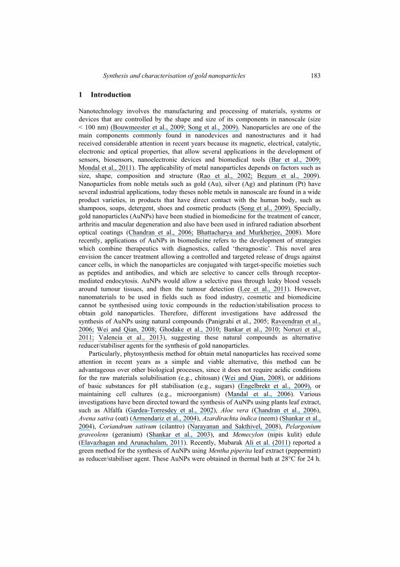
UV-Vis spectra for the colloids obtained to 1.0 and 1.5% of leaf extract concentrations shown a characteristic peak for AuNPs located approximately at 520 nm (Figure 1), and associated to the surface plasmon resonance (SPR) (Khlebtsov and Dykman, 2010). Similar spectra were obtained for 0.5% concentration, however, a decrease in the absorbance values and a shift in the wavelength maximum position (550 nm < λmax < 556 nm) were observed.
Figure 1 UV-Vis spectra for gold nanoparticles synthesised to different leaf extract concentrations and heating at 90°C for 1 min (see online version for colours)
Note: All samples were diluted 90%.
186 G.A. Valencia et al.
The pH for extract solution was monitored but it was not set at any fixed value, avoiding the addition of any other reagent.
AuNPs colloids to 0.5%, 1.0% and 1.5% had pH values of 2.48 ± 0.02, 2.70 ± 0.02 and 3.14 ± 0.02 respectively. Synthesis of AuNPs could be followed by naked eye, thereby Mentha piperita leaf extracts with HAuCl4 solution had a yellow colour that changed to red colour during the thermal treatment (10 min).
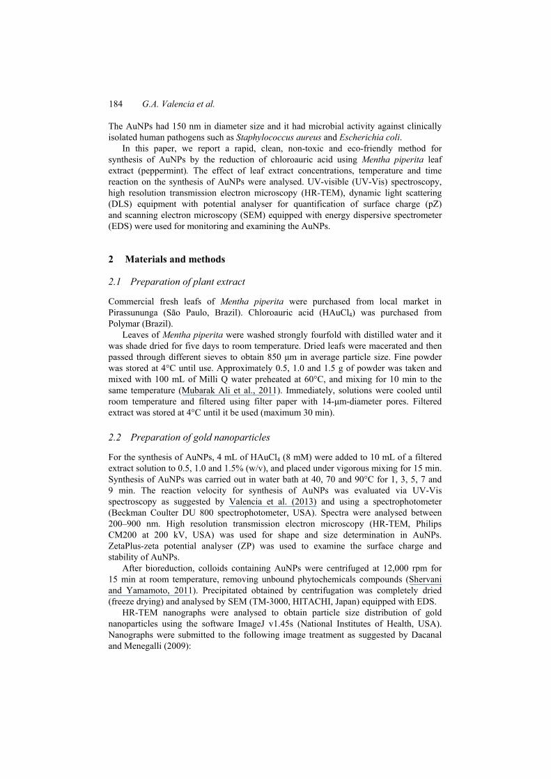
Since better synthesis conditions could be attained combining different temperatures and reaction times, comparative experiments were carried out to examine the effect of different concentrations of Mentha piperita leaf extract, temperature and heating times as reported by Valencia et al. (2013). Reaction rate can be related to the maximum absorbance (Amax) observed by UV-Vis spectra and it is correlated with the reaction rate for AuNPs synthesis (Dubey et al., 2010). Reaction rate for each concentration was drastically different and it indicated that the AuNPs synthesis was very sensitive to changes of leaf extract concentration, temperature and time (Figure 2).
Figure 2 Representative plot of the maximum absorbance vs. time at 40°C (□), 70°C (o) and 90°C (Δ) for gold nanoparticles synthesised to different leaf extract concentrations, (a) 0.5% (b) 1.0% (c) 1.5% (see online version for colours)
Note: All samples were diluted 90%
For the 0.5% samples [Figure 2(a)], no significant variation were observed when the synthesis occurs at 40°C or 90°C, even when a slightly increase could be presumed in the former case and the reaction was more efficient in the latter. The sample processed at
Synthesis and characterisation of gold nanoparticles 187
70°C behaves differently; in this case a noted increase of about 60% in Amax was observed during the first 9 minutes of heating, reaching an absorbance similar to that obtained at 90°C. This fact indicates that for this concentration the lower temperatures were insufficient to activate the reaction, whereas at the higher temperature the reaction does not depend on time.
For a concentration of 1.0% [Figure 2(b)], the reaction at 90°C continues to be more efficient, however, the curves for 40°C and 70°C change their behaviour. At the lower temperatures a clear monotonic increase was observed, whereas the curve at 70°C, the absorbance exhibit a maximum at 5 min, with an absorbance value similar that at 90°C, and then begins to decay.
When the AuNPs were processed with a concentration of 1.5% of leaf extract [Figure 2(c)], the sample at 40°C exhibits an increase at the beginning (shorter times) and, reaches a maximum and decays, that sample at 90°C apparently decay sand the sample at 70°C exhibits a maximum for 3 min, but with higher absorbance than the sample processed at 90°C with any concentration and then decays.
This set of figures shows the inter-related role that these parameters play. The highest values of absorbance, and then the most efficient reaction, were obtained synthesis of AuNPs at 70°C during the first 3 min of processing. In all cases, longer heating times exhibited decaying values of Amax, which were more evident for higher concentrations of leaf extract. These results agree with the fact which to long heating was observed small precipitates at the bottom of the pipettes. This behaviour could be due to the fusion of menthol in the leaf extract, avoiding the AuNPs stabilisation and then precipitate (Soottitantawat et al., 2005).
Control chloroauric acid solution (without leaf extract) and control leaf extract solution (without chloroauric acid solution) were availed for the synthesis of AuNPs at the similar process conditions. In all cases, the results indicated that the synthesis of AuNPs under these reaction conditions did not occur, indicating that AuNPs only can be synthesised in presence of both solutions. These control samples did not develop colour changes and any SPR peak was observed in UV-Vis spectra, confirming the absence of gold nanoparticles.
3.2 Surface charge and stability of the AuNPs
Zeta potential (ZP) for AuNPs colloids had values between –21.8 and –24.1 mV, these values indicated that these systems can be considered as colloids with high stability (Valencia, 2013). These results agree with the reported by Dubey et al. (2010) for AuNPs directly synthesised from tansy fruit extract.
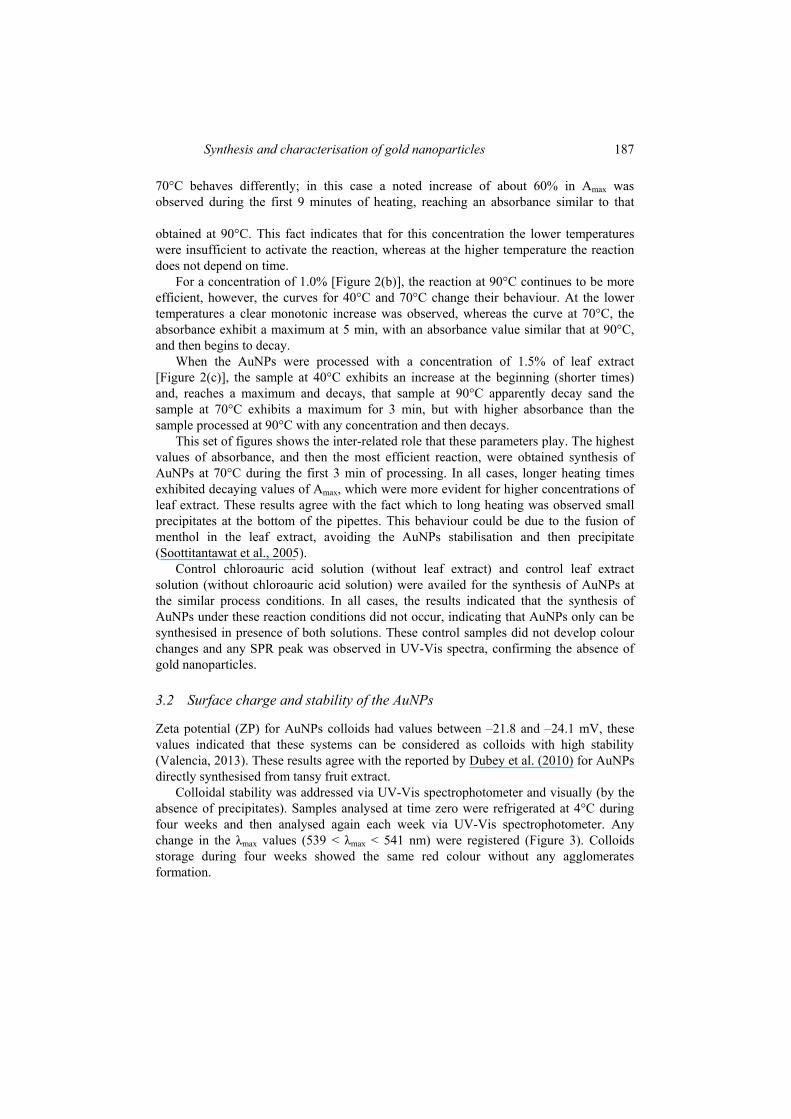
Colloidal stability was addressed via UV-Vis spectrophotometer and visually (by the absence of precipitates). Samples analysed at time zero were refrigerated at 4°C during four weeks and then analysed again each week via UV-Vis spectrophotometer. Any change in the λmax values (539 < λmax < 541 nm) were registered (Figure 3). Colloids storage during four weeks showed the same red colour without any agglomerates formation.
188 G.A. Valencia et al.
Figure 3 UV-Vis spectra for gold nanoparticles to 1.5% of leaf extract concentration and heating at 70°C for 3 min (see online version for colours)
Notes: Effect of storage time on the stability of the sample. All samples were diluted 90%.
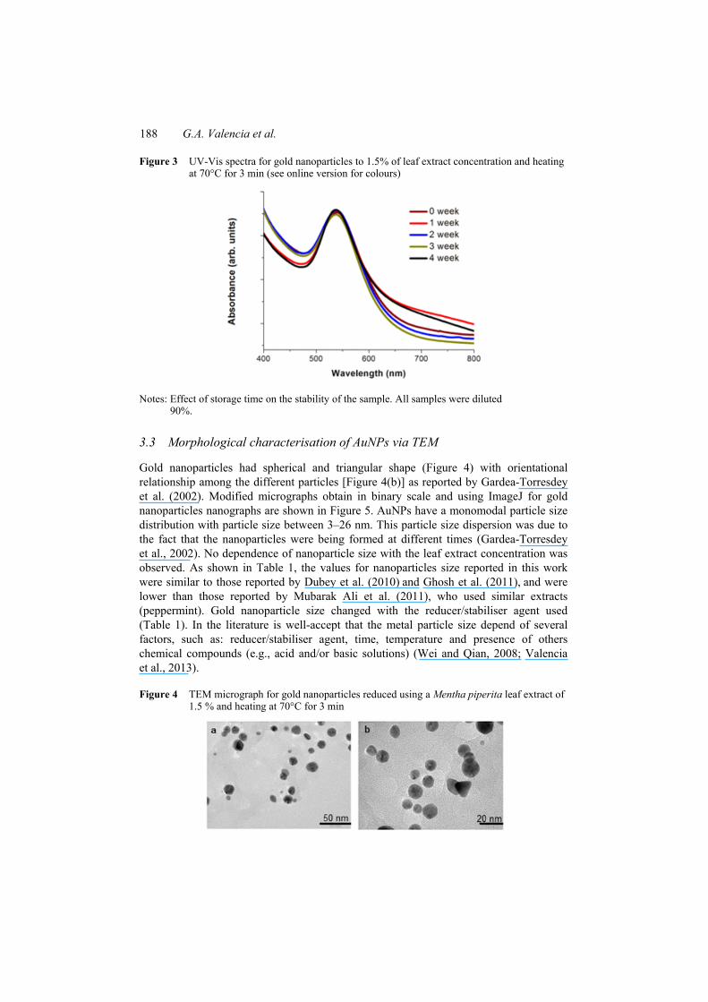
3.3 Morphological characterisation of AuNPs via TEM
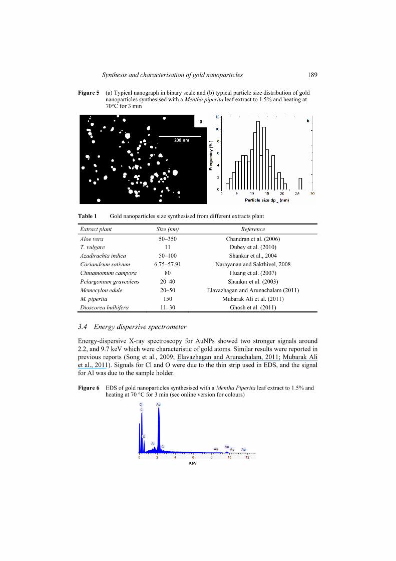
Gold nanoparticles had spherical and triangular shape (Figure 4) with orientational relationship among the different particles [Figure 4(b)] as reported by Gardea-Torresdey et al. (2002). Modified micrographs obtain in binary scale and using ImageJ for gold nanoparticles nanographs are shown in Figure 5. AuNPs have a monomodal particle size distribution with particle size between 3–26 nm. This particle size dispersion was due to the fact that the nanoparticles were being formed at different times (Gardea-Torresdey et al., 2002). No dependence of nanoparticle size with the leaf extract concentration was observed. As shown in Table 1, the values for nanoparticles size reported in this work were similar to those reported by Dubey et al. (2010) and Ghosh et al. (2011), and were lower than those reported by Mubarak Ali et al. (2011), who used similar extracts (peppermint). Gold nanoparticle size changed with the reducer/stabiliser agent used (Table 1). In the literature is well-accept that the metal particle size depend of several factors, such as: reducer/stabiliser agent, time, temperature and presence of others chemical compounds (e.g., acid and/or basic solutions) (Wei and Qian, 2008; Valencia et al., 2013).
Figure 4 TEM micrograph for gold nanoparticles reduced using a Mentha piperita leaf extract of 1.5 % and heating at 70°C for 3 min
Synthesis and characterisation of gold nanoparticles 189
Figure 5 (a) Typical nanograph in binary scale and (b) typical particle size distribution of gold nanoparticles synthesised with a Mentha piperita leaf extract to 1.5% and heating at 70°C for 3 min
Table 1 Gold nanoparticles size synthesised from different extracts plant
Extract plant Size (nm) Reference
Aloe vera 50–350 Chandran et al. (2006) T. vulgare 11 Dubey et al. (2010) Azadirachta indica 50–100 Shankar et al., 2004 Coriandrum sativum 6.75–57.91 Narayanan and Sakthivel, 2008 Cinnamomum campora 80 Huang et al. (2007) Pelargonium graveolens 20–40 Shankar et al. (2003) Memecylon edule 20–50 Elavazhagan and Arunachalam (2011) M. piperita 150 Mubarak Ali et al. (2011) Dioscorea bulbifera 11–30 Ghosh et al. (2011)
3.4 Energy dispersive spectrometer
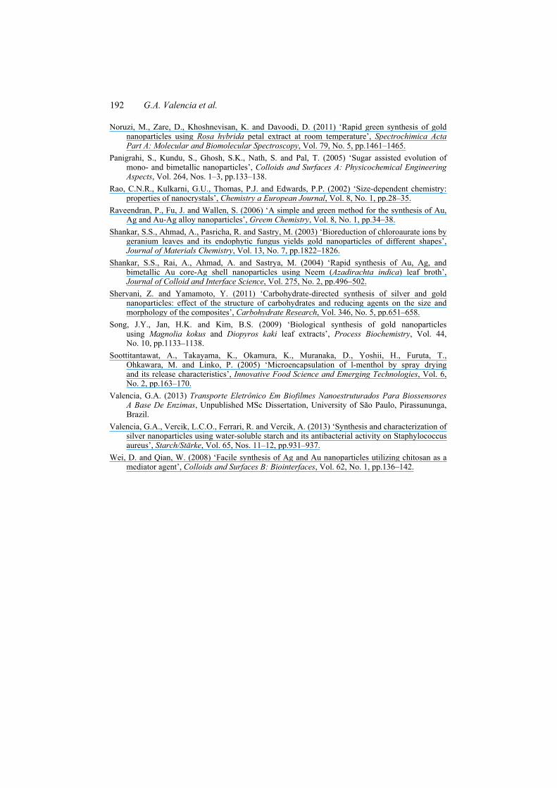
Energy-dispersive X-ray spectroscopy for AuNPs showed two stronger signals around 2.2, and 9.7 keV which were characteristic of gold atoms. Similar results were reported in previous reports (Song et al., 2009; Elavazhagan and Arunachalam, 2011; Mubarak Ali et al., 2011). Signals for Cl and O were due to the thin strip used in EDS, and the signal for Al was due to the sample holder.
Figure 6 EDS of gold nanoparticles synthesised with a Mentha Piperita leaf extract to 1.5% and heating at 70 °C for 3 min (see online version for colours)

190 G.A. Valencia et al.
In summary, above results suggested that gold nanoparticles have been successfully synthesised using Mentha piperita leaf extract as reducing/stabiliser agent. The presence of a peak between 539–556 nm observed by UV-Vis spectroscopy revealed the existence of gold nanoparticles which were later confirmed with HR-TEM micrographs and EDS spectrum.
4 Conclusions
Gold nanoparticles were synthesised using Mentha piperita leaf extract as reducing/stabiliser agent. The synthesis of AuNPs was affect by the leaf extract concentration, time and temperature. The best bioreduction was achieved using concentrations of 1.5% (leaf extract) and heating at 70°C between 1–3 min, followed by extract to 1% and heating at 90°C for 5 min. Gold nanoparticles had a monomodal particle size distribution, with particle sizes between 3–26 nm, and with spherical and triangular shape. Mentha piperita leaf extract appears to be an eco-friendly and economic candidate as reducing/stabiliser agent for the synthesis of gold nanoparticles.
The crucial difference between the present study and the early reports were the small gold nanoparticle sizes that were obtained in a short time process.
Acknowledgements
The authors gratefully acknowledge the financial support from Fundação de Apoio à Pesquisa do Estado de São Paulo (FAPESP), grant number 2010/50424-3 and the laboratories of Chemistry Biology, Colloids Macromolecules and Functionality and Food Technology, from the Animal Science and Food Engineering College, University of São Paulo, for measuring of UV-Vis spectra, size and surface charge, SEM and energy-dispersive X-ray spectroscopy. TEM facility was provided by the IQ-UNESP.
References Armendariz, V., Herrera, I., Videa, J.R., Yacaman, M.J., Troiani, H., Santiago, P. and
Torresdey, J.L.G. (2004) ‘Size controlled gold nanoparticle formation by Avena sativa biomass: use of plants in nanobiotechnology’, Journal of Nanoparticle Research, Vol. 6, No. 4, pp.377–382.
Bankar, A., Joshi, B., Kumar, A.R. and Zinjarde, S. (2010) ‘Banana peel extract mediated synthesis of gold nanoparticles’, Colloids and Surfaces B: Biointerfaces, Vol. 80, No. 1, pp.45–50.
Bar, H., Bhui, D.K., Sahoo, G.P., Sarkar, P., Sankar, P.D. and Misra, A. (2009) ‘Green synthesis of silver nanoparticles using latex of Jatropha curcas’, Colloids and Surfaces A: Physicochemical and Engineering Aspects, Vol. 339, Nos. 1–3, pp.134–139.
Begum, N.A., Mondal, S., Basu, S., Laskar, R.A. and Mandal, D. (2009) ‘Biogenic synthesis of Au and Ag nanoparticles using aqueous solutions of Black Tea leaf extracts’, Colloids and Surfaces B: Biointerfaces, Vol. 71, No. 1, pp.113–118.
Bhattacharya, R. and Murkherjee, P. (2008) ‘Biological properties of naked metal nanoparticles’, Advanced Drug Delivery Reviews, Vol. 60, No. 11, pp.1289–1306.

Synthesis and characterisation of gold nanoparticles 191
Bouwmeester, H., Dekkers, S., Maryvon, Y.N., Werner, I.H., Astrid, S.B., Heer, C., Sandra, E.C.G., Voorde, T., Susan, W.P.W., Hans, J.P.M. and Adriënne, J.A.M.S. (2009) ‘Review of health safety aspect of nanotechnologies in food production’, Regulatory Toxicology and Pharmacology, Vol. 53, No. 1, pp.52–62.
Chandran, S.P., Chaudhary, M., Pasricha, R., Ahmad, A. and Sastry, M. (2006) ‘Synthesis of gold nanotriangles and silver nanoparticles using Aloe vera plant extract’, Biotechnology Progress, Vol. 22, No. 2, pp.577–583.
Dacanal, G.C. and Menegalli, F.C. (2009) ‘Experimental study and optimization of the agglomeration of acerola powder in a conical fluid bed’, Powder Technology, Vol. 188, No. 3, pp.187–194.
Dubey, S.P., Lahtinen, M. and Sillanpää, M. (2010) ‘Tansy fruit mediated greener synthesis of silver and gold nanoparticles’, Process Biochemistry, Vol. 45, No. 7, pp.1065–1071.
Elavazhagan, T. and Arunachalam, K.D. (2011) ‘Memecylon edule leaf extract mediated green synthesis of silver and gold nanoparticles’, International Journal of Nanomedicine, Vol. 6, pp.1265–1278.
Engelbrekt, C., Sørensen, K.H., Zhang, J., Welinder, A.C., Jensen, P.S. and Ulstrup, J. (2009) ‘Green synthesis of gold nanoparticles with starch-glucose and applications in bioelectrochemistry’, Journal of Materials Chemistry, Vol. 19, No. 1, pp.7839–7847.
Gardea-Torresdey, J.L., Parsons, J.G., Gomez, E., Videa, J.P., Troiani, H.E., Santiago, P. and Yacaman, M.J. (2002) ‘Formation and growth of Au nanoparticles inside live alfalfa plants’, Nano Letters, Vol. 2, No. 4, pp.397–401.
Ghodake, G.S., Deshpande, N.G., Lee, Y.P. and Jin, E.S. (2010) ‘Pear fruit extract-assisted room-temperature biosynthesis of gold nanoplates’, Colloids and Surfaces B: Biointerfaces, Vol. 75, No. 2, pp.584–589.
Ghosh, S., Patil, S., Ahire, M., Kitture, R., Jabgunde, A., Kale, S., Pardesi, K., Bellare, J., Dhavale, D.D. and Chopade, B.A. (2011) ‘Synthesis of gold nanoanisotrops using Dioscorea bulbifera tuber extract’, Journal of Nanomaterials, pp.1–8, ID: 354793, 8 [online] http://www.hindawi.com/journals/jnm/2011/354793/.
Huang, J., Li, Q., Sun, D., Lu, Y., Su, Y., Yang, X., Wang, H., Wang, Y., Shao, W., He, N., Hong, J. and Chen, C. (2007) ‘Biosynthesis of silver and gold nanoparticles by novel sundried Cinnamomum camphora leaf’, Nanotechnology, Vol. 18, No. 10, pp.1–11.
Khlebtsov, N.G. and Dykman, L.A. (2010) ‘Optical properties and biomedical applications of plasmonic nanoparticle’, Journal of Quantitative Spectroscopy and Radiative Transfer, Vol. 111, No. 1, pp.1–35.
Lee, K., Lee, H., Lee, K.W. and Park, T.G. (2011) ‘Optical imaging of intracellular reactive oxygen species for the assessment of the cytotoxicity of nanoparticles’, Biomaterials, Vol. 32, No. 10, pp.2556–2565.
Mandal, D., Bolander, M.E., Mukhopadhyay, D., Sarkar, G. and Mukherjee, P. (2006) ‘The use of microorganism for the formation of metal nanoparticles and their application’, Applied Microbiology and Biotechnology, Vol. 69, No. 5, pp.485–492.
Mondal, S., Roy, N., Laskar, R.A., Ismail, S.K., Basu, S., Mandal, D. and Begum, N.A. (2011) ‘Biogenic synthesis of Ag, Au and bimetallic Au/Ag alloy nanoparticles using aqueous extract of mahogany (Swietenia mahogany JACQ.) leaves’, Colloids and Surfaces B: Biointerfaces, Vol. 82, No. 2, pp.497–504.
Mubarak Ali, D., Thajuddin, N., Jeganathan, K. and Gunasekaran, M. (2011) ‘Plant extract mediated synthesis of silver and gold nanoparticles and its antibacterial activity against clinically isolated pathogens’, Colloids and Surfaces B: Biointerfaces, Vol. 85, No. 2, pp.360–365.
Narayanan, K.B. and Sakthivel, N. (2008) ‘Coriander leaf mediated biosynthesis of gold nanoparticles’, Materials Letters, Vol. 62, No. 30, pp.4588–4590.
192 G.A. Valencia et al.
Noruzi, M., Zare, D., Khoshnevisan, K. and Davoodi, D. (2011) ‘Rapid green synthesis of gold nanoparticles using Rosa hybrida petal extract at room temperature’, Spectrochimica Acta Part A: Molecular and Biomolecular Spectroscopy, Vol. 79, No. 5, pp.1461–1465.
Panigrahi, S., Kundu, S., Ghosh, S.K., Nath, S. and Pal, T. (2005) ‘Sugar assisted evolution of mono- and bimetallic nanoparticles’, Colloids and Surfaces A: Physicochemical Engineering Aspects, Vol. 264, Nos. 1–3, pp.133–138.
Rao, C.N.R., Kulkarni, G.U., Thomas, P.J. and Edwards, P.P. (2002) ‘Size-dependent chemistry: properties of nanocrystals’, Chemistry a European Journal, Vol. 8, No. 1, pp.28–35.
Raveendran, P., Fu, J. and Wallen, S. (2006) ‘A simple and green method for the synthesis of Au, Ag and Au-Ag alloy nanoparticles’, Greem Chemistry, Vol. 8, No. 1, pp.34–38.
Shankar, S.S., Ahmad, A., Pasricha, R. and Sastry, M. (2003) ‘Bioreduction of chloroaurate ions by geranium leaves and its endophytic fungus yields gold nanoparticles of different shapes’, Journal of Materials Chemistry, Vol. 13, No. 7, pp.1822–1826.
Shankar, S.S., Rai, A., Ahmad, A. and Sastrya, M. (2004) ‘Rapid synthesis of Au, Ag, and bimetallic Au core-Ag shell nanoparticles using Neem (Azadirachta indica) leaf broth’, Journal of Colloid and Interface Science, Vol. 275, No. 2, pp.496–502.
Shervani, Z. and Yamamoto, Y. (2011) ‘Carbohydrate-directed synthesis of silver and gold nanoparticles: effect of the structure of carbohydrates and reducing agents on the size and morphology of the composites’, Carbohydrate Research, Vol. 346, No. 5, pp.651–658.
Song, J.Y., Jan, H.K. and Kim, B.S. (2009) ‘Biological synthesis of gold nanoparticles using Magnolia kokus and Diopyros kaki leaf extracts’, Process Biochemistry, Vol. 44, No. 10, pp.1133–1138.
Soottitantawat, A., Takayama, K., Okamura, K., Muranaka, D., Yoshii, H., Furuta, T., Ohkawara, M. and Linko, P. (2005) ‘Microencapsulation of l-menthol by spray drying and its release characteristics’, Innovative Food Science and Emerging Technologies, Vol. 6, No. 2, pp.163–170.
Valencia, G.A. (2013) Transporte Eletrônico Em Biofilmes Nanoestruturados Para Biossensores A Base De Enzimas, Unpublished MSc Dissertation, University of São Paulo, Pirassununga, Brazil.
Valencia, G.A., Vercik, L.C.O., Ferrari, R. and Vercik, A. (2013) ‘Synthesis and characterization of silver nanoparticles using water-soluble starch and its antibacterial activity on Staphylococcus aureus’, Starch/Stärke, Vol. 65, Nos. 11–12, pp.931–937.
Wei, D. and Qian, W. (2008) ‘Facile synthesis of Ag and Au nanoparticles utilizing chitosan as a mediator agent’, Colloids and Surfaces B: Biointerfaces, Vol. 62, No. 1, pp.136–142.