Green Synthesis of Gold Nanoparticles Capped with ... - MDPI
24
biomolecules Article Green Synthesis of Gold Nanoparticles Capped with Procyanidins from Leucosidea sericea as Potential Antidiabetic and Antioxidant Agents Umar M. Badeggi 1 , Enas Ismail 1,† , Adewale O. Adeloye 1 , Subelia Botha 2 , Jelili A. Badmus 3 , Jeanine L. Marnewick 3 , Christopher N. Cupido 4 and Ahmed A. Hussein 1, * 1 Chemistry Department, Cape Peninsula University of Technology, Symphony Rd., Bellville 7535, South Africa; [email protected] (U.M.B.); [email protected] (E.I.); [email protected] (A.O.A.) 2 Electron Microscope Unit, University of the Western Cape, Bellville 7535, South Africa; [email protected] 3 Oxidative Stress Research Centre, Institute of Biomedical and Microbial Biotechnology, Cape Peninsula University of Technology, Symphony Rd., Bellville 7535, South Africa; [email protected] (J.A.B.); [email protected] (J.L.M.) 4 Department of Botany, University of Fort Hare, Private Bag X1314, Alice 5700, South Africa; [email protected] * Correspondence: [email protected]; Tel.: +27-21-959-6193; Fax: +27-21-959-3055 † Permanent Affiliation: Physics Department, Faculty of Science (Girls Branch), Al Azhar University, Nasr City, Cairo 11884, Egypt. Received: 5 January 2020; Accepted: 2 March 2020; Published: 13 March 2020 Abstract: In this study, procyanidins fractions of dimers and trimers (F1–F2) from the Leucosidea sericea total extract (LSTE) were investigated for their chemical constituents. The total extract and the procyanidins were employed in the synthesis of gold nanoparticles (Au NPs) and fully characterized. Au NPs of 6, 24 and 21 nm were obtained using LSTE, F1 and F2 respectively. Zeta potential and in vitro stability studies confirmed the stability of the particles. The enzymatic activity of LSTE, F1, F2 and their corresponding Au NPs showed strong inhibitory alpha-amylase activity where F1 Au NPs demonstrated the highest with IC 50 of 1.88 μg/mL. On the other hand, F2 Au NPs displayed the strongest alpha-glucosidase activity at 4.5 μg/mL. F2 and F2 Au NPs also demonstrated the highest antioxidant activity, 1834.0 ± 4.7 μM AAE/g and 1521.9 ± 3.0 μM TE/g respectively. The study revealed not only the ability of procyanidins dimers (F1 and F2) in forming biostable and bioactive Au NPs but also, a significant enhancement of the natural products activities, which could improve the smart delivery in future biomedical applications. Keywords: Leucosidea sericea; alpha-amylase; alpha-glucosidase; diabetics; gold nanoparticles 1. Introduction Unlike the physical and chemical protocols, the green synthesis offers a variety of advantages in line with the principles of green chemistry [1]. For instance, the resources employed are often plant materials that are readily available, accessible and affordable [2]. Plants contain complex structures that can be used in the reduction and stabilization of the nanoparticles, requiring no external stabilizers [3]. The solvents employed are usually eco-friendly, making the environment safe from remnants of toxic chemicals, and the products are comparatively less toxic [4]. Other advantages include cost-effectiveness as sophisticated instrumentation is not required [5]. Small quantities (in grams) from part of the plant, e.g., the leaves, are usually enough for synthesis [5,6]. Furthermore, the synthesis is generally fast [6]. It takes a few minutes to a couple of hours and the new materials have exciting characteristics that Biomolecules 2020, 10, 452; doi:10.3390/biom10030452 www.mdpi.com/journal/biomolecules
-
Upload
khangminh22 -
Category
Documents
-
view
1 -
download
0
Transcript of Green Synthesis of Gold Nanoparticles Capped with ... - MDPI

biomolecules
Article
Green Synthesis of Gold Nanoparticles Capped withProcyanidins from Leucosidea sericea as PotentialAntidiabetic and Antioxidant Agents
Umar M. Badeggi 1 , Enas Ismail 1,† , Adewale O. Adeloye 1 , Subelia Botha 2 ,Jelili A. Badmus 3 , Jeanine L. Marnewick 3, Christopher N. Cupido 4 andAhmed A. Hussein 1,*
1 Chemistry Department, Cape Peninsula University of Technology, Symphony Rd., Bellville 7535, SouthAfrica; [email protected] (U.M.B.); [email protected] (E.I.); [email protected] (A.O.A.)
2 Electron Microscope Unit, University of the Western Cape, Bellville 7535, South Africa; [email protected] Oxidative Stress Research Centre, Institute of Biomedical and Microbial Biotechnology, Cape Peninsula
University of Technology, Symphony Rd., Bellville 7535, South Africa; [email protected] (J.A.B.);[email protected] (J.L.M.)
4 Department of Botany, University of Fort Hare, Private Bag X1314, Alice 5700, South Africa;[email protected]
* Correspondence: [email protected]; Tel.: +27-21-959-6193; Fax: +27-21-959-3055† Permanent Affiliation: Physics Department, Faculty of Science (Girls Branch), Al Azhar University, Nasr
City, Cairo 11884, Egypt.
Received: 5 January 2020; Accepted: 2 March 2020; Published: 13 March 2020�����������������
Abstract: In this study, procyanidins fractions of dimers and trimers (F1–F2) from the Leucosideasericea total extract (LSTE) were investigated for their chemical constituents. The total extract and theprocyanidins were employed in the synthesis of gold nanoparticles (Au NPs) and fully characterized.Au NPs of 6, 24 and 21 nm were obtained using LSTE, F1 and F2 respectively. Zeta potential andin vitro stability studies confirmed the stability of the particles. The enzymatic activity of LSTE, F1,F2 and their corresponding Au NPs showed strong inhibitory alpha-amylase activity where F1 AuNPs demonstrated the highest with IC50 of 1.88 µg/mL. On the other hand, F2 Au NPs displayedthe strongest alpha-glucosidase activity at 4.5 µg/mL. F2 and F2 Au NPs also demonstrated thehighest antioxidant activity, 1834.0 ± 4.7 µM AAE/g and 1521.9 ± 3.0 µM TE/g respectively. The studyrevealed not only the ability of procyanidins dimers (F1 and F2) in forming biostable and bioactiveAu NPs but also, a significant enhancement of the natural products activities, which could improvethe smart delivery in future biomedical applications.
Keywords: Leucosidea sericea; alpha-amylase; alpha-glucosidase; diabetics; gold nanoparticles
1. Introduction
Unlike the physical and chemical protocols, the green synthesis offers a variety of advantages inline with the principles of green chemistry [1]. For instance, the resources employed are often plantmaterials that are readily available, accessible and affordable [2]. Plants contain complex structures thatcan be used in the reduction and stabilization of the nanoparticles, requiring no external stabilizers [3].The solvents employed are usually eco-friendly, making the environment safe from remnants of toxicchemicals, and the products are comparatively less toxic [4]. Other advantages include cost-effectivenessas sophisticated instrumentation is not required [5]. Small quantities (in grams) from part of the plant,e.g., the leaves, are usually enough for synthesis [5,6]. Furthermore, the synthesis is generally fast [6].It takes a few minutes to a couple of hours and the new materials have exciting characteristics that
Biomolecules 2020, 10, 452; doi:10.3390/biom10030452 www.mdpi.com/journal/biomolecules

Biomolecules 2020, 10, 452 2 of 24
had made them the focus of modern-day research [7]. It was thought that their increased surface area,size and shape contributed to their properties [8,9]. Among other great benefits of synthesizing andapplying nanoparticles through the green route is their biocompatibility [5]. Many green synthesizednanoparticles have demonstrated interesting biological activities [5,10] including antimicrobial andantidiabetic properties.
Diabetes mellitus has undoubtedly become a serious health challenge. More worrisome is typeII diabetes mellitus (T2DM), which accounts for 90% of diabetes mellitus. It occurs as a result of theinefficient processing of insulin [11]. As of 2017, the population of adults between the ages of 20and 79 that suffered from diabetes was 425 million [12]. This is equivalent to 9.9% of the world’spopulation [12]. By estimation, this disease would have drastically increased by 48% in 28 years ifnot properly managed [13]. Although drugs like miglitol, vigliobose as well as acarbose are availablein the market, they are costly and their continuous use is associated with side effects like diarrhea,dropsy, heart failure, damage to the liver, weight gain, abdominal pain, hyperglycemia, and flatulence,necessitating the need for more potent and newer remedies [14–16].
It is well established that bioactive compounds in various plants possess significant effectsin delaying and management of T2DM [17]. Extracts from different plants have been reported asalpha-glucosidase and alpha-amylase inhibitors [18–27]. Additionally, biologically synthesized Au NPsusing plant extracts showed interesting antidiabetic activity. The extracts of Chamalcostus cuspidatus [28],Gymnema sylvestre [29], Cassia fistula stem bark [30], Hericium erinaceus [31], Turbinaria conoides [32],Sambucus nigra [33] and Sargassum swartzi [34] displayed antidiabetic activities in various investigations.Silver/gold NPs of Ocimum basilicum [35] and cinnamon extract [36] also lowers glucose levels. The useof single molecules as reducing/stabilizing agents for nanoparticle formation with certain bioactivityhas been reported. Plant polyphenols are among the preferred candidates [37,38]. It has beenshown that gallic and protocatechuic acids possess reducing abilities in forming nanoparticles [39–42].Hesperidin [43,44], diosmin, and naringin [44], curcumin [45], guavanoic acid [46], phloridzin, anantidiabetic agent found in fruits and its aglycon [47], escin [48], resveratrol [49], and gymnemicacid [50] were found to be responsible for the biosynthesis of Au NPs. Other compounds that havebeen used in the formation of Au NPs with antidiabetic properties include chitosan, chondroitin sulfate,tyrosine and tryptophan [51–53].
Like the metal-reducing ability of plant extracts, the antioxidants activity has been associatedwith the plants’ phenolics [54]. Pu and colleagues [55] argued that antioxidants possess free radicalscavenging properties, hence, they play role in promoting health and preventing diseases. In 2019alone, several antioxidant activities [56–63] have been carried out on gold nanoparticles through thegreen route. The results demonstrated encouraging scavenging activities.
Leucosidea sericea (Rosaceae family) is the only species belonging to the genus Leucosidea in theSouthern part of Africa [64]. Family Rosaceae consists of approximately 300 species out of which onlynine are native to Southern African countries like Zimbabwe, Lesotho and South Africa. It is a verypopular plant among the South Africans with names like ‘Ouhout’ by the Afrikaans and ‘umTshitshi’according to the Zulu people [65]. Traditionally, it is used as protection against charm, vermifuge,astringent, for expelling parasitic worms and as a treatment for ophthalmia [66,67]. Extracts fromthis economic plant have been prepared from different solvents and have shown anti-inflammatory,antioxidant, antiparasitic, antimicrobial, anthelmintic, antibacterial, antiacne and acetylcholinesteraseinhibitory activities [64,67–70]. A hand full of active compounds have been identified and isolatedfrom L. sericea including triterpenes [66] and phloroglucinols [70]. As far as we know, procyanidinshave not been identified from the plant previously and no nanoparticle synthesis reported.
Gold nanoparticles is one of the most widely used nanomaterials because of an array of interestingproperties including the ease of its surface chemistry [40]. Among the importance of Au NPs isthe ease of synthesis, characterization, surface modification, low toxicity, tunable surface plasmonresonance, biostability and biocompatibility [5,33,40,47,56,58]. The advantage that comes with theseis the applications in biomedicals. Due to its small size, Au NPs can penetrate cells to interact with

Biomolecules 2020, 10, 452 3 of 24
different molecules without causing damage making it the preferred candidate in drug delivery, cancertherapy among other applications [47,56].
Therefore, the aim of the present work was to identify procyanidins, a class of phytochemicals withunique structure from Leucosidea sericea, and use same for the fabrication and characterization of goldnanoparticles in order to understand the involvement of the functional groups in the gold nanoparticleformation. Biological activities of the intact fractions and nanoparticles were also evaluated throughenzymatic and antioxidant studies.
2. Materials and Methods
2.1. General
Organic solvents, methanol (HPLC grade), ethanol, ethyl acetate and hexane, were supplied byMerck (Cape Town, WC, South Africa). All solvents used for extraction and column chromatographywere general-purpose reagents. Silica gel 60 H (0.040–0.063 mm particle size supplied by Merck(Gauteng, Modderfontein, South Africa) and Sephadex LH-20 supplied by Sigma-Aldrich (Cape Town,WC, South Africa) were used as stationary phases, supported with a glass column of different diameters.The 1H, 13C and DEPT-135 Nuclear Magnetic Resonance spectra were run on a Bruker spectrometeroperating at 400 (for H)/100 (for C) MHz. Polystyrene 96-well microtitre plates were obtained fromGreiner bio-one GmbH (Frickenhausen, BY, Germany). Sodium tetrachloroaurate (III) dihydrate,iron (III) chloride hexahydrate, 2,4,6-Tris(2-pyridyl)-s-triazine, hydrochloric acid (HCl) and sodiumchloride were procured from Sigma-Aldrich (Cape Town, WC, South Africa). N-Acetyl-L-cysteine andFolin–Ciocalteu’s phenol reagent were purchased from Boehringer Mannheim GmbH (Mannheim,BW, Germany). Glycine and phosphate buffered saline (PBS) were purchased from Lonza (CapeTown, WC, South Africa). BSA was procured from Miles Laboratories (Pittsburgh, PA, UnitedStates of America). Alpha-glucosidase (Saccharomyces cerevisiae), alpha-amylase (procaine pancreas),3,5-dinitro salicylic acid (DNS), p-nitrophenyl-α-D-glucopyranoside (p-NPG), sodium carbonate(Na2CO3), sodium dihydrogen phosphate, and disodium hydrogen phosphate, Trolox (6-hydroxyl-2,5, 7, 8- tetramethylchroman-2-carboxylic acid), 2,2-azino-bis (3-ethylbenzothiazoline-6-sulfonic acid)(ABTS) diammonium salt, potassium peroxodisulphate, gallic acid and vitamin C were purchasedfrom Sigma-Aldrich (Cape Town, WC, South Africa). A polar star Omega microtitre plate reader(BMG Labtech, Ortenberg, BW, Germany) was used to monitor the characteristic peaks of the Au NPs.High-Resolution Transmission Electron Microscopy (FEI Tecnai G2 F20 S-Twin HRTEM, operatedat 200 kV) was used to study the morphology of the Au NPs. A few drops of the gold suspensionwere dropped on a carbon-coated copper grid and allowed to dry completely at room temperature.An Oxford EDS system inside a Zeiss Auriga Field Emission Scanning Electron Microscope was usedfor elemental analysis. A few drops of the gold suspension were dropped on a Mica glass substrateand allowed to dry. The crystal structures of the samples were determined by X-ray diffraction (XRD;X-ray diffraction Model Bruker AXS D8 advance) with radiation at λkCuka1 = 1.5406 Å. DynamicLight Scattering (DLS) analysis was done using a Malvern Zetasizer Instrument (Malvern Ltd., UnitedKingdom) at 25 ◦C and a 90◦ angle. Zetasizer software version 7.11 was used to analyze the data.The absorbance readings of biological activities were measured at 540 nm using Multiplate Reader(Multiska Thermo scientific, version 1.00.40)
2.2. Extraction and Purification of F1 and F2 from LSTE
The powder of the aerial parts of Leucosidea sericea (107.40 g) was extracted with 50%aqueous-ethanol (1.2 L). The extraction process was conducted for 72 hr under room temperature withoccasional manual agitation. The filtrate after concentration in vacuo afforded 36.20 g (33.71%). About35.30 g was dissolved in 0.5 L of water and successively partitioned with 0.6 L hexane, dichloromethane(1.0 L) and 1.0 L of ethyl acetate. Ethyl acetate fraction (2.00 g) was applied to the silica gel column andeluted with a mixture of hexane-DCM/DCM-EtOAc/EtOAc-MeOH of increasing polarity. Collected

Biomolecules 2020, 10, 452 4 of 24
fractions were pooled together according to their profiles (on the TLC) to afford 6 major fractions codedas LST1–6. Fraction LST 4 (79.2 mg) was further subjected to the Sephadex column (LH-20) using 80%toluene-ethanol and resulted in the isolation of one single spot (62.3 mg) and named F1. The morepolar fraction LST 6 was subjected to Sephadex column using 20% aqueous ethanol and resulted in onesingle spot (18.0 mg), more polar than the previous one and coded as F2. The analysis was achieved by1H, 13C, DEPT-135 NMR (Supplementary data, Figures S1–S6) and LC–MS.
2.3. Liquid Chromatography–Mass Spectrometry (LC–MS) Analysis
A Waters Synapt G2 quadrupole time-of-flight (QTOF) mass spectrometer (MS) connected to aWaters Acquity ultra-performance liquid chromatography (UPLC; Waters, Milford, MA, United Statesof America) was used for high-resolution UPLC–MS analysis. The method and conditions described inreference [71].
2.4. Green Synthesis of Gold Nanoparticles
The Au NPs was synthesized by dissolving 0.01 g of the total extract, F1 or F2, in 1 mL of deionizedMilli-Q water and vortexed for 10 min giving a clear yellowish solution. This solution was emptiedinto a pre-heated 1.0 mM sodium chloroaurate solution at about 90–100 ◦C. An instantaneous changeof color from light yellow to reddish indicated successful synthesis of Au NPs. Source of heat wasturned off, while reaction mixture continued to stir for additional 60 s. UV-Vis, particle size and zetapotential measurements were done upon cooling to room temperature.
2.5. Stability Study
The method description of [72] was adopted. Slight changes were only in the monitoring period,which was 0, 12 and 24 h. The stability was evaluated as soon as the solutions were mixed with thenanoparticles and after incubation at 37 ◦C.
2.6. Dilution Study
To get the Au NPs in the powdered form, 100 µL of the sample was freeze-dried after severalwashing and centrifuge steps. Of the synthesized Au NPs, 200 µL at different concentrations (100, 80,60, 40, 20 and 10 µg/mL) were prepared using deionized water in a 96-well microtitre plate and theabsorbance was taken by using a polar star Omega microtitre plate reader. From the absorbance curve,the intensity was plotted against different concentrations to examine the relationship and stability ofthe nanoparticles.
2.7. In-Vitro Methods Employed in Antidiabetic Studies
2.7.1. Alpha-Amylase Inhibitory Activity
The alpha-amylase inhibitory activity of the Leucosidea sericea total extract (LSTE), F1, F2 andtheir corresponding gold nanoparticles were carried out according to the standard method with slightmodifications [73]. In a 96-well plate, 50 µL phosphate buffer (100 mM, pH = 6.9) was added followedby 20 µL alpha-amylase (2 U/mL) and 20 µL of varying concentrations of above solutions (200, 100, 50,25 and 12.5 µg/mL) were pre-incubated at 37 ◦C for 20 min. Thereafter, 20 µL of 1% soluble starch(100 mM phosphate buffer pH 6.9) was added as a substrate and incubated again at 37 ◦C for 30 min.Then, 100 µL of the DNS color reagent was added and boiled for 10 min. The absorbance of theresulting mixture was measured at 540 nm using a plate reader (Multiskan Thermo scientific, version1.00.40). Acarbose at various concentrations (0.1–0.5 mg/mL) was used as a standard. Without test(solutions of extract, F1, F2 and nanoparticles) were set up in parallel as control and each experimentwas performed in triplicate. The results were expressed as percentage inhibition, which was calculatedusing the formula below;
Inhibitory activity (%) = (1 − A/B) × 100

Biomolecules 2020, 10, 452 5 of 24
where A is the absorbance in the presence of test substance and B is the absorbance of control.
2.7.2. Alpha-Glucosidase Inhibitory Activity
In addition to the procedure of [74], all other sample preparations are the same as in Section 2.7.1.
2.8. Antioxidant Activity
2.8.1. Ferric Reducing Antioxidant Power (FRAP) Assay
FRAP assay was done on LSTE, F1 and F2 and their corresponding Au NPs. The method of [75]was followed with a slight adjustment. Absorbance was measured at 593 nm and the results wereexpressed as µM ascorbic acid equivalents per milligram of dry weight (µM AAE/g DW) of the samples.
2.8.2. Total Phenolic Content
This evaluation of amount phenolics in LSTE, F1 and F2, as well as their corresponding Au NPs,was done by the method of Salar and colleagues [76] with slight modification. The plate was read at593 nm and the results expressed as gallic acid equivalent.
2.8.3. 2′-Azino-Bis-3-Ethylbenzotiazolin-6- Sulfonic Acid (ABTS) Assay
The ABTS assay was conducted adopting a combined procedure reported in [77,78].
3. Results and Discussion
3.1. The L. sericea Constituents and Gold Nanoparticle Formation
Chromatographic manipulation of the LSTE using different chromatographic techniques resultedin the purification of two fractions (F1 and F2) with potent Au NPs formation. The total extractand purified fractions were investigated for their chemical constituents using different techniques.Although the NMR spectroscopy is the most important technique for structural elucidation in naturalproducts, the LC–MS techniques are the best and fastest method in case of the complicated mixtureand to unveil the nature of the chemical constituents.
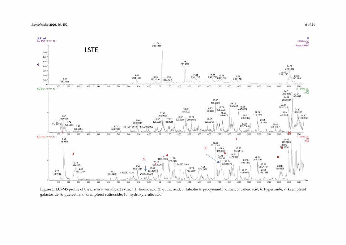
The LC–MS analysis of the LSTE (Figure 1) showed wide range of m/z (M-H) ions, whichindicated the presence of different phenolic compounds including phenolic acids (ferulic (m/z195/Rt 1.52 min)/quinic (191/2.72)/hydroxyferulic (209/24.26) and caffeic (177/17.18) acids); flavonones((luteolin (285/9.76), quercetin (301/23.09), hyperoside (463/17.34), kaempferol galactoside (447/18.95)and kaempferol rutinoside (593/23.95)) and procyanidin dimers (577/10.89, 11.98; B type). Thesetypes of compounds have a strong ability to donate electrons and are playing a key role in theoxidation–reduction process; the removal of the electrons results in a stable quinonoidal structure(Scheme 1).
Biomolecules 2020, 10, 452 6 of 24
Figure 1. LC–MS profile of the L. sericea aerial part extract. 1: ferulic acid; 2: quinic acid; 3: luteolin 4: procynanidin dimer; 5: caffeic acid; 6: hyperoside, 7: kaempferolgalactoside; 8: quercetin; 9: kaempferol rutinoside; 10: hydroxyferulic acid.

Biomolecules 2020, 10, 452 7 of 24
Scheme 1. The proposed reduction of gold salt and encapsulation of the hybrid gold NPs in theF1 and F2 matrix. In addition to the chelating power of the oxygen atom to the metal surface, theintermolecular hydrogen bonding between procyanidins can also make double and triple cappingshells and contributing to the stability of the NPs. The reduction power of the phenolic structures withortho-OH, can be facilitated by the formation of stable ortho-quinone structure.
Attempted purification of fraction (F1) showed both dimers (Rt 10.51; 10.85; 12.02; 12.36) andtrimers (Rt: 11.30; 12.67; 13.91; 14.94; 15.96; 16.78), however, the relative quantity of dimers was higherthan trimers (Figure 2).
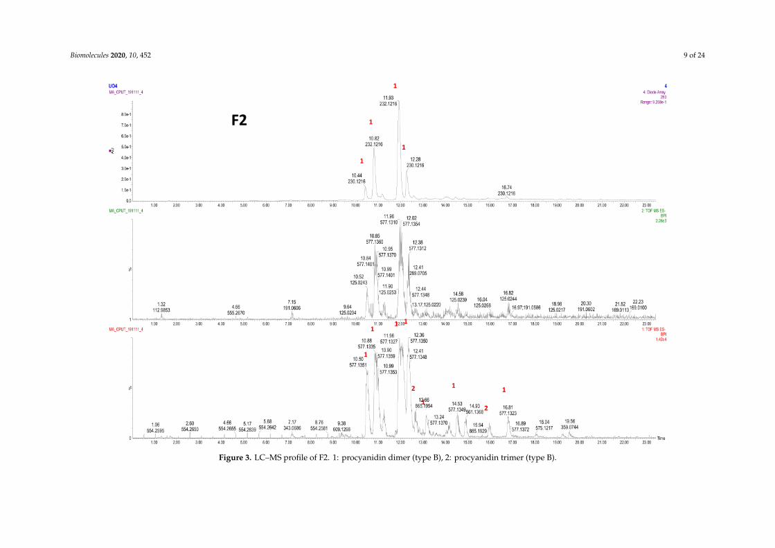
Further purification of fraction LSTE4 produces a single spot, which initially was believed to be apure compound. However, the NMR (1H and 13C NMR), and LC–MS analysis showed an isomericmixture. The LC–MS of F2 (Figure 3) showed procyanidin dimer peaks B-type (m/z 577) at Rt of 10.50;10.88; 11.96; 12.36; 13.24; 14.53 and 16.81; the first four peaks were major while the others were minor.The trimers (B-type; m/z 865) were also detected in minute quantities at 12.66 and 15.94 (Figure 3).
LSTE, F1 and F2 were then used to successfully form Au NPs. The polyphenolic nature of thesefractions makes them hydrophilic, a requirement for green synthesis. The reducing and capping abilityof polyphenolics has been associated with the presence of electronegative atoms [79]. The lone pairs ofelectrons on oxygen further provide a suitable chelation site for binding with gold. The arrangementof certain functional groups in the structure of polyphenolics is also of great significance in theirreactivity. Hydroxyl groups at adjacent positions may offer two or more binding sites, which mayenhance the easy reduction of gold and subsequent capping of the particles that are formed. Oxidationreactions are common to phenolic compounds where the hydroxyl groups at ortho and para positionsconvert to carbonyl groups (ortho and/or para quinones). In the case of the procyanidins, the oxidationwill occur at ortho positions (Scheme 1) when 4 and 6 electrons are lost for the dimer and trimerrespectively. Subsequent interaction of the particles with the molecule probably brought about weakforces of attraction and encapsulation in the F1 and F2 matrix (Scheme 1). As would be discussed later,the particles are covered with negative charges providing stability and confirming the negative zetapotential values as measured by zetasizer.

Biomolecules 2020, 10, 452 8 of 24
Figure 2. LC–MS profile of F1. 1: procyanidin dimer (type B); 2: procyanidin trimer (type B); 3: coumaric acid.
Biomolecules 2020, 10, 452 9 of 24
Figure 3. LC–MS profile of F2. 1: procyanidin dimer (type B), 2: procyanidin trimer (type B).
Biomolecules 2020, 10, 452 10 of 24
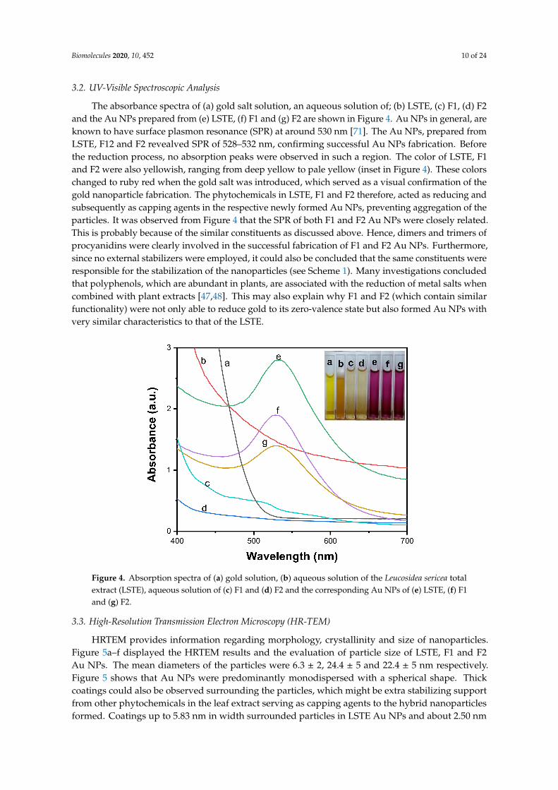
3.2. UV-Visible Spectroscopic Analysis
The absorbance spectra of (a) gold salt solution, an aqueous solution of; (b) LSTE, (c) F1, (d) F2and the Au NPs prepared from (e) LSTE, (f) F1 and (g) F2 are shown in Figure 4. Au NPs in general, areknown to have surface plasmon resonance (SPR) at around 530 nm [71]. The Au NPs, prepared fromLSTE, F12 and F2 revealved SPR of 528–532 nm, confirming successful Au NPs fabrication. Beforethe reduction process, no absorption peaks were observed in such a region. The color of LSTE, F1and F2 were also yellowish, ranging from deep yellow to pale yellow (inset in Figure 4). These colorschanged to ruby red when the gold salt was introduced, which served as a visual confirmation of thegold nanoparticle fabrication. The phytochemicals in LSTE, F1 and F2 therefore, acted as reducing andsubsequently as capping agents in the respective newly formed Au NPs, preventing aggregation of theparticles. It was observed from Figure 4 that the SPR of both F1 and F2 Au NPs were closely related.This is probably because of the similar constituents as discussed above. Hence, dimers and trimers ofprocyanidins were clearly involved in the successful fabrication of F1 and F2 Au NPs. Furthermore,since no external stabilizers were employed, it could also be concluded that the same constituents wereresponsible for the stabilization of the nanoparticles (see Scheme 1). Many investigations concludedthat polyphenols, which are abundant in plants, are associated with the reduction of metal salts whencombined with plant extracts [47,48]. This may also explain why F1 and F2 (which contain similarfunctionality) were not only able to reduce gold to its zero-valence state but also formed Au NPs withvery similar characteristics to that of the LSTE.
Figure 4. Absorption spectra of (a) gold solution, (b) aqueous solution of the Leucosidea sericea totalextract (LSTE), aqueous solution of (c) F1 and (d) F2 and the corresponding Au NPs of (e) LSTE, (f) F1and (g) F2.
3.3. High-Resolution Transmission Electron Microscopy (HR-TEM)
HRTEM provides information regarding morphology, crystallinity and size of nanoparticles.Figure 5a–f displayed the HRTEM results and the evaluation of particle size of LSTE, F1 and F2Au NPs. The mean diameters of the particles were 6.3 ± 2, 24.4 ± 5 and 22.4 ± 5 nm respectively.Figure 5 shows that Au NPs were predominantly monodispersed with a spherical shape. Thickcoatings could also be observed surrounding the particles, which might be extra stabilizing supportfrom other phytochemicals in the leaf extract serving as capping agents to the hybrid nanoparticlesformed. Coatings up to 5.83 nm in width surrounded particles in LSTE Au NPs and about 2.50 nm

Biomolecules 2020, 10, 452 11 of 24
for F1 Au NPs (Figure 5b). Nevertheless, the F2 Au NPs seeds had a layer of 1.97 nm thick, mostlyon the spherical particles. The approximate similar size of Au NPs conjugated with F1 and F2 can beexplained in terms of the similarity in the structures and capping layers packing of F1 and F2, which(both) contain procyanidin dimers (F2) or a mixture of dimers and trimers (F1). On the other hand, thesmaller size obtained by the total extract may indicate the strength of the reducing agents. It couldalso be due to the presence of smaller sized molecules that have the ability to perfectly shield theAu NPs. Similar coatings have been observed previously having a width between 2 and 3 nm andit was concluded that it prevents the gold nanoparticles from agglomeration [80]. Smaller particlesare predominantly round-shaped, others are polygonals including traingular and some five sidedshapes. On the other hand, particles with relatively larger sizes were mostly hexagonal and truncatedtriangular. This combination of shapes is characteristic of Au NPs and it has been associated with thedifferent chemical constituents forming the Au NPs [81,82]. Furthermore, it is interesting to note thatthe mixture of shapes is common to the HRTEM results of the Au NPs but much more pronounced forF1 and F2 Au NPs. This further serves as evidence of the involvemnet of similar functional groups informing the two particles. The inset in Figure 5a–c shows the selected area electron diffraction (SAED)pattern of LSTE, F1 and F2 Au NPs respectively. The bright circular rings indicate that the particles arepolycrystalline, and can be linked to the (111), (200), (220) and (311) planes of a face-centered (FCC)structure of gold [83].
Figure 5. HRTEM images for LSTE, F1 and F2 Au NPs are represented as (a–c) and the correspondingparticle size distributions as (d–f) respectively. The inset in a, b and c, showing the bright rings are theSAED of the respective TEM images.
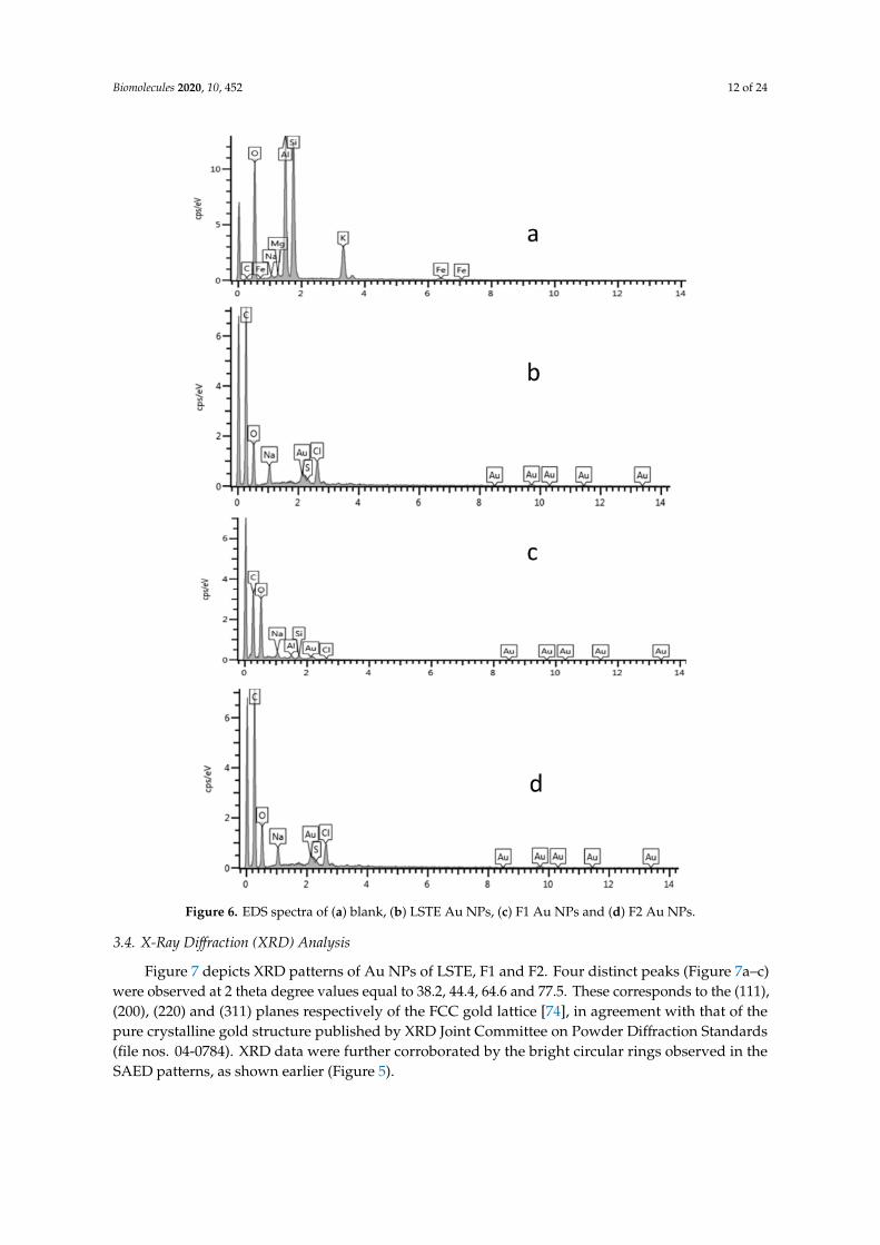
The EDS spectra of the blank/glass substrate (Figure 6a) and the Au NPs (Figure 6b–d) confirmedthe presence of Au in the solution. Other elemental peaks such as C, O and Na (Figure 6a–d) were likelyfrom the sample preparation/glass substrate used to prepare the samples for the analysis. The smallsulfur peak (Figure 6b) could be a contaminant while Cl and perhaps some of the Na were possiblyfrom the sodium chloroaurate solution used as a precursor.
Biomolecules 2020, 10, 452 12 of 24
Figure 6. EDS spectra of (a) blank, (b) LSTE Au NPs, (c) F1 Au NPs and (d) F2 Au NPs.
3.4. X-Ray Diffraction (XRD) Analysis
Figure 7 depicts XRD patterns of Au NPs of LSTE, F1 and F2. Four distinct peaks (Figure 7a–c)were observed at 2 theta degree values equal to 38.2, 44.4, 64.6 and 77.5. These corresponds to the (111),(200), (220) and (311) planes respectively of the FCC gold lattice [74], in agreement with that of thepure crystalline gold structure published by XRD Joint Committee on Powder Diffraction Standards(file nos. 04-0784). XRD data were further corroborated by the bright circular rings observed in theSAED patterns, as shown earlier (Figure 5).

Biomolecules 2020, 10, 452 13 of 24
Figure 7. XRD patterns of the (a) LSTE Au NPs, (b) F1 Au NPs and (c) F2 Au NPs.
3.5. Dynamic Light Scattering (DLS) Measurement of Au NPs
Among other important factors to be considered before the application of nanoparticles is thephase behavior. Light scattering is one of the techniques to understand this phenomenon. DLS,a non-intrusive technique, was employed to measure the size and zeta potential of the Au NPs.The average size of LSTE, F1 and F2 Au NPs was found to be 21.85, 26.91 and 27.20 nm respectively(Figure 8). Size characterization of nanoparticles is of immense importance in nanomedicine. However,because of the physical properties that are measured, and the weighted averages determined in eachcase, the DLS measurement for size is often not the same as those measured by electron microscopy [84].The size variation between DLS and TEM could therefore be due to the sampling volume usedduring analysis.
Figure 8. Hydrodynamic size of (a) LSTE, (c) F1, (e) F2 and zeta potential of (b) LSTE, (d) F1 and (f) F2Au NPs measured using Dynamic Light Scattering (DLS).

Biomolecules 2020, 10, 452 14 of 24
The zeta potentials (ZP) taken immediately after particle formation are displayed in Table 1. Valuesof −33.59, −32.5 and −29.1 mV (Table 1) were obtained for the LSTE-, F1- and F2 Au NPs respectively.ZP is related to the surface charge of nanoparticles, and particles with ZP values -30 mV and below aretaken to be mostly covered with negatively charged ions. The opposite is true for positive ZP values.The extent of ZP is therefore, a subject of repulsion and attraction that has been used to estimate howlong the nanoparticle dispersions would remain stable. The negative zeta-potential values obtained forthe Au NPs fall in the range that is typical of stable colloidal dispersions [72]. The ZP measurement forthe nanomaterials was repeated on the seventh day through to the fourteenth day showing only slightchanges. From the DLS measurement, a slightly higher value of ZP for LSTE Au NPs could be due tothe synergistic effect of other compounds present, probably providing enhanced capping ability. Boththe hydrodynamic sizes and ZP values for F1 and F2 can be observed to be approximately the sameand therefore can be explained by the similarity of phytochemicals involved. This was also supportedby their HRTEM results, where the average particle sizes were 24.4 and 22.4 nm for F1- and F2 Au NPsrespectively. Interestingly, their ZP values equally indicated that the particles are stable to a similarextent, further confirming the presence of similar capping agents. Significant changes were not noticedin the ZP values for the given days, thereby suggesting no agglomeration as they repelled each other.Therefore, the particles were quite stable.
Table 1. Particle size and zeta potential for Au NPs obtained from DLS.
Sample Hydrodynamic Size (nm) Zeta Potential (mV)
LSTE Au NPs 21.85 −33.9F1 Au NPs 27.20 −32.5F2 Au NPs 26.91 −29.1
3.6. In Vitro Stability Study
The effectiveness of nanoparticles in medical applications depends on its stability in biologicalsolutions for a reasonable period. Figure 9 displayed the evaluation of stability when 0.5% bovineserum albumin (BSA), glycine (GLY), sodium chloride (NaCl), cysteine (CYS) and phosphate buffers atpH 7 and 9 were mixed with LSTE, F1 and F2 particles [85]. These were carried out before and afterincubation at 37 ◦C. Even though the pH 9 solution is higher than the pH of human body fluids, it wasconsidered to get additional information about whether nanoparticles are still stable at such a high pH.The results showed that the SPR remained the same in all the formulations. However, a noticeableshift by BSA especially from the 12th h is a behavior of certain proteins. BSA has been reported tohave preference of binding to negatively charged surfaces [86]. This has been ascribed to the presenceof over 50 surface lysine groups, which enable easy electrostatic interactions with negative surfaces.Consequently, the negative ZP values of our particles implies that the surfaces were largely covered bynegative ions, hence more interaction with BSA. Overall, the shifts of less than 1.0 nm in all solutionswas minimal, the λmax remained intact thereby affirming the stability of the particles in the solutions atthe tested period.

Biomolecules 2020, 10, 452 15 of 24
Figure 9. Stability of gold nanoparticles at given time intervals.

Biomolecules 2020, 10, 452 16 of 24
3.7. Dilution Study
Some biomedical applications require different concentrations of gold nanoparticles. Figure 10shows the absorbance of the hybrid (a) LSTE-, (b) F1- and (c) F2 Au NPs at different concentrations.To confirm that dilution of nanoparticles into different concentrations does not affect their stabilityand does not alter their physical and chemical properties in vivo, a dilution study was carried out atdifferent concentrations of the nanoparticles (100, 80, 60, 40, 20 and 10 µg/mL), and UV-Vis spectroscopy(350–850 nm) was recorded for each concentration. From the UV-Vis spectra, the SPR wavelength hadthe same value for all the solutions of a given particle. This means that the dilution did not have an effecton the properties of LSTE-, F1- and F2-Au NPs, and the nanoparticles remained stable. The relationshipbetween various concentrations of the nanoparticles and the absorbance was further examined andfound to be linear. Figure 10Q shows that the intensity of absorption had linear dependence on theconcentration of respective nanoparticles. This is additional evidence that the particles were stable andthat they did not agglomerate even as the concentrations change.
Figure 10. Surface plasmon resonance wavelength (λmax) of diluted concentrations of LSTE-, F1- and F2-Au NPs, while (Q) represents a plot of absorbance against various concentrations, showing the linearproportionality of intensity with increasing concentration. R2 = 0.9455 (LSTE Au NPs), R2 = 0.9908 (F1Au NPs) and R2 = 0.9848 (F2 Au NPs).
3.8. In-Vitro Antidiabetic Studies
Since T2DM is associated with neurological and cardiovascular complications as a result ofmetabolic disorders emanating from hyperglycemia, the most critical remedy is to keep the blood sugarwithin the normal range. Two special enzymes, alpha-glucosidase and -amylase have good records ofbreaking down the long-chains of carbohydrates thereby enhancing the breakup of the glucose unitfrom the disaccharide. Therefore, the inhibition of these enzymes has been among the popular therapiesfor T2DM [87]. The antidiabetic properties of plant extracts have been reported before [88]. In thisstudy, Leucosidea sericea leaves extract, the fractions (F1 and F2) and gold nanoparticles biosynthesizedfrom them were studied and their potential as antidiabetic and antioxidant agents evaluated.
For the enzymatic studies, strong inhibitory activities were displayed by the constituents ofLeucosidea sericea as well as the fabricated Au NPs (Table 2). F1 and F2 are fractions from the extractand demonstrated high alpha-glucosidase activity. IC50 values of 8.1, 7.3 and 7.1 µg/mL were obtained

Biomolecules 2020, 10, 452 17 of 24
for LSTE, F1 and F2 respectively. The alpha-amylase inhibition resulted in 3.5 µg/mL for LSTE and18.9 µg/mL for F2. F1 did not show any activity at the tested concentrations. In general, the resultsshowed that LSTE, F1, F2 and their Au NPs had the potential to be used as antidiabetic agents.Pure isolated phytochemicals have demonstrated activities against alpha-glucosidase in previousstudies [47,48]. Recently, Etsassala et al. [89] reported a strong antidiabetic activity of ten abietanediterpenes. In their study, the antidiabetic activity was due to the type and position of different functionalgroups in the compounds. Additionally, some compounds displayed either alpha-glucosidase oralpha-amylase activity and not both. For instance, 11,12-dehydroursolic acid lactone displayedmoderate alpha-glucosidase activity but showed no activity for amylase [89]. Although, both fractions(F1 and F2) have similar structures, F1 did not show activity against alpha-amylase compared to F2,this may be due to the antagonistic effect of one of the trimers, and this effect disappeared when thiscompound(s) conjugated with Au NPs and showed enhanced activities. Furthermore, among thethree popular antidiabetic standard drugs currently in the market, only acarbose inhibits both alphaglucosidase and alpha amylase. Vigliobose showed little effect on amylase whereas miglitol does nothave any effects on amylase [90] Therefore, the high activity displayed by F1 on alpha-glucosidase andnot for amylase agrees with the above studies. Furthermore, a close examination of the IC50 of LSTE,F1 and F2 for alpha-glucosidase indicates a similar functionality as mentioned above. Almost the samevalues were obtained for F1 and F2, which further supports the occurrence of the dimers and trimers ofprocyanidins, as rightly suggested by the NMR and LC–MS analysis. Procyanidins possessed similararrangements of the hydroxyl groups as well as the aromatic ring systems and therefore, might havesimilar behaviors. However, the mechanism of action of alpha-glucosidase and alpha-amylase mightnot necessarily be the same. This has been demonstrated by the reports of many assays where differentvalues were recorded for the two enzymes, even though the aim of inhibition is the same [91].
Table 2. Inhibitory activities of LSTE, F1, F2 and Au NPs on alpha-glucosidase and alpha-amylase.
Items Alpha-Glucosidase IC50 (µg/mL) Alpha-Amylase IC50 (µg/mL)
LSTE 8.1 ± 0.6 3.5 ± 0.7LSTE Au NPs 14.5 ± 0.8 3.0 ± 0.3
F1 7.3 ± 0.5 NAF1 Au NPs 7.3 ± 0.3 1.8 ± 0.3
F2 7.1 ± 0.4 18.9 ± 0.2F2 Au NPs 4.5 ± 0.6 10.5 ± 0.1Acarbose 610 ± 2.6 10.2 ± 0.6
NA: not active at the tested concentrations, Acarbose: positive control.
Au NPs of LSTE, F1 and F2 demonstrated activities that compete well with the intactextract/fractions. Although the activity of F1 Au NPs was identical to that of F1 for alpha-glucosidase,an enhanced alpha-amylase inhibition was recorded for F1 Au NPs. This behavior is similar to thatreported by Shamprasad et al. [58], where Escin-Au NPs showed better activity compared to Escin intheir study, thereby confirming our submission on the activity of F1 and F2 Au NPs. The activitiesin each case were almost two-fold of the corresponding precursor. This continuous resemblanceof activity may stem from the starting materials of the nanoparticles. As earlier stated, F1 andF2 fractions are composed of dimer and trimer procyanidins as the major constituents. Therefore,closely related behaviors in the same experimental conditions are obvious. Accordingly, the improvedantidiabetic performance of bimetallic Ag-Au NPs over their precursor extracts and acarbose has beenreported [45,46]. The IC50 values of alpha-amylase on LSTE and its gold form also showed improvedactivity in agreement with previous studies [45]. The inhibiting powers of the nanomaterials may be afunction of size and shape. As noted earlier, our Au NPs had a size range of 6–24 nm. These values aresimilar to the sizes in previous investigations [58]. Similarly, Niikura et al. [92] affirmed that spherical

Biomolecules 2020, 10, 452 18 of 24
Au NPs of sizes 20 and 40 nm in diameter induced the west Nile virus better than those of other sizesand shapes. This may be the reason for an improved enzymatic activity of F1 and F2 Au NPs.
3.9. Antioxidant Activity/Total Phenolic Content
Oxidative stress has been linked to the cause of many deadly diseases like diabetes, the managementof which is costly. Thus, plant extracts and nanoparticles with both antidiabetic and antioxidantproperties will be greatly beneficial [93]. Therefore, the need to search for antioxidants with enhancedreducing abilities is crucial. Antioxidants are substances that can inhibit or delay the oxidationof a substrate when present in low concentrations. Due to the relationship of oxidative stressto other diseases, we, therefore, investigated the antioxidant capacities of LSTE, F1, F2 and thecorresponding Au NPs. Three essays; Ferric reducing antioxidant power (FRAP), Folin–Ciocalteu (FC)and 2,2′-azino-bis-3-ethylbenzotiazolin-6- sulfonic acid (ABTS) were carried out and the results arepresented in Table 3.
Table 3. Antioxidant activities of F 2, LSTE, F 1 and the corresponding Au NPs.
Items FRAP (µM AAE/g) ABTS (µM TE/g) FC (µM GAE/g) FC% (Au NPs)
LSTE 1113.2 ± 6.7 814.9 ± 6.1 602.6 ± 6.1LSTE Au NPs 113.8 ± 9.5 1059.4 ± 7.4 179.8 ± 6.2 29.3
F1 1834.0 ± 4.7 818.2 ± 7.7 889.6 ± 6F1 Au NPs 748.6 ± 1.4 1521.9 ± 3.0 356.7 ± 6.6 40.1
F2 1166.0 ± 2.1 816.9 ± 8.6 685.7 ± 6.7F2 Au NPs 1083.8 ±1.2 861.9 ± 5.3 523.1 ± 4.4 76.3Standard 3976.8 ± 3.8 * 7525.0 ± 4.9 **
* = vitamin C, ** = trolox. The results are expressed as mean ± SD for n = 3.
The mechanism with which FRAP operates is known as single electron transfer (SET), wherebyan antioxidant transfers an electron to the corresponding cation, which would neutralize it [94].In Table 3, strong activities were exhibited on FRAP with F1 displaying the highest activity (1834.0 ±4.7 µM AAE/g). From the NMR and LC–MS analysis, F1 was found to contain a dimer and trimer ofprocyanidins that might have acted together in synergy to bring about this high activity. They possesshydroxyl groups attached to aromatic rings, which will perfectly participate in oxidation during theprocess. Similar activities were recorded for LSTE and F2 because they contained phenolic compoundsof similar functionality. It is well known that phenolics have strong antioxidant activities [95]. The hugepresence of many phenolics in LSTE has been demonstrated previously by LC–MS analysis, thereforeits high activity is reasonable, while similar compounds, although different proportions were found inF2, accounted for its activity as well. However, since the FRAP’s mechanism is by electron transfer,the hydroxyl groups of the compound might be interacting with the nanoparticles, thereby limitingthe site for the oxidation process. This may contribute to slightly reduced activity. In contrast, highactivities in close ranges were demonstrated for all samples in FC assay except for LSTE Au NPs. Size,shape and the surrounding environment/medium is critical to the activity of nanoparticles. Owing tothe type and number of phytochemicals taking part in the fabrication of LSTE Au NPs, there is a largesize difference from those of F1 and F2 and the stability was also slightly higher. Therefore, it is expectedto behave somewhat differently since the smaller sized particles have a higher surface area. In addition,more phytochemicals are present in LSTE, which creates a different surrounding environment forthe NPs. Previously, nanoparticles with smaller sizes were reported to show enhanced activity incomparison to relatively larger ones [96]. On the other hand, a clear trend can be observed for the ABTSassay. This is probably because of the difference in the mechanism of operation between the assays.ABTS is largely operating on hydrogen atom transfer (HAT). The trend in the ABTS results is such thatindividual Au NPs demonstrated better antioxidant capacity relative to their respective precursors.

Biomolecules 2020, 10, 452 19 of 24
Recent research reports [54,57,58,95] supported the above submission. Au NPs biosynthesized fromHalymenia dilatata also demonstrated higher antioxidant activity than the starting plant extract [97].
However, results of antioxidant activities often vary from one assay to the other, probably becauseof the difference in mechanism of operation where some operate by SET or HAT or both. The nature ofthe sample, the medium of operation and the functionality might account for the variations in ourresults from one assay to the other (Table 3).
3.10. Quantification of the Total Phenolic Content in the Au NPs from the FC Assay
Results from Table 3 revealed the phenolic content of the intact fractions (F1 and F2) and the totalextracts, as well as the phenolic content (FC) of the corresponding Au NPs. According to the HRTEMand DLS analysis, the average diameter was smaller in case of the LSTE Au NPs relative to the others.Hence, the higher surface area available for interaction with the compounds may explain the smallerphenolic content (29.3%) observed. However, an increase in the percentage FC was observed in the caseof F1 and F2, with maximum concentration of F2 (76.3%). The high payload of F2 may be explained interms of the homogeneity of the compound in that fraction as well as its high packing power.
4. Conclusions
The preliminary screening of the L. sericea total extract showed the potential of generating stableAu NPs. An intent made to purify the pure compound(s) responsible for such activity resulted in thepurification of two inseparable procyanidins fractions namely F1 and F2. The first fraction, F1, containeda mixture of procyanidins dimer (B series) and trimers, while the second fraction, F2, contained fourmajor procyanidin dimers belonging to B series. These fractions successfully formed stable Au NPsconfirming the ability of these class of phytochemicals to act as reducing agents. DLS measurementsand in vitro stability examinations further affirmed their stability in physiological conditions withoutthe introduction of any external stabilizers. This means that the compounds also doubled as cappingagents that prevented agglomeration for the given period. Thereafter, biological activities were carriedout. LSTE constituents and hybrid nanoparticles showed interesting inhibitory activities on bothalpha-glucosidase and alpha-amylase at low concentrations. F1 and its Au NPs demonstrated enhancedalpha-glucosidase activities compared to LSTE and LSTE Au NPs. For alpha-amylase, F2 Au NPsshowed the highest inhibitory activities, which were another interesting behavior that calls for furtherattention. The particles also exhibited interesting antioxidant activity thereby buttressing their potentialbiological applications since this activity has been linked to some diseases. As far as we know, thiswork is the first scientific report on the identification of procyanidins from the aerial parts of Leucosideasericea, and the use thereof in nanoparticle synthesis. The results suggest that procyanidins had theability to reduce gold to form biostable and bioactive gold nanoparticles with potential antidiabeticand antioxidant applications.
Supplementary Materials: The following are available online at http://www.mdpi.com/2218-273X/10/3/452/s1,Figure S1: 1H NMR of Fraction F1; S2: 13C NMR of Fraction F1; S3: 13C –DEPT-135 NMR of Fraction F1; S4: 1HNMR of Fraction F2; S5: 13C NMR of Fraction F2; S6: 13C –DEPT-135 NMR of Fraction F2.
Author Contributions: A.A.H. conceived the research idea. U.M.B. carried out the synthesis of Au NPs, dilutionstudies, UV-Vis and DLS measurements under the guidance of A.A.H. and S.B. A.O.A. carried out extraction,purification of F1 and F2. J.A.B. carried out enzymatic studies under the guidance of J.L.M. while S.B. carried outTEM, SAED and EDS analysis. C.N.C. identified, collected and prepared the plant material. E.I. was responsiblefor stability studies and coordinated the writing of the manuscript. All authors read, edited and contributed to themanuscript. A.A.H. and S.B. are U.M.B.’s supervisors. All authors have read and agreed to the published versionof the manuscript.
Funding: The Tertiary Education Trust Fund (IBB University, Lapai 2016 intervention), Nigeria was used for thisresearch. Cost of chemicals was covered by NRF with a call number (106055) under Professor Ahmed A. Hussein.
Acknowledgments: We would like to appreciate Hamza Elsayed Mohammed of iThemba labs for XRD analysis,Electron microscope unit, UWC for TEM, EDS and SAED analysis. Also, we would like to thank Dr Enas Ismail isalso affiliated to the Physics department, Faculty of Science (girls branch), Al Azhar University, Cairo, Egypt.

Biomolecules 2020, 10, 452 20 of 24
Conflicts of Interest: The authors declare no conflict of interest in this work.
References
1. Duan, H.; Wang, D.; Li, Y. Green chemistry for nanoparticle synthesis. Chem. Soc. Rev. 2015, 44, 5778–5792.[CrossRef] [PubMed]
2. Saravanakumar, A.; Peng, M.M.; Ganesh, M.; Jayaprakash, J.; Mohankumar, M.; Jang, H.T. Low-cost andeco-friendly green synthesis of silver nanoparticles using Prunus japonica (Rosaceae) leaf extract and theirantibacterial, antioxidant properties. Artif. Cells Nanomed. Biotechnol. 2017, 45, 1165–1171. [CrossRef]
3. Jha, A.K.; Prasad, K. Green synthesis of silver nanoparticles using Cycas leaf. Int. J. Green Nanotechnol. Phys.Chem. 2010, 1, P110–P117. [CrossRef]
4. Liu, J.; Qin, G.; Raveendran, P.; Ikushima, Y. Facile “green” synthesis, characterization, and catalytic functionof β-D-glucose-stabilized Au nanocrystals. Chem. Eur. J. 2006, 12, 2131–2138. [CrossRef] [PubMed]
5. Kumar, V.; Mohan, S.; Singh, D.K.; Verma, D.K.; Singh, V.K.; Hasan, S.H. Photo-mediated optimized synthesisof silver nanoparticles for the selective detection of Iron (III), antibacterial and antioxidant activity. Mater.Sci. Eng. C 2017, 71, 1004–1019. [CrossRef] [PubMed]
6. Tekale, S.U.; Kauthale, S.S.; Pagore, V.P.; Jadhav, V.B.; Pawar, R.P. ZnO nanoparticle-catalyzed efficientone-pot three-component synthesis of 3, 4, 5-trisubstituted furan-2 (5H)-ones. J. Iran. Chem. Soc. 2013, 10,1271–1277.
7. Ergin, A.D.; Bayindir, Z.S.; Yüksel, N. Characterization and optimization of colon targetedS-adenosyl-L-methionine loaded chitosan nanoparticles. Marmara Pharm. J. 2019, 23, 914–926. [CrossRef]
8. Liu, Y.; Welch, M.J. Nanoparticles labeled with positron emitting nuclides: Advantages, methods, andapplications. Bioconj. Chem. 2012, 23, 671–682. [CrossRef]
9. Xie, Y.; Kocaefe, D.; Chen, C.; Kocaefe, Y. Review of research on template methods in preparation ofnanomaterials. J. Nanomater. 2016, 2016, 10.
10. Singh, P.; Kim, Y.J.; Zhang, D.; Yang, D.C. Biological synthesis of nanoparticles from plants and microorganisms.Trends Biotechnol. 2016, 34, 588–599. [CrossRef]
11. Lysy, P.A.; Corritore, E.; Sokal, E.M. New insights into diabetes cell therapy. Curr. Diabetes Rep. 2016, 16, 38.[CrossRef] [PubMed]
12. Renner, S.; Blutke, A.; Clauss, S.; Deeg, C.A.; Kemter, E.; Merkus, D.; Wanke, R.; Wolf, E. Porcine modelsfor studying complications and organ crosstalk in diabetes mellitus. Cell Tissue Res. 2020, 1–38. [CrossRef][PubMed]
13. Cho, N.; Shaw, J.E.; Karuranga, S.; Huang, Y.; da Rocha Fernandes, J.D.; Ohlrogge, A.W.; Malanda, B.IDF diabetes atlas: Global estimates of diabetes prevalence for 2017 and projections for 2045. Diabetes Res.Clin. Pract. 2018, 138, 271–281. [CrossRef] [PubMed]
14. Lorenzati, B.; Zucco, C.; Miglietta, S.; Lamberti, F.; Bruno, G. Oral hypoglycemic drugs: Pathophysiologicalbasis of their mechanism of action. Pharmaceuticals 2010, 3, 3005–3020. [CrossRef] [PubMed]
15. Santos, M.S.C.; Azevedo, R.B.; Teixeira, P.R.; Sales, M.J.A.; Báo, S.N.; Paterno, L.G.; Silva, A.L.G.Photochemically-assisted synthesis of non-toxic and biocompatible gold nanoparticles. Colloids Surf.B Biointerfaces 2016, 148, 317–323.
16. Ahmad, T.; Azmi, M.; Irfan, M.; Moniruzzaman, M.; Asghar, A.; Bhattacharjee, S. Green synthesis of stabilizedspherical shaped gold nanoparticles using novel aqueous Elaeis guineensis (Oil palm) leaves extract. J. Mol.Struct. 2018, 1159, 167–173. [CrossRef]
17. Zhang, G.; Li, Y.; Gao, X. An asynchronous-alternating merging-zone flow-injection gold nanoparticles probemethod for determination of Antidiabetic pioglitazone hydrochloride medicine. New J. Chem. 2018, 42,4337–4343. [CrossRef]
18. Yilmazer-Musa, M.; Griffith, A.M.; Michels, A.J.; Schneider, E.; Frei, B. Grape seed and tea extracts andcatechin 3-gallates are potent inhibitors of α-amylase and α-glucosidase activity. J. Agric. Food Chem. 2012,60, 8924–8929. [CrossRef]
19. Tanko, Y.; Yerima, M.; Mahdi, M.A.; Yaro, A.H.; Musa, K.Y.; Mohammed, A. Hypoglycemic activity ofmethanolic stem bark of adansonnia digitata extract on blood glucose levels of streptozocin-induced diabeticwistar rats. Int. J. Appl. Res. Nat. Prod. 2008, 1, 32–36.

Biomolecules 2020, 10, 452 21 of 24
20. Van de Venter, M.; Roux, S.; Bungu, L.C.; Louw, J.; Crouch, N.R.; Grace, O.M.; Folb, P. Anti-diabetic screeningand scoring of 11 plants traditionally used in South Africa. J. Ethnopharmacol. 2008, 119, 81–86. [CrossRef]
21. Eidi, A.; Eidi, M.; Esmaeili, E. Anti-diabetic effect of garlic (Allium sativum L.) in normal andstreptozotocin-induced diabetic rats. Phytomedicine 2006, 13, 624–629. [CrossRef] [PubMed]
22. Rizvi, M.M.A.; El Hassadi, I.M.G.; Younis, S.B. Bioefficacies of Cassia fistula: An Indian labrum. Afr. J. Pharm.Pharmacol. 2009, 3, 287–292.
23. Sethi, J.; Sood, S.; Seth, S.; Talwar, A. Evaluation of hypoglycemic and antioxidant effect of Ocimum sanctum.Indian J. Clin. Biochem. 2004, 19, 152–155. [CrossRef] [PubMed]
24. Nammi, S.; Boini, M.; Lodagala, S.; Behara, R. The juice of fresh leaves of Catharanthus roseus Linn. reducesblood glucose in normal and alloxan diabetic rabbits. BMC Complement. Altern. Med. 2003, 3, 1–4. [CrossRef][PubMed]
25. Yagi, A.; Hegazy, S.; Kabbash, A.; Abd-El Wahab, E. Possible hypoglycemic effect of Aloe vera L. highmolecular weight fractions on type 2 diabetic patients. Saudi Pharm. J. 2009, 17, 209–215. [CrossRef]
26. Yalçın, S.; Erkan, M.; Ünsoy, G.; Parsian, M.; Kleeff, J.; Gündüz, U. Effect of gemcitabine and retinoic acidloaded PAMAM dendrimer-coated magnetic nanoparticles on pancreatic cancer and stellate cell lines. BioMedPharm. 2014, 68, 737–743. [CrossRef]
27. Kaleem, M.; Sheema, S.H.; Bano, B. Protective effects of Piper nigrum and Vinca rosea in alloxan induceddiabetic rats. Indian J. Physiol. Pharmacol. 2005, 49, 65–71.
28. Ponnanikajamideen, M.; Rajeshkumar, S. In vivo type 2 diabetes and wound-healing effects of antioxidantgold nanoparticles synthesized using the insulin plant Chamaecostus cuspidatus in albino rats. Can. J. Diabetes2019, 43, 82–89. [CrossRef]
29. Khalil, M. Biosynthesis of gold nanoparticles using extract of grape (Vitis vinifera) leaves and seeds.Prog. Nanotechnol. Nanomater. 2016, 3, 1–12.
30. Daisy, P.; Saipriya, K. Biochemical analysis of Cassia fistula aqueous extract and phytochemically synthesizedgold nanoparticles as hypoglycemic treatment for diabetes mellitus. Int. J. Nanomed. 2012, 7, 1189–1202.[CrossRef]
31. John, P.A.; Zhijian, C.; Naidu, M. Neurite outgrowth stimulatory effects of myco¬synthesized Au NPs fromHericium erinaceus (Bull.: Fr.) Pers. on pheochromocytoma (PC-12) cells. Int. J. Nanomed. 2015, 10, 5853–5863.
32. Venkatraman, A.; Yahoob, S.A.M.; Nagarajan, Y.; Harikrishnan, S.; Vasudevan, S.; Murugasamy, T.Pharmacological activity of biosynthesized gold nanoparticles from brown algae-Seaweed turbinaria conoides.Nanowrold J. 2018, 4, 17–22. [CrossRef]
33. Opris, R.; Tatomir, C.; Olteanu, D.; Moldovan, R.; Moldovan, B.; David, L.; Adriana, G. The effect of Sambucusnigra L. extract and phytosinthesized gold nanoparticles on diabetic rats. Colloids Surf. B Biointerfaces 2017,150, 192–200. [CrossRef]
34. Dhas, T.S.; Kumar, V.G.; Karthick, V.; Vasanth, K.; Singaravelu, G.; Govindaraju, K. Enzyme and microbialtechnology effect of biosynthesized gold nanoparticles by Sargassum swartzii in alloxan induced diabetic rats.Enzyme Microb. Technol. 2016, 95, 100–106. [CrossRef]
35. Malapermal, V.; Mbatha, N.; Gengan, R.; Anand, K. Biosynthesis of bimetallic Au-Ag nanoparticles usingOcimum basilicum (L.) with anti-diabetic and antimicrobial properties. Adv. Mater. Lett. 2015, 6, 1050–1057.[CrossRef]
36. Elobeid, M.A. Amelioration of streptozotocin induced diabetes in rats by eco-friendly compositenano-cinnamon extract. Pak. J. Zool. 2016, 48, 645–650.
37. Ovais, M.; Khalil, A.T.; Islam, N.U.; Ahmad, I.; Ayaz, M.; Saravanan, M.; Mukherjee, S. Role of plantphytochemicaLSTE and microbial enzymes in biosynthesis of metallic nanoparticles. Appl. Microbiol.Biotechnol. 2018, 102, 6799–6814. [CrossRef]
38. Khan, M.A.; Raza, A.; Ovais, M.; Sohail, M.F.; Ali, S. Current state and prospects of nano-delivery systemsfor sorafenib. Int. J. Polym. Mater. Polym. Biomater. 2018, 67, 1105–1115. [CrossRef]
39. Ali, M.; Khan, T.; Fatima, K.; Aliqul, A.; Ovais, M.; Khalil, A.T.; Idrees, M. Selected hepatoprotective herbalmedicines: Evidence from ethnomedicinal applications, animal models and possible mechanism of actions.Phytother. Res. 2018, 32, 199–215. [CrossRef]
40. Aromal, S.A.; Vidhu, V.K.; Philip, D. Green synthesis of well-dispersed gold nanoparticles using Macrotylomauniflorum. Spectrochim. Acta Part A 2012, 85, 99–104. [CrossRef]

Biomolecules 2020, 10, 452 22 of 24
41. Edison, T.J.I.; Sethuraman, M.G. Instant green synthesis of silver nanoparticles using Terminalia chebula fruitextract and evaluation of their catalytic activity on reduction of methylene blue. Process Biochem. 2012, 47,1351–1357. [CrossRef]
42. Mohan Kumar, K.; Mandal, B.K.; Sinha, M.; Krishnakumar, V. Terminalia chebula mediated green and rapidsynthesis of gold nanoparticles. Spectrochim. Acta Part A 2012, 86, 490–494. [CrossRef]
43. Stephen, A.; Seethalakshmi, S. Phytochemical synthesis and preliminary characterization of silvernanoparticles using hesperidin. J. Nanosci. 2013. [CrossRef]
44. Sahu, N.; Soni, D.; Chandrashekhar, B.; Satpute, D.B.; Saravanadevi, S.; Sarangi, B.K.; Pandey, R.A. Synthesisof silver nanoparticles using flavonoids: Hesperidin, naringin and diosmin, and their antibacterial effectsand cytotoxicity. Int. Nano Lett. 2016, 6, 173–181. [CrossRef]
45. Bisht, S.; Feldmann, G.; Soni, S.; Ravi, R.; Karikar, C.; Maitra, A.; Maitra, A. Polymeric nanoparticle-encapsulated curcumin (‘nanocurcumin’): A novel strategy for human cancer therapy. J. Nanobiotechnol.2007, 5, 1–18. [CrossRef]
46. Khaleel, S.; Govindaraju, K.; Manikandan, R.; Seog, J.; Young, E.; Singaravelu, G. Phytochemical mediatedgold nanoparticles and their PTP 1B inhibitory activity. Colloids Surf. B Biointerfaces 2010, 75, 405–409.[CrossRef]
47. Payne, J.N.; Badwaik, V.D.; Waghwani, H.K.; Moolani, H.V.; Tockstein, S.; Thompson, D.H.;Dakshinamurthy, R. Development of dihydrochalcone-functionalized gold nanoparticles for augmentedantineoplastic activity. Int. J. Nanomed. 2018, 13, 1917–1926. [CrossRef]
48. Shamprasad, B.R.; Keerthana, S.; Megarajan, S.; Lotha, R.; Aravind, S.; Veerappan, A. Photosynthesized escinstabilized gold nanoparticles exhibit anti-diabetic activity in L6 rat skeletal muscle cells. Mater. Lett. 2019,241, 198–201. [CrossRef]
49. Dong, Y.; Wan, G.; Yan, P.; Qian, C.; Li, F.; Peng, G. Biology fabrication of resveratrol coated gold nanoparticlesand investigation of their effect on diabetic retinopathy in streptozotocin induced diabetic rats. J. Photochem.Photobiol. B Biol. 2019, 195, 51–57. [CrossRef]
50. Rajarajeshwari, T.; Shivashri, C.; Rajasekar, P. Synthesis and characterization of biocompatible gymnemicacid—Gold nanoparticles: A study on glucose uptake stimulatory effect in 3T3-L1 adipocytes. RSC Adv.2014, 4, 63285. [CrossRef]
51. Bhumkar, D.R.; Joshi, H.M.; Sastry, M.; Pokharkar, V.B. Chitosan reduced gold nanoparticles as novel carriersfor transmucosal delivery of insulin. Pharm. Res. 2007, 24, 1415–1426. [CrossRef] [PubMed]
52. Cho, H.J.; Oh, J.; Choo, M.K.; Ha, J.I.; Park, Y.; Maeng, H.J. Chondroitin sulfate-capped gold nanoparticles forthe oral delivery of insulin. Int. J. Biol. Macromol. 2014, 63, 15–20. [CrossRef] [PubMed]
53. Dubey, K.; Anand, B.G.; Badhwar, R.; Bagler, G. Tyrosine and tryptophan coated gold nanoparticles inhibitamyloid aggregation of insulin. Amino Acids 2015, 47, 2551–2560. [CrossRef] [PubMed]
54. Khoshnamvand, M.; Ashtiani, S.; Huo, C.; Saeb, S.P.; Liu, J. Use of Alcea rosea leaf extract for biomimeticsynthesis of gold nanoparticles with innate free radical scavenging and catalytic activities. J. Mol. Struct.2019, 1179, 749–755. [CrossRef]
55. Pu, S.; Li, J.; Sun, L.; Zhong, L.; Ma, Q. An in vitro comparison of the antioxidant activities of chitosan andgreen synthesized gold nanoparticles. Carbohydr. Polym. 2019, 211, 161–172. [CrossRef] [PubMed]
56. Torabi, N.; Nowrouzi, A.; Ahadi, A.; Vardasbi, S.; Etesami, B. Green synthesis of gold nanoclusters using seedaqueous extract of Cichorium intybus L. and their characterization. SN Appl. Sci. 2019, 1, 981. [CrossRef]
57. Nakkala, J.R.; Bhagat, E.; Suchiang, K.; Sadras, S.R. Comparative study of antioxidant and catalytic activityof silver and gold nanoparticles synthesized from Costus pictus leaf extract. J. Mater. Sci. Technol. 2015, 31,986–994. [CrossRef]
58. Benedec, D.; Oniga, I.; Cuibus, F.; Sevastre, B.; Stiufiuc, G.; Duma, M.; Lucaciu, C.M. Origanum vulgaremediated green synthesis of biocompatible gold nanoparticles simultaneously possessing plasmonic,antioxidant and antimicrobial properties. Int. J. Nanomed. 2018, 13, 1041. [CrossRef]
59. Zhaleh, M.; Zangeneh, A.; Goorani, S.; Seydi, N.; Zangeneh, M.M.; Tahvilian, R.; Pirabbasi, E. In vitroand in vivo evaluation of cytotoxicity, antioxidant, antibacterial, antifungal, and cutaneous wound healingproperties of gold nanoparticles produced via a green chemistry synthesis using Gundelia tournefortii L. asa capping and reducing agent. Appl. Organomet. Chem. 2019, 33, e5015. [CrossRef]
60. Patil, M.P.; Seo, Y.B.; Lim, H.K.; Kim, G.D. Biofabrication of gold nanoparticles using Agrimonia pilosaextract and their antioxidant and cytotoxic activity. Green Chem. Lett. Rev. 2019, 12, 208–216. [CrossRef]

Biomolecules 2020, 10, 452 23 of 24
61. Veena, S.; Devasena, T.; Sathak, S.S.M.; Yasasve, M.; Vishal, L.A. Green Synthesis of gold nanoparticles fromvitex negundo leaf extract: Characterization and in vitro evaluation of antioxidant-antibacterial activity.J. Clust. Sci. 2019, 30, 1591–1597. [CrossRef]
62. Bharathi, D.; Bhuvaneshwari, V. Evaluation of the cytotoxic and antioxidant activity of phyto-synthesizedsilver nanoparticles using Cassia angustifolia flowers. J. Bionanosci. 2019, 9, 155–163. [CrossRef]
63. Zayed, M.F.; Mahfoze, R.A.; El-kousy, S.M.; Al-Ashkar, E.A. In-vitro antioxidant and antimicrobial activitiesof metal nanoparticles biosynthesized using optimized Pimpinella anisum extract. Colloid Surf. A Phys. Eng.2020, 585, 124167. [CrossRef]
64. Aremu, A.O.; Amoo, S.O.; Ndhlala, A.R.; Finnie, J.F.; Van Staden, J. Antioxidant activity, acetylcholinesteraseinhibition, iridoid content and mutagenic evaluation of Leucosidea sericea. Food Chem. Toxicol. 2011, 49,1122–1128. [CrossRef]
65. Pendota, S.C.; Aremu, A.O.; Slavetínská, L.P.; Rárová, L.; Grúz, J.; Doležal, K.; Van Staden, J. Identificationand characterization of potential bioactive compounds from the leaves of Leucosidea sericea. J. Ethnopharmacol.2018, 220, 169–176. [CrossRef]
66. Nair, J.J.; Aremu, A.O.; Van Staden, J. Anti-inflammatory effects of Leucosidea sericea (Rosaceae) andidentification of the active constituents. S. Afr. J. Bot. 2012, 80, 75–76. [CrossRef]
67. Mafole, T.C.; Aremu, A.O.; Mthethwa, T.; Moyo, M. An overview on Leucosidea sericea Eckl. & Zeyh.:A multi-purpose tree with potential as a phytomedicine. J. Ethnopharmacol. 2017, 203, 288–303.
68. Adamu, M.; Mukandiwa, L.; Awouafack, M.D.; Ahmed, A.S.; Eloff, J.N.; Naidoo, V. Ultrastructure changesinduced by the phloroglucinol derivative agrimol G isolated from Leucosidea sericea in Haemonchus contortus.Exp. Parasitol. 2019, 207, 107780. [CrossRef]
69. Sharma, R.; Kishore, N.; Hussein, A.; Lall, N. The potential of Leucosidea sericea against Propionibacteriumacnes. Phytochem. Lett. 2014, 7, 124–129. [CrossRef]
70. Bosman, A.A.; Combrinck, S.; Roux-Van der Merwe, R.; Botha, B.M.; McCrindle, R.I.; Houghton, P.J. Isolationof an anthelmintic compound from Leucosidea sericea. S. Afr. J. Bot. 2004, 70, 509–511. [CrossRef]
71. Stander, M.A.; Van Wyk, B.E.; Taylor, M.J.; Long, H.S. Analysis of phenolic compounds in rooibos tea(Aspalathus linearis) with a comparison of flavonoid-based compounds in natural populations of plantsfrom different regions. J. Agric. Food Chem. 2017, 65, 10270–10281. [CrossRef] [PubMed]
72. Elbagory, A.M.; Meyer, M.; Cupido, C.N.; Hussein, A.A. Inhibition of bacteria associated with wound infectionby biocompatible green synthesized gold nanoparticles from South African plant extracts. Nanomaterials2017, 7, 417. [CrossRef] [PubMed]
73. Ademiluyi, A.O.; Oboh, G. Experimental and toxicologic pathology soybean phenolic-rich extracts inhibitkey-enzymes linked to type 2 diabetes (α-amylase and α-glucosidase) and hypertension (angiotensin Iconverting enzyme) in vitro. Exp. Toxicol. Pathol. 2013, 65, 305–309. [CrossRef] [PubMed]
74. Jeremia, L. Inhibitory effects of five medicinal plants on rat alpha-glucosidase: Comparison with their effectson yeast alpha-glucosidase. J. Med. Plant Res. 2014, 5, 2863–2867.
75. Ranjan Sarker, S.; Polash, S.A.; Boath, J.; Kandjani, A.E.; Poddar, A.; Dekiwadia, C.; Bhargava, S.K.Functionalization of elongated tetrahexahedral Au nanoparticles and their antimicrobial activity assay.ACS Appl. Mater. Interfaces 2019, 11, 13450–13459. [CrossRef]
76. Salar, R.K.; Certik, M.; Brezova, V. Modulation of phenolic content and antioxidant activity of maize bysolid state fermentation with Thamnidium elegans CCF 1456. Biotechnol. Bioprocess Eng. 2012, 17, 109–116.[CrossRef]
77. Arts, M.J.; Haenen, G.R.; Voss, H.P.; Bast, A. Antioxidant capacity of reaction products limits the applicabilityof the Trolox Equivalent Antioxidant Capacity (TEAC) assay. Food Chem. Toxicol. 2004, 42, 45–49. [CrossRef]
78. Re, R.; Pellegrini, N.; Proteggente, A.; Pannala, A.; Yang, M.; Rice-Evans, C. Antioxidant activity applying animproved ABTS radical cation decolorization assay. Free Radic. Biol. Med. 1999, 26, 1231–1237. [CrossRef]
79. Raveendran, P.; Fu, J.; Wallen, S.L. Completely ‘Green’ synthesis and stabilization of metal nanoparticles.J. Am. Chem. Soc. 2003, 125, 13940–13941. [CrossRef]
80. Elbagory, A.M.; Cupido, C.N.; Meyer, M.; Hussein, A.A. Large scale screening of southern African plantextracts for the green synthesis of gold nanoparticles using microtitre-plate method. Molecules 2016, 21, 1498.[CrossRef]
81. Elia, P.; Zach, R.; Hazan, S.; Kolusheva, S.; Porat, Z.; Zeiri, Y. Green synthesis of gold nanoparticles usingplant extracts as reducing agents. Int. J. Nanomed. 2014, 9, 4007–4021.

Biomolecules 2020, 10, 452 24 of 24
82. Chen, R.; Wu, J.; Li, H.; Cheng, G.; Lu, Z.; Che, C.M. Fabrication of gold nanoparticles with differentmorphologies in HEPES buffer. Rare Metals 2010, 29, 180–186. [CrossRef]
83. Foss, C.A.; Hornyak, G.L.; Stockert, J.A.; Martin, C.R. Template-synthesized nanoscopic gold particles:Optical spectra and the effects of particle size and shape. J. Phys. Chem. 1994, 98, 2963–2971. [CrossRef]
84. Fang, C.; Ma, Z.; Chen, L.; Li, H.; Jiang, C.; Zhang, W. Biosynthesis of gold nanoparticles, characterizationand their loading with zonisamide as a novel drug delivery system for the treatment of acute spinal cordinjury. J. Photochem. Photobiol. B Biol. 2019, 190, 72–75. [CrossRef] [PubMed]
85. Krishnamurthy, S.; Esterle, A.; Sharma, N.C.; Sahi, S.V. Yucca-derived synthesis of gold nanomaterial andtheir catalytic potential. Nanoscale Res. Lett. 2014, 9, 627. [CrossRef] [PubMed]
86. Brewer, S.H.; Glomm, W.R.; Johnson, M.C.; Knag, M.K.; Franzen, S. Probing BSA binding to citrate-coatedgold nanoparticles and surfaces. Langmuir 2005, 21, 9303–9307. [CrossRef] [PubMed]
87. Thilagam, E.; Parimaladevi, B.; Kumarappan, C.; Mandal, S.C. a-Glucosidase and a-Amylase inhibitoryactivity of senna surattensis. J. Acupunct. Meridian Stud. 2013, 6, 24–30. [CrossRef]
88. Sofowora, A.; Ogunbodede, E.; Onayade, A.; Dentistry, C. The role and place of medicinal plants in thestrategies for disease. J. Afr. Tradit. Complement. 2013, 10, 210–229. [CrossRef]
89. Etsassala, N.G.; Badmus, J.A.; Waryo, T.T.; Marnewick, J.L.; Cupido, C.N.; Hussein, A.A.; Iwuoha, E.I.Alpha-glucosidase and alpha-amylase inhibitory activities of novel abietane diterpenes from Salviaafricana-lutea. Antioxidants 2019, 8, 421. [CrossRef]
90. Coman, C.; Rugină, O.D.; Socaciu, C. Plants and natural compounds with antidiabetic action. Not. Bot. HortiAgrobo 2012, 40, 314–325. [CrossRef]
91. Niikura, K.; Matsunaga, T.; Suzuki, T.; Kobayashi, S.; Yamaguchi, H.; Orba, Y.; Sawa, H. Gold nanoparticlesas a vaccine platform: Influence of size and shape on immunological responses in vitro and in vivo. ACSNano 2013, 7, 3926–3938. [CrossRef] [PubMed]
92. Virk, P. Anti-diabetic activity of green gold-silver nanocomposite with trigonella foenum graecum l. seedsextract on streptozotocin-induced diabetic rats. Pak. J. Zool. 2018, 50. [CrossRef]
93. Perez-Fons, L.; GarzÓn, M.T.; Micol, V. Relationship between the antioxidant capacity and effect of rosemary(Rosmarinus officinalis L.) polyphenoLSTE on membrane phospholipid order. J. Agric. Food Chem. 2009, 58,161–171. [CrossRef]
94. Duletic-Lauševic, S.; Aradski, A.A.; Kolarevic, S.; Vukovic-Gacic, B.; Oalđe, M.; Živkovic, J.; Šavikin, K.;Marin, P.D. Antineurodegenerative, antioxidant and antibacterial activities and phenolic components ofOriganum majorana L.(Lamiaceae) extracts. J. Appl. Bot. Food Qual. 2018, 91, 126–134.
95. BarathManiKanth, S.; Kalishwaralal, K.; Sriram, M.; Pandian, S.R.K.; Youn, H.S.; Eom, S.; Gurunathan, S.Anti-oxidant effect of gold nanoparticles restrains hyperglycemic conditions in diabetic mice. J. Nanobiotechnol.2010, 8, 16. [CrossRef]
96. Ajitha, B.; Reddy, Y.A.K.; Reddy, P.S. Enhanced antimicrobial activity of silver nanoparticles with controlledparticle size by pH variation. Powder Technol. 2015, 269, 110–117. [CrossRef]
97. Vinosha, M.; Palanisamy, S.; Muthukrishnan, R.; Selvam, S.; Kannapiran, E.; You, S.; Prabhu, N.M. Biogenicsynthesis of gold nanoparticles from Halymenia dilatata for pharmaceutical applications: Antioxidant,anti-cancer and antibacterial activities. Process Biochem. 2019, 85, 219–229. [CrossRef]
© 2020 by the authors. Licensee MDPI, Basel, Switzerland. This article is an open accessarticle distributed under the terms and conditions of the Creative Commons Attribution(CC BY) license (http://creativecommons.org/licenses/by/4.0/).