Stable gold nanoparticles obtained in pure acetone by laser ablation with different wavelengths
13
RESEARCH PAPER Stable gold nanoparticles obtained in pure acetone by laser ablation with different wavelengths Emilia Giorgetti • Maurizio Muniz-Miranda • Paolo Marsili • David Scarpellini • Francesco Giammanco Received: 21 July 2011 / Accepted: 13 November 2011 Ó Springer Science+Business Media B.V. 2012 Abstract We prepared gold nanoparticles (NPs) by ps laser ablation in pure acetone and water with 532 and 1,064 nm wavelengths. The NPs obtained in pure acetone are stable for years and, depending on the fabrication conditions, they can be very small, quasi monodisperse and fluorescent. These properties are not lost when they are transferred from acetone to water. Post-irradiation tests of the colloids with 532 nm pulses, before and after phase transfer to water, and surface enhanced Raman spectroscopy (SERS), either on liquid and on dried samples, suggest that the stabilization mechanism in acetone is related to the light-induced formation on the gold surface of enolate which, in some cases, can undergo degradation with formation of amorphous carbon. Micro-SERS tests were also used to demonstrate that functionali- zation of the particles with 1,10-phenanthroline or adenine is possible after transfer to the water phase, which opens the way to the use of such structures for biological and medical applications, such as biocom- patible fluorescent or Raman markers. Keywords Gold nanoparticles Á Laser ablation Á SERS Á Fluorescence Introduction The ultra fast development of the possibilities of matter manipulation down to the nanoscale, which occurred in the past 20 years, makes now available several types of metal/dielectric nanostructured mate- rials. Among them, nanoparticles (NPs) of coinage metals and, above all, of gold, represent an important class, largely investigated up to date, due to their perspective applications to several fields, including medical diagnostic, sensing, drug delivery, and devel- opment of smart materials (Jain et al. 2006, 2009; Kumar et al. 2008 and references therein). In spite of Electronic supplementary material The online version of this article (doi:10.1007/s11051-011-0648-9) contains supplementary material, which is available to authorized users. E. Giorgetti (&) INSTM and Istituto dei Sistemi Complessi, Consiglio Nazionale delle Ricerche, Via Madonna del Piano 10, Sesto Fiorentino, 50019 Florence, Italy e-mail: emilia.giorgetti@fi.isc.cnr.it M. Muniz-Miranda Dipartimento di Chimica ‘‘Ugo Schiff’’, Universita ` di Firenze, Via della Lastruccia 3, 50019 Florence, Italy M. Muniz-Miranda European Laboratory for Non-linear Spectroscopy (LENS), Via N. Carrara 1, 50019 Florence, Italy P. Marsili Á D. Scarpellini Istituto dei Sistemi Complessi, Consiglio Nazionale delle Ricerche, Via Madonna del Piano 10, Sesto Fiorentino, 50019 Florence, Italy F. Giammanco Department of Physics ‘‘E. Fermi’’, University of Pisa, Largo Bruno Pontecorvo 3, 56127 Pisa, Italy 123 J Nanopart Res (2012) 14:648 DOI 10.1007/s11051-011-0648-9
Transcript of Stable gold nanoparticles obtained in pure acetone by laser ablation with different wavelengths

RESEARCH PAPER
Stable gold nanoparticles obtained in pure acetone by laserablation with different wavelengths
Emilia Giorgetti • Maurizio Muniz-Miranda •
Paolo Marsili • David Scarpellini •
Francesco Giammanco
Received: 21 July 2011 / Accepted: 13 November 2011
� Springer Science+Business Media B.V. 2012
Abstract We prepared gold nanoparticles (NPs) by
ps laser ablation in pure acetone and water with 532
and 1,064 nm wavelengths. The NPs obtained in pure
acetone are stable for years and, depending on the
fabrication conditions, they can be very small, quasi
monodisperse and fluorescent. These properties are
not lost when they are transferred from acetone to
water. Post-irradiation tests of the colloids with
532 nm pulses, before and after phase transfer to
water, and surface enhanced Raman spectroscopy
(SERS), either on liquid and on dried samples, suggest
that the stabilization mechanism in acetone is related
to the light-induced formation on the gold surface of
enolate which, in some cases, can undergo degradation
with formation of amorphous carbon. Micro-SERS
tests were also used to demonstrate that functionali-
zation of the particles with 1,10-phenanthroline or
adenine is possible after transfer to the water phase,
which opens the way to the use of such structures for
biological and medical applications, such as biocom-
patible fluorescent or Raman markers.
Keywords Gold nanoparticles � Laser ablation �SERS � Fluorescence
Introduction
The ultra fast development of the possibilities of
matter manipulation down to the nanoscale, which
occurred in the past 20 years, makes now available
several types of metal/dielectric nanostructured mate-
rials. Among them, nanoparticles (NPs) of coinage
metals and, above all, of gold, represent an important
class, largely investigated up to date, due to their
perspective applications to several fields, including
medical diagnostic, sensing, drug delivery, and devel-
opment of smart materials (Jain et al. 2006, 2009;
Kumar et al. 2008 and references therein). In spite of
Electronic supplementary material The online version ofthis article (doi:10.1007/s11051-011-0648-9) containssupplementary material, which is available to authorized users.
E. Giorgetti (&)
INSTM and Istituto dei Sistemi Complessi, Consiglio
Nazionale delle Ricerche, Via Madonna del Piano 10,
Sesto Fiorentino, 50019 Florence, Italy
e-mail: [email protected]
M. Muniz-Miranda
Dipartimento di Chimica ‘‘Ugo Schiff’’, Universita di
Firenze, Via della Lastruccia 3, 50019 Florence, Italy
M. Muniz-Miranda
European Laboratory for Non-linear Spectroscopy
(LENS), Via N. Carrara 1, 50019 Florence, Italy
P. Marsili � D. Scarpellini
Istituto dei Sistemi Complessi, Consiglio Nazionale delle
Ricerche, Via Madonna del Piano 10, Sesto Fiorentino,
50019 Florence, Italy
F. Giammanco
Department of Physics ‘‘E. Fermi’’, University of Pisa,
Largo Bruno Pontecorvo 3, 56127 Pisa, Italy
123
J Nanopart Res (2012) 14:648
DOI 10.1007/s11051-011-0648-9

the high degree of versatility, the currently used
preparation methods based on chemical reduction are
seldom compatible with applications to biology or
nanomedicine, due to the presence of contaminants
coming from reaction by-products (Chen et al. 2003;
Alloisio et al. 2009; Seo et al. 2006; Loo et al. 2005;
Chen et al. 2005; Trigari et al. 2011). In this sense, the
method of pulsed laser ablation of a metallic target in
liquid environment, which has been widely studied for
at least 10 years (see for example Amendola and
Meneghetti 2009 and references therein), although
more expensive, slow and less versatile in terms of
particle shape, is extremely simple, fully compatible
with different solvents and, above all, it exhibits the
enormous advantage of the purity of the final products.
Beyond purity, important issues concerning the
preparation of metallic NPs and their subsequent
application are the long-term stability and the possi-
bility of surface functionalization. Although high
long-term stability can be obtained by using proper
capping agents, as sodium-dodecyl-phosphate (SDS)
or ethylendiamine-core poly(amidoamine) (PAMAM-
G5) (Mafune et al. 2001; Giusti et al. 2007), their
presence on the particle surface could impair adsorp-
tion of functionalization compounds. Therefore, it is
important to investigate the possibility to obtain stable
metal NPs in pure solvents.
In the case of gold, by properly choosing the
working conditions, it is possible to obtain AuNPs
which are stable for months in pure water (Silvestre
et al. 2004). However, it is still to be clarified how
preparation conditions (laser wavelength, energy of
the single pulse, fluence, focusing conditions, atmo-
sphere, temperature, and pH), characteristics of the
suspension (concentration, particle dimensions) and
post-irradiation for reshaping purposes can influence
the long-term stability of these products (Giammanco
et al. 2010; Werner et al. 2008). In contrast, acetone
seems to be a more promising solvent for the
preparation of stable uncapped AuNPs. There are
several reports in the literature describing the use of
acetone to obtain colloidal suspensions of uncapped
Au or Ag NPs with high long-term stability (Kawasaki
and Nishimura 2006; Kazakevich et al. 2006; Tilaki
et al. 2006; Tarasenko et al. 2005; Bozon-Verduraz
et al. 2003, Burakov et al. 2005). In the paper by
Kawasaki and Nishimura (2006), where laser ablation
of Au flakes in pure acetone is described, the authors
speculate that the stabilization would be due to a
charge transfer between the C=O group of the solvent
and the metal. Although some hypotheses have been
made, an investigation on the mechanism at the basis
of the stabilization process is still lacking. Further-
more, direct application of such products to drug
delivery or in vivo diagnostic is impaired by the
presence of acetone. Therefore, in order to increase the
biocompatibility of the samples, it is important to
assess the possibility of their phase transfer to water
for subsequent functionalization (Boisselier and
Astruc 2009).
In this article, we describe the preparation of
AuNPs by laser ablation in pure acetone. This article
provides a spectroscopic and morphologic character-
ization and a comparison with analogous systems
obtained in the same experimental conditions, but in
different pure solvents, namely deionized water and
ethanol. A Raman study on the samples of AuNPs in
acetone is carried out both to assess the stabilization
mechanism and, after phase transfer to water, to check
the possibility of functionalization of the metallic
surface. Lastly, post-irradiation experiments with
532-nm pulses are reported to elucidate the solvent
role on the stability of the colloids.
Experimental methods
We prepared AuNPs by laser ablation of a gold target
in liquid environment with the fundamental
(1,064 nm) or the second harmonic (532 nm) of a
mode-locked Nd-YAG laser (EKSPLA PL2143A: rep.
rate 10 Hz, pulse width 25 ps at 1064 nm and 20 ps at
532 nm). The laser beam was focused with a lens
having 20 cm focal length. The target was placed in a
1 cm 9 1 cm-quartz cuvette and was kept 2 cm in
front of the focal plane. The diameter of the laser spot
on the target was fixed at 1.4 mm. We also performed
post-irradiation tests by illuminating some of the
obtained suspensions in absence of the target with the
second harmonic from the same laser. The pulse
energy (15 mJ) and the focusing conditions of the
laser beam were maintained constant through all
ablation and post-irradiation experiments. The vol-
umes of the liquid used for the ablation and post-
irradiation tests were 2 and 1 ml, respectively.
The ablation process or the post irradiation of the
NPs were monitored by measuring in situ the visible
spectra with an Ocean Optics fibre spectrophotometer
Page 2 of 13 J Nanopart Res (2012) 14:648
123

and a deuterium–tungsten lamp. The sampling beam
was perpendicular to the ps laser beam and crossed the
quartz cuvette 0.5 cm above the bottom of the cell. We
also recorded UV–Vis spectra some days after the
preparation of the suspensions with a double beam
spectrophotometer (Perkin Elmer mod. Lambda19)
and fluorescence spectra with a Jasco FP-750
spectrofluorimeter.
AuNPs suspensions were prepared in deionized water
(18.2 MX cm at 25�C), ethanol (Carlo Erba [ 99.8%),
or acetone (Carlo Erba [ 99.8%). The gold target was
purchased from Goodfellow (high purity: 99.95%).
We transferred AuNPs obtained in pure acetone
into deionized water, by adding water to the colloids
(*2:1 or 1:1 acetone/water volume ratio) and letting
acetone to evaporate. We performed phase transfer at
room temperature under fume-hood. The process
typically takes several hours. We functionalized
aqueous colloids with 1,10-phenanthroline (phen) or
adenine by diluting a water solution of phen or adenine
with the Au hydrosol, in order to achieve around
10-4 M concentration of ligands.
TEM samples were obtained by dipping carbon-
coated copper grids in the suspensions and the images
were recorded with a Philips CM12, 120 kV. Particle
mean diameter and dispersivity were determined by
fitting the measured statistical distributions by an
asymmetric Gaussian, i.e., a Gaussian with different
standard deviations below (r-) and above (r?) the
average particle diameter (Giorgetti et al. 2011).
The f-potential was measured with a Zetasizer
Nano ZS90, Malvern Instruments. In this apparatus,
the f-potential is automatically calculated from the
electrophoretic mobility on the basis of the Helm-
holtz–Smoluchowski relation.
The surface enhanced Raman spectra (SERS) of Au
colloids were recorded using the 514.5 nm line of a
Coherent Argon ion laser (power: 50 mW), a Jobin-
Yvon HG2S monochromator equipped with a cooled
RCA-C31034A photomultiplier and a data acquisition
facility. Power density measurements were performed
with a power meter instrument (model 362; Scientech,
Boulder, CO, USA), giving *5% accuracy in the
300–1000 nm spectral range.
The SERS spectra of dried Au NPs were measured
using a Renishaw RM2000 micro-Raman apparatus,
coupled with a diode laser source emitting at 785 nm
or a laser source emitting at 514.5 nm. Sample
irradiation was accomplished by using the 950
microscope objective of a Leica Microscope DMLM.
The beam power was *3 mW, the laser spot diameter
was adjusted between 1 and 3 lm. Raman scattering
was filtered by a double holographic Notch filters
system and collected by an air cooled CCD detector.
The acquisition time for each measurement was 10 s.
All spectra were calibrated with respect to a silicon
wafer at 520 cm-1.
Results and discussion
Preparation of AuNPs in pure solvents
Figure 1 shows typical UV–Vis spectra obtained with
infrared (1,064 nm) and green (532 nm)-light ablation
in three different pure solvents. In all cases, we kept
the energy of the single pulse constant at 15 mJ and
stopped the ablation when the same level of absor-
bance in the plasmon peak was reached, namely, 1.1
and 0.9 in the infrared (IR) and green cases, respec-
tively. The spectra were taken in situ during the
ablation and those reported in the figure represent the
last recording after laser switch-off.
While all spectra of Fig. 1a are identical, big
differences are observed among those of Fig. 1b. The
spectra pertaining to pure water exhibit a well resolved
plasmon band and no major changes with ablating
wavelength (gray lines in Fig. 1a, b). In contrast,
acetone and ethanol undergo big changes. In both
cases, the poor resolution of the plasmon band in
Fig. 1b suggests that green-light ablation in acetone
and ethanol produces much smaller particles with
respect to IR-light ablation. Furthermore, in the case of
ethanol (dashed black line in Fig. 1b), the appearance
of an absorption tail in the red, during the ablation
process itself, is evidence of particle aggregation and
instability. As a matter of facts, uncapped AuNPs in
ethanol rapidly precipitate after fabrication. This is the
reason why, in the following, we will limit ourselves to
description of results obtained with water and acetone.
The previous considerations were confirmed by
TEM analysis. In the case of IR-light ablation, TEM
images obtained with the three solvents are quite
similar. The particles are spherical and polydispersed,
with a non negligible amount of particles with
diameter of the order of 10 nm or more. In the case
of water, reported in Fig. 2a, the particle mean
diameter obtained with IR light is 4.5 nm, and the
J Nanopart Res (2012) 14:648 Page 3 of 13
123

statistical distribution is fitted by a strongly asymmet-
ric Gaussian with r- = 1.4 nm and r? = 12 nm. The
polydispersivity can be considerably reduced by
fabrication with 532 nm light. In this case, the results
for water are reported in Fig. 2b. The main difference
with respect to Fig. 2a is in the absence of ‘‘big’’
particles, which results in a narrowing of the statistical
distribution. Indeed, the mean diameter keeps 4.5 nm,
but the distribution is fitted by a symmetric Gaussian
with r = 1.5 nm. Another difference is represented
by the formation of particle aggregates, which the
circles in Fig. 2b put in evidence. In the light of
several studies on the interaction between AuNPs and
short pulses at 532 nm (Giammanco et al. 2010 and
references therein), such features are not surprising.
Indeed, high intensity green-light irradiation of AuN-
Ps, either during the production process itself or,
subsequently, due to accidental post-irradiation of
particles which happen to be in the path of the ablating
beam, can lead to electron ejection and, in general, to a
strong perturbation of the surface electrostatic equi-
librium, which affects particle growth and stability
(Giammanco et al. 2010; Werner et al. 2008).
In the case of acetone, the particles obtained with IR
ablation (Fig. 2c) exhibit 4 nm average diameter and
the statistical distribution is well fitted by an asym-
metric Gaussian with r- = 2 nm and r? = 5.2 nm.
As expected on the basis of the spectrum of Fig. 1b,
the NPs obtained by green-light ablation are much
smaller. Although the resolution of our TEM equip-
ment impaired an accurate evaluation of average
diameter and statistical distribution, however, the
particles dimensions appear quite uniform and around
2.5–3 nm (Fig. 2d). Such samples are extremely
stable, with no evidence of spectral changes, aggre-
gation or formation of precipitate over periods longer
than 2 years.
Figure 3 shows the effect of green-light post-
irradiation on AuNPs in pure acetone, namely on the
samples of Fig. 2c, d. In general, it is well known that
when stabilizer-capped Ag or AuNPs are exposed to
532 nm laser pulses ‘‘permanent’’ photofragmentation
occurs, namely laser pulses fragment the particles and,
due to the presence of excess stabilizer in suspension,
the fragments cannot reaggregate. In this way, it is
possible to reach shot-by-shot thorough bleaching of a
suspension, down to dissolution of gold and disap-
pearance of the interband absorption (Giorgetti et al.
2011). Such processes are well known and are due to
the coexistence of several entangled effects related to
the resonance of the impinging wavelength with the
absorption spectra of the metal NPs, i.e., heating with
subsequent melting or evaporation, and multiphoton
extraction of electrons with subsequent Coulomb
explosion (Kamat et al. 1998; Mafune et al. 2002;
Giammanco et al. 2010). The NPs of Fig. 3 are
uncapped. This means that, in principle, fragments can
reaggregate (Boyer et al. 2010) and form new NPs,
whose dimensions can be different from the initial
ones, or collapse due to perturbation of the electro-
static equilibrium among particles (Werner et al.
2008).
According to Fig. 3, the spectrum of the sample
obtained by 1,064 nm ablation (thick black line)
underwent dramatic changes after 2,400 laser shots
(thin black line), with almost thorough suppression of
the plasmon band. Such spectrum is typical of very
small particles, whose diameter does not reach the
0.0
0.2
0.4
0.6
0.8
1.0(a)
(b)
ethanol
water
acetone
1064nm
abso
rban
ce
wavelength (nm)400 500 600 700 800
400 500 600 700 8000.0
0.2
0.4
0.6
0.8
1.0 ethanol
water
acetone
532nm
abso
rban
ce
wavelength (nm)
Fig. 1 UV–Vis spectra of suspensions of AuNPs obtained in
pure water (solid gray lines), ethanol (dashed black lines), or
acetone (solid black lines). a Ablation with 1064 nm and 15 mJ
per pulse; b ablation with 532 nm and 15 mJ per pulse. Optical
path length (OPL) = 1 cm
Page 4 of 13 J Nanopart Res (2012) 14:648
123

critical dimension for plasmon resonance. However,
the unchanged value of the absorbance around
400 nm, which is typically used as a measure of the
quantity of metallic gold contained in the colloid
(Kreibig and Vollmer 1995) and which is expected to
decrease progressively during bleaching processes,
means that such post-irradiation does not end up with
particle disintegration and subsequent dissolution. In
contrast, post-irradiation leads to a rearrangement of
particle concentration, average dimension and statis-
tical distribution, which reach a new, highly stable
equilibrium condition. Indeed, Mie-fitting of the
spectra recorded during the bleaching process (Giorg-
etti et al. 2011) shows that, after 800 shots (spectrum
not shown in Fig. 3), the number of particles (N) per
unit volume increased from 1.1 9 1013 up to
6.6 9 1013 N/cm3, while particle average diameter
decreased from 4 down to 2.4 nm, with statistical
distribution characterized by r- = r? = 1 nm. An
analogous behavior is observed with the sample
obtained by 532 nm ablation. The initial spectrum
(thick gray line in Fig. 3) is progressively bleached in
the plasmon region, which is again consistent with an
overall reduction of particle average dimensions.
However, also in this case, no decrease of the
absorbance at 400 nm is observed in the final spectrum
(thin gray line) recorded after 5,200 laser shots,
confirming that no particle dissolution occurs and that
the mechanism at the basis of particle stability in pure
acetone keeps active during green-light post-irradia-
tion. In conclusion, not only green light produces in
Fig. 2 Typical TEM images of some of the suspensions of
AuNPs of Fig. 1: a 1064 nm ablation in pure water; b 532 nm
ablation in pure water; c 1064 nm ablation in pure acetone;
d 532 nm ablation in pure acetone. The insets in a–c represent the
statistical distribution of particle diameters. The inset in
d represents a magnification of the area surrounded by the square
400 500 600 700 800
0.2
0.4
0.6
0.8
1.0
1.2
abso
rban
ce
wavelength (nm)
post-irradiation532 nm 15mJ
pure acetone
Fig. 3 UV–Vis absorption of the samples of Fig. 2c (blacklines) and d (gray lines) before (thick lines) and after (thin lines)
post-irradiation with 532 nm and 15 mJ per pulse
J Nanopart Res (2012) 14:648 Page 5 of 13
123

acetone much smaller particles than IR light, with
narrower statistical distribution, but it does not favor
aggregation nor precipitation of the colloid.
Fluorescence spectroscopy
According to Wilcoxon et al. 1998, the small particle
dimensions of the AuNPs obtained in pure acetone by
532 nm ablation should be accompanied by a consid-
erable fluorescence. To verify that, we carried out
fluorescence spectroscopy tests with different samples
of AuNPs obtained in pure acetone. Some results are
reported in Fig. 4, which compares the excitation and
emission properties of pure acetone (dashed black
lines), of a sample of AuNPs in acetone obtained by IR
ablation (solid black lines) and of a sample of AuNPs
in acetone obtained by 532 nm ablation (solid gray
lines). Only the last sample, that is the one character-
ized by average size far below 5 nm and low
dispersivity, exhibits fluorescence, with excitation
and emission peaks at 400 and 480 nm, respectively.
Furthermore, fluorescence was not detected for sam-
ples prepared in the same ablating conditions in water,
probably due to their larger average size (see Fig. 2).
Phase transfer to water of AuNPs obtained in pure
acetone and subsequent functionalization
with water-soluble compounds
Many applications of our AuNPs in acetone in fields
related to biology or medicine, strongly depend on the
possibility of phase transfer to water, without signif-
icant loss of their properties. In order to assess this
point, we first added ultra pure water to colloidal
suspensions of AuNPs in acetone and let acetone to
evaporate. Then, we checked the presence of acetone
residues by Raman spectroscopy. At the end of the
process, when acetone evaporation was complete, we
recorded UV–Vis and, in the case of samples obtained
by 532 nm ablation, fluorescence spectra of the water-
dissolved AuNPs. Beyond remaining stable over
months, after phase transfer, the gold particles in
water did not exhibit any change of emission or
absorption properties. As an example, a comparison of
the UV–Vis and fluorescence spectra before (black
lines) and after (gray lines) phase transfer from
acetone to water is reported in Fig. 5a, b. In the case
of absorption, the observed difference in the intensity
of the plasmon band is related to dilution.
The second step is particle functionalization. In
order to demonstrate that functionalization is possible,
we used different test compounds, such as adenine and
350 400 450 500 550 600 650
fluo/
exc
(a.u
.)
wavelength (nm)
Fig. 4 Excitation (for emission at 480 nm) and emission (for
excitation at 390 nm) spectra of: pure acetone (dashed blacklines); colloids of AuNPs in pure acetone obtained with
1064 nm, 15 mJ per pulse (solid black lines); colloids of AuNPs
in pure acetone obtained with 532 nm, 15 mJ per pulse (solidgray lines)
abso
rptio
n (a
.u.)
wavelength (nm)400
(a)
(b)
500 600 700 800
450 500 550 600
fluor
esce
nce
(a.u
.)
wavelength (nm)
excitation390 nm
Fig. 5 UV–Vis (a) and fluorescence (b) spectra of AuNPs
obtained by 532 nm, 15 mJ per pulse laser ablation in acetone
before (black lines) and after (gray lines) phase transfer to water.
OPL = 1 cm
Page 6 of 13 J Nanopart Res (2012) 14:648
123
phen. Indeed, adenine is one of the basic components
of RNA and DNA and, consequently, a reference
compounds for the functionalization of medicine-
oriented metallic nanoparticulates (Boisselier and
Astruc 2009; Nam et al. 2004; Elghanian et al.
1997). Phen, which is soluble in different solvents,
including water, can adsorb easily onto metallic
surfaces like Ag, Au, Cu, Ni, Co, Fe (Muniz-Miranda
2000; Zawada and Bukowska 2000; Nikoobakht and
El-Sayed 2003; Peng et al. 2005; Andrade and
Temperini 2007; Muniz-Miranda et al. 2010, 2011;
Sarkar et al. 2010). In this sense, it offers a good test
bed for any study on metal functionalization.
Therefore, we added a water solution of phen to the
Au hydrosol of Fig. 5, in order to have around 10-4 M
concentration and then we checked the functionaliza-
tion by micro-Raman spectroscopy. We deposited a
drop of the colloidal suspension onto a solid substrate
and, after drying, we observed its Raman response
with 785 nm exciting line, whose employment usually
avoids interference of fluorescence. The results are
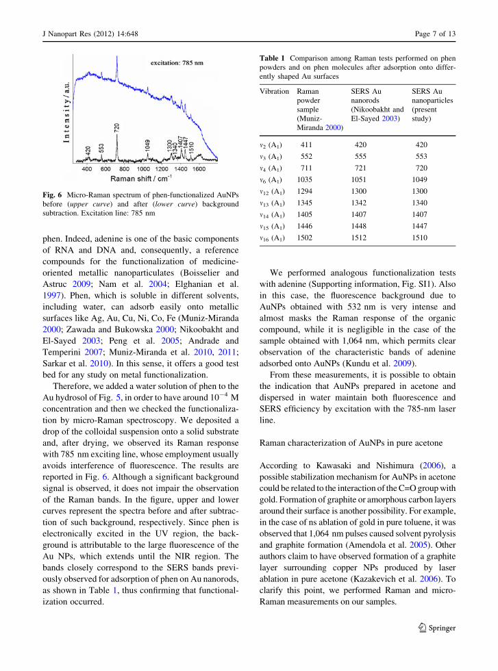
reported in Fig. 6. Although a significant background
signal is observed, it does not impair the observation
of the Raman bands. In the figure, upper and lower
curves represent the spectra before and after subtrac-
tion of such background, respectively. Since phen is
electronically excited in the UV region, the back-
ground is attributable to the large fluorescence of the
Au NPs, which extends until the NIR region. The
bands closely correspond to the SERS bands previ-
ously observed for adsorption of phen on Au nanorods,
as shown in Table 1, thus confirming that functional-
ization occurred.
We performed analogous functionalization tests
with adenine (Supporting information, Fig. SI1). Also
in this case, the fluorescence background due to
AuNPs obtained with 532 nm is very intense and
almost masks the Raman response of the organic
compound, while it is negligible in the case of the
sample obtained with 1,064 nm, which permits clear
observation of the characteristic bands of adenine
adsorbed onto AuNPs (Kundu et al. 2009).
From these measurements, it is possible to obtain
the indication that AuNPs prepared in acetone and
dispersed in water maintain both fluorescence and
SERS efficiency by excitation with the 785-nm laser
line.
Raman characterization of AuNPs in pure acetone
According to Kawasaki and Nishimura (2006), a
possible stabilization mechanism for AuNPs in acetone
could be related to the interaction of the C=O group with
gold. Formation of graphite or amorphous carbon layers
around their surface is another possibility. For example,
in the case of ns ablation of gold in pure toluene, it was
observed that 1,064 nm pulses caused solvent pyrolysis
and graphite formation (Amendola et al. 2005). Other
authors claim to have observed formation of a graphite
layer surrounding copper NPs produced by laser
ablation in pure acetone (Kazakevich et al. 2006). To
clarify this point, we performed Raman and micro-
Raman measurements on our samples.
Fig. 6 Micro-Raman spectrum of phen-functionalized AuNPs
before (upper curve) and after (lower curve) background
subtraction. Excitation line: 785 nm
Table 1 Comparison among Raman tests performed on phen
powders and on phen molecules after adsorption onto differ-
ently shaped Au surfaces
Vibration Raman
powder
sample
(Muniz-
Miranda 2000)
SERS Au
nanorods
(Nikoobakht and
El-Sayed 2003)
SERS Au
nanoparticles
(present
study)
m2 (A1) 411 420 420
m3 (A1) 552 555 553
m4 (A1) 711 721 720
m6 (A1) 1035 1051 1049
m12 (A1) 1294 1300 1300
m13 (A1) 1345 1342 1340
m14 (A1) 1405 1407 1407
m15 (A1) 1446 1448 1447
m16 (A1) 1502 1512 1510
J Nanopart Res (2012) 14:648 Page 7 of 13
123

Figure 7 compares the Raman spectrum of pure
acetone with that of the colloid of Fig. 2d, which
exhibits a weak band at 1,560 cm-1, not ascribed to
acetone vibrations. The detection of such band is
justified by the SERS effect, due to the resonance of
the laser exciting line (514.5 nm) with the plasmon
resonance band of the AuNPs (see Fig. 1). Similar
results (not shown) were obtained with the colloid of
Fig. 2c.
The band at 1,560 cm-1 can be attributed to the
formation of enolate ions, which can adsorb on the
surface of the positively charged gold particles,
according to the schema reported in Fig. 8a. Actually,
enolate cannot be present as free anion in acetone, but
only adsorbed on metal. Acetone enolate is character-
ized by two resonant forms, where C=O or C=C bond
(not present in pure acetone) occur (Fig. 8b). Typi-
cally, the alkenic C=C stretching modes should occur
in the 1,600–1,650 cm-1 spectral region. However, in
the case of enolate, the electronic conjugation with the
nearby C=O bond should move the band to lower
frequency, making it compatible with the observed
band at 1,560 cm-1. This latter, in conclusion, can be
confidently attributed to a mixing of C=O and C=C
stretching modes.
This assignment agrees with previous conclusions
reported in the literature. The existence of surface-
bound acetone enolate was postulated on the basis of
infrared spectroscopic studies of acetone adsorption
on various metal oxides. Absorption bands reported at
1,558, 1,540, and 1,543–1,595 cm-1 have been
assigned to m(CO) of acetone enolate on NiO (Miyata
et al. 1974), Fe2O3 (Busca and Lorenzelli 1982) and
Al2O3 (Hanson et al. 1987; Zaki et al. 2000).
Organometallic complexes of Ru and Pd containing
acetone enolate are also known, with characteristic
frequencies observed at 1,579 cm-1 (Hartwig et al.
1991) and at 1,554–1,565 cm-1 (Vicente et al. 1998),
respectively. On the other hand, for acetone adsorbed
on Pt (Vannice et al. 1991), vibrational bands in the
1,530–1,585 cm-1 spectral region were detected by
RAIRS and HREELS experiments and attributed to
acetone enolate, as successive DFT studies confirmed
(Jeffery et al. 2005). Finally, acetone enolate bound to
Ni surface was attributed by DFT studies to the species
showing a RAIRS band at 1,545 cm-1 (Sim et al.
2002).
To have a deeper insight into the nature of the
observed band, we also performed micro-Raman
measurements by drying relevant amounts of the same
colloid used for Fig. 7 in a steel spoon and adopting
long accumulation times (10 9 200 s). The resulting
spectrum is reported in Fig. 9. Two broad bands at
about 1,590 and 1,350 cm-1 are assigned as G and D
bands of graphitic carbon, whereas that occurring at
1,555 cm-1 corresponds to the C=O/C=C stretching
mode of acetone enolate observed in the acetone
suspension (see Fig. 7). The bands observed between
1,200 and 1,500 cm-1 are, instead, attributable to
those deriving from amorphous carbon (sp2 or sp3
type), intensified by SERS effect, as widely reported in
Fig. 7 Raman spectrum of pure acetone (lower curve) and a
colloid of AuNPs obtained in pure acetone by 532 nm, 15 mJ
per pulse laser ablation (upper curve). Exciting line: 514.5 nm
Fig. 8 a Adsorption of acetone on a gold cluster; b resonant
forms of acetone enolate
Page 8 of 13 J Nanopart Res (2012) 14:648
123
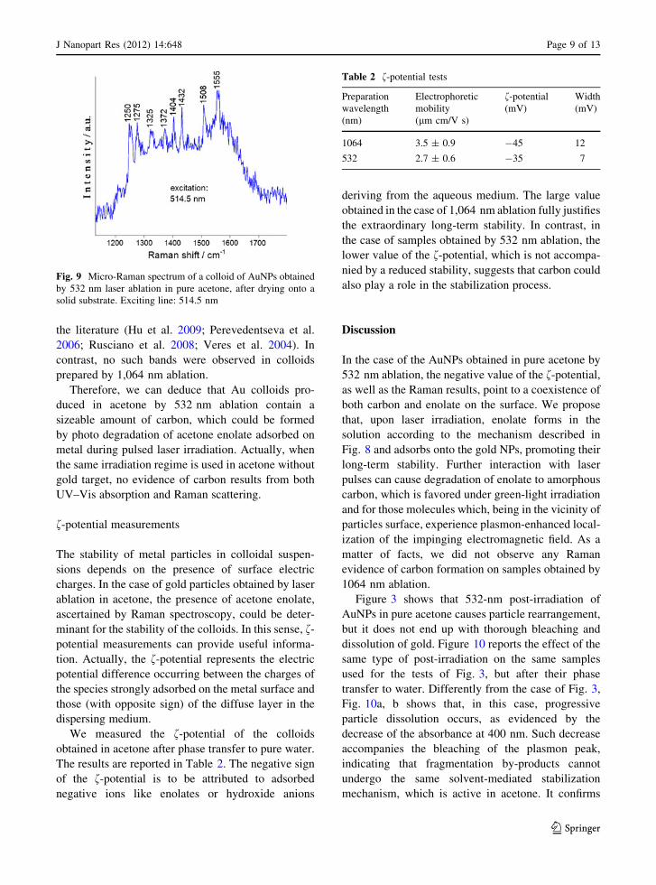
the literature (Hu et al. 2009; Perevedentseva et al.
2006; Rusciano et al. 2008; Veres et al. 2004). In
contrast, no such bands were observed in colloids
prepared by 1,064 nm ablation.
Therefore, we can deduce that Au colloids pro-
duced in acetone by 532 nm ablation contain a
sizeable amount of carbon, which could be formed
by photo degradation of acetone enolate adsorbed on
metal during pulsed laser irradiation. Actually, when
the same irradiation regime is used in acetone without
gold target, no evidence of carbon results from both
UV–Vis absorption and Raman scattering.
f-potential measurements
The stability of metal particles in colloidal suspen-
sions depends on the presence of surface electric
charges. In the case of gold particles obtained by laser
ablation in acetone, the presence of acetone enolate,
ascertained by Raman spectroscopy, could be deter-
minant for the stability of the colloids. In this sense, f-
potential measurements can provide useful informa-
tion. Actually, the f-potential represents the electric
potential difference occurring between the charges of
the species strongly adsorbed on the metal surface and
those (with opposite sign) of the diffuse layer in the
dispersing medium.
We measured the f-potential of the colloids
obtained in acetone after phase transfer to pure water.
The results are reported in Table 2. The negative sign
of the f-potential is to be attributed to adsorbed
negative ions like enolates or hydroxide anions
deriving from the aqueous medium. The large value
obtained in the case of 1,064 nm ablation fully justifies
the extraordinary long-term stability. In contrast, in
the case of samples obtained by 532 nm ablation, the
lower value of the f-potential, which is not accompa-
nied by a reduced stability, suggests that carbon could
also play a role in the stabilization process.
Discussion
In the case of the AuNPs obtained in pure acetone by
532 nm ablation, the negative value of the f-potential,
as well as the Raman results, point to a coexistence of
both carbon and enolate on the surface. We propose
that, upon laser irradiation, enolate forms in the
solution according to the mechanism described in
Fig. 8 and adsorbs onto the gold NPs, promoting their
long-term stability. Further interaction with laser
pulses can cause degradation of enolate to amorphous
carbon, which is favored under green-light irradiation
and for those molecules which, being in the vicinity of
particles surface, experience plasmon-enhanced local-
ization of the impinging electromagnetic field. As a
matter of facts, we did not observe any Raman
evidence of carbon formation on samples obtained by
1064 nm ablation.
Figure 3 shows that 532-nm post-irradiation of
AuNPs in pure acetone causes particle rearrangement,
but it does not end up with thorough bleaching and
dissolution of gold. Figure 10 reports the effect of the
same type of post-irradiation on the same samples
used for the tests of Fig. 3, but after their phase
transfer to water. Differently from the case of Fig. 3,
Fig. 10a, b shows that, in this case, progressive
particle dissolution occurs, as evidenced by the
decrease of the absorbance at 400 nm. Such decrease
accompanies the bleaching of the plasmon peak,
indicating that fragmentation by-products cannot
undergo the same solvent-mediated stabilization
mechanism, which is active in acetone. It confirms
Fig. 9 Micro-Raman spectrum of a colloid of AuNPs obtained
by 532 nm laser ablation in pure acetone, after drying onto a
solid substrate. Exciting line: 514.5 nm
Table 2 f-potential tests
Preparation
wavelength
(nm)
Electrophoretic
mobility
(lm cm/V s)
f-potential
(mV)
Width
(mV)
1064 3.5 ± 0.9 -45 12
532 2.7 ± 0.6 -35 7
J Nanopart Res (2012) 14:648 Page 9 of 13
123

that the stabilization mechanism of AuNPs in pure
acetone is strictly related to laser-solvent interaction.
Emission from small gold NPs has been observed
previously by several authors (Giorgetti et al. 2009,
2011; Wilcoxon et al. 1998; Zheng et al. 2003; Tran
et al. 2006; Zheng et al. 2004). However, the reliability
of such studies is often impaired by the difficulty in
separating the contribution of the capping agents from
the overall emission of the colloid. For example, in the
case of highly fragmented PAMAMG5-capped AuN-
Ps, the emission was rather to be attributed to metal-
enhanced fluorescence of PAMAMG5 than to gold
nano- or subnano-clusters (Giorgetti et al. 2011). In
the case of AuNPs obtained in pure acetone by 532 nm
ablation, no stabilizing molecule was intentionally
added to the colloid. In this sense, such systems are the
closest ones to perfectly uncapped AuNPs. Although
we could easily exclude any contribution of pure
acetone by checking solvent fluorescence before and
after laser irradiation, this was not the case for the
products of light-induced degradation of acetone at
particles surface, like acetone enolate or carbon. These
latter species could also provide some contribution to
the fluorescence emission which, however, can hardly
be isolated and evaluated separately from that of gold.
For example, some species of amorphous carbon emit
at 468 nm albeit excited at the shorter wavelength of
193 nm (Lormes et al. 1998; Damm et al. 2001;
Rholfing 1988). However, a more in-depth analysis
devoted to disentangle the role of carbon, if any, was
beyond the scope of this work.
Conclusions
We prepared AnNPs in pure acetone by ps laser
ablation at 1,064 and 532 nm. Green-light ablation
permits production of very small particles, with
narrow statistical distribution. Moreover, the extre-
mely small average size (2–3 nm) makes them fluo-
rescent. This property is not detected in AuNPs
prepared in pure water, which result bigger. In general,
the particles are characterized by an extraordinary
long-term stability, which we attribute to formation of
acetone enolate on particle surface, which gives them
a negative repulsive charge. Differently from the water
case, such stabilization mechanism impairs thorough
bleaching of the suspensions under ps post-irradiation
and increases colloid resistance to 532-nm short
pulses. Indeed, the effect of post-irradiation is limited
to a rearrangement of particle average size and
statistical distribution, with no precipitation or disso-
lution of gold.
Due to their extraordinary stability, AuNPs
obtained in pure acetone can be stored on-shelf for
years, transferred to the water phase and subsequently
functionalized with different organic compounds on
demand. The phase transfer maintains stability and,
when present, fluorescence. Moreover, after phase
transfer, the NPs still exhibit SERS activity, which we
tested by near IR (785 nm) excitation, that is the most
convenient one in biological applications, since it
avoids any interference deriving from the biological
environment. As a whole, our systems exhibit prom-
ising characteristics for the development of biocom-
patible fluorescent or Raman markers to be used in in
vitro, as well as in in vivo detection of molecule/
particle nanohybrids.
0.5
1.0
1.5(a)
(b)
post-irradiation
532 nm 15mJ
abso
rban
ce
wavelength (nm)
water phase
400 500 600 700 800
400 600 800
0.4
0.8
1.2 post-irradiation
532 nm 15mJ
abso
rban
ce
wavelength (nm)
water phase
Fig. 10 Post-irradiation with 532 nm, 15 mJ per pulse of the
samples of Figs. 2c (a) and d (b), after phase transfer to water.
Thick black lines initial spectra; thick gray lines final spectra
obtained after 2,400 (a) and 5,200 (b) shots. Intermediate thinlines, from top to bottom: spectra recorded for increasing
number of shots
Page 10 of 13 J Nanopart Res (2012) 14:648
123

Acknowledgments Funding from the project NABLA (Decree
n.4508—September 1, 2010 by Regione Toscana, Italy, PAR FAS
2007–2013 funds, Action 1.1.a.3) and project PRIN2009 Novelplasmon-based processes and materials for sensor applications of
the Italian Ministry of Research is acknowledged. The authors
also wish to thank Gabriella Caminati (Chemistry Department of
the University of Firenze, Italy) and Alessio Rindi (Department
of Chemistry and Industrial Chemistry of the University of
Genova, Italy) for technical assistance with f-potential and TEM
characterizations, respectively.
References
Alloisio M, Demartini A, Cuniberti C, Dellepiane G, Jadhav S,
Thea S, Giorgetti E, Gellini C, Muniz-Miranda M (2009)
Polydiacetylene-functionalized noble metal nanocages.
J Phys Chem C 113:19475–19481. doi:10.1021/jp905787h
Amendola V, Meneghetti M (2009) Laser ablation synthesis in
solution and size manipulation of noble metal nanoparti-
cles. Phys Chem Chem Phys 11:3805–3821. doi:10.1039/
B900654K
Amendola V, Rizzi G, Polizzi S, Meneghetti M (2005) Synthesis
of gold nanoparticles by laser ablation in toluene:
quenching and recovery of the surface plasmon absorption.
J Phys Chem B 109:23125–23128. doi:10.1021/jp055783v
Andrade GFS, Temperini MLA (2007) 1,10-Phenanthroline
adsorption on iron electrode monitored by surface-
enhanced raman scattering (SERS). Comparison to SERS
of Phen and its transition metal complex on silver electrode.
J Phys Chem C 111:13821–13830. doi:10.1021/jp072383u
Boisselier E, Astruc D (2009) Gold nanoparticles in nanomed-
icine: preparations, imaging, diagnostics, therapies and
toxicity. Chem Soc Rev 38:1759–1782. doi:10.1039/
b806051g
Boyer P, Menard D, Meunier M (2010) Nanoclustered Co–Au
particles fabricated by femtosecond laser fragmentation in
liquids. J Phys Chem C 114:13497–13500. doi:10.1021/
jp1037552
Bozon-Verduraz F, Brayner R, Voronov VV, Kirichenko NA,
Simakin AV, Shafeev GA (2003) Production of nanopar-
ticles by laser-induced ablation of metals in liquids.
Quantum Electron 33:714–720. doi:10.1070/QE2003v033
n08ABEH002484
Burakov VS, Tarasenko NV, Butsen AV, Rozantsev VA,
Nedel’ko NI (2005) Formation of nanoparticles during
double-pulse laser ablation of metals in liquids. Eur Phys J
Appl Phys 30:107–112. doi:10.1051/epjap:2005016
Busca G, Lorenzelli V (1982) Infrared study of the reactivity of
acetone and hexachloroacetone adsorbed on haematite.
J Chem Soc Faraday Trans I 78:2911–2919. doi:10.1039/
F19827802911
Chen S, Wang ZL, Ballato J, Foulger SH, Carroll DL (2003)
Rotational isomerism in acetic acid: the first experimental
observation of the high-energy conformer. J Am Chem Soc
125:16186–16187. doi:10.1021/ja038341a
Chen J, Saeki F, Wiley BJ, Cang H, Cobb MJ, Li Z, Au L, Zhang
H, Kimmey MB, Xia Y (2005) Gold nanocages: biocon-
jugation and their potential use as optical imaging contrast
agents. Nano Lett 5:473–477. doi:10.1021/nl047950t
Damm CJ, Lucas D, Sawyer RF, Koshland CP (2001) Real-time
measurement of combustion generated particles with
photofragmentation-fluorescence. Appl Spectrosc 55:
1478–1482. doi:10.1366/0003702011953892
Elghanian R, Storhoff JJ, Mucic RC, Letsinger RL, Mirkin CA
(1997) Selective colorimetric detection of polynucleotides
based on the distance-dependent optical properties of gold
nanoparticles. Science 277:1078–1081. doi:10.1126/
Science.277.5329.1078
Giammanco F, Giorgetti E, Marsili P, Giusti A (2010) Experi-
mental and theoretical analysis of photofragmentation of
Au nanoparticles by picosecond laser radiation. J Phys
Chem C 114:3354–3363. doi:10.1021/jp908964t
Giorgetti E, Giusti A, Giammanco F, Marsili P, Laza S (2009)
Dendrimer-capped nanoparticles prepared by picosecond
laser ablation in liquid environment. Molecules 14:
3731–3753. doi:10.3390/molecules14093731
Giorgetti E, Giammanco F, Marsili P, Giusti A (2011) Effect of
picosecond postirradiation on colloidal suspensions of
differently capped AuNPs. J Phys Chem C 115:5011–5020.
doi:10.1021/jp108042m
Giusti A, Giorgetti E, Laza S, Marsili P, Giammanco F (2007)
Multiphoton fragmentation of PAMAM G5-capped gold
nanoparticles induced by picosecond laser irradiation at
532 nm. J Phys Chem C 111:14984–14991. doi:10.1021/
jp072611k
Hanson BE, Wieserman LF, Wagner GW, Kaufman RA (1987)
Identification of acetone enolate on c-alumina: implications
for the oligomerization and polymerization of adsorbed
acetone. Langmuir 3:549–555. doi:10.1021/la00076a019
Hartwig F, Bergman RG, Anderson RA (1991) Oxygen- and
carbon-bound ruthenium enolates: migratory insertion,
reductive elimination, beta-hydrogen elimination, and cy-
clometalation reactions. Organometallics 10:3326–3344.
doi:10.1021/om00055a060
Hu A, Sanderson J, Zhou Y, Duley WW (2009) Formation of
diamond-like carbon by fs laser irradiation of organic liq-
uids. Diam Relat Mater 18:999–1001. doi:10.1016/j.
diamond.2009.02.019
Jain PK, Lee KS, El-Sayed MA (2006) Calculated absorption
and scattering properties of gold nanoparticles of different
size, shape, and composition: applications in biological
imaging and biomedicine. J Phys Chem B 110:7238–7248.
doi:10.1021/jp057170o
Jain PK, Yeo BS, Syadler J, Schmid T, Zenobi R, Zhang W
(2009) Tip-enhanced Raman spectroscopy—its status,
challenges and future directions. Chem Phys Lett 472:
1–13. doi:10.1016/j.cplett.2009.02.023
Jeffery EL, Mann RK, Hutchings GJ, Taylor SH, Willock DJ
(2005) A density functional theory study of the adsorption
of acetone to the (1 1 1) surface of Pt: implications for
hydrogenation catalysis. Catal Today 105:85–92. doi:
10.1016/j.cattod.2005.04.013
Kamat PV, Flumiani M, Hartland GV (1998) Picosecond
dynamics of silver nanoclusters. Photoejection of electrons
and fragmentation. J Phys Chem B 102:3123–3128. doi:10.
1021/jp980009b
Kawasaki M, Nishimura N (2006) 1064-nm laser of thin Au and
Ag flakes in acetone for highly productive pathway to
stable metal nanoparticles. Appl Surf Sci 253:2208–2216.
doi:10.1016/j.apsusc.2006.04.024
J Nanopart Res (2012) 14:648 Page 11 of 13
123

Kazakevich PV, Simakin AV, Voronov VV, Shafeev GA (2006)
Laser induced synthesis of nanoparticles in liquids. Appl
Surf Sci 252:4373–4380. doi:10.1016/j.apsusc.2005.06.
059
Kreibig U, Vollmer M (1995) Optical properties of metal
clusters. Springer-Verlag, Berlin
Kumar A, Vemula P, Ajayan PM, John G (2008) Silver-nano-
particle-embedded antimicrobial paints based on vegetable
oil. Nat Mater 7:236–241. doi:10.1038/nmat2099
Kundu JO, Neumann O, Janesko BG, Zhang D, Lal S, Barhoumi
A, Scuseria GE, Halas NJ (2009) Adenine and adenosine
monophosphate (AMP) gold binding interactions studied
by surface-enhanced raman and infrared spectroscopies.
J Phys Chem C 113:14390–14397. doi:10.1021/jp903126f
Loo C, Lowery A, Halas N, West J, Drezek R (2005) Immu-
notargeted nanoshells for integrated cancer imaging and
therapy. Nano Lett 5:709–711. doi:10.1021/nl050127s
Lormes W, Hundhausen M, Ley L (1998) Time resolved pho-
toluminescence of amorphous hydrogenated carbon.
J Noncryst Solids 227–230:570–573. doi:10.1016/S0022-
3093(98)00272-5
Mafune F, Khono J, Takeda Y, Kondow T, Sawabe H (2001)
Formation of gold nanoparticles by laser ablation in
aqueous solution of surfactant. J Phys Chem B
105:5114–5120. doi:10.1021/jp0037091
Mafune’ F, Kohno J, Takeda Y, Kondow T (2002) Full physical
preparation of size-selected gold nanoparticles in solution:
laser ablation and laser-induced size control. J Phys Chem
B 106:7575–7577. doi:10.1021/jp020577y
Miyata H, Toda Y, Kubokawa Y (1974) Infrared studies of
adsorption of acetone on MgO and NiO. J Catal
32:155–158. doi:10.1016/0021-9517(74)90170-5
Muniz-Miranda M (2000) Surface enhanced raman scattering
and normal coordinate analysis of 1,10-phenanthroline
adsorbed on silver sols. J Phys Chem A 104:7803–7810.
doi:10.1021/jp001578y
Muniz-Miranda M, Pergolese B, Bigotto A (2010) SERS and
DFT investigation on the adsorption of 1,10-phenanthro-
line on transition metal surfaces. Phys Chem Chem Phys
12:1145–1151. doi:10.1039/B913014D
Muniz-Miranda M, Gellini C, Giorgetti E (2011) Surface-
enhanced Raman scattering from copper nanoparticles
obtained by laser ablation. J Phys Chem C 115:5021–5027.
doi:10.1021/jp1086027
Nam J, Stoeva SI, Mitkin CA (2004) Bio-bar-code-based DNA
detection with PCR-like sensitivity. J Am Chem Soc
126:5932–5933. doi:10.121/ja049384
Nikoobakht B, El-Sayed MA (2003) Surface-enhanced Raman
scattering studies on aggregated gold nanorods. J Phys
Chem A 107:3372–3378. doi:10.1021/jp026770?
Peng Y, Niu Z, Huang W, Chen S, Li Z (2005) Surface-
enhanced Raman scattering studies of 1,10-phenanthroline
adsorption and its surface complexes on a gold electrode.
J Phys Chem B 109:10880–10885. doi:10.1021/jp0443198
Perevedentseva E, Karmenyan A, Chung PH, He YT, Cheng CL
(2006) Surface enhanced Raman spectroscopy of carbon
nanostructures. Surf Sci 600:3723–3728. doi:10.1016/
j.susc.2006.01.074
Rholfing EA (1988) Optical emission studies of atomic,
molecular, and particulate carbon produced from a laser
vaporization cluster source. J Chem Phys 89:6103–6112.
doi:10.1063/1.455426
Rusciano G, De Luca AC, D’Alessio A, Minatolo P, Pesce G,
Sasso A (2008) Surface-enhanced Raman scattering study
of nano-sized organic carbon particles produced in com-
bustion processes. Carbon 46:335–341. doi:10.1016/
j.carbon.2007.11.052
Sarkar S, Pradhan M, Sinha AK, Basu M, Pal T (2010) Chelate
effect in surface enhanced Raman scattering with transition
metal nanoparticles. J Phys Chem Lett 1:439–444. doi:
10.1021/jz900272u
Seo D, Park JC, Song H (2006) Polyhedral gold nanocrystals
with Oh symmetry: from octahedra to cubes. J Am Chem
Soc 128:14863–14870. doi:10.1021/ja062892u
Silvestre JP, Poulin S, Kabashin AV, Sacher E, Meunier M,
Luong JHT (2004) Surface chemistry of gold nanoparticles
produced by laser ablation in aqueous media. J Phys Chem
B 108(16864):16869. doi:10.1021/jp047134?
Sim WS, Li TC, Yang PX, Yeo BS (2002) Isolation and iden-
tification of surface-bound acetone enolate on Ni(111).
J Am Chem Soc 124:4970–4971. doi:10.1021/ja025749j
Tarasenko NV, Butsen AV, Nevar EA (2005) Laser-induced
modification of metal nanoparticles formed by laser abla-
tion technique in liquids. Appl Surf Sci 247:418–422. doi:
10.1016/j.apsusc.2005.01.093
Tilaki RM, Irajizad A, Mahdavi SM (2006) Fabrication of
microchip based on UV transparent polymer for DNA
electrophoresis by F2 laser ablation. Appl Phys A
84:251–255. doi:10.1007/s00339-006-3618-9
Tran ML, Zvyagin AV, Plakhotnik T (2006) Synthesis and
spectroscopic observation of dendrimer-encapsulated gold
nanoclusters. Chem Commun 22:2400–2401. doi:10.
1039/B602079H
Trigari S, Rindi A, Margheri G, Sottini S, Dellepiane G,
Giorgetti E (2011) Synthesis and modelling of gold nano-
stars with tunable morphology and extinction spectrum.
J Mater Chem 21:6531–6540. doi:10.1039/C0JM04519E
Vannice MA, Erley W, Ibach H (1991) A RAIRS and HREELS
study of acetone on Pt(111). Surf Sci 254:1–3. doi:10.
1016/0039-6028(91)90632-3
Veres M, Fule M, Toth S, Koos M, Pocsik I (2004) Surface
enhanced Raman scattering (SERS) investigation of
amorphous carbon. Diam Relat Mater 13:1412–1415. doi:
10.1016/j.diamond.2004.01.041
Vicente J, Abad JA, Chicote MT, Abrisqueta MD, Lorca JA, de
Arellano MCR (1998) Synthesis of new ketonyl palla-
dium(II) and platinum(ii) complexes with nitrogen-donor
ligands. Crystal structure of [Pt{CH2C(O)Me}2(bpy)].
Organometallics 17:1564–1568. doi:10.1021/om971028v
Werner D, Hashimoto S, Tomita T, Matsuo S, Makita Y (2008)
In situ spectroscopic measurements of laser ablation-
induced splitting and agglomeration of metal nanoparticles
in solution. J Phys Chem C 112:16801–16808. doi:10.
1021/jp804647a
Wilcoxon JP, Martin JE, Parsapour F, Wiedenman B, Kelly DF
(1998) Photoluminescence from nanosize gold clusters.
J Chem Phys 108:9137–9143. doi:10.1063/1.476360
Zaki MI, Hasan MA, Al-Sagheer FA, Pasupulety L (2000)
Surface chemistry of acetone on metal oxides: IR obser-
vation of acetone adsorption and consequent surface
Page 12 of 13 J Nanopart Res (2012) 14:648
123

reactions on silica–alumina versus silica and alumina.
Langmuir 16:430–436. doi:10.1021/la990739q
Zawada K, Bukowska J (2000) An interaction of 1,10-phe-
nantroline with the copper electrode in neutral and acidic
aqueous solutions: a surface enhanced Raman scattering
study. J Mol Struct 555:425–432. doi:10.1016/S0022-
2860(00)00629-3
Zheng J, Petty JT, Dickson RM (2003) High quantum yield blue
emission from water-soluble Au8 nanodots. J Am Chem
Soc 125:7780–7781. doi:10.1021/ja035473v
Zheng J, Zhang C, Dickson RM (2004) Highly fluorescent,
water-soluble, size-tunable gold quantum dots. Phys Rev
Lett 93:77402. doi:10.1103/PhysRevLett.93.077402
J Nanopart Res (2012) 14:648 Page 13 of 13
123