Adaptor Protein-2 Interaction with Arrestin Regulates GPCR Recycling and Apoptosis
28
Adaptor protein-2 interaction with arrestin regulates GPCR recycling and apoptosis Brant M. Wagener 1 , Nicole A. Marjon 1 , Chetana M. Revankar 1 , and Eric R. Prossnitz 1,2 1 Department of Cell Biology and Physiology and UNM Cancer Research and Treatment Center, University of New Mexico Health Sciences Center, Albuquerque, NM 87131, USA SUMMARY G protein-coupled receptors (GPCRs) are integral to cellular function in nearly all physiologic and many pathologic processes. GPCR signaling represents an intricate balance between receptor activation, inactivation (desensitization, internalization and degradation) and resensitization (recycling and de novo synthesis). Complex formation between phosphorylated GPCRs, arrestins and an ever-increasing number of effector molecules is known to regulate cellular function. Previous studies have demonstrated that, although N-formyl peptide receptor (FPR) internalization occurs in the absence of arrestins, FPR recycling is arrestin-dependent. Furthermore, FPR stimulation in the absence of arrestins leads to receptor accumulation in perinuclear endosomes and apoptosis. In this study, we show that the interaction of GPCR-bound arrestin with Adaptor Protein 2 (AP-2) is a critical anti-apoptotic event. In addition, AP-2 associates with the receptor- arrestin complex in perinuclear endosomes and is required for proper post-endocytic GPCR trafficking. Finally, we observed that depletion of endogenous AP-2 results in the initiation of apoptosis upon stimulation of multiple GPCRs including P2Y purinergic receptors and CXCR2 but not CXCR4. We propose a model in which the abnormal accumulation of internalized GPCR- arrestin complexes in recycling endosomes, resulting from defective arrestin-AP-2 interactions, leads to the specific initiation of aberrant signaling pathways and apoptosis. Keywords apoptosis; GPCR; FPR; arrestin; adaptor protein-2; post-endocytic trafficking; recycling INTRODUCTION G protein-coupled receptors (GPCR) are involved in virtually every aspect of human physiology including cardiovascular (1), immune (2) and neuronal systems (3). An important feature of GPCR signaling is the cycle of receptor activation, desensitization, internalization, down-regulation/degradation, recycling and resensitization. When these processes are interrupted, they can detrimentally affect cellular migration (4), proliferation and cell adhesion (5). In the case of the v2 vasopressin receptor, intracellular accumulation of the receptor through constitutive desensitization and internalization leads to nephrogenic diabetes insipidus (6). GPCRs are activated by a myriad of ligands including amino acids and their derivatives, peptides, proteins, ions, lipids and photons. Ligand-bound GPCRs activate heterotrimeric G proteins, resulting in adenylyl cyclase and phospholipase activation, among other effector 2 Corresponding author: Eric R. Prossnitz, Department of Cell Biology and Physiology, University of New Mexico Health Sciences Center, Albuquerque, NM USA 87131, Tel: (505) 272-5647, Fax: (505) 272-1421, [email protected]. NIH Public Access Author Manuscript Traffic. Author manuscript; available in PMC 2010 September 1. Published in final edited form as: Traffic. 2009 September ; 10(9): 1286–1300. doi:10.1111/j.1600-0854.2009.00957.x. NIH-PA Author Manuscript NIH-PA Author Manuscript NIH-PA Author Manuscript
Transcript of Adaptor Protein-2 Interaction with Arrestin Regulates GPCR Recycling and Apoptosis
Adaptor protein-2 interaction with arrestin regulates GPCRrecycling and apoptosis
Brant M. Wagener1, Nicole A. Marjon1, Chetana M. Revankar1, and Eric R. Prossnitz1,2
1Department of Cell Biology and Physiology and UNM Cancer Research and Treatment Center,University of New Mexico Health Sciences Center, Albuquerque, NM 87131, USA
SUMMARYG protein-coupled receptors (GPCRs) are integral to cellular function in nearly all physiologic andmany pathologic processes. GPCR signaling represents an intricate balance between receptoractivation, inactivation (desensitization, internalization and degradation) and resensitization(recycling and de novo synthesis). Complex formation between phosphorylated GPCRs, arrestinsand an ever-increasing number of effector molecules is known to regulate cellular function.Previous studies have demonstrated that, although N-formyl peptide receptor (FPR) internalizationoccurs in the absence of arrestins, FPR recycling is arrestin-dependent. Furthermore, FPRstimulation in the absence of arrestins leads to receptor accumulation in perinuclear endosomesand apoptosis. In this study, we show that the interaction of GPCR-bound arrestin with AdaptorProtein 2 (AP-2) is a critical anti-apoptotic event. In addition, AP-2 associates with the receptor-arrestin complex in perinuclear endosomes and is required for proper post-endocytic GPCRtrafficking. Finally, we observed that depletion of endogenous AP-2 results in the initiation ofapoptosis upon stimulation of multiple GPCRs including P2Y purinergic receptors and CXCR2but not CXCR4. We propose a model in which the abnormal accumulation of internalized GPCR-arrestin complexes in recycling endosomes, resulting from defective arrestin-AP-2 interactions,leads to the specific initiation of aberrant signaling pathways and apoptosis.
Keywordsapoptosis; GPCR; FPR; arrestin; adaptor protein-2; post-endocytic trafficking; recycling
INTRODUCTIONG protein-coupled receptors (GPCR) are involved in virtually every aspect of humanphysiology including cardiovascular (1), immune (2) and neuronal systems (3). Animportant feature of GPCR signaling is the cycle of receptor activation, desensitization,internalization, down-regulation/degradation, recycling and resensitization. When theseprocesses are interrupted, they can detrimentally affect cellular migration (4), proliferationand cell adhesion (5). In the case of the v2 vasopressin receptor, intracellular accumulationof the receptor through constitutive desensitization and internalization leads to nephrogenicdiabetes insipidus (6).
GPCRs are activated by a myriad of ligands including amino acids and their derivatives,peptides, proteins, ions, lipids and photons. Ligand-bound GPCRs activate heterotrimeric Gproteins, resulting in adenylyl cyclase and phospholipase activation, among other effector
2Corresponding author: Eric R. Prossnitz, Department of Cell Biology and Physiology, University of New Mexico Health SciencesCenter, Albuquerque, NM USA 87131, Tel: (505) 272-5647, Fax: (505) 272-1421, [email protected].
NIH Public AccessAuthor ManuscriptTraffic. Author manuscript; available in PMC 2010 September 1.
Published in final edited form as:Traffic. 2009 September ; 10(9): 1286–1300. doi:10.1111/j.1600-0854.2009.00957.x.
NIH
-PA Author Manuscript
NIH
-PA Author Manuscript
NIH
-PA Author Manuscript
systems. Activated receptors are subsequently phosphorylated in complex patterns onintracellular domains by serine/threonine G protein-coupled receptor kinases (GRKs), whichreduce receptor affinity for G proteins and increase receptor affinity for arrestins (7–10).Arrestin binding sterically blocks receptor-G protein interactions, thereby terminating Gprotein signaling, while simultaneously providing a scaffold to coordinate the recruitment ofinternalization machinery, leading to receptor sequestration (11–13). In this model, basedprimarily on studies of the β2-adrenergic receptor (β2-AR), adaptor protein (AP)-2 andclathrin bind the carboxy terminus of arrestin (14), translocating receptors to clathrin-coatedpits for internalization. Arrestin-recruited Src then phosphorylates dynamin, which pinchesoff the plasma membrane invagination to form an endosome, whereupon arrestin dissociatesfrom the receptor (15). Internalized β2-AR in the Rab5-containing early endosomalcompartment is then sorted to lysosomes (via a Rab7-containing compartment) fordegradation or to the cell surface (via Rab4-containing endosomes) (16).
This classic pathway of GPCR internalization and post-endocytic trafficking is, however,not observed with all GPCRs. For example, internalization of the m2-muscarinicacetylcholine receptor is not dependent on either arrestin or clathrin (17) and the N-formylpeptide receptor (FPR) internalizes in arrestin knockout cell lines, in which β2-ARinternalization is completely inhibited (18,19). In contrast to GPCRs that follow the rab4-mediated, rapid recycling pathway or the rab7-mediated, lysosomal degradation pathway(20), some GPCRs recycle via perinuclear endosomes, associated with Rab11. Suchreceptors include the FPR (18), v2 vasopressin receptor (21), somatostatin receptor 3 (22)and CXCR2 (23). In many cases, such GPCRs have been shown to recycle more slowly andform stable complexes with arrestins, resulting in prolonged endosome-associated arrestin(18,24,25). Although this stable association has been shown to be critical in prolonged ERKactivation in the cytoplasm (25,26), little is known regarding the role of arrestin inregulating intracellular trafficking of GPCRs, particularly via the perinuclear endosomepathway. Arrestin dissociation has however been implicated as an essential step in therecycling and resensitization of the bradykinin B2 receptor (27). Furthermore, in cellslacking both arrestin-2 and arrestin-3, recycling of the FPR is absent, resulting in receptoraccumulation in perinuclear endosomes and suggesting a critical role for arrestin inrecycling of the FPR from this compartment (18). Thus, arrestins appear to mediate multiplediverse aspects of GPCR trafficking.
Subsequent studies of FPR trafficking in arrestin-deficient cells revealed that, in addition toa recycling defect, cells also underwent rapid apoptosis via a cytochrome-dependentpathway (28). A requirement for receptor internalization was demonstrated using asignaling-competent, internalization-defective mutant of the FPR. Furthermore, apoptosiswas prevented by ERK and other signaling inhibitors. This led to the hypothesis that theaccumulation of ligand-activated FPR in recycling endosomes in the absence of arrestinsresults in aberrant signaling, which initiates apoptotic pathways culminating in caspaseactivation. Based on the fact that both the trafficking and signaling defects could be rescuedby the re-introduction of either arrestin-2 or arrestin-3, we hypothesized that these twoapparently distinct defects may be mechanistically linked. Because of the large number ofarrestin-interacting proteins, we speculated that the absence of specific interactions might beresponsible for the observed defects. Therefore, in this work, we undertook a mapping studyto identify the sites within arrestin that when mutated, result in aberrant trafficking andsignaling, culminating in apoptosis. Our results reveal novel mechanisms by which AP-2specifically regulates FPR trafficking from recycling endosomes and prevents apoptosis.
Wagener et al. Page 2
Traffic. Author manuscript; available in PMC 2010 September 1.
NIH
-PA Author Manuscript
NIH
-PA Author Manuscript
NIH
-PA Author Manuscript
RESULTSRescue of FPR-mediated Apoptosis by Arrestin-2 Domains
We have previously described a requirement for arrestins in preventing apoptosis resultingfrom activation of multiple GPCRs, including the FPR, CXCR2 and Angiotensin II (Type1A) receptor (28). Arrestins also play a critical role in the proper post-endocytic intracellulartrafficking of GPCRs such as the FPR, although FPR internalization per se does not requirearrestin. To define the region(s) of arrestin-2 (arr2) responsible for preventing FPR-mediatedapoptosis, we generated four constructs producing large fragments of arr2: amino acids 1–186, 177–418, 319–418 and 1–382. The structure of arr2 is composed of two domains ofbeta-pleated sheets (amino acids 1–172 and 180–352) and a C-terminal “tail” (amino acids357–418) (29). Arr2-(319–418) contains the “tail” that acts as a dominant-negative constructinhibiting β2-AR internalization (30) whereas arr2-(1–382) is a truncated form of arr2 thathas been previously described as constitutively active with respect to receptor binding(7,8,31,32). Mouse embryonic fibroblasts (MEF) deficient in both arr2 and arr3, but stablyexpressing the FPR (arr2−/−/3−/− FPR MEF), were used to assess arr2 mutants withoutcompetition from endogenous arrestins. Arrestin-deficient MEF cells transiently transfectedwith the four GFP-fused arr2 fragments, wild type arr2 and empty GFP vector were assayedfor apoptosis upon FPR stimulation by evaluation of cell rounding, previously shown tocorrelate absolutely with conventional apoptosis markers such as Annexin V/PI positivityand caspase activation (28). As shown in Fig. 1A, unstimulated cells (expressing GFP, wildtype or mutant arrestins) did not exhibit apoptosis. On the contrary, 70–80% of ligand-stimulated cells expressing GFP alone exhibited an apoptotic phenotype, whereas cellsexpressing wild type arr2 were indistinguishable from unstimulated cells, as previouslydescribed (28). Furthermore, none of the four expressed arr2 domains were capable ofpreventing FPR-mediated apoptosis, although the arr2-(177–418) showed a small reductionin this parameter. Of the four arrestin mutants, arr2-(1–382) is the only domain thatdemonstrated association with the FPR upon stimulation (determined by confocalfluorescence microscopy, unpublished data). This is consistent with previous data thatdemonstrated colocalization of arr2-(1–382) with the FPR upon receptor activation (9) andbinding to the FPR in reconstitution assays (8,31). These results demonstrate that thebinding of arr2 alone (i.e. arr2-(1–382)) is insufficient to prevent FPR-mediated apoptosisand furthermore that sequences within the carboxy terminus of arr2 (amino acids 383–418)are essential to prevent apoptosis.
Rescue of FPR-mediated Apoptosis by Arrestin-2 Tail MutantsThe tail of arr2 contains recognition sites for multiple adapter and signaling proteins,including clathrin (30,33,34), AP-2 (14,29) and ERK (35), which phosphorylates a serine atposition 412. Based on the crude mapping results (Fig. 1A), we hypothesized amino acids inarr2 carboxy terminus were responsible for suppressing FPR-mediated apoptosis. To testthis hypothesis, we generated five mutants of arr2 using alanine-scanning/replacementmutagenesis of residues 383–390, 391, 397–400, 401–408 and 409–418 (in which theindicated amino acids were mutated to alanine residues). The arr2-F391A mutant haspreviously been described as reducing AP-2 binding (29) and the arr2-A397–400 mutant(heretowith referred to as arr2-4A) encompasses residues K397, M399 and K400 that haveindividually been shown to regulate AP-2 binding (14). GFP-fusions of the five arr2 tailmutants were tested for their ability to prevent FPR-mediated apoptosis as determined bypropidium iodide staining, which provides a lower basal level of estimated apoptotic cells.Greater than 90% of GFP vector- and arr2-(1–382)-expressing cells were PI positive uponligand (fMLF) stimulation (Fig. 1B), completely consistent with cell rounding results (Fig.1A). Expression of wild type arr2 and mutants encompassing residues 383–390, 401–408and 409–418 rescued cells from FPR-mediated apoptosis; however, the arr2-F391A and
Wagener et al. Page 3
Traffic. Author manuscript; available in PMC 2010 September 1.
NIH
-PA Author Manuscript
NIH
-PA Author Manuscript
NIH
-PA Author Manuscript
−4A mutants were completely ineffective in preventing apoptosis (Fig. 1B). To confirm thatpositive PI staining was the result of FPR-mediated apoptosis, empty GFP, wild type arr2, -(1–382), -F391A and −4A were treated with the pan-caspase inhibitor, zVAD-FMK, prior tostimulation. Transfected cells that underwent apoptosis in response to FPR stimulation failedto do so in the presence of zVAD-FMK (Fig. 1C), demonstrating that the PI staining in thepresence of the arr2 mutants F391A and 4A represents FPR-mediated, caspase-dependentapoptosis consistent with our previous characterization of this apoptotic pathway (28).
Previous results have demonstrated that receptor internalization, which occurs via arrestin-independent mechanisms, is essential for FPR-mediated apoptosis (28). Because AP-2 isrequired for the internalization of certain GPCRs, we hypothesized that overexpression ofarr2 mutants might inhibit FPR internalization, thereby preventing FPR-mediated apoptosis.To examine this, we measured FPR internalization for each arrestin mutant. We determinedthat the extent of FPR internalization varied between 65–80%, with no significant differencebetween the extents or rates (data not shown) of internalization for any of the arrestinconstructs expressed, demonstrating that rescue of FPR-mediated apoptosis by arrestinmutants is not due to inhibition of FPR internalization. In addition, internalization of theF391A mutant is consistent with our previous studies showing that FPR internalization isindependent of clathrin-dependent mechanisms (36).
Arrestin-2 Mutants that Fail to Rescue FPR-mediated Apoptosis Accumulate in PerinuclearEndosomes
As we have previously observed that, in arrestin-deficient cells, FPR-mediated apoptosis isassociated with defective intracellular trafficking, this raises the question as to whether thearr2-F391A and arr2-4A mutants allow normal trafficking of the FPR and to what extentthey remain associated with the receptor as it traffics intracellularly. Localization of the FPR(visualized using an Alexa633 labeled N-formyl-Leucyl-Leucyl-Phenylalanyl-Leucyl-Tyrosinyl-Lysine ligand (633-6pep)) and arr2 mutants (tagged with monomeric redfluorescent protein, mRFP1 (37)) was determined using confocal fluorescence microscopy.The perinuclear endosomal recycling compartment was visualized using a GFP fusion ofRab11 (which has also been shown to localize to the Golgi compartment (38)). Inunstimulated cells, Rab11 is located in a perinuclear region and arr2 (either wild type ormutant) is dispersed throughout the cytoplasm. Activation of the FPR with fluorescentligand (633-6pep) in the absence of arr2 (empty mRFP1 vector) resulted in accumulation ofthe FPR in perinuclear endosomes, exhibiting substantial overlap with Rab11 (Fig. 2).Expression of wild type arr2, while also leading to localization of the FPR in perinuclearendosomes, resulted in a greater proportion of non-perinuclear vesicles containing FPR andarrestin, consistent with normal trafficking and recycling of the FPR (18). To control for thepossible mistrafficking of arrestin-fluorescent protein fusions (due to potential dimerizationfor example), we cotransfected arrestin2-GFP with arrestin2-mRFP (note that AequoreaGFP and Discosoma RFP are distinct proteins that do not cross-dimerize, and that mRFPwas specifically engineered not to dimerize with itself). Internalized ligand-bearing vesiclesshowed a perfect concordance between the GFP and mRFP arrestin fusion proteins,indicating normal binding and trafficking interactions (data not shown).
The non-perinuclear complexes of FPR-633-6pep and arr2 observed followinginternalization of receptor represent both newly internalized as well as recycling endosomes(returning to the plasma membrane) based on the two following independent results. First,following a 10 min stimulation of arrestin-deficient cells with 633-6pep, followed by 50 minchase in the absence of ligand, the FPR is not seen in non-perinuclear vesicles, as it is in thepresence of arrestins (data not shown). The presence of the FPR in vesicles after 50 min ofagonist depletion in wild type cells suggests that this population represents recyclingreceptor, since no such vesicles are seen in the arrestin-deficient cells. Second, FPR
Wagener et al. Page 4
Traffic. Author manuscript; available in PMC 2010 September 1.
NIH
-PA Author Manuscript
NIH
-PA Author Manuscript
NIH
-PA Author Manuscript

internalization experiments performed with 10nM fMLF (the concentration of 633-6pepused in imaging experiments) in the presence of wild type arr2, reveal that the FPR achievesan equilibrium within 10 min wherein only approximately 25–30% of the total receptor isinternalized (compared to 1 µM fMLF, where 75–80% of the FPR is internalized, data notshown). This equilibrium with less total internalized receptor suggests that recycling istaking place under these conditions.
Localization results for the mutant arrestins paralleled their apoptotic phenotype. Expressionof both the arr2-F391A and −4A mutants (which did not rescue FPR-mediated apoptosis)resulted in accumulation of the FPR with the associated mutant arrestin in perinuclarendosomes (Fig. 2, note also the more rounded morphology of non-dividing cellsundergoing apoptosis, cf. arr2-F391A and −4A vs. WT stimulated). All other mutants(which did rescue the apoptotic phenotype) produced FPR trafficking patternsindistinguishable from wild type arr2 (Fig. 2). These results provide additional support forthe link between FPR trafficking defects and the initiation of FPR-mediated apoptosisobserved in the absence of arrestins as well as in the presence of the arr2-F391A and −4Amutants.
Previous reports have demonstrated that recycling of the FPR is impaired in the absence ofarrestins and that this phenotype is rescued by reconstitution with wild type arr2 (18). In thecurrent study, arr2−/−/3−/− FPR MEF cells transfected with EGFP alone recycled littleinternalized FPR (<3%) whereas reconstitution with wild type arr2 significantly increasedthe amount of FPR recycled to 15% of the internalized receptor. Expression of either theF391A or 4A mutants yielded no increase in recycling of the FPR (<3%), confirming thatthe accumulation of the FPR in recycling endosomes corresponds to a lack of recycling.
Arrestin-2 Mutants that Support FPR-mediated Apoptosis Exhibit Altered Associationswith AP-2
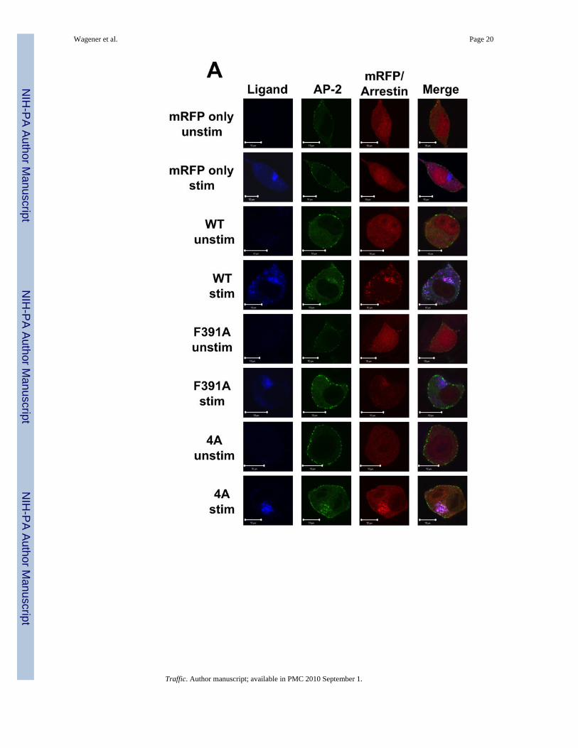
The region of arr2 encompassing amino acids 391–400 has previously been associated withaltered binding to AP-2 (14,29). To better understand the relationship between AP-2association and FPR trafficking, we used confocal fluorescence microscopy to track theinteraction of AP-2 with the FPR-arr2 complex. In unstimulated cells, AP-2 is distributedthroughout the cytoplasm as well as in puncta at the plasma membrane (Fig. 3). In arr2−/−/3−/− FPR MEF cells, following 60 min of stimulation with 633-6pep in the absence oftransfected arrestins, the FPR accumulates in a perinuclear region with Rab11 (Fig. 3, cf.Fig. 2). This observation and the lack of AP-2 association with the FPR in non-perinuclearendosomes at 15 and 30 minutes in the absence of arrestins (Suppl. Fig. 1) are consistentwith a requirement for arr2 in the recruitment of AP-2. Upon expression of wild type arr-2 inthe arr2−/−/3−/− FPR cells, ligand stimulation results in the accumulation of AP-2 with the633-6pep-FPR/arr2 complex in the perinuclear region at 60 min. However, AP-2 is notobserved to associate with non-perinuclear endosomes containing FPR-arr2 complexes,suggesting that AP-2 is not associated with the complex after exiting perinuclear endosomes.At 15 minutes, almost no AP-2 was seen associated with FPR-arr2 complexes in perinuclearendosomes. It is not until 30 minutes that significant levels of AP-2 are concentrated withthe FPR and wild type arr2 in perinuclear endosomes, suggesting that AP-2 does notassociate with FPR-arr2 complexes prior to internalization.
Expression of the F391A and 4A arrestin mutants yield substantially different results withrespect to AP-2 association. In the presence of the arr2-F391A mutant, AP-2 showed noassociation with the 633-6pep-FPR/arr2-F391A complex at any of the time points (Fig. 3Aand Suppl. Fig. 1). This is consistent with published reports that demonstrate decreasedAP-2 binding to arr2-F391A (14). However, while we hypothesized that the same would betrue of the arr2-4A mutant, this was not the case. Following stimulation for 60 minutes,
Wagener et al. Page 5
Traffic. Author manuscript; available in PMC 2010 September 1.
NIH
-PA Author Manuscript
NIH
-PA Author Manuscript
NIH
-PA Author Manuscript
AP-2 showed strong colocalization with the 633-6pep-FPR/arr2-4A complex, in many casesstronger than that observed with wild type arr2. A similar association of AP-2 with the633-6pep-FPR/arr2-4A complex was also observed following 15 and 30 minutesstimulation. Our results suggest that the arr2-4A mutant, in contrast to the arr2-F391Amutant, is capable of binding AP-2. The major difference between wild type arr2 and thearr2-4A mutant is that there is little non-perinuclear 633-6pep-FPR/arr2-4A complex(particularly at 60 min) suggesting an accumulation of this particular complex in recyclingendosomes.
To examine the rate of association of AP-2 with FPR/arrestin complexes, we measured AP-2colocalization with FPR/arr2 complexes over time by visually scoring the fraction of cells inwhich AP-2 was colocalized with perinuclear FPR/arr2 complexes (Fig. 3B). At 15 minutes,20–30% of cells show AP-2 to be clustered with arrestin when either wild type arr2 orarr2-4A is expressed. The percentage of cells showing colocalization increased at the 30 and60 minute time points for both wild type arr2 (~35 and ~80%, respectively) and arr2-4A(~60 and ~95%, respectively). The arr2-F391A mutant however showed essentially no AP-2localization at any time point.
To confirm the above results and validate our use of a GFP-tagged α subunit of AP-2, weexamined U937 cells that express both endogenous arrestins (arr2 and arr3) and were stablytransfected to express the FPR (U937 FPR). U937 cells are a promonocytic cell line usedextensively as a model for FPR function (39,40). Confocal immunofluorescence microscopyof U937 FPR cells transiently expressing Rab11-GFP and stained with antibodies againstendogenous AP-2 confirms our results regarding the subcellular localization of AP-2 (Suppl.Fig. 2A). In unstimulated cells, Rab11-GFP is localized to the perinuclear region while AP-2shows puncta at the cell membrane with limited cytosolic staining. Upon stimulation of theFPR with Alexa 546-6pep (546-6pep) for 30 min, ligand-FPR complexes colocalize withendogenous AP-2 in perinuclear endosomes. In addition, ligand-receptor complexes existoutside the perinuclear region, but with little AP-2 staining, consistent with the idea thatAP-2 associates primarily with the FPR at perinuclear endosomes. Alternatively, thesevesicles may represent recycling endosomes emanating from the perinuclear endosomalrecycling compartment (see AP-1 results below). To confirm that transient expression ofRab11-GFP did not alter the localization of AP-2, we also stained untransfected U937 cellswith antibodies against endogenous AP-2, confirming the perinuclear colocalization of AP-2with ligand upon FPR stimulation (Suppl. Fig. 3A, cf. Suppl. Figs. 2A at 30 min).
To extend our microscopy results, we immunoprecipitated FLAG-tagged arrestins andassessed AP-2 binding over a time course of FPR activation (Fig. 4A). Western blotting withantibodies directed against the β subunit of AP-2 (the subunit that directly binds wild typearr2 (41)) did not detect any β-adaptin following immunoprecipitation with anti-FLAGantibodies using cell lysates from fMLF-stimulated, arrestin-deficient cells. Upon expressionof FLAG-tagged wild type arr2, β-adaptin was detected in the immunoprecipitate as early as5 minutes after ligand stimulation (quantitated in Fig. 4B), but not in unstimulated cells.Although there was virtually no detectable binding of AP-2 to the F391A mutant(particularly at 30 and 60 min), AP-2 bound to the 4A mutant to a much greater extent thanwild type arr2 (~6-fold increase).
The FPR Displays Differential Arrestin-dependent Trafficking Patterns with AP-1AP-1 has been localized to the TGN and endosomes of cells and is required for traffickingof proteins to and from these compartments (42). In addition, the β subunit of AP-1 showssignificant overall homology to the β subunit of AP-2 with amino acids shown to benecessary for arr-2 binding to β2-adaptin (i.e. E849, Y888 and E902) (43) being absolutelyconserved in β1-adaptin (44). To determine whether AP-1 might also play a role in FPR
Wagener et al. Page 6
Traffic. Author manuscript; available in PMC 2010 September 1.
NIH
-PA Author Manuscript
NIH
-PA Author Manuscript
NIH
-PA Author Manuscript

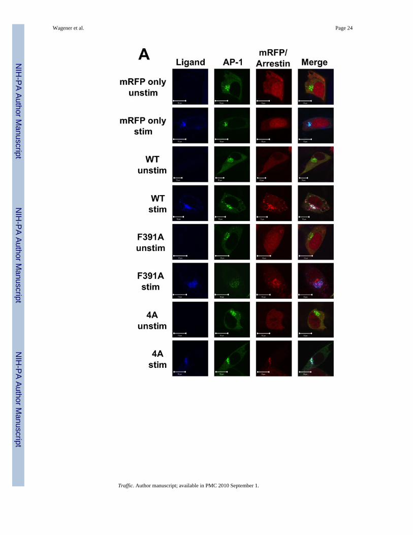
trafficking, we examined the localization of the FPR, arrestins and AP-1 in arr2−/−/3−/− FPRMEF cells using confocal fluorescence microscopy. In unstimulated cells, AP-1 waslocalized primarily in a perinuclear region, but was also present in the cytoplasm. When theFPR was stimulated in arrestin-deficient cells, receptor accumulated in perinuclearendosomes overlapping substantially with AP-1 (Fig. 5A). Upon stimulation of cellsexpressing wild type arr2, the FPR-arr2 complexes were also colocalized with AP-1 in theperinuclear region. However, arr2 was also observed outside this region in cytoplasmicvesicles in association with the FPR and AP-1. This suggests that upon internalization, theFPR traffics to perinuclear endosomes where it associates with AP-1, which may escortreceptor back to the cell surface. The fact that arrestin appears to mediate these latetrafficking events indicates a role for arrestin in receptor trafficking at a much later stagethan previously thought. In contrast to wild type arr2, the F391A and 4A mutants bothaccumulated with the FPR in AP-1-positive endosomes in the perinuclear region with noreceptor, arrestin or AP-1 observed outside this cellular compartment, consistent with thelack of recycling. To further assess AP-1 trafficking with FPR/arr2 complexes, wequantitated the number of cells that showed AP-1 puncta outside of the perinuclear region(Fig. 5B). Only when wild type arr2 is present is AP-1 observed on vesicles outside theperinuclear region following FPR activation. Finally, pulse-chase experiments (10 min pulsewith 633-6pep, 50 min chase) using arr2−/−/3−/− FPR cells show that AP-1 is colocalizedwith 633-6pep-FPR-wild type arr2 in cytoplasmic vesicles following the 50 min chase, butnot in early endosomes immediately following internalization (10 min pulse with no chase,Suppl. Fig. 4).
In order to confirm that the GFP-fused β subunit of AP-1 accurately reflects endogenousAP-1 trafficking, we again used confocal immunofluorescence microscopy of U937 FPRcells transiently transfected only with Rab11-GFP (Suppl. Figs. 2B and 3A). In unstimulatedcells, AP-1 is predominantly colocalized with perinuclear Rab11 with minimal non-perinuclear staining. Upon stimulation of the FPR with 546-6pep, ligand-receptor complexescolocalize with Rab11 and AP-1 (as well as AP-2, Suppl. Fig. 3B) in the perinuclear region.In addition, ligand-receptor complexes found in non-perinuclear endosomes (lacking Rab11)also colocalize significantly with AP-1 consistent with recycling of the FPR to the plasmamembrane.
Based on the colocalization of the FPR, arrestin and AP-1 upon receptor activation, wesought to determine whether arrestin also forms a complex with AP-1. Arr2−/−/3−/− FPRcells were transiently transfected with arr2-WT-FLAG and stimulated prior toimmunoprecipitation with anti-FLAG antibodies. AP-1 was detected in theimmunoprecipitates by blotting for the γ subunit of AP-1. In the absence of arrestins, AP-1was not detected in anti-FLAG immunoprecipitates (Fig. 5C). However, in the presence ofarr2-WT-FLAG, the γ subunit of AP-1 was detected in the anti-FLAG immunoprecipitatesin a stimulation-dependent manner, indicating an association between the FPR-arrestincomplex and AP-1.
AP-2 Regulates FPR Recycling and GPCR-mediated ApoptosisIn order to determine explicitly whether AP-2 and AP-1 regulate FPR recycling and GPCR-mediated apoptosis in general, we used siRNAs to knockdown the µ1A and µ2 subunits ofAP-1 and AP-2, respectively in U937 FPR cells (Fig. 6A). Knockdown of µ1A and µ2subunit expression was >90% in both cases. In addition, expression of the β subunit of AP-2and γ subunit of AP-1 was decreased as well, but not to the same extent (~70% and ~50%respectively, unpublished data). Although the rate of FPR internalization was not affected byknockdown of either AP-1 or AP-2 (half time for internalization: control, 1.4+/−0.2 min;AP-1, 1.5+/−0.2 min; AP-2, 1.6+/−0.2 min), the extent of FPR internalization was modestlyreduced upon AP-2 knockdown (control, 75+/−2%; AP-1, 82+/−3%; AP-2, 57+/−5%). In
Wagener et al. Page 7
Traffic. Author manuscript; available in PMC 2010 September 1.
NIH
-PA Author Manuscript
NIH
-PA Author Manuscript
NIH
-PA Author Manuscript
contrast, knockdown of AP-2 produced a significant decrease of approximately 40% in FPRrecycling, whereas knockdown of AP-1 produced no effect (Fig. 6B). Visualization of FPRtrafficking in AP-2 siRNA-treated U937 cells also demonstrated a partial accumulation ofthe FPR in perinuclear endosomes, whereas AP-1 siRNA treatment yielded a wild typeintracellular FPR distribution (Suppl. Fig. 3C). To determine whether the observed reductionin FPR recycling resulted in an alteration in cell survival, U937 FPR cells treated withsiRNA for AP-1 or AP-2 were stimulated with ligand and evaluated for apoptosis. Onlystimulated cells treated with siRNA directed against AP-2 showed a profound apoptoticresponse with over 90% of the cells undergoing apoptosis (Fig. 6C). Pretreatment with thecaspase inhibitor ZVAD-FMK completely blocked the ligand-dependent apoptosis. Finally,we asked whether this induction of apoptosis was unique to the FPR or occurred with otherGPCRs. To test this, U937 cells stably transfected with either CXCR2 or CXCR4 weretreated with AP-2 siRNA and stimulated with the appropriate ligand. As a control for anendogenously expressed receptor, we also stimulated the U937 FPR cells with ATP, whichis known to activate P2Y purinergic receptors on these cells (45). Ligand dependentapoptosis was observed in both the CXCR2-expressing cells as well as the U937 FPR cellsstimulated with either fMLF or ATP (Fig. 6D). However, ligand stimulation of CXCR4-expressing cells had no effect. These results mirror our previous findings in arr2−/−/3−/−
FPR MEF cells, where stimulation of FPR and CXCR2 but not CXCR4 induced apoptosis(28). These results demonstrate that many but not all GPCRs are capable of initiatingapoptosis in the absence of a functional arrestin-AP-2 interaction.
DISCUSSIONIn summary, we have demonstrated that the ligand-dependent accumulation of GPCR-arrestin complexes in perinuclear recycling endosomes leads to the rapid initiation ofapoptosis. Furthermore, receptor accumulation and apoptosis were induced by the disruptionof the arrestin-AP-2 interaction, either by a single point mutation in arr2 (F391A) or bydepleting AP-2 levels. It is interesting to note that stabilization of the arr2-AP-2 complex inthe 4A mutant also affected receptor trafficking and induced apoptosis, suggesting thatcyclic binding and release of AP-2 is necessary for proper trafficking. This is also the firstreport to describe a role for AP-2 in the recycling of internalized GPCRs. Althoughdepletion of AP-1, best known for mediating vesicle traffic between the Golgi compartmentand endosomes, did not prevent receptor recycling, it was found to be associated withreceptor-arrestin complexes in perinuclear endosomes and non-perinuclear endosomesemanating from the recycling compartment, suggesting a possible role in GPCR re-expression. This observation is consistent with reported roles for AP-1 in the formation ofrecycling vesicles from internalized receptors (46–48). As signaling inhibitors have beenshown to inhibit FPR-mediated apoptosis, our results suggest that inhibition of proper FPRtrafficking also alters the spatial control of GPCR signaling complexes leading to theinitiation of apoptosis within the cell. This is supported by results that show spatial controlof GPCRs can induce or limit their potential to initiate cellular signaling pathways (49).
The F391A arrestin mutant was previously shown to exhibit decreased binding to the βsubunit of AP-2 through in vitro binding assays while its overexpression in cells inhibitedinternalization of the β2-AR (29). While the 4A mutant has not been previously reported, themutation and characterization of individual amino acids within this site (K397, M399 andK400) have been described (14). These individual mutants showed similar bindingproperties with the β subunit of AP-2 and produced similar effects on β2-AR internalizationas the F391A mutant. While the F391A mutant did not significantly colocalize or co-immunoprecipitate with AP-2, the 4A mutant interacted strongly with AP-2 upon FPRactivation. The reason for increased AP-2 binding to an arrestin mutant whose componentmutations show decreased binding is unclear, but may be due to conformational changes
Wagener et al. Page 8
Traffic. Author manuscript; available in PMC 2010 September 1.
NIH
-PA Author Manuscript
NIH
-PA Author Manuscript
NIH
-PA Author Manuscript
within the protein that alter the binding properties of this specific motif or secondary bindingsites. Similar to our arrestin 4A mutant, arrestin mutants I386A and V387A show 5-foldenhanced binding to AP-2 whereas an F388A substitution prevents binding (50).Furthermore, the AP-2 mutant R879A shows enhanced (10-fold) binding to arr2 (51).Together, these results demonstrate that this interaction can be both positively andnegatively modulated by mutations in both arrestin and AP-2. The inhibitory effect ofincreased AP-2 binding by the 4A mutant suggests that appropriate cycling of AP-2 witharrestin (association followed by dissociation) is required for the exit of the FPR fromrecycling endosomes. This idea is supported by evidence demonstrating Src activity as anecessary component for AP-2/arrestin dissociation and internalization of the β2-AR (41).
GPCR-arrestin interactions have also been shown to initiate apoptosis in the Drosophila eye,where alterations in the processing of rhodopsin, through diverse molecular mechanismssuch as deletion of one of the two visual arrestins, leads to light-dependent retinaldegeneration (52). Furthermore, loss-of-function mutants in a Ca2+-dependent serine/threonine phosphatase (rdgC) as well as an eye-specific phospholipase (norpA), whichactivates rdgC, result in the formation of stable internalized rhodopsin-arrestin complexesthat initiate apoptosis in photoreceptor cells resulting in retinal degeneration (53,54).Interestingly, in the norpA Drosophila mutant, disruption of arrestin/AP-2 binding throughthe introduction of arrestin point mutants rescued apoptosis by preventing rhodopsininternalization (52). Support for the conservation of this visual pathway has recently beenreported in mice. A mutant opsin (K296E), associated with autosomal dominant retinitispigmentosa, is hyperphosphorylated and its excessive association with arrestin results in themislocation and accumulation of the complex to the inner segments (55). These results bearmany similarities to our results in non-visual systems. First of all, we have previouslyreported that ligand-dependent GPCR-mediated apoptosis in arrestin-deficient cells requiresreceptor internalization (28). Second, in this study, we demonstrated that the accumulationof GPCR-arrestin complexes in recycling endosomes initiates rapid apoptosis. However, incontrast to the Drosophila system, AP-2 plays a critical role in mediating the appropriatetrafficking of GPCRs out of recycling endosomes, thus preventing the initiation ofapoptosis. Thus, our results demonstrate that arrestins likely play a critical role in themaintenance of cell viability in all eukaryotic cells.
Based on the results of this study, we propose a new model of FPR internalization andrecycling. In this model, following ligand binding the FPR can internalize in an arrestin-independent manner. Before, during or after internalization, the FPR binds to arrestin,resulting in the generation of FPR/arrestin complexes in early endosomes. At some pointduring its trafficking to perinuclear recycling compartment, the FPR-arrestin complexrecruits AP-2. Within or as it exits this compartment, the FPR/arrestin complex releasesAP-2, whereupon AP-1 associates with the FPR/arrestin complex. Along the path to the cellsurface the complex dissociates and the FPR is dephosphorylated. Finally, the FPRcompletes its return to the cell surface in a resensitized form ready to continue signaling.
In this report, we have described a novel mechanism for GPCR post-endocytic traffickingand recycling. This is the first report that identifies a definitive role for arrestin in the latestages of GPCR trafficking as compared to other GPCRs, where arrestins are involved in theearliest events of GPCR trafficking (i.e. internalization). In addition, the observation thatFPR-mediated apoptosis occurs under conditions where arrestin mutants are bound to theFPR demonstrates that the receptor-initiated apoptotic signaling in arrestin-deficient cells isnot purely a result of receptor activation in the absence of arrestins and furthermoreconfirms that receptor-arrestin accumulation in recycling endosomes results in aberrantsignaling that leads to apoptosis. With this report of novel roles for arrestins and adaptorproteins in the trafficking and signaling of GPCRs, new avenues for the targeting of GPCR
Wagener et al. Page 9
Traffic. Author manuscript; available in PMC 2010 September 1.
NIH
-PA Author Manuscript
NIH
-PA Author Manuscript
NIH
-PA Author Manuscript
function are presented that may lead to the development of therapeutic interventions fordisease states.
MATERIALS AND METHODSReagents, Plasmids and Mutagenesis
All reagents are from Sigma unless otherwise noted. With the exception of the arr2-F391Amutant (a gift from Jeffrey Benovic), regions of arr2 were mutated by site-directedmutagenesis and cloned into EGFP-N1 vector or mRFP1 vector using standard subcloningprocedures and HindIII/ApaI restriction sites. These constructs were amplified using PCRwith primers that created HindIII/ApaI restriction sites and subcloned as describe above.Arr2-WT-FLAG was created by digesting Arr2-WT-GFP at the ApaI/NotI restriction sites toremove GFP and inserting a linker that contained the FLAG sequence. Arr2-F391A- andarr2-4A–FLAG were constructed by digesting with HindIII/ApaI sites and subcloning the~1300bp fragment into the HindIII/ApaI restriction sites of Arr2-WT-FLAG. All mutantswere confirmed by DNA sequencing. Rab11-GFP was a gift from Angela Wandinger-Ness.GFP-fused α subunit of AP-2 and GFP-fused γ subunit of AP-1 were gifts from Lois Greene(56).
Cell culture and TransfectionArr2−/−/3−/− FPR cells were grown in DMEM with 10% fetal bovine serum, 100 units/mLpenicillin and 100units/mL streptomycin at 37°C and 5% CO2. U937 cells were grown inRPMI with 10% fetal bovine serum, 100 units/mL penicillin and 100units/mL streptomycinat 37°C and 5% CO2. Transient transfections of mouse embryonic fibroblasts wereperformed with Lipofectamine 2000 according to manufacturer’s instructions. siRNAtransfection of U937 FPR cells was performed using siPORT Electroporation Buffer(Ambion) with a Genepulser Xcell (Bio Rad) according to manufacturer instructions. Cellswere transfected with 20µg of siRNA twice at 72 hour intervals and assayed 72 hours afterthe second transfection. Cell survival was measured using Trypan Blue and was >95% forall transfections. siRNAs used for depletion of µ1A (AP-1) and µ2 (AP-2) were aspreviously described (57). mRNA gene target sequences for siRNA design were as follows:µ1A—GGCAUCAAGUAUCGGAAGA; µ2—GUGGAUGCCUUUCGGGUCA. Anonfunctional siRNA (Ambion) was used as a control.
ApoptosisFor cell rounding assays, arr2−/−/3−/− FPR cells transiently transfected with GFP-fusedarrestins were plated on 12mm-glass coverslips, incubated overnight and serum-starved for30 minutes. Cells were incubated in serum-free medium (SFM) or were stimulated with10nM fMLF in SFM 5 hours at 37°C. Cells were washed twice with PBS, fixed with 2%paraformaldehyde and mounted using Vectashield. Slides were viewed in a blinded mannerby phase-contrast microscopy and random fields were evaluated (at least five) until 100–300GFP-expressing cells were assayed. GFP-expressing cells were counted “positive” for celldeath if they rounded (spherical and refractile cells with no extensions). Data are expressedas a percentage of rounded/GFP-expressing cells. For propidium iodide iodide staining,arr2−/−/3−/− FPR cells were transfected as above. Stimulated MEF and U937 cells werestained with 100ng/mL propidium iodide in SFM at room temperature for 5–10 minutes,washed twice with PBS, fixed with 2% paraformaldehyde and mounted using Vectashield.Slides were viewed in a blinded manner by fluorescence microscopy and random fields wereevaluated (at least five) until 100–300 cells (GFP-expressing for MEFs) were evaluated.Cells were counted “positive” for cell death if they were stained with propidium iodide. Dataare expressed as a percentage of PI-positive/GFP-expressing cells for MEFs and percentageof PI-positive cells for U937 cells.
Wagener et al. Page 10
Traffic. Author manuscript; available in PMC 2010 September 1.
NIH
-PA Author Manuscript
NIH
-PA Author Manuscript
NIH
-PA Author Manuscript
FPR InternalizationArr2−/−/3−/− FPR cells transiently transfected with GFP-fused arrestins were harvested bytrypsinization. U937 FPR cells were electroporated with siRNAs and harvested bycentrifugation. Cells were resuspended in SFM and stimulated for 2, 5, 10, 20 and 30minutes with 1µM fMLF. Cells were then washed extensively with cold SFM to removeexcess unlabelled ligand. Remaining receptors on the cell surface were labeled with 10nM633-6pep. Cells were then assayed by flow cytometry using a Becton-DickinsonFACSCalibur. Cells (100,000) were gated for live cells using forward and side scatterparameters. GFP-fused arrestin mutant-expressing cells were subsequently gated using FL-1and the mean channel fluorescence (MCF) was determined in FL-4 to monitor cell surfaceexpression of the FPR. Non-specific binding was determined by labeling arr2−/−/3−/− cellsnot expressing the FPR with 633-6pep and assaying as described. Non-specific binding wassubtracted before further calculations. MCF from unstimulated cells represented 100% FPRcell surface expression. Cell surface expression from stimulated cells was calculated bydividing the MCF following treatment by the MCF from unstimulated cells. Internalizationdata were then plotted using GraphPad Prism to calculate maximum internalization extentsusing a singe exponential decay. Statistical analysis was performed using Student’s t-test.
Confocal Fluorescence MicroscopyArr2−/−/3−/− FPR cells or U937 cells were transiently transfected with mRFP-fusedarrestins, GFP-fused Rab11, GFP-fused α subunit of AP-2 or GFP-fused γ subunit of AP-1.Arr2−/−/3−/− FPR cells were plated on 25mm-glass coverslips and grown overnight. U937FPR cells were harvested by centrifugation. Cells were serum-starved for 30 minutes andstimulated with 10 nM 633- or 546-6pep in SFM for indicated time at 37°C. Coverslips orharvested cells were rinsed with PBS, fixed with 2% paraformaldehyde at room temperatureand mounted with Vectashield. For experiments using antibody staining, after fixation cellswere incubated in 0.02% Saponin/3% BSA for 15–20 minutes at room temperature. Cellswere washed and incubated in 1:100 primary antibody (100/3 for γ1-adaptin antibody orAP-6 for α-adaptin (Affinity Bioreagents)) in 3% NGS for 30 minutes at room temperature.After further washing, cells were incubated in 1:200 secondary antibody (Cy5-conjugatedDonkey Anti-Mouse (Jackson Immunoresearch)) in 3% NGS for 30 minutes at roomtemperature. Cells were then washed and mounted using Vectashield. Fluorescence imageswere acquired using a Zeiss LSM 510 inverted laser scanning microscope equipped with He-Ne and Kr-Ar lasers. To assess the extent of colocalization, cells with arrestin clusters wereviewed and scored for the presence of any corresponding AP-2 or AP-1 clusters,respectively. Data were expressed as percentage of cells (mean +/− SEM) with colocalizedclusters from >25 cells/experiment. Values were normalized to the wild type arrestinresponse at 60 minutes.
Cell Lysis and ImmunoprecipitationArr2−/−/3−/− FPR MEF cells transiently transfected with FLAG-tagged arrestins were grownto confluence. Cells were serum-starved for 30 min and stimulated for the designated timeswith 10nM fMLF in SFM at 37°C. Medium was removed and cold co-IP lysis buffer (1 mL1% v/v TX-100, 150mM NaCl, 10mM Tris-HCl pH 7.4 supplemented with proteaseinhibitor cocktail (Calbiochem)) was added immediately. Lysates were collected, incubatedon ice for 30 min, and centrifuged at maximum speed for 30 min at 4°C. An aliquot fromeach tube was set aside for determining pre-immunoprecipitation levels of proteins byWestern blot. For immunoprecipitations, 25µL of protein A Sepharose (Pierce) was washedthree times with co-IP lysis buffer. Beads were rotated for at least one hour at 4°C in 250µLof co-IP lysis buffer with 1:1000 M2 Anti-FLAG antibody. Beads were washed with co-IPlysis buffer and lysates were added and rotated overnight at 4°C. The following day, beads
Wagener et al. Page 11
Traffic. Author manuscript; available in PMC 2010 September 1.
NIH
-PA Author Manuscript
NIH
-PA Author Manuscript
NIH
-PA Author Manuscript
were washed again with co-IP lysis buffer, 40µL of 2X Sample Buffer was added andimmunoprecipitated proteins released by boiling for 5 minutes.
Western BlottingLysates or immunoprecipitates were resolved with SDS-PAGE. Proteins were transferred toPVDF membranes and blocked for 1 hour with 5% dry milk in TBS-T. Blotting was carriedout using 1:1000 dilutions of biotinylated M2 mouse anti-FLAG antibody, M2 mouse Anti-FLAG, mouse anti-β-adaptin antibody (BD Biosciences), mouse 100/3 to γ1-adaptinantibody, rabbit RY/1 to µ1A (gift from L. Traub), rabbit R11–29 to µ2 ((58), gift from J.Bonifacino) and 1:4000 mouse anti-GAPDH antibody (Chemicon) in 5% dry milk in TBS-Tovernight at 4°C. Blots were washed with TBS-T and incubated with 1:2500 HRP-streptavidin (in 5% BSA in TBS-T), 1:2500 HRP rabbit anti-mouse antibody or 1:2500 HRPgoat anti-rabbit antibody (in 5% Dry Milk in TBS-T) at room temperature for 1 or 3 hours,respectively. Bands were visualized using SuperSignal West Pico ChemiluminescentSubstrate (Pierce). Densitometry was performed using Quantity One (BioRad) and ratiosexpressed as mean ratio +/− SEM adaptin/arrestin immunoprecipitated and normalized tozero time points for each vector expressed.
FPR RecyclingArr2−/−/3−/− FPR cells transiently transfected with GFP-fused arrestins or U937 FPR cellstransfected with siRNA were harvested and resuspended in SFM. An aliquot was removedto measure total cell surface receptor. The remaining cells were stimulated with 1µM fMLFin SFM at 37°C for 1 hour and were then washed extensively to remove excess unlabelledligand. Half the remaining cells were resuspended in pre-warmed SFM for 30 min at 37°C(20 min for U937 FPR cells) to allow the FPR to recycle. The other half was kept on ice tomeasure post-internalization cell surface receptor levels. All aliquots were then resuspendedin SFM containing 10nM 633-6pep and assayed by flow cytometry using a Becton-Dickinson FACSCalibur. For analysis, assayed cells were gated for live cells using forwardand side scatter parameters. These cells were then gated using FL-1 for GFP-fused arrestinmutant expression and the mean channel fluorescence was measured in FL-4 to monitor cellsurface expression of the FPR. Non-specific binding was determined by labeling arrestinknockout cell lines (or U937 cells lines) that did not express FPR and was subtracted fromall values. To account for differences in total recycling that could be due to differences inthe initial extent of internalization, the fraction of recycled FPR (starting from the finalinternalization time point) was divided by the fraction of internalized FPR. Data areexpressed as a percentage of recycled receptor /internalized receptor.
Supplementary MaterialRefer to Web version on PubMed Central for supplementary material.
AcknowledgmentsWe thank Charlotte Vines and Angela Wandinger-Ness for helpful comments. This work was funded by NIH grantsAI36357 and GM68901 to E.R.P. and pre-doctoral fellowship BC030217 from the Department of Defense BreastCancer Research Program to B.M.W. Flow cytometry data and confocal images in this study were generated in theFlow Cytometry and Fluorescence Microscopy Facilities, respectively, at the University of New Mexico HealthSciences Center, which received support from NCRR 1 S10 RR14668, NSF MCB9982161, NCRR P20 RR11830,NCI P30 CA118100, NCRR S10 RR19287, NCRR S10 RR016918, the University of New Mexico Health SciencesCenter, and the University of New Mexico Cancer Center.
Wagener et al. Page 12
Traffic. Author manuscript; available in PMC 2010 September 1.
NIH
-PA Author Manuscript
NIH
-PA Author Manuscript
NIH
-PA Author Manuscript
Abbreviations used
AP-1 adaptor protein-1 complex
AP-2 adaptor protein-2 complex
Arr2 arrestin-2
Arr2−/−/3−/− FPRMEF
arrestin-2−/−/-3−/− knockout MEF cells stably expressing the FPR
fMLF N-formyl-Methionyl-Leucyl-Phenylalanine
FPR N-formyl peptide receptor
GPCR G protein-coupled receptor
MEF mouse embryonic fibroblast
PI propidium iodide
SFM serum-free medium (DMEM or RPMI)
6pep N-formyl-Leucyl-Leucyl-Phenylalanyl-Leucyl-Tyrosinyl-Lysine
U937 FPR U937 cells stably expressing the FPR
REFERENCES1. Penela P, Murga C, Ribas C, Tutor AS, Peregrin S, Mayor F Jr. Mechanisms of regulation of G
protein-coupled receptor kinases (GRKs) and cardiovascular disease. Cardiovasc Res. 2006; 69(1):46–56. [PubMed: 16288730]
2. Lombardi MS, Kavelaars A, Heijnen CJ. Role and modulation of G protein-coupled receptorsignaling in inflammatory processes. Crit Rev Immunol. 2002; 22(2):141–163. [PubMed:12433131]
3. Premont RT. Once and future signaling: G protein-coupled receptor kinase control of neuronalsensitivity. Neuromolecular Med. 2005; 7(1–2):129–147. [PubMed: 16052042]
4. Moratz C, Harrison K, Kehrl JH. Role of RGS proteins in regulating the migration of Blymphocytes. Arch Immunol Ther Exp (Warsz). 2004; 52(1):27–35. [PubMed: 15053230]
5. Luttrell DK, Luttrell LM. Not so strange bedfellows: G-protein-coupled receptors and Src familykinases. Oncogene. 2004; 23(48):7969–7978. [PubMed: 15489914]
6. Barak LS, Oakley RH, Laporte SA, Caron MG. Constitutive arrestin-mediated desensitization of ahuman vasopressin receptor mutant associated with nephrogenic diabetes insipidus. Proc Natl AcadSci U S A. 2001; 98(1):93–98. [PubMed: 11134505]
7. Key TA, Bennett TA, Foutz TD, Gurevich VV, Sklar LA, Prossnitz ER. Regulation of formylpeptide receptor agonist affinity by reconstitution with arrestins and heterotrimeric G proteins. JBiol Chem. 2001; 276(52):49204–49212. [PubMed: 11598142]
8. Key TA, Foutz TD, Gurevich VV, Sklar LA, Prossnitz ER. N-formyl peptide receptorphosphorylation domains differentially regulate arrestin and agonist affinity. J Biol Chem. 2003;278(6):4041–4047. [PubMed: 12424254]
9. Key TA, Vines CM, Wagener BM, Gurevich VV, Sklar LA, Prossnitz ER. Inhibition ofchemoattractant N-formyl peptide receptor trafficking by active arrestins. Traffic. 2005; 6(2):87–99.[PubMed: 15634210]
10. Tobin AB, Butcher AJ, Kong KC. Location, location, location…site-specific GPCRphosphorylation offers a mechanism for cell-type-specific signalling. Trends Pharmacol Sci. 2008;29(8):413–420. [PubMed: 18606460]
11. DeFea KA. Stop that cell! Beta-arrestin-dependent chemotaxis: a tale of localized actin assemblyand receptor desensitization. Annu Rev Physiol. 2007; 69:535–560. [PubMed: 17002593]
12. DeWire SM, Ahn S, Lefkowitz RJ, Shenoy SK. Beta-arrestins and cell signaling. Annu RevPhysiol. 2007; 69:483–510. [PubMed: 17305471]
Wagener et al. Page 13
Traffic. Author manuscript; available in PMC 2010 September 1.
NIH
-PA Author Manuscript
NIH
-PA Author Manuscript
NIH
-PA Author Manuscript
13. Premont RT, Gainetdinov RR. Physiological roles of G protein-coupled receptor kinases andarrestins. Annu Rev Physiol. 2007; 69:511–534. [PubMed: 17305472]
14. Kim YM, Benovic JL. Differential roles of arrestin-2 interaction with clathrin and adaptor protein2 in G protein-coupled receptor trafficking. J Biol Chem. 2002; 277(34):30760–30768. [PubMed:12070169]
15. Ahn S, Maudsley S, Luttrell LM, Lefkowitz RJ, Daaka Y. Src-mediated tyrosine phosphorylationof dynamin is required for beta2-adrenergic receptor internalization and mitogen-activated proteinkinase signaling. J Biol Chem. 1999; 274(3):1185–1188. [PubMed: 9880482]
16. Cao TT, Deacon HW, Reczek D, Bretscher A, von Zastrow M. A kinase-regulated PDZ-domaininteraction controls endocytic sorting of the beta2-adrenergic receptor. Nature. 1999; 401(6750):286–290. [PubMed: 10499588]
17. Delaney KA, Murph MM, Brown LM, Radhakrishna H. Transfer of M2 muscarinic acetylcholinereceptors to clathrin-derived early endosomes following clathrin-independent endocytosis. J BiolChem. 2002; 277(36):33439–33446. [PubMed: 12093817]
18. Vines CM, Revankar CM, Maestas DC, LaRusch LL, Cimino DF, Kohout TA, Lefkowitz RJ,Prossnitz ER. N-formyl peptide receptors internalize but do not recycle in the absence of arrestins.J Biol Chem. 2003; 278(43):41581–41584. [PubMed: 12947104]
19. Kohout TA, Lin FS, Perry SJ, Conner DA, Lefkowitz RJ. beta-Arrestin 1 and 2 differentiallyregulate heptahelical receptor signaling and trafficking. Proc Natl Acad Sci U S A. 2001; 98(4):1601–1606. [PubMed: 11171997]
20. Rosenfeld JL, Knoll BJ, Moore RH. Regulation of G-protein-coupled receptor activity by rabGTPases. Receptors Channels. 2002; 8(2):87–97. [PubMed: 12448790]
21. Innamorati G, Le Gouill C, Balamotis M, Birnbaumer M. The long and the short cycle. Alternativeintracellular routes for trafficking of G-protein-coupled receptors. J Biol Chem. 2001; 276(16):13096–13103. [PubMed: 11150299]
22. Kreuzer OJ, Krisch B, Dery O, Bunnett NW, Meyerhof W. Agonist-mediated endocytosis of ratsomatostatin receptor subtype 3 involves beta-arrestin and clathrin coated vesicles. JNeuroendocrinol. 2001; 13(3):279–287. [PubMed: 11207943]
23. Fan GH, Lapierre LA, Goldenring JR, Richmond A. Differential regulation of CXCR2 traffickingby Rab GTPases. Blood. 2003; 101(6):2115–2124. [PubMed: 12411301]
24. Oakley RH, Laporte SA, Holt JA, Barak LS, Caron MG. Molecular determinants underlying theformation of stable intracellular G protein-coupled receptor-beta-arrestin complexes after receptorendocytosis. J Biol Chem. 2001; 276(22):19452–19460. [PubMed: 11279203]
25. Tohgo A, Choy EW, Gesty-Palmer D, Pierce KL, Laporte S, Oakley RH, Caron MG, LefkowitzRJ, Luttrell LM. The stability of the G protein-coupled receptor-beta-arrestin interactiondetermines the mechanism and functional consequence of ERK activation. J Biol Chem. 2003;278(8):6258–6267. [PubMed: 12473660]
26. Tohgo A, Pierce KL, Choy EW, Lefkowitz RJ, Luttrell LM. beta-Arrestin scaffolding of the ERKcascade enhances cytosolic ERK activity but inhibits ERK-mediated transcription followingangiotensin AT1a receptor stimulation. J Biol Chem. 2002; 277(11):9429–9436. [PubMed:11777902]
27. Simaan M, Bedard-Goulet S, Fessart D, Gratton JP, Laporte SA. Dissociation of beta-arrestin frominternalized bradykinin B2 receptor is necessary for receptor recycling and resensitization. CellSignal. 2005; 17(9):1074–1083. [PubMed: 15993749]
28. Revankar CM, Vines CM, Cimino DF, Prossnitz ER. Arrestins block G protein-coupled receptor-mediated apoptosis. J Biol Chem. 2004; 279(23):24578–24584. [PubMed: 15051714]
29. Milano SK, Pace HC, Kim YM, Brenner C, Benovic JL. Scaffolding functions of arrestin-2revealed by crystal structure and mutagenesis. Biochemistry. 2002; 41(10):3321–3328. [PubMed:11876640]
30. Krupnick JG, Santini F, Gagnon AW, Keen JH, Benovic JL. Modulation of the arrestin-clathrininteraction in cells. Characterization of beta-arrestin dominant-negative mutants. J Biol Chem.1997; 272(51):32507–32512. [PubMed: 9405462]
Wagener et al. Page 14
Traffic. Author manuscript; available in PMC 2010 September 1.
NIH
-PA Author Manuscript
NIH
-PA Author Manuscript
NIH
-PA Author Manuscript
31. Potter RM, Key TA, Gurevich VV, Sklar LA, Prossnitz ER. Arrestin variants display differentialbinding characteristics for the phosphorylated N-formyl peptide receptor carboxyl terminus. J BiolChem. 2002; 277(11):8970–8978. [PubMed: 11777932]
32. Kovoor A, Celver J, Abdryashitov RI, Chavkin C, Gurevich VV. Targeted construction ofphosphorylation-independent beta-arrestin mutants with constitutive activity in cells. J Biol Chem.1999; 274(11):6831–6834. [PubMed: 10066734]
33. Goodman OB Jr, Krupnick JG, Gurevich VV, Benovic JL, Keen JH. Arrestin/clathrin interaction.Localization of the arrestin binding locus to the clathrin terminal domain. J Biol Chem. 1997;272(23):15017–15022. [PubMed: 9169477]
34. Krupnick JG, Goodman OB Jr, Keen JH, Benovic JL. Arrestin/clathrin interaction. Localization ofthe clathrin binding domain of nonvisual arrestins to the carboxy terminus. J Biol Chem. 1997;272(23):15011–15016. [PubMed: 9169476]
35. Lin FT, Krueger KM, Kendall HE, Daaka Y, Fredericks ZL, Pitcher JA, Lefkowitz RJ. Clathrin-mediated endocytosis of the beta-adrenergic receptor is regulated by phosphorylation/dephosphorylation of beta-arrestin1. J Biol Chem. 1997; 272(49):31051–31057. [PubMed:9388255]
36. Gilbert TL, Bennett TA, Maestas DC, Cimino DF, Prossnitz ER. Internalization of the human N-formyl peptide and C5a chemoattractant receptors occurs via clathrin-independent mechanisms.Biochemistry. 2001; 40(12):3467–3475. [PubMed: 11297412]
37. Campbell RE, Tour O, Palmer AE, Steinbach PA, Baird GS, Zacharias DA, Tsien RY. Amonomeric red fluorescent protein. Proc Natl Acad Sci U S A. 2002; 99(12):7877–7882.[PubMed: 12060735]
38. Chen W, Feng Y, Chen D, Wandinger-Ness A. Rab11 is required for trans-golgi network-to-plasma membrane transport and a preferential target for GDP dissociation inhibitor. Mol Biol Cell.1998; 9(11):3241–3257. [PubMed: 9802909]
39. Browning DD, Pan ZK, Prossnitz ER, Ye RD. Cell type- and developmental stage-specificactivation of NF-kappaB by fMet-Leu-Phe in myeloid cells. J Biol Chem. 1997; 272(12):7995–8001. [PubMed: 9065470]
40. Hsu MH, Chiang SC, Ye RD, Prossnitz ER. Phosphorylation of the N-formyl peptide receptor isrequired for receptor internalization but not chemotaxis. J Biol Chem. 1997; 272(47):29426–29429. [PubMed: 9367998]
41. Fessart D, Simaan M, Laporte SA. c-Src regulates clathrin adapter protein 2 interaction with beta-arrestin and the angiotensin II type 1 receptor during clathrin- mediated internalization. MolEndocrinol. 2005; 19(2):491–503. [PubMed: 15498833]
42. Robinson MS. Adaptable adaptors for coated vesicles. Trends Cell Biol. 2004; 14:167–174.[PubMed: 15066634]
43. Laporte SA, Miller WE, Kim KM, Caron MG. beta-Arrestin/AP-2 interaction in G protein-coupledreceptor internalization: identification of a beta-arrestin binging site in beta 2-adaptin. J BiolChem. 2002; 277(11):9247–9254. [PubMed: 11777907]
44. Lundmark R, Carlsson SR. The beta-appendages of the four adaptor-protein (AP) complexes:structure and binding properties, and identification of sorting nexin 9 as an accessory protein toAP-2. Biochem J. 2002; 362(Pt 3):597–607. [PubMed: 11879186]
45. Cowen DS, Berger M, Nuttle L, Dubyak GR. Chronic treatment with P2-purinergic receptoragonists induces phenotypic modulation of the HL-60 and U937 human myelogenous leukemiacell lines. J Leukoc Biol. 1991; 50(2):109–122. [PubMed: 1649238]
46. Deneka M, Neeft M, Popa I, van Oort M, Sprong H, Oorschot V, Klumperman J, Schu P, van derSluijs P. Rabaptin-5alpha/rabaptin-4 serves as a linker between rab4 and gamma(1)-adaptin inmembrane recycling from endosomes. Embo J. 2003; 22(11):2645–2657. [PubMed: 12773381]
47. Pagano A, Crottet P, Prescianotto-Baschong C, Spiess M. In vitro formation of recycling vesiclesfrom endosomes requires adaptor protein-1/clathrin and is regulated by rab4 and the connectorrabaptin-5. Mol Biol Cell. 2004; 15(11):4990–5000. [PubMed: 15331762]
48. Popa I, Deneka M, van der Sluijs P. Expression and properties of the Rab4, Rabaptin-5alpha, AP-1complex in endosomal recycling. Methods Enzymol. 2005; 403:526–540. [PubMed: 16473617]
Wagener et al. Page 15
Traffic. Author manuscript; available in PMC 2010 September 1.
NIH
-PA Author Manuscript
NIH
-PA Author Manuscript
NIH
-PA Author Manuscript
49. Daaka Y, Luttrell LM, Ahn S, Della Rocca GJ, Ferguson SS, Caron MG, Lefkowitz RJ. Essentialrole for G protein-coupled receptor endocytosis in the activation of mitogen-activated proteinkinase. J Biol Chem. 1998; 273(2):685–688. [PubMed: 9422717]
50. Burtey A, Schmid EM, Ford MG, Rappoport JZ, Scott MG, Marullo S, Simon SM, McMahon HT,Benmerah A. The conserved isoleucine-valine-phenylalanine motif couples activation state andendocytic functions of beta-arrestins. Traffic. 2007; 8(7):914–931. [PubMed: 17547696]
51. Schmid EM, Ford MG, Burtey A, Praefcke GJ, Peak-Chew SY, Mills IG, Benmerah A, McMahonHT. Role of the AP2 beta-Appendage Hub in Recruiting Partners for Clathrin-Coated VesicleAssembly. PLoS Biol. 2006; 4(9)
52. Orem NR, Xia L, Dolph PJ. An essential role for endocytosis of rhodopsin through interaction ofvisual arrestin with the AP-2 adaptor. J Cell Sci. 2006; 119(Pt 15):3141–3148. [PubMed:16835270]
53. Kiselev A, Socolich M, Vinos J, Hardy RW, Zuker CS, Ranganathan R. A molecular pathway forlight-dependent photoreceptor apoptosis in Drosophila. Neuron. 2000; 28(1):139–152. [PubMed:11086990]
54. Alloway PG, Howard L, Dolph PJ. The formation of stable rhodopsin-arrestin complexes inducesapoptosis and photoreceptor cell degeneration. Neuron. 2000; 28(1):129–138. [PubMed:11086989]
55. Chen J, Shi G, Concepcion FA, Xie G, Oprian D, Chen J. Stable rhodopsin/arrestin complex leadsto retinal degeneration in a transgenic mouse model of autosomal dominant retinitis pigmentosa. JNeurosci. 2006; 26(46):11929–11937. [PubMed: 17108167]
56. Wu X, Zhao X, Puertollano R, Bonifacino JS, Eisenberg E, Greene LE. Adaptor and clathrinexchange at the plasma membrane and trans-Golgi network. Mol Biol Cell. 2003; 14(2):516–528.[PubMed: 12589051]
57. Janvier K, Bonifacino JS. Role of the endocytic machinery in the sorting of lysosome-associatedmembrane proteins. Mol Biol Cell. 2005; 16(9):4231–4242. [PubMed: 15987739]
58. Aguilar RC, Ohno H, Roche KW, Bonifacino JS. Functional domain mapping of the clathrin-associated adaptor medium chains mu1 and mu2. J Biol Chem. 1997; 272(43):27160–27166.[PubMed: 9341158]
Wagener et al. Page 16
Traffic. Author manuscript; available in PMC 2010 September 1.
NIH
-PA Author Manuscript
NIH
-PA Author Manuscript
NIH
-PA Author Manuscript
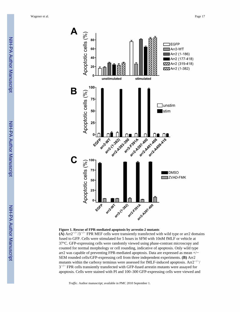
Figure 1. Rescue of FPR-mediated apoptosis by arrestin-2 mutants(A) Arr2−/−/3−/− FPR MEF cells were transiently transfected with wild type or arr2 domainsfused to GFP. Cells were stimulated for 5 hours in SFM with 10nM fMLF or vehicle at37°C. GFP-expressing cells were randomly viewed using phase-contrast microscopy andcounted for normal morphology or cell rounding, indicative of apoptosis. Only wild typearr2 was capable of preventing FPR-mediated apoptosis. Data are expressed as mean +/−SEM rounded cells/GFP-expressing cell from three independent experiments. (B) Arr2mutants within the carboxy terminus were assessed for fMLF-induced apoptosis. Arr2−/−/3−/− FPR cells transiently transfected with GFP-fused arrestin mutants were assayed forapoptosis. Cells were stained with PI and 100–300 GFP-expressing cells were viewed and
Wagener et al. Page 17
Traffic. Author manuscript; available in PMC 2010 September 1.
NIH
-PA Author Manuscript
NIH
-PA Author Manuscript
NIH
-PA Author Manuscript

scored for the presence of PI staining. Data are expressed as mean ± SEM PI positive/GFPcell from three independent experiments. (C) Reversal of FPR-mediated apoptosis by thecaspase inhibitor ZVAD-FMK. Data are expressed as mean ± SEM from three independentexperiments
Wagener et al. Page 18
Traffic. Author manuscript; available in PMC 2010 September 1.
NIH
-PA Author Manuscript
NIH
-PA Author Manuscript
NIH
-PA Author Manuscript
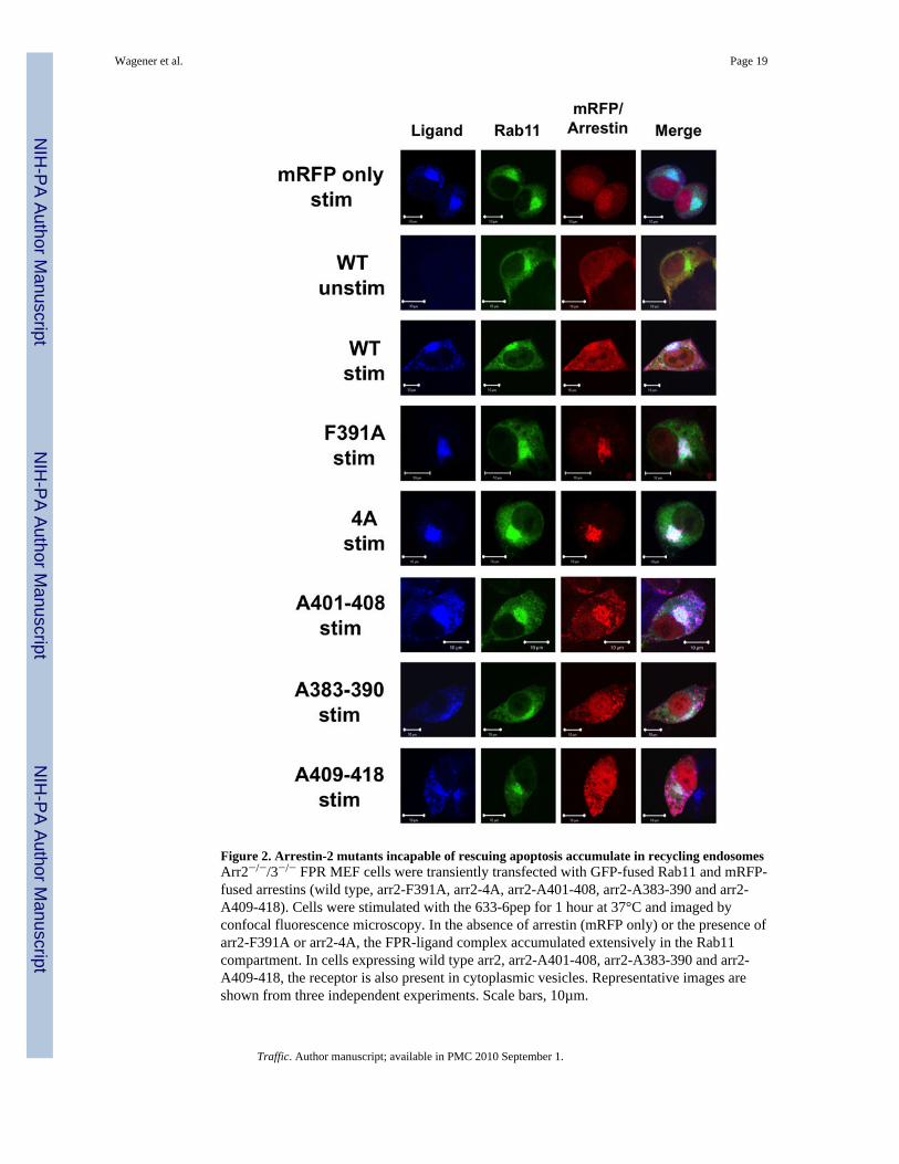
Figure 2. Arrestin-2 mutants incapable of rescuing apoptosis accumulate in recycling endosomesArr2−/−/3−/− FPR MEF cells were transiently transfected with GFP-fused Rab11 and mRFP-fused arrestins (wild type, arr2-F391A, arr2-4A, arr2-A401-408, arr2-A383-390 and arr2-A409-418). Cells were stimulated with the 633-6pep for 1 hour at 37°C and imaged byconfocal fluorescence microscopy. In the absence of arrestin (mRFP only) or the presence ofarr2-F391A or arr2-4A, the FPR-ligand complex accumulated extensively in the Rab11compartment. In cells expressing wild type arr2, arr2-A401-408, arr2-A383-390 and arr2-A409-418, the receptor is also present in cytoplasmic vesicles. Representative images areshown from three independent experiments. Scale bars, 10µm.
Wagener et al. Page 19
Traffic. Author manuscript; available in PMC 2010 September 1.
NIH
-PA Author Manuscript
NIH
-PA Author Manuscript
NIH
-PA Author Manuscript
Wagener et al. Page 20
Traffic. Author manuscript; available in PMC 2010 September 1.
NIH
-PA Author Manuscript
NIH
-PA Author Manuscript
NIH
-PA Author Manuscript
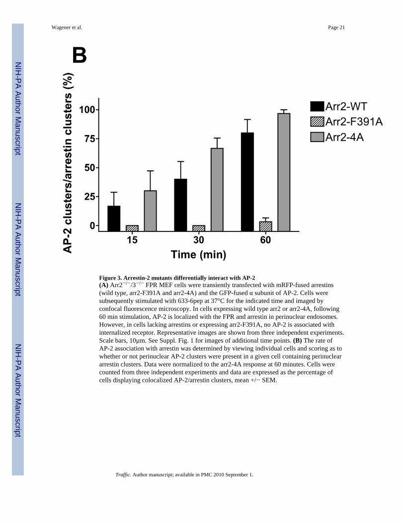
Figure 3. Arrestin-2 mutants differentially interact with AP-2(A) Arr2−/−/3−/− FPR MEF cells were transiently transfected with mRFP-fused arrestins(wild type, arr2-F391A and arr2-4A) and the GFP-fused α subunit of AP-2. Cells weresubsequently stimulated with 633-6pep at 37°C for the indicated time and imaged byconfocal fluorescence microscopy. In cells expressing wild type arr2 or arr2-4A, following60 min stimulation, AP-2 is localized with the FPR and arrestin in perinuclear endosomes.However, in cells lacking arrestins or expressing arr2-F391A, no AP-2 is associated withinternalized receptor. Representative images are shown from three independent experiments.Scale bars, 10µm. See Suppl. Fig. 1 for images of additional time points. (B) The rate ofAP-2 association with arrestin was determined by viewing individual cells and scoring as towhether or not perinuclear AP-2 clusters were present in a given cell containing perinucleararrestin clusters. Data were normalized to the arr2-4A response at 60 minutes. Cells werecounted from three independent experiments and data are expressed as the percentage ofcells displaying colocalized AP-2/arrestin clusters, mean +/− SEM.
Wagener et al. Page 21
Traffic. Author manuscript; available in PMC 2010 September 1.
NIH
-PA Author Manuscript
NIH
-PA Author Manuscript
NIH
-PA Author Manuscript
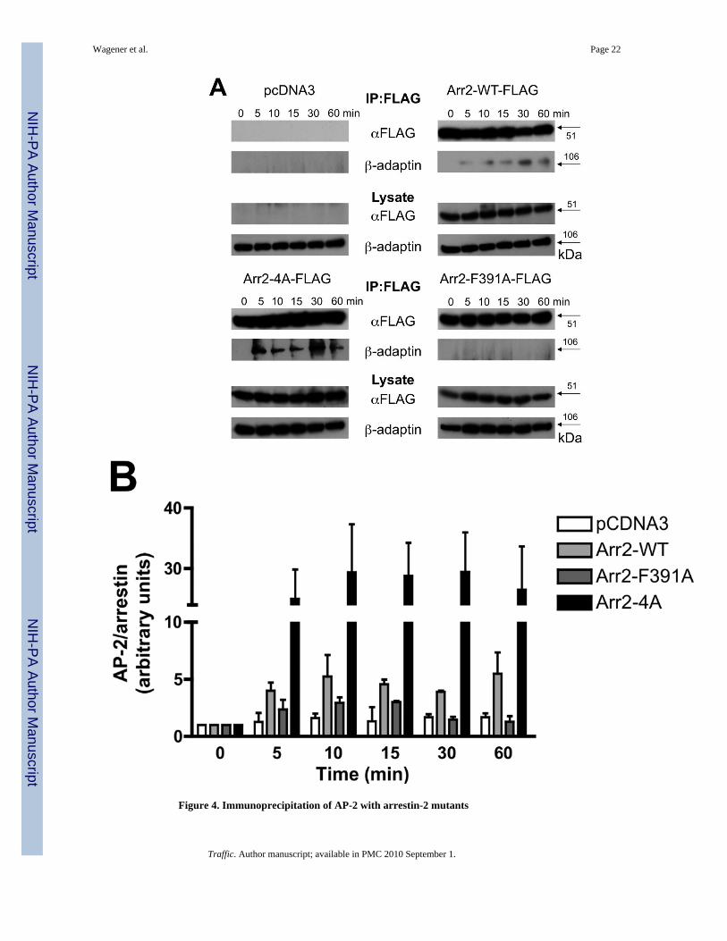
Figure 4. Immunoprecipitation of AP-2 with arrestin-2 mutants
Wagener et al. Page 22
Traffic. Author manuscript; available in PMC 2010 September 1.
NIH
-PA Author Manuscript
NIH
-PA Author Manuscript
NIH
-PA Author Manuscript
(A) Arr2−/−/3−/− FPR MEF cells were transiently transfected with FLAG-tagged arrestins.Cells were stimulated for the indicated times with 10nM fMLF and lysed. Lysates wereimmunoprecipitated with anti-FLAG antibodies, resolved by SDS-PAGE and blotted forFLAG-tagged arrestins and the β-adaptin subunit of AP-2. Representative blots are shown.(B) Quantitation of immunoprecipitated proteins by optometric density. Data are expressedas mean +/− SEM of β-adaptin/arrestin intensity ratios and normalized to respective zerotime points. Data are from three independent experiments.
Wagener et al. Page 23
Traffic. Author manuscript; available in PMC 2010 September 1.
NIH
-PA Author Manuscript
NIH
-PA Author Manuscript
NIH
-PA Author Manuscript
Wagener et al. Page 24
Traffic. Author manuscript; available in PMC 2010 September 1.
NIH
-PA Author Manuscript
NIH
-PA Author Manuscript
NIH
-PA Author Manuscript
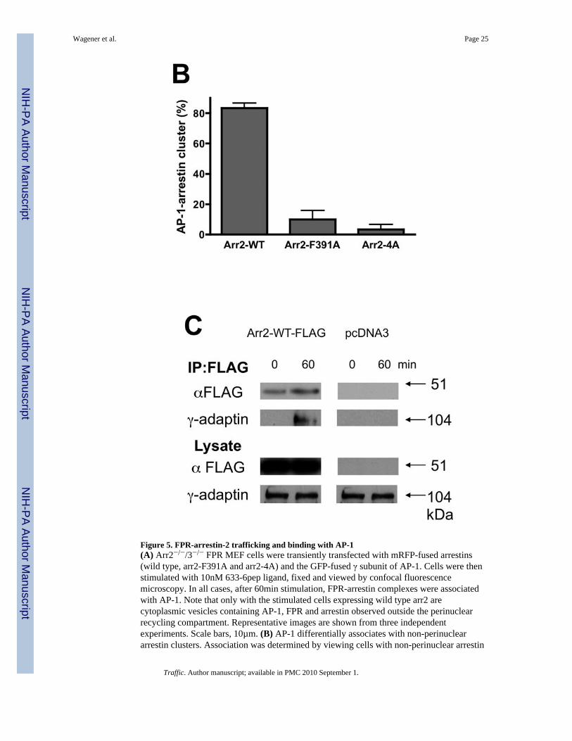
Figure 5. FPR-arrestin-2 trafficking and binding with AP-1(A) Arr2−/−/3−/− FPR MEF cells were transiently transfected with mRFP-fused arrestins(wild type, arr2-F391A and arr2-4A) and the GFP-fused γ subunit of AP-1. Cells were thenstimulated with 10nM 633-6pep ligand, fixed and viewed by confocal fluorescencemicroscopy. In all cases, after 60min stimulation, FPR-arrestin complexes were associatedwith AP-1. Note that only with the stimulated cells expressing wild type arr2 arecytoplasmic vesicles containing AP-1, FPR and arrestin observed outside the perinuclearrecycling compartment. Representative images are shown from three independentexperiments. Scale bars, 10µm. (B) AP-1 differentially associates with non-perinucleararrestin clusters. Association was determined by viewing cells with non-perinuclear arrestin
Wagener et al. Page 25
Traffic. Author manuscript; available in PMC 2010 September 1.
NIH
-PA Author Manuscript
NIH
-PA Author Manuscript
NIH
-PA Author Manuscript
clusters and scoring whether or not AP-1 clusters colocalized. Data are expressed as mean +/− SEM for >30 cells from three independent experiments. (C) Arr2 and AP-1 interact inresponse to FPR activation. Arr2−/−/3−/− FPR cells were transiently transfected with arr2-WT-FLAG or empty vector and immunoprecipitated with anti-FLAG antibodies after FPRactivation. Protein was resolved by SDS-PAGE and blotted as indicated. Representativeblots from three independent experiments are shown.
Wagener et al. Page 26
Traffic. Author manuscript; available in PMC 2010 September 1.
NIH
-PA Author Manuscript
NIH
-PA Author Manuscript
NIH
-PA Author Manuscript
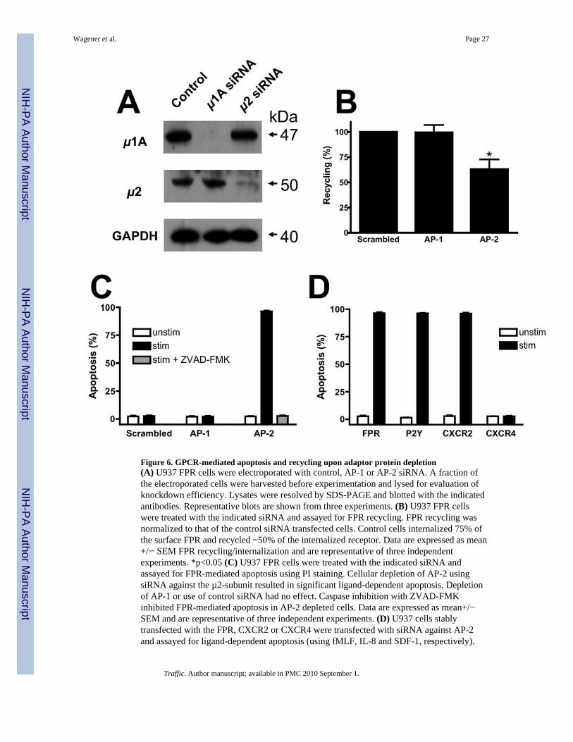
Figure 6. GPCR-mediated apoptosis and recycling upon adaptor protein depletion(A) U937 FPR cells were electroporated with control, AP-1 or AP-2 siRNA. A fraction ofthe electroporated cells were harvested before experimentation and lysed for evaluation ofknockdown efficiency. Lysates were resolved by SDS-PAGE and blotted with the indicatedantibodies. Representative blots are shown from three experiments. (B) U937 FPR cellswere treated with the indicated siRNA and assayed for FPR recycling. FPR recycling wasnormalized to that of the control siRNA transfected cells. Control cells internalized 75% ofthe surface FPR and recycled ~50% of the internalized receptor. Data are expressed as mean+/− SEM FPR recycling/internalization and are representative of three independentexperiments. *p<0.05 (C) U937 FPR cells were treated with the indicated siRNA andassayed for FPR-mediated apoptosis using PI staining. Cellular depletion of AP-2 usingsiRNA against the µ2-subunit resulted in significant ligand-dependent apoptosis. Depletionof AP-1 or use of control siRNA had no effect. Caspase inhibition with ZVAD-FMKinhibited FPR-mediated apoptosis in AP-2 depleted cells. Data are expressed as mean+/−SEM and are representative of three independent experiments. (D) U937 cells stablytransfected with the FPR, CXCR2 or CXCR4 were transfected with siRNA against AP-2and assayed for ligand-dependent apoptosis (using fMLF, IL-8 and SDF-1, respectively).
Wagener et al. Page 27
Traffic. Author manuscript; available in PMC 2010 September 1.
NIH
-PA Author Manuscript
NIH
-PA Author Manuscript
NIH
-PA Author Manuscript
FPR-expressing cells were also stimulated with ATP to activate endogenous P2Y purinergicreceptors. Data are expressed as mean+/− SEM and are representative of three independentexperiments.
Wagener et al. Page 28
Traffic. Author manuscript; available in PMC 2010 September 1.
NIH
-PA Author Manuscript
NIH
-PA Author Manuscript
NIH
-PA Author Manuscript