Roles of Src-like adaptor protein 2 (SLAP-2) in GPVI-mediated platelet activation
10
Regular Article Roles of Src-like adaptor protein 2 (SLAP-2) in GPVI-mediated platelet activation SLAP-2 and GPVI signaling Sayaka Sugihara a , Shinya Katsutani a , Hans Deckmyn b , Kingo Fujimura c , Akiro Kimura a, ⁎ a Department of Hematology and Oncology, Division of Clinical and Experimental Oncology, Research Institute for Radiation Biology and Medicine, Hiroshima University, Japan b Laboratory for Thrombosis Research, KU Leuven campus Kortrijk, Belgium c Division of Clinical Pharmacotherapeutics, Department Pharmaceutical Science, Hiroshima International University, Japan abstract article info Article history: Received 17 February 2010 Received in revised form 12 July 2010 Accepted 14 July 2010 Available online 15 September 2010 Background: Glycoprotein VI (GPVI) /Fc receptor gamma (FcRγ)-chain complex is one of the collagen receptors in platelets and responsible for the majority of the intracellular signaling events through a similar pathway to immune receptors. Src-like adaptor protein 2 (SLAP-2) is a recently characterized adaptor protein predominantly expressed in hematopoietic cells. In T cells, SLAP-2 was reported to associate with several tyrosine phosphorylated proteins, and function as a negative regulator of signaling downstream of T cell antigen receptor by virtue of its interaction with the ubiquitin ligase c-Cbl. But the data regarding the presence and role of SLAP-2 proteins in platelets is limited. Objectives: We describe the characterization of SLAP-2 in human platelets. Methods: Human platelets were analyzed by Western blot analysis, immunoprecipitation, and pull down assay, etc. Results: Immunoprecipitation revealed the presence of two forms of SLAP-2 with approximately 28kD and 25kD, and following stimulation of GPVI, the additional form with approximately 32kD apppeared. We have found that upon GPVI activation, SLAP-2 translocated from the Triton X-100-soluble fraction to the Triton X-100-insoluble cytoskeleton fraction, with concomitant association with Syk, c-Cbl, and LAT. Conclusions: SLAP-2 appears to play a role in regulating signaling pathways by bringing important signaling molecules such as c-Cbl and Syk into proximity of cytoskeletal substrates. In platelets, SLAP-2 may have function as a negative regulator of GPVI-mediated signaling by interacting with c-Cbl, being similar to that reported in T cells. © 2010 Elsevier B.V. All rights reserved. Introduction At sites of vascular injury, platelets come into contact with subendothelial collagen, which triggers their activation and the formation of a hemostatic plug. Collagen binds to at least two different receptors on the platelet membrane, one is α 2 β 1 integrin and the other is the immunoglobulin superfamily member glycoprotein VI (GPVI) [1]. The initial platelet contact with collagen and subsequent initiation of integrin activation are strictly dependent on functional GPVI [2,3]. GPVI is expressed exclusively in platelets and mature megakaryocytes, where it associates with the immunoreceptor tyrosine-based activation motif (ITAM)-containing transmembrane adaptor protein Fc receptor (FcR) γ-chain [4,5]. GPVI signals have the many similarities with that used by immune receptors, such as the T- and B-cell receptors on lymphocytes. Cross-linking of GPVI induces Src kinase-dependent tyrosine phosphorylation of the ITAM in the cytoplasmic tail of FcR γ-chain, leading to recruitment of the tyrosine kinase Syk [3]. Syk undergoes autophosphorylation and phosphory- lation by Src family kinases Fyn and Lyn upon binding to the phosphorylated ITAM and initiates a downstream signaling cascade. Central to this signaling cascade is the formation of a signalosome that is composed of a series of adapter and effector proteins, which have been previously shown to participate in T cell receptor signaling. At the core of this signalosome is the transmembrane adapter LAT and the two cytosolic adapters SLP-76 and Gads [6]. These here proteins associate with a number of signaling molecules to regulate one of the major effecter enzymes in the GPVI signaling cascade, phospholipase C (PLC)γ2 and phosphoinositide (PI)3-kinase [7]. GPVI engagement activates several mechanisms that have been described to attenuate GPVI-mediated signaling. One of them is through regulation of the Cbl family of adaptor proteins [3]. c-Cbl is a ubiquitously expressed protein, initially characterized as an adaptor that functions as a negative regulator of both receptor and nonreceptor tyrosine kinases [8,9]. In addition to its adaptor function, c-Cbl also possesses a RING finger domain and has E3 ubiquitin ligase activity, which promotes the ubiquitination of activated tyrosine kinases [8,9]. Following TCR activation, c-Cbl is recruited to the Thrombosis Research 126 (2010) e276–e285 ⁎ Corresponding author. Department of Hematology and Oncology, Division of Clinical and Experimental Oncology, Research Institute for Radiation Biology and Medicine, Hiroshima University, 1-2-3 Kasumi, Minami-ku, Hiroshima 734-8553, Japan. Tel.: +81 82 257 5861; fax: +81 82 256 7108. 0049-3848/$ – see front matter © 2010 Elsevier B.V. All rights reserved. doi:10.1016/j.thromres.2010.07.010 Contents lists available at ScienceDirect Thrombosis Research journal homepage: www.elsevier.com/locate/thromres
-
Upload
independent -
Category
Documents
-
view
8 -
download
0
Transcript of Roles of Src-like adaptor protein 2 (SLAP-2) in GPVI-mediated platelet activation

Thrombosis Research 126 (2010) e276–e285
Contents lists available at ScienceDirect
Thrombosis Research
j ourna l homepage: www.e lsev ie r.com/ locate / th romres
Regular Article
Roles of Src-like adaptor protein 2 (SLAP-2) in GPVI-mediated platelet activationSLAP-2 and GPVI signaling
Sayaka Sugihara a, Shinya Katsutani a, Hans Deckmyn b, Kingo Fujimura c, Akiro Kimura a,⁎a Department of Hematology and Oncology, Division of Clinical and Experimental Oncology, Research Institute for Radiation Biology and Medicine, Hiroshima University, Japanb Laboratory for Thrombosis Research, KU Leuven campus Kortrijk, Belgiumc Division of Clinical Pharmacotherapeutics, Department Pharmaceutical Science, Hiroshima International University, Japan
⁎ Corresponding author. Department of HematologClinical and Experimental Oncology, Research InstituMedicine, Hiroshima University, 1-2-3 Kasumi, Minami-kTel.: +81 82 257 5861; fax: +81 82 256 7108.
0049-3848/$ – see front matter © 2010 Elsevier B.V. Adoi:10.1016/j.thromres.2010.07.010
a b s t r a c t
a r t i c l e i n f oArticle history:
Received 17 February 2010Received in revised form 12 July 2010Accepted 14 July 2010Available online 15 September 2010Background: Glycoprotein VI (GPVI) /Fc receptor gamma (FcRγ)-chain complex is one of the collagenreceptors in platelets and responsible for the majority of the intracellular signaling events through a similarpathway to immune receptors. Src-like adaptor protein 2 (SLAP-2) is a recently characterized adaptorprotein predominantly expressed in hematopoietic cells. In T cells, SLAP-2 was reported to associate withseveral tyrosine phosphorylated proteins, and function as a negative regulator of signaling downstream of Tcell antigen receptor by virtue of its interaction with the ubiquitin ligase c-Cbl. But the data regarding the
presence and role of SLAP-2 proteins in platelets is limited.Objectives: We describe the characterization of SLAP-2 in human platelets.Methods: Human platelets were analyzed by Western blot analysis, immunoprecipitation, and pull downassay, etc.Results: Immunoprecipitation revealed the presence of two forms of SLAP-2with approximately 28kD and 25kD,and following stimulation of GPVI, the additional formwith approximately 32kD apppeared.We have found thatupon GPVI activation, SLAP-2 translocated from the Triton X-100-soluble fraction to the Triton X-100-insolublecytoskeleton fraction, with concomitant association with Syk, c-Cbl, and LAT.Conclusions: SLAP-2 appears to play a role in regulating signaling pathways by bringing important signalingmolecules such as c-Cbl and Syk into proximity of cytoskeletal substrates. In platelets, SLAP-2may have functionas a negative regulator of GPVI-mediated signaling by interacting with c-Cbl, being similar to that reported in Tcells.© 2010 Elsevier B.V. All rights reserved.
Introduction
At sites of vascular injury, platelets come into contact withsubendothelial collagen, which triggers their activation and theformation of a hemostatic plug. Collagen binds to at least twodifferent receptors on the platelet membrane, one isα2β1 integrin andthe other is the immunoglobulin superfamily member glycoproteinVI (GPVI) [1]. The initial platelet contact with collagen and subsequentinitiation of integrin activation are strictly dependent on functionalGPVI [2,3]. GPVI is expressed exclusively in platelets and maturemegakaryocytes, where it associates with the immunoreceptortyrosine-based activation motif (ITAM)-containing transmembraneadaptor protein Fc receptor (FcR) γ-chain [4,5]. GPVI signals havethemany similarities with that used by immune receptors, such as theT- and B-cell receptors on lymphocytes. Cross-linking of GPVI inducesSrc kinase-dependent tyrosine phosphorylation of the ITAM in the
y and Oncology, Division ofte for Radiation Biology andu, Hiroshima 734-8553, Japan.
ll rights reserved.
cytoplasmic tail of FcR γ-chain, leading to recruitment of the tyrosinekinase Syk [3]. Syk undergoes autophosphorylation and phosphory-lation by Src family kinases Fyn and Lyn upon binding to thephosphorylated ITAM and initiates a downstream signaling cascade.Central to this signaling cascade is the formation of a signalosome thatis composed of a series of adapter and effector proteins, which havebeen previously shown to participate in T cell receptor signaling. Atthe core of this signalosome is the transmembrane adapter LAT andthe two cytosolic adapters SLP-76 and Gads [6]. These here proteinsassociate with a number of signaling molecules to regulate one of themajor effecter enzymes in the GPVI signaling cascade, phospholipaseC (PLC)γ2 and phosphoinositide (PI)3-kinase [7].
GPVI engagement activates several mechanisms that have beendescribed to attenuate GPVI-mediated signaling. One of them isthrough regulation of the Cbl family of adaptor proteins [3]. c-Cbl is aubiquitously expressed protein, initially characterized as an adaptorthat functions as a negative regulator of both receptor andnonreceptor tyrosine kinases [8,9]. In addition to its adaptor function,c-Cbl also possesses a RING finger domain and has E3 ubiquitin ligaseactivity, which promotes the ubiquitination of activated tyrosinekinases [8,9]. Following TCR activation, c-Cbl is recruited to the

e277S. Sugihara et al. / Thrombosis Research 126 (2010) e276–e285
activated TCR complex and tyrosine phosphorylated [8]. The activa-tion of TCR signaling also leads to c-Cbl associationwith the Syk familykinases Syk and ZAP-70. The association between c-Cbl and the Sykfamily kinases results in a decrease in the activities and protein levelsof these kinases [10,11], and thus in an overall down-regulation ofsignaling from the TCR. In platelets, c-Cbl is phosphorylated by CVXand collagen [12,13], and binds to several proteins that are involved inGPVI signaling, such as Src- and Syk-family protein tyrosine kinasesand PI-3 kinase p85 subunit [12,14,15]. In c-Cbl-deficient mouseplatelets, tyrosine phosphorylation of several proteins, including Syk,as a result of GPVI stimulation is enhanced as compared to that inwild-type platelets [12,14]. GPVI-induced aggregation is also en-hanced in c-Cbl-null platelets [12,14]. Therefore, c-Cbl appears to playa negative regulatory role in platelets GPVI signal pathway as well.
Src-like adaptor protein 2 (SLAP-2) is a recently characterized adaptorprotein bearing sequence and structural similarity to the Src-like adaptorprotein (SLAP). SLAP-2 is predominantly expressed in hematopoieticcells [16–18]. SLAP-2 protein is composed of a N-terminalmyristoylationconsensus sequence that mediates SLAP-2 association with membranes,Src homology 3 (SH3) and SH2 domains, and a unique 78 amino acid C-terminal domain that is reported to bind c-Cbl [16–18]. Myristoylation ofSLAP-2 facilitates its associationwith the plasmamembrane and vesicles,while nonmyristoylated isoforms are found primarily within the cytosol[18]. In antigen receptor-stimulated cells, SLAP-2 associates with severaltyrosine phosphorylated proteins, including the ubiquitin ligase Cbl, andhas been demonstrated to function as a negative regulator of T and B-cellantigen receptor signaling by acting together with c-Cbl to mediate theubiquitination and degradation of antigen receptor subunits and/ordownstream effector molecules [17,18]. In recent studies, it wasdemonstrated that SLAP-2 is expressed in murine bone marrow-derivedmacrophages and plays a role in c-Cbl-dependent downregulation of thecolony-stimulating factor-1 receptor (CSF-1R) signaling [19,20].
Little is known about SLAP-2 in platelets. Here, we provide the firstreport on the characterization of SLAP-2 in human platelets.We described that SLAP-2 was present on human platelets and hadsome modification and translocation following CVX stimulation. Fur-thermore, SLAP-2 was found to be capable of binding to c-Cbl, Syk andLAT in CVX-activated platelets. Our data suggest that SLAP-2 may play arole in negative regulation of GPVI-mediated signaling by interactingwith c-Cbl in human platelets.
Materials and Methods
Materials
Polyclonal anti-SLAP-2 antibody, Syk kinase inhibitor Piceatannoland Src family kinase inhibitor PP2 was purchased from Calbiochem.Anti-phosphotyrosine monoclonal antibody 4G10, anti-LAT polyclonalantibody, and anti-ZAP-70 monoclonal antibody were purchased fromUpstate Biotechnology. Anti-c-Cbl polyclonal antibody and anti-Sykmonoclonal antibody were purchased from Santa Cruz Biotechnology.Anti-integrin β3 antibody was purchased from Chemicon Interna-tional. Rabbit anti-mouse antibody, goat anti-rabbit antibody, andprotein A conjugated to horseradish peroxidase (HRP) were purchasedfrom Zymed laboratories and used to detect bound primary antibodies.The goat anti-mouse IgG labeled with Alexa Fluor 546 and the anti-rabbit IgG labeledwithAlexa Fluor 488were purchased from Invitrogen.The synthetic peptide consisting of the sequence Arg-Gly-Asp-Ser(RGDS), PI3-kinase inhibitor wortmannin, apyrase, prostaglandinE1,P2Y1 antagonist MRS2179, and thrombin was purchased from Sigma.
Preparation of human platelets
Bloodwas collected fromdrug-free healthy volunteers into one-ninevolume of 130 mMsodiumcitrate. Platelet-rich plasmawas obtained bycentrifugation at 200×g for 20 min at room temperature, and platelets
were isolated by centrifugation at 1000×g for 10 min in the presence ofprostaglandinE1 (0.1 μg/ml) at room temperature. Platelets wereresuspended in a wash solution(6.85 mM citrate, 130 mM NaCl, 4 mMKCl, 5.5 mM glucose, pH 6.5) [21] in the presence of prostaglandinE1(0.1 μg/ml), recentrifuged at 1000×g for 10 min at room temperature,and resuspended in Hepes buffer (136 mM NaCl, 0.42 mM NaH2PO4,2.7 mMKCl, 12 mMNaHCO3, 5 mMHepes, 5.5 mMglucose, 2 mMCaCl2,2 mM MgCl2, 0.2% bovine serum albumin (BSA;SIGMA), pH 7.4).Aliquots of platelets were stimulated with convulxin (CVX ;ALEXIS).In some experiments, as indicated, platelets were preincubated withinhibitors at 37 °C for 15 min prior to stimulation: 100 ng/ml of RGDS,4U/ml of apyrase, 50 μM of piceatannol, 50 nM of wortmannin, and20 μM of MRS2179.
Subcellular fraction of platelets
Triton X-100 insoluble cytoskeleton was isolated as describedwiththe following modification [22]. Briefly, after platelets were stimulat-ed for the indicated times, resting and stimlated platelets wereimmediately lysed by the addition of an equal volume of Triton lysisbuffer (2% Triton X-100, 10 mM EGTA, 100 mM Tris-HCl, pH 7.4)containing protease inhibitors cocktail (Calbiochem), 2 mM phenyl-methylsulfonyl fluoride (PMSF), and 2 mM sodium orthovanadate.After incubation on ice for 10 min, Triton X-100-insoluble residue wassedimented by centrifugation at 15000×g for 10 min at 4 °C, washedonce, and homogenized with 1x sodium dodecyl sulfate (SDS) samplebuffer (BioLabs) by boiling for 5 min (cytoskeleton-rich fraction). TheTriton X-100-soluble fractions were solubilized in 3x SDS samplebuffer by boiling for 5 min.
Immunoprecipitation
Platelets were lysed in NP-40 lysis buffer (1% NP-40, 150 mMNaCl,10 mM Tris-HCl, 2 mM EDTA, 1 mM EGTA, pH 7.5) containingprotease inhibitors cocktail, 1 mM sodium orthovanadate, and100 μM PMSF. After incubation on ice for 10 min, cell lysates werecleared by centrifugation at 20,000×g at 4 °C for 10 min andprecleared by incubation with 50 μl of 50% protein A-Sepharosebeads (Zymed) at 4 °C for 60 min. Precleared lysates were incubatedwith antibodies and protein A-Sepharose beads and then incubated at4 °C with gentle rotation for 90 min or overnight. Immune complexeswerewashed four times in 1 ml of coldwash buffer (NP-40 lysis buffercontaining 0.1% SDS), and bound proteins were eluted by boiling for5 min in 3x SDS sample buffer. Phosphatase treatment was performedwith 200 unit of lamda protein phosphatase (New England Biolabs) at30 °C for 1 h.
Cloning of human SLAP-2 cDNA
Based on the sequence of human SLAP-2 [16], gene-specific primerwere designed to PCR amplify the full-length human SLAP-2 cDNA(PCR primers: 5'-ATGGGAAGTCTGCCCAGCAG-3' (sense) and 5'-CTAGGCATCATCCAAAGAGA-3' (anti- sense). RT-PCR analysis wasperformed using first-Strand cDNA Synthesis kit (Amersham). Weused pd(N)6 primer and mRNA templates from human lymphocytesand platelets. The PCR reaction was performed in 50 μl volumecontaining 1 μl of cDNA, 5 μl of 10xPCR buffer, 0.2 mM dNTP, 1 μl ofeach primer, and 1.25 units of Taq polymerase (Applied Biosystems).The reaction mixture was denatured by heating at 96 °C for 5 min.Denaturation was followed by 40 cycles of 96 °C for 30 s, 60 °C for1 min, 72 °C for 2 min, and final extension at 72 °C for 7 min. The PCRproducts were analyzed by agarose gel electrophoresis. The fragmentswere visualized by illumination after ethidium bromide staining.

e278 S. Sugihara et al. / Thrombosis Research 126 (2010) e276–e285
In vitro binding assays
Full-length SLAP-2 cDNAs were cloned in frame into pGEX-4 T1(EcoRI/XhoI) in order to produce glutathione S-transferase (GST)fusion proteins in bacteria as described [18]. SLAP-2 expressionconstructs were confirmed by DNA sequencing. GST fusion proteinswere produced in bacteria and purified on glutathione-Sepharosebeads (Amersham). In vitro binding assays were done with plateletslysates either unstimulated or stimulated with CVX. Lysates wereincubated with GST fusion proteins or GST alone for 90 min at 4 °C.Following several washes with cold wash buffer, bound proteins wereeluted in 3x SDS sample buffer.
Western blotting
Eluted proteins were resolved by SDS-polyacrylamide gel electro-phoresis (PAGE) on 8% or 12% gels under reduced conditions. Proteinswere electrophoretically transferred to polyvinylidene difluoride(PVDF) membranes (BIO RAD) and incubated in a blocking solutionof 5% (wt/vol) skimmilk powder in 1x Tris-buffered saline with 0.05%Tween 20 (TBST) for a minimum of 30 min prior to the addition ofantibodies. Blocked membranes were incubated with primary anti-bodies at room temperature for 1 h or at 4 °C overnight. Membraneswere washed 3 times with TBST and incubated for 1 h with anappropriate secondary antibody conjugated to horseradish peroxi-dase. Membranes were washed an additional 3 times and boundantibodies were detected by using ECL Western blotting detectionreagent (Amersham Bioscience).
Immunofluorescence
Platelets were fixed with 2% paraformaldehyde for 10 min at 4 °C.After fixation, platelets were permeabilized with 0.25% n-octyl-glucopyranoside for 15 min and washed in phosphate buffer saline(PBS, NaCl 58.5 mM, KCl 74.55 mM, KH2PO4 136 mM, Na2HPO4
358.4 mM, pH 7.4). Then, platelets for cytospin preparation(1.0×10⁷ cells/slide) were deposited on glass slides using AutoSmear CF-12D (Sakura Finetek, Tokyo, Japan). Cytospin-preparedslides cells were blocked with PBS containing 3% BSA and 10% nomalgoat IgG for 1 h, and were incubated with primary antibodies in 1%BSA-PBS for 1.5 h at 4 °C. After three washes with PBS, the appropriatesecondary antibodies were added in 1% BSA-PBS for 30 min at roomtemperature. Anti-SLAP-2 antibody and anti-integrin β3 antibody
Fig. 1. SLAP-2 is expressed in platelets. (a) RT-PCR of SLAP-2 from purified human platelets antemplates obtained by reverse transcription of mRNAs from platelets and lymphocytes. A 768protein expression by Western blotting. Human platelet lysate(approximately 15ug of protein
were used at a 1:50 dilution, and the secondary antibodies, the goatanti-mouse IgG/Alexa Fluor 546 and the anti-rabbit IgG/Alexa Fluor488, were used at 1:500. Cells were washed and mounted inVectashield media (Vector Laboratories). Cover slips were then sealedwith nail polish. Platelets were observed using a LSM-5 Pascalconfocal laser microscope (Carl Zeiss Co).
Results
Expression of SLAP-2 in human platelets
Given the limited data regarding the presence and role of SLAP-2proteins in platelets, initial experiments sought to determine whichSLAP-2 were present in human platelets. For this, we performedreverse transcription (RT)-PCR in human platelets and lymphocytesas positive control. We were able to detect SLAP-2 transcript as anapproximately 786 base pair PCR product in human platelets as wellas lymphocytes (Fig. 1a, indicated by arrows). Next, to eliminate thepossibility that lymphocytes were contaminated to RT-PCR assay,Western blot analyses were performed with a selective polyclonalantibody against human SLAP-2. In these experiments, whole celllysates of Jurkat T cell was used as a control. In both platelets andJurkat T cells, the anti-SLAP-2 antibodies specifically detected aprotein doublet. The upper bandmigratedwith an apparentmolecularmass of 28 kDa (p28), and the smaller form of SLAP-2 migrated withan apparent molecular mass of 25 kDa (p25) (Fig. 1b, indicated byarrows). It was in agreement with a previous report that alternativetranslation initiation results in the expression of human SLAP-2isoforms of approximately 28 and 25 kDa, respectively [23]. Thesmaller 25 kDa SLAP-2 isoform was reported that lacks both theamino-terminal myristoylation sequence and the serine-rich regionpresent at the amino terminus of the long isoform [18,23]. p25 isexpressed at low levels relative to p28 in platelets. (Fig. 1b) These datademonstrate that SLAP-2 is present in platelets.
SLAP-2 in resting platelets and CVX-stimulated platelets
Immunoprecipitation andWestern blotting of human platelets withanti-SLAP-2 antibody revealed the presence of two bands withapproximately 28 kDa and 25 kDa (Fig. 2a). Following stimulationwith convulxin (CVX), a venom that acts by clustering GPVI, theadditional higher-molecular-weight form with approximately 32 kDa(p32)was observed. This 32 kDa bandwas thought to be the same band
d lymphocytes. Specific SLAP-2 primers were used in a standard PCR reaction with cDNAbp band corresponding to SLAP-2 transcript was detected (arrow). (b) Analysis of SLAP-2) and Jurkat T cell lysate(20ug) were immunoblotted with anti-SLAP-2 antibody.

Fig. 2. p32 of SLAP-2 appeared in CVX-activated platelets. (a) Immunoprecipitates ofSLAP-2 from human platelets. Lysates from platelets unstimulated(0) or stimulatedwith 100 ng/ml CVX for 30 seconds(30) or 5 minutes(300) were immunoprecipitatedand immunoblotted with anti-SLAP-2 antibody. (b) Effect of phosphatase onmodification of SLAP-2. Immunoprecipitates were treated with lamda proteinphosphatase (λPPase)(+) or phosphatase buffer (-) at 30 °C for 30 minutes. Sampleswere Western blotted with anti-SLAP-2 antibody. IP: immunoprecipitation. NC:negative control. This experiment and those shown in all other figures are representiveof three separate experiments.
e279S. Sugihara et al. / Thrombosis Research 126 (2010) e276–e285
that previously observed in COS cells transfected with SLAP-2 [18] or inbonemarrowmacrophages following CSF-1 stumulation [20], and arisefromposttranslationalmodification. The 32 kDa bandwas lostwhen theimmunoprecipitate was treated with lambda protein phosphatase(Fig. 2b) and was not detected by anti-phosphotyrosine antibodies(data not shown). These results suggest that phosphorylation on serineand/or threonine residues in amino terminus contribute to theformation of p32. In phosphoamino acid analysis of bone marrowmacrophages, SLAP-2was reported to be phosphorylated exclusively onserine residues [20]. But, we could not detect this band by Westernblottingwith anti-phosphothreonineand anti-phosphoserin antibodies.As described later, SLAP-2 is in association with c-Cbl in CVX-activatedplatelets. As c-Cbl has ubiquitin ligase activity [8], we investigatedwhether we could detect ubiquitination of p32 SLAP-2 in platelets byimmunoprecipitating SLAP-2, followed by Western blotting for ubiqui-tin, using the anti-ubiquitin antibody P4D1 (Santa Cruz) and FK2(Biomol). However, no ubiquitination of SLAP-2 could be detected inbasal or GPVI-activated platelets. (data not shown)
Translocation of SLAP-2 from the Triton-soluble fraction to thecytoskeleton-rich fraction in platelets
Reorganization of the cytoskeleton and redistribution of plateletstructural proteins and signalingmolecules are thought to be importantin early activation process of platelets. Since SLAP-2 contains an amino-terminal myristoylation sequence that mediates reversible associationof SLAP-2 with plasma membrane, we investigated whether thesubcellular localization of SLAP-2 changed following stimulation ofplatelets with CVX. The Triton-insoluble matrix of the platelet isoperationally defined as the cytoskeleton, based on biochemical andmorphological analysis [22,24]. To assess the detergent solubility ofSLAP-2 in platelets, washed human platelets before and after activationwith CVX were lysed with Triton X-100-containing buffer. Thedetergent-insoluble cytoskeleton fragments were precipitated bycentrifugation at 15000×g. The immunoblots were probed with anti-SLAP-2 antibody (Fig. 3a). SLAP-2 sedimented at 15000×g fromunstimulated platelets was very little. SLAP-2 was detected in theTriton-insoluble fraction 30 s after CVX stimulation, and the levels of
SLAP-2 in this fraction increased throughout the time course of CVXtreatment. Therewas a corresponding decrease in the amount of SLAP-2that was detected in Triton-soluble fraction. Interestingly, isoforms ofSLAP-2 did not behave together. The p32 isoform of SLAP-2 thatappeared following CVX stimulation was detected mainly in Triton-insoluble cytoskeleton fraction, and the increase of p32 was parallelwith the decrease of p28. While, the p25 isoform of SLAP-2 which lacksthemyristoylation sequence in the cytoskeleton fraction little increased.These data suggests that p32 arises from modification of p28, but notp25, and that the sequence present only at p28 participates intranslocation and modification of SLAP-2.
CVX induction of tyrosine phosphorylation of several plateletproteinswas previously shown to be dependent on platelet aggregationmediated by the integrin receptor GPIIb-IIIa. To determine whetherinteraction of SLAP-2 and cytoskeleton proteins was dependent onGPIIb-IIIa receptor occupancy, platelets were activated by CVX in thepresence of the RGDS peptide. This treatment significantly inhibitedplatelets aggregation and translocation of SLAP-2 (Fig. 3b). This resultsuggests that the association of SLAP-2 with the Triton-insolublefraction is GPIIb-IIIa dependent. However, p32 was detected in theTriton-soluble fraction of platelets pretreated with RGDS peptide,suggesting that integrin outside-in signaling is not required for theCVX-mediated SLAP-2 modification. Moreover, SLAP-2 translocationand modification occurred in the presence of apyrase (4U/ml) andMRS2179 (P2Y1 antagonist, 20 μM), indicating that this process is notdependent upon the release of secondary mediators (data not shown).
To see whether inhibition of Src and Syk kinases affects plateletSLAP-2 redistribution,wepretreatedplateletswithPP2 andpiceatannol.The inhibition of the kinases abolished the translocation of SLAP-2 intothe cytoskeleton in CVX-activated platelets (data not shown). Inhibitionof PI3-kinase by wortmannin also greatly decreased the SLAP-2 contentin cytoskeleton in CVX-activated platelets . These treatment inhibitedappearance of p32 SLAP-2. Therefore, it was thought that thetranslocation to cytoskeleton and modification of SLAP-2 wereregulated downstream of these kinases.
An other platelet agonistwas tested to determine if the translocationof SLAP-2 was downstream of their signaling cascades. SLAP-2 wasdetected in the Triton-insoluble fraction of thrombin-stimulatedplatelets, as shown in CVX-stimulated platelets (Fig. 3c). This translo-cation of SLAP-2 was inhibited in the presence of RGDS peptides(Fig. 3d). Thus, interaction of SLAP-2 with cytoskeleton proteins inthrombin-activated platelets was dependent on GPIIb-IIIa. The p32isoform of SLAP-2 was detected in thrombin-stimulated platelets, andthis was independent on GPIIb-IIIa.
We used immunofluorescence to investigate whether plateletactivation might cause changes in SLAP-2 subcellular localization.Anti-integrin β3 stained by Alexa546 (red) was used as platelet surfacemarker. Platelets unstimulated or stimulated by CVX for 30 sec, 10 min,were fixed and stained for immunofluorescence. In resting plateletslabeledwith anti-SLAP-2 and stained by Alexa488 (green), fluorescencewas diffused through the cytoplasm (Fig. 4). On the other hand, inactivatedplatelets, SLAP-2waspredominantly localizednear theplasmamembrane. However, the merged images did not show marked yellowsignals due to co-localization of the green and red signals, andwe couldnot show the direct binding of SLAP-2 with plasma membrane (Fig. 4).These results confirmed the presence of SLAP-2 in human platelets andits translocation following CVX stimulation.
SLAP-2 associates with c-Cbl, Syk and LAT following GPVI cross-linking
It is known that platelet GPVI signals through a similar pathway tothat used by the T cell antigen receptor (TCR) [2,3]. Then, SLAP-2 hasbeen demonstrated to function as a negative regulator of TCR-mediatedsignaling by ability of its interactionwith c-Cbl [16–18,23]. Since SLAP-2is expressed in platelets, it is possible that it plays some roles in plateletGPVI signaling. In order to assess the ability of SLAP-2 to interact with
Fig. 3. Translocation of SLAP-2 to cytoskeleton in CVX-activated platelets requires GPIIb/IIIa. (a) Translocation of SLAP-2 to cytoskeleton following GPVI activation. Suspension ofplatelets (1.5×10⁹ platelets/ml) were incubated with slight agitation in the presence of 100 ng/ml CVX for the indicated times (0-600 s). Incubations were terminated by addition ofan equal volume of Triton X-100 lysis buffer. Lysates were centrifuged for 10 min at 15000×g. The sediment and supernatant were solubilized in an SDS sample buffer. Solubilizedproteins were electrophoresed through SDS-PAGE and transferred to PVDF membrane. Blots were incubated with polyclonal antibodies against the SLAP-2. Densitometric analysiswas shown at the right panel. (b) Effect of RGDS peptide on the translocation of SLAP-2 to the cytoskeleton. Platelets suspensions preincubated with 100 μg/ml RGDS peptide for15 min were stimulated with 100 ng/ml CVX for the indicated times and then lysed in Triton X-100 lysis buffer. The sediment and supernatant were immnoblotted with anti-SLAP-2antibodies. (c)(d) Suspension of platelets were incubated with slight agitation in the presence of 0.2U/ml thrombin for the indicated times (0-600 s). Triton-soluble and Triton-insoluble cytoskeleton fraction from thrombin-activated platelets untreated (c) or treated (d) with 100 μg/ml RGDS peptides were immunoblotted with anti-SLAP-2 antibodies.
e280 S. Sugihara et al. / Thrombosis Research 126 (2010) e276–e285
tyrosine-phosphorylated signaling proteins downstream of the activat-ed GPVI, we first tried to show interaction of endogenous SLAP-2 withother proteins by immunoprecipitation in several detergents, but wefailed. We thought that the interaction between SLAP-2 with the otherproteins is very weak, and detergents disrupted it. Then we maderecombinant GST-SLAP-2 to test in vitro association with endogenousproteins in unstimulated or stimulatedwith CVXplatelet lysates. SLAP-2was interactedwith tyrosine-phosphorylatedproteins of approximately120 kDa, 70 kDa and 36 kDa from CVX-stimulated human platelets(Fig. 5a). In T cell, SLAP-2 has been reported to associate with c-Cbl andZAP-70 [17,18]. Therefore,we thought that the 120 kDaphosphoproteinrepresented c-Cbl, and the 70 kDa protein represented ZAP-70 orthe related family member Syk. Subsequent immunoblotting withvarious antibodies identified which as c-Cbl, Syk and LAT, respectively
(Fig. 5a). ZAP-70 was not detected. In addition, c-Cbl translocated fromTriton X-100 soluble fraction to cytoskeletal fraction after CVXstimulation in similar manner of SLAP-2 (data not shown).
To investigate whether this interaction was truly due to stimula-tion through GPVI or whether it was caused by secondary agonistreleased from activated platelets, we used apyrase to preventactivation via ADP release. Inclusion of apyrase did not significantlyaffect the association of SLAP-2 with the above three proteins.Moreover, we examined the role of integrin outside-in signaling inSLAP-2 interaction. RGDS peptide, a selective blocker for integrins, didnot affect the interaction of SLAP-2 with these proteins in CVX-stimulated platelets (Fig. 5b), where platelet aggregation wascompletely inhibited. These data suggest that CVX-induced GPVIclustering is primarily responsible for the interaction of SLAP-2 with
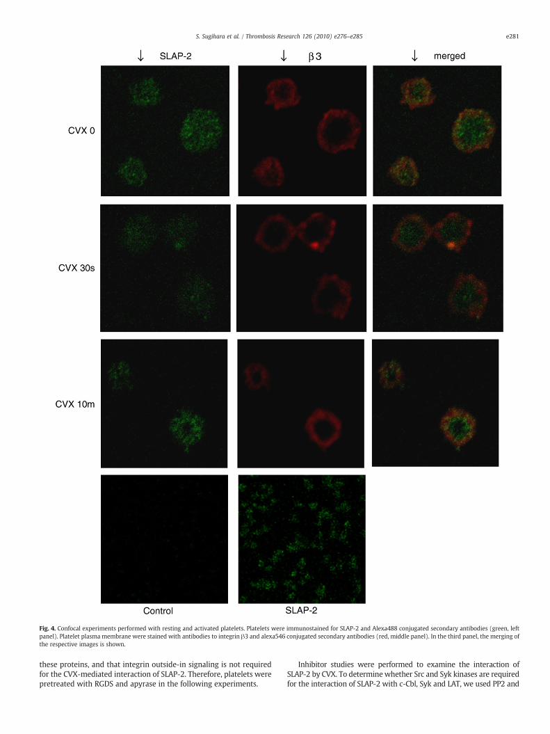
Fig. 4. Confocal experiments performed with resting and activated platelets. Platelets were immunostained for SLAP-2 and Alexa488 conjugated secondary antibodies (green, leftpanel). Platelet plasma membrane were stained with antibodies to integrin β3 and alexa546 conjugated secondary antibodies (red, middle panel). In the third panel, the merging ofthe respective images is shown.
e281S. Sugihara et al. / Thrombosis Research 126 (2010) e276–e285
these proteins, and that integrin outside-in signaling is not requiredfor the CVX-mediated interaction of SLAP-2. Therefore, platelets werepretreated with RGDS and apyrase in the following experiments.
Inhibitor studies were performed to examine the interaction ofSLAP-2 by CVX. To determinewhether Src and Syk kinases are requiredfor the interaction of SLAP-2 with c-Cbl, Syk and LAT, we used PP2 and

Fig. 5. SLAP-2 associates with tyrosine-phosphorylated c-Cbl, Syk and LAT following GPVI activation in platelets. (a) SLAP-2 associate with tyrosine-phospholylated proteinsfollowing GPVI activation. Purified immobilized GST fusion proteins with SLAP-2 were incubated with lysates from human platelets that were either unstimulated (-) orstimulated (+) with CVX. Immunoblotting was performed with the indicated antibodies. (b) Effect of apyrase and RGDS peptide on association of SLAP-2 with tyrosinephospholylated proteins. Lysates from platelets pretreated with 4U/ml apyrase and 100 μg/ml RGDS peptide for 15 minutes at 37 °C and unstimulated (-) or stimulated (+)with CVX were incubated with GST fusion protein with SLAP-2. Immunoblotting was performed with the indicated antibodies.
e282 S. Sugihara et al. / Thrombosis Research 126 (2010) e276–e285
piceatannol to inhibit Src and Syk-kinases, respectively, whereinhibition of platelet aggregation induced by CVX was observed (datanot shown). PP2 completely inhibited both tyrosine phosphorylation ofc-Cbl, Syk and LAT, and interaction of SLAP-2 with these proteins(Fig. 6a-d). PP2 also inhibited the p32 SLAP-2 production with CVX andthrombin stimulation (data not shown). On the other hand, Syk kinaseinhibitor Piceatannol had no effect on interaction of SLAP-2 with theseproteins. PI3-kinase plays a central role in the regulation of manycellular event, including recruitment of various proteins to cellmembrane and regulation of the activity of a number of tyrosinekinases [3]. Pretreatment of platelets with wortmannin, whichinactivates the catalytic p110 subunit of PI3-kinase, inhibited plateletaggregation induced by CVX [25]. However this treatment did not affectthe interaction of SLAP-2 with c-Cbl, Syk and LAT. Interestingly,piceatanol and wortmannin inhibited the p32 SLAP-2 production withCVX stimulation and translocation to cytoskeletal fraction. These resultsdemonstrated that association of SLAP-2 with these proteinsthrough the GPVI activation is regulated predominantly downstreamof Src-family kinases, and upstream of Syk kinases and PI3-kinase.
In order to determine whether interaction of SLAP-2 with theseproteins is a general phenomenon downstream of all platelet agonist,we stimulated platelets with thrombin (Fig. 7). SLAP-2 associated withsome tyrosine phosphorylated proteins. However, in contrast to GPVI-dependent agonists, immunoblotting experiment identified the pro-teins other thanc-Cbl, Sykand LAT. This data suggest that thrombindoesnot cause the association of SLAP-2 with these proteins.
Discussion
In the present study, we have investigated the potential role ofSLAP-2 in regulation of signaling by the GPVI in human platelets.
Human SLAP-2 is predominantly expressed in leukocytes, lymphnodes, spleen, thymus, lung [16,17], and platelets (M.Tomlinson andS.P.W., July 2006, unpublished). We have now demonstrated that twoprotein isoforms of SLAP-2, p28 and p25, are expressed in humanplatelets. These isoforms are products of alternative translationinitiation, the smaller isoform lacking both the myristoylationsequence and serine-rich region present at the amino terminus ofthe long isoform [18,23]. The p28 and p25 isoforms have been shownto be differentially localized to the membrane and cytoplasm,respectively [18]. The appearance of a higher molecular mass speciesof SLAP-2 (P32) following CVX stimulation suggested that SLAP-2becomes phosphorylated in response to activation of GPVI. This ideawas supported by the finding that protein phosphatase treatment ofanti-SLAP-2 immunoprecipitates of SLAP-2 from CVX-stimulatedplatelets resulted in the disappearance of the P32 SLAP-2 and aconcomitant increase of the smaller SLAP-2 species. Anti-phosphotyr-osine western blotting of anti-SLAP-2 immunoprecipitates failed toreveal CVX-induced tyrosine phosphorylation of SLAP-2. This findingsuggested that SLAP-2 became phosphorylated on serine and/orthreonine residues in response to CVX stimulation. Anti-phosphoser-ine and phoshothreoninmight not be sensitive enough to confirm thisphosphorylation by Western blotting. A recent study by Loreto et al.[18] suggested that SLAP-2 undergoes phosphorylation when tran-siently expressed in COS cells. In addition, SLAP-2 is phosphorylated inserine residues [20] or tyrosine residues [19] in response to activationof the CSF-1 receptor when ectopically expressed in bone marrowmacrophages or FD-Fms cells. However, this is the first report todescribe receptor-induced phosphorylation of endogenous SLAP-2.The increase of p32 isoform that appeared following CVX stimulationwas parallel with the decrease of p28, while the p25 isoform of SLAP-2which lacks the myristoylation sequence slightly decreased. These

blot:anti-pYPP2
150
100
75
50
37
Piceatannol
37
100
75
150
50
Wortmannin
(a)
100
75
50
37
CVX GST SLAP-2 WCL GST SLAP-2 WCL
GST SLAP-2 WCL
GST SLAP-2 WCL
- + - + - +
blot:anti-Syk
blot:anti-c-Cbl
PP2
100
150Piceatannol
75
Wortmannin
PP2
Piceatannol
Wortmannin
100
150
75
100
150
75
(b)
(c)
CVX - + - + - +
CVX - + - + - +
PP2
Piceatannol
Wortmannin
37
37
blot:anti-LAT
37
(d)CVX - + - + - +
Fig. 6. Effect of inhibition of Src kinase, Syk kinase and PI3-kinase on association of SLAP-2with c-Cbl, Syk and LAT.Washed platelets were treatedwith 100 ng/ml of CVXwith orwithoutPP2 (Src kinase inhibitor, 10 μM), piceatannol (Syk kinase inhibitor, 50 μM), and wortmannin (PI3-kinase inhibitor, 50nM) and incubated with GST fusion protein with SLAP-2.Immunoblotting was performed with the indicated antibodies. (a) anti-phosphotyrosine antibodies. (b) anti-c-Cbl antibodies. (c) anti-Syk antibodies. (d) anti-LAT antibodies.
e283S. Sugihara et al. / Thrombosis Research 126 (2010) e276–e285
data suggests that p32 arises from modification of p28, but not p25,and that the sequence present only at p28 is participates inphosphorylation of SLAP-2. It was in agreement with previous reportsthat the phosphorylation site in SLAP-2 was likely to reside within itsN-terminus [18,20]. However, the increase of no less than 4 kDa ofmolecular mass could not be caused by phosphorylation alone. Thisphosphorylation may induce a conformational change in SLAP-2 thatalters its electrophoretic mobility or trigger another modification ofSLAP-2.
Various proteins in signal transduction pathways are myristoy-lated. In general, N-myristoylation is an irreversible protein modifi-cation that occurs co-translationally following removal of the initiatormethionine residue by cellular methionylaminopeptidases [26]. N-myristoylation promotes weak and reversible protein-membrane andprotein-protein interactions [27]. Many N-myristoylated proteins aremembrane bound, and can be found in the plasmamembrane or otherintracellular membranes in eukaryotic cells. The binding energyprovided by myristate is relatively weak and not sufficient to fully
anchor a peptide or protein to a cellular membrane [27]. A secondsignal within the N-myristoylated protein is therefore required forefficient membrane binding [28]. This signal can be provided inseveral ways such as a polybasic domain or palmitate. A cluster ofbasic residues can provide electrostatic interactions with acidicmembrane phospholipids. Palmitoylation is reversible, providing anadditional hydrophobic interaction with the bilayer. Moreover, aconformational change of a protein regulates exposure of themyristate moiety. The transition between these two states isregulated by a mechanism known as the ‘myristoyl switch’. Thereforemyristoyl switch is mechanism for reversible membrane binding [28].The presence of myristoylation sites within N-terminal of SLAP-2suggests a potential mechanism for reversing its association withmembranes. The proteins that are dually fatty acylated with myristateand palmitate nearly all contain the consensus sequence Met-Gly-Cysat their N-terminal [28]. SLAP-2 does not contain this palmitate motif,so the association of SLAP-2 with the plasma membrane is notregulated by palmitoylation. It is unclear whether the conformation of

Fig. 7. SLAP-2 did not associate with c-Cbl, Syk and LAT in thrombin-actibated platelets.Purified immobilized GST fusion proteins with SLAP-2 were incubated with lysatesfrom human platelets that were either unstimulated (-) or stimulated (+) with 0.2U/ml of thrombin. Immunoblotting was performed with the indicated antibodies.
e284 S. Sugihara et al. / Thrombosis Research 126 (2010) e276–e285
SLAP-2 is changed, and therefore, it participates in the membranebinding or not. On the other hands, the N-terminus of SLAP-2 containsserine-rich region in positively charge. Phosphorylation of serinereduces the net charge of the basic cluster, altering its electrostaticpotential and weakening its electrostatic binding to membranescontaining acidic lipids. We showed that a fraction of SLAP-2translocated to the Triton X-100-insoluble cytoskeletal fractionupon CVX stimulation of platelets. These data lead us to put forwardthe following hypothesis. In resting platelets, SLAP-2 exists mainly incytoplasm. SLAP-2 can be attached to cytoskeleton with its myristate,but easily detached without second switch. Following activation ofplatelets by CVX stimulation, phosphorylation of the N-terminalserine works as a second switch, and results in redistribution of SLAP-2 to the Triton X-100-insoluble fraction(cytoskeleton). We could notdetect the direct binding of SLAP-2 to actin by immunofluorescencestudy (data not shown), SLAP-2 may associate with some othercytoskeletal proteins or with actin indirectly. Upon activation, a lot ofstructural proteins and signaling molecules are recruited to thecytoskeleton [25,29–33]. Our results now show that SLAP-2 is also oneof the components to associate to the cytoskeleton in response to CVXstimulation and facilitate signal transductions. Signaling by CVXinvolves rapid tyrosine phosphorylation of a number of proteinsincluding Src, Syk and PI3-kinase [3]. The inhibitors of thesemoleculesinhibited CVX-induced cytoskeletal translocation of SLAP-2. At thesame time, these inhibitors inhibited CVX-induced phosphorylation ofSLAP-2. These results are consistent with the hypothesis thatphosphorylation of SLAP-2 results in translocation of SLAP-2 fromthe membrane to the cytoskeleton. However, RGDS blocked theassociation of SLAP-2 with the cytoskeleton in CVX-induced platelets,although it did not inhibit phosphorylation of SLAP-2. Thus, any stepin the CVX signaling leading to GP IIb/IIIa activation is thought to becritical for CVX-induced interaction of SLAP-2 with cytoskeleton.
A function of SLAP-2 in lymphocytes is to negatively regulateantigen receptor signaling, most probably by facilitating the c-Cbl-directed ubiquitination of target proteins [16–18]. By analogy, SLAP-2could potentially regulate GPVI signaling negatively by promoting the
c-Cbl-directed ubiquitination of the GPVI and/or downstream signal-ing proteins. Consistent with studies in lymphocytes [18], our in vitrobinding assays with GST fusion proteins of SLAP-2 revealed that SLAP-2 was capable of associating with c-Cbl and Syk, in place of Syk familytyrosine kinase ZAP-70, following GPVI stimulation. In T cells, ZAP-70is tyrosine phosphorylated and binds to SLAP-2 following TCRstimulation [18]. In the process of c-Cbl-dependent downregulationof TCR, ZAP-70 associates with and is negatively regulated by c-Cbl[34]. In platelets, Syk is tyrosine phosphorylated, associatedwith c-Cbland ubiquitinated in platelets activated with agonists that activate theGPVI dependent pathway [14]. The function of Syk in platelets maytherefore be similar to that of ZAP-70 in T cell activation. On the otherhand, we have observed an inducible association between SLAP-2 andtransmembrane adaptor protein LAT, as was not previously reportedin T cells [18], although the significance of this is unclear.Interestingly, in T cells, SLAP, but not SLAP-2, appears to induciblyassociate with LAT following anti-CD3 stimulation [35,36]. Theinteraction of SLAP-2 and these proteins have several potentialfunctions in cellular signaling. Adapter proteins form an intracellularscaffold that regulates and targets effector proteins to appropriateregions of the cell [3]. The function of the adaptor-cytoskeletoninteraction is thought to be to bring the effector protein into closeproximity to their molecular substrates that are involved in the signalevents. Thus, if SLAP-2 is associated with components of thecytoskeleton in platelets, this might be important in bringing theeffector protein into proximity with cytoskeletal substrates. Followingstimulation of human platelets with CVX, SLAP-2 interacts with c-Cbl,Syk and LAT, and subsequently they translocate to the cytoskeleton.Therefore, SLAP-2 may play a role in promoting signaling pathway bybringing these signaling proteins to the cytoskeletal substrates.
We have reported in the present study that SLAP-2 is expressed inhuman platelets and has potentially functions as a novel componentof a GPVI regulated signaling pathway. Indeed, activation of the GPVIinduces phosphorylation of SLAP-2 and interaction with some signalproteins. Thus further studies that establish the specific role of SLAP-2in signaling by the GPVI are required.
Conflict of interest statement
None of the authors have any conflicts of interest associated withthis study.
Acknowledgements
This work was carried out at the Analysis Center of Life Science,Hiroshima University.
References
[1] Nieswandt B, Watson SP. Platelet-collagen interaction: is GPVI the centralreceptor? Blood 2003;102:449–61.
[2] Asazuma N,Wilde JI, Berlanga O, Leduc M, Leo A, Schweighoffer E, et al. Interactionof linker for activation of T cells with multiple adapter proteins in plateletsactivated by the glycoprotein VI-selective ligand, convulxin. J Biol Chem 2000;275:33427–34.
[3] Watson SP, Auger JM, McCarty OJ, Pearce AC. GPVI and integrin alphaIIb beta3signaling in platelets. J Thromb Haemost 2005;3:1752–62.
[4] Gibbins JM, Okuma M, Farndale R, Barnes M, Watson SP. Glycoprotein VI is thecollagen receptor in platelets which underlies tyrosine phosphorylation of the Fcreceptor gamma-chain. FEBS Lett 1997;413:255–9.
[5] Berlanga O, Bobe R, Becker M, Murphy G, Leduc M, Bon C, et al. Expression of thecollagen receptor glycoprotein VI during megakaryocyte differentiation. Blood2000;96:2740–5.
[6] Watson SP, Asazuma N, Atkinson B, Berlanga O, Best D, Bobe R, et al. The role ofITAM- and ITIM-coupled receptors in platelet activation by collagen. ThrombHaemost 2001;86:276–88.
[7] Pasquet JM, Gross B, Quek L, Asazuma N, Zhang W, Sommers CL, et al. LAT isrequired for tyrosine phosphorylation of phospholipase cgamma2 and plateletactivation by the collagen receptor GPVI. Mol Cell Biol 1999;19:8326–34.
[8] Swaminathan G, Tsygankov AY. The Cbl family proteins: ring leaders in regulationof cell signaling. J Cell Physiol 2006;209:21–43.

e285S. Sugihara et al. / Thrombosis Research 126 (2010) e276–e285
[9] Thien CB, LangdonWY. Cbl: many adaptations to regulate protein tyrosine kinases.Nat Rev Mol Cell Biol 2001;2:294–307.
[10] Murphy MA, Schnall RG, Venter DJ, Barnett L, Bertoncello I, Thien CB, et al. Tissuehyperplasia and enhanced T-cell signalling via ZAP-70 in c-Cbl-deficient mice. MolCell Biol 1998;18:4872–82.
[11] Rao N, Lupher Jr ML, Ota S, Reedquist KA, Druker BJ, Band H. The linkerphosphorylation site Tyr292 mediates the negative regulatory effect of Cbl onZAP-70 in T cells. J Immunol 2000;164:4616–26.
[12] Auger JM, Best D, Snell DC, Wilde JI, Watson SP. c-Cbl negatively regulates plateletactivation by glycoprotein VI. J Thromb Haemost 2003;1:2419–26.
[13] Polgar J, Clemetson JM, Kehrel BE, Wiedemann M, Magnenat EM, Wells TN, et al.Platelet activation and signal transduction by convulxin, a C-type lectin fromCrotalus durissus terrificus (tropical rattlesnake) venom via the p62/GPVIcollagen receptor. J Biol Chem 1997;272:13576–83.
[14] Dangelmaier CA, Quinter PG, Jin J, Tsygankov AY, Kunapuli SP, Daniel JL. Rapidubiquitination of Syk following GPVI activation in platelets. Blood 2005;105:3918–24.
[15] Saci A, Rendu F, Bachelot-Loza C. Platelet alpha IIb-beta 3 integrin engagementinduces the tyrosine phosphorylation of Cbl and its association with phosphoi-nositide 3-kinase and Syk. Biochem J 2000;3:669–76 351 Pt.
[16] Holland SJ, Liao XC, Mendenhall MK, Zhou X, Pardo J, Chu P, et al. Functionalcloning of Src-like adapter protein-2 (SLAP-2), a novel inhibitor of antigenreceptor signaling. J Exp Med 2001;194:1263–76.
[17] Pandey A, Ibarrola N, Kratchmarova I, FernandezMM, Constantinescu SN, Ohara O,et al. A novel Src homology 2 domain-containing molecule, Src-like adapterprotein-2 (SLAP-2), which negatively regulates T cell receptor signaling. J BiolChem 2002;277:19131–8.
[18] Loreto MP, Berry DM, McGlade CJ. Functional cooperation between c-Cbl and Src-like adaptor protein 2 in the negative regulation of T-cell receptor signaling. MolCell Biol 2002;22:4241–55.
[19] Pakuts B, Debonneville C, Liontos LM, Loreto MP, McGlade CJ. The Src-like adaptorprotein 2 regulates colony-stimulating factor-1 receptor signaling and down-regulation. J Biol Chem 2007;282:17953–63.
[20] Manes GA, Masendycz P, Nguyen T, Achuthan A, Dinh H, Hamilton JA, et al. Apotential role for the Src-like adapter protein SLAP-2 in signaling by the colonystimulating factor-1 receptor. FEBS J 2006;273:1791–804.
[21] Jung SM, Moroi M. Signal-transducing mechanisms involved in activation of theplatelet collagen receptor integrin alpha(2)beta(1). J Biol Chem 2000;275:8016–26.
[22] Phillips DR, Jennings LK, Edwards HH. Identification of membrane proteinsmediating the interaction of human platelets. J Cell Biol 1980;86:77–86.
[23] Loreto MP, McGlade CJ. Cloning and characterization of human Src-like adaptorprotein 2 and a novel splice isoform, SLAP-2-v. Oncogene 2003;22:266–73.
[24] Fox JE, Boyles JK, Berndt MC, Steffen PK, Anderson LK. Identification of amembrane skeleton in platelets. J Cell Biol 1988;106:1525–38.
[25] Lu Q, Clemetson JM, Clemetson KJ. Translocation of GPIb and Fc receptor gamma-chain to cytoskeleton in mucetin-activated platelets. J Thromb Haemost 2005;3:2065–76.
[26] Wolven A, Okamura H, Rosenblatt Y, Resh MD. Palmitoylation of p59fyn isreversible and sufficient for plasma membrane association. Mol Biol Cell 1997;8:1159–73.
[27] Peitzsch RM, McLaughlin S. Binding of acylated peptides and fatty acids tophospholipid vesicles: pertinence to myristoylated proteins. Biochemistry1993;32:10436–43.
[28] Resh MD. Fatty acylation of proteins: new insights into membrane targeting ofmyristoylated and palmitoylated proteins. Biochim Biophys Acta 1999;1451:1–16.
[29] Clark EA, Brugge JS. Redistribution of activated pp 60c-src to integrin-dependentcytoskeletal complexes in thrombin-stimulated platelets. Mol Cell Biol 1993;13:1863–71.
[30] Fox JE. Cytoskeletal proteins and platelet signaling. Thromb Haemost 2001;86:198–213.
[31] Tohyama Y, Yanagi S, Sada K, Yamamura H. Translocation of p72syk to thecytoskeleton in thrombin-stimulated platelets. J Biol Chem 1994;269:32796–9.
[32] Fox JE, Lipfert L, Clark EA, Reynolds CC, Austin CD, Brugge JS. On the role of theplatelet membrane skeleton in mediating signal transduction. Association of GPIIb-IIIa, pp 60c-src, pp62c-yes, and the p21ras GTPase-activating protein with themembrane skeleton. J Biol Chem 1993;268:25973–84.
[33] Horvath AR, Muszbek L, Kellie S. Translocation of pp 60c-src to the cytoskeletonduring platelet aggregation. EMBO J 1992;11:855–61.
[34] Naramura M, Kole HK, Hu RJ, Gu H. Altered thymic positive selection andintracellular signals in Cbl-deficient mice. Proc Natl Acad Sci USA 1998;95:15547–52.
[35] Sosinowski T, Pandey A, Dixit VM, Weiss A. Src-like adaptor protein (SLAP) is anegative regulator of T cell receptor signaling. J Exp Med 2000;191:463–74.
[36] Tang J, Sawasdikosol S, Chang JH, Burakoff SJ. SLAP, a dimeric adapter protein,plays a functional role in T cell receptor signaling. Proc Natl Acad Sci USA 1999;96:9775–80.