NHE1 promotes invadopodial ECM proteolysis through acidification of the peri-invadopodial space

2727Research Article
IntroductionInvasion and metastasis are hallmarks of tumour progression andthe major cause of cancer mortality, but the underlying mechanismsof these devastating processes are complex and poorly understood.Minimally, for a tumour to disseminate, malignant cells must detachfrom the primary lesion, acquire a motile phenotype and penetratethe ECM of basement membranes and stroma which physicallyseparate the tumour from the circulation (Condeelis and Segall,2003; Friedl and Wolf, 2003). Depending on matrix density, tumourcells may exploit proteolytic and non-proteolytic mechanisms tobreach ECM barriers (Friedl and Wolf, 2003; Friedl and Wolf,2008). Destruction of ECM components is facilitated by secretedand membrane-bound extracellular proteases, including matrixmetalloproteases (MMPs) and serine proteases actingindependently or in proteolytic cascades (Basbaum and Werb, 1996;Linder, 2007).
In many tumour cells, enzymes responsible for matrixdegradation are targeted to dynamic F-actin-enriched structurestermed invadopodia, which are closely related molecularly andfunctionally to ventral adhesive structures called podosomes thatare formed by several normal cell types (e.g. osteoclasts) as wellas Src-transformed fibroblasts (Weaver, 2006; Linder, 2007; Gimonaet al., 2008). Formation of invadopodia is enhanced by matrixrigidity (Alexander et al., 2008), some growth factors (Tague et al.,2004; Yamaguchi et al., 2005) or expression of wild-type oractivated forms of Src tyrosine kinase (Chen et al., 1984; Artym etal., 2006). Numerous proteins and a subset of phosphoinositidesare recruited to invadopodia and have roles in processes such asECM degradation and adhesion, signal transduction, actin network
assembly and membrane remodelling (Artym et al., 2006; Weaver,2006; Linder, 2007). Extensive molecular cross-talk in invadopodiais suggested by the fact that depletion, inhibition or sequestrationof individual constituents is usually sufficient to abrogateinvadopodia formation (Bowden et al., 1999; Baldassarre et al.,2003; Chuang et al., 2004; Hashimoto et al., 2004; Tague et al.,2004; Onodera et al., 2005; Seals et al., 2005; Yamaguchi et al.,2005; Artym et al., 2006; Clark et al., 2007; Oikawa et al., 2008).Consistent with this, several physical interactions betweeninvadopodia-localized proteins are critical for invadopodia function,such as those involving cortactin and AMAP1 (also known asASAP1) (Onodera et al., 2005) and N-WASP and WIP (Yamaguchiet al., 2005) among others. Live cell studies also show thatinvadopodia assemble sequentially. For example, recruitment ofcortactin precedes and is necessary for membrane type 1(MT1)-MMP accumulation at invadopodia and ensuing matrix degradation(Artym et al., 2006; Clark et al., 2007).
Several Src substrates are phosphorylated in invadopodiaincluding cortactin (Ayala et al., 2008), AMAP1 (Bharti et al., 2007),paxillin (Badowski et al., 2008) and p130Cas (Alexander et al.,2008). Moreover, tyrosine phosphorylation of some of these proteinsappears to be critical for their functional activity in regulatinginvadopodia and/or podosome dynamics and organization. Tks5(also known as SH3PXD2A; formerly FISH) is a large adaptormolecule containing a phox homology (PX) and five SH3 domains(Lock et al., 1998). Although Tks5 was identified as a Src substrateand is highly phosphorylated in Src-transformed cells, thesignificance of Tks5 phosphorylation is unknown. Tks5 localizesin and its expression is critical for podosome formation as well as
Invadopodia are actin-based projections enriched withproteases, which invasive cancer cells use to degrade theextracellular matrix (ECM). The Phox homology (PX)-Srchomology (SH)3 domain adaptor protein Tks5 (also known asSH3PXD2A) cooperates with Src tyrosine kinase to promoteinvadopodia formation but the underlying pathway is not clear.Here we show that Src phosphorylates Tks5 at Y557, inducingit to associate directly with the SH3-SH2 domain adaptorproteins Nck1 and Nck2 in invadopodia. Tks5 mutants unableto bind Nck show reduced matrix degradation-promotingactivity and recruit actin to invadopodia inefficiently.Conversely, Src- and Tks5-driven matrix proteolysis and actinassembly in invadopodia are enhanced by Nck1 or Nck2
overexpression and inhibited by Nck1 depletion. We show thatclustering at the plasma membrane of the Tks5 inter-SH3 regioncontaining Y557 triggers phosphorylation at this site, facilitatingNck recruitment and F-actin assembly. These results identify aSrc-Tks5-Nck pathway in ECM-degrading invadopodia thatshows parallels with pathways linking several mammalian andpathogen-derived proteins to local actin regulation.
Supplementary material available online athttp://jcs.biologists.org/cgi/content/full/122/15/2727/DC1
Key words: Invadopodia, Podosome, Src, Tks5, Nck, ECMdegradation
Summary
Nck adaptor proteins link Tks5 to invadopodia actinregulation and ECM degradationStanley S. Stylli1, Stacey T. T. I1, Anne M. Verhagen2, San San Xu1, Ian Pass3, Sara A. Courtneidge3 andPeter Lock1,*,‡
1Department of Surgery, University of Melbourne, Level 5, Clinical Sciences Building, Royal Melbourne Hospital, Parkville, Victoria 3052, Australia2Walter and Eliza Hall Institute of Medical Research, Parkville, Victoria 3052, Australia3Burnham Institute for Medical Research, Torrey Pines Road, La Jolla, CA 92037, USA*Present address: Biochemistry Department, La Trobe University, Victoria 3086, Australia‡Author for correspondence (e-mail: [email protected])
Accepted 5 May 2009Journal of Cell Science 122, 2727-2740 Published by The Company of Biologists 2009doi:10.1242/jcs.046680
Jour
nal o
f Cel
l Sci
ence
JCS ePress online publication date 13 July 2009
2728
the invasive potential of Src-transformed cells and several humantumour cell lines (Abram et al., 2003; Seals et al., 2005).Recruitment of Tks5 to podosomes is facilitated by the PX domain,which binds the phosphoinositides, PtdIns(3)P and PtdIns(3,4)P2
in vitro (Abram et al., 2003; Oikawa et al., 2008). In keeping withthis, PtdIns(3,4)P2 accumulates in the early phases of podosomeformation, in a Src-, PI 3-kinase- and synaptojanin-2-dependentmanner, preceding Tks5 recruitment and actin assembly (Oikawaet al., 2008; Symons, 2008). Consistent with a role as an adaptorprotein Tks5 can interact with multiple proteins found in podosomesincluding ADAM-family metalloproteases (Abram et al., 2003), theadhesion receptor, dystroglycan (Thompson et al., 2008), theSH3/SH2 adaptor, Grb2 and the nucleation promoting factor, N-WASP (Oikawa et al., 2008). Although it is not clear if theseinteractions occur simultaneously or sequentially or via direct orindirect associations, collectively these results suggest that Tks5has a central role in invadopodia and podosome regulation. Here,we explored the mechanism by which Tks5 facilitates invadopodiaformation in tumour cells.
ResultsTks5 synergises with Src to promote invadopodia and matrixproteolysisA clear functional link between Src and Tks5 in podosome andinvadopodia regulation has been demonstrated (Seals et al., 2005;Oikawa et al., 2008) but the extent to which these proteins cooperateto stimulate ECM degradation has not been quantified. To examinethis, we first compared the abilities of metastatic B16F10 melanomacells transiently expressing wild-type or activated Src (Src and SrcY527F) and/or FLAG-epitope-tagged Tks5 to degrade matrices ofFITC-labelled gelatin during a 24-hour period. B16F10 cells wereselected for this analysis because they express relatively high levelsof endogenous Tks5, form robust invadopodia enriched with bothTks5 and F-actin (supplementary material Fig. S1A,B) and exhibita reproducibly high transfection efficiency (40-60%, based on GFPmarker studies; data not shown). In the 24-hour matrix degradationassay used, the area of cleared gelatin, normalized for cell number,was determined in multiple independent microscope fields. Valuesare expressed as mean degradation area (μm2)/cell and provide ameasure of the average matrix degradation activity of a cell in thesample population, irrespective of transfection status, cellmovements or individual activities of cells during the assay. Zonesof gelatin degradation are therefore not assigned to specific cellsas in some other assays. The average levels of transiently expressedand endogenous proteins are assessed in parallel by immunoblotanalysis.
As shown in Fig. 1A-C, the basal matrix degradation activity ofthe parental B16F10 cells was not affected by transfection with anempty vector but was boosted approximately three- and fivefold incells expressing Src and Src Y527F, respectively. Expression of N-or C-terminal FLAG-Tks5 alone also resulted in significant (sixfold)enhancement of matrix degradation, whereas coexpression of Tks5proteins in the presence of wild-type or active Src, resulted in super-additive increases in matrix degradation (up to 15- and 17-fold,respectively). We observed a similar functional cooperativitybetween Src and Tks5 in MDA-MB-231 breast carcinoma cells(supplementary material Fig. S1C,D). Expression of Src or SrcY527F in these cells has been reported to enhance invadopodiaformation and overall matrix degradation (Artym et al., 2006). Inour hands, expression of Src Y527F and, to a greater extent, N-FLAG-Tks5, enhanced the basal matrix degradation activity of
MDA-MB-231 cells, whereas coexpression of Src Y527F and Tks5produced a synergistic increase in matrix degradation.
The effect of reduced Tks5 expression on matrix degradationwas also tested in parental and Src Y527F-expressing B16F10 cellsusing RNAi. Endogenous Tks5 levels were strongly suppressedby Tks5 siRNA treatment and coinciding with this, basal andSrc-induced gelatin degradation was markedly diminished(supplementary material Fig. S1E,F). These results support earlierwork showing that Tks5 is needed for podosome formation andmatrix degradation in Src-transformed fibroblasts and tumour cells(Seals et al., 2005).
To confirm that the effects of altered Tks5 levels on Src-drivenmatrix degradation are related to invadopodia formation, we useda 4-hour matrix degradation assay to compare the functionalactivity and subcellular localization of a full-length Tks5-GFP fusionprotein versus GFP in Src Y527F-expressing B16F10 cells. Unlikethe 24-hour matrix degradation assay, only degradation associatedwith individual GFP-positive cells was analysed. Whereas Tks5-GFP colocalized extensively with Src in invadopodia of the SrcY527F-expressing cells, GFP was distributed throughout the nucleusand cytoplasm of cells and not recruited to invadopodia (Fig. 1E).Correlating with Tks5-GFP recruitment to invadopodia andconfirming its biological activity, Tks5-GFP expression significantlyenhanced the matrix degradation activity of cells compared withGFP alone (Fig. 1F).
Identification of Nck1/2 in association with Tks5We postulated that Tks5 is dependent on interactions with cellulartarget proteins for its activity and that such interactions may be Srcdependent. The isolated SH3 and PX domains of Tks5 have beenused previously as affinity reagents to identify Tks5-interactingproteins (Abram et al., 2003; Seals et al., 2005; Oikawa et al., 2008).However, a potential disadvantage of this approach is that itexcludes the ‘linker’ regions between the PX and SH3 domains(supplementary material Fig. S2A), which contain numerouspolyproline motifs and suspected phosphorylation sites that couldpotentially interface with other proteins (Lock et al., 1998). Toidentify proteins in association with intact Tks5, full length N- andC-FLAG-Tks5 were coexpressed with Src in 60�15 cm dishes of293T cells (supplementary material Fig. S2A). A Tks5 variantlacking the PX domain, FLAG-ΔPX-Tks5, was also included, basedon evidence that the PX domain interferes with SH3 domain binding(Abram et al., 2003). 293T cells lack endogenous Tks5 as assessedby immunoblot analysis (not shown). FLAG-Tks5 complexes werepurified from cell lysates by binding to immobilized FLAGantibody, then eluted with a FLAG peptide, resolved by SDS-PAGEand stained with Sypro Ruby (supplementary material Fig. S2B)and proteins identified by mass spectrometry (supplementarymaterial Table S1).
A number of candidate proteins with known or potential roles ininvadopodia, actin regulation or tyrosine kinase signalling appearedto associate specifically with Tks5 (supplementary material TableS1) but not with Tks5-unrelated FLAG-tagged proteins screenedseparately (A.M.V., unpublished data). Among them, were cortactin,Nck1, Nck2, Grb2, CrkL, drebrin and SH3P7. In co-immunoprecipitation assays designed to validate some of theseinteractions, however, we could not confirm a stable interaction ofTks5 with cortactin, CrkL (Fig. 2A) or SH3P7 (data not shown).The reasons for this are unclear but may be related to the highsensitivity of the mass spectrometry versus the relatively highstringency of the co-immunoprecipitation assay. We note that Grb2
Journal of Cell Science 122 (15)
Jour
nal o
f Cel
l Sci
ence

2729A Src-Tks5-Nck pathway in invadopodia
was identified as a Tks5-binding protein recently (Oikawa et al.,2008).
In contrast to the other candidate proteins tested we found thatendogenous Nck co-immunoprecipitated readily with each of theFLAG-Tks5 bait proteins (Fig. 2A,D), associating preferentiallywith Tks5 isolated from Src-expressing cells and correlating withincreased Tks5 tyrosine phosphorylation. We also noticed that thePX domain of Tks5 appears to be inhibitory for Nck binding:significantly more Nck co-immunoprecipitated with FLAG-ΔPX-Tks5 than with either N-FLAG-Tks5 (Fig. 2A,B) or C-FLAG-Tks5(not shown). The p47phox adaptor protein is a cytosolic subunit ofthe NADPH oxidase complex which shares a similar architecture(PX-SH3-SH3) and significant homology with the N-terminal PXdomain and first two SH3 domains of Tks5 (Lock et al., 1998).Structural and biochemical studies have suggested that the secondSH3 domain of p47phox, either alone or in tandem with the first
SH3 domain, can interact intramolecularly with proline-containingmotifs in the p47phox PX domain or C-terminus, respectively, andthereby promote an auto-inhibited conformation (Karathanassis etal., 2002; Ago et al., 2003; Groemping et al., 2003). On this basiswe examined the role of the second Tks5 SH3 domain in Nckbinding to Tks5. Inactivation of the SH3 domain by the substitution,W260R (Karathanassis et al., 2002), resulted in increased co-immunoprecipitation of Nck with N-FLAG-Tks5 to levels similarto those of FLAG-ΔPX-Tks5 (Fig. 2A). Quantification of Nck andphosphotyrosine immunoblots in two independent experimentsrevealed that the increased binding of Nck to both ΔPX-Tks5 andthe full-length W260R mutant relative to wild-type Tks5 wascorrelated to a large extent with increased Tks5 tyrosinephosphorylation (Fig. 2B). These data suggest that the PX andsecond SH3 domains of Tks5 may partially impede Tks5 tyrosinephosphorylation and Nck binding.
Fig. 1. Src and Tks5 induce invadopodiasynergistically. (A) Representativeconfocal microscopy images showingstrong induction of degradation (blackareas in bottom row) of FITC-gelatin films(green) by co-transfection of B16F10melanoma cells with Src and Tks5. F-actin(red) and nuclei (blue) were stained withTRITC-phalloidin and DAPI, respectively.pEFBOS, control vector. Scale bars:20 μm. (B) Immunoblot showing Tks5, Srcand active (Y416-P)-Src expression levelsin B16F10 cells transiently transfectedwith the indicated constructs (correspondsto groups shown in A). (C) Quantificationof the average matrix degradation activityof the indicated B16F10 transfectantsreveals that Src and Tks5 promote matrixdegradation cooperatively. Graph showsthe mean degradation area/cell + s.e.m.n=41 fields (>600 cells total)/group, threeexperiments; P<0.01 for all pairwisecomparisons except B16F10 versuspEFBOS and N- versus C-FLAG-Tks5.(D) Confocal images showing examples ofsingle B16F10 cells coexpressing SrcY527F (blue) and GFP or Tks5-GFP(green) 4 hours after seeding onRhodamine-gelatin (red). Arrows indicatecolocalization of Src and Tks5-GFP ininvadopodia. Scale bars: 10 μm.(E,F) Quantification of (E) the GFP punctacolocalizing with Src and (F) matrixdegradation reveals significant Tks5-GFPrecruitment to Src-containing invadopodiaand enhanced matrix degradation.Columns and error bars represent means +s.e.m. (n=10 cells/group). **P<0.01 forindicated comparisons.
Jour
nal o
f Cel
l Sci
ence

2730
Src phosphorylates Tks5 at Y557 and promotes NckassociationTks5 contains three candidate Src phosphorylation sites (Y552,Y557 and Y619; Fig. 3A) (Lock et al., 1998), two of which (Y552and Y557) conform to consensus Nck SH2 domain binding sites{Y(P)DE[P/V/D]} (Songyang et al., 1993; Jones et al., 2006). Y557and Y619 were confirmed to be Src phosphorylation sites intransfected Src-transformed NIH 3T3 cells (I.P., S.A.C. and P.L.,unpublished data). To determine the contribution of these tyrosinesto Nck binding, we generated FLAG-ΔPX-Tks5 constructs withY552F, Y557F or Y619F substitutions, expressed them with orwithout Src in 293T cells and performed co-immunoprecipitation
assays using a FLAG antibody. Endogenous Nck co-immunoprecipitated in a Src-inducible manner with wild-type andY552F and Y619F mutant forms of FLAG-ΔPX-Tks5 but did notassociate detectably with the Y557F mutant (Fig. 3B). Binding ofNck to full length C- and N-FLAG-Tks5 was also abolishedby Y557F substitutions (Fig. 3C). Additionally, co-immunoprecipitation of Nck with N-FLAG-Tks5 could becompetitively inhibited by pre-incubating lysates with aphosphopeptide containing the Nck binding site (Fig. 4A), but notby an unphosphorylated peptide of the same sequence, even at 15-fold higher concentrations (Fig. 3D). Together, these data show thatTks5 Y557 phosphorylation is critical for Nck binding.
Nck1 and Nck2 are closely related adaptor proteins containingthree SH3 domains and a single SH2 domain (Fig. 3E). They sharea broad and overlapping tissue distribution in the mouse embryoand appear to be functionally redundant in vivo, as mice deficientfor Nck1 or Nck2 are viable, whereas those lacking both isoformsdevelop with severe abnormalities and die in utero (Bladt et al.,2003). These studies suggest that Nck1 and 2 may be largelyinterchangeable. To map the region(s) of Nck that facilitate bindingto Tks5, HA-tagged human Nck1, Nck2 or Nck2 mutants withinactivated SH2 (Nck2 R311K) or SH3 domains (Nck2 W3K; Fig.3E) were expressed with Src and wild-type or mutant forms of N-FLAG-Tks5 in 293T cells. Like endogenous Nck (Fig. 3C), HA-Nck1 and HA-Nck2 co-precipitated efficiently with wild-type Tks5and were principally dependent for this interaction on the Y557residue of Tks5 (Fig. 3F). However, we did observe some relativelyweak binding of HA-Nck2 to Tks5 Y557F (Fig. 3F,G) althoughthis interaction was not evident for Nck2 W3K, indicating that theSH3 domains of Nck2 mediate this association (Fig. 3F). Consistentwith phosphorylated Y557 functioning as a docking site for the NckSH2 domain, its inactivation markedly inhibited HA-Nck2 bindingto wild-type N-FLAG-Tks5, although the much weaker (presumablySH3-mediated) interaction of Nck2 R311K with Tks5 Y557Fremained (Fig. 3G; NB, the immunoblot is overexposed todemonstrate the interaction of HA-Nck2 with Tks5 Y557F. Thisexposure also reveals binding of endogenous Nck1/2, exclusivelyto wild-type Tks5).
To further probe the requirements of the Tks5-Nck interactionwe performed an in vitro binding assay, incubating lysate from 293Tcells coexpressing wild-type or mutant forms of Tks5 (plus or minusactivated Src) with a GST-Nck1 SH2 domain fusion protein.Notably, the Nck1 and Nck2 SH2 domains show equivalentphosphopeptide binding specificities (Frese et al., 2006). SH2binding proteins were precipitated on glutathione and analysed byimmunoblotting with Tks5 and phosphotyrosine antibodies. Fig. 3Hshows that wild-type, Y552F and Y619F variants were efficientlyprecipitated in a Src-inducible manner by the SH2 domain whereas,the Y557F mutants were not (Fig. 3H). Immunoblot analysis witha phosphotyrosine antibody provided an internal control for theintegrity of the SH2 domain; endogenous 60 and 130 kDa proteinswere efficiently precipitated from lysate of cells expressing the Tks5Y557F mutants.
Lastly, we tested whether the Nck SH2 domain binds directly toTks5 by using a far western (overlay) assay (Nollau and Mayer,2001). Purified GST-Nck1 SH2 domain was incubated with theimmunoblot shown in Fig. 3C and was found to bind specificallyto immunoprecipitated C- and N-FLAG-Tks5 but did not bind thecorresponding Y557F variants. Significantly, the relative levels ofNck SH2 domain bound to C- and N-FLAG-Tks5 in the overlayassay are in close agreement with the amounts of Nck co-
Journal of Cell Science 122 (15)
Fig. 2. Association of Nck with Tks5 is stimulated by Src but suppressed bythe PX and second SH3 domains. (A) Co-immunoprecipitation andimmunoblot analysis of extracts of 293T cells expressing N-terminal FLAG-tagged ΔPX-, full length or W260R Tks5 reveals that endogenous Nck1/2 (butnot cortactin or CrkL) associate with all Tks5 variants in a Src-dependentmanner. Nck binding is further enhanced by PX domain removal or mutationof the second SH3 domain. IP, immunoprecipitation, WCL, whole cell lysate.pY, phosphotyrosine. (B) Densitometry analysis of relative Tks5 tyrosinephosphorylation and Nck binding (normalized for Tks5 levels), reveals thatΔPX and W260R mutants are more phosphorylated and bind Nck moreefficiently than N-FLAG-Tks5. Graphs show means + s.e.m. (n=2). Values forFLAG-ΔPX-Tks5 + Src were set arbitrarily at 1.
Jour
nal o
f Cel
l Sci
ence

2731A Src-Tks5-Nck pathway in invadopodia
Fig. 3. Phosphorylation at Y557, but notY552 or Y619, promotes a directinteraction between Tks5 and the NckSH2 domain. (A) Diagram of Tks5showing the location of candidatephosphorylation sites. (B).Immunoprecipitation analysis ofextracts of 293T cells expressingFLAG-ΔPX-Tks5 or the indicatedvariants, identifies Y557 as a criticaldeterminant of Src-induced Nckassociation. Re-probing for endogenousN-WASP shows that Y557 also enablesN-WASP association, but is notsufficient in the Y619F mutant to permitmaximal N-WASP binding.(C) Immunoprecipitation experimentshowing requirement by full lengthTks5 proteins for an intact Y557 residuefor Nck co-immunoprecipitation in293T cells. Overlay (far western)analysis with recombinant GST-Nck1SH2 domain reveals that Nck binds theY557 site directly. (D) Peptidecompetition experiment showing thatincubation of 293T lysates with a Tks5phosphopeptide based on the predictedNck binding site of human Tks5 (Fig.4A), but not its unphosphorylatedcounterpart, blocks Src-induced co-immunoprecipitation of endogenousNck with FLAG-Tks5. (E) Schematicdiagram of HA-Nck1, HA-Nck2 andNck2 variants with inactivating pointmutations in the SH2 (R311K) and allthree SH3 domains (W3K). HA tag(diamond) and point mutations (verticalbars) are indicated. (F) Co-immunoprecipitation experimentshowing that association of HA-Nck1and HA-Nck2 with Tks5 in 293T cellsrelies primarily on Tks5 Y557; a weakY557-independent interaction of Nck2with Tks5 requires the Nck2 SH3domains. Inexplicably, this interactionwas not apparent with another antibody(‘Nck1’) which also recognises Nck2.C, antibody from Cell SignalingTechnology; M, from Millipore. (G) Co-immunoprecipitation analysis showingthat Y557-dependent, but not-independent association of HA-Nck2with Tks5 requires an intact SH2domain. (H) Pull down assay usingrecombinant GST Nck1 SH2 domainand extracts of 293T cells expressingthe indicated proteins shows that Src-stimulated precipitation of Tks5 by theSH2 domain is Y557 dependent.
Jour
nal o
f Cel
l Sci
ence

2732
immunoprecipitated in each case. Collectively, these data indicatethat Nck associates directly with Tks5 and the primary determinantsof this interaction are the Y557 phosphorylation site and the Nck1/2SH2 domain.
Evidence that Src promotes a Tks5–Nck–N-WASP ternarycomplexN-WASP is critical for podosome assembly in Src-transformed cellsand EGF-induced invadopodia in mammary carcinoma cells(Mizutani et al., 2002; Yamaguchi et al., 2005; Co et al., 2007).Recently, N-WASP was identified as a Tks5 SH3-domain-bindingprotein and proposed to regulate Tks5-mediated actin assembly inpodosomes (Oikawa et al., 2008). As Nck can also interact with N-WASP via its SH3 domains and can activate N-WASP to stimulate
actin polymerization in vitro and in cells (Rohatgi et al., 2001; Riveraet al., 2004; Tehrani et al., 2007) we were interested to examinewhether the interaction of N-WASP with Tks5 is affected by Nckbinding. Reprobing the immunoblot shown in Fig. 3B revealed aSrc-dependent interaction of endogenous N-WASP with FLAG-ΔPX-Tks5. This interaction was abolished by mutation of Y557,the Nck binding site, but intriguingly, was also partially reducedby mutation of Y619 without a noticeable effect on Nck binding.Although these experiments utilise a mutant form of Tks5 lackingthe PX domain, this form of Tks5 contains all five SH3 domains.These data provide evidence that association of Tks5 with N-WASPis regulated by Y557 and Y619 phosphorylation and suggest thatN-WASP could associate indirectly with Tks5 at least in part, viaNck.
Tks5 is phosphorylated at Y557/8 in vivo and associates withendogenous NckTo assess whether Y557 and its counterpart in human Tks5 (Y558)are phosphorylation sites in vivo we raised an antibody against a17 residue phosphopeptide based on human Tks5 (Fig. 4A). Thehuman sequence differs from mouse Tks5 at the Y+2 position (Vin mouse Tks5, I in human Tks5). The affinity-purified antibody,p-Tks5, was judged to be specific as it recognisedimmunoprecipitated N-FLAG-Tks5, but not the Y557F variant inlysates from Src-expressing 293T cells and was minimally reactiveagainst Tks5 in the absence of Src co-expression (Fig. 4A).Furthermore, the antibody showed no reactivity towards anotherphosphorylated tyrosine in N-FLAG-Y557F detected with a generalphosphotyrosine antibody.
Analysis of endogenous Tks5 Y557/8 phosphorylation in mouseB16F10 and human MDA-MB-231 cells revealed that this site isminimally phosphorylated in unstimulated cells (Fig. 4B-D). AsTks5 tyrosine phosphorylation is responsive to cell treatment withthe actin polymerisation inhibitor cytochalasin D (Lock et al., 1998)we tested whether cytochalasin D or an unrelated actinpolymerisation inhibitor, latrunculin B, affected Tks5 Y557/8-specific phosphorylation. Both inhibitors enhanced Tks5 Y557/8
Journal of Cell Science 122 (15)
Fig. 4. Phosphorylation of endogenous Tks5 at Y557/8 and association withendogenous Nck are stimulated by inhibitors of actin polymerization andtyrosine phosphatases. (A) Tks5 phosphopeptide used to generate p-Tks5antibody. Immunoprecipitation experiment showing specific recognition byaffinity-purified p-Tks5 antibody of FLAG-Tks5 (but not Y557F) isolatedfrom 293T cells coexpressing Src. (B) Time course experiment showing Y557-specific phosphorylation of immunoprecipitated endogenous Tks5 is inducedin B16F10 cells with cytochalasin D (CytD) or latrunculin B (LatB) but notDMSO. (C,D) Immunoprecipitation experiments using (C) mouse B16F10 and(D) human MDA-MB-231 cells treated for 30 minutes with various inhibitorsshow that Tks5 Y557-specific (mouse) and Y558-specific (human)phosphorylation are boosted by cytochalasin D, latrunculin A and B andpervanadate. Concentrations used: latrunculin A (1 μM); latrunculin B (5 μM);swinholide A (0.1 μM); jasplakinolide (0.5 μM); brefeldin A (10 nM);nocodazole (5 μM); cytochalasin D (1 μM); wortmannin (0.1 μM);pervanadate (40 μM). Mouse and human Tks5 were immunoprecipitated with6G1 or 2F4 antibodies, respectively. PrG, Protein G.(E,F) Immunoprecipitation experiments using B16F10 cells grown for 3 hourson 0.1% gelatin and treated (+) or not treated (–) with cytochalasin D (1 μM,1 hour, E) or pervanadate (PV, 40 mM, 15 minutes, F), show increased co-immunoprecipitation of endogenous Nck with endogenous Tks5 from lysatesof the CytD- and PV-treated cells. Nck co-immunoprecipitation was stronglyor partially inhibited by pre-incubation of cell lysates with Tks5phosphopeptide compared with a control peptide (both 10 μg/ml).
Jour
nal o
f Cel
l Sci
ence
2733A Src-Tks5-Nck pathway in invadopodia
phosphorylation in B16F10 and MDA-MB-231 cells compared withvehicle alone (Fig. 4B-D). By contrast, treatment of B16F10 cellswith swinholide A, an F-actin severing agent, or jasplakinolide,which induces actin polymerisation, had little effect on Tks5 Y557phosphorylation (Fig. 4C). Y557 phosphorylation was alsounaffected by treatment of cells with brefeldin A, nocodazole orwortmannin. Perhaps not surprisingly, incubation of cells withpervanadate, to inhibit tyrosine phosphatases, strongly potentiatedTks5 Y557/8 phosphorylation (Fig. 4C,D). These data suggest thatthe Y557/8 residue is a phosphorylation site in vivo but imply thatits phosphorylation is maintained at low steady state levels in cellsby mechanisms, currently unknown, requiring the activities oftyrosine phosphatase(s) and/or actin polymerization.
We investigated the interaction of endogenous Tks5 and Nck inB16F10 melanoma cells. Cells were allowed to attach to gelatin-coated dishes for 3 hours to maximize recruitment of Tks5 toinvadopodia puncta (supplementary material Fig. S1B; and data notshown) and then treated (or left untreated) with cytochalasin D orpervanadate. Cell lysates were then prepared and Tks5immunoprecipitated. Endogenous Tks5-Nck complexes were barelydetectable or were absent in untreated cells but could beimmunoprecipitated from both cytochalasin D- and pervanadate-treated cells (Fig. 4E,F). Significantly, co-immunoprecipitation of Nckwas selectively inhibited by the Tks5 phosphopeptide containing theNck SH2 binding site but not by a control unphosphorylated peptide(Fig. 4E,F). These results confirm a specific association betweenendogenous Tks5 and Nck in cells, mediated primarily by the NckSH2 domain and the Tks5 Y557 phosphorylation site.
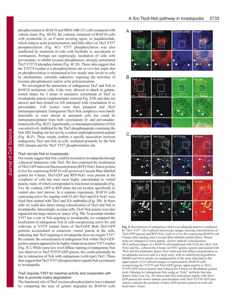
Tks5 recruits Nck to invadopodiaOur results suggest that Nck could be recruited to invadopodia througha physical interaction with Tks5. We first examined the localizationof Tks5-GFP and a red fluorescent protein (RFP)-Nck1 fusion proteinin live Src-expressing B16F10 cells grown on Cascade Blue-labelledgelatin for 4 hours. Tks5-GFP and RFP-Nck1 were present in thecytoplasm of cells but were most highly concentrated in ventralpuncta, many of which corresponded to functional invadopodia (Fig.5A). By contrast, GFP or RFP alone did not localize specifically toventral sites (not shown). In a separate experiment, B16F10 cellsexpressing active Src together with FLAG-Tks5 and HA-Nck1 werefixed then stained with Tks5 and HA antibodies (Fig. 5B). In thesecells we could also detect strong colocalization of Tks5 and Nck ininvadopodia. Interestingly, in some cells, Tks5-Nck puncta were alsoorganized into large clusters or ‘arrays’ (Fig. 5B). To ascertain whetherY557 has a role in Nck targeting to invadopodia, we compared thelocalization of endogenous Nck in cells coexpressing active Src andwild-type or Y557F mutant forms of Tks5-GFP. Both Tks5-GFPproteins accumulated in numerous ventral puncta in the cells,indicating that Tks5 targeting to invadopodia does not require Y557.By contrast, the colocalization of endogenous Nck within Tks5-GFP-positive puncta appeared to be highly reliant on an intact Y557 residue(Fig. 5C). While some low level diffuse staining of endogenous Nckwas observed in Tks5-Y557F-GFP puncta, this could potentially bedue to interaction of Nck with endogenous (wild-type) Tks5. Thesedata suggest that Tks5 Y557 phosphorylation signals Nck recruitmentto invadopodia.
Tks5 requires Y557 for maximal activity and cooperates withNck to promote matrix degradationThe functional role of Tks5 tyrosine phosphorylation was evaluatedby comparing the area of gelatin degraded by B16F10 cells
Fig. 5. Recruitment of endogenous Nck to invadopodia puncta is mediatedby Tks5 Y557. (A) Confocal microscopy images showing colocalization ofTks5-GFP (green) and RFP-Nck1 (red) in a live Src-expressing B16F10 cell4 hours after seeding onto Cascade Blue-labelled gelatin (blue). Boxedareas are enlarged in lower panels. Arrows indicate colocalization.(B) Confocal images of a B16F10 cell transfected with N-FLAG-Tks5, HA-Nck1 and Src, cultured for 4 hours on FITC-gelatin (green) and stained withTks5 and HA antibodies shows Tks5 (red) and Nck (blue) colocalization ininvadopodia (arrows) and in a large array with no underlying degradation.Middle and lower panels are enlargements of the areas indicated in theupper panels. (C) Confocal images of representative B16F10 cellscoexpressing Src Y527F and Tks5-GFP (green, upper panels) or Tks5-Y557F-GFP (lower panels) and cultured for 4 hours on Rhodamine-gelatin(red). Staining for endogenous Nck using an ‘Nck1’ antibody that alsodetects Nck2 (see Fig. 3F) shows that Nck colocalizes tightly with Tks5-GFP puncta but shows limited colocalization with Tks5-Y557F-GFP sites.Arrows indicate the positions of three GFP-positive puncta in each cell.Scale bars: 10 μm.
Jour
nal o
f Cel
l Sci
ence
2734
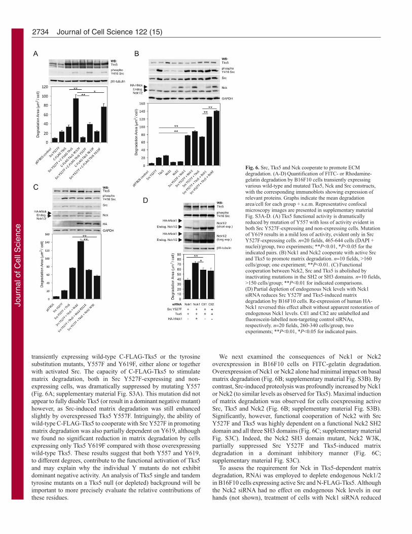
transiently expressing wild-type C-FLAG-Tks5 or the tyrosinesubstitution mutants, Y557F and Y619F, either alone or togetherwith activated Src. The capacity of C-FLAG-Tks5 to stimulatematrix degradation, both in Src Y527F-expressing and non-expressing cells, was dramatically suppressed by mutating Y557(Fig. 6A; supplementary material Fig. S3A). This mutation did notappear to fully disable Tks5 (or result in a dominant negative mutant)however, as Src-induced matrix degradation was still enhancedslightly by overexpressed Tks5 Y557F. Intriguingly, the ability ofwild-type C-FLAG-Tks5 to cooperate with Src Y527F in promotingmatrix degradation was also partially dependent on Y619, althoughwe found no significant reduction in matrix degradation by cellsexpressing only Tks5 Y619F compared with those overexpressingwild-type Tks5. These results suggest that both Y557 and Y619,to different degrees, contribute to the functional activation of Tks5and may explain why the individual Y mutants do not exhibitdominant negative activity. An analysis of Tks5 single and tandemtyrosine mutants on a Tks5 null (or depleted) background will beimportant to more precisely evaluate the relative contributions ofthese residues.
We next examined the consequences of Nck1 or Nck2overexpression in B16F10 cells on FITC-gelatin degradation.Overexpression of Nck1 or Nck2 alone had minimal impact on basalmatrix degradation (Fig. 6B; supplementary material Fig. S3B). Bycontrast, Src-induced proteolysis was profoundly increased by Nck1or Nck2 (to similar levels as observed for Tks5). Maximal inductionof matrix degradation was observed for cells coexpressing activeSrc, Tks5 and Nck2 (Fig. 6B; supplementary material Fig. S3B).Significantly, however, functional cooperation of Nck2 with SrcY527F and Tks5 was highly dependent on a functional Nck2 SH2domain and all three SH3 domains (Fig. 6C; supplementary materialFig. S3C). Indeed, the Nck2 SH3 domain mutant, Nck2 W3K,partially suppressed Src Y527F and Tks5-induced matrixdegradation in a dominant inhibitory manner (Fig. 6C;supplementary material Fig. S3C).
To assess the requirement for Nck in Tks5-dependent matrixdegradation, RNAi was employed to deplete endogenous Nck1/2in B16F10 cells expressing active Src and N-FLAG-Tks5. Althoughthe Nck2 siRNA had no effect on endogenous Nck levels in ourhands (not shown), treatment of cells with Nck1 siRNA reduced
Journal of Cell Science 122 (15)
Fig. 6. Src, Tks5 and Nck cooperate to promote ECMdegradation. (A-D) Quantification of FITC- or Rhodamine-gelatin degradation by B16F10 cells transiently expressingvarious wild-type and mutated Tks5, Nck and Src constructs,with the corresponding immunoblots showing expression ofrelevant proteins. Graphs indicate the mean degradationarea/cell for each group + s.e.m. Representative confocalmicroscopy images are presented in supplementary materialFig. S3A-D. (A) Tks5 functional activity is dramaticallyreduced by mutation of Y557 with loss of activity evident inboth Src Y527F-expressing and non-expressing cells. Mutationof Y619 results in a mild loss of activity, evident only in SrcY527F-expressing cells. n=20 fields, 465-644 cells (DAPI +nuclei)/group, two experiments; **P<0.01, *P<0.05 for theindicated pairs. (B) Nck1 and Nck2 cooperate with active Srcand Tks5 to promote matrix degradation. n=10 fields, >160cells/group; one experiment; **P<0.01. (C) Functionalcooperation between Nck2, Src and Tks5 is abolished byinactivating mutations in the SH2 or SH3 domains. n=10 fields,>150 cells/group; **P<0.01 for indicated comparisons.(D) Partial depletion of endogenous Nck levels with Nck1siRNA reduces Src Y527F and Tks5-induced matrixdegradation by B16F10 cells. Re-expression of human HA-Nck1 reversed this effect albeit without apparent restoration ofendogenous Nck1 levels. Ctl1 and Clt2 are unlabelled andfluoroscein-labelled non-targeting control siRNAs,respectively. n=20 fields, 260-340 cells/group, twoexperiments; **P<0.01, *P<0.05 for indicated pairs.
Jour
nal o
f Cel
l Sci
ence
2735A Src-Tks5-Nck pathway in invadopodia
total Nck levels in B16F10 cells by approximately 55% whencompared with cells treated with either of two different control non-targeting siRNAs, Ctl1 and Ctl2 (Fig. 6D; supplementary materialFig. S3D; and data not shown. NB, Nck antibody detects both Nck1and Nck2). The fluoroscein-conjugated Ctl2 siRNA was used toestimate the average siRNA transfection efficiency at 61%.Treatment with the Nck1 siRNA resulted in a partial but significantreduction in matrix degradation by cells and this effect wasovercome by coexpression of siRNA-resistant human HA-Nck1.Paradoxically, the relative levels of HA-Nck1 in cells appear to betoo low to account for the observed phenotypic rescue; however,highly similar results were obtained in three separate experiments(Fig. 6D; and data not shown). One possible explanation is that theHA-epitope tag interferes with recognition of HA-Nck1 by the Nckantibody and underestimates its real levels. We conclude from thedata presented in Fig. 6 that Src, Tks5 and Nck cooperate in B16F10cells to stimulate matrix degradation.
Nck links Tks5 to invadopodial actin regulationNck adaptor proteins physically couple diverse mammalian andpathogen-encoded proteins to local actin regulation (Frischknechtet al., 1999; Gruenheid et al., 2001; Woodring et al., 2004; Joneset al., 2006; Verma et al., 2006). Current models propose that Nckadaptors are recruited to tyrosine phosphorylated target proteins via
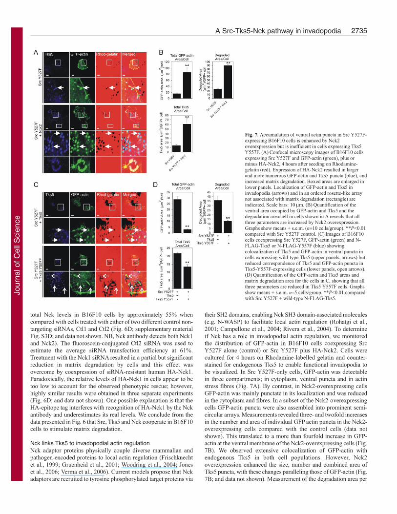
their SH2 domains, enabling Nck SH3 domain-associated molecules(e.g. N-WASP) to facilitate local actin regulation (Rohatgi et al.,2001; Campellone et al., 2004; Rivera et al., 2004). To determineif Nck has a role in invadopodial actin regulation, we monitoredthe distribution of GFP-actin in B16F10 cells coexpressing SrcY527F alone (control) or Src Y527F plus HA-Nck2. Cells werecultured for 4 hours on Rhodamine-labelled gelatin and counter-stained for endogenous Tks5 to enable functional invadopodia tobe visualized. In Src Y527F-only cells, GFP-actin was detectablein three compartments; in cytoplasm, ventral puncta and in actinstress fibres (Fig. 7A). By contrast, in Nck2-overexpressing cellsGFP-actin was mainly punctate in its localization and was reducedin the cytoplasm and fibres. In a subset of the Nck2-overexpressingcells GFP-actin puncta were also assembled into prominent semi-circular arrays. Measurements revealed three- and twofold increasesin the number and area of individual GFP actin puncta in the Nck2-overexpressing cells compared with the control cells (data notshown). This translated to a more than fourfold increase in GFP-actin at the ventral membrane of the Nck2-overexpressing cells (Fig.7B). We observed extensive colocalization of GFP-actin withendogenous Tks5 in both cell populations. However, Nck2overexpression enhanced the size, number and combined area ofTks5 puncta, with these changes paralleling those of GFP-actin (Fig.7B; and data not shown). Measurement of the degradation area per
Fig. 7. Accumulation of ventral actin puncta in Src Y527F-expressing B16F10 cells is enhanced by Nck2overexpression but is inefficient in cells expressing Tks5Y557F. (A) Confocal microscopy images of B16F10 cellsexpressing Src Y527F and GFP-actin (green), plus orminus HA-Nck2, 4 hours after seeding on Rhodamine-gelatin (red). Expression of HA-Nck2 resulted in largerand more numerous GFP-actin and Tks5 puncta (blue), andincreased matrix degradation. Boxed areas are enlarged inlower panels. Localization of GFP-actin and Tks5 ininvadopodia (arrows) and in an ordered rosette-like arraynot associated with matrix degradation (rectangle) areindicated. Scale bars: 10 μm. (B) Quantification of theventral area occupied by GFP-actin and Tks5 and thedegradation area/cell in cells shown in A reveals that allthree parameters are increased by Nck2 overexpression.Graphs show means + s.e.m. (n=10 cells/group). **P<0.01compared with Src Y527F control. (C) Images of B16F10cells coexpressing Src Y527F, GFP-actin (green) and N-FLAG-Tks5 or N-FLAG-Y557F (blue) showingcolocalization of Tks5 and GFP-actin in ventral puncta incells expressing wild-type Tks5 (upper panels, arrows) butreduced correspondence of Tks5 and GFP-actin puncta inTks5-Y557F-expressing cells (lower panels, open arrows).(D) Quantification of the GFP-actin and Tks5 areas andmatrix degradation area for the cells in C, showing that allthree parameters are reduced in Tks5 Y557F cells. Graphsshow means + s.e.m. n=5 cells/group. **P<0.01 comparedwith Src Y527F + wild-type N-FLAG-Tks5.
Jour
nal o
f Cel
l Sci
ence

2736
GFP-actin-positive cell showed that the Nck2-overexpressing cellsdegraded over fourfold more gelatin than control Src Y527F-expressing cells. Collectively, these results show that Nck2cooperates with Src and Tks5 to stimulate actin accumulation ininvadopodia.
We next compared GFP-actin distribution in B16F10 cellsexpressing constitutively active Src and either N-FLAG-Tks5 orthe Y557F mutant lacking the Nck binding site. Cells were culturedon Rhodamine-gelatin for 4 hours then fixed and stained with aTks5 antibody to identify cells that clearly overexpressed Tks5.Although wild-type Tks5 colocalized prominently with GFP-actin,the Y557F mutant showed limited colocalization with GFP-actin(Fig. 7C, arrows, lower panels). Evaluation of the number, size andcumulative area of GFP-actin puncta showed that these parameterswere markedly reduced in cells expressing Tks5 Y557F comparedwith those expressing wild-type Tks5 (Fig. 7D; and data not shown).By comparison, the size of Tks5 puncta was similar in wild-typeand Y557F mutant-expressing cells (not shown) and the numbersand combined area of Tks5 puncta, although less in these cells,were not reduced to the same extent as GFP-actin. Consistent withour earlier results (Fig. 6A), cells expressing Src Y527F and Tks5Y557F degraded the gelatin matrix less efficiently than thoseexpressing Src Y527F and wild-type Tks5. Overall, these resultssuggest that Y557 couples Tks5 to actin regulation in invadopodia.
Clustering of the Tks5 linker region containing Y557 at theplasma membrane stimulates actin assemblyTo determine if the inter-SH3 region of Tks5, containing the Nckdocking site, is capable of directing actin regulation in cells, weadopted a receptor chimera strategy (Campellone et al., 2004; Riveraet al., 2004; Jones et al., 2006). Three receptor chimeras wereconstructed in which the CD16 extracellular and CD7transmembrane domains were fused to GFP (control) or to the linkerregion of Tks5 containing the intact (or mutated) Nck binding sitefollowed by GFP (supplementary material Fig. S4A). In this
system, cross linking of receptors by incubation of cells with a CD16antibody and a fluorescently labelled secondary antibody is usedto manipulate (and visualize) receptor aggregation on the cell surface(supplementary material Fig. S4B). In control experiments weconfirmed that incubation of B16F10 cells expressing theCD16/CD7-GFP chimera with cross-linking antibodies, but notsecondary antibody alone, induced receptor clustering that couldbe detected by CD16 and GFP colocalization (supplementarymaterial Fig. S4C). Furthermore, analysis of CD16/CD7-Tks5-GFPchimera phosphorylation with the p-Tks5 antibody showed thatY557 phosphorylation was strongly enhanced by receptoraggregation (supplementary material Fig. S4D).
We next evaluated the effect of receptor aggregation on thedistribution of an RFP-Nck1 fusion protein. RFP-Nck1 was recruitedto the clustered CD16/CD7-Tks5-GFP receptor at multiple sites atthe cell periphery but did not colocalize with either the CD16/CD7-Y577F-GFP or CD16/CD7-GFP receptors (supplementary materialFig. S4E). To determine the impact of receptor clustering on actinregulation, CD16/CD7 receptor chimeras were expressed in B16F10cells with HA-Nck2, cells were treated with cross linking antibodies,then fixed and stained with TRITC-phalloidin to visualize F-actin.Each of the receptor chimeras underwent extensive clustering inresponse to antibody treatment as indicated by colocalization of theanti-CD16 and GFP signals (Fig. 8). Strikingly, however, whereasthe clustered CD16/CD7-Tks5-GFP receptors colocalized withnumerous F-actin structures (white signal), the CD16/CD7-Y557F-GFP and the control CD16/CD7-GFP receptors did not. These dataindicate the Nck binding region of Tks5 can mediate actinrecruitment in the absence of the Tks5 SH3 domains.
DiscussionWe describe a pathway used by invasive tumour cells to promoteinvadopodia and ECM proteolysis. Collectively our results and thoseof others suggest the following model (Fig. 9). Activation of Src,for example, in response to integrin-mediated adhesion to ECM
Journal of Cell Science 122 (15)
Fig. 8. Clustering of chimericCD16/CD7 receptors containing theTks5 inter-SH3 regionencompassing Y557 promotes actinrecruitment. Confocal imagesshowing examples of B16F10 cellsexpressing CD16/CD7-Tks5-GFP,CD16/CD7-Y557F-GFP or controlreceptor chimeras (green) and HA-Nck2 after treatment with cross-linking antibodies (anti-CD16 plusAlexa Fluor 405 anti-mouse IgG;blue) and F-actin staining (red).Receptor clustering is induced onall cells (turquoise signal) butsignificant F-actin colocalizationwith receptors is only apparent inthe cell expressing CD16/CD7-Tks5-GFP (white signal). Scalebars: 10 μm.
Jour
nal o
f Cel
l Sci
ence

2737A Src-Tks5-Nck pathway in invadopodia
substrates (Mitra and Schlaepfer, 2006), promotes thephosphorylation of Tks5 at Y557/8 to generate a specific bindingsite for the SH2 domains of the Nck adaptor proteins. As Y557 wasdispensable for Tks5-GFP localization in invadopodia, but importantfor Nck colocalization, we propose that Tks5 Y557 phosphorylationoccurs in invadopodia and provides a signal for Nck recruitmentto these sites. The initial targeting of Tks5 to invadopodia probablyinvolves binding of its PX domain to membrane-localizedPtdIns(3,4)P2, as occurs in the initial phases of podosome assemblyin Src-transformed fibroblasts (Oikawa et al., 2008; Symons, 2008).Once recruited to invadopodia, Nck proteins are postulated toregulate actin network assembly via Nck SH3 domain-associatedproteins such as N-WASP and its key target, the Arp2/3 actinnucleation complex (Rohatgi et al., 2001; Rivera et al., 2004). Tks5-Nck-mediated actin assembly in invadopodia could potentiallyregulate protrusive or vesicular trafficking events in invadopodia.How local actin assembly promoted by Tks5-Nck complexes islinked to increased peri-cellular ECM degradation awaits furtherinvestigations, but presumably this requires increased focalexpression of proteases such as MT1-MMP (Artym et al., 2006;Clark et al., 2007).
Recent evidence suggests that Tks5 can bind constitutively toN-WASP by all or a subset of its SH3 domains (Oikawa et al., 2008).In this manner, Tks5–N-WASP complexes could conceivablyregulate actin nucleation in invadopodia without any requirementfor Nck adaptors. Although our data are not directly comparablewith those of Oikawa et al. (Oikawa et al., 2008), which showedthat Tks5 and N-WASP interact equally in Src-transformed oruntransformed 3T3 cells, we found that association of endogenous
N-WASP with a FLAG-ΔPX-Tks5 construct lacking the PX domain(but replete with SH3 domains) was entirely Src dependent.Furthermore, binding of N-WASP was abolished by mutation ofthe Y557 phosphorylation site that mediates Nck binding or reduced(without affect on Nck binding) by mutation of Y619, anotherphosphorylation site. These two tyrosine residues lie in the linkerregion between the third and fourth SH3 domains and althoughmutating them would not be expected to affect SH3 domainintegrity, their phosphorylation could perhaps effect conformationalchanges that expose Tks5 SH3 domains to permit N-WASP binding.An alternative possibility is that N-WASP association with Tks5requires, in addition to Nck, another adaptor protein binding topY619. The SH3-SH2-SH3 adaptor Grb2, identified in this workand by others, as a Tks5-interacting protein (Oikawa et al., 2008),is a strong candidate. Grb2 association with Tks5 is potentiallyphosphorylation dependent, being elevated in Src-transformed cellsand inhibited by the Src kinase inhibitor PP2 (Oikawa et al., 2008).Although the binding site has not been mapped, it is noteworthythat Y619 lies within a Grb2 SH2 domain-binding motif [Y(P)xNx](Songyang et al., 1994). Also, Grb2 SH3 domains can bind to N-WASP and promote N-WASP-dependent actin nucleation (Carlieret al., 2000; Rohatgi et al., 2001; Scaplehorn et al., 2002).Compellingly, evidence of functional cooperativity between Grb2and Nck has also been reported in other model systems (Scaplehornet al., 2002; Garg et al., 2007). For example, studies of actin-basedmotility of vaccinia virus (see also below) have shown that Grb2and Nck are coordinately recruited to tandem phosphorylation sitesin the viral A36R protein and cooperate to bind N-WASP andpromote virus motility (Scaplehorn et al., 2002). Our finding thatY619 is necessary for maximal N-WASP binding by ΔPX-Tks5 andfor maximal Tks5-mediated matrix degradation in Src-expressingB16F10 cells is intriguing in this context. The potential for Tks5to form both Nck-dependent and -independent complexes with N-WASP (Fig. 9), perhaps during different stages of invadopodiagenesis, remains an area for further investigations.
Our observation that inhibitors of tyrosine phosphatases and actinpolymerization in cells enhanced Tks5 Y557 phosphorylation andassociation with Nck from low basal levels indicates that theseevents are tightly controlled. The effect of cytochalasin D is counter-intuitive, however, as Tks5 Y557/8 phosphorylation and Nckrecruitment to invadopodia puncta seem to facilitate increased actinassembly. These results may reflect a negative feedback loop,dependent on actin polymerization, that down-modulates Tks5phosphorylation and Nck binding. Indeed, actin polymerizationinhibitors can trigger the activation of Src- or Abl-family kinasesin some cells (Lock et al., 1998; Maher, 2000; Woodring et al.,2004). It seems unlikely that inhibition of actin polymerization isrequired for Nck recruitment to invadopodia since recruitment ofendogenous Nck to Tks5-GFP puncta in B16F10 cells did notrequire pre-treatment of cells with cytochalasin D, but wasdependent on Y557 and expression of active Src. Studies inmonocytic-lineage cells indicate key roles for microtubules andassociated molecular motors (e.g. kinesin KIF1C) in regulating theformation, location and dynamics of podosomes, including theirassembly in osteoclasts into podosome belts (Linder, 2007; Gimonaet al., 2008). Future experiments utilizing inhibitors/stabilisers ofmicrotubule polymerization will assess the role of a functionalmicrotubule network in Nck (and Tks5) localization in invadopodia.Possible roles of Arf-mediated vesicle trafficking and PI 3-kinasesignalling in Nck recruitment to invadopodia will also beinvestigated.
Fig. 9. Model: a Src-Tks5-Nck signalling pathway in invadopodia. Interactionof tumour cells with the ECM activates Src and PtdIns(3,4)P2-dependenttargeting of Tks5 to nascent invadopodia (Oikawa et al., 2008).Phosphorylation of Tks5 at Y557/8 promotes the recruitment of Nck adaptorproteins via their SH2 domains. Nck-associated N-WASP (possibly linked viathe intermediate WIP) activates Arp2/3-mediated actin network assembly,potentially facilitating protrusive and/or trafficking events. How and whetherthese processes are linked to ECM degradation is not known. Tks5 can alsoassociate with N-WASP via its SH3 domains (Oikawa et al., 2008) or possibly,via Y619 and Grb2. Inhibitors of tyrosine phosphatases and actinpolymerization provoke Y557 phosphorylation and Nck association.
Jour
nal o
f Cel
l Sci
ence

2738
The proposed role of Nck in invadopodia is consistent with itsrole as a key intermediate in localized actin assembly in a variety ofphysiological and pathological settings. For example, in kidneypodocytes, Nck1 and 2 are recruited to specific docking sites in thecytoplasmic domain of the nephrin receptor and regulate themaintenance of actin-based cellular processes involved in glomerularfiltration (Jones et al., 2006; Verma et al., 2006). Tyrosinephosphorylation of Dok1 by Abl kinase also induces Nck bindingand is linked to the formation of filopodia during cell spreading(Woodring et al., 2004). In vaccinia-virus-infected cells, discussedbriefly already, the A36R protein binds Nck (via Y112-P), recruitingWIP and N-WASP and triggering the assembly of Arp2/3-dependentF-actin ‘comets’ that promote motility of extracellular viral particles(Frischknecht et al., 1999). Grb2, recruited to a second phosphorylatedresidue, Y132-P, cooperates with Nck in this process (Scaplehorn etal., 2002). Another pathogen, enteropathogenic E. coli (EPEC), alsoexploits Nck-dependent actin regulation in host cells. The EPECtransmembrane protein Tir is phosphorylated and binds to endogenousNck, and N-WASP associated with Nck promotes the formation ofactin ‘pedestals’ at the plasma membrane (Gruenheid et al., 2001).Using a receptor-clustering approach similar to that developedpreviously (Campellone et al., 2004; Rivera et al., 2004; Jones et al.,2006; Verma et al., 2006), we confirmed that the Nck binding regionof Tks5, excluding its SH3 domains, but not a Y557-mutated variant,can promote F-actin assembly in the presence of transiently expressedNck2 in cells. Nck SH3 domains bind directly or indirectly to severalproteins involved in actin polymerization including WIP and itsbinding partner N-WASP (Moreau et al., 2000; Rohatgi et al., 2001;Benesch et al., 2002). Moreover in cell-free assays, purified Nckcooperates with N-WASP, WIP, cortactin and Arp2/3 to stimulateactin polymerization (Rohatgi et al., 2001; Tehrani et al., 2007)providing compelling evidence that these proteins are criticalcomponents of a multi-subunit actin-regulatory complex. Consistentwith our results, others found that Nck1 (along with N-WASP, WIPand Arp2/3) is critical for EGF-induced invadopodia formation inMTLn3 mammary carcinoma cells (Yamaguchi et al., 2005). It wasunclear from this study how EGF receptor signalling targets Nck toinvadopodia but it is interesting to speculate that Src and Tks5 areimportant intermediates in this pathway.
Materials and MethodsAntibodies and reagentsThe following antibodies were used: anti-FLAG M2 agarose (Sigma), anti-Nck (06-288) and anti-cortactin (4F11; Millipore), anti-N-WASP, anti-CrkL, anti-GAPDH,anti-phosphotyrosine (P-Tyr-100), anti-phospho Src family (TyrY416), anti-HA tag(6E2), anti-Nck1 (Cell Signaling Technology), anti-Fish (Tks5; M-300, Santa CruzBiotechnology Inc.) and anti-CD16 (Beckman Coulter). Anti-Tks5 (Tks5.2) and anti-Src (2-17) antibodies have been described previously (Lock et al., 1998). Tks5monoclonal antibodies (6G1 and 2F4) were generated in mice by immunisation witha GST-Tks5 fusion protein containing the fourth SH3 domain of mouse Tks5. Thephosphospecific p-Tks5 antibody was raised in rabbits at the WEHI BiotechnologyCentre (Melbourne) by immunisation with a synthetic human phosphopeptide,KLKYEEPEY558(P)DIPAFGFD coupled to diphtheria toxin (Auspep). Antisera wereaffinity-purified by removal of antibodies binding to an immobilized unphosphorylatedTks5 peptide and collection of those binding the phosphopeptide immunogen. AlexaFluor 405, 488 and 546 anti-mouse and anti-rabbit IgGs and Cascade Blue were fromInvitrogen. DAPI, TRITC-phalloidin, Rhodamine B isothiocyanate, 300 Bloomgelatin, DMSO, cytochalasin D, brefeldin A, nocodazole, swinholide A, wortmannin,latrunculin A and B and sodium orthovanadate, were purchased from Sigma.Jasplakinolide was obtained from Merck Biosciences and FITC from MP Biomedicals.
Plasmids and siRNADNA constructs encoding Tks5 and ΔPX-Tks5 (lacking the PX domain) with N-and/or C-terminal FLAG tags were generated by PCR using mouse Tks5 DNAtemplate (Lock et al., 1998) and pEFBOS FLAG vectors (I et al., 2004). The Tks5-GFP construct was generated by PCR on mouse Tks5 (minus stop codon) and insertion
in the pEGFP-C1 vector (Clontech). Plasmids encoding C-terminal 3x HA-taggedhuman Nck1 and Nck2 were generated by RT-PCR from a human glioma cDNAlibrary (kindly provided by Nathan Godde, Dept. of Surgery, University of Melbourne,Victoria, Australia) and insertion into pcDNA3 (Invitrogen). Constructs encodingCD16/CD7 receptor chimeras were generated as follows: CD16 extracellular andCD7 transmembrane sequences were amplified by PCR using a CD16.7 plasmidkindly provided by Brian Seed (Harvard Medical School). The CD16/CD7 sequencewas inserted into pEGFP-C1 (to generate CD16/CD7-GFP) and the PCR-amplifiedTks5 region encoding residues 506-835 (with/without the Y557F mutation;supplementary material Fig. S3) was inserted into this backbone. Tks5 phosphorylationsite mutants (Y552F, Y557F and Y619F) and Nck2 point mutants (R311K and W3K)were generated with a QuickChange XL mutagenesis kit (Stratagene). All PCR-generated regions were sequenced (Australian Genome Research Facility Ltd) andconfirmed to be error-free. The following siRNA duplexes were purchased from SantaCruz Inc: mouse Tks5/Fish (sc-35377), mouse Nck1 (sc-40968) and non-targetingcontrol (sc-37007). Fluoroscein-conjugated control siRNA was from New EnglandBiolabs.
Cell culture and transfectionMurine melanoma B16F10, human glioma D270, D54, D566, LN18 and mouse gliomaSMA560 cells were gifts from Ulrike Novak (Dept of Surgery, University ofMelbourne, Victoria, Australia). Human breast carcinoma MDA-MB-231, MDA-MB-435 and T47D cells and human melanoma MEL007, ME4405 and 624.38 cells werekindly provided by Phil Darcy (Peter McCallum Institute, Victoria, Australia). RatC6 glioma and human U118MG, U251MG and U373MG glioma cells were obtainedfrom ATCC. Cells were cultured as described previously (I et al., 2004). For DNAtransfections, 1�106 (B16F10, MDA-MB-231) or 2�106 (293T) cells were seededin 6 cm dishes and the following day transfected with 1 μg total DNA using EffecteneTransfection Reagent (Qiagen). For siRNA transfections, 5�105 B16F10 cells/wellwere seeded in 12-well plates and cells transfected the next day with 50 nM siRNA(with or without 1 μg plasmid DNA). Cells were incubated for 16-18 hours (DNA)or 40-42 hours (siRNA) before matrix degradation and invadopodia assays wereperforming.
Identification of Tks5-interacting proteinsPlasmids encoding Src and FLAG-Tks5 proteins (supplementary material Fig. S2A)were co-transfected into subconfluent cultures of 293T cells in 60�15 cm dishes. 24hours later, cells were washed with a solution of TBS, vanadate and 2 mM DTT andsolubilized in lysis buffer (1% Triton X-100, 10% glycerol, 150 mM NaCl, 20 mMTris pH 7.5, 2 mM EDTA, 10 μg/ml aprotinin, 10 μg/ml leupeptin and 1 mM PMSF).Clarified cell lysates were passed twice through columns of FLAG-M2 antibody beads(Sigma) as described previously (Verhagen et al., 2000). Following extensivewashing, FLAG-Tks5 and associated proteins were eluted with 3� FLAG peptide(Sigma), the eluate concentrated by ultrafiltration using an Amicon ultrafiltration unit(10 kDa cut-off) and proteins separated by SDS-PAGE on 12% long (22 cm) gels(Hoefer). Proteins were visualised by staining the gels with Sypro Ruby (Invitrogen),and Phast-Blue Coomassie (Pharmacia). Protein bands were excised, digested in situwith trypsin and analysed by tandem mass spectrometry (Verhagen et al., 2000). Themass spectrometry and peptide sequence analysis was performed at the JointProteomicS Facility (JPSF), Melbourne.
ImmunofluorescenceGelatin was labelled with FITC or Rhodamine, applied to glass coverslips (25 mmdiameter, 0.17±0.01 mm thickness; Menzel) and fixed with glutaraldehyde asdescribed previously (Bowden et al., 2001). Cells (1�105) were seeded onto thecoverslips in 6-well plates and incubated for 4 hours (to visualize invadopodia) or24 hours (to assess cumulative matrix degradation). Cells were fixed in 4%paraformaldehyde in PBS for 15 minutes, washed with PBS then permeabilized in0.2% Triton X-100 in PBS for 5 minutes. Washed cells were incubated with primaryantibodies in 2% BSA in TBS containing 0.1% Tween 20 at RT for 1 hour. Cellswere then washed and incubated with Alexa Fluor-conjugated anti-IgGs (Invitrogen)or stained for F-actin with TRITC-Phalloidin (Sigma) for 1 hour at room temperature.Following three further washes with PBS, coverslips were mounted on microscopeslides with Vecta-Shield (Vector Laboratories). For detection of nuclei, DAPI (5 μg/ml)was included in the first of the final PBS washes.
Imaging and quantification of matrix degradation and punctaassaysConfocal microscopy images were acquired using a Nikon TE2000-E invertedmicroscope with Plan Apochromat VC �60.0 and �100 oil immersion, 1.40 NA,objectives. The microscope was linked to a Nikon C1 confocal system (equippedwith 405, 488 and 532 nm lasers) and EZ-C1 acquisition software. Images were savedin Nikon ids format and converted using the Nikon EZ-C1 FreeViewer program toTIFF files for image analysis using ImageJ (v1.38) software (NIH). Threshold andregion tools were used to define total regions of matrix degradation in a given field(fluorescence-negative areas) and the area of degradation computed using the particlecounter function. Values obtained in pixels were converted to μm2 and normalizedwith respect to the number of cells (DAPI-positive nuclei) in the field. Ten or more
Journal of Cell Science 122 (15)
Jour
nal o
f Cel
l Sci
ence

2739A Src-Tks5-Nck pathway in invadopodia
fields of at least 150 cells/group were analysed and data expressed as degradationarea (μm2)/cell (mean ± s.e.m.). For the 4-hour matrix degradation assay, only fieldswith Tks5-GFP or GFP-expressing cells (n=10) were analysed. Results were expressedaccordingly as degradation area (μm2)/GFP-positive cell. For puncta determinations,the ImageJ region tool was used to trace the outline of all ventral puncta in a givencell and areas computed as above in 10 cells. Total GFP-actin (or Tks5) puncta area/cellwas calculated by summing the values for each punctum in the cell.
StatisticsFor analysis of four or more groups a global ANOVA analysis was performed followedby post-hoc Tukey-Kramer analysis. Experiments with two or three comparison groupswere analysed using an unpaired t-test. Differences were considered significant ifP<0.05.
Chimeric CD16/CD7-Tks5 receptor experimentsCD16/CD7-GFP receptor constructs were transiently expressed in B16F10 cellsgrowing on coverslips. Medium was removed 24 hours after transfection and receptorclustering was induced by incubation of cells with anti-CD16 antibody (1 μg/ml)and/or Alexa Fluor 405 anti-mouse IgG (1:1,000) for various times at roomtemperature (Verma et al., 2006).
We thank Stephen Cody for confocal microscopy advice and training,Darren Seals for protocols to prepare labelled gelatin coverslips, BrianSeed, Ulrike Novak, Nathan Godde and Phil Darcy for plasmids andcell lines. This work was supported by grants from the NHMRC(Australia), the National Cancer Institute of the USA (NIH) and the JohnT. Reid Charitable Trusts. Deposited in PMC for release after 12 months.
ReferencesAbram, C. L., Seals, D. F., Pass, I., Salinsky, D., Maurer, L., Roth, T. M. and
Courtneidge, S. A. (2003). The adaptor protein fish associates with members of theADAMs family and localizes to podosomes of Src-transformed cells. J. Biol. Chem.278, 16844-16851.
Ago, T., Kuribayashi, F., Hiroaki, H., Takeya, R., Ito, T., Kohda, D. and Sumimoto,H. (2003). Phosphorylation of p47phox directs phox homology domain from SH3 domaintoward phosphoinositides, leading to phagocyte NADPH oxidase activation. Proc. Natl.Acad. Sci. USA 100, 4474-4479.
Alexander, N. R., Branch, K. M., Parekh, A., Clark, E. S., Iwueke, I. C., Guelcher, S.A. and Weaver, A. M. (2008). Extracellular matrix rigidity promotes invadopodiaactivity. Curr. Biol. 18, 1295-1299.
Artym, V. V., Zhang, Y., Seillier-Moiseiwitsch, F., Yamada, K. M. and Mueller, S. C.(2006). Dynamic interactions of cortactin and membrane type 1 matrix metalloproteinaseat invadopodia: defining the stages of invadopodia formation and function. Cancer Res.66, 3034-3043.
Ayala, I., Baldassarre, M., Giacchetti, G., Caldieri, G., Tete, S., Luini, A. andBuccione, R. (2008). Multiple regulatory inputs converge on cortactin to controlinvadopodia biogenesis and extracellular matrix degradation. J. Cell Sci. 121, 369-378.
Badowski, C., Pawlak, G., Grichine, A., Chabadel, A., Oddou, C., Jurdic, P., Pfaff,M., Albiges-Rizo, C. and Block, M. R. (2008). Paxillin phosphorylation controlsinvadopodia/podosomes spatiotemporal organization. Mol. Biol. Cell 19, 633-645.
Baldassarre, M., Pompeo, A., Beznoussenko, G., Castaldi, C., Cortellino, S., McNiven,M. A., Luini, A. and Buccione, R. (2003). Dynamin participates in focal extracellularmatrix degradation by invasive cells. Mol. Biol. Cell 14, 1074-1084.
Basbaum, C. B. and Werb, Z. (1996). Focalized proteolysis: spatial and temporal regulationof extracellular matrix degradation at the cell surface. Curr. Opin. Cell Biol. 8, 731-738.
Benesch, S., Lommel, S., Steffen, A., Stradal, T. E., Scaplehorn, N., Way, M., Wehland,J. and Rottner, K. (2002). Phosphatidylinositol 4,5-biphosphate (PIP2)-induced vesiclemovement depends on N-WASP and involves Nck, WIP, and Grb2. J. Biol. Chem. 277,37771-37776.
Bharti, S., Inoue, H., Bharti, K., Hirsch, D. S., Nie, Z., Yoon, H. Y., Artym, V., Yamada,K. M., Mueller, S. C., Barr, V. A. et al. (2007). Src-dependent phosphorylation ofASAP1 regulates podosomes. Mol. Cell. Biol. 27, 8271-8283.
Bladt, F., Aippersbach, E., Gelkop, S., Strasser, G. A., Nash, P., Tafuri, A., Gertler, F.B. and Pawson, T. (2003). The murine Nck SH2/SH3 adaptors are important for thedevelopment of mesoderm-derived embryonic structures and for regulating the cellularactin network. Mol. Cell. Biol. 23, 4586-4597.
Bowden, E. T., Barth, M., Thomas, D., Glazer, R. I. and Mueller, S. C. (1999). Aninvasion-related complex of cortactin, paxillin and PKCmu associates with invadopodiaat sites of extracellular matrix degradation. Oncogene 18, 4440-4449.
Bowden, E. T., Coopman, P. J. and Mueller, S. C. (2001). Invadopodia: unique methodsfor measurement of extracellular matrix degradation in vitro. Methods Cell Biol. 63,613-627.
Campellone, K. G., Rankin, S., Pawson, T., Kirschner, M. W., Tipper, D. J. and Leong,J. M. (2004). Clustering of Nck by a 12-residue Tir phosphopeptide is sufficient totrigger localized actin assembly. J. Cell Biol. 164, 407-416.
Carlier, M. F., Nioche, P., Broutin-L’Hermite, I., Boujemaa, R., Le Clainche, C., Egile,C., Garbay, C., Ducruix, A., Sansonetti, P. and Pantaloni, D. (2000). GRB2 linkssignaling to actin assembly by enhancing interaction of neural Wiskott-Aldrich syndromeprotein (N-WASp) with actin-related protein (ARP2/3) complex. J. Biol. Chem. 275,21946-21952.
Chen, W. T., Olden, K., Bernard, B. A. and Chu, F. F. (1984). Expression oftransformation-associated protease(s) that degrade fibronectin at cell contact sites. J.Cell Biol. 98, 1546-1555.
Chuang, Y. Y., Tran, N. L., Rusk, N., Nakada, M., Berens, M. E. and Symons, M.(2004). Role of synaptojanin 2 in glioma cell migration and invasion. Cancer Res. 64,8271-8275.
Clark, E. S., Whigham, A. S., Yarbrough, W. G. and Weaver, A. M. (2007). Cortactinis an essential regulator of matrix metalloproteinase secretion and extracellular matrixdegradation in invadopodia. Cancer Res. 67, 4227-4235.
Co, C., Wong, D. T., Gierke, S., Chang, V. and Taunton, J. (2007). Mechanism of actinnetwork attachment to moving membranes: barbed end capture by N-WASP WH2domains. Cell 128, 901-913.
Condeelis, J. and Segall, J. E. (2003). Intravital imaging of cell movement in tumours.Nat. Rev. Cancer 3, 921-930.
Frese, S., Schubert, W. D., Findeis, A. C., Marquardt, T., Roske, Y. S., Stradal, T. E.and Heinz, D. W. (2006). The phosphotyrosine peptide binding specificity of Nck1 andNck2 Src homology 2 domains. J. Biol. Chem. 281, 18236-18245.
Friedl, P. and Wolf, K. (2003). Tumour-cell invasion and migration: diversity and escapemechanisms. Nat. Rev. Cancer 3, 362-374.
Friedl, P. and Wolf, K. (2008). Tube travel: the role of proteases in individual and collectivecancer cell invasion. Cancer Res. 68, 7247-7249.
Frischknecht, F., Moreau, V., Rottger, S., Gonfloni, S., Reckmann, I., Superti-Furga,G. and Way, M. (1999). Actin-based motility of vaccinia virus mimics receptor tyrosinekinase signalling. Nature 401, 926-929.
Garg, P., Verma, R., Nihalani, D., Johnstone, D. B. and Holzman, L. B. (2007). Neph1cooperates with nephrin to transduce a signal that induces actin polymerization. Mol.Cell. Biol. 27, 8698-8712.
Gimona, M., Buccione, R., Courtneidge, S. A. and Linder, S. (2008). Assembly andbiological role of podosomes and invadopodia. Curr. Opin. Cell Biol. 20, 235-241.
Groemping, Y., Lapouge, K., Smerdon, S. J. and Rittinger, K. (2003). Molecular basisof phosphorylation-induced activation of the NADPH oxidase. Cell 113, 343-355.
Gruenheid, S., DeVinney, R., Bladt, F., Goosney, D., Gelkop, S., Gish, G. D., Pawson,T. and Finlay, B. B. (2001). Enteropathogenic E. coli Tir binds Nck to initiate actinpedestal formation in host cells. Nat. Cell Biol. 3, 856-859.
Hashimoto, S., Onodera, Y., Hashimoto, A., Tanaka, M., Hamaguchi, M., Yamada, A.and Sabe, H. (2004). Requirement for Arf6 in breast cancer invasive activities. Proc.Natl. Acad. Sci. USA 101, 6647-6652.
I, S. T., Nie, Z., Stewart, A., Najdovska, M., Hall, N. E., He, H., Randazzo, P. A. andLock, P. (2004). ARAP3 is transiently tyrosine phosphorylated in cells attaching tofibronectin and inhibits cell spreading in a RhoGAP-dependent manner. J. Cell Sci. 117,6071-6084.
Jones, N., Blasutig, I. M., Eremina, V., Ruston, J. M., Bladt, F., Li, H., Huang, H.,Larose, L., Li, S. S., Takano, T. et al. (2006). Nck adaptor proteins link nephrin to theactin cytoskeleton of kidney podocytes. Nature 440, 818-823.
Karathanassis, D., Stahelin, R. V., Bravo, J., Perisic, O., Pacold, C. M., Cho, W. andWilliams, R. L. (2002). Binding of the PX domain of p47(phox) to phosphatidylinositol3,4-bisphosphate and phosphatidic acid is masked by an intramolecular interaction.EMBO J. 21, 5057-5068.
Linder, S. (2007). The matrix corroded: podosomes and invadopodia in extracellular matrixdegradation. Trends Cell Biol. 17, 107-117.
Lock, P., Abram, C. L., Gibson, T. and Courtneidge, S. A. (1998). A new method forisolating tyrosine kinase substrates used to identify fish, an SH3 and PX domain-containing protein, and Src substrate. EMBO J. 17, 4346-4357.
Maher, P. A. (2000). Disruption of cell-substrate adhesion activates the protein tyrosinekinase pp60(c-src). Exp. Cell Res. 260, 189-198.
Mitra, S. K. and Schlaepfer, D. D. (2006). Integrin-regulated FAK-Src signaling in normaland cancer cells. Curr. Opin. Cell Biol. 18, 516-523.
Mizutani, K., Miki, H., He, H., Maruta, H. and Takenawa, T. (2002). Essential role ofneural Wiskott-Aldrich syndrome protein in podosome formation and degradation ofextracellular matrix in src-transformed fibroblasts. Cancer Res. 62, 669-674.
Moreau, V., Frischknecht, F., Reckmann, I., Vincentelli, R., Rabut, G., Stewart, D.and Way, M. (2000). A complex of N-WASP and WIP integrates signalling cascadesthat lead to actin polymerization. Nat. Cell Biol. 2, 441-448.
Nollau, P. and Mayer, B. J. (2001). Profiling the global tyrosine phosphorylation state bySrc homology 2 domain binding. Proc. Natl. Acad. Sci. USA 98, 13531-13536.
Oikawa, T., Itoh, T. and Takenawa, T. (2008). Sequential signals toward podosomeformation in NIH-src cells. J. Cell Biol. 182, 157-169.
Onodera, Y., Hashimoto, S., Hashimoto, A., Morishige, M., Mazaki, Y., Yamada, A.,Ogawa, E., Adachi, M., Sakurai, T., Manabe, T. et al. (2005). Expression of AMAP1,an ArfGAP, provides novel targets to inhibit breast cancer invasive activities. EMBO J.24, 963-973.
Rivera, G. M., Briceno, C. A., Takeshima, F., Snapper, S. B. and Mayer, B. J. (2004).Inducible clustering of membrane-targeted SH3 domains of the adaptor protein Ncktriggers localized actin polymerization. Curr. Biol. 14, 11-22.
Rohatgi, R., Nollau, P., Ho, H. Y., Kirschner, M. W. and Mayer, B. J. (2001). Nck andphosphatidylinositol 4,5-bisphosphate synergistically activate actin polymerizationthrough the N-WASP-Arp2/3 pathway. J. Biol. Chem. 276, 26448-26452.
Scaplehorn, N., Holmstrom, A., Moreau, V., Frischknecht, F., Reckmann, I. and Way,M. (2002). Grb2 and Nck act cooperatively to promote actin-based motility of vacciniavirus. Curr. Biol. 12, 740-745.
Seals, D. F., Azucena, E. F., Jr, Pass, I., Tesfay, L., Gordon, R., Woodrow, M., Resau,J. H. and Courtneidge, S. A. (2005). The adaptor protein Tks5/Fish is required forpodosome formation and function, and for the protease-driven invasion of cancer cells.Cancer Cell 7, 155-165.
Jour
nal o
f Cel
l Sci
ence

2740
Songyang, Z., Shoelson, S. E., Chaudhuri, M., Gish, G., Pawson, T., Haser, W. G.,King, F., Roberts, T., Ratnofsky, S., Lechleider, R. J. et al. (1993). SH2 domainsrecognize specific phosphopeptide sequences. Cell 72, 767-778.
Songyang, Z., Shoelson, S. E., McGlade, J., Olivier, P., Pawson, T., Bustelo, X. R.,Barbacid, M., Sabe, H., Hanafusa, H., Yi, T. et al. (1994). Specific motifs recognizedby the SH2 domains of Csk, 3BP2, fps/fes, GRB-2, HCP, SHC, Syk, and Vav. Mol. Cell.Biol. 14, 2777-2785.
Symons, M. (2008). Cell biology: watching the first steps of podosome formation. Curr.Biol. 18, R925-R927.
Tague, S. E., Muralidharan, V. and D’Souza-Schorey, C. (2004). ADP-ribosylation factor6 regulates tumor cell invasion through the activation of the MEK/ERK signaling pathway.Proc. Natl. Acad. Sci. USA 101, 9671-9676.
Tehrani, S., Tomasevic, N., Weed, S., Sakowicz, R. and Cooper, J. A. (2007). Srcphosphorylation of cortactin enhances actin assembly. Proc. Natl. Acad. Sci. USA 104,11933-11938.
Thompson, O., Kleino, I., Crimaldi, L., Gimona, M., Saksela, K. and Winder, S. J.(2008). Dystroglycan, Tks5 and Src mediated assembly of podosomes in myoblasts.PLoS ONE 3, e3638.
Verhagen, A. M., Ekert, P. G., Pakusch, M., Silke, J., Connolly, L. M., Reid, G. E.,Moritz, R. L., Simpson, R. J. and Vaux, D. L. (2000). Identification of DIABLO, amammalian protein that promotes apoptosis by binding to and antagonizing IAP proteins.Cell 102, 43-53.
Verma, R., Kovari, I., Soofi, A., Nihalani, D., Patrie, K. and Holzman, L. B. (2006).Nephrin ectodomain engagement results in Src kinase activation, nephrinphosphorylation, Nck recruitment, and actin polymerization. J. Clin. Invest. 116, 1346-1359.
Weaver, A. M. (2006). Invadopodia: specialized cell structures for cancer invasion. Clin.Exp. Metastasis 23, 97-105.
Woodring, P. J., Meisenhelder, J., Johnson, S. A., Zhou, G. L., Field, J.,Shah, K., Bladt, F., Pawson, T., Niki, M., Pandolfi, P. P. et al. (2004). c-Ablphosphorylates Dok1 to promote filopodia during cell spreading. J. Cell Biol. 165,493-503.
Yamaguchi, H., Lorenz, M., Kempiak, S., Sarmiento, C., Coniglio, S., Symons, M.,Segall, J., Eddy, R., Miki, H., Takenawa, T. et al. (2005). Molecular mechanisms ofinvadopodium formation: the role of the N-WASP-Arp2/3 complex pathway and cofilin.J. Cell Biol. 168, 441-452.
Journal of Cell Science 122 (15)
Jour
nal o
f Cel
l Sci
ence
Copyright © 2022 FDOKUMEN