The Nck-interacting kinase phosphorylates ERM proteins for formation of lamellipodium by growth...
6
The Nck-interacting kinase phosphorylates ERM proteins for formation of lamellipodium by growth factors Martin Baumgartner* †‡ , Amy L. Sillman* † , Elizabeth M. Blackwood § , Jyoti Srivastava*, Nikki Madson*, James W. Schilling § , Jocelyn H. Wright § , and Diane L. Barber* ¶ *Department of Cell and Tissue Biology, University of California, San Francisco, CA 94143; and § Sugen, Inc., South San Francisco, CA 94080 Communicated by Henry R. Bourne, University of California, San Francisco, CA, July 14, 2006 (received for review March 29, 2006) The mammalian Ste20-like Nck-interacting kinase (NIK) and its orthologs Misshapen in Drosophila and Mig-15 in Caenorhabditis elegans have a conserved function in regulating cell morphology, although through poorly understood mechanisms. We report two previously unrecognized actions of NIK: regulation of lamellipo- dium formation by growth factors and phosphorylation of the ERM proteins ezrin, radixin, and moesin. ERM proteins regulate cell morphology and plasma membrane dynamics by reversibly an- choring actin filaments to integral plasma membrane proteins. In vitro assays show that NIK interacts directly with ERM proteins, binding their N termini and phosphorylating a conserved C-termi- nal threonine. In cells, NIK and phosphorylated ERM proteins localize at the distal margins of lamellipodia, and NIK activity is necessary for phosphorylation of ERM proteins induced by EGF and PDGF, but not by thrombin. Lamellipodium extension in response to growth factors is inhibited in cells expressing a kinase-inactive NIK, suppressed for NIK expression with siRNA oligonucleotides, or expressing ezrin T567A that cannot be phosphorylated. These data suggest that direct phosphorylation of ERM proteins by NIK con- stitutes a signaling mechanism controlling growth factor-induced membrane protrusion and cell morphology. ezrin moesin ste20 kinase membrane protrusion T he Nck-interacting kinase (NIK) is a member of the germinal center kinase subfamily of Ste20MAP4K serinethreonine kinases (1). Closely related to NIK are the mammalian kinases TNIK (2), MINK (3), and NRKNESK (4) and orthologs Misshapen (Msn) in Drosophila (5) and MIG-15 in Caenorhab- ditis elegans (6). NIK and its orthologs share a common function in regulating cell shape and migration. In mice, homozygous knockout of NIK results in early embryonic lethality with defects in mesoderm migration (7), and expression of kinase-inactive NIK attenuates epithelial cell invasion (8). Msn functions in determining epithelial polarity, dorsal closure. and neuronal targeting (5, 9, 10), and MIG-15 controls axonal navigation (6). NIK (1) and Msn (5) also share a conserved activation of the JNK pathway. NIK, however, does not directly phosphorylate JNK, nor do NIK nullizygous embryos precisely phenocopy mice lacking JNK1 or JNK2 (7). Additionally, activation of JNK is not associated with dynamic changes in cell morphology. Hence, NIK substrates that control cell morphogenesis have not been identified. We now report that the ERM proteins ezrin, radixin, and moesin are substrates for NIK. ERM proteins regulate cell morphology by cross-linking actin filaments to the plasma mem- brane. The N-terminal FERM (4.1 ERM) domain of ERM proteins binds to integral plasma membrane proteins and the C terminus binds F-actin (11). ERM proteins control cell shape primarily by regulating membrane protrusions and cell-substrate adhesion. In epithelial cells ERM proteins are necessary for the formation of apical microvilli (12–14). In fibroblasts ERM proteins regulate the assembly of focal adhesions (15) and dynamic membrane protrusions, including filopodia and lamel- lipodia (16, 17). Inactive ERM proteins are retained in the cytosol in a closed conformation by an intramolecular association of N- and C- terminal domains, which masks a C-terminal F-actin binding site. ERM proteins are coincidence detectors, with activation and release of the N- and C-terminal interaction requiring two sequential regulatory events: binding of phosphatidylinositol 4,5-bisphosphate to the N terminus and phosphorylation of a C-terminal threonine residue (18), conserved in ezrin (T567), radixin (T564), and moesin (T558). Phosphorylation of ERM proteins at specific membrane domains may be regulated by different kinases. ERM proteins are phosphorylated by myo- tonic dystrophy kinase-related Cdc42-binding kinase in filopodia (19), protein kinase C in membrane protrusions (20), and the Rho-associated kinase (ROCK) in microvilli (21), although this latter finding is controversial (22). Kinases mediating phosphor- ylation of ERM proteins by growth factors to regulate lamelli- podium formation, however, have not been identified. We found that NIK directly phosphorylates the conserved C-terminal threonine in ERM proteins, and that NIK activity and phosphorylation of ezrin T567 are necessary for lamellipo- dium extension induced by growth factors. NIK binds ERM proteins, localizes with phosphorylated ERM (pERM) proteins at the distal margins of lamellipodia, and is necessary for increased phosphorylation of ERM proteins in response to growth factors. These data suggest that NIK activity and its direct phosphorylation of ERM proteins control growth factor- induced changes in cell morphology. Results NIK Activity Is Necessary for Lamellipodium Extension by EGF. Be- cause NIK acts downstream of receptor tyrosine kinases (23, 24), we asked whether its kinase activity regulates dynamic changes in cell morphology in response to growth factors. In rat mam- mary epithelial MTLn3 cells, which extend broad lamellipodia with addition of EGF (25), NIK activity was necessary for increased membrane protrusion by EGF. In MTLn3 cells in- fected with control adenovirus (Ad) vector, EGF (25 nM) rapidly induced lamellipodia extending around the cell body (Fig. 1A Top and see Movie 1, which is published as supporting information on the PNAS web site). The area of lamellipodium protrusion with EGF increased significantly by 2 min (P 0.1; n 6), reaching a maximum 3-fold increase at 10 min (Fig. 1B). Conflict of interest statement: No conflicts declared. Abbreviations: NIK, Nck-interacting kinase; Msn, Misshapen; ERM, ezrin, radixin, moesin; pERM, phosphorylated ERM; Ad, adenovirus. † M.B. and A.L.S. contributed equally to this work. ‡ Present address: Institute of Medical Virology, University of Zu ¨ rich, CH-8006 Zu ¨ rich, Switzerland. ¶ To whom correspondence should be addressed. E-mail: [email protected]. © 2006 by The National Academy of Sciences of the USA www.pnas.orgcgidoi10.1073pnas.0605950103 PNAS September 5, 2006 vol. 103 no. 36 13391–13396 CELL BIOLOGY
-
Upload
independent -
Category
Documents
-
view
0 -
download
0
Transcript of The Nck-interacting kinase phosphorylates ERM proteins for formation of lamellipodium by growth...
The Nck-interacting kinase phosphorylates ERMproteins for formation of lamellipodiumby growth factorsMartin Baumgartner*†‡, Amy L. Sillman*†, Elizabeth M. Blackwood§, Jyoti Srivastava*, Nikki Madson*,James W. Schilling§, Jocelyn H. Wright§, and Diane L. Barber*¶
*Department of Cell and Tissue Biology, University of California, San Francisco, CA 94143; and §Sugen, Inc., South San Francisco, CA 94080
Communicated by Henry R. Bourne, University of California, San Francisco, CA, July 14, 2006 (received for review March 29, 2006)
The mammalian Ste20-like Nck-interacting kinase (NIK) and itsorthologs Misshapen in Drosophila and Mig-15 in Caenorhabditiselegans have a conserved function in regulating cell morphology,although through poorly understood mechanisms. We report twopreviously unrecognized actions of NIK: regulation of lamellipo-dium formation by growth factors and phosphorylation of the ERMproteins ezrin, radixin, and moesin. ERM proteins regulate cellmorphology and plasma membrane dynamics by reversibly an-choring actin filaments to integral plasma membrane proteins. Invitro assays show that NIK interacts directly with ERM proteins,binding their N termini and phosphorylating a conserved C-termi-nal threonine. In cells, NIK and phosphorylated ERM proteinslocalize at the distal margins of lamellipodia, and NIK activity isnecessary for phosphorylation of ERM proteins induced by EGF andPDGF, but not by thrombin. Lamellipodium extension in responseto growth factors is inhibited in cells expressing a kinase-inactiveNIK, suppressed for NIK expression with siRNA oligonucleotides, orexpressing ezrin T567A that cannot be phosphorylated. These datasuggest that direct phosphorylation of ERM proteins by NIK con-stitutes a signaling mechanism controlling growth factor-inducedmembrane protrusion and cell morphology.
ezrin � moesin � ste20 kinase � membrane protrusion
The Nck-interacting kinase (NIK) is a member of the germinalcenter kinase subfamily of Ste20�MAP4K serine�threonine
kinases (1). Closely related to NIK are the mammalian kinasesTNIK (2), MINK (3), and NRK�NESK (4) and orthologsMisshapen (Msn) in Drosophila (5) and MIG-15 in Caenorhab-ditis elegans (6). NIK and its orthologs share a common functionin regulating cell shape and migration. In mice, homozygousknockout of NIK results in early embryonic lethality with defectsin mesoderm migration (7), and expression of kinase-inactiveNIK attenuates epithelial cell invasion (8). Msn functions indetermining epithelial polarity, dorsal closure. and neuronaltargeting (5, 9, 10), and MIG-15 controls axonal navigation (6).NIK (1) and Msn (5) also share a conserved activation of theJNK pathway. NIK, however, does not directly phosphorylateJNK, nor do NIK nullizygous embryos precisely phenocopy micelacking JNK1 or JNK2 (7). Additionally, activation of JNK is notassociated with dynamic changes in cell morphology. Hence,NIK substrates that control cell morphogenesis have not beenidentified.
We now report that the ERM proteins ezrin, radixin, andmoesin are substrates for NIK. ERM proteins regulate cellmorphology by cross-linking actin filaments to the plasma mem-brane. The N-terminal FERM (4.1 ERM) domain of ERMproteins binds to integral plasma membrane proteins and the Cterminus binds F-actin (11). ERM proteins control cell shapeprimarily by regulating membrane protrusions and cell-substrateadhesion. In epithelial cells ERM proteins are necessary for theformation of apical microvilli (12–14). In fibroblasts ERMproteins regulate the assembly of focal adhesions (15) and
dynamic membrane protrusions, including filopodia and lamel-lipodia (16, 17).
Inactive ERM proteins are retained in the cytosol in a closedconformation by an intramolecular association of N- and C-terminal domains, which masks a C-terminal F-actin binding site.ERM proteins are coincidence detectors, with activation andrelease of the N- and C-terminal interaction requiring twosequential regulatory events: binding of phosphatidylinositol4,5-bisphosphate to the N terminus and phosphorylation of aC-terminal threonine residue (18), conserved in ezrin (T567),radixin (T564), and moesin (T558). Phosphorylation of ERMproteins at specific membrane domains may be regulated bydifferent kinases. ERM proteins are phosphorylated by myo-tonic dystrophy kinase-related Cdc42-binding kinase in filopodia(19), protein kinase C� in membrane protrusions (20), and theRho-associated kinase (ROCK) in microvilli (21), although thislatter finding is controversial (22). Kinases mediating phosphor-ylation of ERM proteins by growth factors to regulate lamelli-podium formation, however, have not been identified.
We found that NIK directly phosphorylates the conservedC-terminal threonine in ERM proteins, and that NIK activityand phosphorylation of ezrin T567 are necessary for lamellipo-dium extension induced by growth factors. NIK binds ERMproteins, localizes with phosphorylated ERM (pERM) proteinsat the distal margins of lamellipodia, and is necessary forincreased phosphorylation of ERM proteins in response togrowth factors. These data suggest that NIK activity and itsdirect phosphorylation of ERM proteins control growth factor-induced changes in cell morphology.
ResultsNIK Activity Is Necessary for Lamellipodium Extension by EGF. Be-cause NIK acts downstream of receptor tyrosine kinases (23, 24),we asked whether its kinase activity regulates dynamic changesin cell morphology in response to growth factors. In rat mam-mary epithelial MTLn3 cells, which extend broad lamellipodiawith addition of EGF (25), NIK activity was necessary forincreased membrane protrusion by EGF. In MTLn3 cells in-fected with control adenovirus (Ad) vector, EGF (25 nM)rapidly induced lamellipodia extending around the cell body(Fig. 1A Top and see Movie 1, which is published as supportinginformation on the PNAS web site). The area of lamellipodiumprotrusion with EGF increased significantly by 2 min (P � 0.1;n � 6), reaching a maximum �3-fold increase at 10 min (Fig. 1B).
Conflict of interest statement: No conflicts declared.
Abbreviations: NIK, Nck-interacting kinase; Msn, Misshapen; ERM, ezrin, radixin, moesin;pERM, phosphorylated ERM; Ad, adenovirus.
†M.B. and A.L.S. contributed equally to this work.
‡Present address: Institute of Medical Virology, University of Zurich, CH-8006 Zurich,Switzerland.
¶To whom correspondence should be addressed. E-mail: [email protected].
© 2006 by The National Academy of Sciences of the USA
www.pnas.org�cgi�doi�10.1073�pnas.0605950103 PNAS � September 5, 2006 � vol. 103 � no. 36 � 13391–13396
CELL
BIO
LOG
Y
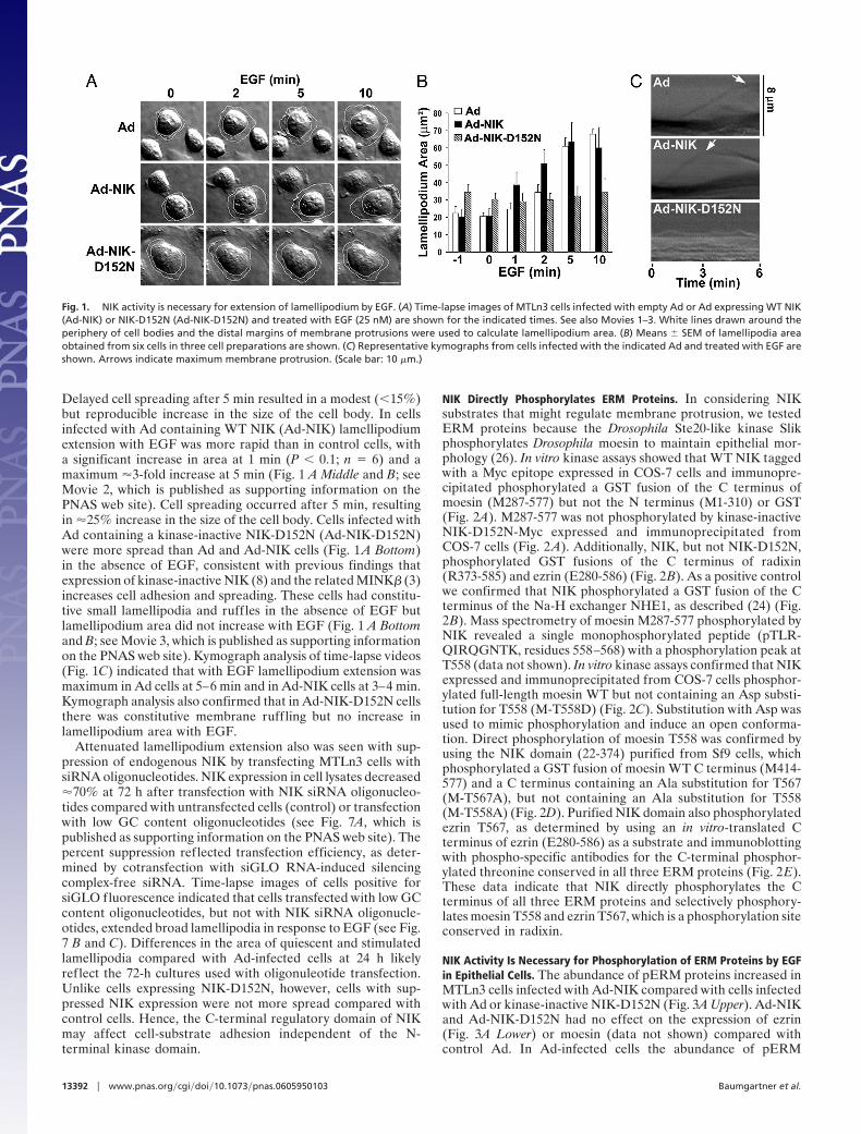
Delayed cell spreading after 5 min resulted in a modest (�15%)but reproducible increase in the size of the cell body. In cellsinfected with Ad containing WT NIK (Ad-NIK) lamellipodiumextension with EGF was more rapid than in control cells, witha significant increase in area at 1 min (P � 0.1; n � 6) and amaximum �3-fold increase at 5 min (Fig. 1 A Middle and B; seeMovie 2, which is published as supporting information on thePNAS web site). Cell spreading occurred after 5 min, resultingin �25% increase in the size of the cell body. Cells infected withAd containing a kinase-inactive NIK-D152N (Ad-NIK-D152N)were more spread than Ad and Ad-NIK cells (Fig. 1 A Bottom)in the absence of EGF, consistent with previous findings thatexpression of kinase-inactive NIK (8) and the related MINK� (3)increases cell adhesion and spreading. These cells had constitu-tive small lamellipodia and ruffles in the absence of EGF butlamellipodium area did not increase with EGF (Fig. 1 A Bottomand B; see Movie 3, which is published as supporting informationon the PNAS web site). Kymograph analysis of time-lapse videos(Fig. 1C) indicated that with EGF lamellipodium extension wasmaximum in Ad cells at 5–6 min and in Ad-NIK cells at 3–4 min.Kymograph analysis also confirmed that in Ad-NIK-D152N cellsthere was constitutive membrane ruffling but no increase inlamellipodium area with EGF.
Attenuated lamellipodium extension also was seen with sup-pression of endogenous NIK by transfecting MTLn3 cells withsiRNA oligonucleotides. NIK expression in cell lysates decreased�70% at 72 h after transfection with NIK siRNA oligonucleo-tides compared with untransfected cells (control) or transfectionwith low GC content oligonucleotides (see Fig. 7A, which ispublished as supporting information on the PNAS web site). Thepercent suppression reflected transfection efficiency, as deter-mined by cotransfection with siGLO RNA-induced silencingcomplex-free siRNA. Time-lapse images of cells positive forsiGLO fluorescence indicated that cells transfected with low GCcontent oligonucleotides, but not with NIK siRNA oligonucle-otides, extended broad lamellipodia in response to EGF (see Fig.7 B and C). Differences in the area of quiescent and stimulatedlamellipodia compared with Ad-infected cells at 24 h likelyreflect the 72-h cultures used with oligonuleotide transfection.Unlike cells expressing NIK-D152N, however, cells with sup-pressed NIK expression were not more spread compared withcontrol cells. Hence, the C-terminal regulatory domain of NIKmay affect cell-substrate adhesion independent of the N-terminal kinase domain.
NIK Directly Phosphorylates ERM Proteins. In considering NIKsubstrates that might regulate membrane protrusion, we testedERM proteins because the Drosophila Ste20-like kinase Slikphosphorylates Drosophila moesin to maintain epithelial mor-phology (26). In vitro kinase assays showed that WT NIK taggedwith a Myc epitope expressed in COS-7 cells and immunopre-cipitated phosphorylated a GST fusion of the C terminus ofmoesin (M287-577) but not the N terminus (M1-310) or GST(Fig. 2A). M287-577 was not phosphorylated by kinase-inactiveNIK-D152N-Myc expressed and immunoprecipitated fromCOS-7 cells (Fig. 2 A). Additionally, NIK, but not NIK-D152N,phosphorylated GST fusions of the C terminus of radixin(R373-585) and ezrin (E280-586) (Fig. 2B). As a positive controlwe confirmed that NIK phosphorylated a GST fusion of the Cterminus of the Na-H exchanger NHE1, as described (24) (Fig.2B). Mass spectrometry of moesin M287-577 phosphorylated byNIK revealed a single monophosphorylated peptide (pTLR-QIRQGNTK, residues 558–568) with a phosphorylation peak atT558 (data not shown). In vitro kinase assays confirmed that NIKexpressed and immunoprecipitated from COS-7 cells phosphor-ylated full-length moesin WT but not containing an Asp substi-tution for T558 (M-T558D) (Fig. 2C). Substitution with Asp wasused to mimic phosphorylation and induce an open conforma-tion. Direct phosphorylation of moesin T558 was confirmed byusing the NIK domain (22-374) purified from Sf9 cells, whichphosphorylated a GST fusion of moesin WT C terminus (M414-577) and a C terminus containing an Ala substitution for T567(M-T567A), but not containing an Ala substitution for T558(M-T558A) (Fig. 2D). Purified NIK domain also phosphorylatedezrin T567, as determined by using an in vitro-translated Cterminus of ezrin (E280-586) as a substrate and immunoblottingwith phospho-specific antibodies for the C-terminal phosphor-ylated threonine conserved in all three ERM proteins (Fig. 2E).These data indicate that NIK directly phosphorylates the Cterminus of all three ERM proteins and selectively phosphory-lates moesin T558 and ezrin T567, which is a phosphorylation siteconserved in radixin.
NIK Activity Is Necessary for Phosphorylation of ERM Proteins by EGFin Epithelial Cells. The abundance of pERM proteins increased inMTLn3 cells infected with Ad-NIK compared with cells infectedwith Ad or kinase-inactive NIK-D152N (Fig. 3A Upper). Ad-NIKand Ad-NIK-D152N had no effect on the expression of ezrin(Fig. 3A Lower) or moesin (data not shown) compared withcontrol Ad. In Ad-infected cells the abundance of pERM
Fig. 1. NIK activity is necessary for extension of lamellipodium by EGF. (A) Time-lapse images of MTLn3 cells infected with empty Ad or Ad expressing WT NIK(Ad-NIK) or NIK-D152N (Ad-NIK-D152N) and treated with EGF (25 nM) are shown for the indicated times. See also Movies 1–3. White lines drawn around theperiphery of cell bodies and the distal margins of membrane protrusions were used to calculate lamellipodium area. (B) Means � SEM of lamellipodia areaobtained from six cells in three cell preparations are shown. (C) Representative kymographs from cells infected with the indicated Ad and treated with EGF areshown. Arrows indicate maximum membrane protrusion. (Scale bar: 10 �m.)
13392 � www.pnas.org�cgi�doi�10.1073�pnas.0605950103 Baumgartner et al.
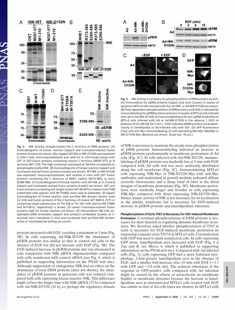
proteins increased with EGF, reaching a maximum at 5 min (Fig.3B). In cells expressing Ad-NIK-D152N the abundance ofpERM proteins was similar to that in control Ad cells in theabsence of EGF but did not increase with EGF (Fig. 3B). TheEGF-induced increase in pERM proteins also was attenuated incells transfected with NIK siRNA oligonucleotides comparedwith cells transfected with control siRNA (see Fig. 8, which ispublished as supporting information on the PNAS web site).Although suppression of endogenous NIK had no effect on theabundance of total ERM proteins (data not shown), the abun-dance of pERM proteins in quiescent cells was reduced com-pared with cells expressing kinase-inactive NIK. This differencemight reflect the longer time with NIK siRNA (72 h) comparedwith Ad-NIK-D152N (24 h), or perhaps the regulatory domain
of NIK is necessary to maintain the steady-state phosphorylationof ERM proteins. Immunolabeling indicated an increase inpERM proteins predominantly in membrane protrusions of Adcells (Fig. 3C). In cells infected with Ad-NIK-D152N, immuno-labeling of pERM proteins was markedly less at 5 min with EGFcompared with Ad cells and was more uniformly distributedalong the cell membrane (Fig. 3C). Immunolabeling MTLn3cells expressing NIK-Myc or NIK-D152N-Myc with anti-Mycantibodies and maintained in growth medium indicated diffusecytosolic f luorescence and a sharp band of label at the distalmargins of membrane protrusions (Fig. 3D). Membrane protru-sions were markedly longer and broader in cells expressingNIK-Myc compared with those expressing NIK-D152N-Myc.Hence, kinase activity of NIK is not necessary for its localizationat the plasma membrane but is necessary for EGF-inducedincrease in pERM proteins and lamellipodium extension.
Phosphorylation of Ezrin T567 Is Necessary for EGF-Induced MembraneProtrusion. C-terminal phosphorylation of ERM proteins is nec-essary for their function in regulating dynamic membrane struc-tures. We therefore asked whether phosphorylation of T567 inezrin is necessary for EGF-induced membrane protrusion byexpressing a mutant ezrin T567A in MTLn3 cells. Cotransfectionwith GFP was used to mark transfected cells. In cells expressingGFP alone, lamellipodium area increased with EGF (Fig. 4 ATop and B; see Movie 4, which is published as supportinginformation on the PNAS web site). Compared with Ad-infectedcells (Fig. 1), cells expressing GFP had a more flattened mor-phology, 2-fold greater lamellipodium area in the absence ofEGF, and a smaller fold-increase after 10 min with EGF (�1.7with GFP and �3.0 with Ad). The reduced robustness of theresponse in GFP-positive cells compared with Ad infectionmight be caused by the effects of nucleofectin on membranefluidity or membrane dynamics because the increase in lamel-lipodium area in untransfected MTLn3 cells treated with EGFwas similar to that of Ad cells (data not shown). In MTLn3 cells
Fig. 2. NIK directly phosphorylates the C terminus of ERM proteins. (A)Autoradiograms of kinase reaction (Upper) and Coomassie-stained fusionproteins (Lower) are shown. Myc-tagged WT NIK or NIK-D152N was expressedin COS-7 cells, immunoprecipitated, and used for in vitro kinase assays withGST or GST-fusion proteins containing moesin C terminus (M287-577) or Nterminus (M1-310). The high-molecular-mass band at 160 kDa corresponds toautophosphorylated NIK. (B) Autoradiograms of kinase reaction (Upper) andCoomassie-stained fusion proteins (Lower) are shown. WT NIK or NIK-D152Nwas expressed, immunoprecipitated, and reacted in vitro with GST fusionproteins containing the C terminus of NHE1, radixin (R373-585), or ezrin(E280-586). (C) Autoradiograms of kinase reaction with WT NIK as in A and B(Upper) and Coomassie-stained fusion proteins (Lower) are shown. GST andfusion proteins containing full-length moesin WT (M-WT) or moesin with T558substituted with aspartic acid (M-T558D) were used as substrates. (D Upper)Autoradiogram of kinase reaction with purified NIK domain (amino acids22–374) and fusion proteins of the C terminus of moesin WT (M414–577) orcontaining single substitutions of Thr-558 or Thr-567 with alanine (M-T558Aand M-T567A, respectively) is shown. (D Lower) Coomassie-stained fusionproteins used for kinase reaction are shown. (E) Immunoblots (IB) with an-tiphospho-ERM antibodies (Upper) and antiezrin antibodies (Lower) of C-terminal ezrin translated in vitro and incubated with purified NIK domainactive or inactivated by boiling are shown.
Fig. 3. NIK activity is necessary for phosphorylation of ERM proteins by EGF.(A) Immunoblots for pERM proteins (Upper) and ezrin (Lower) in lysates ofquiescent MTLn3 cells infected with Ad, Ad-NIK, or Ad-NIK-D152N are shown.(B) Time-dependent phosphorylation of ERM proteins with EGF is indicated byimmunoblotting for pERM proteins and ezrin in lysates of MTLn3 cells infectedwith Ad or Ad-NIK-D152N. (C) Immunolabeling with anti-pERM antibodies ofMTLn3 cells infected with Ad or Ad-NIK-D152N in the absence (�EGF) orpresence of 25 nM EGF for 5 min (�EGF) indicates pERM proteins are predom-inantly in lamellipodia in Ad-infected cells with EGF. (D) GFP fluorescence(Top) and anti-Myc immunolabeling of cells expressing NIK-Myc (Middle) orNIK-D152N-Myc (Bottom) are shown. (Scale bar: 10 �m.)
Baumgartner et al. PNAS � September 5, 2006 � vol. 103 � no. 36 � 13393
CELL
BIO
LOG
Y

transiently expressing WT ezrin, the morphology was similar tothat of cells expressing GFP alone, but lamellipodium area wasgreater in the absence and presence of EGF (Fig. 4 A Middle andB; see Movie 5, which is published as supporting information onthe PNAS web site). Cells expressing ezrin T567A (Fig. 4ABottom, white lines) were flatter and more spread, like cellsexpressing NIK-D152N (Fig. 1 A Bottom), but unlike Ad-NIK-D152N cells, they did not have increased lamellipodium area inthe absence of EGF. Adjacent untransfected cells (Fig. 4ABottom, red lines) also were more spread compared with cellstransfected with GFP or WT ezrin, perhaps because ezrin T567Ainduced changes in cell–cell adhesion. In cells expressing ezrinT567A the increase in lamellipodium area with EGF was delayedand attenuated (Fig. 4 A Bottom and B; see Movie 6, which ispublished as supporting information on the PNAS web site).These data indicate that inactive ezrin, like inactive NIK, inhibitsEGF-induced lamellipodia.
NIK Activity Is Necessary for Phosphorylation of ERM Proteins by PDGFbut Not Thrombin in Fibroblasts. Previous studies found that growthfactors induce Tyr phosphorylation of ezrin (27, 28), and throm-bin induces phosphorylation of the C-terminal conserved thre-onine in ERM proteins (29) we identified as a NIK phosphor-ylation site. To further confirm the action and specificity of NIKactivity we examined the kinetics of phosphorylation of theconserved threonine in ERM proteins in CCL39 fibroblasts,which express receptors for PDGF and thrombin (24). PDGFinduced a rapid increase in the abundance of pERM proteins inCCL39 cells infected with control Ad (Fig. 5A). In contrast to thesustained increase in pERM proteins in MTLn3 cells with EGFat 10 min (Fig. 3B), the response in CCL39 cells was moretransient, returning to unstimulated levels at 5 min. The abun-dance of pERM proteins in quiescent cells expressing Ad-NIK-D152N was similar to that in Ad controls but did not increasewith PDGF (Fig. 5A). Thrombin also increased phosphorylation
of the C-terminal conserved threonine of ERM proteins inCCL39 cells infected with control Ad that was sustained at 5 min,and this increase was not attenuated in cells infected withAd-NIK-D152N (Fig. 5B). NIK activity also was not necessaryfor activation of ERK by PDGF. The abundance of phosphor-ylated ERK, as indicated by immunoblotting cell lysates withphospho-specific antibodies for Thr-202�Tyr-204 of ERK1 andERK2, was similar in quiescent and PDGF-stimulated Ad andAd-NIK-D152N cells (Fig. 5C). These data indicate that NIKactivity is necessary for increased phosphorylation of ERMproteins by growth factors (EGF and PDGF) but not by throm-bin and suggest that NIK mediates the PDGF response by eitheracting downstream or independently of an ERK-mediatedpathway.
NIK Binds the N Terminus of Moesin. The localization of NIK andpERM proteins at the distal margins of lamellipodia and theability of NIK to phosphorylate full-length moesin in vitro (Fig.2C), which is predicted to be in closed conformation, suggest thatNIK might bind ERM proteins. An in vitro binding assayconfirmed that in vitro-translated 35S-labeled NIK bound a GSTfusion of the N terminus of moesin (M1-310) but not the Cterminus (M287-577) or GST (Fig. 6A). NIK also bound the Nterminus of ezrin (data not shown), and although less strongly,full-length moesin WT and T558D (Fig. 6B). To determine themoesin-binding site in NIK, we incubated GST-M1-310 with invitro-translated NIK containing truncations in the C-terminalregulatory domain. Binding equivalent to that with full-lengthNIK was found for truncated NIK containing residues 1–321.However, NIK (residues 1–288), which contains only the kinase
Fig. 4. Phosphorylation of T567 in ezrin is necessary for lamellipodiumextension with EGF. (A) Time-lapse images of MTLn3 cells transfected withempty vector (Control) or vector encoding WT ezrin (Ezrin) or ezrin T567A(EzrinT567A) and treated with EGF (25 nM) are shown for the indicated times.See also Movies 4–6. Transfected cells (outlined in white) were identified bycoexpression of GFP. Lamellipodium area was determined as in Fig. 1. (B)Mean � SEM of lamellipodium area of six cells in three cell preparations isshown. (Scale bar: 10 �m.)
Fig. 5. NIK activity is necessary for phosphorylation of ERM proteins by PDGFbut not by thrombin. Representative immunoblots of lysates prepared fromCCL39 fibroblasts infected with Ad or Ad-NIK-D152N and treated for theindicated times with PDGF (50 ng�ml) (A Upper and C) or thrombin (30 nM) (BUpper) are shown. Blots were probed with anti-pERM antibodies (A and B),antiphospho-ERK antibodies (C), or antiezrin antibodies (A–C). Data normal-ized from five (A Lower) and three (B Lower) cell preparations are expressedas the mean � SEM of fold-change relative to abundance of pERM proteins inAd-infected cells at time 0. The immunoblot in C is representative of five cellpreparations.
13394 � www.pnas.org�cgi�doi�10.1073�pnas.0605950103 Baumgartner et al.

domain, did not bind GST-M1-310 (Fig. 6C). These data indicatethat the N-terminal 33 aa of the regulatory domain of NIK arenecessary for binding ERM proteins.
Transient expression of NIK1-340 in CCL39 fibroblasts wassufficient to increase phosphorylation of ERM proteins at theplasma membrane (Fig. 6D). NIK1-340 does not contain bindingsites for Nck (proline-rich motifs between residues 392 and 618)and MEKK1 (amino acids 908-1233), which suggests that NIKbinding to Nck or MEKK1 is not necessary for NIK recruitmentto the plasma membrane to phosphorylate ERM proteins.Because the attachment of cells expressing only the kinasedomain of NIK (residues 1–288) was markedly impaired, we wereunable to determine whether the moesin-binding domain wasnecessary to increase phosphorylation of ERM proteins in cells.We also confirmed that NIK associates with endogenous ezrin.However, ezrin coprecipitated with transiently expressed NIK-D152N (Fig. 6E) but not with WT NIK (data not shown),suggesting that ERM proteins may be allosterically regulated byNIK-induced phosphorylation.
DiscussionNIK and ERM proteins have been shown independently toregulate cell morphology, but their functional interaction incontrolling plasma membrane dynamics was not previouslyrecognized. We found that ERM proteins are substrates for NIK,NIK and pERM proteins localize at the distal margins oflamellipodia, and NIK activity is necessary for phosphorylationof ERM proteins in response to EGF and PDGF. Additionally,NIK activity and phosphorylation of ezrin T567 are necessary forincreased lamellipodium extension by growth factors. Collec-tively, these data suggest that phosphorylation of ERM proteins
by NIK may constitute a signaling mechanism controlling growthfactor-induced membrane protrusion and cell morphology.
Substrates for NIK and its orthologs in pathways regulating cellmorphology remain poorly understood. NIK and its Drosophilaortholog Msn increase phosphorylation of JNK (1) and BSK (30),respectively. Phosphorylation of JNK by NIK, however, is not directbut mediated through NIK binding to a scaffolding complex withMEKK1. Msn also increases phosphorylation of Bicaudal-D (31) toregulate epithelial planar polarity (9); however, whether Bicau-dal-D is directly phosphorylated by Msn is unknown. Moesin, thesingle ERM protein in Drosophila, also is necessary to maintainepithelial integrity (32), and it will be interesting to determinewhether Dmoesin is a substrate for Msn. HGK, the human homologof NIK, acts downstream of receptors for hepatocyte growth factorto increase phosphorylation of STAT3 (8). Although STAT3coprecipitates with HGK, its direct phosphorylation is undeter-mined. In mammalian fibroblasts NIK binds to the Na-H exchangerNHE1 and directly phosphorylates NHE1 to increase H� efflux inresponse to PDGF (24). ERM proteins also bind to NHE1 (33), andby anchoring actin filaments ERM proteins maintain the localiza-tion of NHE1 at the leading edge of migrating fibroblasts. Becausea leading-edge H� efflux by NHE1 is necessary for membraneprotrusion and directed migration (34), the possibility of a struc-tural and functional link between NIK, ERM proteins, and NHE1in promoting membrane protrusion is intriguing.
Activation of ERM proteins requires two sequential steps(18). First, N-terminal binding to phosphatidylinositol 4,5-bisphosphate recruits ERM proteins from the cytosol to theplasma membrane and permits N-terminal binding to transmem-brane proteins. ERM proteins can be retained at the plasmamembrane but not bound to F-actin (33). A second activationstep, C-terminal phosphorylation, is necessary for F-actin bind-ing. The required second activation step of phosphoryation mayrestrict actin tethering by ERM proteins to specialized mem-brane domains, such as protruding membranes. Although sev-eral kinases, including myotonic dystrophy kinase-relatedCdc42-binding kinase (19), protein kinase C� (20), and Rho-associated kinase (ROCK) (21), phosphorylate the conservedC-terminal threonine in ERM proteins, they have not beenshown to mediate growth factor-induced phosphorylation ofERM proteins at lamellipodia.
Because activation of ERM proteins promotes F-actin anchoringto the plasma membrane, their phosphorylation by NIK likelystabilizes extending lamellipodia. However, we predict that NIKalso regulates membrane dynamics through mechanisms indepen-dent of ERM proteins. MTLn3 cells expressing NIK-D152N, butnot ezrin T567A, had constitutive, albeit small, ruffles. Substrates,including NHE1 (24), and possibly gelsolin or cofilin, which arephosphorylated by the closely related kinases TNIK (2) and NRK(4), respectively, might contribute to NIK-dependent membraneprotrusion. Additionally, NIK phosphorylation of ERM proteins orother substrates might act coordinately with Nck to promote orstabilize membrane protrusions. Our finding that NIK activity isnecessary to phosphorylate ERM proteins in response to EGF andPDGF, but not to thrombin, is consistent with NIK binding to theSrc homology 3 domain of Nck (1), an adaptor protein associatedwith receptor tyrosine kinases, and with Msn binding to DOCK(10), the Drosophila ortholog of Nck. Nck also binds and activatesthe Wiskott-Aldrich syndrome protein WASP (35) and the WASPfamily verprolin homologous protein WAVE (36), which promoteactin assembly by the Arp2�3 complex and membrane protrusion.Although NIK may act coordinately with Nck to regulate mem-brane dynamics, its phosphorylation of ERM proteins can occurindependently of Nck because truncated NIK 1-321 lacking theC-terminal Nck-binding domain was sufficient to increase phos-phorylation of ERM proteins in quiescent cells. Additionally,kinase inactive NIK-D152N did not block activation of ERK1�ERK2 by PDGF, suggesting that NIK regulates ERM protein
Fig. 6. NIK 1-310 binds the FERM domain of moesin and is sufficient tophosphorylate ERM proteins in cells. (A Upper) Autoradiogram of binding assaywithfull-lengthNIKtranslated invitro in thepresenceof [35S]methionineandGSTand GST-moesin FERM domain (M1-310) and C terminus (M287-577) is shown. (ALower) Coomassie-stained fusion proteins are shown. (B) NIK binding to theindicated fusion proteins relative to binding GST-M1-310 is shown. (C) Schematicrepresentation of full-length (resides 1–1289) and C-terminal-truncated NIK usedin binding assays is shown. ��, domains with comparable binding to GST-moesin1–310. (D) Expression of an EGFP fusion of truncated NIK-1-340 is sufficient toincrease phosphorylation of endogenous ERM proteins in CCL39 fibroblasts.Phosphorylation of ERM proteins was determined by immunolabeling with anti-pERMantibody.*, cellsexpressingNIK-1-340-EGFPare identifiedbyfluorescence.(E) Ezrin coprecipitates with Myc-tagged kinase-inactive NIK (mNIK-D152N). (Topand Middle) Myc immunoprecipitates from lysates of CCL39 fibroblasts trans-fected with empty vector (�) or vector containing Myc-NIK-D152N (�) wereprobed with antibodies against Myc (Top) and ezrin (Middle). (Bottom) Ezrin incell lysates was determined by immunoblotting. IP, immunoprecipitation; IB,immunoblotting.
Baumgartner et al. PNAS � September 5, 2006 � vol. 103 � no. 36 � 13395
CELL
BIO
LOG
Y
phosphorylation downstream or independently of an ERK-mediated pathway, the latter possibility being consistent with NIKacting independently of Nck.
Our findings indicate that activation of ERM proteins by NIKis a cellular mechanism to promote local alterations in cellmorphology in response to growth factors. This mechanism islikely important in migrating cells because NIK activity isnecessary for growth factor-induced phosphorylation of ERMproteins in lamellipodia. Because activation of NIK (8) and ezrin(37) is implicated in processes related to tumor cell dissemina-tion with aberrant growth factor signaling, a functional interac-tion between NIK and ERM proteins might play a previouslyunrecognized role in tumor cell metastasis.
Materials and MethodsMaterials and methods for cell culture, transfections, in vitrotranslation, in vitro kinase and binding assays, immunoblotting,immunolabeling, and immunoprecipitation have been described (8,24, 25, 33). All methods are described in detail in Supporting Text,which is published as supporting information on the PNAS web site.
Ad and siRNA Oligonucleotides. To generate NIK and NIK-D152NAd, full-length WT mouse NIK and NIK-D152N (24) wereexcised by HindIII�NotI digestion from pCRII-TOPO, ligatedinto the same sites in pAD-Track-CMV, and amplified inEscherichia coli XL1-blue. Positive clones were identified byanalytical restriction enzyme digestion, and plasmids from theseclones were linearized by Pme1 digestion, gel-purified, and usedto transform the BJ5183-AD-1 strain of E. coli (Stratagene, LaJolla, CA) carrying the pADEasy-1 plasmid that encodes theAd-5 genome. Plasmid DNA was purified from kanamycin-resistant colonies and analyzed by PacI digestion. 293Ad cellswere transfected with recombined pTrack-CMV andpADEasy-1, and 8 days after transfection spreading Ad withinthe 293Ad cell monolayer was determined by plaque formationand fluorescence of GFP was encoded by pADEasy-1. Ad was
collected by repetitive freeze-thaw cycles followed by centrifu-gation at 12,000 � g for 10 min at 4°C and used for subsequentinfection of 293Ad cells. Optimal titers for cell infection reached90–100% infection after 48 h, determined by GFP fluorescence.
The siRNA oligonucleotide duplex (5�-UAA UGA AAG CACCAU AGU AUG UUG C-3�) to rat NIK was synthesized byInvitrogen, Carlsbad, CA (Stealth Select RNAi). Stealth RNAiNegative Control Low GC (low GC content; Invitrogen) was usedas a control. siGLO RNA-induced silencing complex-free siRNA(Dharmacon, Lafayette, CO) was used to monitor transfectionefficiency. MTLn3 cells were cotransfected with 200 nm NIK orcontrol siRNA and 150 nm siGLO by using Oligofectamine (In-vitrogen) according to the manufacturer’s protocol. After 48 h cellswere replated and maintained in the absence of FBS for 4 h beforeimaging or harvesting at 72 h posttransfection.
Video Microscopy. MTLn3 cells in six-well plates were placed on apreheated, climate-regulated stage mounted on a Zeiss (Thorn-wood, NY) Axiovert S-100 microscope. Images were capturedevery 20 s, beginning 2 min before adding EGF and ending 10 minafter adding EGF (25 nM; Invitrogen) by using a Spot CCD camerathrough a �40 Hoffmann modulation objective and operated withOpenlab software. Lamellipodium area was designated as theregion from the cell body to the periphery of the plasma membrane.Area calculations performed with Openlab software were made bysubtracting the area of the cell body from the total cell area.
We thank Hitesh Patel for assistance with video microscopy; NeetuGupta for help with transfection of MTLn3 cells; Torsten Wittmann forhelp with kymography; and Brandon LaMere for technical assistance.This work was supported by a Swiss National Science Foundationfellowship (to M.B.) and National Institutes of Health (NIH) Grants T32DE07204 (to A.L.S.) and GM47413 (to D.L.B.). This investigation wasconducted in a facility constructed with support from Research FacilitiesImprovement Program Grant C06 RR16490 from the National Centerfor Research Resources (NIH). Work conducted at Sugen was funded bythe Pfizer Corporation.
1. Su, Y. C., Han, J., Xu, S., Cobb, M. & Skolnik, E. Y. (1997) EMBO J. 16,1279–1290.
2. Fu, C. A., Shen, M., Huang, B. C., Lasaga, J., Payan, D. G. & Luo, Y. (1999)J. Biol. Chem. 274, 30729–30737.
3. Hu, Y., Leo, C., Yu, S., Huang, B. C., Wang, H., Shen, M., Luo, Y.,Daniel-Issakani, S., Payan, D. G. & Xu, X. (2004) J. Biol. Chem. 279,54387–54397.
4. Nakano, K., Kanai-Azuma, M., Kanai, Y., Moriyama, K., Yazaki, K., Hayashi,Y. & Kitamura, N. (2003) Exp. Cell Res. 287, 219–227.
5. Su, Y. C., Maurel-Zaffran, C., Treisman, J. E. & Skolnik, E. Y. (2000) Mol. CellBiol. 20, 4736–4744.
6. Poinat, P., De Arcangelis, A., Sookhareea, S., Zhu, X., Hedgecock, E. M.,Labouesse, M. & Georges-Labouesse, E. (2002) Curr. Biol. 12, 622–631.
7. Xue, Y., Wang, X., Li, Z., Gotoh, N., Chapman, D. & Skolnik, E. Y. (2001)Development (Cambridge, U.K.) 128, 1559–1572.
8. Wright, J. H., Wang, X., Manning, G., LaMere, B. J., Le, P., Zhu, S., Khatry,D., Flanagan, P. M., Buckley, S. D., Whyte, D. B., et al. (2003) Mol. Cell Biol.23, 2068–2082.
9. Paricio, N., Feiguin, F., Boutros, M., Eaton, S. & Mlodzik, M. (1999) EMBOJ. 18, 4669–4678.
10. Ruan, W., Pang, P. & Rao, Y. (1999) Neuron 24, 595–605.11. Bretscher, A., Edwards, K. & Fehon, R. G. (2002) Nat. Rev. Mol. Cell Biol. 3,
586–599.12. Takeuchi, K., Sato, N., Kasahara, H., Funayama, N., Nagafuchi, A., Yonemura,
S. & Tsukita, S. (1994) J. Cell Biol. 125, 1371–1384.13. Crepaldi, T., Gautreau, A., Comoglio, P. M., Louvard, D. & Arpin, M. (1997)
J. Cell Biol. 138, 423–434.14. Saotome, I., Curto, M. & McClatchey, A. I. (2004) Dev. Cell 6, 855–864.15. Mackay, D. J., Esch, F., Furthmayr, H. & Hall, A. (1997) J. Cell Biol. 138,
927–938.16. Lamb, R. F., Ozanne, B. W., Roy, C., McGarry, L., Stipp, C., Mangeat, P. &
Jay, D. G. (1997) Curr. Biol. 7, 682–688.17. Castelo, L. & Jay, D. G. (1999) Mol. Biol. Cell 10, 1511–1520.18. Fievet, B. T., Gautreau, A., Roy, C., Del Maestro, L., Mangeat, P., Louvard,
D. & Arpin, M. (2004) J. Cell Biol. 164, 653–659.
19. Nakamura, N., Oshiro, N., Fukata, Y., Amano, M., Fukata, M., Kuroda, S.,Matsuura, Y., Leung, T., Lim, L. & Kaibuchi, K. (2000) Genes Cells 5, 571–581.
20. Ng, T., Parsons, M., Hughes, W. E., Monypenny, J., Zicha, D., Gautreau, A.,Arpin, M., Gschmeissner, S., Verveer, P. J., Bastiaens, P. I. & Parker, P. J.(2001) EMBO J. 20, 2723–2741.
21. Oshiro, N., Fukata, Y. & Kaibuchi, K. (1998) J. Biol. Chem. 273, 34663–34666.22. Matsui, T., Yonemura, S. & Tsukita, S. (1999) Curr. Biol. 9, 1259–1262.23. Becker, E., Huynh-Do, U., Holland, S., Pawson, T., Daniel, T. O. & Skolnik,
E. Y. (2000) Mol. Cell Biol. 20, 1537–1545.24. Yan, W., Nehrke, K., Choi, J. & Barber, D. L. (2001) J. Biol. Chem. 276,
31349–31356.25. Chan, A. Y., Raft, S., Bailly, M., Wyckoff, J. B., Segall, J. E. & Condeelis, J. S.
(1998) J. Cell Sci. 111, 199–211.26. Hipfner, D. R., Keller, N. & Cohen, S. M. (2004) Genes Dev. 18, 2243–2248.27. Bretscher, A. (1989) J. Cell Biol. 108, 921–930.28. Srivastava, J., Elliott, B. E., Louvard, D. & Arpin, M. (2005) Mol. Biol. Cell 16,
1481–1490.29. Nakamura, F., Huang, L., Pestonjamasp, K., Luna, E. J. & Furthmayr, H.
(1999) Mol. Biol. Cell 10, 2669–2685.30. Su, Y. C., Treisman, J. E. & Skolnik, E. Y. (1998) Genes Dev. 12, 2371–2380.31. Houalla, T., Hien Vuong, D., Ruan, W., Suter, B. & Rao, Y. (2005) Mech. Dev.
122, 97–108.32. Speck, O., Hughes, S. C., Noren, N. K., Kulikauskas, R. M. & Fehon, R. G.
(2003) Nature 421, 83–87.33. Denker, S. P., Huang, D. C., Orlowski, J., Furthmayr, H. & Barber, D. L. (2000)
Mol. Cell 6, 1425–1436.34. Denker, S. P. & Barber, D. L. (2002) J. Cell Biol. 159, 1087–1096.35. Rohatgi, R., Nollau, P., Ho, H. Y., Kirschner, M. W. & Mayer, B. J. (2001)
J. Biol. Chem. 276, 26448–26452.36. Eden, S., Rohatgi, R., Podtelejnikov, A. V., Mann, M. & Kirschner, M. W.
(2002) Nature 418, 790–793.37. Yu, Y., Khan, J., Khanna, C., Helman, L., Meltzer, P. S. & Merlino, G. (2004)
Nat. Med. 10, 175–181.
13396 � www.pnas.org�cgi�doi�10.1073�pnas.0605950103 Baumgartner et al.