Clathrin Adaptor epsinR Is Required for Retrograde Sorting on Early Endosomal Membranes
14
Developmental Cell, Vol. 6, 525–538, April, 2004, Copyright 2004 by Cell Press Clathrin Adaptor epsinR Is Required for Retrograde Sorting on Early Endosomal Membranes routes is commonly termed clathrin-independent endo- cytosis (Nichols and Lippincott-Schwartz, 2001; Johan- nes and Lamaze, 2002; Conner and Schmid, 2003). The Agne ` s Saint-Pol, 1,6 Bele ´ n Ye ´ lamos, 1,6 Mohamed Amessou, 1 Ian G. Mills, 4,7 Marc Dugast, 3 Danie ` le Tenza, 2 Peter Schu, 5 molecular mechanisms of clathrin-independent endocy- Claude Antony, 4 Harvey T. McMahon, 4 tosis remain to be established. Recent data have shown Christophe Lamaze, 1 and Ludger Johannes 1, * that multiple pathways exist. Some of the clathrin-inde- 1 Laboratoire Trafic et Signalisation pendent uptake pathways, including IL2 receptor endo- 2 Unite ´ de Microscopie Electronique cytosis (Lamaze et al., 2001) and SV40 endocytosis UMR144 Curie/CNRS (Pelkmans and Helenius, 2002), are controlled by the 3 INSERM U520 GTPase dynamin. Other uptake pathways, including Institut Curie those used by some GPI-anchored proteins, are inde- 26 rue d’Ulm pendent of the commonly used dynamin isoforms 75248 Paris Cedex 05 (Skretting et al., 1999; Sabharanjak et al., 2002). France Many endocytosed molecules reach the conventional 4 MRC Laboratory of Molecular Biology transferrin receptor (TfR)-positive early endosome (EE), Neurobiology Division which serves as a sorting station (Gruenberg and Max- Hills Road field, 1995; Mellman, 1996). Some receptors are recycled Cambridge CB2 2QH to the plasma membrane to be recharged with ligands, United Kingdom whereas others enter the late endocytic pathway for 5 Georg-August-Universita ¨ t Go ¨ ttingen degradation. The retrograde route bypasses the recy- Zentrum fu ¨ r Biochimie und Molekulare Zellbiologie cling and degradation pathways, allowing specific pro- Heinrich-Du ¨ ker-Weg 12 teins and lipids to reach other intracellular compart- 37073 Go ¨ ttingen ments such as the trans-Golgi network (TGN), the Golgi Germany cisternae, the endoplasmic reticulum and, in some in- stances, the cytosol (Mallard et al., 1998; for a review, see Johannes and Goud, 1998). This pathway has been Summary studied in detail to explain the cellular entry of some protein toxins, such as cholera toxin, ricin, and Shiga Retrograde transport links early/recycling endosomes toxin (Lord and Roberts, 1998; Sandvig and van Deurs, to the trans-Golgi network (TGN), thereby connecting 2000). Other studies have shown that the cellular pro- the endocytic and the biosynthetic/secretory path- teins TGN38/46 (Mallard et al., 1998, 2002; Ghosh et ways. To determine how internalized molecules are al., 1998), GPP130, and GP73 (Puri et al., 2002), all of targeted to the retrograde route, we have interfered unknown function, use the retrograde route. with the function of clathrin and that of two proteins In some cases, clathrin-independent endocytosis has that interact with it, AP1 and epsinR. We found that been described to be linked to endosomes that are de- the glycosphingolipid binding bacterial Shiga toxin en- void of classical markers of the early endocytic pathway tered cells efficiently when clathrin expression was (Pelkmans et al., 2001; Nichols et al., 2001). The relation- inhibited. However, retrograde transport of Shiga toxin ship between theses structures and the conventional to the TGN was strongly inhibited. This allowed us to EE has remained debatable (see Nichols, 2002, versus show that for Shiga toxin, retrograde sorting on early/ Sabharanjak et al., 2002). recycling endosomes depends on clathrin and epsinR, Although the conventional EE is known to be a sorting but not AP1. EpsinR was also involved in retrograde station, the molecular mechanisms underlying the differ- transport of two endogenous proteins, TGN38/46 and ential distribution of internalized molecules have not mannose 6-phosphate receptor. In conclusion, our been studied in detail. COP-type coats are localized on work reveals the existence of clathrin-independent the EE and implicated in sorting into the late endocytic and -dependent transport steps in the retrograde pathway (Whitney et al., 1995; Aniento et al., 1996). The route, and establishes a function for clathrin and ep- role of clathrin in the early endocytic pathway is more sinR at the endosome-TGN interface. uncertain, even though ultrastructural imaging has clearly shown that it is present on this compartment (Stoorvogel et al., 1996; Mallard et al., 1998; Sachse et Introduction al., 2002). Clathrin may play a role in transferrin (Tf) recycling (Bennett et al., 2001; Wettey et al., 2002), pos- Clathrin plays a central role in endocytosis at the plasma sibly via perinuclear recycling endosomal (RE) tubules membrane. However, alternative internalization path- (van Dam and Stoorvogel, 2002). Several studies have ways also exist. Cellular uptake via these alternative suggested that clathrin coats containing the adaptor protein 1 (AP1) are involved in retrograde transport (Mal- *Correspondence: [email protected] lard et al., 1998; Meyer et al., 2000; Folsch et al., 2001; 6 These authors contributed equally to this work. Crump et al., 2001; Valdivia et al., 2002). 7 Present address: Department of Oncology, University of Cam- EpsinR, one of the proteins that interacts with AP1, bridge, c/o Hutchinson/MRC Cancer Research Centre, Hills Road, Cambridge CB2 2XZ, United Kingdom. can also directly associate with clathrin (Kalthoff et al.,
-
Upload
wwwunistra -
Category
Documents
-
view
0 -
download
0
Transcript of Clathrin Adaptor epsinR Is Required for Retrograde Sorting on Early Endosomal Membranes

Developmental Cell, Vol. 6, 525–538, April, 2004, Copyright 2004 by Cell Press
Clathrin Adaptor epsinR Is Requiredfor Retrograde Sorting on Early Endosomal Membranes
routes is commonly termed clathrin-independent endo-cytosis (Nichols and Lippincott-Schwartz, 2001; Johan-nes and Lamaze, 2002; Conner and Schmid, 2003). The
Agnes Saint-Pol,1,6 Belen Yelamos,1,6
Mohamed Amessou,1 Ian G. Mills,4,7
Marc Dugast,3 Daniele Tenza,2 Peter Schu,5
molecular mechanisms of clathrin-independent endocy-Claude Antony,4 Harvey T. McMahon,4
tosis remain to be established. Recent data have shownChristophe Lamaze,1 and Ludger Johannes1,*that multiple pathways exist. Some of the clathrin-inde-1Laboratoire Trafic et Signalisationpendent uptake pathways, including IL2 receptor endo-2 Unite de Microscopie Electroniquecytosis (Lamaze et al., 2001) and SV40 endocytosisUMR144 Curie/CNRS(Pelkmans and Helenius, 2002), are controlled by the3 INSERM U520GTPase dynamin. Other uptake pathways, includingInstitut Curiethose used by some GPI-anchored proteins, are inde-26 rue d’Ulmpendent of the commonly used dynamin isoforms75248 Paris Cedex 05(Skretting et al., 1999; Sabharanjak et al., 2002).France
Many endocytosed molecules reach the conventional4 MRC Laboratory of Molecular Biologytransferrin receptor (TfR)-positive early endosome (EE),Neurobiology Divisionwhich serves as a sorting station (Gruenberg and Max-Hills Roadfield, 1995; Mellman, 1996). Some receptors are recycledCambridge CB2 2QHto the plasma membrane to be recharged with ligands,United Kingdomwhereas others enter the late endocytic pathway for5Georg-August-Universitat Gottingendegradation. The retrograde route bypasses the recy-Zentrum fur Biochimie und Molekulare Zellbiologiecling and degradation pathways, allowing specific pro-Heinrich-Duker-Weg 12teins and lipids to reach other intracellular compart-37073 Gottingenments such as the trans-Golgi network (TGN), the GolgiGermanycisternae, the endoplasmic reticulum and, in some in-stances, the cytosol (Mallard et al., 1998; for a review,see Johannes and Goud, 1998). This pathway has beenSummarystudied in detail to explain the cellular entry of someprotein toxins, such as cholera toxin, ricin, and ShigaRetrograde transport links early/recycling endosomestoxin (Lord and Roberts, 1998; Sandvig and van Deurs,to the trans-Golgi network (TGN), thereby connecting2000). Other studies have shown that the cellular pro-
the endocytic and the biosynthetic/secretory path-teins TGN38/46 (Mallard et al., 1998, 2002; Ghosh et
ways. To determine how internalized molecules are al., 1998), GPP130, and GP73 (Puri et al., 2002), all oftargeted to the retrograde route, we have interfered unknown function, use the retrograde route.with the function of clathrin and that of two proteins In some cases, clathrin-independent endocytosis hasthat interact with it, AP1 and epsinR. We found that been described to be linked to endosomes that are de-the glycosphingolipid binding bacterial Shiga toxin en- void of classical markers of the early endocytic pathwaytered cells efficiently when clathrin expression was (Pelkmans et al., 2001; Nichols et al., 2001). The relation-inhibited. However, retrograde transport of Shiga toxin ship between theses structures and the conventionalto the TGN was strongly inhibited. This allowed us to EE has remained debatable (see Nichols, 2002, versusshow that for Shiga toxin, retrograde sorting on early/ Sabharanjak et al., 2002).recycling endosomes depends on clathrin and epsinR, Although the conventional EE is known to be a sortingbut not AP1. EpsinR was also involved in retrograde station, the molecular mechanisms underlying the differ-transport of two endogenous proteins, TGN38/46 and ential distribution of internalized molecules have notmannose 6-phosphate receptor. In conclusion, our been studied in detail. COP-type coats are localized onwork reveals the existence of clathrin-independent the EE and implicated in sorting into the late endocyticand -dependent transport steps in the retrograde pathway (Whitney et al., 1995; Aniento et al., 1996). Theroute, and establishes a function for clathrin and ep- role of clathrin in the early endocytic pathway is moresinR at the endosome-TGN interface. uncertain, even though ultrastructural imaging has
clearly shown that it is present on this compartment(Stoorvogel et al., 1996; Mallard et al., 1998; Sachse etIntroductional., 2002). Clathrin may play a role in transferrin (Tf)recycling (Bennett et al., 2001; Wettey et al., 2002), pos-Clathrin plays a central role in endocytosis at the plasmasibly via perinuclear recycling endosomal (RE) tubulesmembrane. However, alternative internalization path-(van Dam and Stoorvogel, 2002). Several studies haveways also exist. Cellular uptake via these alternativesuggested that clathrin coats containing the adaptorprotein 1 (AP1) are involved in retrograde transport (Mal-
*Correspondence: [email protected] et al., 1998; Meyer et al., 2000; Folsch et al., 2001;6 These authors contributed equally to this work.Crump et al., 2001; Valdivia et al., 2002).7 Present address: Department of Oncology, University of Cam-
EpsinR, one of the proteins that interacts with AP1,bridge, c/o Hutchinson/MRC Cancer Research Centre, Hills Road,Cambridge CB2 2XZ, United Kingdom. can also directly associate with clathrin (Kalthoff et al.,
Developmental Cell526
2002; Wasiak et al., 2002; Mills et al., 2003; Hirst et To address the function of clathrin in STxB trafficking,al., 2003). In addition, epsinR can bind to phosphatidyl we designed an RNA interference (RNAi) tool that al-inositol (PtdIns) lipids via its ENTH domain. Like AP1, lowed us to inhibit clathrin heavy chain (CHC) expressionepsinR has been localized to Golgi membranes and pe- to 20% of control levels (Figure 1F). We carried outripheral structures that are most likely endosomes (Mills whole-mount analysis of clathrin RNAi cells and mock-et al., 2003; Hirst et al., 2003). However, this localization transfected cells after 15 min internalization of STxB/of epsinR does not depend on AP1 expression (Hirst et HRP at 37�C. This confirmed that clathrin was presental., 2003). Unlike epsins involved in coated pit formation on STxB-containing early/recycling endosomal buds ofat the plasma membrane, epsinR is strongly enriched different sizes both at low temperatures (Figure 1E) andin clathrin-coated vesicles and stably associates with at 37�C (Figure 1G, left). Interestingly, even in CHC RNAi-them (Mills et al., 2003). treated cells, in which clathrin immunolabeling was al-
In this study, we showed that the bacterial Shiga toxin most lost, STxB/HRP still entered the endosomal systemcan enter cells efficiently even when clathrin expression (Figure 1G, left). STxB-containing endosomal mem-was inhibited. However, the retrograde transport of the branes were strongly labeled for the �-adaptin subunitShiga toxin from EE/RE to the TGN was strongly reduced of the clathrin adaptor AP1, even in CHC RNAi-treatedin these conditions. EpsinR was localized to endosomal cells that were recognized via thick tubular elements inmembranes, and involved in the retrograde transport of their cytoplasm (Figure 1G, middle). Similar observa-exogenous Shiga toxin and endogenous TGN38/46 and tions were made for the clathrin, AP1, and PtdIns lipidmannose 6-phosphate receptor of 300 kDa (MPR300, binding protein epsinR (Kalthoff et al., 2002; Wasiak etalso termed cation-independent MPR). Thus, our study al., 2002; Mills et al., 2003; Hirst et al., 2003), anotherestablishes a role for epsinR/clathrin in retrograde sort- candidate for recruiting clathrin to internal membranesing at the EE/RE-TGN interface. (Figure 1G, right). These data clearly show that a specific
clathrin machinery is present on STxB-containing early/Results recycling endosomal membranes.
A Specific Clathrin Machinery on Endosomal Functional Analysis of ClathrinMembranes of the Retrograde Route in the Retrograde RouteThe whole-mount technique developed by Stoorvogel We first studied the effect of CHC RNAi treatment onand colleagues allows the in-depth inspection of cyto- the retrograde transport of STxB by using immunofluo-plasmic membranes by using an ultrastructural method rescence. In control conditions, Tf was efficiently inter-(Stoorvogel et al., 1996). For the analysis of the retro- nalized and STxB was transported to the TGN (Figuregrade route, horseradish peroxidase (HRP) was cova- 2A). In CHC RNAi-treated cells, the uptake of Tf waslently linked to a variant of the Shiga toxin B-subunit strongly inhibited (Figure 2B), as expected. In these con-(STxB) that was specifically constructed for site-directed ditions, STxB appeared in tubular and vesicular struc-chemical coupling (Haicheur et al., 2003). STxB is a non- tures, instead of being concentrated in perinuclear Golgitoxic protein used as a bona fide retrograde transport membranes (Figure 2B). The tubular structures con-marker (see Johannes, 2002, for a review). Immunofluo- tained TfR (Figure 2C), suggesting that they were ofrescence (Figure 1A) and immunoelectron microscopy
early/recycling endosomal origin. The TfR cell surface(Figures 1B–1C) showed that HRP-coupled STxB was
signal was 8-fold stronger in these conditions than intargeted normally to the TGN and Golgi cisternae. This
control conditions, indicating that a significant fractionconfirms that the trafficking of STxB was not altered byof the receptor was blocked at the plasma membranechemical crosslinking.(data not shown).We then used the whole-mount technique (Stoorvogel
To characterize further the TfR-positive compartmentet al., 1996) to compare the intracellular distribution ofin which STxB accumulated in CHC RNAi cells, we usedSTxB/HRP to that of Tf/HRP, which has previously beenthree known early and recycling endosomal markers.used for these studies. Both Tf/HRP and STxB/HRPThe STxB-containing tubules were strongly labeled withwere internalized by HeLa cells at 19.5�C, i.e., in condi-anti-Rab11 antibody and with GFP-tagged Rab4 (Fig-tions in which retrograde transport of STxB to the TGN/ures 2D–2E), clearly indicating that they belong to theGolgi is inhibited (Mallard et al., 1998). Cells were pre-recycling branch of the early endocytic pathway (Son-pared for whole-mount analysis and stained with anti-nichsen et al., 2000). Consistently, they were only lightlyclathrin antibody. Membranes containing HRP-coupledlabeled by EEA1 (Figure 2F), a PtdIns(3)P binding proteinligands appeared dark due to the deposit of a polymer-that interacts with Rab5 on the EE (Simonsen et al.,ization product. Some Tf/HRP was detected in early1998).endosomal vacuoles while most was found in tubules
Morphological analysis suggested that the transportand in 60 nm clathrin-coated buds (Figure 1D, arrows),of STxB to the TGN/Golgi, but not endocytosis intoas described before (Stoorvogel et al., 1996). In contrast,EE/RE, was inhibited in CHC RNAi-treated cells. Welarge amounts of STxB/HRP were found in clathrin-con-used quantitative biochemical techniques to test thistaining early endosomal vacuoles (Figure 1E, arrow-hypothesis. When measuring endocytosis with biotinyl-heads), endosomal tubules, and clathrin-coated budsated ligands (i.e., Tf or STxB), we observed that in CHCof different size (arrows). Thus, although STxB and TfRNAi-treated cells, Tf uptake was inhibited (Figure 3A)colocalize extensively when examined by immunofluo-to a similar extent to cells in which the clathrin generescence microscopy (Mallard et al., 1998), both pro-was genetically inactivated (Wettey et al., 2002). In con-teins appeared to be differentially enriched in subdo-trast to that of Tf, the endocytosis of STxB was onlymains of the early/recycling endosomal membrane
system. slightly affected in these conditions (Figure 3B). The

EpsinR/Clathrin Coat for Retrograde Sorting527
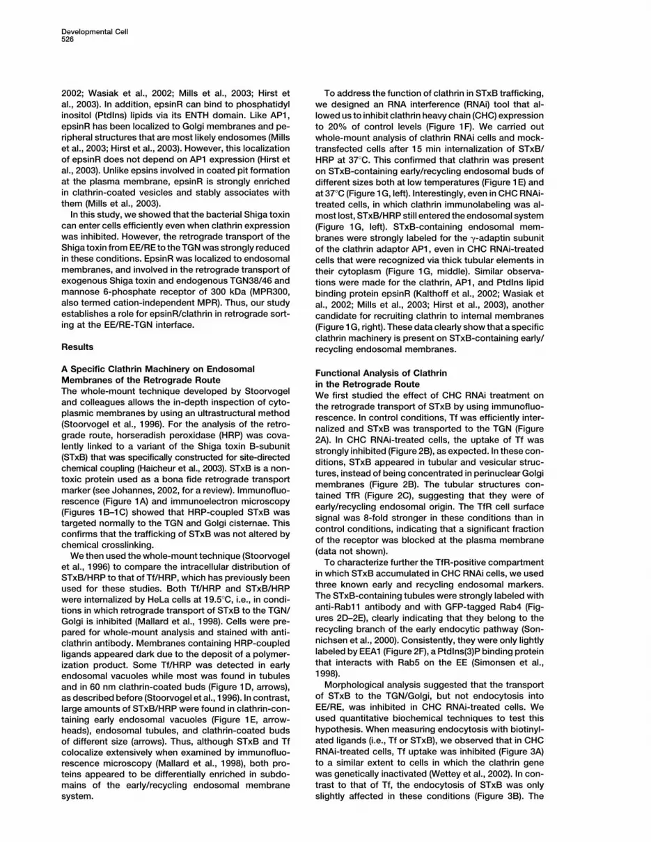
Figure 1. Analysis of the Clathrin Machinery on the STxB-Containing Early/Recycling Endosome
(A) HRP was chemically coupled to a STxB variant. The coupling product was bound to HeLa cells on ice and incubated for 45 min at 37�Cbefore fixation and immunolabeling with antibodies against HRP and the Golgi/TGN marker Rab6. Note that when coupled to STxB, HRPefficiently accumulated in perinuclear Golgi membranes. Bar � 10 �m.(B and C) STxB/HRP was internalized into HeLa cells for 45 min at 37�C. After cryosectioning, cells were fixed and immunolabeled. Note thatHRP and STxB were readily detected in the TGN and in the cisternae of the Golgi apparatus. Bar � 100 nm.(D and E) Whole-mount preparations with (D) Tf/HRP and (E) STxB/HRP internalized at 19.5�C. STxB-HRP- or Tf/HRP-containing membranesappear dark due to the formation of the DAB precipitate. Clathrin: 10 nm gold particles. Bar � 200 nm.(F) RNAi efficiently down-modulated CHC expression in HeLa cells as detected by Western blot analysis. The histogram shows the mean oftwo determinations.(G) Whole-mount analysis of clathrin (CHC), �-adaptin, and epsinR (EpsR) localization on EE/RE of HeLa cells. STxB/HRP was internalizedfor 15 min at 37�C into mock-transfected (CTL) or CHC RNAi-transfected cells before sample preparation. Note that �-adaptin and epsinRlabeling on membranes could be detected independently of clathrin expression. Bar � 100 nm.
initial rate of STxB uptake and the plateau reached after 2001; Nichols et al., 2001), meaning that it is possibleto study its intracellular fate in these conditions.about 20 min were largely unaltered compared to control
cells transfected with the empty vector. These data con- Sulfation analysis was chosen to quantify retrogradetransport of sulfation site-carrying STxB to the TGN (Jo-firm that STxB can enter cells even when clathrin-depen-
dent endocytosis is severely impaired (Falguieres et al., hannes et al., 1997; Mallard et al., 1998, 2002). In CHC

Developmental Cell528
Figure 2. Immunofluorescence Analysis of Retrograde Transport of STxB in CHC RNAi HeLa Cells
(A–F) STxB was internalized for 45 min at 37�C by mock-transfected (A) or CHC RNAi-transfected (B–F) HeLa cells. During the last 15 min, Tfwas added continuously and accumulated in EE/RE. In (E), the cells were also transfected with GFP-Rab4. The cells were then stained forthe indicated markers. Bars � 10 �m.
RNAi cells, only a small fraction of sulfation site-carrying inhibited in cells expressing the CHC Hub fragment (Ben-nett et al., 2001) (Figure 3C), whereas STxB endocytosisSTxB was transported to the TGN (9% of that observed
in control cells) (Figure 3C). A correlation was observed again was unaffected in these conditions (data notshown).between the level of CHC expression and retrograde
transport efficiency (data not shown). To confirm the To determine whether the observed effects of CHCdepletion on the retrograde transport of STxB wereeffect of the CHC RNAi tool, we showed that the retro-
grade transport of STxB to the TGN was also strongly direct or indirect, we used an assay that measures

EpsinR/Clathrin Coat for Retrograde Sorting529
Figure 3. Biochemical Analysis of Endocytosis and Retrograde Transport under Clathrin Dysfunction Conditions
(A and B) HeLa cells were transfected with control plasmid (CTL) or RNAi plasmid against CHC (RNAi). The following experiments were donein the indicated conditions: (A) endocytosis of Tf; (B) endocytosis of STxB. Means of three independent experiments are shown. Note that inCHC RNAi-treated cells, Tf uptake was strongly inhibited, as opposed to STxB uptake.(C) HeLa cells were transfected with empty vector (CTL) or CHC RNAi (RNAi). Alternatively, HeLa Hub cells were induced (I) or not (NI).Retrograde transport to the TGN (20 min at 37�C) was measured by sulfation analysis on intact cells.(D) Permeabilized cell approach. STxB-Sulf2 was internalized by HeLa cells at low temperatures. The cells were permeabilized and sulfationanalysis was done in the presence of the indicated concentrations of X22 or 0.4 mg/ml control mouse IgGs.(E) In the same experimental conditions as shown in (D), cytosol-dependent degradation of EGF was not inhibited by 0.4 mg/ml X22,demonstrating the specificity of the observed effect on retrograde transport. (C–E) Means of three to seven independent experiments.
EE/RE-to-TGN transport on permeabilized cells (Mallard the AP1 subunit �-adaptin by using RNAi. No effect wasdetected on STxB transport to the TGN in cells thatet al., 2002). In this assay, STxB is internalized into the
EE/RE of HeLa cells at low temperatures. The cells are expressed as little as 20% of endogenous �-adaptin(Figure 4A), as shown by quantitative sulfation analysisthen permeabilized and retrograde transport to the TGN
measured in the presence or absence of exogenous (Figure 4B). To exclude the possibility that RNAi-medi-ated �-adaptin downmodulation was not efficient enoughmolecules. The interfering anti-clathrin antibody X22
(Doxsey et al., 1987) had a strong dose-dependent inhib- to produce an effect on retrograde transport, we usedfibroblasts from mice in which the �1A subunit of AP1itory effect in this assay, whereas even the highest con-
centration of the control antibody had no effect (Figure had been genetically inactivated (Meyer et al., 2000).Again, no effect was detected on the retrograde trans-3D). STxB was not relocalized to the late endocytic path-
way in the presence of X22, as indicated by the fact that port of STxB to the TGN, as observed by immunofluores-cence (Figure 4C). Transport in �1A-positive controlSTxB was not degraded over time (data not shown). As
a further control, we showed that the degradation of fibroblasts was identical (data not shown). In the perme-abilized cell assay described above, concentrations ofepidermal growth factor (EGF) was not affected by the
X22 antibody (Figure 3E) in identical experimental condi- up to 3 mg/ml of the anti-�-adaptin antibody 100/3 hadno effect on retrograde transport to the TGN (data nottions to those used in Figure 3D. These observations
demonstrate that clathrin directly and specifically con- shown). We concluded that AP1 could not be the onlyadaptor involved in clathrin-dependent transport inter-trols retrograde transport at the EE/RE-TGN interface.mediate formation on EE/RE.
AP1 Is Not Required for the RetrogradeTransport of STxBOur previous data (Mallard et al., 1998) and the whole- The Clathrin Binding Protein epsinR Is Involved
in Retrograde Transport to the TGNmount analysis presented in this study (Figure 1G, mid-dle) revealed the presence of the clathrin adaptor AP1 The clathrin binding protein epsinR is localized on the
STxB-containing endosome (Figure 1G, right). To ad-on STxB-containing early/recycling endosomal mem-branes. To test whether AP1 is required for the retro- dress its function in retrograde transport, we overex-
pressed myc-tagged wild-type epsinR and analyzedgrade transport of STxB, we inhibited the expression of

Developmental Cell530
Figure 4. Analysis of the Role of AP1 in Retrograde Transport of STxB
(A) Expression of the AP1 subunit �-adaptin was efficiently down-modulated using RNAi, as determined by Western blotting (see inset).(B) In conditions of RNAi-mediated inhibition of �-adaptin expression, retrograde transport was not inhibited, as assessed quantitatively usingsulfation analysis on intact cells (20 min at 37�C).(C) In fibroblasts from �1A knockout mice, STxB was efficiently transported to the Golgi apparatus, labeled with Rab6. Bar � 10 �m.
STxB transport to the Golgi apparatus by immunofluo- To address this issue, we used mutations that interferewith AP1 or clathrin interactions. The D422R mutantrescence. This approach is based on the previous ob-
servation that overexpression of epsinR impairs pro- shows a reduced binding affinity for clathrin, whereasits interaction with adaptors is not affected (Mills etcathepsin D trafficking (Mills et al., 2003). In cells that
strongly expressed myc-epsinR, STxB could not be de- al., 2003). The expression of this mutant inhibited STxBtransport to the TGN and led to the accumulation oftected in perinuclear Golgi membranes labeled with the
Golgi marker CTR433 (Figure 5A) or the TGN marker STxB in TfR-positive membranes that were also decor-ated by the mutant (Figure 5D). Combining the D34G/Rab6 (data not shown). STxB accumulated in membrane
structures that contained the TfR (Figure 5B) or cointer- R67L mutations with D422R yielded a triple mutant withstrongly reduced inhibitory activity on retrograde trans-nalized Tf (see below), showing that they were of endo-
somal origin. Sulfation analysis of these cells confirmed port (Figure 5E). Similar observations were made withthe D349R mutant, which is selectively deficient in adap-that retrograde transport was indeed inhibited (data
not shown). tor binding but can still interact with clathrin (data notshown). These data suggest that the ENTH domain isThe capacity of epsinR to bind the PtdIns lipids
PtdIns(4)P and PtdIns(5)P via its ENTH domain (Kalthoff necessary for localizing epsinR to endosomal mem-branes and that concomitant inactivation of lipid andet al., 2002; Mills et al., 2003; Hirst et al., 2003) positions
the molecule as a potential adaptor between mem- clathrin or lipid and adaptor binding is required to re-lease the inhibitory impact of myc-epsinR overexpres-branes and clathrin. To understand the contribution of
the different interactions to the observed overexpres- sion on retrograde transport.To characterize further the endosomal TfR-positivesion phenotype, we used epsinR mutants with selec-
tively affected binding to specific partners, as described structures that accumulate in myc-epsinR-expressingcells, we double labeled cells for endosomal markers.previously (Mills et al., 2003). The D34G/R67L double
mutation in the ENTH domain drastically reduces bind- The early endosomal antigen EEA1 was clearly detectedon myc-epsinR membranes (Figure 6A). Only partialing to PtdIns lipids (Mills et al., 2003). When the double
mutant was expressed in HeLa cells, STxB transport to overlap was observed with Rab11 (Figure 6B), a markerthat is enriched in recycling endosomal membranesthe TGN was inhibited (Figure 5C). The mutant pheno-
type was somewhat less robust, when compared to (Sheff et al., 1999), and GFP-Rab4 (Figure 6C). Thus,myc-epsinR membranes appear to be mostly of earlywild-type transfected cells, and the peripheral STxB-
positive structures contained the TfR but were not dec- endosomal origin. Strikingly, they were also stronglydecorated with clathrin and the AP1 subunit �-adaptinorated by the epsinR mutant (Figure 5C). This suggests
that the mutant inhibits transport through interaction(s) (Figures 6D–6E), in agreement with a role for epsinR asa coat-recruiting protein.with cytosolic partners. By analogy, an epsin1 lipid bind-
ing mutant inhibits Tf uptake by mislocalizing adaptors The effect of myc-epsinR overexpression was not re-stricted to the retrograde transport of STxB. Indeed,from the membrane (Ford et al., 2002).

EpsinR/Clathrin Coat for Retrograde Sorting531
Figure 5. Morphological Analysis of epsinR Function in Retrograde Transport
In all conditions, HeLa cells were transfected for 1 day with the indicated myc-tagged epsinR mutants. STxB was internalized for 45 min at37�C with these cells, before fixation and immunostaining, as indicated.(A) In cells expressing myc-tagged epsinR, retrograde transport of STxB to the Golgi apparatus, labeled with CTR433, was inhibited.(B) In these cells, STxB accumulated in membranes of early/recycling endosomal origin, labeled with TfR.(C–E) Mutational analysis of epsinR interactions with membranes and coat components. (C) In cells expressing the ENTH domain doublemutation, D34G/R67L, the transport of STxB to the Golgi apparatus was inhibited and STxB staining substantially overlapped with TfR staining.Unlike in cells overexpressing wild-type epsinR (see above), no mutant protein was detected on STxB-containing membranes. (D) In cellsexpressing the clathrin interaction-deficient D422R mutant, STxB did not reach the Golgi apparatus and accumulated on TfR-positive mem-branes that also were decorated by the D422R mutant. (E) When combined with the D34G/R67L double mutation, D422R was no longerinhibitory. Bar � 10 �m.
endogenous MPR300 and TGN46 were relocalized to these conditions was not addressed directly. In antibodyuptake experiments on rat TGN38-expressing HeLamyc-epsinR-containing endosomal membranes (Fig-
ures 6F and 6G). The labeling for endogenous TGN46 cells, antibodies to TGN38 also accumulated in myc-epsinR membranes, whereas they were transported towas lost in the most strongly myc-epsinR-expressing
cells, similar to the apparent disappearance of the pro- the TGN in control conditions (data not shown).Thus, proteins that depend on endosome-to-TGNtein in CHC and �-adaptin RNAi-treated cells (data not
shown). Whether the protein is actually degraded in transport for their intracellular localization, i.e., Shiga

Developmental Cell532
Figure 6. Characterization of Membranes Accumulating in Cells Overexpressing myc-epsinR
(A–E) HeLa cells were transfected with myc-epsinR for 24 hr and stained for the indicated markers. In (C), the cells were cotransfected withGFP-Rab4. In all conditions, myc-epsinR-positive membranes contained internalized STxB (data not shown). Note that myc-epsinR membranesare well recognized by anti-EEA1 antibody (A) and are strongly decorated with CHC (D) and the AP1 subunit �-adaptin (E).(F and G) Endogenous MPR300 and TGN46 relocalized from their steady-state localization at the TGN to myc-epsinR membranes.(H and I) Fluorophore-labeled EGF (H) or Tf (I) were internalized by myc-epsinR-transfected HeLa cells for 40 min. The cells were then washedand fixed (H) or chased for 40 min (I) before fixation. Bar � 10 �m.
toxin, TGN38/46, and MPR300, accumulate in myc-epsinR shown). In agreement with these observations, we foundthat the endogenous EGF receptor was not relocalizedmembranes. Golgi markers that are not known to cycle,
i.e., CTR433 and Rab6, were not relocalized. To explore to the myc-epsinR compartment (data not shown). Whencells that had accumulated intracellular Tf were washedthe specificity of the transport block, fluorophore-la-
beled Tf and EGF were internalized by myc-epsinR- and then chased for 40 min, Tf labeling decreased intransfected and nontransfected cells, indicating that re-transfected and nontransfected cells. The distribution
of EGF was similar in both conditions, and the fraction cycling was not inhibited. In the conditions that wereused to detect the Tf remaining in these cells (Figureof the EGF labeling that was found in or close to myc-
epsinR membranes was low (Figure 6H). Upon chase, 6I), it became apparent that in nontransfected cells, Tfwas located in short tubular membranes distributedEGF labeling disappeared in all conditions (data not

EpsinR/Clathrin Coat for Retrograde Sorting533
throughout the cell, characteristic of the RE in HeLa clathrin-independent endocytosis followed by clathrin-dependent retrograde sorting at the level of the early/cells (Lin et al., 2002). In transfected cells, Tf was still
in the myc-epsinR membranes and appeared to recycle recycling endosome. Our data suggest that epsinR actsas a structural adaptor between lipids and clathrin onfrom there. These morphological data suggest that nei-
ther access to the late endocytic pathway nor access endosomal membranes, and that it is involved in theretrograde transport of Shiga toxin and the endogenousto the recycling pathway is inhibited when epsinR func-
tion is perturbed. These conclusions were supported by proteins TGN38/46 and MPR300 to the TGN.quantitative biochemical techniques (see below).
The permeabilized cell assay showed that epsinR Uncoupling of Clathrin-Independent and Clathrin-plays a direct role in transport from endosomal mem- Dependent Transport Steps in thebranes to the TGN. A polyclonal antibody against full- Retrograde Routelength epsinR significantly inhibited retrograde trans- The molecular demonstration of the existence of clathrin-port, whereas the corresponding preimmune serum did independent endocytosis raised the question of whethernot (Figure 7A). The recombinant clathrin/adaptor bind- different internalization routes lead to the same EE anding N3 fragment of epsinR (Mills et al., 2003) also dis- couple to the same intracellular pathways (Johannesplayed significant inhibitory activity in this assay when and Lamaze, 2002). It appears possible that clathrin-fused with glutathione-S-transferase or after cleavage, independent endocytosis connects to intracellular routes,whereas GST had no effect (Figure 7A). The purified bypassing the conventional TfR-positive EE/RE reachedepsinR ENTH domain was not inhibitory at the concen- by many markers of the endocytic clathrin pathwaytrations used (up to 50 �M; data not shown). (Pelkmans et al., 2001; Nichols, 2002; Le and Nabi, 2003).
To address whether epsinR is required for retrograde Unlike the above-mentioned studies, our data show thattransport, we used siRNA to inhibit its expression. Ep- in the retrograde route to the Golgi apparatus, the con-sinR expression was 60% lower in siRNA-treated cells ventional EE/RE can be reached by clathrin-indepen-than in control cells (Figure 7B, inset). Stronger reduc- dent endocytosis. These observations are similar totion resulted in reduced growth (data not shown). Sul- those described by Pagano and colleagues on the cellu-fation analysis (Figure 7B) showed that STxB transport lar uptake of fluorophore-labeled sphingomyelin (Puri etto the TGN was inhibited in epsinR siRNA-treated cells al., 2001). Strikingly, the endosomal targeting of endoge-(45% residual transport), strongly suggesting that ep- nous and exogenous markers to the retrograde routesinR is a necessary component of the clathrin machinery depends on clathrin function. Thus, Shiga toxin is re-involved in retrograde sorting. Immunofluorescence cruited either into clathrin-coated or non-clathrin-coatedstudies showed that when compared to control cells in structures depending on its molecular environment.which STxB had efficiently accumulated in the Golgi Earlier studies had suggested that clathrin located at(Figure 7C, left), STxB was still present in peripheral the plasma membrane is involved in Shiga toxin endocy-membranes in epsinR siRNA-treated cells where it colo- tosis (Sandvig et al., 1989; but see also Falguieres etcalized with the TfR (Figure 7C, right). al., 2001; Nichols et al., 2001). Our data do not contradict
We developed a method that allowed us to follow the these findings. However, they show that when theretrograde transport of cellular proteins to the TGN by clathrin pathway is blocked, Shiga toxin can use othercoupling specific antibodies to a sulfation site peptide. endocytic routes, although the exact molecular natureWe found that the retrograde transport of antibodies of these routes remains to be established.to GFP-tagged MPR300 (Waguri et al., 2003) and to The role of clathrin/adaptors at the level of the EE/REendogenous TGN46 was inhibited in epsinR siRNA- is a controversial matter (Stoorvogel et al., 1996; Mallardtreated cells (Figures 7D–7E). This was expected for et al., 1998; Futter et al., 1998; Meyer et al., 2000; BennettTGN38/46 as this protein uses the same molecular ma- et al., 2001; Valdivia et al., 2002; Wettey et al., 2002;chinery as STxB to reach the TGN (Mallard et al., 2002). Deneka et al., 2003). Our study clearly shows thatHowever, it was more surprising for MPR300 (see Dis- clathrin plays a functional role in retrograde transport.cussion). Therefore, we showed that siRNAs to syntaxin For endogenous cargo proteins of the retrograde route,16, the heavy chain t-SNARE involved in EE/RE-to-TGN such as TGN38/46 and VAMP4, coupling to the clathrintransport (Mallard et al., 2002), not only inhibited retro- machinery may occur via AP1-interacting peptide sig-grade transport of STxB but also that of antibodies to nals in their cytosolic tails (Rapoport et al., 1998; PedenMPR300 (Figure 7D). In contrast to the inhibition of retro- et al., 2001; Hinners et al., 2003; see below). Such directgrade trafficking of STxB, TGN46, and MPR300, degra- interactions with elements of the cytosolic sorting ma-dation of EGF was barely affected in epsinR siRNA- chinery are not possible for Shiga toxin, which is associ-treated cells (Figure 7F), and Tf recycling was not affected ated with the exoplasmic leaflet. Shiga toxin may inter-at all (Figure 7G). This suggests that epsinR plays a act directly or indirectly via coclustering in membranespecific role in the clathrin-dependent retrograde trans- microdomains (Falguieres et al., 2001) with trans-mem-port of endogenous and exogenous proteins and lipids brane domain proteins that are themselves targeted toat the EE/RE-TGN interface. the retrograde route. Alternatively, a specific lipid com-
position of the cytoplasmic leaflet of membrane micro-domains that are implicated in Shiga toxin targeting toDiscussionthe retrograde route could allow the recruitment ofclathrin, possibly via PtdIns lipid-binders such asUsing exogenous Shiga toxin as a marker for the retro-
grade route, we observed an unexpected sequence of epsinR.

Developmental Cell534
Figure 7. Biochemical Analysis of epsinR Function in Retrograde Transport
(A) Use of the permeabilized cell assay (see Figure 3D) with different epsinR-specific tools: GST-tagged N3 domain (residues 291–426; 1mg/ml), GST (1 mg/ml), N3 domain (0.3 mg/ml), preimmune serum (CTL), and anti-epsinR serum Ra43 at 4-fold dilutions. As a control, theefficiency of retrograde transport in the absence of exogenous cytosol is shown. Means of three independent experiments.(B) Use of siRNA to down-modulate epsinR expression. In siRNA-transfected cells, retrograde transport of STxB to the TGN (20 min at 37�C)was efficiently inhibited, as indicated by sulfation analysis. Means of five independent experiments. Inset: Western analysis of epsinR expressionin mock-transfected (CTL) and epsinR siRNA-transfected cells (siEpsR).(C) In epsinR siRNA-transfected cells (siEpsR, right), STxB was retained in peripheral TfR-positive membranes, whereas the protein wasefficiently transported to the TGN/Golgi in control cells (CTL, left). Transfected cells were recognized using Ra43 anti-epsinR serum (data notshown). Bar � 10 �m.(D) Retrograde transport to the TGN (60 min at 37�C) of sulfation site-tagged anti-GFP antibody (MPR) and of sulfation site-tagged STxB wasinhibited in HeLa GFP-MPR300 cells in which epsinR (siEpsR) or syntaxin 16 (siSyn16) expression were down-modulated using specific siRNAs.(E) Similar to (D), sulfation site-tagged anti-TGN46 antibody was used to show that retrograde transport (60 min at 37�C) of this marker wassimilarly inhibited to STxB in epsinR siRNA-transfected cells.(F) Radiolabeled EGF was bound to mock-transfected (CTL) and epsinR siRNA-transfected (siEpsR) HeLa cells on ice. These were then shiftedto 37�C for the indicated times and degradation of EGF was measured. Means of three independent experiments. Note that error bars arenot visible under the symbols.(G) Biotinylated Tf was internalized by HeLa cells for 40 min at 37�C. The cells were washed, and incubated at 37�C for the indicated times.Cell-associated Tf was determined in cells using ELISA. Means of two independent experiments.

EpsinR/Clathrin Coat for Retrograde Sorting535
A Specific Clathrin Machinery (Chidambaram et al., 2004), and whether epsinR hason Endosomal Membranes other functions in transport intermediate formation, re-We found that epsinR is a clathrin binding protein pres- mains to be established.ent on endosomal membranes where it functions in ret- In summary, our study reveals the existence of anrograde sorting of endogenous and exogenous proteins. epsinR/clathrin coat on endosomes involved in retro-We mutated the interactions with PtdIns lipids, clathrin, grade sorting. Future work will focus on the functionaland clathrin adaptors to release the inhibition of retro- interaction of epsinR with other endosomal clathringrade transport that was observed upon overexpression adaptors and cargo proteins, and on the lipid organiza-of epsinR, suggesting that epsinR functions as a tion that underlies epsinR/clathrin-dependent retro-multivalent linker between membranes and coats. grade sorting.
Several ENTH domain proteins exist in yeast, includ-Experimental Proceduresing a potential epsinR ortholog (Duncan et al., 2003;
Chidambaram et al., 2004). Deletion of these proteinsCellsresults in distinct phenotypes at the TGN-endosomeHeLa cells stably transfected with GFP-CI-MPR (B. Hoflack, Dres-
interface. Interestingly, a double deletion including the den, Germany) and HeLa-T7Hub cells (F. Brodsky, UC San Fran-potential epsinR ortholog Ent3p and the ENTH protein cisco) were grown in the presence of 0.5 mg/ml G418, or 0.2 mg/mlEnt5p results in the secretion of precursor � factor (Dun- G418 and 0.4 mg/ml hygromycin (Life Technologies), respectively.
The expression of the Hub domain was induced with 2 �g/ml ofcan et al., 2003). This observation could be explained ifdoxycycline (Sigma). �1A knockout fibroblasts were grown as de-the processing protease Kex2p failed to recycle fromscribed previously (Meyer et al., 2000). �1A knockout fibroblastsendosomes to the TGN to meet its substrate. However,stably expressing Gb3 synthase were obtained after selection in the
other mutant combinations involving Ent3p result in al- presence of G418 (2 mg/ml).tered vacuolar transport pathways (Duncan et al., 2003;Chidambaram et al., 2004), similar to the cathepsin D Recombinant Proteins and Antibodies
STxB-Cy3, Tf-Cy5, and STxB-Sulf2, monoclonal (13C4) and poly-processing phenotype observed in mammalian cells thatclonal anti-STxB, and polyclonal anti-epsinR antibodies were ob-overexpress myc-epsinR (Mills et al., 2003).tained as described previously (Johannes et al., 1997; Mallard etThe finding that retrograde sorting of MPR300 in-al., 1998; Mills et al., 2003). Polyclonal anti-Tf antibodies, polyclonalvolves epsinR is intriguing in the light of recent data anti-Rab11 antibodies, and monoclonal antibodies against clathrin
that showed that MPR300 was relocalized to peripheral (X-22) were kindly provided by E. Smythe (Sheffield, United King-cytoplasmic structures in cells in which PtdIns 4-kinase dom), Bruno Goud (Institut Curie, Paris), and F. Brodsky (UC San
Francisco), respectively. The polyclonal anti-Rab6 antibody (SantaII� was inactivated (Wang et al., 2003), suggesting thatCruz), the monoclonal antibodies against GFP (Roche), TfR (H68.4,the endosomal retrieval of the receptor is deficient inZymed), clathrin (BD Bioscience), and anti-CD71, Sigma, actinthese conditions. We also showed that retrograde traf-(AC-74, Sigma), TGN46 (Serotec), and FITC-, Cy3-, Cy5-, or AMCA-ficking of MPR300 depends on the TGN t-SNARE syn-coupled secondary antibodies (Jackson Immunoresearch) were pur-
taxin 16, which we have previously shown to be involved chased from the indicated suppliers.in the retrograde route of Shiga toxin (Mallard et al.,2002). This surprising result is in agreement with other RNA Interference
Synthetic oligonucleotides (64-mers) containing the human clathrinrecent studies (Medigeshi and Schu, 2003; Umeda et(AAGACCAAUUUCAGCAGACAG) or �-adaptin (AAACCGAAUUAAal., 2003; Lin et al., 2003), and suggests that MPR300GAAAGUGGU) target sequences for cloning into pSUPER were syn-uses two different pathways to cycle between endo-thesized (MWG-Biotech, Germany), annealed, and ligated into thesomes and the TGN: the late endosome-to-TGN path-pSUPER construct, as described previously (Brummelkamp et al.,
way described by Pfeffer and colleagues (Lombardi et 2002). HeLa cells (8 � 106) were transfected with pSUPER vectorsal., 1993), and the EE/RE-to-TGN pathway that we and by electroporation using OptiMIX according to the manufacturer’sothers have identified (Johannes, 2002). instructions (Ozyme). Cells were split after 2 days and used 4 days
after transfection. CHC siRNA-transfected cells were not apoptoticUnlike epsinR, AP1 is not required for retrogradewhen used for experiments, as shown by intact nuclear morphologytransport of Shiga toxin, despite its localization on theand the absence of annexin V labeling (data not shown). At laterShiga toxin-containing EE/RE (Mallard et al., 1998, andtime points, cell survival was compromised, as described previouslythis study). This is in contrast to earlier studies on AP1 (Wettey et al., 2002). For epsinR, synthetic siRNA duplexes were
function in the retrograde transport of endogenous pro- purchased from Dharmacon (Lafayette, CO). The sequence was:teins (Meyer et al., 2000; Folsch et al., 2001; Crump et AAGUGCCAGAGAACACAUUUA (Hirst et al., 2003). HeLa cells wereal., 2001; Valdivia et al., 2002). Notably, the resialylation transfected using oligofectamine (Invitrogen) according to the man-
ufacturer’s recommendations. Experiments were carried out 3 daysof MPR46 (also termed cation-dependent MPR) is inhib-after transfection.ited in cells in which the �1A subunit of AP1 is genetically
inactivated, strongly suggesting that the return ofImmunoelectron Microscopy and Whole-Mount Analysis
MPR46 from endosomes to the TGN is severely impaired STxB-Cys (Haicheur et al., 2003) was conjugated to horseradishin these cells (Meyer et al., 2000). One explanation for peroxidase by chemical coupling with the heterobifunctional cross-this apparent discrepancy is that AP1 might not be es- linker Sulfo-m-maleimidobenzoyl-N-hydroxysuccinimide ester (Sulfo-sential for the formation of retrograde transport interme- MBS, Pierce). Cryosectioning and immunoelectron microscopy
were done as described previously (Johannes et al., 1997). In thediates, similarly to the recent observation that AP2 hascase of mAbs, a rabbit anti-mouse linker antibody was used (DAKO-cargo-specific functions at the plasma membrane (Mot-PATTS AB, Alvsjo, Sweden).ley et al., 2003). The role of these adaptors may be
For whole-mount analysis, HeLa cells were cultured directly onto recruit trans-membrane proteins to budding sites, electron microscopy grids (Jose Delville Technology), and STxB/similar to the role of arrestins in G protein-coupled re- HRP or Tf/HRP (Jackson Immunoresearch) was internalized afterceptor internalization (Goodman et al., 1996). Whether binding on ice, as described in the figure legends. The cells were
then put on ice again and incubated in DAB-containing buffer to fixepsinR is another cargo adaptor, as suggested recently

Developmental Cell536
HRP-containing compartments with DAB-polymer (Stoorvogel et al., Graca Raposo for helpful discussions. This work was supported bygrants from the Ligue Nationale contre le Cancer, Association pour la1996). In brief, cells were washed and incubated on ice in freshly
prepared DAB buffer (1.5 mg/ml DAB, 70 mM NaCl, 50 mM ascorbic Recherche sur le Cancer (n�5177), Fondation de France, and Actionconcertee incitative – Jeune chercheurs (n�5233) to L.J. and C.L.,acid, 20 mM HEPES [pH 7.0], adjusted to 300 mosM with NaCl, and
supplemented with 0.02% H2O2). After HRP-mediated crosslinking, and grants European Union Marie Curie Fellowship (HPMF-CT-202-01745) and Association pour la Recherche sur le Cancer to B.Y. andsoluble cytosolic proteins were removed by permeabilizing the cells
on ice with 0.5 mg/ml saponin in PBS containing 1 mM EGTA, 0.5 A.S.-P., respectively.mM MgCl2. Cells were fixed for with 1% paraformaldehyde in PBS,and free reactive aldehyde groups were blocked by incubation in Received: August 25, 2003PBS, 20 mM glycine. The immunolabeling was done in blocking Revised: February 19, 2004buffer (PBS, 0.5 mg/ml saponin, 20 mM glycine, 0.1% cold water Accepted: February 20, 2004fish gelatin, and 0.02% NaN3). Labeling was performed using 10 Published: April 12, 2004nm Protein A colloidal gold particles. Monoclonal antibodies weredetected using rabbit anti-mouse Ig as an intermediate step. After
Referencesimmunolabeling and fixation, the cells were extensively washed withH2O, dehydrated in ethanol, and critical point dried. The grids were
Aniento, F., Gu, F., Parton, R.G., and Gruenberg, J. (1996). An endo-examined using a Philips CN120 transmission electron microscope.somal beta COP is involved in the pH-dependent formation of trans-port vesicles destined for late endosomes. J. Cell Biol. 133, 29–41.
Endocytosis and Recycling AssayBennett, E.M., Lin, S.X., Towler, M.C., Maxfield, F.R., and Brodsky,STxB and human diferric transferrin were biotinylated using NHS-F.M. (2001). Clathrin hub expression affects early endosome distri-SS-Biotin (Pierce). Endocytosis was measured as described pre-bution with minimal impact on receptor sorting and recycling. Mol.viously (Mallard and Johannes, 2002) with the following modifica-Biol. Cell 12, 2790–2799.tions. Serum-starved cells were detached from plates with 2 mM
EDTA in PBS and incubated in the presence of 1 �M biotin-STxB Brummelkamp, T.R., Bernards, R., and Agami, R. (2002). A systemand 200 nM biotin-Tf for 30 min on ice. After washing, cells were for stable expression of short interfering RNAs in mammalian cells.incubated at 30�C for the indicated times (1.5 � 105 cells per data Science 296, 550–553.point). The biotin on cell surface-exposed STxB or transferrin was Chidambaram, S., Mullers, N., Wiederhold, K., Haucke, V., and voncleaved by subsequent treatment with 100 mM nonmembrane per- Mollard, G.F. (2004). Specific interaction between SNAREs andmeable reducing agent sodium 2-mercaptoethanesulfonic acid ENTH domains of epsin-related proteins in TGN to endosome trans-(MESNA) on ice for 20 min. After washing, excess MESNA was port. J. Biol. Chem. 279, 4175–4179.quenched with 150 mM iodoacetamide for 20 min. Cells were lysed
Conner, S.D., and Schmid, S.L. (2003). Regulated portals of entryin blocking buffer (10 mM Tris [pH 7.4], 1 mM EDTA, 50 mM NaCl,into the cell. Nature 422, 37–44.0.2% BSA, 0.1% SDS, and 1% Triton X-100) before loading onCrump, C.M., Xiang, Y., Thomas, L., Gu, F., Austin, C., Tooze, S.A.,ELISA plates coated either with anti-Tf or anti-STxB antibody (13C4).and Thomas, G. (2001). PACS-1 binding to adaptors is required forBiotinylated STxB or transferrin was detected using streptavidin-acidic cluster motif-mediated protein traffic. EMBO J. 20, 2191–HRP (Roche). For the transferrin recycling experiments, biotinylated2201.Tf was internalized into HeLa cells for 40 min at 37�C. After washing
on ice, the cells were incubated at 37�C for the indicated times. The Deneka, M., Neeft, M., Popa, I., van Oort, M., Sprong, H., Oorschot,cells were directly lysed in blocking buffer, and Tf was quantified V., Klumperman, J., Schu, P., and van der Sluijs, P. (2003). Rabaptin-by ELISA. 5alpha/rabaptin-4 serves as a linker between rab4 and gamma(1)-
adaptin in membrane recycling from endosomes. EMBO J. 22, 2645–Transfection and Immunofluoresence 2657.HeLa cells were transfected with myc-tagged epsinR and mutants Doxsey, S.J., Brodsky, F.M., Blank, G.S., and Helenius, A. (1987).using FUGENE reagent according to the manufacturer’s instructions Inhibition of endocytosis by anti-clathrin antibodies. Cell 50,(Roche). Immunofluorescence experiments were carried out 24 hr 453–463.after transfection, as described previously (Johannes et al., 1997).
Duncan, M.C., Costaguta, G., and Payne, G.S. (2003). Yeast epsin-In Figure 2, Tf was not stripped before fixation.related proteins required for Golgi-endosome traffic define agamma-adaptin ear-binding motif. Nat. Cell Biol. 5, 77–81.
Retrograde Transport and EGF Degradation Assays on IntactFalguieres, T., Mallard, F., Baron, C.L., Hanau, D., Lingwood, C.,Cells and in SLO-Permeabilized HeLa CellsGoud, B., Salamero, J., and Johannes, L. (2001). Targeting of ShigaAnti-GFP (Roche) and anti-TGN46 (Serotec) antibodies were conju-toxin B-subunit to retrograde transport route in association withgated to a peptide carrying a tandem sulfation site. The experimentaldetergent resistant membranes. Mol. Biol. Cell 12, 2453–2468.procedure will be described elsewhere. Sulfation analysis on intact
or SLO-permeabilized cells was carried out as described previously Folsch, H., Pypaert, M., Schu, P., and Mellman, I. (2001). Distribution(Mallard et al., 2002). Sulfation of endogenous proteins was used and function of AP-1 clathrin adaptor complexes in polarized epithe-to normalize data obtained in various conditions. To measure degra- lial cells. J. Cell Biol. 152, 595–606.dation of EGF on intact cells, 125I-EGF (Amersham) was bound to Ford, M.G., Mills, I.G., Peter, B.J., Vallis, Y., Praefcke, G.J., Evans,cells on ice. These were then shifted to 37�C for the indicated times. P.R., and McMahon, H.T. (2002). Curvature of clathrin-coated pitsTCA-precipitable counts were determined in the culture medium driven by epsin. Nature 419, 361–366.and cell lysate. To measure EGF degradation on SLO permeabilized
Futter, C.E., Gibson, A., Allchin, E.H., Maxwell, S., Ruddock, L.J.,cells, 125I-EGF was internalized at 19.5�C, as described in the perme-Odorizzi, G., Domingo, D., Trowbridge, I.S., and Hopkins, C.R. (1998).abilized cell retrograde transport protocol (Mallard et al., 2002). AfterIn polarized MDCK cells basolateral vesicles arise from clathrin-permeabilization and incubation at 37�C for 30 min, TCA-precipitablegamma-adaptin-coated domains on endosomal tubules. J. Cell Biol.counts were determined in the conditions indicated in the figure141, 611–623.legends.Ghosh, R.N., Mallet, W.G., Soe, T.T., McGraw, T.E., and Maxfield,F.R. (1998). An endocytosed TGN38 chimeric protein is delivered toAcknowledgmentsthe TGN after trafficking through the endocytic recycling compart-ment in CHO cells. J. Cell Biol. 142, 923–936.We would like to thank the following people for kindly providing
reagents: Frances Brodsky, Joelle Wiels, Georges Banting, Bruno Goodman, O.B., Jr., Krupnick, J.G., Santini, F., Gurevich, V.V., Penn,R.B., Gagnon, A.W., Keen, J.H., and Benovic, J.L. (1996). �-arrestinGoud, Ernst Ungewickell, Elizabeth Smythe, and Bernard Hoflack.
We thank Lucien Cabanie for protein purification, Aimee Wiltz for acts as a clathrin adaptor in endocytosis of the �2-adrenergic recep-tor. Nature 383, 447–450.the initial studies on STxB endocytosis, and Philippe Benaroch and

EpsinR/Clathrin Coat for Retrograde Sorting537
Gruenberg, J., and Maxfield, F.R. (1995). Membrane transport in the Mills, I.G., Praefcke, G.J., Vallis, Y., Peter, B.J., Olesen, L.E., Gallop,J.L., Butler, P.J., Evans, P.R., and McMahon, H.T. (2003). EpsinR:endocytic pathway. Curr. Opin. Cell Biol. 7, 552–563.an AP1/clathrin interacting protein involved in vesicle trafficking. J.Haicheur, N., Benchetrit, F., Amessou, M., Leclerc, C., Falguieres,Cell Biol. 160, 213–222.T., Fayolle, C., Bismuth, E., Fridman, W.H., Johannes, L., and Tartour,
E. (2003). The B-subunit of Shiga toxin coupled to full-size protein Motley, A., Bright, N.A., Seaman, M.N., and Robinson, M.S. (2003).Clathrin-mediated endocytosis in AP-2-depleted cells. J. Cell Biol.elicits humoral and cellular immune responses associated with a
TH1 dominant polarization. Int. Immunol. 15, 1161–1171. 162, 909–918.
Nichols, B.J. (2002). A distinct class of endosome mediates clathrin-Hinners, I., Wendler, F., Fei, H., Thomas, L., Thomas, G., and Tooze,S.A. (2003). AP-1 recruitment to VAMP4 is modulated by phosphory- independent endocytosis to the Golgi complex. Nat. Cell Biol. 4,
374–378.lation-dependent binding of PACS-1. EMBO Rep. 4, 1182–1189.
Hirst, J., Motley, A., Harasaki, K., Peak Chew, S.Y., and Robinson, Nichols, B.J., and Lippincott-Schwartz, J. (2001). Endocytosis with-out clathrin coats. Trends Cell Biol. 11, 406–412.M.S. (2003). EpsinR: an ENTH domain-containing protein that inter-
acts with AP-1. Mol. Biol. Cell 14, 625–641. Nichols, B.J., Kenworthy, A.K., Polishchuk, R.S., Lodge, R., Roberts,T.H., Hirschberg, K., Phair, R.D., and Lippincott-Schwartz, J. (2001).Johannes, L. (2002). The Shiga toxin B-subunit system: Retrograde
transport, intracellular vectorization, and more. Am. J. Physiol. Gas- Rapid cycling of lipid raft markers between the cell surface andGolgi complex. J. Cell Biol. 153, 529–541.trointest. Liver Physiol. 283, G1–G7.
Johannes, L., and Goud, B. (1998). Surfing on a retrograde wave: Peden, A.A., Park, G.Y., and Scheller, R.H. (2001). The di-leucinemotif of vesicle-associated membrane protein 4 is required for itshow does Shiga toxin reach the endoplasmic reticulum? Trends Cell
Biol. 8, 158–162. localization and AP-1 binding. J. Biol. Chem. 276, 49183–49187.
Pelkmans, L., and Helenius, A. (2002). Endocytosis via caveolae.Johannes, L., and Lamaze, C. (2002). Clatrin-dependent or not: isit still the question? Traffic 3, 443–451. Traffic 3, 311–320.
Pelkmans, L., Kartenbeck, J., and Helenius, A. (2001). Caveolar en-Johannes, L., Tenza, D., Antony, C., and Goud, B. (1997). Retrogradetransport of KDEL-bearing B-fragment of Shiga toxin. J. Biol. Chem. docytosis of simian virus 40 reveals a new two-step vesicular-trans-
port pathway to the ER. Nat. Cell Biol. 3, 473–483.272, 19554–19561.
Kalthoff, C., Groos, S., Kohl, R., Mahrhold, S., and Ungewickell, E.J. Puri, S., Bachert, C., Fimmel, C.J., and Linstedt, A.D. (2002). Cyclingof early Golgi proteins via the cell surface and endosomes upon(2002). Clint: a novel clathrin-binding ENTH-domain protein at the
golgi. Mol. Biol. Cell 13, 4060–4073. lumenal pH disruption. Traffic 3, 641–653.
Puri, V., Watanabe, R., Singh, R.D., Dominguez, M., Brown, J.C.,Lamaze, C., Dujeancourt, A., Baba, T., Lo, C.G., Benmerah, A., andDautry-Varsat, A. (2001). Interleukin 2 receptors and detergent-resis- Wheatley, C.L., Marks, D.L., and Pagano, R.E. (2001). Clathrin-
dependent and -independent internalization of plasma membranetant membrane domains define a clathrin-independent endocyticpathway. Mol. Cell 7, 661–671. sphingolipids initiates two Golgi targeting pathways. J. Cell Biol.
154, 535–547.Le, P.U., and Nabi, I.R. (2003). Distinct caveolae-mediated endocyticpathways target the Golgi apparatus and the endoplasmic reticulum. Rapoport, I., Chen, Y.C., Cupers, P., Shoelson, S.E., and Kirch-
hausen, T. (1998). Dileucine-based sorting signals bind to the betaJ. Cell Sci. 116, 1059–1071.chain of AP-1 at a site distinct and regulated differently from theLin, S.X., Gundersen, G.G., and Maxfield, F.R. (2002). Export fromtyrosine-based motif-binding site. EMBO J. 17, 2148–2155.pericentriolar endocytic recycling compartment to cell surface de-
pends on stable, detyrosinated (glu) microtubules and kinesin. Mol. Sabharanjak, S., Sharma, P., Parton, R.G., and Mayor, S. (2002).GPI-anchored proteins are delivered to recycling endosomes via aBiol. Cell 13, 96–109.distinct cdc42-regulated, clathrin-independent pinocytic pathway.Lin, S.X., Mallet, W.G., Huang, A.Y., and Maxfield, F.R. (2003). Endo-Dev. Cell 2, 411–423.cytosed cation-independent mannose 6-phosphate receptor traffics
via the endocytic recycling compartment en route to the trans-Golgi Sachse, M., Urbe, S., Oorschot, V., Strous, G.J., and Klumperman,J. (2002). Bilayered clathrin coats on endosomal vacuoles are in-network and a sub-population of late endosomes. Mol. Biol. Cell
15, 721–733. Published online October 31, 2003. 10.1091/mbc.E03- volved in protein sorting toward lysosomes. Mol. Biol. Cell 13, 1313–1328.07-0497
Lombardi, D., Soldati, T., Riederer, M.A., Goda, Y., Zerial, M., and Sandvig, K., and van Deurs, B. (2000). Entry of ricin and shiga toxininto cells: molecular mechanisms and medical perspectives. EMBOPfeffer, S.R. (1993). Rab9 functions in transport between late endo-
somes and the trans Golgi network. EMBO J. 12, 677–682. J. 19, 5943–5950.
Sandvig, K., Olsnes, S., Brown, J.E., Petersen, O.W., and van Deurs,Lord, J.M., and Roberts, L.M. (1998). Toxin entry: retrograde trans-port through the secretory pathway. J. Cell Biol. 140, 733–736. B. (1989). Endocytosis from coated pits of Shiga toxin: a glycolipid-
binding protein from Shigella dysenteriae 1. J. Cell Biol. 108, 1331–Mallard, F., and Johannes, L. (2002). Shiga toxin B-subunit as a tool1343.to study retrograde transport. In Methods Mol. Med. Shiga Toxin
Methods and Protocols, Vol. 73, Chapter 17, D. Philpott and F. Ebel, Sheff, D.R., Daro, E.A., Hull, M., and Mellman, I. (1999). The receptorrecycling pathway contains two distinct populations of early endo-eds. (Totowa, NJ: Humana Press), pp. 209–220.somes with different sorting functions. J. Cell Biol. 145, 123–139.Mallard, F., Tenza, D., Antony, C., Salamero, J., Goud, B., and Johan-
nes, L. (1998). Direct pathway from early/recycling endosomes to Simonsen, A., Lippe, R., Christoforidis, S., Gaullier, J.-M., Brech, A.,Callaghan, J., Toh, B.-H., Murphy, C., Zerial, M., and Stenmark, H.the Golgi apparatus revealed through the study of Shiga toxin
B-fragment transport. J. Cell Biol. 143, 973–990. (1998). EEA1 links PI(3)K function to Rab5 regulation of endosomefusion. Nature 394, 494–498.Mallard, F., Tang, B.L., Galli, T., Tenza, D., Saint-Pol, A., Yue, X.,
Antony, C., Hong, W.J., Goud, B., and Johannes, L. (2002). Early/ Skretting, G., Torgersen, M.L., van Deurs, B., and Sandvig, K. (1999).Endocytic mechanisms responsible for uptake of GPI-linked diph-recycling endosomes-to-TGN transport involves two SNARE com-
plexes and a Rab6 isoform. J. Cell Biol. 156, 653–664. theria toxin receptor. J. Cell Sci. 112, 3899–3909.
Sonnichsen, B., De Renzis, S., Nielsen, E., Rietdorf, J., and Zerial, M.Medigeshi, G.R., and Schu, P. (2003). Characterization of the in vitroretrograde transport of MPR46. Traffic 4, 802–811. (2000). Distinct membrane domains on endosomes in the recycling
pathway visualized by multicolor imaging of Rab4, Rab5, and Rab11.Mellman, I. (1996). Endocytosis and molecular sorting. Annu. Rev.J. Cell Biol. 149, 901–914.Cell Dev. Biol. 12, 575–625.Stoorvogel, W., Oorschot, V., and Geuze, H.J. (1996). A novel classMeyer, C., Zizioli, D., Lausmann, S., Eskelinen, E.L., Hamann, J.,of clathrin-coated vesicles budding from endosomes. J. Cell Biol.Saftig, P., von Figura, K., and Schu, P. (2000). �1A-adaptin-deficient132, 21–33.mice: lethality, loss of AP-1 binding and rerouting of mannose
6-phosphate receptors. EMBO J. 19, 2193–2203. Umeda, A., Fujita, H., Kuronita, T., Hirosako, K., Himeno, M., and

Developmental Cell538
Tanaka, Y. (2003). Distribution and trafficking of MPR300 is normal incells with cholesterol accumulated in late endocytic compartments:evidence for early endosome-to-TGN trafficking of MPR300. J. LipidRes. 44, 1821–1832.
Valdivia, R.H., Baggott, D., Chuang, J.S., and Schekman, R.W.(2002). The yeast clathrin adaptor protein complex 1 is required forthe efficient retention of a subset of late Golgi membrane proteins.Dev. Cell 2, 283–294.
van Dam, E.M., and Stoorvogel, W. (2002). Dynamin-dependenttransferrin receptor recycling by endosome-derived clathrin-coatedvesicles. Mol. Biol. Cell 13, 169–182.
Waguri, S., Dewitte, F., Le Borgne, R., Rouille, Y., Uchiyama, Y.,Dubremetz, J.F., and Hoflack, B. (2003). Visualization of TGN toendosome trafficking through fluorescently labeled MPR and AP-1in living cells. Mol. Biol. Cell 14, 142–155.
Wang, Y.J., Wang, J., Sun, H.Q., Martinez, M., Sun, Y.X., Macia, E.,Kirchhausen, T., Albanesi, J.P., Roth, M.G., and Yin, H.L. (2003).Phosphatidylinositol 4 phosphate regulates targeting of clathrinadaptor AP-1 complexes to the Golgi. Cell 114, 299–310.
Wasiak, S., Legendre-Guillemin, V., Puertollano, R., Blondeau, F.,Girard, M., de Heuvel, E., Boismenu, D., Bell, A.W., Bonifacino, J.S.,and McPherson, P.S. (2002). Enthoprotin: a novel clathrin-associ-ated protein identified through subcellular proteomics. J. Cell Biol.158, 855–862.
Wettey, F.R., Hawkins, S.F., Stewart, A., Luzio, J.P., Howard, J.C.,and Jackson, A.P. (2002). Controlled elimination of clathrin heavy-chain expression in DT40 lymphocytes. Science 297, 1521–1525.
Whitney, J.A., Gomez, M., Sheff, D., Kreis, T.E., and Mellman, I.(1995). Cytoplasmic coat proteins involved in endosome function.Cell 83, 703–713.