Question 22: What is the evidence that weight loss directly ...
Upload
thuvientailieuCategory
view
3download
0
Washington University School of MedicineDigital Commons@Becker
Open Access Publications
1-1-2012
The Legionella IcmSW complex directly interactswith DotL to mediate translocation of adaptor-dependent substratesMolly C. SutherlandWashington University School of Medicine in St. Louis
Thuy Linh NguyenWashington University School of Medicine in St. Louis
Victor TsengNew York Medical College
Joseph P. VogelWashington University School of Medicine in St. Louis
Follow this and additional works at: http://digitalcommons.wustl.edu/open_access_pubs
This Open Access Publication is brought to you for free and open access by Digital Commons@Becker. It has been accepted for inclusion in OpenAccess Publications by an authorized administrator of Digital Commons@Becker. For more information, please contact [email protected].
Recommended CitationSutherland, Molly C.; Nguyen, Thuy Linh; Tseng, Victor; and Vogel, Joseph P., ,"The Legionella IcmSW complex directly interactswith DotL to mediate translocation of adaptor-dependent substrates." PLoS Pathogens.8,9. e1002910. (2012).http://digitalcommons.wustl.edu/open_access_pubs/1300
The Legionella IcmSW Complex Directly Interacts withDotL to Mediate Translocation of Adaptor-DependentSubstratesMolly C. Sutherland1, Thuy Linh Nguyen1, Victor Tseng2, Joseph P. Vogel1*
1 Department of Molecular Microbiology, Washington University School of Medicine, St. Louis, Missouri, United States of America, 2 New York Medical College, Valhalla,
New York, United States of America
Abstract
Legionella pneumophila is a Gram-negative bacterium that replicates within human alveolar macrophages by evasion of thehost endocytic pathway through the formation of a replicative vacuole. Generation of this vacuole is dependent upon thesecretion of over 275 effector proteins into the host cell via the Dot/Icm type IVB secretion system (T4SS). The type IVcoupling protein (T4CP) subcomplex, consisting of DotL, DotM, DotN, IcmS and IcmW, was recently defined. DotL isproposed to be the T4CP of the L. pneumophila T4SS based on its homology to known T4CPs, which function as inner-membrane receptors for substrates. As a result, DotL is hypothesized to play an integral role(s) in the L. pneumophila T4SSfor the engagement and translocation of substrates. To elucidate this role, a genetic approach was taken to screen for dotLmutants that were unable to survive inside host cells. One mutant, dotLY725Stop, did not interact with the type IV adaptorproteins IcmS/IcmW (IcmSW) leading to the identification of an IcmSW-binding domain on DotL. Interestingly, thedotLY725Stop mutant was competent for export of one class of secreted effectors, the IcmSW-independent substrates, butexhibited a specific defect in secretion of IcmSW-dependent substrates. This differential secretion illustrates that DotLrequires a direct interaction with the type IV adaptor proteins for the secretion of a major class of substrates. Thus, byidentifying a new target for IcmSW, we have discovered that the type IV adaptors perform an additional role in the export ofsubstrates by the L. pneumophila Dot/Icm T4SS.
Citation: Sutherland MC, Nguyen TL, Tseng V, Vogel JP (2012) The Legionella IcmSW Complex Directly Interacts with DotL to Mediate Translocation of Adaptor-Dependent Substrates. PLoS Pathog 8(9): e1002910. doi:10.1371/journal.ppat.1002910
Editor: Ralph R. Isberg, Tufts University School of Medicine, United States of America
Received June 8, 2012; Accepted August 1, 2012; Published September 13, 2012
Copyright: � 2012 Sutherland et al. This is an open-access article distributed under the terms of the Creative Commons Attribution License, which permitsunrestricted use, distribution, and reproduction in any medium, provided the original author and source are credited.
Funding: This work was supported by NIH grant AI48052 to JPV. The funders had no role in study design, data collection and analysis, decision to publish, orpreparation of the manuscript.
Competing Interests: The authors have declared that no competing interests exist.
* E-mail: [email protected]
Introduction
Legionella pneumophila is a ubiquitous Gram-negative bacterium
that is able to survive and replicate in freshwater protozoa and in
human alveolar macrophages, where its proliferation can result in
a severe pneumonia known as Legionnaires’ Disease [1–3].
Following entry into the host cell, the Legionella containing vacuole
(LCV) evades the host endocytic pathway and avoids fusion with
the bactericidal lysosomes. Concurrently, it forms a protective
niche by recruitment of endoplasmic reticulum-derived vesicles,
including associated markers such as Sec22b and Rab1 (reviewed
in [4,5]).
Intracellular replication of L. pneumophila is dependent on the
translocation of at least two hundred seventy-five effector proteins
into the host cell [6–11]. Deciphering the specific functions of
individual substrates has been hampered by the lack of detectable
homology of many of the effectors to known toxins and the
apparent functional redundancy among the substrates [4,5].
Nevertheless, the mechanisms of action for some substrates have
been determined and they include activities such as recruitment
and activation of Arf1 and Rab1 at the LCV, subsequent
inactivation of Rab1, prevention of LCV acidification, up-
regulation of the NF-kB pathway, interference with host cell
pro-apoptotic pathways among others (reviewed in [5,12]).
Secretion of these proteins is dependent upon a large class of
genes called dot (defect in organelle trafficking) and icm (intracel-
lular multiplication defect) [13,14]. The dot/icm genes encode a
large, membrane-spanning apparatus that is classified as a type
IVB secretion system (T4SS) [15,16]. The Dot/Icm T4SS is made
up of twenty-seven proteins that include two large subcomplexes
[17,18]. The first is called the transmembrane subcomplex and
includes the inner membrane proteins DotF(IcmG) and Dot-
G(IcmE), the presumed outer membrane pore DotH(IcmK) and
two associated outer membrane lipoproteins DotC and DotD [17].
The second sub-assembly, the DotL T4CP subcomplex, is made
up of DotL(IcmO), DotM(IcmP), DotN(IcmJ), IcmS and IcmW
[18].
DotL is of particular interest because it has been proposed to
function as the type IV coupling protein (T4CP) for the Legionella
Dot/Icm secretion system [19]. T4CPs are inner membrane
components of T4SSs, they are known to interact with substrates
prior to their secretion and with other membrane proteins of the
T4SSs, thus ‘‘coupling’’ substrates to the T4SS apparatus
(reviewed in [20]). In addition, they contain a Walker box motif
for ATP hydrolysis and are believed to provide energy to actively
drive the export of substrates across the inner and outer
membranes of the bacterial cell wall.
PLOS Pathogens | www.plospathogens.org 1 September 2012 | Volume 8 | Issue 9 | e1002910

The DotL T4CP subcomplex includes four other proteins in
addition to DotL. Although DotM and DotN lack homology to
well conserved T4SS components, they are known to associate
with the inner membrane and are required to stabilize DotL [17].
In contrast, more is known about IcmS and IcmW (IcmSW).
These are two small, acidic proteins that interact to form a
heterodimer pair and have been called type IV adaptors [18,21].
They have been shown to bind to a subset of Dot/Icm substrates
and are required for their export [22–28]. Based on these
properties, IcmSW appear to function similarly to the well-
characterized T3SS specialized secretion chaperones.
Based on DotL’s similarity to T4CPs, we hypothesized that DotL
performs an integral role in substrate secretion. To determine
DotL’s role(s) in effector translocation, a collection of dotL mutants
that were defective for the intracellular growth of L. pneumophila were
isolated. Characterization of one dotL mutant elucidated a specific
interaction with one component of the Dot/Icm T4SS and revealed
novel information about the differential mechanism of secretion of
IcmSW-dependent and IcmSW-independent substrates by the L.
pneumophila type IV secretion system.
Results
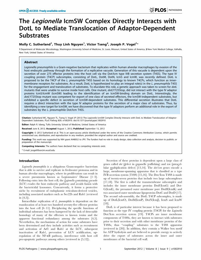
Isolation of DotL mutants with an intracellular growthdefect
To ascertain DotL’s role in the export of L. pneumophila T4SS
substrates, we isolated a collection of dotL mutants that were unable
to replicate inside Acanthamoeba castellanii. However, this was
challenging as an easy and unbiased method to select for dot/icm
mutants that are replication incompetent within host cells did not
currently exist. In addition, the most common null mutants consist of
proteins that are not stable, which would not be useful for structure-
function analysis. Therefore, to eliminate unstable proteins we
exploited a unique property of dotL, known as DdotL lethality, that
exists in the Legionella strain Lp02 [19]. In this strain background, dotL
is not only essential for growth within host cells but, unlike most other
dot/icm genes, is also required for growth on bacteriologic media
[19,29]. Since these traits are not linked, it was possible to first select
for clones from an error-prone PCR generated dotL library that were
able to complement DdotL lethality, thus ensuring that they made
stable, functional DotL protein. Subsequently, the library was
screened for clones that failed to complement a L. pneumophila DdotL
mutant for growth within amoebae.
Based on this strategy, ,100 dotL mutants with an intracellular
growth defect were identified by a visual screen from a pool of
3800 plasmids. To eliminate clones with multiple mutations, the
plasmids were sequenced revealing twelve dotL mutants that
contained a single amino acid change (Fig. 1A). The mutants were
distributed throughout the protein but none were identified in
either the amino-terminal transmembrane domain of DotL or in
the canonical Walker A box motif at amino acids 133–140 of DotL
(GSTGSGKT) [19,30]. Eleven of the mutants contained a single
amino acid change, while one resulted in a truncation of the C-
terminus of DotL at amino acid 725 due to the insertion of a
nonsense mutation (Fig. 1A).
In order to validate our screen, the twelve dotL mutants were
assayed for their ability to complement DdotL lethality and to
express normal levels of DotL protein. Similar to a strain
expressing wild-type dotL, each of the dotL mutants was able to
restore viability to a DdotL mutant when grown on plates (Fig.
S1A). Likewise, each of the mutants restored DotL proteins levels
in a DdotL mutant to that observed with wild-type dotL (Fig. S1B).
In addition, we confirmed that expression of another Dot/Icm
component, DotM, was not affected as DotL and DotM require
each other for their own stability [17,18] (Fig. S1B). Therefore,
based on these criteria, we conclude that our strategy successfully
identified dotL mutants with an intracellular growth defect and that
their attenuated growth phenotype was not due to reduced DotL
or DotM levels.
DotL plays a conserved role in intracellular growthThe dotL mutants were originally identified as being defective
for intracellular growth by a visual assessment at forty-eight hours.
To measure the extent of their growth defect more precisely, the
mutants were assayed for intracellular replication within A.
castellanii by a quantitative growth curve. In this assay, both the
wild-type strain Lp02 and a DdotL mutant expressing dotL+ from a
plasmid grew approximately 3,000-fold over forty-two hours
(Fig. 1B, filled squares and inverted triangles). In contrast, a strain
with a mutation in dotA that renders the T4SS non-functional
(Lp03), was defective for intracellular growth (Fig. 1B, filled
triangles). Similarly, none of the dotL mutants could complement
the DdotL mutant for replication in A. castellanii, further validating
our screen conditions (two representative mutants are shown in
Fig. 1B and the remaining are shown in Fig. S2A–B).
To examine if the dotL mutants were defective in other hosts,
their ability to grow and replicate was assayed in mouse A/J bone
marrow derived macrophages (BMM) [31] and in the human
monocytic cell line U937 [32]. Similar to amoebae, the wild-type
strain Lp02 grew robustly in BMMs and in U937 cells, whereas
the T4SS-deficient strain Lp03 was unable to grow in either
(Fig. 1C and data not shown). Likewise, all twelve of the dotL
mutants exhibited a dramatic defect in their ability to replicate
within BMM cells (Fig. 1C and Fig. S2C–D) and were attenuated
for growth within U937 cells (data not shown). Therefore, the
intracellular growth defect of the dotL mutants was not restricted to
amoebae and occurred in a variety of host cells.
dotL mutants’ interaction with other components of theDot/Icm T4SS
To identify the underlying molecular mechanism(s) for the dotL
mutants’ intracellular growth defect, their ability to assemble the
Author Summary
Many pathogens are able to survive and grow withineukaryotic host cells. One such pathogen, Legionellapneumophila, is able to replicate within macrophages,resulting in a form of pneumonia called Legionnaires’Disease. One key to L. pneumophila’s capacity to causedisease is its ability to translocate several hundred proteinsinto the host cell. These proteins, typically referred to as‘‘effectors’’, function to alter the host cell to create ahospitable environment for the bacteria. L. pneumophilaeffectors are exported by a specialized export apparatus,which is encoded by the dot/icm genes. However, themechanism of secretion for these substrates is poorlyunderstood. It is known that a subset of these effectorsrequires assistance from the type IV adaptor proteins IcmSand IcmW for transport out of the bacterium. It has beenshown that IcmSW binds adaptor-dependent secretedproteins in the bacterial cytoplasm prior to their export.Here we report that DotL, an inner membrane componentof the Dot/Icm secretion system, also binds IcmSW and thisinteraction is required for the export of adaptor-depen-dent substrates. This defines a new role for the type IVadaptors IcmSW and furthers our understanding of howLegionella exports substrates into its host cell.
IcmSW Interacts with DotL
PLOS Pathogens | www.plospathogens.org 2 September 2012 | Volume 8 | Issue 9 | e1002910
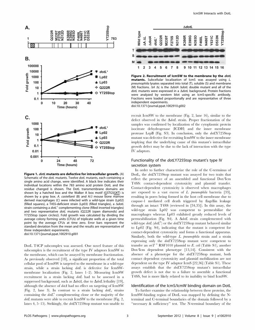
DotL T4CP subcomplex was assessed. One novel feature of this
subcomplex is the recruitment of the type IV adaptors IcmSW to
the membrane, which can be assayed by membrane fractionation.
As previously observed [18], a significant proportion of the total
cellular pool of IcmSW is targeted to the membrane in a wild-type
strain, while a strain lacking dotL is defective for IcmSW-
membrane localization (Fig. 2, lanes 1–2). Measuring IcmSW
recruitment in a strain lacking dotL had to be assessed in a
suppressed background, such as DdotA, due to DdotL lethality [19],
although the absence of dotA had no effect on targeting of IcmSW
(Fig. 2, lane 3). In contrast to a strain lacking dotL, strains
containing the dotL+ complementing clone or the majority of the
dotL mutants were able to recruit IcmSW to the membrane (Fig. 2,
lanes 4, 5–15). Strikingly, the dotLY725Stop mutant was unable to
recruit IcmSW to the membrane (Fig. 2, lane 16), similar to the
defect observed in the DdotL strain. Proper fractionation of the
samples was confirmed by localization of the cytoplasmic protein
isocitrate dehydrogenase (ICDH) and the inner membrane
protease LepB (Fig. S3). In conclusion, only the dotLY725Stop
mutant was defective for recruiting IcmSW to the inner membrane
implying that the underlying cause of this mutant’s intracellular
growth defect may be due to the lack of interaction with the type
IV adaptors.
Functionality of the dotLY725Stop mutant’s type IVsecretion system
In order to further characterize the role of the C-terminus of
DotL, the dotLY725Stop mutant was assayed for two traits that
reflect the presence of an assembled and functional Dot/Icm
T4SS: contact-dependent cytotoxicity and plasmid transfer.
Contact-dependent cytotoxicity is observed when macrophages
are exposed to a vast excess of L. pneumophila bacteria [33],
resulting in pores being formed in the host cell membrane due to
caspase-1 mediated cell death triggered by flagellin leakage
through an intact T4SS (reviewed in [34,35]). In this assay, the
wild-type strain Lp02 was competent to permeabilize the
macrophages whereas Lp03 exhibited greatly reduced levels of
permeabilization (Fig. S4). A DdotL strain complemented with
wild-type dotL (dotL+) or the dotLY725Stop mutant behaved similar
to Lp02 (Fig. S4), indicating that the mutant is competent for
contact-dependent cytotoxicity and forms a functional apparatus.
Similarly, both the wild-type L. pneumophila strain and a strain
expressing only the dotLY725Stop mutant were competent to
transfer an oriT + RSF1010 plasmid to E. coli (Table S1), another
Dot/Icm dependent phenotype [13,14]. Consistent with the
absence of a phenotype for the dotLY725Stop mutant, both
contact dependent cytotoxicity and plasmid mobilization are not
dependent on the type IV adaptor IcmS [22,36] (Table S1). These
assays establish that the dotLY725Stop mutant’s intracellular
growth defect is not due to a failure to assemble a functional
T4SS, but is more likely related to its inability to bind IcmSW.
Identification of the IcmS/IcmW binding domain on DotLTo further examine the relationship between these proteins, the
IcmSW-binding region of DotL was mapped by defining the N-
terminal and C-terminal boundaries of the domain followed by a
‘‘necessary & sufficiency’’ test. The N-terminal boundary of the
Figure 1. dotL mutants are defective for intracellular growth. (A)Schematic of the dotL mutants. Twelve dotL mutants, each containing asingle amino acid change, were identified. A black line indicates theirindividual locations within the 783 amino acid protein DotL and theresidue changed is shown. The DotL transmembrane domains areshown by a hatched box and the Walker A box motif (GSTGSGKT) isshown by a gray box. A. castellanii (B) and A/J mouse bone marrowderived macrophages (C) were infected with a wild-type strain (Lp02)(filled squares), a T4SS-deficient strain (Lp03) (filled triangles), a DdotLstrain containing a dotL+ complementing clone (filled inverted triangles)and two representative dotL mutants (Q222R (open diamonds) andY725Stop (open circles)). Fold growth was calculated by dividing theaverage colony forming units (CFUs) of triplicate wells at a given timepoint by the average CFUs at time zero. Error bars represent thestandard deviation from the mean and the results are representative ofthree independent experiments.doi:10.1371/journal.ppat.1002910.g001
Figure 2. Recruitment of IcmSW to the membrane by the dotLmutants. Subcellular localization of IcmS was assayed using L.pneumophila lysates separated into total (T), soluble (S) and membrane(M) fractions. DA DL is the DdotA DdotL double mutant and all of thedotL mutants were expressed in a DdotL background. Protein fractionswere analyzed by western blot using an IcmS-specific antibody.Fractions were loaded proportionally and are representative of threeindependent experiments.doi:10.1371/journal.ppat.1002910.g002
IcmSW Interacts with DotL
PLOS Pathogens | www.plospathogens.org 3 September 2012 | Volume 8 | Issue 9 | e1002910

IcmSW-binding domain of DotL was determined by analyzing
interactions between histidine tagged-IcmS/IcmW (His:SW) and a
series of DotL truncations fused to glutathione S-transferase
(GST:DotL). The His:SW heterodimer was recovered with nickel-
nitrilotriacetic acid (Ni-NTA) resin and interaction with the
GST:DotL fragments was monitored by immunoblot with a GST-
specific antibody. In this analysis, the minimal DotL fragment that
could bind IcmSW consisted of 113 amino acids from residues
671–783 of DotL (Fig. 3A). The interactions were specific as none
of the GST:DotL fragments were recovered from reactions
containing the empty His vector (Fig. S5A) and the failure to see
an interaction was not due to an inability to bind the Ni-NTA
resin as His:SW was recoverable in all reactions (Fig. S5B).
The C-terminal boundary of the IcmSW-binding domain of
DotL was determined by assaying recruitment of IcmSW to the
membrane as described in Figure 2. Truncations of 10 to 30
amino acids did not affect IcmSW localization to the membrane
(Fig. 3B with representative controls in Fig. S5C). In contrast,
removal of the last 40 or 50 amino acids of DotL resulted in vastly
diminished recruitment of IcmSW (Fig. 3B), although these
constructs did express slightly decreased levels of DotL (Fig.
S5D), which could be partially responsible for their defect. Based
on the above analysis, the N-terminal boundary of the IcmSW-
binding domain on DotL begins around amino acid 671 and ends
near amino acid 753. Deletion of this domain from wild-type DotL
(removal of amino acids 671 to 753) resulted in no IcmSW
recruitment to the membrane (Fig. 3C), thus validating the
mapped boundaries in Figures 3A and 3B.
To further refine the binding domain, a necessary and
sufficiency test was performed. A series of internal ten amino acid
deletions were constructed throughout the mapped boundaries in
full-length DotL. Only two deletions, one removing amino acids
730–739 and the other deleting amino acids 735–744, affected
IcmSW binding (Fig. 4A), thus potentially identifying one domain
necessary for the interaction between DotL and the adaptors.
Since these deletions may affect the fold of this domain, we cannot
conclude that DotL binds IcmSW directly via these residues. To
further refine the binding domain(s), a set of larger deletions
consisting of twenty amino acids revealed a potential second
binding domain (Fig. 4B). The first deletion, spanning amino acids
735–753, overlapped the domain identified with the smaller
deletions. The second deletion, spanning amino acids 695–714,
was located twenty amino acids closer to the N-terminus. Thus,
there appears to be at least two critical domains within this region
Figure 3. Identification of the IcmSW-binding domain on DotL. (A) E. coli lysates co-expressing His:SW and various GST:DotL fragmentstruncated at their amino terminus were bound to a Ni-NTA column. The presence of the indicated GST:DotL fusions was assessed in the total (T), flow-through (F), and bound fractions (B) in a GST western. A schematic of the C-terminal amino acids 631–783 of DotL illustrates the indicated deletions.(B) L. pneumophila lysates expressing C-terminal deletions of DotL were separated into total (T), soluble (S) and membrane (M) fractions. Membranerecruitment of IcmS was determined by western blot using an IcmS-specific antibody. (C) IcmS recruitment was assayed for a dotL mutant containinga deletion of the putative IcmSW-binding domain on DotL (amino acids 671–753). Results are representative of three independent experiments.doi:10.1371/journal.ppat.1002910.g003
IcmSW Interacts with DotL
PLOS Pathogens | www.plospathogens.org 4 September 2012 | Volume 8 | Issue 9 | e1002910

of DotL that are necessary for IcmSW recruitment to the
membrane.
Finally, we attempted to identify the minimal fragment of DotL
sufficient to interact with IcmSW. The minimal fragment
identified in Fig. 3A consisted of 113 amino acids from residues
671 to 783. Although small deletions from the C-terminus of this
fragment could bind IcmSW, larger deletions were unstable (Fig.
S6A). Therefore, a series of truncations were constructed using a
slightly larger DotL fragment containing amino acids 661–783.
Removal of 3 or 10 amino acids from the C-terminus of this
fragment had no effect, whereas a deletion of 20 amino acids was
partially defective and removal of the last 30 amino acids
completely prevented DotL binding to IcmSW (Fig. 4C with
controls in Fig. S6B–C). In summary, two DotL fragments
consisting of 113 amino acids (671–783 or 661–773) were both
fully sufficient to interact with IcmSW. Taken as a whole, we
propose the minimal domain of DotL that is sufficient to interact
with IcmSW likely consists of amino acids 671–773 and this
includes two regions (695–714 and 730–753) that are critically
important for the interaction (schematic shown in Fig. 4D).
Direct interaction between DotL and IcmS/IcmWThe previous data mapped an IcmSW-binding domain on
DotL, and strongly suggested that the connection was direct, but
did not rule out the possibility that the interaction was mediated
via an intermediate protein that binds both IcmSW and DotL. To
Figure 4. Identification of the domain(s) of DotL that is necessary and sufficient to bind IcmSW. DotL domains necessary for bindingIcmSW were identified by assaying L. pneumophila strains expressing 10 amino acid (A) and 20 amino acid (B) internal deletions of DotL. Membranerecruitment of IcmS was determined by western blot using an IcmS-specific antibody against total (T), soluble (S) and membrane (M) fractions. (C) TheDotL domain sufficient to bind IcmSW was identified by binding lysates co-expressing His:SW and various GST:DotL fragments to a Ni-NTA column.The presence of GST:DotL in the total (T), flow-through (F), and bound fractions (B) was determined by western blotting using a GST-specificantibody. Results are representative of three independent experiments. (D) A schematic of DotL containing amino acids 631–783 is shown toillustrate the properties of the DotL IcmSW-binding domain. The sufficient domain is indicated by a gray box and the necessary domains are denotedby striped boxes.doi:10.1371/journal.ppat.1002910.g004
IcmSW Interacts with DotL
PLOS Pathogens | www.plospathogens.org 5 September 2012 | Volume 8 | Issue 9 | e1002910
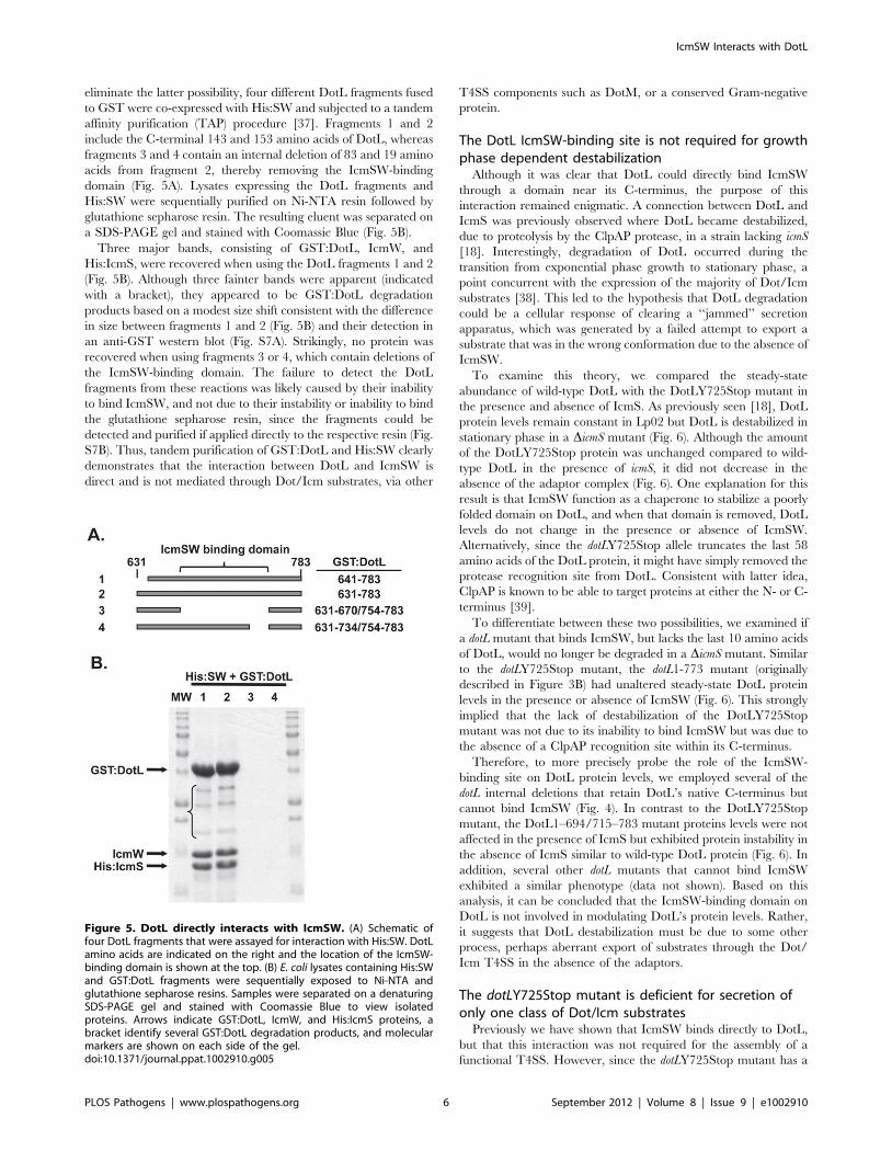
eliminate the latter possibility, four different DotL fragments fused
to GST were co-expressed with His:SW and subjected to a tandem
affinity purification (TAP) procedure [37]. Fragments 1 and 2
include the C-terminal 143 and 153 amino acids of DotL, whereas
fragments 3 and 4 contain an internal deletion of 83 and 19 amino
acids from fragment 2, thereby removing the IcmSW-binding
domain (Fig. 5A). Lysates expressing the DotL fragments and
His:SW were sequentially purified on Ni-NTA resin followed by
glutathione sepharose resin. The resulting eluent was separated on
a SDS-PAGE gel and stained with Coomassie Blue (Fig. 5B).
Three major bands, consisting of GST:DotL, IcmW, and
His:IcmS, were recovered when using the DotL fragments 1 and 2
(Fig. 5B). Although three fainter bands were apparent (indicated
with a bracket), they appeared to be GST:DotL degradation
products based on a modest size shift consistent with the difference
in size between fragments 1 and 2 (Fig. 5B) and their detection in
an anti-GST western blot (Fig. S7A). Strikingly, no protein was
recovered when using fragments 3 or 4, which contain deletions of
the IcmSW-binding domain. The failure to detect the DotL
fragments from these reactions was likely caused by their inability
to bind IcmSW, and not due to their instability or inability to bind
the glutathione sepharose resin, since the fragments could be
detected and purified if applied directly to the respective resin (Fig.
S7B). Thus, tandem purification of GST:DotL and His:SW clearly
demonstrates that the interaction between DotL and IcmSW is
direct and is not mediated through Dot/Icm substrates, via other
T4SS components such as DotM, or a conserved Gram-negative
protein.
The DotL IcmSW-binding site is not required for growthphase dependent destabilization
Although it was clear that DotL could directly bind IcmSW
through a domain near its C-terminus, the purpose of this
interaction remained enigmatic. A connection between DotL and
IcmS was previously observed where DotL became destabilized,
due to proteolysis by the ClpAP protease, in a strain lacking icmS
[18]. Interestingly, degradation of DotL occurred during the
transition from exponential phase growth to stationary phase, a
point concurrent with the expression of the majority of Dot/Icm
substrates [38]. This led to the hypothesis that DotL degradation
could be a cellular response of clearing a ‘‘jammed’’ secretion
apparatus, which was generated by a failed attempt to export a
substrate that was in the wrong conformation due to the absence of
IcmSW.
To examine this theory, we compared the steady-state
abundance of wild-type DotL with the DotLY725Stop mutant in
the presence and absence of IcmS. As previously seen [18], DotL
protein levels remain constant in Lp02 but DotL is destabilized in
stationary phase in a DicmS mutant (Fig. 6). Although the amount
of the DotLY725Stop protein was unchanged compared to wild-
type DotL in the presence of icmS, it did not decrease in the
absence of the adaptor complex (Fig. 6). One explanation for this
result is that IcmSW function as a chaperone to stabilize a poorly
folded domain on DotL, and when that domain is removed, DotL
levels do not change in the presence or absence of IcmSW.
Alternatively, since the dotLY725Stop allele truncates the last 58
amino acids of the DotL protein, it might have simply removed the
protease recognition site from DotL. Consistent with latter idea,
ClpAP is known to be able to target proteins at either the N- or C-
terminus [39].
To differentiate between these two possibilities, we examined if
a dotL mutant that binds IcmSW, but lacks the last 10 amino acids
of DotL, would no longer be degraded in a DicmS mutant. Similar
to the dotLY725Stop mutant, the dotL1-773 mutant (originally
described in Figure 3B) had unaltered steady-state DotL protein
levels in the presence or absence of IcmSW (Fig. 6). This strongly
implied that the lack of destabilization of the DotLY725Stop
mutant was not due to its inability to bind IcmSW but was due to
the absence of a ClpAP recognition site within its C-terminus.
Therefore, to more precisely probe the role of the IcmSW-
binding site on DotL protein levels, we employed several of the
dotL internal deletions that retain DotL’s native C-terminus but
cannot bind IcmSW (Fig. 4). In contrast to the DotLY725Stop
mutant, the DotL1–694/715–783 mutant proteins levels were not
affected in the presence of IcmS but exhibited protein instability in
the absence of IcmS similar to wild-type DotL protein (Fig. 6). In
addition, several other dotL mutants that cannot bind IcmSW
exhibited a similar phenotype (data not shown). Based on this
analysis, it can be concluded that the IcmSW-binding domain on
DotL is not involved in modulating DotL’s protein levels. Rather,
it suggests that DotL destabilization must be due to some other
process, perhaps aberrant export of substrates through the Dot/
Icm T4SS in the absence of the adaptors.
The dotLY725Stop mutant is deficient for secretion ofonly one class of Dot/Icm substrates
Previously we have shown that IcmSW binds directly to DotL,
but that this interaction was not required for the assembly of a
functional T4SS. However, since the dotLY725Stop mutant has a
Figure 5. DotL directly interacts with IcmSW. (A) Schematic offour DotL fragments that were assayed for interaction with His:SW. DotLamino acids are indicated on the right and the location of the IcmSW-binding domain is shown at the top. (B) E. coli lysates containing His:SWand GST:DotL fragments were sequentially exposed to Ni-NTA andglutathione sepharose resins. Samples were separated on a denaturingSDS-PAGE gel and stained with Coomassie Blue to view isolatedproteins. Arrows indicate GST:DotL, IcmW, and His:IcmS proteins, abracket identify several GST:DotL degradation products, and molecularmarkers are shown on each side of the gel.doi:10.1371/journal.ppat.1002910.g005
IcmSW Interacts with DotL
PLOS Pathogens | www.plospathogens.org 6 September 2012 | Volume 8 | Issue 9 | e1002910

profound intracellular growth defect, it must be defective for a vital
activity such as the translocation of Dot/Icm substrates into host
cells. To test if the dotLY725Stop mutant was defective for this
process, secretion was assayed in the wild-type strain Lp02, a
DicmS mutant, the dotLY725Stop mutant and a double mutant
containing both the dotLY725Stop and the icmS deletion.
Translocation was measured using reporter fusions consisting of
Bordetella pertussis adenylate cyclase toxin (CyaA) fused to repre-
sentative Dot/Icm T4SS effectors, and export was monitored by
measuring increased cAMP production by the host cell [40]. In
contrast to a previous report [18], secretion was monitored using
late exponential cultures to avoid indirect effects due to diminished
levels of DotL in the absence of IcmS (see Materials & Methods).
As previously shown [18,23,25], three IcmSW-dependent
substrates (SdeA, SidD and VipA) are secreted from a wild-type
strain, but exhibit a severe defect in export from a strain lacking
icmS, confirming their IcmSW-dependence (Fig. 7A–C). Interest-
ingly, secretion of these substrates was severely attenuated in the
dotLY725Stop mutant (Fig. 7A–C). Decreased export of the
substrates was not due to lowered amounts of IcmSW (data not
shown) nor did diminished expression levels of the reporters cause
it (Fig. S8A). Moreover, the secretion defect remained in a double
mutant consisting of the dotLY725Stop mutant and the icmS
deletion, indicating that the export deficiency of the dotLY725Stop
mutant was not due to an inhibitory effect of expressing IcmSW in
the absence of the IcmSW-binding site on DotL.
In contrast to the IcmSW-dependent substrates, three IcmSW-
independent substrates (RalF, LnaB, and LidA) [18,41,42] were
secreted at the same level in all strains, notably including the
dotLY725Stop mutant (Fig. 7D–F). A similar result was obtained
when export was assayed using a second dotL mutant (1–734/754–
783 described in Fig. 4B), that contains a smaller, twenty amino
Figure 6. Growth phase dependent instability of DotL in the absence of icmS. DotL levels were analyzed by western blotting using a DotL-specific antibody on samples harvested at various stages of growth. Samples include Lp02 or DicmS strains expressing wild-type DotL, theDotLY725Stop mutant, DotL1–773, or a DotL mutant lacking a domain necessary for IcmSW-binding (DotL1–694/715–783). Samples ranged frommid-exponential phase (lane 1, OD600 ,2.5) to late stationary phase (lane 8, OD600 ,3.5). Results are representative of three independentexperiments.doi:10.1371/journal.ppat.1002910.g006
Figure 7. The dotLY725Stop mutant has a specific secretion defect for IcmSW-dependent substrates. Export of CyaA fusions to thefollowing Dot/Icm substrates was assayed: A. SdeA, B. SidD, C. VipA, D. RalF, E. LnaB, and F. LidA. The CyaA:substrate fusions were expressed in a wild-type strain (black bars), a strain lacking icmS (white bars), a strain with the dotLY725Stop mutant integrated onto the chromosome (striped bars), anda dotLY725Stop DicmS double mutant (gray bars). cAMP was measured by ELISA from triplicate wells and error bars represent the standard deviationfrom the mean.doi:10.1371/journal.ppat.1002910.g007
IcmSW Interacts with DotL
PLOS Pathogens | www.plospathogens.org 7 September 2012 | Volume 8 | Issue 9 | e1002910

acid deletion in the IcmSW-binding site of DotL (Fig. S8B–E).
This result confirms that removal of the IcmSW-binding domain
on DotL did not prevent the proper assembly or ability to export
some substrates from the L. pneumophila Dot/Icm T4SS. In
addition, the secretion data conclusively demonstrates that the
IcmSW-binding site on DotL performs an essential role in the
export of IcmSW-dependent substrates, which is independent of
the role the adaptors play in directly binding substrates.
Discussion
In this study, we extensively characterized one dotL mutant,
dotLY725Stop, which revealed that DotL directly binds to the type
IV adaptors IcmSW via a domain that is located near the C-
terminus of DotL. Removal of the IcmSW-binding domain from
DotL did not prevent the assembly of a functional T4SS, nor did it
affect the growth phase instability of DotL that occurs in the
absence of IcmS. However, dotL mutants unable to bind IcmSW
were defective specifically for the export of IcmSW-dependent
substrates, thus revealing an additional role for the type IV
adaptors in the translocation of a major class of Dot/Icm
substrates into the cytoplasm of host cells.
Although the IcmSW complex is critically important for the
intracellular replication of L. pneumophila, the molecular role of
these two proteins has remained elusive. Based on the type IV
adaptors resemblance to type III secretion chaperones, including
their small size, acidic nature, and ability to dimerize, it was
initially proposed that IcmS and IcmW might function as
chaperones to mediate the export of Dot/Icm substrates [22,24].
It was subsequently shown that secretion of several substrates,
including SdeA, SidG, SidH, and WipA, was more dependent on
IcmSW than the substrate RalF, whose export was largely
IcmSW-independent [23,24]. Moreover, consistent with their
proposed role as secretion chaperones, several of the substrates
that require IcmSW for their optimal export were also shown to
bind IcmSW [22–25]. Conversely, RalF does not require IcmSW
for its export and was unable to bind to IcmSW [22–25]. To date,
24 out of 34 Dot/Icm substrates examined require IcmS and/or
IcmW for their export (summarized in Table S2). However, it is
worth noting that this collection includes only a small fraction of
the ,300 known Dot/Icm substrates [5]. In addition, only six
substrates have been assayed for their ability to bind the type IV
adaptors (Table S2).
Although a good correlation exists between these traits within
this limited data set, it is not known how the adaptors mediate
export by binding to IcmSW-dependent substrates. Direct binding
of substrates by the type IV adaptors is reminiscent of one or more
of the proposed roles performed by type III secretion chaperones
[43,44]. For example, IcmSW-binding of substrates could
potentially stabilize or prevent aggregation of the substrates in
the bacterial cytoplasm. Though, this does not appear to be a
general task for the L. pneumophila type IV adaptors as SidE family
members and SidG are stable and do not aggregate in the absence
of IcmSW [23,25].
Alternatively, IcmSW could function to target substrates to the
T4SS apparatus. However, IcmSW binds SidG at a location
distinct from its C-terminal signal sequence and removal of the
IcmSW-binding domain(s) allows SidG to be exported in type IV
adaptor-independent manner, inconsistent with IcmSW function-
ing directly as a targeting factor [25]. Instead Cambronne et al.
proposed that the type IV adaptors might regulate secretion by
preventing occlusion of the C-terminal signal sequence in a
partially folded domain of the substrate [25]. While this is a novel
and interesting concept, no experimental evidence exists that
IcmSW-dependent substrates fail to engage the T4SS apparatus in
the absence of the type IV adaptors. In contrast, our data on DotL
protein levels suggests that IcmSW-dependent substrates can
interact with DotL in the absence of icmS, leading to the
degradation of DotL.
Therefore, IcmSW likely perform an alternative role in
mediating export of many Dot/Icm substrates. Since IcmSW
appear not to be required for targeting, and are not exported along
with the Dot/Icm substrates, they likely act at the membrane
complex perhaps by directly assisting in the export of the proteins.
Consistent with this notion, we have now discovered that IcmSW
bind directly to DotL and this interaction is critical for the export
of IcmSW-dependent substrates. Precedence exists for an interac-
tion between T3SS chaperones and their associated ATPases
during targeting of substrates to the secretion apparatus. In
addition, some T3SS chaperones are able to interact with their
corresponding ATPase in the absence of their cognate substrate,
although it is not clear why they interact nor has the interaction
been shown to be required for secretion (reviewed in [43,45]).
Therefore, taking all of the existing data into context, we
propose a new model for the export of IcmSW-dependent
substrates by the L. pneumophila Dot/Icm T4SS (Fig. 8A). In this
unified model, IcmSW-dependent substrates are bound by the
type IV adaptors in the cytoplasm in order to maintain the
substrates in a translocation competent form. Substrates are
targeted to the inner membrane via their C-terminal signal
sequence and likely interact directly with the type IV coupling
protein DotL. After the substrates engage the translocation
apparatus, IcmSW need to be removed from the substrates and
this may be mediated by transfer to the IcmSW-binding domain
on DotL, perhaps while the substrates are being actively pumped
out of the cell. Later, IcmSW would dissociate from DotL,
disengage from the inner membrane, and transit back to the
cytoplasm in order to interact with new substrates, thus completing
an export cycle (Fig. 8A). Although this model incorporates all of
the existing data, and provides a reasonable explanation for how
IcmSW could function while independently binding substrates and
DotL, there are a number of potential caveats. For example, the
model predicts a large amount of IcmSW would be required in the
cell in order to bind the vast number of type IV-adaptor
dependent substrates. This has not been experimentally estab-
lished nor has the direct transfer of IcmSW from substrates to
DotL.
Nevertheless, our working model allows us to speculate why
IcmSW-dependent substrates are not exported in different
mutant backgrounds that are compromised for type IV adaptor
activity. For example, a strain that expresses a dotL mutant that is
unable to bind IcmSW (dotL2S/W) is not capable of exporting
IcmSW-dependent substrates (Fig. 8B). In this case, IcmSW-
dependent substrates would still be bound by the adaptors and
maintained in a translocation competent form. Since targeting is
dependent on their C-terminal signal sequence, these substrates
would still bind to inner membrane components of the T4SS.
However, as the substrates engage the T4SS apparatus, the
proposed transfer of IcmSW from the substrate onto DotL would
fail because no acceptor site would be present. As a result, the
substrate might be rejected from the apparatus, a result
previously observed with T3SS substrates that are fused to
proteins that are difficult to be unfolded such as DHFR and
ubiquitin (reviewed in [46]). This model of rejection is consistent
with our observation that DotL2S/W is not degraded in wild-type
strains expressing IcmS.
In contrast, the phenotypes associated with a DicmS mutant are
more complicated because both IcmSW-dependent substrates and
IcmSW Interacts with DotL
PLOS Pathogens | www.plospathogens.org 8 September 2012 | Volume 8 | Issue 9 | e1002910

DotL would not bind the type IV adaptors (Fig. 8C). Although the
dotL2S/W mutant and a DicmS mutant are each unable to export
IcmSW-dependent substrates, we propose that their molecular
defects differ. In the case of a strain lacking icmS, IcmSW-
dependent substrates would still interact with DotL via their C-
terminal signal sequence, however they would not be in a
translocation competent form due to the absence of the type IV
adaptors. As a result, the substrates might engage the apparatus in
an aberrant manner and become ‘‘jammed’’. This would likely
induce membrane stress resulting in removal of the DotL/
substrate aggregate via the ClpAP protease, which would also be
consistent with our results on DotL protein levels in the absence of
icmS. Furthermore, this model predicts that substrates can interact
with DotL in the absence of the type IV adaptors, thus supporting
the idea that IcmSW do not perform a role in targeting of
substrates to the Dot/Icm apparatus.
Figure 8. Model for secretion of IcmS/IcmW-dependent effectors by DotL. The transmembrane-spanning L. pneumophila T4SS complex isrepresented by a purple cylinder, components of the T4CP subcomplex (DotL, DotM, IcmS, and IcmW) are labeled and the inner and outermembranes are indicated as IM and OM, respectively. (A) Export of IcmSW-dependent substrates in a wild type strain. (B) Rejection of IcmSW-dependent substrates in a dotL mutant strain that lacks the IcmSW-dependent binding domain (dotL2S/W). (C) Jamming of IcmSW-dependentsubstrates in a DicmS mutant, leads to DotL degradation by ClpAP.doi:10.1371/journal.ppat.1002910.g008
IcmSW Interacts with DotL
PLOS Pathogens | www.plospathogens.org 9 September 2012 | Volume 8 | Issue 9 | e1002910

By characterizing the dotLY725Stop mutant, we discovered that
DotL directly binds to the type IV adaptors and that the
interaction is required for the specific export of IcmSW-dependent
substrates. This mutant provides a key reagent for the future
dissection of the role of IcmSW, as the phenotypes of the DicmS
mutant are clearly pleiotropic. Moreover, as IcmSW-dependent
substrates appear to constitute the major class of Dot/Icm
proteins, understanding how L. pneumophila controls and regulates
the secretion of several hundred proteins will be vital to
understanding how this pathogen is able to replicate inside host
cells and cause disease.
Materials and Methods
Ethics statementThis study was carried out in strict accordance with the
recommendations in the Guide for the Care and Use of
Laboratory Animals of the National Institutes of Health. The
protocol was approved by the Institutional Animal Care and Use
Committee at the Washington University School of Medicine
(Assurance Number: A3381-01). All efforts were made to minimize
suffering.
Bacterial strains and mediaStrains used in this study are provided in Table S3. All Legionella
pneumophila strains having a JV number are derived from the wild
type derivative, L. pneumophila Philadelphia Lp02 (hsdR rpsL thyA), of
the clinical isolate L. pneumophila Philadelphia-1 [47]. L. pneumophila
strains were grown in yeast extract broth (AYE) or on solid media
consisting of charcoal yeast agar (CYE) both buffered with N-(2-
acetamido)-2-aminoethanesulfonic acid (ACES) [48]. Antibiotics,
sucrose (5%), and thymidine (100 mg/ml) were added as needed.
Screen for dotL mutantsTwelve dotL mutant libraries were generated by PCR using Taq
DNA polymerase, the primers JVP1038 (CCCGAATTCGGA-
ATTAGAGCCATGATGCG) and JVP1039 (CCCGCATGC-
CACTTCTACCTCCAATTGCCG) and a dotL complementing
clone (pJB3151) as the template. The PCR fragment was cloned into
an IPTG inducible vector (pJB4858), transformants were pooled
into libraries, and plasmid DNA was isolated. Mutant libraries were
transformed into a dotL/DdotL::CmR merodiploid strain (JV1003).
Integrants were pooled and plated on CYE plates containing 5%
sucrose, 0.1 mM IPTG, 2 mg/ml chloramphenicol to select for dotL
plasmids that allowed resolution of the merodiploid to DdotL::CmR.
The putative dotL mutants were used to infect Acanthamoeba castellanii
and a visual assessment for growth was performed at 24 and
48 hours. dotL mutants that did not replicate in A. castellanii were
recovered and confirmed for plasmid linkage. Approximately 100
dotL mutant plasmids that had an attenuated growth phenotype
were sequenced at the Harvard Biopolymer Facility to identify
amino acid changes responsible for the growth defect. Twelve
clones contained one amino acid change and the remaining
contained two or more amino acid changes; only mutants with a
single amino acid change were pursued.
Complementation of DdotL lethality by the dotL mutantsdotL mutants were transformed into the dotL merodiploid strain,
Lp02 dotL/DdotL::CmR (JV1003), and an OD600 of 1.0 suspension
from a two day patch was diluted and plated on CYE sucrose
IPTG chloramphenicol plates to select for resolution to
DdotL::CmR and on CYE + IPTG to determine the total number
of cells. The frequency of resolution to DdotL was determined by
dividing the number of cells that resolved to DdotL::CmR by the
total number of cells.
Growth of L. pneumophila in host cellsL. pneumophila liquid cultures were grown with appropriate
selection to the point of infectivity as determined by OD600 and
motility. Bacteria were washed, resuspended at an OD600 of 1.0
and diluted 1:10,000 for infection. A. castellanii were infected, lysed
with 5% saponin in phosphate buffered saline (PBS) at 0, 20, 31,
and 42 hours, and dilutions were plated on CYE + IPTG to
determine rates of multiplication. Primary bone marrow derived
macrophages were prepared as previously described [31] and
infected as described with the A. castellanii. Macrophages were
lysed at 24 hour intervals with water and fold growth was
determined by plating dilutions on CYE + IPTG.
Immunoblot analysisSamples were collected, resuspended in Laemmli sample buffer,
boiled for 5 minutes, separated by SDS-PAGE gel electrophoresis
and transferred to PVDF membranes. Membrane was blocked
with PBS containing 5% non-fat dry milk, washed with 0.05%
Tween 20 in PBS, and incubated for 1 hour with primary
antibody diluted in the non-fat dry milk solution. Blots were then
washed and incubated for 1 hour with secondary goat anti-rabbit
or goat anti-mouse conjugated to horseradish peroxidase (Sigma)
diluted in non-fat dry milk solution. Finally, blots were washed and
developed using an ECL detection kit (Amersham Biosciences).
Determination of cellular location by membranefractionation
Membrane fractionation was performed as previously described
[17]. Briefly, L. pneumophila liquid cultures were grown to the point of
infectivity as above and 20 OD600 of cells were collected. Cell pellets
was resuspended in lysis buffer (50 mM Tris pH 8.0, 0.2 mg/ml
lysozyme, and protease inhibitor cocktail (Sigma) and lysed by
French press (14,000 PSI). Unlysed cells were removed by
centrifugation at 10,000 g for 10 minutes. Cytoplasmic and mem-
brane proteins were distinguished by ultracentrifugation at 100,000 g
for 1 hour. Membrane proteins were resuspended in 16 Laemmli
sample buffer and all other fractions were diluted with 26Laemmli
sample buffer. Fractionations were analyzed by immunoblot analysis
as above. Fractionation quality was assessed by immunoblot analysis
with antibodies to the cytoplasmic protein isocitrate dehydrogenase
(ICDH) and the inner-membrane protein LepB.
Interaction of GST:DotL fragments with His:SWHis and GST fusion plasmids were co-expressed in E. coli,
induced with IPTG for four hours at room temperature, and 10
OD600 of cells were collected. Cell pellets were resuspended in
lysis buffer (50 mM Tris-pH 8, 150 mM NaCl, 1% TritonX-100,
1 mM DTT, 1 mM PMSF, 10 mM imidazole) and lysed by
sonication. Unlysed cells were removed by centrifugation at
10,000 g for 20 minutes. Cell lysates were added to a 50 ml Ni-
NTA (Qiagen) bed volume and incubated with rotation for
2 hours at 4 C. Ni-NTA was washed twice in wash buffer (lysis
buffer with 30 mM imidazole) and proteins were eluted in 26Laemmli sample buffer by boiling for 5 minutes. Fractions were
loaded proportionally and analyzed by immunoblot analysis as
above with specific His and GST antibodies.
Contact dependent cytotoxicityContact dependent cytotoxicity was performed as previously
described [33,49]. Briefly, L. pneumophila cultures were grown to
IcmSW Interacts with DotL
PLOS Pathogens | www.plospathogens.org 10 September 2012 | Volume 8 | Issue 9 | e1002910
the point of infectivity as described above, washed, and pelleted
onto BMM by centrifugation at a multiplicity of infection (MOIs)
of 5, 50, or 500. BMM were infected for one hour, washed, stained
with a 5:1 mixture of ethidium bromide and acridine orange to
assess cell viability and immediately examined by immunofluores-
cent microscopy. The ability to induce contact-dependent
cytotoxicity is expressed as the percentage of ethidium bromide
positive cells and calculated by dividing the total number of
ethidium bromide positive cells by the total number of cells in
three randomly selected fields.
Plasmid transfer assayPlasmid transfer assays were performed as previously described
[13]. Briefly, L. pneumophila strains were grown in liquid media to
the point of infectivity as described above. L. pneumophila strains
were mixed with the E. coli recipient ER1821 in a 10:1 ratio on a
0.45 mm filter (Millipore) on non-selective CYET media for two
hours. Bacteria were washed from the mating membrane with
sterile water and assayed for the number of plasmid transfer events
by plating on selective media. Rate of plasmid transfer was
determined by dividing the number of E. coli recipients by the total
number of L. pneumophila donors.
DotL protein levelsL. pneumophila strains were grown in liquid media. When cultures
reached early exponential phase growth rate was monitored by
OD600 and 1 OD600 of cells were collected every hour for fifteen
hours. Protein samples were prepared as above and assayed by
western blot with a specific DotL antibody to determine protein
levels.
Adenylate cyclase reporter assays for substrate secretionReporter assays were performed as described [18,23] with the
following modification. In Vincent et al, it was shown that DotL
becomes destabilized in stationary phase in the absence of IcmS
resulting in diminished export of IcmSW-independent substrates
such as RalF and this defect could be suppressed by over-
producing DotL. This indirect defect could be avoided by assaying
export of substrates in late exponential phase, prior to the point
where DotL levels decrease. Measuring secretion of substrates at
either point in growth is feasible since the Dot/Icm system is
constitutively expressed and is functional at all phases of growth.
Specifically, CyaA fusions were transformed into the L.
pneumophila strains Lp02, Lp02 DicmS (JV1962), Lp02 dotLY725-
Stop (JV7325) and Lp02 dotLY725Stop DicmS (JV7645). The
strains were grown in liquid media and induced for 2.5 hours with
IPTG at early exponential phase. 1 OD600 of cells was collected,
washed, diluted 1:200 in RPMI-1640 (HyClone) supplemented
with fetal bovine serum 10% (FBS) (HyClone) for infection of
U937 cells. Bacterial cultures were pelleted onto differentiated
U937 cells for 5 minutes at 1000 g at room temperature and
infections were allowed to proceed for 1 hour at 37 C. Cells were
washed 36 in warm PBS, lysed (50 mM HCl, 0.1% Triton X-100)
and immediately boiled for 5 minutes. Subsequently HCl was
neutralized with NaOH and cAMP was extracted was extracted
with 95% ethanol. Extracted cAMP was desiccated and analyzed
via a competitive ELISA (cAMP Biotrak Enzymeimmunoassay
System, Amersham or Cyclic AMP EIA Kit, Cayman Chemical).
Supporting Information
Figure S1 The dotL mutants complement DdotL lethal-ity and express stable protein. (A) A dotL merodiploid strain
(dotL/DdotL::CmR) containing a dotL complementing plasmid,
vector, or the dotL mutants was monitored for its ability to resolve
to the DdotL::CmR locus. The experiment was performed in
triplicate and error bars represent the standard deviation from the
mean. (B) Westerns using antibodies specific for DotL or DotM
were performed on each of the dotL mutants. Westerns to the
cytoplasmic housekeeping protein ICDH serve as a loading
control. Results are representative of three independent experi-
ments.
(TIF)
Figure S2 The dotL mutant’s exhibit an intracellulargrowth defect inside host cells. L. pneumophila was used to
infect A. castellanii (A–B) or BMMs (C–D). Growth is shown for a
wild-type strain (Lp02) (filled squares), a T4SS-deficient strain
(Lp03) (filled triangles), and a DdotL strain containing a dotL
complementing clone (filled inverted triangles) or the dotL mutants
(open symbols). Error bars represent the standard deviation from
the mean and the results are representative of three independent
experiments.
(TIF)
Figure S3 Controls for Legionella fractionations show-ing IcmSW recruitment to the membrane. L. pneumophila
cultures were lysed and fractionated as described in the Materials
& Methods. Protein fractions were analyzed by western blotting
with the specified antibody to ensure the quality of the separation
technique. (A) Westerns detecting the cytoplasmic housekeeping
protein isocitrate dehydrogenase (ICDH) demonstrate that no
cytoplasmic proteins are present in the membrane fractions. (B)
Westerns detecting the inner membrane signal peptidase, LepB,
demonstrate that membrane proteins are not present in the
cytoplasmic fractions. The control in lane 1 is a total protein
fraction from the wild-type strain loaded in the T, S, and M blots
to illustrate protein transfer to the membrane occurred.
(TIF)
Figure S4 dotLY725Stop exhibits contact-dependentcytotoxicity. BMMs were challenged with four different L.
pneumophila cultures (wild-type strain Lp02, the dotA mutant Lp03,
a DdotL::CmR strain containing a wild-type dotL+ complementing
clone or the dotLY725Stop clone). Infections were done using
various amounts of bacteria including: uninfected (light gray bar),
MOI of 5 (black bar), MOI of 50 (white bar), and MOI of 100
(gray bar). Host cell permeabilization was assayed using a live/
dead stain consisting of acridine orange and ethidium bromide
(EtBr). Results are displayed as the percentage of EtBr-positive
macrophages and were determined by taking the number of EtBr
positive cells divided by the total number of host cells in three
random fields. The results are representative of several indepen-
dent experiments and the standard deviation from the mean is
shown for each sample.
(TIF)
Figure S5 Controls for delineation of the IcmSW-binding domain of DotL. (A) GST:DotL fragments that
interacted with His:SW in Fig. 3A were tested to confirm that the
interaction was specific. GST:DotL fragments were co-expressed
with empty His fusion vector assayed for non-specific binding to
Ni-NTA resin. Protein from the total (T), flow-through (F), and
bound fractions (B) were assessed by western blotting with a GST-
specific antibody. (B) Samples from Fig. 3A were blotted with an
anti-His antibody to demonstrate near complete retention of
His:SW to the Ni-NTA resin. (C) Selected samples from Fig. 3B
were examined to confirm the quality of the membrane
fractionation technique. As described in Fig. S4, ICDH is a
cytoplasmic protein and LepB is an inner membrane protease. (D)
IcmSW Interacts with DotL
PLOS Pathogens | www.plospathogens.org 11 September 2012 | Volume 8 | Issue 9 | e1002910
Samples from Figures 3B, 3C, 4A and 4B were analyzed by
western blotting with DotL-specific antibodies to assay the total
amount of DotL protein in the cells. ICDH blots served as a
loading control.
(TIF)
Figure S6 Controls for identifying the IcmSW-domainthat is sufficient to bind DotL. (A) Longer fragments of GST-
DotL starting at amino acid 671 bind to IcmSW but shorter
fragments become unstable. GST-DotL was detected by western
blots using a GST-specific antibody. (B) GST-DotL fragments
from Fig. 4C were tested to confirm that the interaction was
specific as described in Fig. S5A. (C) Samples from Fig. 4C were
blotted with an anti-His antibody to demonstrate near complete
retention of His:SW to the Ni-NTA resin.
(TIF)
Figure S7 DotL and IcmSW directly interact. (A) Non-
stoichiometric bands from the tandem affinity purification are
GST:DotL degradation products. Samples used in Fig. 5 were
separated on an SDS-PAGE gel, probed with a GST specific
antibody, revealing several degradation products. Fragment 2 was
also loaded on the right side of the gel as a control. (B–D) Lysates
containing His:SW and the four fragments from Fig. 5 were
purified on either a Ni-NTA column (Ni) or a glutathione
sepharose column (G). Elutions were run on SDS-PAGE gel and
stained with Coomassie (B), probed with anti-His antibody to
detect His-IcmS (C) or probed with anti-IcmW antibody (D).
His:SW is able to bind Ni-NTA in all cases and all 4 GST:DotL
fragments are able to bind glutathione sepharose. GST:DotL
fragments 1 & 2 co-purify from the Ni column, but fragments 3 &
4 do not. His:SW co-purifies from the glutathione sepharose
column when using fragments 1 & 2 but not with fragments 3 & 4.
The * indicates a His:SW dimer that was not denatured by the
SDS-PAGE gel and can be detected in the westerns (C & D).
(TIF)
Figure S8 The DotL1–734/754–783 mutant is defectivefor secretion of only IcmSW-dependent substrates asobserved for the original DotLY725Stop mutant. (A)
Protein levels of CyaA fusions to Dot/Icm substrates used in
Figure 7 were assessed by western blot with a CyaA-specific
antibody and shown to produce equivalent levels of protein. (B &
C) Export of CyaA fusions to IcmSW-dependent effectors (SdeA,
SidD, VipA) and IcmSW-independent effectors (RalF, LnaB,
LidA) were assayed in a wild-type strain (black bars), DicmS mutant
(white bars), and a strain with DotL1–734/754–783 integrated
onto the chromosome (striped bars). cAMP was measured from
triplicate wells and error bars represent the standard deviation
from the mean. (D & E) Protein samples from Fig. S8B and S8C
were analyzed as in Fig. S8A.
(TIF)
Table S1 dotLY725Stop is able to transfer an RSF1010mobilizable plasmid. Mobilization of the RSF1010 plasmid by
the L. pneumophila T4SS to E. coli recipients was determined. The
rate of transfer was calculated by dividing the average number of
recipients by the average total number of donor cells.
(PDF)
Table S2 IcmSW dependence of Dot/Icm substrates.
(PDF)
Table S3 Relevant strains, plasmids, and primersemployed in this study.
(PDF)
Table S4 Construction of plasmids employed in thisstudy.
(PDF)
Acknowledgments
We thank Emily Buford, Rebecca O’Connell, and Malissa Wolfgang for
technical assistance. We thank L. David Sibley and Petra A. Levin for
helpful suggestions and critical review of the manuscript.
Author Contributions
Conceived and designed the experiments: MCS JPV. Performed the
experiments: MCS TLN VT. Analyzed the data: MCS JPV. Contributed
reagents/materials/analysis tools: MCS TLN VT JPV. Wrote the paper:
MCS JPV.
References
1. McDade JE, Shepard CC, Fraser DW, Tsai TR, Redus MA, et al. (1977)
Legionnaires’ disease: isolation of a bacterium and demonstration of its role in
other respiratory disease. N Engl J Med 297: 1197–1203.
2. Horwitz MA, Silverstein SC (1980) Legionnaires’ disease bacterium (Legionella
pneumophila) multiples intracellularly in human monocytes. J Clin Invest 66: 441–
450.
3. Fields BS, Benson RF, Besser RE (2002) Legionella and Legionnaires’ disease: 25
years of investigation. Clin Microbiol Rev 15: 506–526.
4. Isberg RR, O’Connor TJ, Heidtman M (2009) The Legionella pneumophila
replication vacuole: making a cosy niche inside host cells. Nat Rev Microbiol 7:
13–24.
5. Hubber A, Roy CR (2010) Modulation of host cell function by Legionella
pneumophila type IV effectors. Annu Rev Cell Dev Biol 26: 261–283.
6. Burstein D, Zusman T, Degtyar E, Viner R, Segal G, et al. (2009) Genome-scale
identification of Legionella pneumophila effectors using a machine learning
approach. PLoS Pathog 5: e1000508.
7. de Felipe KS, Glover RT, Charpentier X, Anderson OR, Reyes M, et al. (2008)
Legionella eukaryotic-like type IV substrates interfere with organelle trafficking.
PLoS Pathog 4: e1000117.
8. Gomez-Valero L, Rusniok C, Cazalet C, Buchrieser C (2011) Comparative and
functional genomics of Legionella identified eukaryotic like proteins as key players
in host-pathogen interactions. Front Microbiol 2: 208.
9. Heidtman M, Chen EJ, Moy MY, Isberg RR (2009) Large-scale identification of
Legionella pneumophila Dot/Icm substrates that modulate host cell vesicle
trafficking pathways. Cell Microbiol 11: 230–248.
10. Huang L, Boyd D, Amyot WM, Hempstead AD, Luo ZQ, et al. (2011) The E
Block motif is associated with Legionella pneumophila translocated substrates. Cell
Microbiol 13: 227–245.
11. Zhu W, Banga S, Tan Y, Zheng C, Stephenson R, et al. (2011) Comprehensive
identification of protein substrates of the Dot/Icm type IV transporter of
Legionella pneumophila. PLoS One 6: e17638.
12. Ge J, Shao F (2011) Manipulation of host vesicular trafficking and innate
immune defence by Legionella Dot/Icm effectors. Cell Microbiol 13: 1870–1880.
13. Vogel JP, Andrews HL, Wong SK, Isberg RR (1998) Conjugative transfer by the
virulence system of Legionella pneumophila. Science 279: 873–876.
14. Segal G, Purcell M, Shuman HA (1998) Host cell killing and bacterial
conjugation require overlapping sets of genes within a 22-kb region of the
Legionella pneumophila genome. Proc Natl Acad Sci U S A 95: 1669–1674.
15. Christie PJ, Vogel JP (2000) Bacterial type IV secretion: conjugation systems
adapted to deliver effector molecules to host cells. Trends Microbiol 8: 354–360.
16. Nagai H, Kubori T (2011) Type IVB Secretion Systems of Legionella and Other
Gram-Negative Bacteria. Front Microbiol 2: 136.
17. Vincent CD, Friedman JR, Jeong KC, Buford EC, Miller JL, et al. (2006)
Identification of the core transmembrane complex of the Legionella Dot/Icm type
IV secretion system. Mol Microbiol 62: 1278–1291.
18. Vincent CD, Friedman JR, Jeong KC, Sutherland MC, Vogel JP (2012)
Identification of the DotL coupling protein subcomplex of the Legionella Dot/
Icm type IV secretion system. Mol Microbiol 85: 378–391.
19. Buscher BA, Conover GM, Miller JL, Vogel SA, Meyers SN, et al. (2005) The
DotL protein, a member of the TraG-coupling protein family, is essential for
viability of Legionella pneumophila strain Lp02. J Bacteriol 187: 2927–2938.
20. Fronzes R, Christie PJ, Waksman G (2009) The structural biology of type IV
secretion systems. Nat Rev Microbiol 7: 703–714.
21. Vincent CD, Vogel JP (2006) The Legionella pneumophila IcmS-LvgA protein
complex is important for Dot/Icm-dependent intracellular growth. Mol
Microbiol 61: 596–613.
IcmSW Interacts with DotL
PLOS Pathogens | www.plospathogens.org 12 September 2012 | Volume 8 | Issue 9 | e1002910
22. Coers J, Kagan JC, Matthews M, Nagai H, Zuckman DM, et al. (2000)
Identification of Icm protein complexes that play distinct roles in the biogenesis
of an organelle permissive for Legionella pneumophila intracellular growth. Mol
Microbiol 38: 719–736.
23. Bardill JP, Miller JL, Vogel JP (2005) IcmS-dependent translocation of SdeA into
macrophages by the Legionella pneumophila type IV secretion system. Mol
Microbiol 56: 90–103.
24. Ninio S, Zuckman-Cholon DM, Cambronne ED, Roy CR (2005) The Legionella
IcmS-IcmW protein complex is important for Dot/Icm-mediated protein
translocation. Mol Microbiol 55: 912–926.
25. Cambronne ED, Roy CR (2007) The Legionella pneumophila IcmSW complex
interacts with multiple Dot/Icm effectors to facilitate type IV translocation.
PLoS Pathog 3: e188.
26. Chen J, Reyes M, Clarke M, Shuman HA (2007) Host cell-dependent secretion
and translocation of the LepA and LepB effectors of Legionella pneumophila. Cell
Microbiol 9: 1660–1671.
27. Ninio S, Celli J, Roy CR (2009) A Legionella pneumophila effector protein encoded
in a region of genomic plasticity binds to Dot/Icm-modified vacuoles. PLoS
Pathog 5: e1000278.
28. Habyarimana F, Price CT, Santic M, Al-Khodor S, Kwaik YA (2010) Molecular
characterization of the Dot/Icm-translocated AnkH and AnkJ eukaryotic-like
effectors of Legionella pneumophila. Infect Immun 78: 1123–1134.
29. Vincent CD, Buscher BA, Friedman JR, Williams LA, Bardill P, et al. (2006)
Identification of non-dot/icm suppressors of the Legionella pneumophila deletion dotL
lethality phenotype. J Bacteriol 188: 8231–8243.
30. Walker JE, Saraste M, Runswick MJ, Gay NJ (1982) Distantly related sequences
in the alpha- and beta-subunits of ATP synthase, myosin, kinases and other
ATP-requiring enzymes and a common nucleotide binding fold. EMBO J 1:
945–951.
31. Swanson MS, Isberg RR (1995) Association of Legionella pneumophila with the
macrophage endoplasmic reticulum. Infect Immun 63: 3609–3620.
32. Sundstrom C, Nilsson K (1976) Establishment and characterization of a human
histiocytic lymphoma cell line (U-937). Int J Cancer 17: 565–577.
33. Kirby JE, Vogel JP, Andrews HL, Isberg RR (1998) Evidence for pore-forming
ability by Legionella pneumophila. Mol Microbiol 27: 323–336.
34. Miao EA, Andersen-Nissen E, Warren SE, Aderem A (2007) TLR5 and Ipaf:
dual sensors of bacterial flagellin in the innate immune system. Semin
Immunopathol 29: 275–288.
35. Persson J, Vance RE (2007) Genetics-squared: combining host and pathogen
genetics in the analysis of innate immunity and bacterial virulence. Immuno-genetics 59: 761–778.
36. Zuckman DM, Hung JB, Roy CR (1999) Pore-forming activity is not sufficient
for Legionella pneumophila phagosome trafficking and intracellular growth. MolMicrobiol 32: 990–1001.
37. Puig O, Caspary F, Rigaut G, Rutz B, Bouveret E, et al. (2001) The tandemaffinity purification (TAP) method: a general procedure of protein complex
purification. Methods 24: 218–229.
38. Ninio S, Roy CR (2007) Effector proteins translocated by Legionella pneumophila:strength in numbers. Trends Microbiol 15: 372–380.
39. Baker TA, Sauer RT (2006) ATP-dependent proteases of bacteria: recognitionlogic and operating principles. Trends Biochem Sci 31: 647–653.
40. Sory MP, Cornelis GR (1994) Translocation of a hybrid YopE-adenylate cyclasefrom Yersinia enterocolitica into HeLa cells. Mol Microbiol 14: 583–594.
41. Nagai H, Cambronne ED, Kagan JC, Amor JC, Kahn RA, et al. (2005) A C-
terminal translocation signal required for Dot/Icm-dependent delivery of theLegionella RalF protein to host cells. Proc Natl Acad Sci U S A 102: 826–831.
42. Losick VP, Haenssler E, Moy MY, Isberg RR (2010) LnaB: a Legionella
pneumophila activator of NF-kappaB. Cell Microbiol 12: 1083–1097.
43. Galan JE, Wolf-Watz H (2006) Protein delivery into eukaryotic cells by type III
secretion machines. Nature 444: 567–573.44. Fattori J, Prando A, Martins AM, Rodrigues FH, Tasic L (2011) Bacterial
secretion chaperones. Protein Pept Lett 18: 158–166.45. Wilharm G, Dittmann S, Schmid A, Heesemann J (2007) On the role of specific
chaperones, the specific ATPase, and the proton motive force in type IIIsecretion. Int J Med Microbiol 297: 27–36.
46. Sorg JA, Miller NC, Schneewind O (2005) Substrate recognition of type III
secretion machines–testing the RNA signal hypothesis. Cell Microbiol 7: 1217–1225.
47. Berger KH, Isberg RR (1993) Two distinct defects in intracellular growthcomplemented by a single genetic locus in Legionella pneumophila. Mol Microbiol 7:
7–19.
48. Feeley JC, Gibson RJ, Gorman GW, Langford NC, Rasheed JK, et al. (1979)Charcoal-yeast extract agar: primary isolation medium for Legionella pneumophila.
J Clin Microbiol 10: 437–441.49. Sexton JA, Yeo HJ, Vogel JP (2005) Genetic analysis of the Legionella pneumophila
DotB ATPase reveals a role in type IV secretion system protein export. MolMicrobiol 57: 70–84.
IcmSW Interacts with DotL
PLOS Pathogens | www.plospathogens.org 13 September 2012 | Volume 8 | Issue 9 | e1002910
Copyright © 2022 FDOKUMEN