Expression of 1-integrins and N-cadherin in bladder cancer and melanoma cell lines

Cell, Vol. 87, 733–743, November 15, 1996, Copyright 1996 by Cell Press
The Adaptor Protein ShcCouples a Class of Integrinsto the Control of Cell Cycle Progression
Kishore K. Wary,*† Fabrizio Mainiero,*‡ nine b1 and four av subunit–containing heterodimers (Ru-oslahti, 1991; Hynes, 1992). Individual integrins can rec-Steven J. Isakoff,§ Eugene E. Marcantonio,‖
and Filippo G. Giancotti*† ognize several extracellular matrix molecules; con-versely, one extracellular matrix molecule usually binds*Department of Pathology and Kaplan Cancer Center
§Sackler Institute of Graduate Biomedical Sciences to several integrins. Despite this high degree of apparentredundancy, several integrin gene knockouts cause de-New York University School of Medicine
New York, New York 10016 velopmental abnormalities. In particular, mutation ofeach of the three major fibronectin-binding integrins‖Department of Pathology
College of Physicians and Surgeons produces a distinct phenotype, suggesting that integrinswith overlapping ligand binding specificities carry outColumbia University
New York, New York 10032 distinct functions (Hynes, 1996). Although differences inligand binding affinity and association with the cytoskel-eton have been demonstrated, the observation that theextracellular matrix can promote either proliferation orSummarygrowth arrest and differentiation, depending on its com-position and the cell type involved (Lin and Bissell, 1993),Weprovide evidence thata class of integrins combinessuggests the existence of signaling differences amongwith the adaptor Shc and thereby with Grb2. Coimmu-integrins.noprecipitation and mutagenesis experiments indi-
The intracellular signals elicited by b1 and av integrinscate that the recruitment of Shc is specified by theresemble those induced by receptor tyrosine kinases,extracellular or transmembrane domain of integrin asuggesting that tyrosine phosphorylation plays a crucialsubunit and suggest that this process is mediated byrole in integrin signaling (Clark and Brugge, 1995). Re-caveolin. Mutagenesis and dominant-negative inhibi-cent studies on integrin signaling have focused on thetion studies reveal that Shc is necessary and sufficienttyrosine kinase named focal adhesion kinase (FAK)for activation of the MAP kinase pathway in response(Schaller and Parsons, 1994). FAK can interact with sig-to integrin ligation. Mitogens and Shc-activating inte-naling molecules capable of regulating gene expressiongrins cooperate to promote transcription from the Fos(Chen and Guan, 1994; Schlaepfer et al., 1994; Vuori etserum response element and transit through G1. Inal., 1996). However, the effects of disrupting FAK bycontrast, adhesion mediated by integrins not linked togene targeting or dominant-negative inhibition appearShc results in cell cycle arrest and apoptosis even into be limited to the cytoskeleton (Ilic et al., 1995; Rich-presence of mitogens. These findings indicate that theardson and Parsons, 1996). Furthermore, because all b1association of specific integrins with Shc regulatesand av integrins are able to stimulate FAK, the activationcell survival and cell cycle progression.of FAK does not explain the differential effects of extra-cellular matrix on cellular function.
Introduction In this paper, we demonstrate that a subset of b1 andav integrins are linked to the MAP kinase pathway by
In addition to promoting cell adhesion, the extracellular the adaptor protein Shc, and we provide evidence thatmatrix exerts complex and often divergent effects on this signaling mechanism regulates cell survival and cellcellular behavior. Normal cells require anchorage to the cycle progression in response to the extracellularextracellular matrix in order to proliferate, and loss of matrix.this growth control mechanism is a hallmark of neoplas-tic cells (Giancotti and Mainiero, 1994). In a number
Resultsof settings, however, interaction with the extracellularmatrix promotes exit from the cell cycle and differentia-
Association of b1 Integrins with Shc and Grb2tion (Lin and Bissell, 1993). Recent results suggest thatPreliminary experiments indicated that cell adhesion toadhesion to theextracellular matrix may also be requiredfibronectin results in tyrosine phosphorylation of pro-for cell survival (Ruoslahti and Reed, 1994). These di-teins with apparent molecular masses of 125 kDa, 80verse activities of the extracellular matrix are likely tokDa, 52 kDa, and 46 kDa. While the 125 kDa and 80 kDabe mediated by the ability of integrins to activate intra-proteins comigrated with FAK and paxillin, the 52 kDacellular signaling pathways, but the mechanisms in-and 46 kDa proteins displayed a mobility similar to thatvolved are incompletely understood.of the two major isoforms of Shc. Shc is a SH2–In vertebrates, cell adhesion to the extracellular matrixphosphotyrosine-binding (PTB) domain adaptor thatis mediated by at least 15 distinct integrins, includinglinks tyrosine kinases to Ras signaling by recruitingthe Grb2–mSOS complex to the plasma membrane in a
† Present address: Cellular Biochemistry and Biophysics Program, tyrosine phosphorylation–dependent manner (Pawson,Memorial Sloan–Kettering Cancer Center, 1275 York Avenue, New 1995). To test whether antibody-mediated ligation of b1York, New York 10021.
integrins resulted in tyrosine phosphorylation of Shc‡ Present address: Dipartimento di Patologia Sperimentale e Medi-and association of Shc with Grb2, A431 cells were incu-cina, Universita La Sapienza, Viale Regina Elena 324, 00161 Rome,
Italy. bated in suspension with polystyrene beads coated with

Cell734
Figure 2. Spectrum of Integrins Linked to Shc
Cells were stimulated in suspension with W6.32 (c), 4B4 (b1), P4H9(b2), LM609 (avb3), TS2/7 (a1), PIE6 (a2), PIB5 (a3), PID6 (a5), or GoH3(a6) MAb-coated beads, immunoprecipitated with anti-Shc serum,and subjected to immunoblotting with anti-P-Tyr MAb PY20.
with anti-b1 but not anti-MHC beads (Figure 1). In accor-dance with the hypothesis that the association of Grb2with b1 integrins is predominantly mediated by Shc, wedetected a lower amount of Grb2 in anti-b1 than anti-Shc immunoprecipitates (compare Figure 1A with Fig-ure 1B).
Since the b1 tail contains two tyrosines (Y783 andY795) that upon phosphorylation could provide a bind-ing site for the PTB domain of Shc (N-P-X-pY), we testedwhether the interaction of b1 integrins with Shc wasmediated by tyrosine phosphorylation of the b1 tail. How-ever, experiments of immunoblotting with anti-P-Tyrantibodies failed to demonstrate tyrosine phosphoryla-tion of b1 integrins in cells stimulated with anti-b1 beads(Figure 1B). In addition, Sepharose beads carrying GSTfusion proteins encoding the PTB or SH2 domains ofShc did not bind the b1 subunit from SDS-denaturedextracts of anti-b1-stimulated cells (data not shown).
Figure 1. Recruitment of Shc and Grb2 by b1 Integrins Taken together, these results are consistent with the(A) A431 cells were stimulated in suspension with 4B4 (anti-b1) or hypothesis that b1 integrins interact with Shc indirectly.W6.32 (anti-MHC) MAb–coated beads for the indicated times. Equal Since Shc potentially links b1 integrins to Ras signal-amounts of proteins were immunoprecipitated with an antiserum ing, we examined whether ligation of b1 integrins acti-reacting predominantly with p52shc and subjected to immunoblotting
vated Ras. In vivo Ras–GTP loading experiments indi-with anti-b1 cytoplasmic domain serum, anti-P-Tyr MAb PY20, orcated that adhesion of primary human keratinocytesanti-Grb2 MAb EL6.to anti-MHC MAb–coated dishes does not result in a(B) A431 cells were stimulated as above, immunoprecipitated with
anti-b1 MAb TS2/16, and subjected to immunoblotting with anti- significant activation of Ras (18.5% GTP/GTP plus GDP).P-Tyr MAb PY20, anti-Shc serum, or anti-Grb2 serum. In contrast, adhesion to anti-b1 MAb–coated dishes
caused activation of Ras to a level (43% GTP/GTP plusGDP) comparable to that induced by treatment with
the anti-b1 monoclonal antibody (MAb) 4B4 or the con- tetradecanoyl phorbol acetate (TPA) and ionomycintrol anti–major histocompatibility complex (MHC) MAb (49% GTP/GTP plus GDP). These findings suggest thatW6.32 and immunoprecipitated with an antiserum re- the recruitment of Shc by b1 integrins contributes to theacting predominantly with the 52 kDa isoform of Shc. activation of Ras in response to cell adhesion.Immunoblotting with anti-phosphotyrosine (anti-P-Tyr)and anti-Grb2 antibodies indicated that cross-linking of The Association with Shc Defines a Classb1 integrins, but not MHC molecules, results in tyrosine of Integrinsphosphorylation of p52shc and association of Shc with To determine the spectrum of integrins linked to Shc,Grb2 (Figure 1A). we examined Jurkat cells, which express various b1 and
To examine whether b1 integrins formed a complex b2 integrins, and MG-63 cells, which express the promis-with Shc, anti-Shc immunoprecipitates were subjected cuous integrins avb3 and a3b1, the collagen/laminin re-to immunoblotting with anti-b1 antibodies, and anti-b1 ceptors a1b1 and a2b1, the fibronectin receptor a5b1, andimmunoprecipitates were probed by immunoblotting the laminin receptor a6b1. As shown in Figure 2, anti-with anti-Shc antibodies. The results indicated that b1 body-mediated cross-linking of a1b1, a5b1, and avb3
caused tyrosine phosphorylation of Shc. In contrast,integrins associate with Shc in response to stimulation
Integrin Signaling Mediated by Shc735
ligation of a2b1, a3b1, a6b1, and b2 integrins did not inducethis event. Since the beads coated with anti-a2, -a3, and-a6 MAbs were able to induce tyrosine phosphorylationof FAK (Figure 4C; data not shown), it is unlikely thatthey mediate inefficient cross-linking or interfere withsome other aspect of integrin signaling. Furthermore,beads carrying three different MAbs to the ectodomainof a6, including two that interfere with the adhesive func-tion of a6b1 and one that does not, failed to inducetyrosine phosphorylation of Shc, while beads coatedwith three MAbs to a5, including both adhesion-blockingand -nonblocking reagents, consistently induced thisevent (data not shown). These results suggest that a2b1,a3b1, and a6b1 areunable tocombine with Shc and induceits tyrosine phosphorylation. In accordance with thisconclusion, coimmunoprecipitation analysis indicatedthat a1b1, a5b1, and avb3, but not a2b1, a3b1, and a6b1,associate with Shc upon antibody-mediated cross-link-ing (see Figures 4A and 4C; data not shown). Thesefindings indicate that integrins with overlapping bindingspecificity can be distinguished on the basis of theirability to combine with Shc and induce its phosphory-lation.
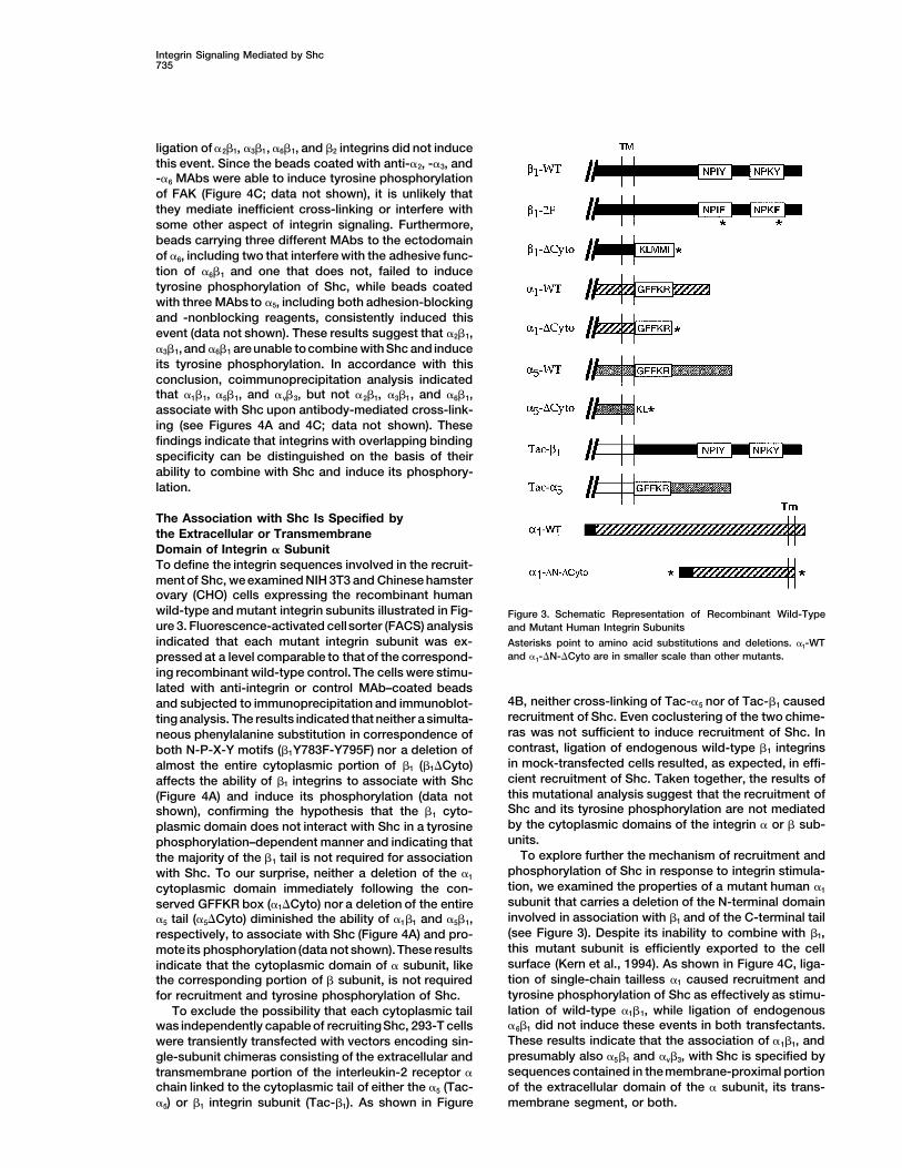
The Association with Shc Is Specified bythe Extracellular or TransmembraneDomain of Integrin a SubunitTo define the integrin sequences involved in the recruit-ment of Shc, we examined NIH 3T3 and Chinese hamsterovary (CHO) cells expressing the recombinant humanwild-type and mutant integrin subunits illustrated in Fig- Figure 3. Schematic Representation of Recombinant Wild-Typeure 3. Fluorescence-activated cellsorter (FACS) analysis and Mutant Human Integrin Subunitsindicated that each mutant integrin subunit was ex- Asterisks point to amino acid substitutions and deletions. a1-WT
and a1-DN-DCyto are in smaller scale than other mutants.pressed at a level comparable to that of the correspond-ing recombinant wild-type control. The cells were stimu-lated with anti-integrin or control MAb–coated beads
4B, neither cross-linking of Tac-a5 nor of Tac-b1 causedand subjected to immunoprecipitation and immunoblot-recruitment of Shc. Even coclustering of the two chime-ting analysis. The results indicated that neither a simulta-ras was not sufficient to induce recruitment of Shc. Inneous phenylalanine substitution in correspondence ofcontrast, ligation of endogenous wild-type b1 integrinsboth N-P-X-Y motifs (b1Y783F-Y795F) nor a deletion ofin mock-transfected cells resulted, as expected, in effi-almost the entire cytoplasmic portion of b1 (b1DCyto)cient recruitment of Shc. Taken together, the results ofaffects the ability of b1 integrins to associate with Shcthis mutational analysis suggest that the recruitment of(Figure 4A) and induce its phosphorylation (data notShc and its tyrosine phosphorylation are not mediatedshown), confirming the hypothesis that the b1 cyto-by the cytoplasmic domains of the integrin a or b sub-plasmic domain does not interact with Shc in a tyrosineunits.phosphorylation–dependent manner and indicating that
To explore further the mechanism of recruitment andthe majority of the b1 tail is not required for associationphosphorylation of Shc in response to integrin stimula-with Shc. To our surprise, neither a deletion of the a1
tion, we examined the properties of a mutant human a1cytoplasmic domain immediately following the con-subunit that carries a deletion of the N-terminal domainserved GFFKR box (a1DCyto) nor a deletion of the entireinvolved in association with b1 and of the C-terminal taila5 tail (a5DCyto) diminished the ability of a1b1 and a5b1,(see Figure 3). Despite its inability to combine with b1,respectively, to associate with Shc (Figure 4A) and pro-this mutant subunit is efficiently exported to the cellmote its phosphorylation (data not shown).These resultssurface (Kern et al., 1994). As shown in Figure 4C, liga-indicate that the cytoplasmic domain of a subunit, liketion of single-chain tailless a1 caused recruitment andthe corresponding portion of b subunit, is not requiredtyrosine phosphorylation of Shc as effectively as stimu-for recruitment and tyrosine phosphorylation of Shc.lation of wild-type a1b1, while ligation of endogenousTo exclude the possibility that each cytoplasmic taila6b1 did not induce these events in both transfectants.was independently capable of recruitingShc, 293-T cellsThese results indicate that the association of a1b1, andwere transiently transfected with vectors encoding sin-presumably also a5b1 and avb3, with Shc is specified bygle-subunit chimeras consisting of the extracellular andsequences contained in themembrane-proximal portiontransmembrane portion of the interleukin-2 receptor aof the extracellular domain of the a subunit, its trans-chain linked to the cytoplasmic tail of either the a5 (Tac-
a5) or b1 integrin subunit (Tac-b1). As shown in Figure membrane segment, or both.

Cell736
Association of Integrins with CaveolinBecause Shc isa cytosolic protein, the results describedabove suggest that the association of integrins with Shcis mediated by an intermediary membrane component.Caveolin is a 22 kDa transmembrane protein that linksa variety of cell-surface receptors lacking a cytoplasmicdomain to intracellular signaling pathways (Lisanti et al.,1994). We wished therefore to examine whether caveolinassociated with b1 integrins and Shc. The A431 cellswere incubated with anti-MHC or anti-b1 beads and im-munoprecipitated with antibodies to caveolin, Shc, andb1 integrins. The samples were probed by immunoblot-ting with antibodies reacting with the b1 subunit or thethree isoforms of Shc. The results revealed that caveolinis constitutively associated with b1 integrins and formsa complex with Shc upon stimulation with anti-b1 beads(Figure 5A). The anti-caveolin immunoprecipitates con-tained similar amounts of the precursor and mature formof b1, suggesting that b1 integrins associate with caveolinin the endoplasmic reticulum. To obtain evidence thatthe association of b1 integrins with caveolin was not anartifact of cell lysis, we performed immunofluorescentstaining of primary human fibroblasts. The results indi-cated that a significant fraction of caveolin is not dif-fusely distributed at the cell surface, but coaligns withb1 integrins, fibronectin fibrils, and actin stress fibers atextracellular matrix contact sites (data not shown).These findings indicate that b1 integrins associate withcaveolin.
To explore further the hypothesis that the recruitmentof Shc by integrins was mediated by caveolin, we exam-ined whether the association with caveolin and the re-cruitment of Shc required the same integrin sequences.NIH 3T3 cells expressing either the human recombinantwild-type a1 subunit or single-chain tailless a1 subunitwere immunoprecipitated with the anti–human a1 MAbTS2/7 or the control anti-MHC MAb W6.32, and the sam-ples were subjected to immunoblotting with anti-caveo-lin antibodies. The results showed that the wild-typea1b1 subunit and the single-chain tailless a1 subunit as-sociate to similar extent with the 22 kDa isoform ofcaveolin (Figure 5B). Prolonged exposure of the autora-diograph indicated that they both also form a complexwith the 23 kDa minor isoform of caveolin. These resultsindicate that the association of a1b1 with caveolin isspecified by sequences contained in the membrane- Figure 4. The Recruitment of Shc Is Specified by the Extracellularproximal portion of the extracellular domain of the a Domain of the a Subunit, Its Transmembrane Domain, or Bothsubunit, its transmembrane segment, or both, i.e., the (A) NIH 3T3-b1-WT, NIH 3T3-b1-2F, CHO-b1-WT, and CHO-b1-DCyto
cells were stimulated with W6.32 (c) or 4B4 (b1) MAb–coated beads,same sequences involved in the recruitment of Shc.immunoprecipitated with anti-b1 MAb TS2/16, and probed by immu-Taken together, these findings identify caveolin as anoblotting with anti-Shc serum. CHO-a5-WT, CHO-a5-DCyto, NIHpotential mediator of the interaction between integrins3T3-a1-WT, and NIH 3T3-a1-DCyto cells were stimulated with W6.32
and Shc. (c), P1D6 (a5), or TS2/7 (a1) MAb–coated beads, immunoprecipitatedwith MAb P1D6 or TS2/7, and probed by immunoblotting with anti-Shc serum.
Role of Shc in MAP Kinase Activation by Integrins (B) 293-T cells were transfected with Tac-b1 and Tac-a5, eitherseparately or in combination, stimulated with anti-Tac MAb 4E3,Since the recruitment of Shc is mediated by the integrinand immunoprecipitated with the same antibody. Mock-transfecteda subunit, while the activation of FAK requires the bcells were stimulated with W6.32 (c) or 4B4 (b1) MAbs and immuno-subunit, we sought to examine whether the activationprecipitated with MAb 4B4. Samples were probed with anti-Shcof Shc and FAK were independent phenomena. Experi-serum.
ments of immunoprecipitation with anti-FAK antibodies (C) After stimulation with W6.32 (c), TS2/7 (a1), or GoH3 (a6) MAbs,followed by immunoblotting with anti-P-Tyr antibodies NIH 3T3-a1-WT and NIH 3T3-a1-DN-DCyto cells were immunoprecip-
itated with the same antibodies and probed with anti-Shc serum,indicated that ligation of single-chain tailless a1 does

Integrin Signaling Mediated by Shc737
a1 subunit were transiently transfected with a hemagglu-tinin (HA)-tagged Erk2 vector, stimulated with beadscoated with fibronectin, various anti-integrin MAbs, orfetal calf serum (FCS), and subjected to immune com-plex kinase assays. As shown in Figure 6A, antibody-mediated ligation of single-chain tailless a1 or endoge-nous a5b1 activated MAP kinase to a level similar to thatinduced by fibronectin and serum. In contrast, cross-linking of endogenous a6b1 did not induce this activity.Since the single-chain tailless a1 subunit is capable ofrecruiting Shc, but not activating FAK, while cross-link-ing of a6b1 can stimulate FAK, but does not induce asso-ciation with Shc, these results suggest that Shc, andnot FAK, plays a crucial role in the activation of MAPkinase by integrins.
The role of Shc in the activation of MAP kinase byintegrins was further examined by testing the effect ofwild-type and dominant-negative Shc (Y317F). This mu-tant can exert a dominant-negative effect because it isable to combine with tyrosine-phosphorylated proteinsbut not with Grb2 (Salcini et al., 1994; Chen et al., 1996).The introduction of wild-type Shc led to a dose-depen-dent increase in Erk2 activation in response to ligationof wild-type a1b1 or single-chain tailless a1. Conversely,dominant-negative Shc suppressed the activation ofMAP kinase in response to ligation of both moleculesas effectively as dominant-negative Ras (N17) (Figure6B). Since full activation of FAK may require cell spread-ing on theextracellular matrix and cytoskeletal organiza-tion, we also examined the effect of dominant-negativeShc on MAP kinase activation in NIH 3T3 cells freshlyplated on fibronectin. The introduction of dominant neg-ative Shc and dominant-negative Ras prevented the ac-Figure 5. Association of b1 Integrins with Caveolintivation of Erk2 in response to cell adhesion to fibronec-(A) A431 cells were stimulated with W6.32 (anti-MHC) or 4B4 (anti-
b1) MAbs for 10 min, immunoprecipitated with MAb W6.32 (c), C060 tin (Figure 6C). We concluded that Shc plays a crucial(Cav), clone 8 (Shc), or TS2/16 (b1), and probed with anti-b1 cyto- role in the activation of the Ras–MAP kinase pathwayplasmic domain serum or affinity-purified antibodies to Shc. by integrins.(B) NIH 3T3-a1-WT and NIH 3T3 a1-DN-DCyto cells were immunopre-
The observation that only a subset of integrins cancipitated with W6.32 (c) or TS2/7 (a1) MAbs followed by immunoblot-combine with Shc suggests that cell adhesion to theting with anti-caveolin antibodies.extracellular matrix may or may not activate Ras signal-ing, depending on the repertoire of integrins involved.
not induce tyrosine phosphorylation of FAK, while stimu- To test this hypothesis, we examined primary humanlation of wild-type a1b1 and endogenous a6b1 induces umbilical vein endothelial cells (HUVECs), because it issignificant tyrosine phosphorylation of FAK (see Figure known that they adhere to fibronectin primarily through4C). Together with the data of Figure 2, these results a5b1 (Conforti et al., 1989), to vitronectin through avb3
indicate that the single-chain tailless a1 subunit can re- (Cheresh, 1987), and to laminin 1 and laminin 4 throughcruit Shc, but not activate FAK; wild-type a6b1 can acti- a2b1 (Languino et al., 1989). In addition, FACS analysisvate FAK, but not recruit Shc; and a1b1 can both recruit indicated that they do not express the a1b1 collagen/Shc and activate FAK. Thus, the recruitment and tyro- laminin receptor. The HUVECs were either kept in sus-sine phosphorylation of Shc and the activation of FAK pension or plated on dishes coated with 10 mg/ml poly-are separable phenomena. L-lysine, fibronectin, vitronectin, laminin 1, or laminin 4.
To assess the relative contribution of Shc and FAK Under these conditions, the cells adhered to and spreadon each extracellular matrix substratum to the sameto the activation of the Ras–mitogen-activated proteinextent. As shown in Figure 6D, adhesion of HUVECs to(MAP) kinase pathway by integrins, we examined thefibronectin and vitronectin resulted in tyrosine phos-ability of various integrins to activate the MAP kinasephorylation of the three isoforms of Shc, association ofErk2. NIH 3T3 cells expressing the single-chain taillessShc with Grb2, and activation of MAP kinase. In contrast,adhesion to laminin 1 or laminin 4 did not induce theseevents. These results are in accordance with the obser-immunoprecipitated with anti-Shc serum and probed with a mixturevation that a5b1 and avb3, but not a2b1, can combineof anti-P-Tyr MAbs PY20 and 4G10, or immunoprecipitated withwith Shc and activate the Ras–MAP kinase pathway andanti-FAK polyclonal antibodies and probed with a mixture of anti-
P-Tyr MAbs PY-20 and 4G10. indicate that the extracellular matrix may have selective

Cell738
Figure 6. Role of Shc in Activation of MAPKinase by Integrins
(A) NIH 3T3-a1-DN-DCyto cells were tran-siently transfected with HA-tagged Erk2 plas-mid, growth factor–starved, and stimulated insuspension with beads coated with poly-L-lysine (PL), fibronectin (Fn), or MAbs W6.32(c), TS2/7(a1), 5H10-27 (a5), and GoH3 (a6). Asa control, the cells were stimulated with 10%FCS (plus) or left untreated (minus). HA–Erk2was immunoprecipitated and subjected to invitro kinase assay.(B) NIH 3T3-a1-WT and NIH 3T3-a1-DN-DCytocells were transiently transfected with 3 mgof Erk2-HA plasmid alone or in combinationwith 2.5, 5.0, and 10 mg of wild-type Shc plas-mid (WT-Shc), 2.5 and 5.0 mg of dominantnegative Shc plasmid (Dn-Shc), and 2.5 and5.0 mg of dominant-negative Ras plasmid(Dn-Ras). The cells were stimulated withMAbs W6.32 (c) or TS2/7 (a1). Immunoprecipi-tated HA–Erk2 was subjected to in vitrokinase assay with MBP as a substrate. Trans-fection efficiencies were verified by immu-noblotting aliquots of total proteins with anti-HA antibodies.
(C) NIH 3T3 cells were transiently transfected with 3 mg of HA–Erk2 plasmid alone or in combination with 2.5, 5.0, and 10 mg of dominant-negative Shc (Dn-Shc) or Ras plasmid (Dn-Ras). After starvation, the cells were either kept in suspension or plated on fibronectin-coateddishes. HA–Erk2 was immunoprecipitated and subjected to in vitro kinase assay with MBP as a substrate. Transfection efficiencies wereverified as above.(D) HUVECs were growth factor–starved and either kept in suspension or plated on dishes coated with poly-L-lysine (PL), fibronectin (Fn),vitronectin (Vn), laminin 1 (L1), or laminin 4 (L4). Lysates were immunoprecipitated with anti-Shc MAb clone 8 followed by immunoblottingwith anti-P-Tyr MAb RC-20 (top) or anti-Grb2 serum (middle). HUVECs were transiently transfected with HA–Erk2 plasmid prior to plating onthe various substrata. HA–Erk2 was immunoprecipitated and subjected to in vitro kinase assay with MBP as a substrate (bottom). Transfectionefficiencies were verified as above.
effects on intracellular signaling depending on the inte- to Shc, such as a5b1 or avb3, is required for induction ofFos SRE–dependent transcription in cells exposed togrins to which it binds.mitogenic growth factors.
To examine whether the integrins linked to Shc playedAdhesion Mediated by Integrins Linked to ShcPromotes Transcription from the Fos Serum a role in cell cycle progression, HUVECs were syncron-
ized in G0 by growth factor starvation and then platedResponse Element and CellCycle Progression in presence of mitogens on plastic wells coated with
extracellular matrix proteins or poly-L-lysine. Entry intoWe next examined whether the coupling of specific inte-grins to Shc played a role in the control of immediate- the S phase was examined by 59-bromo-29-deoxy-uri-
dine (BrdU) incorporation and anti-BrdU staining. Theearly gene expression. Since Erk1 and Erk2 regulatetranscription from the Fos serum response element large majority of HUVECs adhering to fibronectin
(96.9%6 2.7%) and vitronectin (96.2% 6 2.1%) entered(SRE) by phosphorylating the ternary complex factorsElk-1 and SAP-1 (Treisman, 1995), we examined the the S phase during the 24 hr of the assay. In contrast,
only a modest percentage of cells plated on laminin 1effect of integrin ligation on the Fos SRE. HUVECs weretransiently transfected with a vector containing the Fos (11.8% 6 4.4%), laminin 4 (21.2% 6 9%), or poly-L-
lysine (0.8% 6 1.3%) entered into S phase under theSRE promoter element linked to the luciferase reportergene and plated on dishes coated with extracellular same conditions (Figure 7B). Since this percentage did
not increase over an additional 24-hr period, it is unlikelymatrix proteins or poly-L-lysine. The results of the lucif-erase assay indicated that adhesion to fibronectin and that adhesion to poly-L-lysine or laminins simply delays
entry into S phase. In addition, because the HUVECsvitronectin causes elevation of Fos SRE–dependenttranscription in HUVECs, while adhesion to poly-L- acquired and maintained a well-spread morphology on
laminins (Figure 7B), their inability to enter into S phaselysine, laminin 1, or laminin 4 does not (Figure 7A). Thisresult indicates that ligation of a5b1 and avb3, but not on these substrata isnot the result of insufficient spread-
ing. Interestingly, if the endothelial cells were plated ona2b1, is sufficient to promote transcription from the FosSRE. Interestingly, treatment with basic fibroblast poly-L-lysine or laminins in the presence of 10% FCS,
which contains fibronectin and vitronectin, they pro-growth factor (bFGF) induced a significant elevation ofFos SRE activity in HUVECs plated on fibronectin and gressed normally through G1 and entered the S phase.
The results of these experiments indicate that attach-vitronectin, but caused a remarkably modest effect incells adhering to poly-L-lysine, laminin 1, or laminin 4 ment and spreading on the extracellular matrix are not
sufficient for progression of primary cells through G1 in(Figure 7A), suggesting that ligation of integrins linked

Integrin Signaling Mediated by Shc739
Figure 7. Control of Fos-SRE-DependentTranscription, Cell Survival, and Cell CycleProgression by the Extracellular Matrix
(A) HUVECs were transiently transfected withFos-SRE-Luc plasmid. After growth factorstarvation, the cells were either kept in sus-pension (S) or plated onto dishes coated with10 mg/ml poly-L-lysine (PL), fibronectin (Fn),laminin 1 (Lm1), or laminin 4 (Lm4) for 20 min.The cells were then either left untreated(hatched bars) or exposed to 10 ng/ml bFGFand 1 mg/ml heparin (closed bars) for 5 min.Cell lysates containing equal amounts of totalproteins were subjected to luciferase assay.Values are expressed in arbitrary units.(B) HUVECs were growth factor–starved andplated for 24 hr in defined medium containing10 mM BrdU onto wells coated with 10 mg/ml fibronectin (Fn), vitronectin (Vn), laminin 1(Lm1), or laminin 4 (Lm4). After immunostain-ing with anti-BrdU monoclonal antibodiesand alkaline phosphatase–conjugated sec-ondary antibodies, the cells were lightlycounterstained with hematoxylin.(C) HUVECs were transiently transfected with3 mg of HA–Erk2 plasmid alone or in combina-tion with 10, 5, and 2.5 mg of plasmid encod-ing dominant-negative Shc (Dn-Shc). Aftergrowth factor starvation, the cells were de-tached and kept in suspension (c) or platedon fibronectin-coated dishes (Fn), or theywere not detached but were treated with 25ng/ml bFGF and 1 mg/ml heparin for 5 min(bFGF). Transfection efficiencies were veri-
fied by immunoblotting with anti-HA MAb. Immunoprecipitated HA–Erk2 was subjected to in vitro kinase assay with MBP as a substrate (top).HUVECs were transiently transfected with 10, 5, and 2.5 mg of plasmids encoding HA-tagged b-galactosidase (b-Gal), FLAG-tagged dominantnegative Shc (Dn-Shc), or FLAG-tagged wild-type Shc (WT-Shc). The percentage of transfected cells entering into S was determined asdescribed in Experimental Procedures. The diagram shows the mean value and standard deviation from triplicate samples (bottom).(D) G0-synchronized HUVECs were plated on wells coated with 10 mg/ml poly-L-lysine (PL), fibronectin (Fn), laminin 1 (Lm1), or laminin 4(Lm4). Adherent cells were incubated in defined medium for the indicated times. At each time point, attached and unattached cells werecombined and stained in suspension with Hoechst dye. The percentage of apoptotic nuclei was determined by scoring at least 500 cells fromfive different microscopic fields. Diagram indicates the mean and standard deviation from triplicate samples.
response to mitogens, but that this process requires high-level expression of the inhibitory molecule over therelatively prolonged duration of G1. It is unlikely thatligation of integrins, such as a5b1 and avb3, which are
coupled to Ras signaling by Shc. dominant-negative Shc interferes with mitogen stimula-tion of HUVECs, because it affected the activation ofTo examine further the role of integrin signaling in
cell proliferation, we examined the effect of dominant- Erk2 by fibronectin, but not by bFGF (Figure 7C, top).These results are consistent with the observation thatnegative or wild-type Shc on HUVEC progression
through G1. Varying concentrations of vectors encoding the FGF receptor 1 is linked to Ras by both Shc-depen-dent and Shc-independent mechanisms (MohammadiFLAG-tagged dominant-negative or wild-type Shc were
transfected in HUVECs. After growth factor starvation et al., 1996) and support the notion that the coupling ofintegrins to Ras signaling mediated by Shc regulatesand restimulation with mitogens, the entry into S phase
of cells expressing FLAG-tagged Shc molecules was cell cycle progression.monitored by double immunofluorescent staining withanti-FLAG and anti-BrdU antibodies. As a control,HUVECs were transfected with varying doses of a vector Adhesion Mediated by Integrins Not Linked
to Shc Results in Apoptotic Deathencoding HA-tagged b-galactosidase, and the entry intoS phase of these cells was evaluated by double immuno- A large fraction of endothelial cells, which initially ad-
hered on poly-L-lysine or laminins, subsequently lostfluorescent staining with anti-HA and anti-BrdU antibod-ies. As shown in Figure 7C (bottom), the introduction phase density and detached, despite being exposed to
optimal concentrations of growth factors. We thereforeof dominant-negative Shc caused a dose-dependentinhibition of HUVEC entry into Sphase. Conversely,wild- examined whether ligation of integrins linked to Shc
protected endothelial cells from apoptosis. HUVECstype Shc accelerated entry into S, although to a limitedextent. The inhibition of cell cycle progression caused by were plated at low density in the presence of mitogens
on dishes coated with extracellular matrix molecules ordominant-negative Shc was significant but incomplete,perhaps because transient transfection does not allow poly-L-lysine. At various times after plating, adherent

Cell740
and floating cells were removed, combined, and exam- response to the extracellular matrix. First, ligation ofa1b1, a5b1, and avb3, which are linked to Shc, results inined for features of apoptotic death by staining with
Hoechst dye. As shown in Figure 7D, only a small number MAP kinase activation, but ligation of other integrinsdoes not produce this effect, despite stimulating FAK.of HUVECs plated on fibronectin or vitronectin under-
went apoptosis during the 24 hr of the assay. In contrast, Second, cross-linking of the single-chain tailless a1 sub-unit causes recruitment and tyrosine phosphorylationthe majority of cells plated on poly-L-lysine or laminins
became apoptotic by the end of the assay. Direct stain- of Shc and activation of MAP kinase without inducingFAK activation. Third, a dominant-negative version ofing of cellsadhering to poly-L-lysine or laminins revealed
that most of them had become apoptotic by 12 hr, sug- Shc suppresses MAP kinase activation in response tointegrin ligation. Fourth, while adhesion of endothelialgesting that detachment follows apoptosis and not vice
versa. These findings suggest that ligation of integrins cells to fibronectin and vitronectin, which is mediatedby the Shc-linked a5b1 and avb3 integrins, activates MAPlinked to Shc is required for endothelial cell survival.kinase and induces transcription from the Fos SRE, theirinteraction with laminin 1 and laminin 4, which is medi-
Discussion ated by a2b1, does not cause these effects. Taken to-gether, these results indicate that Shc plays a crucial
The results of this study indicate that a subset of b1 role in the activation of the Ras–MAP kinase pathwayintegrins and avb3 are linked to Ras signaling and imme- and Fos gene expression in response to the extracellulardiate-early gene expression by the adaptor protein Shc. matrix.Ligation of integrins linked to Shc enables primary endo- Our findings suggest that the association of specificthelial cells to progress through G1 in response to mito- integrins with Shc regulates cell cycle progression ingens, whereas ligation of other integrins, under the same normal cells. In fact, engagement of integrins linked toconditions, results in exit from the cell cycle. On the Shc activates SRE-dependent transcription and pro-basis of these findings and the ability of dominant-nega- motes progression through G1 in response to growthtive Shc to inhibit cell cycle progression without affect- factors. In contrast, ligation of other integrins resultsing mitogen signaling, we propose that the association in cell cycle arrest even in the presence of otherwiseof specific integrins with Shc controls cell cycle progres- mitogenic concentrations of growth factors. Normalsion in response to the extracellular matrix. Since exit cells require a signal from the extracellular matrix infrom the cell cycle is a prerequisite for cell differentia- order to proliferate. Recent studies have indicated thattion, our results may also explain why interaction with cell adhesion is necessary for the induction of cyclin D1
the extracellular matrix in some settings promotesdiffer- and activation of the cyclin E–cdk2 complex in early toentiation. mid-G1 (Fang et al., 1996; Zhu et al., 1996), suggesting
Shc is a SH2-PTB domain adaptor protein that links that mitogen- and cell adhesion–dependent signals arevarious tyrosine-phosphorylated signal transducers to integrated prior to the induction of cyclin D1. In accor-Ras (Pawson, 1995). We have previously shown that dance with this hypothesis, our findings suggest thatligation of the a6b4 integrin, which is associated with a integrin- and growth factor–dependent signals convergetyrosine kinase, causes phosphorylation of the b4 tail on Ras.and direct recruitment of Shc (Mainiero et al., 1995). In It is likely that a simultaneous stimulation of Ras bythis study, we provide evidence that specific b1 integrins integrins and growth factor receptors is needed to reachand avb3 also combine with Shc, but by a distinct and the threshold level of MAP kinase activation requirednovel mechanism. In this case, the recruitment of Shc for optimal transcription of immediate-early responseis mediated by the membrane-proximal portion of the genes. Since mitogenic growth factors induce a rapidextracellular domain of the integrin a subunit, its trans- and short-lived stimulation of MAP kinase, while cellmembrane segment, or both. Coimmunoprecipitation adhesion produces a long-lasting activation of the en-experiments revealed that this region of the a subunit zyme (Zhu and Assoian, 1995), the two stimuli may alsointeracts constitutively with caveolin, a two-pass trans- cooperate kinetically. This model is consistent with themembrane adaptor involved in linking a variety of cell- observation that overexpression of Shc (Pelicci et al.,surface receptors to intracellular signaling pathways (Li- 1992) and constitutive activation of MAP kinase (Cowleysanti et al., 1994), and indicated that Shc associates et al., 1994) lead to anchorage-independent cell growth.with caveolin in response to integrin ligation. Although Furthermore, since most dominant oncogenes trans-a definitive demonstration that the recruitment of Shc form cells by activating the Ras–MAP kinase pathway,to b1 integrins and avb3 is mediated by caveolin will it also explains why most transformed cells display an-require further biochemical and mutational analysis, this chorage-independent growth.model is intriguing, because caveolin is phosphorylated Recent studieshave revealed that normal cells deniedon tyrosine in cells transformed by v-Src (Glenney and anchorage to the extracellular matrix undergo apoptosisSoppet, 1992) and has also been shown to interact with (Ruoslahti and Reed, 1994). The findings of this studyc-Fyn (Corley Mastick et al., 1995). Thus, caveolin may suggest that the ability of the extracellular matrix toprovide both the adaptor and the tyrosine kinase neces- promote cell survival is mediated by the coupling ofsary for the recruitment and tyrosine phosphorylation specific integrins to Shc. Since inhibition of MAP kinaseof Shc in response to integrin ligation. or activation of Jun kinase cause apoptosis (Xia et al.,
There are several reasons to believe that the associa- 1995), it is possible that the integrins that combine withtion of specific integrins with Shc mediates activation Shc promote cell survival by elevating the activity of
MAP kinase, thereby increasing the ratio of MAP kinaseof the MAP kinase pathway and transcription of Fos in

Integrin Signaling Mediated by Shc741
to Jun kinase activity. The anti-apoptotic function of (Sastry et al., 1996). Furthermore, recent studies haveindicated that the binding of fibronectin to a5b1, butintegrins linked to Shc is consistent with the results of
previous studies. For example, antibodies and synthetic not a4b1, results in induction of the collagenase gene insynovial fibroblasts, suggesting that the same matrixpeptides, which interfere with the adhesive function of
avb3, induce endothelial cell apoptosis and thereby blunt molecule may or may not induce gene expression de-pending on the integrin to which it binds (Huhtala et al.,angiogenesis in vivo (Brooks et al., 1994). We suggest,
on the basis of our results, that these reagents do not 1995). Taken together, our findings suggest that thedifferential responses of a given cell type to extracellulartrigger intracellular signals that lead to apoptosis, but
rather prevent the activation of Shc signaling caused by matrices of different composition and of different celltypes to the same extracellular matrix protein may allavb3 ligation. The ability of fibronectin to protect CHO
cells from apoptosis in response to serum withdrawal depend on the ability of a class of integrins to activateShc signaling.(Zhang et al., 1995) and that of basement membrane
components, butnot type I collagen, to promote survivalExperimental Proceduresof breast epithelial cells (Pullan et al., 1996) may also
depend on the engagement of specific integrins linkedAntibodies and Extracellular Matrix Moleculesto Shc.The MAbs TS2/7, FW-14–14–15, and TS2/16 were obtained from
We have observed that intercellular contact can res- the American Type Culture Collection, P1E6, P1B5, P1D6, and P4H9cue primary endothelial cells plated on laminins from from GIBCO BRL, 5H10–27 from Pharmingen, GoH3 from Immuno-apoptotic death (unpublished data). It has been reported tech, 4B4 from Coulter, 4E3 and 12CA5 from Boehringer, M2 from
Eastman-Kodak, 4G10 from Upstate Biotechnology, Incorporated,that cell-to-cell contact exerts a similar protective effectand PY20, RC-20, clone 8, and C060 from Transduction Labora-in primary breast epithelial cells (Pullan et al., 1996).tories. The anti-b1 cytoplasmic domain serum and serum 410, whichAlthough the mechanism by which physical contact be-reacts predominantly with the 52 kDaisoform of Shc, were described
tween cells protects primary cells from apoptosis is not (Giancotti and Ruoslahti, 1990; Mainiero et al., 1995). The MAbsknown, these observations suggest that multiple posi- AIIB2 and BIE5 were provided by C. Damsky, LM609 by D. Cheresh,tional signals contribute to cell survival in vivo. It can and 135–13C by S. Kennel. The anti-P-Tyr serum 72 and anti-Grb2
MAb EL-6 were generated in the laboratory of J. Schlessinger. Affin-be envisioned that in vivo these signals promote theity-purified rabbit antibodies to Shc and caveolin were from Upstatesurvival of those cells that have, as part of their naturalBiotechnology and Transduction Laboratories, respectively. Anti-life cycle, lost contact with a matrix capable of activatingGrb2 and anti-Erk2 sera were from Santa Cruz Biotechnology. The
the Shc pathway. This mechanism may ensure that only anti-FAK peptide serum was generated in the laboratory of G. Tar-cells displaced from their natural environment are elimi- one. Human fibronectin, vitronectin, laminin 4 (placental merosin;nated. Spinardi et al., 1995), and mouse laminin 1 were purchased from
GIBCO BRL.What is the fate of those cells that survive despitelacking the Shc signal? The observation that extracellu-
Cell Lines, Constructs, and Transfectionslar matrix recognition by integrins that fail to activateCHO cells expressing b1-WT and b1-DCyto were generated in the
Shc results in cell cycle exit even in the presence of laboratory of G. Tarone (University of Torino, Turin, Italy). CHO cellsmitogens suggests that these cells may be induced to expressing a5-WT or a5-DCyto and NIH 3T3 cells expressing a1-WT,differentiate. It is known that withdrawal from the cell a1-DCyto, or a1-DN-DCyto were previously described (Bauer et al.,
1993; Briesewitz etal., 1993; Kernet al., 1994). NIH 3T3 cells express-cycle is a prerequisite for differentiation, and severaling b1-WT and b1-2F were generated as previously described (Gian-mechanisms ensure that proliferation and differentiationcotti et al., 1994). Transient transfection of vectors encoding Tac-are mutually exclusive. For example, active cyclin D1–a5Cyto and Tac-b1Cyto (LaFlamme et al., 1992) in 293-T cells was
cdk complexes suppress MyoD function in proliferating also done as described (Giancotti et al., 1994). The Fos(SRE)-Lucmyoblasts, thereby preventing the expression of mus- reporter plasmid (from J. Schlessinger) and vectors encoding HA-cle-specific genes (Skapek et al., 1995). Conversely, tagged Erk2, HA-tagged b-galactosidase, FLAG-tagged and un-
tagged wild-type or dominant-negative p52shc (Y317F), and dominantMyoD may maintain the G0 arrest of differentiated skele-negative Ras (N17) (from E. Scolnik, New York University Schooltal muscle by acting on Rb (Gu et al., 1993). These obser-of Medicine) were transiently transfected in NIH 3T3 cells by thevations suggest that those integrins that do not activatelipofectamine method and in HUVECs by the lipofectin method
the Shc pathway may promote differentiation primarily (GIBCO BRL).because they do not induce cyclin D1–cdk levels suffi-cient to block the function of transcription factors in- Biochemical Methodsvolved in differentiation. After ligation of integrins (Mainiero et al., 1995), cells were extracted
and subjected to immunoprecipitation followed by immunoblottingThe existence of two classes of integrins with distinctor kinase assay. To immunoprecipitate Shc, integrins, caveolin, andsignaling properties explains a number of previous ob-FAK, cells were extracted in 50 mM HEPES (pH 7.5), 150 mM NaCl,servations. In various cell types, interaction with fibro-1% Triton X-100. To immunoprecipitate Erk2, cells were extracted
nectin promotesproliferation and inhibits differentiation, with 50 mM HEPES (pH 7.5), 150 mM NaCl, 1% NP-40, 1 mM EDTA.while adhesion to laminin promotes cell cycle with- Immunoprecipitation, immunoblotting, and GST fusion protein bind-drawal and morphological and functional differentiation. ing experiments were performed as previously described (Mainiero
et al., 1995). Erk2 immune-complex kinase assays were performedFor example, endothelial cells plated on fibronectin pro-in 50 mM Tris (pH 7.5), 10 mM MgCl2 containing 5 mCi of [g-32P]ATPliferate, but on a laminin-rich matrix they cease growing(4500 Ci/mmol, ICN Biomedicals, Incorporation) and 2.5 mg of MHC-and rapidly form capillary-like structures (Kubota et al.,binding protein (MBP). Ras–GTP loading assays were performed as
1988). Similarly, myoblasts proliferate on fibronectin, but described (Gale et al., 1993). To measure SRE-dependent transcrip-fuse to form myotubes on laminin (von der Mark and tion, HUVECs were transiently transfected with Fos-SRE-Luc. AfterOcalan, 1989). These two opposing functions have been growth factor starvation, the cells were detached, plated on dishes
coated with 10 mg/ml poly-L-lysine, fibronectin, vitronectin, lamininlinked to the expression of a5b1 and a6b1, respectively

Cell742
1, or laminin 4, andthen subjected to luciferaseassay. At this coating Clark, E.A., andBrugge, J.S. (1995). Integrins andsignal transductionpathways: the road taken. Science 268, 233–239.concentration, all extracellular matrix proteins promoted similar lev-
els of adhesion and spreading in HUVECs. Conforti, G., Zanetti, A., Colella, S., Abbadini, M., Marchisio, P.C.,Pytela, R., Giancotti, F.G., Tarone, G., Languino, L.R., and Dejana,
Measurement of Cell Cycle Progression and Apoptosis E. (1989). Interaction of fibronectin with cultured human endothelialTo monitor progression through G1 and entry into S phase, HUVECs cells: characterization of the specific receptor. Blood 73, 1576–were synchronized in G0 by growth factor starvation, detached, and 1585.plated at low density on microtiter plates or glass coverslips coated Corley Mastick, C., Brady, M.J., and Saltiel, A.R. (1995). Insulin stim-with poly-L-lysine or extracellular matrix molecules. After 24 hr of ulates the tyrosine phosphorylation of caveolin. J. Cell Biol. 129,incubation in defined medium (M199 supplemented with 25 ng/ml 1523–1531.bFGF, 1 mg/ml heparin, 6.25 mg/ml insulin, 6.25 mg/ml transferrin,
Cowley, S., Patterson, H., Kemp, P., and Marshall, C.J. (1994). Acti-0.625 ng/ml selenous acid, 1.25 mg/ml BSA, and 5.35 mg/ml linoleicvation of MAP kinase kinase is necessary and sufficient for PC12acid) containing 10 mM BrdU, cells were stained with anti-BrdU MAbdifferentiation and for transformation of NIH 3T3 cells. Cell 77, 841–and AP-conjugated anti-mouse IgGs (Boehringer).852.To test the effect of dominant-negative and wild-type Shc onFang, F., Orend, G., Watanabe, N., Hunter, T., and Ruoslahti, E.cell cycle progression, HUVECs were transiently transfected with(1996). Dependence of cyclin E–CDK 2 kinase activity on cellanchor-vectors encoding FLAG-tagged dominant-negative Shc, FLAG-age. Science 271, 499–502.tagged wild-type Shc, or the control protein HA-b-galactosidase.
After incubation in complete medium for 8 hr, the cells were starved Gale, N.W., Kaplan, S., Lowenstein, E.J., Schlessinger, J., and Bar-for 24 hr and then incubated in defined medium containing 10 mM Sagi, D. (1993). Grb2 mediates the EGF-dependent activation ofBrdU for 24 hr. Entry into S phaseof transfected cells was monitored guanine nucleotide exchange on Ras. Nature 363, 88–92.by double immunofluorescent staining with anti-FLAG MAb M2 or Giancotti, F.G., and Mainiero, F. (1994). Integrin-mediated adhesionanti-HA MAb 12CA5 followed by Texas red–conjugated anti-mouse and signaling in tumorigenesis. Biochem. Biophys. Acta 1198,IgGs and FITC-conjugated anti-BrdU antibodies. 47–64.
To measure apoptosis, G0-synchronized HUVECs were plated onGiancotti, F.G., and Ruoslahti, E. (1990). Elevated levels of the a5b1wells or coverslips coated with poly-L-lysine or extracellular matrixfibronectin receptor suppress the transformed phenotype of Chi-proteins and incubated in defined medium for the indicated times.nese hamster ovary cells. Cell 60, 849–859.Attached and unattached cells were combined and stained in sus-Giancotti, F.G., Spinardi, L., Mainiero, F., and Sanders, R. (1994).pension with Hoechst dye.Expression of heterologous integrin genes in cultured eukaryoticcells. Methods Enzymol. 245, 297–316.AcknowledgmentsGlenney, J.R., and Soppet, D. (1992). Sequence and expressionCorrespondence should be addressed to F. G. G. We thank D. Bar-of caveolin, a protein component of caveolae plasma membraneSagi, D. Cheresh, C. Damsky, R. Juliano, S. Kennel, S. LaFlamme,domains phosphorylated on tyrosine in Rous sarcoma virus–S. Retta, E. Scolnik, J. Schlessinger, G. Tarone, and K. Yamada fortransformed fibroblasts. Proc. Natl. Acad. Sci. USA. 89, 10517–reagents, B. Gumbiner and E. Scolnick for reviewing the manuscript,10521.M. Palmieri for the analysis of cells expressing mutant b1 subunits,Gu, W., Schneider, J.W., Condorelli, G., Kaushal, S., Mahdavi, V.,F. David and A. Kern for FACS analysis, and A. Pepe and M. Epsteinand Nadal-Ginard, B. (1993). Interaction of myogenic factors andfor technical assistance. This work was supported by Departmentthe retinoblastoma protein mediates muscle cell commitment andof Army Medical Defense grant 17-94-J4306 (F. G. G.) and Nationaldifferentiation. Cell 72, 309–324.Institutes of Health (NIH) grants CA-58976 (F. G. G.), GM-44585
(E. E. M.), and CA–16087 (Cancer Center). K. K. W. was supported Huhtala, P., Humphries, M.J., McCarthy, J.B., Tremble, P.M., Werb,by NIH training grant CA-09161. F. G. G. is an Established Investiga- Z., and Damsky, C.H. (1995). Cooperative signaling by a5b1 and a4b1
tor of the American Heart Association (AHA). E. E. M. is an Estab- integrins regulate metalloproteinase gene expression in fibroblastslished Scientist of the New York City Affiliate of the AHA. adhering to fibronectin. J. Cell Biol. 129, 867–879.
Hynes, R.O. (1992). Integrins: versatility, modulation, and signalingReceived June 26, 1996; revised September 27, 1996. in cell adhesion. Cell 69, 11–25.
Hynes, R.O. (1996). Targetted mutations in cell adhesion genes:what have we learned from them? Dev. Biol., in press.
ReferencesIlic, D., Fruta, Y., Kanazawa, S., Takeda, N., Sobue, K., Nakatsuji,N., Nomura, S., Fujimoto, J., Okada, M., Yamamoto, T., and Aizawa,Bauer, J.S., Varner, J., Schreiner, C., Kornberg, L., Nicholas, R., andS. (1995). Reduced cell motility and enhanced focal adhesion con-Juliano, R.L. (1993). Functional role of the cytoplasmic domain oftact formation in cells from FAK-deficient mice. Nature 377, 539–the integrin a5 subunit. J. Cell Biol. 122, 209–221.544.
Briesewitz, R., Kern, A., and Marcantonio, E.E. (1993). Ligand-Kern, A., Briesewitz, R., Bank, I., and Marcantonio, E.E. (1994). Thedependent and -independent integrin focal contact localization: therole of the I domain in ligand binding of the human integrin a1b1. J.role of the a chain cytoplasmic domain. Mol. Biol. Cell 4, 593–604.Biol. Chem. 269, 22811–22816.
Brooks, P.C., Montgomery, A.M.P., Rosenfeld, M., Reisfeld, R.A.,Kubota, W., Kleinman, H.K., Martin, G.R., and Lawley, T.J. (1988).Hu, T., Klier, G., and Cheresh, D.A. (1994). Integrin avb3 antagonistsRole of laminin and basement membrane in the morphological differ-promote tumor regression by inducing apoptosis of angiogenicentiation of human endothelial cells into capillary-like structures. J.blood vessels. Cell 79, 1157–1164.Cell Biol. 107, 1589–1598.
Chen, H.-C., and Guan, J.-L. (1994). Association of focal adhesionLaFlamme, S.E., Akiyama, S.K., and Yamada, K.M. (1992). Regula-kinase with its potential substrate phosphatidylinositol 3-kinase.tion of fibronectin receptor distribution. J. Cell Biol. 117, 437–447.Proc. Natl. Acad. Sci. USA. 91, 10148–10152.Languino, L.R., Gehlsen, K.R., Wayner, E., Carter, W.G., Engvall, E.,Chen, Y., Grall, D., Salcini, A.E., Pelicci, P.G., Pouyssegur, J., andand Ruoslahti, E. (1989). Endothelial cells use a2b1 integrins as aVan Obberghen-Schilling, E. (1996). Shc adaptor proteins are keylaminin receptor. J. Cell Biol. 109, 2455–2462.transducers of mitogenic signaling mediated by the G protein–
coupled thrombin receptor. EMBO J. 15, 1037–1044. Lin, C.Q., and Bissell, M.J. (1993). Multi-faceted regulation of celldifferentiation via extracellular matrix. FASEB J. 7, 737–743.Cheresh, D.A. (1987). Human endothelial cells synthesize and ex-
press an Arg-Gly-Asp-directed adhesion receptor involved in at- Lisanti, M.P., Scherer, P.E., Tang, Z., and Sargiacomo, M. (1994).Caveolae, caveolin, and caveolin-rich membrane domains: a signal-tachment to fibrinogen and Von Willebrand factor. Proc. Natl. Acad.
Sci. USA. 84, 6471–6475. ing hypothesis. Trends Cell Biol. 4, 231–235.

Integrin Signaling Mediated by Shc743
Mainiero, F., Pepe, A., Wary, K.K., Spinardi, L., Mohammadi, M.,Schlessinger, J., and Giancotti, F.G. (1995). Signal transduction bythe a6b4 integrin: distinct b4 subunit sites mediate recruitment of Shc/Grb2 and association with the cytoskeleton of hemidesmosomes.EMBO J. 14, 4470–4481.
Mohammadi, M., Dikic, I., Sorokin, A., Burgess, W.H., Jaye, M., andSchlessinger, J. (1996). Identification of six novel autophosphoryla-tion sites on fibroblast growth factor receptor 1 and elucidation oftheir importance in receptor activation and signal transduction. Mol.Cell. Biol. 16, 977–989.
Pawson, T. (1995). Protein modules and signaling networks. Nature373, 573–580.
Pelicci, G., Lanfrancone, L., Grignani, F., McGlade, J., Cavallo, F.,Forni, G., Nicoletti, I., Grignani, F., Pawson, T., and Pelicci, P.G.(1992) A novel transforming protein (Shc) with an SH2 domain isimplicated in mitogenic signal transduction. Cell 70, 93–104.
Pullan, S., Wilson, J., Metcalfe, A., Edwards, G.M., Goberdhan, N.,Tilly, J.,Hickman, J.A., Dive, C., and Streuli, C.H. (1996). Requirementof basement membrane for the suppression of programmed celldeath in mammary epithelium. J. Cell Sci. 109, 631–642.
Richardson, A., and Parsons, T. (1996). A mechanism for regulationof the adhesion-associated protein tyrosine kinase pp125FAK. Nature380, 538–540.
Ruoslahti, E. (1991). Integrins. J. Clin. Invest. 87, 1–5.
Ruoslahti, E., and Reed, J.C. (1994). Anchorage dependence, inte-grins, and apoptosis. Cell 77, 477–478.
Salcini, A.E., McGlade, J., Pelicci, G., Nicoletti, I., Pawson, T., andPelicci, P.G. (1994). Formation of Shc–Grb2 complexes is necessaryto induce neoplastic transformation by overexpression of Shc pro-teins. Oncogene 9, 2827–2836.
Sastry, S.K., Lakonishok, M., Thomas, D.A., Muschler, J., and Hor-witz, A.F. (1996). Integrin a subunit ratios, cytoplasmic domains,and growth factor synergy regulate muscle proliferation and differ-entiation. J. Cell Biol. 133, 169–184.
Schaller, M.D., and Parsons, J.T. (1994). Focal adhesion kinase andassociated proteins. Curr. Opin. Cell Biol. 6, 705–710.
Schlaepfer, D.D., Hanks, S.K., Hunter,T., and van der Geer, P. (1994).Integrin-mediated signal transduction linked to Ras pathway byGRB2 binding to focal adhesion kinase. Nature 372, 786–791.
Skapek, S., Rhee, J., Spicer, D.B., and Lassar, A.B. (1995). Inhibitionof myogenic differentiation in proliferating myoblasts by cyclin D1–dependent kinase. Science 267, 1022–1024.
Spinardi, L., Einheber, S., Cullen., T., Milner, T.A., and Giancotti,F. G. (1995). A recombinant tail-less integrin a6b4 subunit disruptshemidesmosomes, but does not suppress a6b4-mediated cell adhe-sion to laminins. J. Cell Biol. 129, 473–487.
Treisman, R. (1995). Journey to the surface of the cell: Fos regulationand SRE. EMBO J. 14, 4905–4913.
von der Mark, K., and Ocalan, M. (1989). Antagonistic effects oflaminin and fibronectin on the expression of the myogenic pheno-type. Differentiation 40, 150–157.
Vuori, K., Hirai, H., Aizawa, S., and Ruoslahti, E. (1996). Inductionof p130CAS signaling complex formation upon integrin-mediated celladhesion: a role for src familykinases. Mol. Cell. Biol. 16, 2606–2613.
Xia, Z., Dickens, M., Raingeaud, J., Davis, R.J., and Greenberg,M.E. (1995). Opposing effects of Erk and Jnk–p38 MAP kinases onapoptosis. Science 270, 1326–1331.
Zhang, Z., Vuori, K., Reed, J.C., and Ruoslahti, E. (1995). The a5b1
integrin supports survival of cells on fibronectin and up-regulatesBcl-2 expression. Proc. Natl. Acad. Sci. USA. 92, 6161–6165.
Zhu, X., and Assoian, R.K. (1995). Integrin-dependent activation ofMAP kinase: a link to shape-dependent cell proliferation. Mol. Biol.Cell 6, 273–282.
Zhu, X., Ohtsubo, M., Bohmer, R., Roberts, J.M., and Assoian, R.K.(1996). Adhesion dependent cell cycle progression linked to theexpression of cyclin D1, activation of cyclin E–cdk2, and phosphory-lation of the retinoblastoma protein. J. Cell Biol. 133, 391–403.
Copyright © 2022 FDOKUMEN