Cooperative Signaling by and a4/ l Integrins Regulates Metalloproteinase Gene Expression in...
13
Cooperative Signaling by and a4/ l Integrins Regulates Metalloproteinase Gene Expression in Fibroblasts Adhering to Fibronectin Pirkko Huhtala,* Martin J. Humphries, IIJames B. McCarthy,1 Patrice M. Tremble,§ Zena Werb,*§ and Caroline H. Damsky** Departments of *Stomatology and *Anatomy, and § Laboratory of Radiobiology and Environmental Health, University of California, San Francisco, California 94143; II School of Biological Sciences, University of Manchester, Manchester M139PT, United Kingdom; and I Department of Laboratory Medicine and Pathology, University of Minnesota, Minneapolis, Minnesota 55455 Abstract. Rabbit synovial fibroblasts (RSF) express basal levels of the metalloproteinases (MMP) col- lagenase, stromelysin-1 and 92-kD gelatinase when plated on intact fibronectin (FN), but elevated levels when plated on either the central RGD-containing cell-binding region of FN (120FN) or antibody against the ot5/~l integrin, suggesting that domains outside 120FN may suppress the induction of MMP (Werb, Z., P. M. Tremble, O. Behrendtsen, E. Crowley, and C. H. Damsky. 1989. J. Cell Biol. 109:877-889). We therefore attempted to reconstitute the basal signaling of intact FN by plating RSF on 120FN together with domains of FN outside this region. Large COOH-ter- minal fragments containing both the heparin-binding and IIICS domains suppressed MMP when combined with 120FN. To map the active sequences, peptides from this region and larger fragments that did, or did. not, include the CS-1 portion of IIICS were tested. Only CS-1 peptide, or larger fragments containing CS-1, suppressed MMP expression induced by 120FN. In contrast, peptide V from the heparin-binding region, shown previously to stimulate focal contact formation, further enhanced MMP expression by RSF when pres- ent on the substrate with 120FN. RSF expressed ot4B1 integrin, the receptor for CS-1, and the anti-tx4 mAb blocked the ability of CS-1 to suppress MMP induc- tion by 120FN. These results show that signals mod- ulating MMP expression and focal contact assembly are regulated independently, and that cooperative sig- naling by ot5/51 and ot4~l integrins plays a dominant role in regulating expression of these extracellular ma- trix-remodeling genes in response to FN. This work demonstrates directly the modular way in which infor- mation in the extracellular matrix is detected and processed by cell surface receptors. C ELLS interact with the extracellular matrix (ECM) ~ via several classes of cell surface receptors (reviewed in Albelda and Buck, 1990; Yamada, 1991; Hynes, 1992). Their responses to ECM are varied and include adhe- sion, spreading, migration, and differentiation. In addition to these morphogenetic effects, recent studies have shown that the composition and organization of the ECM can regu- Address all correspondence to P. Huhtala, HSW 604, Department of Stomatology, 513 Parnassus Avenue, University of California, San Fran- cisco, CA 94143-0512. Ph.: (415)476-8921. Fax: (415)476-4204. 1. Abbreviations used in this paper: CM, conditioned medium; CS, con- netting segment; CSPG, chondroitin sulfate proteoglycan; ECM, extra- cellular matrix; FN, fibronecfin, GAPDH, glyceraldehyde-3-phosphate dehydrogenase; HBD, heparin-bindingdomain; HSPG, heparan sulfate pro- teoglycan; LH, lactalbumin hydrolysate; MMP, matrix metalloproteinase; OA, ovalbumin; RGD, Arg-Gly-Asp; RSF, rabbit synovial fibroblast; 120FN, 120-kD fragment of fibroneetin; 33166FN, 33- and 66-kD COOH- terminal fragments of FN; 29FN, 29-kD fragment of FN. late the expression of genes that play important roles in tissue-specific differentiation, growth, pH regulation, and matrix remodeling (reviewed in Damsky and Werb, 1992; Adams and Watt, 1993; Ashkenas et al., 1994). The information encoded in ECM is transduced by cells through combinations of interactions between individual ECM ligand-binding sites and cell surface receptors. The ECM as a whole is extremely complex, with many different components exhibiting the ability to bind to cells as well as to other ECM constituents. Recently it has become clear that cell contact with even one specific ECM component, such as fibronectin (FN), initiates multiple signals affecting both cell behavior and gene expression. Little is known about how ceils process the complex input from the engagement of different combinations of receptors and binding sites on an individual ECM ligand. However, the non-redundant nature of the multiple interactions between different receptors and a single ECM component has been underscored recently by © The Rockefeller University Press, 0021-9525195/05/867/13 $2.00 The Journal of Cell Biology, Volume 129, Number 3, May 1995 867-879 867 on November 10, 2014 jcb.rupress.org Downloaded from Published May 1, 1995
Transcript of Cooperative Signaling by and a4/ l Integrins Regulates Metalloproteinase Gene Expression in...

Cooperative Signaling by and a4/ l Integrins Regulates Metalloproteinase Gene Expression in Fibroblasts Adhering to Fibronectin Pirkko Huhtala ,* Mar t in J. Humphr ies , II James B. McCarthy,1 Patrice M. Tremble,§ Zena Werb,*§ and Carol ine H. Damsky**
Departments of * Stomatology and * Anatomy, and § Laboratory of Radiobiology and Environmental Health, University of California, San Francisco, California 94143; II School of Biological Sciences, University of Manchester, Manchester M139PT, United Kingdom; and I Department of Laboratory Medicine and Pathology, University of Minnesota, Minneapolis, Minnesota 55455
Abstract. Rabbit synovial fibroblasts (RSF) express basal levels of the metalloproteinases (MMP) col- lagenase, stromelysin-1 and 92-kD gelatinase when plated on intact fibronectin (FN), but elevated levels when plated on either the central RGD-containing cell-binding region of FN (120FN) or antibody against the ot5/~l integrin, suggesting that domains outside 120FN may suppress the induction of MMP (Werb, Z., P. M. Tremble, O. Behrendtsen, E. Crowley, and C. H. Damsky. 1989. J. Cell Biol. 109:877-889). We therefore attempted to reconstitute the basal signaling of intact FN by plating RSF on 120FN together with domains of FN outside this region. Large COOH-ter- minal fragments containing both the heparin-binding and IIICS domains suppressed MMP when combined with 120FN. To map the active sequences, peptides from this region and larger fragments that did, or did. not, include the CS-1 portion of IIICS were tested.
Only CS-1 peptide, or larger fragments containing CS-1, suppressed MMP expression induced by 120FN. In contrast, peptide V from the heparin-binding region, shown previously to stimulate focal contact formation, further enhanced MMP expression by RSF when pres- ent on the substrate with 120FN. RSF expressed ot4B1 integrin, the receptor for CS-1, and the anti-tx4 mAb blocked the ability of CS-1 to suppress MMP induc- tion by 120FN. These results show that signals mod- ulating MMP expression and focal contact assembly are regulated independently, and that cooperative sig- naling by ot5/51 and ot4~l integrins plays a dominant role in regulating expression of these extracellular ma- trix-remodeling genes in response to FN. This work demonstrates directly the modular way in which infor- mation in the extracellular matrix is detected and processed by cell surface receptors.
C ELLS interact with the extracellular matrix (ECM) ~ via several classes of cell surface receptors (reviewed in Albelda and Buck, 1990; Yamada, 1991; Hynes,
1992). Their responses to ECM are varied and include adhe- sion, spreading, migration, and differentiation. In addition to these morphogenetic effects, recent studies have shown that the composition and organization of the ECM can regu-
Address all correspondence to P. Huhtala, HSW 604, Department of Stomatology, 513 Parnassus Avenue, University of California, San Fran- cisco, CA 94143-0512. Ph.: (415)476-8921. Fax: (415)476-4204.
1. Abbreviations used in this paper: CM, conditioned medium; CS, con- netting segment; CSPG, chondroitin sulfate proteoglycan; ECM, extra- cellular matrix; FN, fibronecfin, GAPDH, glyceraldehyde-3-phosphate dehydrogenase; HBD, heparin-binding domain; HSPG, heparan sulfate pro- teoglycan; LH, lactalbumin hydrolysate; MMP, matrix metalloproteinase; OA, ovalbumin; RGD, Arg-Gly-Asp; RSF, rabbit synovial fibroblast; 120FN, 120-kD fragment of fibroneetin; 33166FN, 33- and 66-kD COOH- terminal fragments of FN; 29FN, 29-kD fragment of FN.
late the expression of genes that play important roles in tissue-specific differentiation, growth, pH regulation, and matrix remodeling (reviewed in Damsky and Werb, 1992; Adams and Watt, 1993; Ashkenas et al., 1994).
The information encoded in ECM is transduced by cells through combinations of interactions between individual ECM ligand-binding sites and cell surface receptors. The ECM as a whole is extremely complex, with many different components exhibiting the ability to bind to cells as well as to other ECM constituents. Recently it has become clear that cell contact with even one specific ECM component, such as fibronectin (FN), initiates multiple signals affecting both cell behavior and gene expression. Little is known about how ceils process the complex input from the engagement of different combinations of receptors and binding sites on an individual ECM ligand. However, the non-redundant nature of the multiple interactions between different receptors and a single ECM component has been underscored recently by
© The Rockefeller University Press, 0021-9525195/05/867/13 $2.00 The Journal of Cell Biology, Volume 129, Number 3, May 1995 867-879 867
on Novem
ber 10, 2014jcb.rupress.org
Dow
nloaded from
Published May 1, 1995

the observation that targeted null mutations in the genes for FN (George et al., 1993), or for either of its two major inte- grin receptors, ot5/31 (Yang et at., 1993) and c~4~1 (Yang et al., 1994), display distinct phenotypes, and they are all lethal early in development.
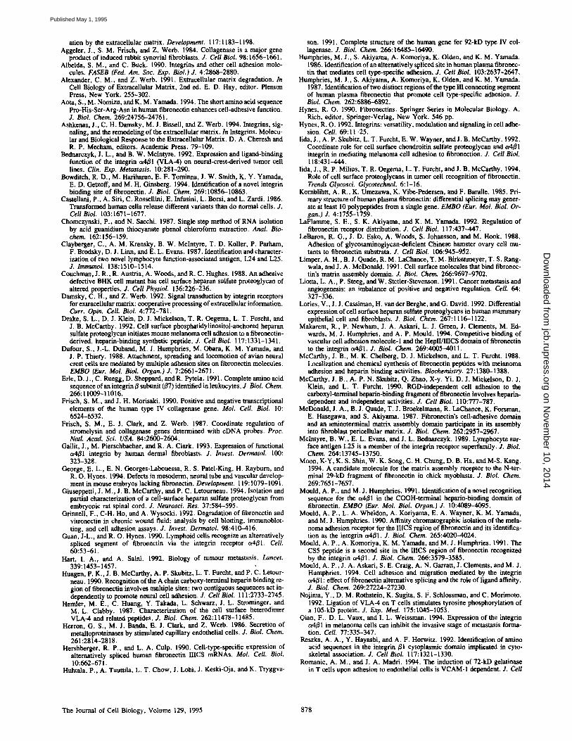
Dissection of the structure-function relationships of a sin- gle ECM component is most advanced for FN. FN com- prises a family of polypeptides that results from the alterna- tive splicing of a single RNA (reviewed in Hynes, 1990; Schwarzbauer, 1991). FN can interact extensively with itself, and with other matrix components through its collagen-, fibrin- and glycosaminoglycan-binding domains. Remark- ably, at least eight peptide sequences with varying cell sur- face binding activities have been mapped to different regions of FN (see Fig. 1). These sites on FN have been shown to interact with at least two distinct classes of cell surface receptors: integrins and membrane-associated proteogly- cans (reviewed in Yamada, 1991; Damsky and Werb, 1992). The NH2-terminal 29-kD domain, containing the first five type I repeats (II-~), is required for assembly of a fibrillar FN matrix. It has fibrin- and weak heparin-binding activi- ties and it binds weakly to the cell surface (Limper et al., 1991; Moon et al., 1994). However, no specific cell surface receptor has been identified. A large, chymotrypsin-derived, central cell-binding region with relatively high cell-binding activity (approximately III 3-~') contains the Arg-Gly-Asp (RGD) sequence in III '° and additional synergy sequences in Ill 9 that are important for receptor specificity and bind- ing activity (Aota et al., 1994; Bowditch et at., 1994). This region is recognized by several integrin family members, in- cluding the "classical" ct5fll FN receptor, c¢3/31, c~v/31, ave3, and ctvB6 integrins (reviewed in Yamada, 1991; Damsky and Werb, 1992). Several sites that are COOH-terminal to the central cell-binding domain of FN also interact with the cell surface. Several peptides from the major heparin-binding domain (HBD) of FN (III ~2-~4) support cell attachment with varying affinities, and specific cell surface heparan sulfate proteoglycans (HSPG) and chondroitin sulfate proteoglycans (CSPG) have been identified as putative receptors (McCar- thy et al., 1988; Drake et at., 1992; Iida et at., 1992; Lories et al., 1992; Woods et al., 1993; Giuseppetti et al., 1994). The III '4 also has a low affinity binding site for ot4~l inte- grin (Mould and Humphries, 1991). Finally, COOH-terminal to the HBD is the so-called connecting segment (IIICS) or variable (V) region, which has a complex pattern of alterna- tive splicing (reviewed in Hynes, 1990; Schwarzbauer, 1991). It contains ot4Bl-binding sites, CS-1 and CS-5, of which CS-1 is a high affinity binding sequence (Wayner et al., 1989; Guan and Hynes, 1990; Mould et al., 1990, 1991). The CS-1 site is also recognized by the c¢4/~7 integrin (Erie et al., 1991; Ruegg et at., 1992).
For the information encoded in the different domains of FN to have an impact on the cell, receptors on the cell sur- face must be able to detect and integrate the diverse cues arising from the interaction with FN. One example of their ability to do so comes from Woods et al. (1986), who showed that formation of focal contacts by human primary fibro- blasts is a result of signals from both the central cell- and heparin-binding regions of FN.
Previous studies from our group have shown that rabbit sy- novial fibroblasts (RSF) respond to subtle changes in the composition of the ECM by regulating their expression of
several ECM-degrading matrix metalloproteinases (MMP; Werb et at., 1989; Tremble et at., 1993, 1994). For example, these cells express basal levels of interstitial collagenase (MMP-1) and stromelysin-1 (MMP-3) following synthesis of an endogenous matrix in culture or after plating on intact FN. However, expression of these enzymes is induced when cells are plated on immobilized RGD-containing peptides, on the chymotryptic central cell-binding fragment of fibro- nectin (120FN), or on an immobilized antibody to the or5 subunit of the ot5~/1 fibronectin receptor (BIIG2; Werb et at., 1989). Taken together, these data suggest the hypothesis that the 120FN-c~5~I interaction provides an inductive signal for MMP expression which is overridden by additional signal(s) arising from the interaction of domain(s) of fibronectin out- side the central cell-binding region with as yet unidentified cell surface receptor(s).
In the present study we tested this hypothesis and now re- port that the presence of the CS-1 sequence from the IIICS region of FN on the substrate along with 120FN provides sufficient information for maintaining the low basal levels of MMP expression characteristic of RSF in contact with intact FN. Furthermore, we show that this regulation is mediated by the interaction of the ot4/31 integrin with the CS-1 region. Finally, we have found that a distinct sequence, peptide V, within the COOH-terminal HBD, shown previously to stim- ulate focal contact formation by normal fibroblasts (Woods et at., 1993), can stimulate MMP expression above the level induced by 120FN alone.
Materials and Methods
Ce//s
RSFs were isolated as described previously (Aggeler el al., 1984) and cul- tured in DME (Cell Culture Facility, University of California, San Fran- cisco, CA) supplemented with 10% FBS (Hyclone, Denver, CO). RSF were used between passages 2 and 7, and they were subcoltured 48 h before ex- perimental procedures. At least five different cell lines derived from differ- ent rabbits were used in these experiments.
ECM Ligands The FN fragments and peptides used, and their position in intact plasma FN, are shown in Fig. 1. Human plasma FN was purchased from Boehringer-Mannheim Biochemicals (Indianapolis, IN), and chymotryptic 120-kD fragment of FN (120FN) was purchased from GIBCO BRL (Gaithersburg, MD). Both were reconstituted according to the manufac- turers' instructions and stored at -700C in single-use aliquots. When checked with SDS-PAGE and Coomassie blue staining, more than 95% of the protein in each product had the molecular mass of intact FN or of the 120FN fragment, respectively. The COOH-terminal FN fragments used were from two different sources. A mixture of 33- and 66-kD fragments of FN (33/66FN), the region COOH-terminai to the cell-binding domain, was produced from human plasma FN using trypsin and cathepsin D and purified as described previously (McCarthy et al., 1988). This region con- tains both the COOH-terminal HBD (IIP 2-~4) and a part of the IIICS cell- binding segment. Purified recombinant fragments containing the COOH- terminal heparin-binding domain with or without the adjacent IIICS region (H120 and HO), or without either the CS-1 (H95) or CS-5 (H89) sequences, were expressed and purified as described previously (Makarem et al., 1994; Mould et al., 1994). These fragments all contain repeats II112-15.
The NHe-terminal 29-kD fragment of FN (29FN) was purified from conditioned medium (CM) of CHO cells transfected with a construct con- taining the rat 29-kD NHz-terminal fragment under control of a metal- lothionein promoter. These cells were a giR from P. Johnson (University of California, San Francisco, CA). Briefly, CHO cells were cultured in FI2 medium with 10% FBS until subconfluency. Cells were washed with PBS
The Journal of Cell Biology, Volume 129, 1995 868
on Novem
ber 10, 2014jcb.rupress.org
Dow
nloaded from
Published May 1, 1995

l-lepazin Ma t r i x Co l~&en ass~bly C~.hU~ CeU ~ X'~artn n i t s
~ . [ l I I I 1 2 3 4 S 6 7 I 9 I X 1 " COOH
DH]I I coo
~FN
[ ~ ] - - - - COOH
1 ~ type I repeat type n repeat
type ITI repeat
[3 H95
H120
t . t t t t t t nl IV v I II CS-I CS-S
Figure 1. Schemat ic d iagram of intact p lasma FN and location of f ragments and synthetic peptides used in this study. Locat ions of pept ides are shown by arrows. See Mater ia ls and Methods for de- tails of origins of peptides and fragments. Binding activities of the individual F N domains are indicated.
to remove serum and changed to serum-free FI2 medium. CM was col- lected after 3 d, dialyzed in 500 ml fractions in TE (50 mM Tris-HCl, 1 mM EDTA, pH 7.5), and then passed through 20 mi of DEAE-Sepharose (Pharmacia, Piscataway, NJ) equilibrated in TE. The flow-through, con- taining the 29FN, was applied subsequently to a 10-ml CM-Sepharose column (Pharmacia, Piscataway, NJ). After the column was washed with several column volumes of TE to remove all unbound protein, the 29FN was eluted with DME containing 150 mM NaCI. SDS-PAGE and Coomas- sic brilliant blue staining and Western analysis with polyclonal anti-FN an- tibody (Telios Corporation, La Jolla, CA) were used to monitor the purification of the fragment. The 29FN-containing fractions were dialyzed against DME. The purification protocol resulted in preparations showing two closely spaced proteins of 29 kD, both of which stained with anti-FN antibody (data not shown). Fragment preparations were further filtered through 0.2-tLm-pore Millex filters (Miilipore Corp., Bedford, MA) and stored in aliquots at -70°C. Purified fragments from four different prepara- tions were used in the experiments.
The synthetic peptides corresponding to particular regions of the COOH- terminal HBD and IIICS included: peptide I (FN-C/HI; YEKPGSPPRE- VVPRPRPGV), peptide II (FN-C/HII; KNNQKSEPLIGRKKT), peptide III (FN-C/HIII; YRVRVTPKEKTGPMKE), peptide IV (FN-C/HIV; SPP- RRARVT), peptide V (FN-C/HV; WQPPRARI) (McCarthy et al., 1988; Iida et al., 1994), CS-1 (DELPQLVTLPHPNLHGPEILDVPST) (Hum- phries et al., 1986), and scrambled versions of peptides CS-1 (scr-CS-I) and peptide V (scr-peptide V). The positions of these peptides in intact plasma FN are shown in Fig. 1. Peptides were chemically conjugated to ovalbumin (OA) using l-ethyl-3(3-dimethylaminopropyl)carbodiimide hydrochloride (EDC) (Sigma Chem. Co.) as described previously (Humphries et al., 1987; Haugen et al., 1990). Briefly, equal amounts of peptide and OA were solubilized in water and mixed with a 10-fold excess (by weight) of EDC dissolved in water. The mixture was rotated overnight at 4°C. The coupled peptides were dialyzed (10-14-kD exclusion dialysis membrane; Spectrum Medical Industries, Houston, TX) extensively against PBS to remove ex- cess EDC. The dialyzed peptides were filtered, aliquoted and stored at -70°C. At least two conjugates of each peptide were prepared, and each pair had the same biological activity in assays. A control conjugate was pre- pared by cross-linking OA to itself.
Preparation of ECM Ligand Substrates and Assay Conditions Culture dishes (24- or 48-well culture dishes; Costar Corp., Cambridge,
MA) were incubated with FN or 120FN at 30/~g/mi in PBS for 8-16 h at 4"C. Wells were ~irashed three times with PBS, and nonspocific adhesion to the tissue culture dish was blocked with 0.2% BSA in PBS for 1 h. The wells were washed three times with PBS prior to plating cells. To prepare mixed substrates, other fragments of FN (29FN, 33/66FN, H120, H0, H89, H95) were co-coated with 120FN and FN in various concentrations (10-30 /~g/ml). Synthetic poptides conjugated to OA were also co-coated with FN and 120FN, at concentrations of 10-200/~g/ml, using the protocol shown for FN. RSF were plated on substrate-coated wells at a density of 0.5-1 x 10~/cm 2 in serum-free DME supplemented with 0.2% lactalbumin hydrolysate (DME-LH). Cells were incubated on the substrates for 18 h and supernatants were collected. In some experiments RSF were pretreated with antibodies (5 ~g/mi) for 30 min before plating.
Antibodies
Mouse mAbs against rabbit collagenase have been described previously (Werb et al., 1989). Mouse mAb against stromelysin-1 (Wilhelm et al., 1992) was a gift from Dr. Scott Wilhelm (Miles Research, West Haven, CT). Rabbit polyclonal antibodies were generated against COOH-terminal FN fragments (33/66FN) by injecting New Zealand White rabbits first with 10 ~g of fragment emulsified in complete Freund's adjuvant, followed by biweekly injections of antigen in incomplete Freund's adjuvant. The rabbits were bled, IgG was purified by ion exchange chromatography, and the immunologic reactivity of the purified IgG was verified by ELISA as de- scribed previously (McCarthy et al., 1990). Mouse mAb against human u4 subunit (L25) (Clayberger et al., 1987; Mclntyre et al., 1989) was kindly provided by B. Mclntyre (MD Anderson Cancer Center, University of Texas, Houston, TX). Mouse mAb against human u5 subunit (IVF4) was a gift from R. Isberg (Tufts University, Boston, MA). Anti-vinculin mAb was purchased from Sigma Chem. Co.
Analysis of Specific Proteinases Samples of CM (30/zl) were separated by SDS-PAGE, and proteins were transferred to nitrocellulose membranes (BioRad Laboratories, Hercules, CA). Collagenase and stromelysin were detected by using specific mAbs with enhanced chemiluminescence as described previously (Tremble et al., 1994). Specific bands were scanned with LKB densitometer and GSXL software (LKB-Pharmacia, Piscataway, NJ). In many experiments a slot-blot filtration manifold (BioRad Laboratories) was used to apply serial dilutions of CM on membranes to allow better quantification of collagenase levels.
In some experiments, RSF were cultured in DME-LH for 16 h, after which CM was removed and RSF were metabolically labeled by incubation with 50-100/zCi/ml [35S]methionine (Express Label; New England Nu- clear, Boston, MA) for 4-5 h in methionine-free DME. CM was collected, and radiolabeled secreted proteins were precipitated with quinine sulfate and SDS or immunoprecipitated with anti-collaganase antibodies as de- scribed previously (Werb et al., 1989). Precipitated proteins were analyzed by SDS-PAGE followed by autoradiography.
Gelatinase activities present in 25-/~1 aliquots of CM were assayed by zymography as described previously (Herron et al., 1986; Werb et al., 1989).
RNA Isolation, Hybridization, and cDNA Probes RSF were cultured for 24 h on substrates on 60-mm cell culture dishes. To- tal RNA was isolated using the isothiocyanate-phenol-chloroform extraction method (Chomczynski and Sacchi, 1987). Northern blot hybridization anal- yses were performed according to standard procedures (Sambrook et al., 1989) with QuickHyb hybridization solution (Stratagene Corp., La Jolla, CA) and Redivue DNA labeling system with Redivue [32p]dCTP (Amer- sham Corp.). Membranes were probed with a 32p-labeled rabbit col- lagenase cDNA clone pCL1 (Frisch et al., 1987). A cDNA probe specific for glyceraldehyde-3-phosphate dehydrogenase (GAPDH) was used as an internal control.
Immunof luorescence
RSF were plated on acid-washed coverslips coated with various ECM ligand substrates as described above. Coverslips were washed with PBS, fixed with 3% paraformaldehyde, permeabilized with acetone, and stained with anti- body as described previously (Werb et al., 1989; Tremble et al., 1993).
Huhtala et al. lntegrins Regulate Metalloproteinase Expression 869
on Novem
ber 10, 2014jcb.rupress.org
Dow
nloaded from
Published May 1, 1995

Figure 2. Collagenase expression by RSF plated on fragments of FN. (A) RSF were plated in DME-LH in wells that had been coated with FN (lanes I and 2), 120FN (lanes 3 and 4), or 120FN + 33/66FN (lanes 5 and 6) and incubated for 18 h. Samples of CM containing secreted proteins were separated by SDS-PAGE and the proteins were transferred to membranes and analyzed by immunoblotting with anti-collagenase mAb. Collagenase is indicated by arrows. Molecular weight markers (xl0 -3) are indicated at the left. (B) RSF were plated on FN, FN + 33/66FN, FN + 29FN, 120FN, 120FN + 33/66FN, and 120FN + 29FN. Samples of CM from cultures grown on these substrates for 18 h were analyzed by immunoblotting with anti-collagenase mAb and quantified by densitometry. The data are expressed as fold induction, where expression of collagenase by RSF cultured on FN was normalized to 1. The data are mean + SEM from five to seven different experiments, each in duplicate. (C) Collagenase expression by RSF plated on FN in the presence of polyclonal antibodies against the COOH-terminal regions of FN. RSF were plated in wells coated with FN and incubated for 18 h with antibodies. Cultures were metabolically labeled with [35S]methionine for 5 h and radiolabeled secreted proteins were analyzed by SDS-PAGE. RSF with control antibody at 100 #g/ml (lanes 1 and 2), without antibodies (lanes 3 and 4), or with anti-33/66FN Ab at 100 ~g/ml (lanes 5 and 6). Proteins immunoprecipitated with collagenase mAb from CM from cultures grown on FN (lane 7), 120FN (lane 8), and on FN with anti-33/66FN antibody (lane 9). The collagenase doublet is indicated by arrows. Molecular weight markers (xl0 -3) are indi- cated at the left. (D) Regulation of coUagenase mRNA expression in RSF plated on various FN substrates for 24 h. Total RNA from RSF plated on FN (lane 1), 120FN (lane 2), and 120FN + 33/66FN (lane 3) was hybridized with 32p-labeled collagenase and GAPDH cDNAs. The amount of collagenase mRNA was normalized with the GAPDH mRNA, and the amount of collagenase mRNA on 120FN was set to 1.
The Journal of Cell Biology, Volume 129, 1995 870
on Novem
ber 10, 2014jcb.rupress.org
Dow
nloaded from
Published May 1, 1995

Figure 3. Expression of 92-kD gelatinase and stromelysin-1 by RSF plated on a mixed substrate of 120FN and 33/66FN. RSF were plated in DME-LH in wells coated with FN (lanes 1 and 2), 120FN (lanes 3 and 4), and 120FN + 33/66FN (lanes 5 and 6) and incubated for 18 h. (A) The CM was analyzed by gelatin zymography. Proteolytic degradation of gelatin appears as clear zones. 92- and 72-kD gelatinases are indicated by arrows. Molecular weight markers (xl0 -3) are indicated at the left. (B) The CM was analyzed by SDS-PAGE and immunoblotting with stromelysin-1 mAb. Stromelysin-1 is indicated by an arrow.
Biotinylation of Cells and Immunoprecipitations
RSF were cultured in DME supplemented with 10% FBS until confluency. Cells were surface labeled with biotin and cell lysates prepared as described previously (Stephens et al., 1993). Lysates from 1 x 10⁶ cells were immu- noprecipitated with anti-u4 and -c~5 antibodies, and immunoprecipitated biotinylated proteins were separated by SDS-PAGE, blotted, and visualized using enhanced chemiluminescence according to the manufacturer's in- structions (Amersharn Corp.).
Cell Adhesion Assays 96-well plates were coated with OA-conjugated CS-1 and V peptides at 50 ~g/ml in PBS for overnight at 4"C. Wells were washed and nonspecific adhesion blocked as described above. RSF were trypsinized, washed with medium containing 0.2% soybean trypsin inhibitor (Sigma Chem. Co.), and then incubated with or without anti-~x4 antibodies (5/xg/rnl) for 30 min prior to plating on substrates. Plated RSF were incubated for 30 min to per- mit attachment, shaken on an orbital shaker at 250 rpm for 30 s and washed with PBS. Cells on substrates were photographed both before and after shaking.
Results
A Mixed Substratum ofl2OFN and the COOH-termina133/66FN Region of FN, but Not the NHz-termina129FN Region, Reconstitutes the Suppressive Effect of FN for MMP Expression The first goal of this study was to ascertain which regions of FN outside the central cell-binding domain were impor- tant in determining MMP gene expression in RSE Because intact FN is not inductive for collagenase expression whereas 120FN is (Werb et al., 1989), we first determined whether regions NH2- or COOH-terminal to the 120FN central cell- binding domain added in trans could restore the response of RSF to that displayed on intact FN. We plated cells on intact FN, or on a substrate consisting of an equimolar mixture (250 nM) of 120FN and either the NH~-terminal 29FN or the COOH-terminal 33/66FN, and analyzed secreted col- lagenase after 18 h. RSF plated on 120FN secreted fivefold more collagenase than RSF plated on intact FN. However,
when cells were plated on a mixed substrate of the COOH- terminal 33/66FN and 120FN, the collagenase expression resembled that of cells on intact FN (Fig. 2, A and B). These data indicate that the COOH-terminal region contains infor- mation that can alter the signaling capacity of 120FN. In contrast, a mixed substrate containing the NH2-terminal 29-kD matrix assembly domain (29FN) and I20FN was un- able to reconstitute the phenotype produced by intact FN, and collagenase expression remained elevated (Fig. 2 B). Neither 29FN nor 33/66FN affected collagenase expression when presented on the substrate together with intact FN.
Because the 29FN fragment was unable to affect the sig- naling capacity of 120FN, we verified that the purified re- combinant fragment was biologically active in a well-estab- lished matrix assembly assay (McDonald et al., 1987). We found that the 29FN was able to block accumulation of a pericellular FN matrix by RSF cells (data not shown) under conditions previously demonstrated for human fibroblasts (McDonald et al., 1987), indicating that our recombinant 29FN had biological activity.
To determine whether the 33/66FN region is active in regulating collagenase expression in cis in the intact FN mol- ecule, we added antibodies raised against this region to RSF plated on intact FN. When polyclonal anti-33/66FN anti- bodies were included in the medium at a concentration of 100 #g/ml, the RSF were clearly affected, upregulating ex- pression of a 57-kD protein which was confirmed to be col- lagenase (MMP-I) by immunoprecipitation with collagenase antibody (Fig. 2 C).
Northern blot analysis showed that the decreased expres- sion of collagenase protein detected on a mixed substrate of 33/66FN and 120FN was due to a decrease in steady state collagenase mRNA, rather than to changes in production or secretion of the protein. Collagenase mRNA in RSF plated on 120FN + 33/66FN for 24 h was much lower than that present in ceils plated on 120FN, and was similar to the ex- pression level in cells plated on intact FN (Fig. 2 D).
The observation that the 33/66FN COOH-terminal region
Huhtala et al. Integrins Regulate Metalloproteinase Expression 871
on Novem
ber 10, 2014jcb.rupress.org
Dow
nloaded from
Published May 1, 1995

Figure 4. Expression of collagenase by RSF on a mixed substratum of 120FN and COOH-terminal fragments and peptides of FN. (A) RSF were plated in DME-LH in wells coated with FN (lane 1 ), 120FN (lane 2), 120FN + H120 (lane 3), 120FN + H95 (lane 4), 120FN + H89 (lane 5), or 120FN + H0 (lane 6), and CM were analyzed by SDS-PAGE and immunoblotting with anti-collagenase mAb. Col- lagenase is indicated by arrows. Molecular weight markers (xl0 -3) are indicated at the left. (B) RSF were plated in DME-LH in wells coated with FN (lane 1), 120FN + 33/66FN (lane 2), 120FN (lane 3), 120FN + CS-I (lanes 4-6, with CS-I-OA added at 10, 50, and 200/~g/ml) and 120FN + scrambled CS-1-OA (lanes 7and 8, with peptide [scr-CS-1] added at 50 and 200/~g/ml) and incubated for 18 h. CM was analyzed by immunoblotting with anti-collagenase mAb. (C) RSF were plated on mixed substrates of 120FN and COOH-terminal fragments or CS-1-OA peptide. Collagenase present in CM was analyzed by immunoblotting with collagenase mAb, and collagenase expres- sion was quantified by scanning densitometry from three to five different experiments, each in duplicate, and are shown as mean 5: SEM. (D) RSF were plated on a mixture of 120FN and peptides from the COOH-terminal HBD. Wells were coated with FN, 120FN, mixed substrates with 120FN, and OA-conjugated peptides I, II, III, IV, V, scrambled version of peptide V (scr-V), OA alone, and with peptide V-OA alone. CM was analyzed by immunoblotting with anti-collagenase mAb. Collagenase expression was quantified by densitometry from four separate experiments.
of FN downregulated expression of collagenase induced by 120FN was also true for the expression of two other MMPs induced by 120FN. The 120FN-stimulated expression of 92-kD gelatinase (gelatinase B; MMP-9) (Fig. 3 A) and
stromelysin-1 (MMP-3) (Fig. 3 B) was decreased to basal levels when the 33/66FN was present along with 120FN on the substrate. In contrast 72-kD gelatinase (gelatinase A; MMP-2), which has distinct regulation of its promoter
The Journal of Cell Biology, Volume 129, 1995 872
on Novem
ber 10, 2014jcb.rupress.org
Dow
nloaded from
Published May 1, 1995

(Frisch and Morisaki, 1990; Huhtala et al., 1991) and is ex- pressed constitutively in RSF, even on intact FN, was not affected by the presence of the 33/66FN, demonstrating the specificity of this fragment's effect for certain genes.
The CS-1 Peptide from the IIICS Region of FN Is Sufficient to Suppress the Induction of MMP by 120FN
The 33/66FN region capable of suppressing the inductive effect of 120FN on MMP expression has several sequences that interact with the cell surface. These include the CS-1 se- quence of the I/ICS domain and several beparan sulfate- and chondroitin sulfate-binding sequences in repeats II112-14 (Fig.
1, see Introduction section). To analyze the potential contri- butions of these sequences, we tested recombinant fragments and synthetic peptides from the major HBD and the IIICS regions for their ability to suppress induction of collagenase expression by RSF plated on 120FN.
We first used recombinant fragments of the COOH- terminal HBD region with and without the complete IIICS sequence. On substrates prepared with 120FN and HBD fragments that included the CS-1 region (H120 and H89), RSF had low collagenase expression. However, substrates containing a HBD lacking the IIICS domain (H0), or a recombinant fragment (H95) lacking the CS-1 sequence but including the CS-5 sequence, were not able to suppress col- lagenase expression, indicating that the CS-I sequence is in- volved in the suppression (Fig. 4, A and C).
We next used CS-1 peptide conjugated to OA as a substrate along with 120FN. The CS-1 peptide from the IIICS domain was able to suppress 120FN-induced collagenase expression (Fig. 4, B and C), an effect that was concentration depen- dent: at the highest coating concentration (200 #g/ml) col- lagenase expression decreased to the same low level seen with cells on intact FN. At this concentration a control, scrambled version of CS-1 peptide (scr-CS-1) had no detect- able effect on collagenase expression. The unconjugated CS-1 peptide had no activity when immobilized on the substrate. Interestingly, when RSF were plated on a substrate consist- ing only of CS-1-OA, cells attached, spread and expressed low levels of MMP, similar to those found in cells plated on intact FN (Fig. 4 C). Thus, CS-1 affects the ability of 120FN to stimulate MMP expression, but does not have an indepen- dent effect on MMP levels.
A Peptide Within the COOH-terminal HBD I~ype III Repeat 14 Can Stimulate MMP Expression above the Level Induced by 120FN Alone
We next tested other synthetic peptides from the major COOH-terminal HBD (III '2-~4) for their ability to affect col- lagenase expression by RSF plated on 120FN. One of the six peptides tested, (peptide V) from the III j~ repeat, increased collagenase expression twofold over the already elevated ex- pression level found in cells plated on 120FN alone (Fig. 4 D). This 8-amino acid peptide has strong heparin-binding activity and it has been shown to promote assembly of focal adhesions (Woods et al., 1993). The stimulatory effect of peptide V-OA was seen both when it was added in solution (200./~g/ml) and when it was coated as a substrate (200 /~g/ml) with 120FN. The unconjugated peptide V also re- tained its activity when bound to the substrate (data not shown). Interestingly, however, when cells were plated on
peptide V-OA as the only substrate, they attached, spread, but expressed basal levels of MMP similar to those ex- pressed by cells plated on intact FN. Also, addition of pep- tide-OA alone to a substrate of intact FN could not induce elevated levels of MMP (data not shown). The other four peptides from repeats 12 and 13 had no significant stimula- tory or suppressive effects on collagenase expression.
Focal Contact Formation and MMP Expression Are Not Coordinately Regulated
Previous studies have shown that primary human embryo fibroblasts can distinguish intact FN and large cell-binding fragments of FN, as evidenced by their ability to form focal contacts (Woods et al., 1986). Because primary RSF are able to distinguish between intact FN and the large central cell- binding fragment of FN, as determined by induction of col- lagenase expression, we next determined whether there was any correlation between the extent of focal contact formation and expression of collagenase by RSF plated on specific combinations of FN fragments. We used immunocytochem- istry with anti-vinculin antibody to evaluate focal contact formation in cells plated for 18 h on substrates that affected the expression of collagenase (Fig. 5). RSF plated on 120FN, which resulted in increased production of col- lagenase, had far fewer focal contacts than cells plated on in- tact FN. In contrast RSF plated on a mixed substrate con- taining 120FN and 33/66FN, which resulted in lower collagenase expression than that on 120FN alone, formed an extensive array of focal contacts, similar to that present in cells plated on intact FN. The 33/66FN contains both CS-1, which suppresses MMP expression when present in a sub- strate with 120FN, and peptide V, which stimulated MMP above the level expressed by RSF on 120FN alone. Peptide V also accounts for the focal contact-promoting activity of the COOH-terminal HBD (Woods et al., 1993). We there- fore compared the focal contact-promoting activities of CS-1 and peptide V in RSE Addition of either OA-conjugated pep- tide V or CS-1 to 120FN restored formation of focal contacts in RSE although the focal contacts seen with CS-1 were weaker and thinner than those seen with 33/66FN or peptide V in combination with 120FN (Fig. 5). These data indicate that although both peptides V and CS-1 are able to restore focal contacts, only CS-1 is able to suppress collagenase ex- pression when present in the substrate with 120FN. Further- more, MMP expression was induced in RSF under condi- tions in which they did (120FN + peptide V) and did not (120FN) form focal contacts. Therefore these two phenom- ena, the signaling mechanisms regulating collagenase ex- pression and formation of focal contacts, are not coupled.
The ~4{31 Integrin Present on RSF Recognizes CS-1 and Mediates the Effects of CS-I on Collagenase Expression
Our next goal was to identify the receptor involved in main- taining basal expression of MMP in RSE The ct4/31 integrin is the receptor known to interact with CS-1, and thus the most likely candidate to mediate the CS-l-dependent sup- pression ofcollagenase expression induced by I20FN. How- ever, c~4/~1 expression has mainly been detected in mela- noma cells, cells of neural crest origin and lymphocytes, and there is little information on its presence in fibroblasts (Hem-
Huhtala et al. lntegrins Regulate Metalloproteinase Expression 873
on Novem
ber 10, 2014jcb.rupress.org
Dow
nloaded from
Published May 1, 1995
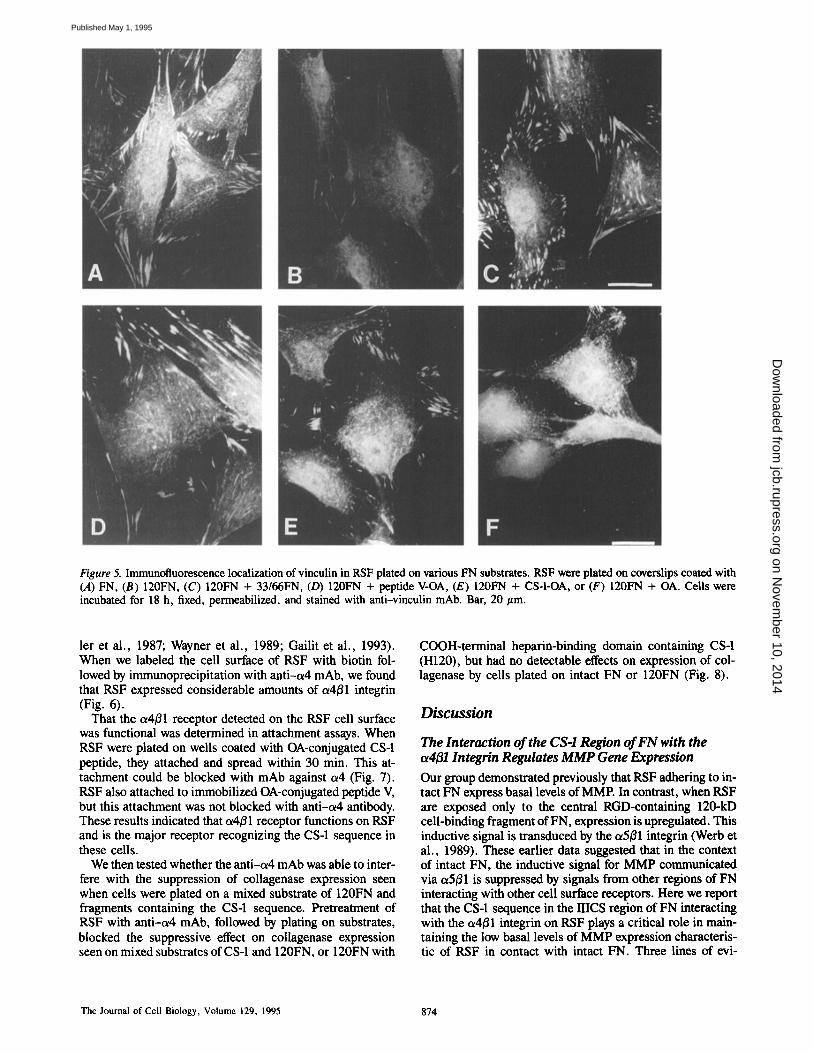
Figure 5. Immunofluorescence localization of vinculin in RSF plated on various FN substrates. RSF were plated on coverslips coated with (A) FN, (B) 120FN, (C) 120FN + 33/66FN, (D) 120FN + peptide V-OA, (E) 120FN + CS-I-OA, or (F) 120FN + OA. Cells were incubated for 18 h, fixed, permeabilized, and stained with anti-vinculin mAb. Bar, 20/zm.
ler et al., 1987; Wayner et al., 1989; Gailit et al., 1993). When we labeled the cell surface of RSF with biotin fol- lowed by immunoprecipitation with anti-or4 mAb, we found that RSF expressed considerable amounts of a4B1 integrin (Fig. 6).
That the ot4B1 receptor detected on the RSF cell surface was functional was determined in attachment assays. When RSF were plated on wells coated with OA-conjugated CS-1 peptide, they attached and spread within 30 min. This at- tachment could be blocked with mAb against a4 (Fig. 7). RSF also attached to immobilized OA-conjugated peptide V, but this attachment was not blocked with anti-tx4 antibody. These results indicated that ot4/31 receptor functions on RSF and is the major receptor recognizing the CS-1 sequence in these cells.
We then tested whether the anti-or4 mAb was able to inter- fere with the suppression of collagenase expression seen when cells were plated on a mixed substrate of 120FN and fragments containing the CS-1 sequence. Pretreatment of RSF with anti-or4 mAb, followed by plating on substrates, blocked the suppressive effect on collagenase expression seen on mixed substrates of CS-1 and 120FN, or 120FN with
COOH-terminal heparin-binding domain containing CS-1 (H120), but had no detectable effects on expression of col- lagenase by cells plated on intact FN or 120FN (Fig. 8).
Discussion
The Interaction of the CS-1 Region of FN with the a4~l Integrin Regulates MMP Gene Expression Our group demonstrated previously that RSF adhering to in- tact FN express basal levels of MME In contrast, when RSF are exposed only to the central RGD-eontaining 120-kD cell-binding fragment of FN, expression is upregulated. This inductive signal is transduced by the o~5/~1 integrin Obrerb et al., 1989). These earlier data suggested that in the context of intact FN, the inductive signal for MMP communicated via cz5/~1 is suppressed by signals from other regions of FN interacting with other cell surface receptors. Here we report that the CS-1 sequence in the RICS region of FN interacting with the c~4/~1 integrin on RSF plays a critical role in main- taining the low basal levels of MMP expression characteris- tic of RSF in contact with intact FN. Three lines of evi-
The Journal of Cell Biology, Volume 129, 1995 874
on Novem
ber 10, 2014jcb.rupress.org
Dow
nloaded from
Published May 1, 1995

Figure 6. Immunoprecipitation of biotinylated surface proteins with anti-a4 and -a5 antibodies. RSF were cultured for 72 h in DME supplemented with 10% FBS; ,~ ceils were biotinylated and surface- "~ labeled proteins analyzed by in- i ~ munoprecipitation with anti-c~4 (lane 1) and anti-a5 (lane 2) mAbs, followed by non-reducing ~ ~ SDS-PAGE and detection by en- ~ m hanced chemiluminescence. The a _~_~ subunits are indicated by asterisks (*). Both the intact and proteolyti- cally processed forms of a4 are in- dicated. Molecular weight markers are indicated at the left.
dence support these conclusions. First, the presence of the 25-amino acid CS-1 peptide in the substrate, along with 120FN, is sufficient to promote a low level of MMP expres- sion by RSF, similar to that found when cells are plated on intact FN. Second, when larger COOH-terminal fragments are present on the substrate along with 120FN, only those containing the CS-1 sequence promote the reduced level of MMP expression. Finally, RSF express a known receptor for the CS-1 sequence, the a4/31 integrin, and an antibody against the a4 subunit interferes with the ability of CS-1 to regulate MMP expression.
The IIICS region of FN is a 120-amino acid segment with a complex alternative splicing pattern (Kornblihtt et al., 1985; reviewed in Schwarzbauer, 1991). IIICS was identified originally as an attachment region for mouse melanoma cells (Humphries et al., 1986), but was subsequently shown also to support adhesion of neural crest ceils and their derivatives (Dufour et al., 1988; Berdnarczyk and Mclntyre, 1992), lymphocytes (Wayner et al., 1989; Guan and Hynes, 1990), and dermal fibroblasts (Gailit et al., 1993). IIICS has two binding sites for the a4/~l integrin: CS-1 and CS-5 sites, of
I I
Figure 8. Anti-c~4 mAb blocks the ability of CS-I and HI20 to sup- press induction by 120FN of collagenase expression in RSE RSF were treated with anti-a4 mAb (5 t~g/ml) for 30 min before plating them in DME-LH in wells coated with various substrates. RSF with or without anti-a4 treatment were plated on FN, 120FN, 120FN + CS-1-OA, and 120FN + H120. CM was analyzed by SDS-PAGE and immunoblotting with anti-collagenase mAb. Collagenase ex- pression was quantified by densitometry from six separate experi- ments, each in duplicate, and is shown as mean + SEM.
which the CS-1 has the higher binding affinity (Mould et al., 1991). Both of these regions can be independently included or excluded from the IIICS region as a consequence of alter- native splicing. The IIICS region containing CS-1 is included in >50% of the FN secreted by fibroblasts in culture, and transformed fibroblasts have been shown to exhibit even higher levels of FN containing CS-1 (Castellani et al., 1986;
Figure 7. RSF adhesion to CS-I and peptide V. RSF were treated with anti-a4 mAb prior to plating in wells coated with OA- conjugated CS-I or peptide V. Cells were incubated for 30 min and plates were shaken at 250 rpm for 30 s. (,4) Control RSF on CS-I-OA, (B) RSF with anti-a4 mAb on CS-1-OA after shaking, (C) control RSF on peptide V-OA, (D) RSF with anti-a4 mAb on peptide V-OA after shaking.
Huhta|a eta|. lntegrins Regulate Metalloproteinoze Expression 875
on Novem
ber 10, 2014jcb.rupress.org
Dow
nloaded from
Published May 1, 1995

Hershberger and Culp, 1990), Even though all FN isoforms also contain another low-affinity 0~4/31-binding site, HI in the IH t4 segment of the COOH-terminal HBD (Mould and Humphries, 1991), our present studies with H95, H89 and H0 fragments reported here suggest that MMP regulation via a4/31 involves only the CS-1 sequence.
The c~4/51 integrin is not expressed ubiquitously. This cell surface receptor is present in melanoma cells, lymphocytes and neural crest ceils, although previous reports have shown that some cultured fibroblasts express o~4/~1 (Gailit et al., 1993). Our data suggest that regulated expression of ot4131, concomitant with selective expression of FN splice variants that contain binding sites for ot4~31, could contribute to the regulation of MMP expression spatially and temporally.
Several Regions of F~bronectin Contribute to Regulating the Level of MMP Expression in RSF Our data document critical and antagonistic roles for CS-1 and the RGD binding region of the central cell-binding do- main (120FN) in the regulation of MMP gene expression by FN. However, ot4/~l mAb, which was able to interfere with the ability of CS-1 to regulate MMP expression on mixed substrates of 120FN + CS-1 and 120FN + H120, could not overcome the suppressive effects on MMP expression of in- tact FN, suggesting that other regions of FN, at least in cis, may play a role in the regulation of MMP expression on in- tact FN.
Interestingly the 33/66FN, comprised of both the COOH- terminal HBD (repeats III ~2-~4) and IUCS, also contains MMP-inductive (peptide V) as well as MMP-suppressive regions (CS-1). Peptide V, an 8-amino acid sequence in III ~4, stimulated MMP expression further when present in the substrate along with 120FN. The observation that the whole 33/66FN is suppressive, whereas recombinant COOH- terminal fragments lacking CS-1 and containing peptide V are inductive, suggests that in the context of the whole COOH- terminal region the suppressive signal from CS-1 is domi- nant, when presented on the substrate with 120FN. In addi- tion, peptide V was not inductive by itself when plated in the absence of 120FN, suggesting that its effect on MMP expres- sion is indirect. Addition of peptide V in the context of intact FN did not induce elevated levels of MMP. A receptor that binds to the peptide V sequence has not been identified.
In addition to c~4B1 and 0~5/~1 integrins RSF express other receptors for FN, including otv-containing integrins, which also recognize the RGD sequence, and cell-surface pro- teoglycans. Anti-otv/~3 mAb (LM609) and anti-c~5 mAb (BIIG2) can block cell adhesion to 120FN (Tremble et al., 1994) but not to intact FN, which allows the additional inter- action between c~4/~1 and CS-1. However, ~v/~3 is prob- ably not involved in the inductive signal that stimulates MMP expression in RSF plated on 120FN, because RSF in- teraction with immobilized anti-~5 mAb, but not with im- mobilized anti-c~v/~3 mAb, stimulates MMP (Werb et al., 1989; Tremble, P. M., and Z. Werb, unpublished observa- tions). Whether otv/~3 or other integrins can participate, in addition to c~4fll, in the regulation of MMP by intact FN was not addressed, but cannot be ruled out.
Integrins are not the only cell-surface receptors that medi- ate the effects of FN on the regulation of MMP expression in RSE Treatment of RSF with heparan sulfate, chondroitin sulfate, or heparatinase III before plating them on intact FN
increases the expression of collagenase to the level seen with 120FN (Tremble et al., 1994; and unpublished observa- tions). This suggests that both cell surface heparan sulfate and chondroitin sulfate may be involved in the suppression of MMP expression on intact FN. However, neither the whole I-IBD (II112-t4) without the CS-1 sequence, nor any of the peptides containing heparan- and chondroitin sulfate- binding sites in the COOH-terminal HBD region that we tested were able to suppress MMP expression when added in trans with 120FN, Previous studies have shown that mela- noma cell adhesion to CS-1 mediated by ot4131 can be partly inhibited by interfering with CSPG synthesis or expression. Since these molecules do not bind CS-1, they may instead modify the function and activity of ot4B1 integrin (Iida et al., 1992). Thus, it is possible that proteoglycans participate in collagenase regulation indirectly by modifying integrin ac- tivities. Cell surface proteoglycans do, however, appear to be directly involved in the regulation of focal contact assembly (Couchman et al., 1988; LeBaron et al., 1988; Woods et al., 1993).
Focal Contact Formation and Collagenase Expression Are Regulated Independently in RSF Although the physiological role of the peptide V sequence in intact FN is not clear, our studies with this peptide and CS-l-containing fragments of FN have allowed us to demon- strate that two important responses to FN-assembly of focal contacts and regulation of MMP gene expression-involve distinct signaling processes. Peptide V has been shown pre- viously to be the active focal adhesion-promoting peptide in the lII ~2-~4 HBD of FN (Woods et al., 1993). Our present studies show that peptide V both stimulated MMP expres- sion and restored focal contact formation in RSF plated on 120FN. Furthermore, CS-1 also promoted focal contact for- mation by RSF when presented in trans with 120FN, but it suppressed MMP levels. Thus, RSF can assemble focal con- tacts under plating conditions that promote low (intact FN or 120FN + CS-1) or high (120FN, or 120FN + peptide V) MMP expression levels (see Table I). Previous studies have shown clearly that localization of integrins to focal contacts is mediated via the/5'1 subunit of c~/~l complexes (LaFlamme et al., 1992; Reszka et al., 1992), with ~ subunits playing a permissive, but not instructive role. Our present studies suggest that the integrin c~ subunits may play active roles in regulating signaling pathways leading to altered MMP gene expression.
Cell Surface Receptors Collaborate in Regulating MMP Expression In the present study we show that signaling by the ¢x5/~1 and oe4/~l integrins, which recognize distinct domains on FN, regulates MMP expression in response to FN. When exam- ined individually, these two interactions signal opposing effects on MMP expression. When only ot5/~l is engaged by 120FN or immobilized anti-~5 antibody, MMP expression is stimulated. When only a4/~l is engaged by CS-1, expres- sion is low. However, when both these receptors interact with their respective domains, MMP expression is also low. We hypothesize that engagement of c~4/~1 with its ligand inter- feres with the inductive signal from the otS/~I-RGD interac- tion. One possibility is that the engagement of ¢x4~1 with CS-1
The Journal of Cell Biology, Volume 129, 1995 876
on Novem
ber 10, 2014jcb.rupress.org
Dow
nloaded from
Published May 1, 1995
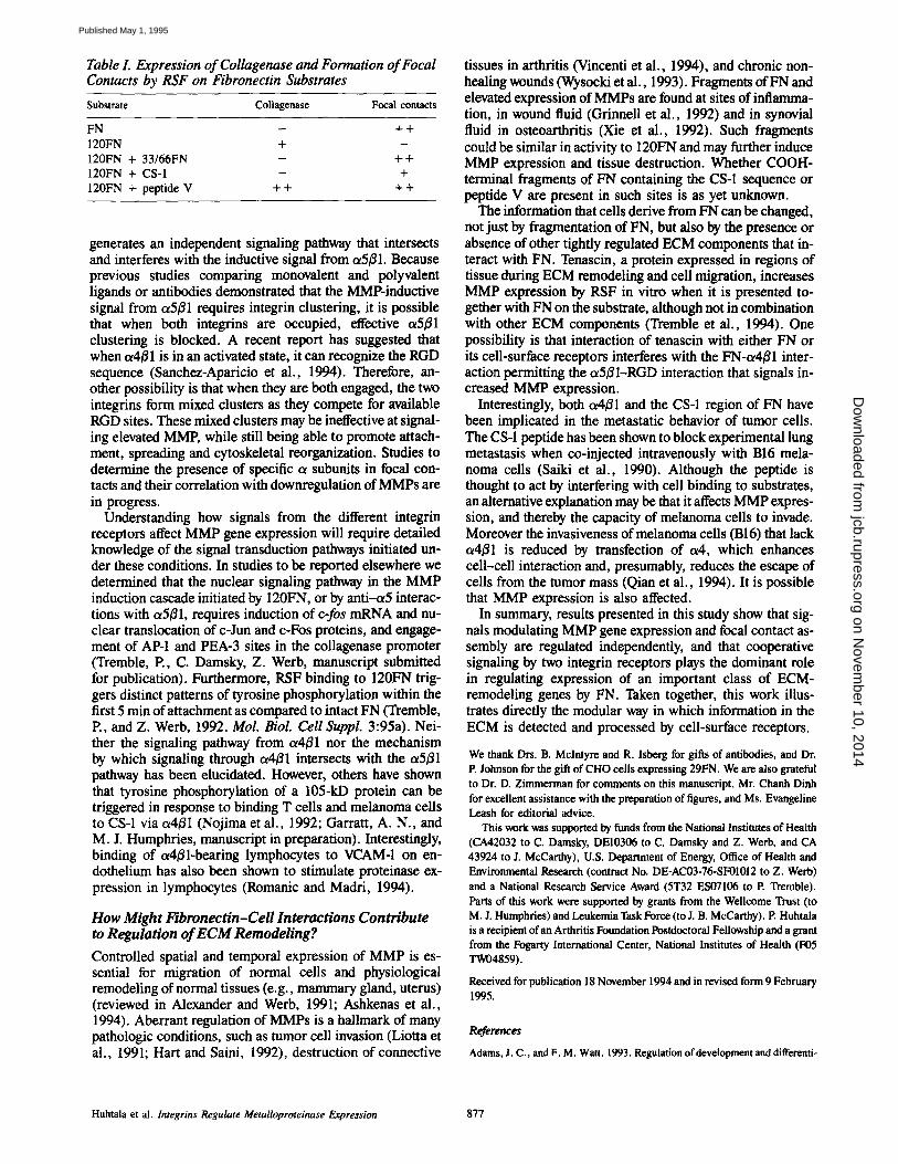
Table I. Expression of Collagenase and Formation of Focal Contacts by RSF on Fibronectin Substrates
Substrate Collagenase Focal contacts
FN - + + 120FN + - 120FN + 33/66FN - + + 120FN + CS-1 - + 120FN + peptide V + + + +
generates an independent signaling pathway that intersects and interferes with the inductive signal from ot5fll. Because previous studies comparing monovalent and polyvalent ligands or antibodies demonstrated that the MMP-inductive signal from ot5/$1 requires integrin clustering, it is possible that when both integrins are occupied, effective ot5fll clustering is blocked. A recent report has suggested that when u4~l is in an activated state, it can recognize the RGD sequence (Sanchez-Aparicio et al., 1994). Therefore, an- other possibility is that when they are both engaged, the two integrins form mixed clusters as they compete for available RGD sites. These mixed clusters may be ineffective at signal- ing elevated MMP, while still being able to promote attach- ment, spreading and cytoskeletal reorganization. Studies to determine the presence of specific o~ subunits in focal con- tacts and their correlation with downregulation of MMPs are in progress.
Understanding how signals from the different integrin receptors affect MMP gene expression will require detailed knowledge of the signal transduction pathways initiated un- der these conditions. In studies to be reported elsewhere we determined that the nuclear signaling pathway in the MMP induction cascade initiated by 120FN, or by anti-or5 interac- tions with a5/~1, requires induction of c-fos mRNA and nu- clear translocation of c-Jun and c-Fos proteins, and engage- ment of AP-1 and PEA-3 sites in the collagenase promoter (Tremble, E, C. Damsky, Z. Werb, manuscript submitted for publication). Furthermore, RSF binding to 120FN trig- gers distinct patterns of tyrosine phosphorylation within the first 5 min of attachment as compared to intact FN (Tremble, E, and Z. Werb, 1992. Mol. Biol. Cell Suppl. 3:95a). Nei- ther the signaling pathway from t~4(31 nor the mechanism by which signaling through ol4/~1 intersects with the ct5/~1 pathway has been elucidated. However, others have shown that tyrosine phosphorylation of a 105-kD protein can be triggered in response to binding T cells and melanoma cells to CS-1 via c~4B1 (Nojima et al., 1992; Garratt, A. N., and M. J. Humphries, manuscript in preparation). Interestingly, binding of ct4/31-bearing lymphocytes to VCAM-1 on en- dothelium has also been shown to stimulate proteinase ex- pression in lymphocytes (Romanic and Madri, 1994).
How Might Fibronectin-Cell Interactions Contribute to Regulation of ECM Remodeling?
Controlled spatial and temporal expression of MMP is es- sential for migration of normal cells and physiological remodeling of normal tissues (e.g., mammary gland, uterus) (reviewed in Alexander and Werb, 1991; Ashkenas et al., 1994). Aberrant regulation of MMPs is a hallmark of many pathologic conditions, such as tumor cell invasion (Liotta et al., 1991; Hart and Saini, 1992), destruction of connective
tissues in arthritis (Vincenti et al., 1994), and chronic non- healing wounds (Wysocki et al., 1993). Fragments of FN and elevated expression of MMPs are found at sites of inflamma- tion, in wound fluid (Grinnell et al., 1992) and in synovial fluid in osteoarthritis (Xie et al., 1992). Such fragments could be similar in activity to 120FN and may further induce MMP expression and tissue destruction. Whether COOH- terminal fragments of FN containing the CS-1 sequence or peptide V are present in such sites is as yet unknown.
The information that cells derive from FN can be changed, not just by fragmentation of FN, but also by the presence or absence of other tightly regulated ECM components that in- teract with FN. Tenascin, a protein expressed in regions of tissue during ECM remodeling and cell migration, increases MMP expression by RSF in vitro when it is presented to- gether with FN on the substrate, although not in combination with other ECM components (Tremble et al., 1994). One possibility is that interaction of tenascin with either FN or its cell-surface receptors interferes with the FN-ot4/$1 inter- action permitting the ~5fll-RGD interaction that signals in- creased MMP expression,
Interestingly, both ot4~l and the CS-1 region of FN have been implicated in the metastatic behavior of tumor cells. The CS-1 peptide has been shown to block experimental lung metastasis when co-injected intravenously with B16 mela- noma cells (Saiki et al., 1990). Although the peptide is thought to act by interfering with cell binding to substrates, an alternative explanation may be that it affects MMP expres- sion, and thereby the capacity of melanoma cells to invade. Moreover the invasiveness of melanoma cells (B16) that lack ot4fll is reduced by transfection of or4, which enhances cell-cell interaction and, presumably, reduces the escape of ceils from the tumor mass (Qian et al., 1994). It is possible that MMP expression is also affected.
In summary, results presented in this study show that sig- nals modulating MMP gene expression and focal contact as- sembly are regulated independently, and that cooperative signaling by two integrin receptors plays the dominant role in regulating expression of an important class of ECM- remodeling genes by FN. Taken together, this work illus- trates directly the modular way in which information in the ECM is detected and processed by cell-surface receptors.
We thank Drs. B. Mclntyre and R. Isberg for gifts of antibodies, and Dr. P. Johnson for the gift of CHO cells expressing 29FN. We ate also grateful to Dr. D. Zimmerman for comments on this manuscript, Mr. Chanh Dinh for excellent assistance with the preparation of figures, and Ms. Evangeline Leash for editorial advice.
This work was supported by funds from the National Institutes of Health (CA42032 to C. Damsky, DEI0306 to C. Darnsky and Z. Werb, and CA 43924 to I. McCarthy), U.S. Department of Energy, Office of Health and Environmental Research (contract No. DE-AC03-76-SF01012 to Z. Werb) and a National Research Service Award (5T32 ES07106 to P. Tremble). Parts of this work were supported by grants from the Wellcome Trust (to M. J. Humphries) and Leukemia 1~tsk Force (to J. B. McCarthy). P. Huhtala is a recipient of an Arthritis Foundation Postdoctoral Fellowship and a grant from the Fogarty International Center, National Institutes of Health (F05 TWO4859).
Received for publication 18 November 1994 and in revised form 9 February 1995.
References
Adams, J. C., and F. M. Watt. 1993. Regulation of development and differenti-
Huhtala et al. lntegrins Regulate Metalloproteinase Expression 877
on Novem
ber 10, 2014jcb.rupress.org
Dow
nloaded from
Published May 1, 1995
ation by the extracellular matrix. Development. 117:1183-1198. Aggeler, J., S. M. Friseh, and Z. Werb. 1984. Collagunase is a major gene
product of induced rabbit synovial fibroblasts. J. Cell Biol. 98:1656-1661. Albelda, S. M., and C. Buck. 1990. Integrins and other cell adhesion mole-
cules. FASEB (Fed. Am. Soc. Exp. Biol.) J. 4:2868-2880. Alexander, C. M., and Z. Werb. 1991. Extracellular matrix degradation. In
Cell Biology of Extracellular Matrix, 2nd ed. E. D. Hay, editor. Plenum Press, New York. 255-302.
Aota, S., M. Nomizu, and K. M. Yamada. 1994. The short amino acid sequence Pro-His-Ser-Arg-Asn in human fibronectin enhances cell-adhesive function. J. Biol. Chem. 269:24756-24761.
Ashkenas, J., C. H. Damsky, M. J. Bissell, and Z. Werb. 1994. Integrins, sig- naling, and the remodeling of the extracellular matrix. In Integrins. Molecu- lar and Biological Response to the Extracellular Matrix. D. A. Cheresh and R. P. Mecham, editors. Academic Press. 79-109.
Bednarczyk, J. L., and B. W. MeIntyre. 1992. Expression and ligand-binding function of the integrin et4/31 (VLA-4) on neural-crest-derived tumor cell lines. Clio. Exp. Metastasis. 10:281-290.
Bowditeh, R. D., M. Hariharan, E. P. Tominna, J. W. Smith, K. Y. Yamada, E. D. Getzoff, and M. H. Ginsberg. 1994. Identification of a novel integrin binding site of fibronectin. J. Biol. Chem. 269:10856-10863.
Castellani, P., A. Sift, C. Rossellini, E. fufusini, L. Borsi, and L. Zardi. 1986. Transformed human cells release different variants than do normal cells. J. Cell Biol. 103:1671-1677.
Chomczynski, P., and N. Sacchi. 1987. Single step method of RNA isolation by acid guanidium thiocyanate phenol chloroform extraction. Anal. Bio- chem. 162:156-159.
Clayberger, C., A. M. Krensky, B. W. McIntyre, T. D. Koller, P. Parham, F. Brodsky, D. J. Linn, and E. L. Evans. 1987. Identification and character- ization of two novel lymphocyte function-associated antigen, L24 and L25. J. Immunol. 138:1510-1514.
Couchman, J. R., R. Austria, A. Woods, and R. C. Hughes. 1988. An adhesive defective BHK cell mutant has cell surface heparan sulfate proteoglycan of altered properties. J. Cell Physiol. 136:226-236.
Damsky, C. H., and Z. Werb. 1992. Signal transduction by integrin receptors for extracellular matrix: cooperative processing of extracellular information. Curr. Opin. Cell. Biol. 4:772-781.
Drake, S. L., D. J. Klein, D. J. Mickelson, T. R. Oegema, L. T. Furcht, and J. B. McCarthy. 1992. Cell surface phosphatidylinositol-anehored beparan sulfate proteoglycan initiates mouse melanoma cell adhesion to a fibronectin- derived, heparin-binding synthetic peptide. J. Cell Biol. 117:1331-1341.
Dufour, S., J.-L. Duband, M. J. Humphries, M. Obara, K. M. Yamada, and J. P. Thiery. 1988. Attachment, spreading and locomotion of avian neural crest cells are mediated by multiple adhesion sites on fibronectin molecules. EMBO (Fur. Mol. Biol. Organ.) J, 7:2661-2671.
Erle, D. J., C. Ruegg, D. Sheppard, and R. Pytela. 1991. Complete amino acid sequence of an integrin/~ subunit (/$7) identified in leakocytes. J. Biol. Chem. 266: 11009-11016.
Frisch, S. M., and J. H. Morisaki. 1990. Positive and negative transcriptional elements of the human type IV collagenase gene. Mol. Cell. Biol. 10: 6524-6532.
Frisch, S. M., E. J. Clark, and Z. Werb. 1987. Coordinate regulation of stromelysin and collagenase genes determined with eDNA probes. Proc. Natl. Acad. Sci. USA. 84:2600-2604.
Gailit, J., M. Pierschbacher, and R. A. Clark. 1993. Expression of functional a4/31 integrin by human dermal fibroblasts. J. Invest. Dermatol. 100: 323-328.
George, E. L., E. N. Georges-Labouessa, R. S. Patel-King, H. Rayburn, and R. O. Hynes. 1994. Defects in mesoderm, neural tube and vascular develop- ment in mouse embryos lacking fibronectin. Development. 119:1079-1091.
Giuseppetti, J. M., J. B. McCanhy, and P. C. Letourneau. 1994. Isolation and partial characterization of a cell-surface heparan sulfate proteoglycan from embryonic rat spinal cord. J. Neurosci. Res. 37:584-595.
Grinnell, F., C-H. Ho, and A. Wysocki. 1992. Degradation of fibrnnectin and vitronectin in chronic wound fluid: analysis by cell blotting, immunoblot- ring, and cell adhesion assays. J. Invest. Dermatol. 98:410-416.
Guan, J-L., and R. O. Hynes. 1990. Lymphoid cells recognize an alternatively spliced segment of fibronectin via the integrin receptor a4/~l. Cell. 60:53-61.
Hart, I. A., and A. Saini. 1992. Biology of turnout metastasis. Lancet. 339:1453-1457.
Huagun, P. K., J. B. McCarthy, A. P. Skubitz, L. T. Furcht, and P. C. Letour- neau. 1990. Recognition of the A chain carboxy-terminal heparin binding re- gion of fibronectin involves multiple sit~s: two contiguous sequences act in- dependently to promote neural cell adhesion. J. Cell Biol. 111:2733-2745.
Hemier, M. E., C. Huang, Y. Takada, L. Schwarz, J. L. Strominger, and M.L . Clabby. 1987. Characterization of the cell surface heterodimer VLA-4 and related peptides. J. Biol. Chem. 262:11478-11485.
Hen'on, G. S., M. J. Banda, E. J. Clark, and Z. Werb. 1986. Secretion of metalloproteinases by stimulated capillary endothelial cells. J. Biol. Chem. 261:2814-2818.
Hershberger, R. P., and L. A. Culp. 1990. Cell-type-specific expression of alternatively spliced human fibronectin IIICS mRNAs. Mol. Cell. Biol. 10:662-671.
Huhtala, P., A. Tuuttila, L. T. Chow, J. Lohi, J. Keski-Oja, and K. Tryggva-
son. 1991. Complete structure of the human gene for 92-kD type IV col- lagenase. J. Biol. Chem. 266:16485-16490.
Humphries, M. J., S. Akiyama, A. Komoriya, K. Olden, and K. M. Yamada. 1986. Identification of an alternatively spliced site in human plasma fibronec- tin that mediates cell type-specific adhesion. J. Cell Biol. 103:2637-2647.
Humphries, M. J., S. Akiyama, A. Komoriya, K. Olden, and K. M. Yamada, 1987. Identification of two distinct regions of the type III connecting segment of human plasma fibronectin that promote cell type-specific adhesion. J. Biol. Chem. 262:6886-6892.
Hynes, R. O. 1990. Fibronectins. Springer Series in Molecular Biology. A. Rich, editor, Spftnger-Verlag, New York. 546 pp.
Hynes, R. O. 1992. Integrins: versatility, modulation and signaling in cell adhe- sion. Cell. 69:11-25.
lida, J., A. P. Skubitz, L. T. Furcht, E. W. Wayner, andJ. B. McCarthy. 1992. Coordinate role for cell surface chondroitin sulfate proteoglycan and ct4/31 integrin in mediating melanoma cell adhesion to fibronection. J. Cell Biol. 118:431 ~ .
Iida, I., R. P. Milius, T. R. Oegema, L. T. Furcht, and J. B. McCarthy. 1994. Role of cell surface proteoglycans in tumor cell recognition of fibronectin. Trends Glycosci. Glycotechnol. 6:1-16.
Kornblihtt, A. R., K. Umezawa, K. Vibe-Pedersen, and F. Baralle. 1985. Pri- mary structure of human plasma fibronectin: differential splicing may gener- ate at least 10 polypeptides from a single gene. EMBO (Fur. Mol. Biol. Or- gan.) J. 4:1755-1759.
LaFlamme, S. E., S. K. Akiyama, and K. M. Yamada. 1992. Regulation of fibronectin receptor distribution. J. Cell Biol. 117:437--447.
LeBaron, R. G., J. D. Esko, A. Woods, S. Johanssen, and M. Hook. 1988. Adhesion of glycosaminoglycan-deficient Chinese hamster ovary cell mu- tants to fibronectin substrata. J. Cell Biol. 106:945-952.
Limper, A. H., B. J. Quade, R. M. LaChance, T. M. Birkenmeyer, T. S. Rang- wala, and J. A. McDonald. 1991. Cell surface molecules that bind fibronec- tin's matrix assembly domain. J. Biol. Chem. 266:9697-9702.
Liotta, L. A., P. Stceg, and W. Stetler-Stevenson. 1991. Cancer metastasis and angiogenesis: an imbalance of positive and negative regulation. Cell. 64: 327-336.
Lories, V., J. J. Cassiman, H. van der Berghe, and G. David. 1992. Differential expression of cell surface heparan sulfate proteoglycans in human mammary epithelial cell and fibroblasts. J. Biol. Chem. 267:1116-1122.
Makarem, R., P. Newham, J. A. Askari, L. J. Green, J. Clements, M. Ed- wards, M. J. Humphries, and A. P. Mould. 1994. Competitive binding of vascular cell adhesion molecule-1 and the HeplI/IIICS domain of fibronectin to the integrin u4~l. J. Biol. Chem. 269:4005--4011.
McCarthy, J. B., M. K. Chelberg, D. J. Mickelson, and L. T. Furcht. 1988. Localization and chemical synthesis of fibronectin paptides with melanoma adhesion and heparin binding activities. Biochemistry. 27:1380-1388.
McCarthy, J. B., A. P. N. Skubitz, Q. Zhao, X-y. Yi, D. J. Mickelson, D. J. Klein, and L. T. Furcht. 1990. RGD-independent cell adhesion to the carboxyl-terminal heparin-binding fragment of fibronectin involves heparin- dependent and independent activities. J. Cell Biol. 110:777-787.
McDonald, J. A., B. I. Quade, T. J. Broekelmann, R. LaChance, K. Forsman, E. Hasegawa, and S. Akiyama. 1987. Fibronectin's cell-adhesive domain and an aminoterminal matrix assembly domain participate in its assembly into fibroblast pericellular matrix. J. Biol. Chem. 262:2957-2967.
Mclntyre, B. W., E. L. Evans, and J. L. Bednarezyk. 1989. Lymphocyte sur- face antigen L25 is a member of the integrin receptor superfamily. Z Biol. Chem. 264:13745-13750.
Moon, K-Y, K. S. Shin, W. K. Song, C. H. Chung, D. B. Ha, and M-S. Kang. 1994. A candidate molecule for the matrix assembly receptor to the N-ter- minal 29-kD fragment of fibronectin in chick myoblasts. J. Biol. Chem. 269:7651-7657.
Mould, A. P., and M. J. Humphries. 1991. Identification of a novel recognition sequence for the c~4~1 in the COOH-terminal heparin-binding domain of fibronectin. EMBO (Eur. Mol. Biol. Organ.) J. 10:4089--4095.
Mould, A. P., L. A. Wheldon, A. Koriyama, E. A. Wayner, K. M. Yamada, and M. J. Humphries. 1990. Affinity chromatographic isolation of the mela- noma adhesion receptor for the IIICS region of fibronectin and its identifica- tion as the integrin ct4/~l. J. Biol. Chem. 265:4020--4024.
Mould, A. P., A. Komoriya, K. M. Yamada, and M. J. Humphries. 1991. The CS5 peptide is a second site in the IIICS region of fibronectin recognized by the integrin a4~l. J. Biol. Chem. 266:3579-3585.
Mould, A. P., J. A. Askari, S. E. Craig, A. N. Garratt, J. Clements, and M. J. Humphries. 1994. Cell adhesion and migration mediated by the integrin ct4/31 : effect of fibronectin alternative splicing and the role of ligand affinity. J. Biol. Chem. 269:27224-27230.
Nojima, Y., D. M. Rothstein, K. Sugita, S. F. Schlossman, and C. Morimoto. 1992. Ligation of VLA-4 on T cells stimulates tyrosine phosphorylation of a 105-kD protein. J. Exp. bled. 175:1045-1053.
Qian, F., D. L. Vaux, and 1. L. Weissman. 1994. Expression of the integrin a4/~l in melanoma cells can inhibit the invasive stage of metastasis forma- tion. Cell. 77:335-347.
Reszka, A. A., Y. Hayashi, and A. F. Horwitz. 1992. Identification of amino acid sequences in the integrin El eytuplasmic domain implicated in cyto- skeletal association. J. Cell Biol. 117:1321-1330.
Romanic, A. M., and J. A. Madri. 1994. The induction of 72-kD gelatinase in T cells upon adhesion to endothelial cells is VCAM-I dependent. J. Cell
The Journal of Cell Biology, Volume 129, 1995 878
on Novem
ber 10, 2014jcb.rupress.org
Dow
nloaded from
Published May 1, 1995

Biol. 125:1165-1178. Ruegg, C., A. A. Postigo, E. E. Sikorski, E. C. Butcher, R. Pytela, and D. J.
Erie. 1992. Role of integrin et4~7/~t4EP in lymphocyte adherence to fibronectin and VCAM-I and in homotypic cell clustering. J. Cell Biol. 117:179-189.
Saiki, 1., J. Murata, T. Mukabe, Y. Matsumoto, Y. Ohdate, Y. Kawase, Y. Taguchi, and 1. Kato. 1990. Inhibition of lung metastasis by synthesis and recombinant fragments of human fibronectin with functional domains. Jap. J. Cancer Res. 81:1003-1011.
Sambrook, J. E., E. Fritch, and T. Maniatis. 1989. Molecular Cloning: A Lab- oratory Manual. Cold Spring Harbor Laboratory, Cold Spring Harbor, NY.
Sanchez-Aparicio, P., C. Dominguez-Jimenez, and A. Garcia-Pardo. 1994. Activation of the ct4El integrin through the El subunit induces recognition of the RGDS sequence in fibronectin. J. Cell Biol. 126:271-279.
Schwarzbauer, J. E. 1991. Alternative splicing of fibronection: three variants, three functions. BioEssays. 13:527-533.
Stephens, L. E., J. E. Sonne, M. L. Fitzgerald, and C. H. Damsky. 1993. Tar- geted deletion of E1 integrins in F9 embryonal carcinoma cell affects mor- phological differentiation but not tissue-specific gene expression, J. Cell Biol. 123:1607-1620.
Tremble, P. M., T. F. Lane, E. H. Sage, and Z. Werb. 1993. SPARC, a secreted protein associated with morphogenesis and tissue remodeling, in- duces expression of metalloproteinases in fibroblasts through a novel ex- tracellular matrix-dependent pathway. J. Cell Biol. 121:1433-1444.
Tremble, P., R. Chiquet-Ehrismann, and Z. Werb. 1994. The extracellular ma- trix ligands fibronectin and tenascin collaborate in regulating collagenase gene expression in fibroblasts. MoL Biol. Cell. 5:439-453.
Vincenti, M. P., I. M. Clark, and C. E. Brinckerhoff. 1994. Using inhibitors of metalloproteinases to treat arthritis. Arthr. Rheum. 37:1115-1126.
Wayner, E. A., A. Garcia-Pardo, M. J. Humphries, J. A. McDonald, and W. G. Carter. 1989. Identification and characterization of the T lymphocyte adhesion receptor for an alternative cell attachment domain (CS-1) in plasma fibronectin. J. Cell Biol. 109:1321-1330.
Werb, Z., P. M. Tremble, O. Behrendtsen, E. Crowley, and C. H. Damsky. 1989. Signal transduction through the fibronectin receptor induces col- lagenase and stromelysin gene expression. J, Cell Biol. 109:877-889.
Wilhelm, S. M., D. Wunderlich, C. A. Maniglia, A. Z. Eisen, and G. I. Gold- berg. 1992. Primary structure and function of stromelysin/transin in carti- lage matrix turnover. Matrix Suppl. 1:37--44.
Woods, A., J. R. Couchman, S. Johansson, and M, Hook. 1986. Adhesion and cytoskeletal organization of fibroblasts in response to fibronectin fragments. EMBO (Eur. Mol. Biol. Organ.) J. 5:665-670.
Woods, A., J. B. McCarthy, L. T. Furcht, and J. R. Couchman. 1993. A syn- thetic peptide from the COOH-terminal heparin-binding domain of fibronec- tin promotes focal adhesion formation. Mol. Biol. Cell. 4:605-613.
Wysocki, A., L. Staiano-Coico, and F. Grinnell. 1993. Wound fluid from chronic leg ulcers contains elevated levels of metalloproteinases MMP-2 and MMP-9. J. Invest. Dermatol. 101:64-68.
Xie, D.-L., R. Meyers, and G. A. Homandberg. 1992. Fibronectin fragments in osteoarthritic synovial fluid. J. Rheumatol. 19:1448-1452.
Yamada, K. M, 1991. Adhesive recognition sequences. J. Biol. Chem. 266:12809-12812.
Yang, J. T., H. Rayburn, and R. O. Hynes. 1993. Embryonic mesodermal defects in ~5 integrin-deficient mice. Development. 119:1093-1105.
Yang, J. T., H. Rayburn, and R. O. Hynes. 1995. Cell adhesion events medi- ated by c~4 integrins are essential in placental and cardiac development. De- velopment. 121:549-560.
Huhtala et al. lntegrins Regulate Metalloproteinase Expression 879
on Novem
ber 10, 2014jcb.rupress.org
Dow
nloaded from
Published May 1, 1995