IKAP/Elp1 involvement in cytoskeleton regulation and implication for familial dysautonomia
Upload
independentCategory
view
3download
0
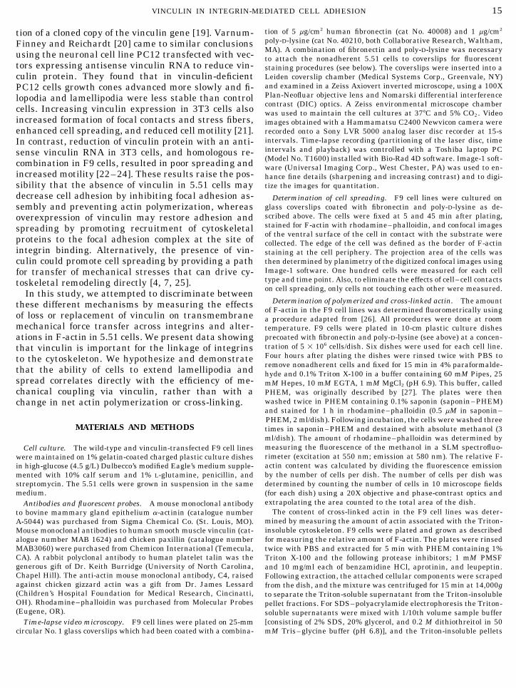
EXPERIMENTAL CELL RESEARCH 231, 14–26 (1997)ARTICLE NO. EX963451
Vinculin Promotes Cell Spreading by Mechanically Coupling Integrinsto the Cytoskeleton
ROBERT M. EZZELL,*,1 WOLFGANG H. GOLDMANN,* NING WANG,†NATESH PARASHARAMA,* AND DONALD E. INGBER‡
*Surgery Research Laboratories, Massachusetts General Hospital, Department of Surgery, Harvard Medical School, Charlestown,Massachusetts 02129; and †Physiology Program, Harvard School of Public Health, ‡Departments of Pathology
and Surgery, Children’s Hospital and Harvard Medical School, Boston, Massachusetts 02115
a complex of proteins that assemble at sites of attach-Mouse F9 embryonic carcinoma 5.51 cells that lack ment of the cell to the extracellular matrix [1]. The
the cytoskeletal protein vinculin spread poorly on ex- transmembrane proteins mediating these contacts aretracellular matrix compared with wild-type F9 cells members of the integrin family of extracellular matrixor two vinculin-transfected clones (5.51Vin3 and Vin4; (ECM) receptors. Integrins are heterodimeric com-Samuels et al., 1993, J. Cell Biol. 121, 909–921). In the plexes in which both chains span the plasma mem-present study, we used this model system to determine brane bilayer once, and the cytoplasmic domain is re-how the presence of vinculin promotes cytoskeletal al- sponsible for linkage of the actin cytoskeleton. A num-terations and associated changes in cell shape. Micro- ber of proteins are found in focal adhesions at thescopic analysis of cell spreading at early times, re- intracellular face of the plasma membrane, includingvealed that 5.51 cells retained the ability to form filo- vinculin, a-actinin, paxillin, and talin [2, 3]. Which pro-podia; however, they could not form lamellipodia,
teins are absolutely required for the formation of focalassemble stress fibers, or efficiently spread over theadhesions is still under investigation, and the orderculture substrate. Detergent (Triton X-100) studies re-with which these proteins bind integrins, actin, or eachvealed that these major differences in cell morphologyother is only partly understood. In addition, changesand cytoskeletal organization did not result from dif-in cell shape and movement may require the transferferences in levels of total polymerized or cross-linkedof mechanical forces between the cytoskeleton andactin. Biochemical studies showed that 5.51 cells, inECM as well as changes in cytoskeletal organizationaddition to lacking vinculin, exhibited slightly re-[4–6]. The focal adhesion complex is a critical pointduced levels of a-actinin and paxillin in their deter-
gent-insoluble cytoskeleton. The absence of vinculin for the regulation of actin organization [1] as well ascorrelated with a decrease in the mechanical stiffness mechanical signal transfer [7]. Recently, Crowley andof the integrin–cytoskeleton linkage, as measured us- Horwitz [8] have shown tyrosine phosphorylation toing cell magnetometry. Furthermore, when vinculin play a role in the signal transduction, the actin-basedwas replaced by transfection in 5.51Vin3 and 5.51Vin4 tension of focal contacts, and the release of cellularcells, the levels of cytoskeletal-associated a-actinin adhesions.and paxillin, the efficiency of transmembrane mechan- We have been studying a mutant F9 embryonic carci-ical coupling, and the formation of actin stress fibers noma cell line (called 5.51) that adheres poorly to sub-were all restored to near wild-type levels. These find-
strates and does not differentiate into a polarized epi-ings suggest that vinculin may promote cell spreadingthelium [9, 10]. The protein vinculin is not detected inby stabilizing focal adhesions and transferring me-5.51 cells. Vinculin is a 117-kDa protein that localizeschanical stresses that drive cytoskeletal remodeling,at focal contacts and intercellular adherens junctionsrather than by altering the total level of actin polymer-[2, 11]. In both types of contacts, vinculin is thought toization or cross-linking. q 1997 Academic Press
form a complex with a number of other componentsthat serve to anchor actin filaments to the membrane.INTRODUCTIONIn focal adhesions, vinculin binds to talin [12], a-ac-
One of the links between the actin cytoskeleton and tinin [13], paxillin [14], actin [15], and forms homodi-the plasma membrane, the focal adhesion, consists of mers [16]. The binding of vinculin to talin and actin is
regulated by polyphosphoinositides [17] and inhibited1 To whom correspondence and reprint requests should be ad- by acidic phospholipids [18]. We have demonstrateddressed at Massachusetts General Hospital, Surgery Research Unit,
the importance of vinculin in the defect in adhesion in149 13th Street, Charlestown, MA 02129. Fax: (617) 726-5414.E-mail: [email protected]. 5.51 cells correcting the mutant phenotype by transfec-
140014-4827/97 $25.00Copyright q 1997 by Academic PressAll rights of reproduction in any form reserved.
AID ECR 3451 / 6i1a$$$$61 02-14-97 13:49:29 eca
15VINCULIN IN INTEGRIN-MEDIATED CELL ADHESION
tion of 5 mg/cm2 human fibronectin (cat No. 40008) and 1 mg/cm2tion of a cloned copy of the vinculin gene [19]. Varnum-poly-D-lysine (cat No. 40210, both Collaborative Research, Waltham,Finney and Reichardt [20] came to similar conclusionsMA). A combination of fibronectin and poly-D-lysine was necessaryusing the neuronal cell line PC12 transfected with vec- to attach the nonadherent 5.51 cells to coverslips for fluorescent
tors expressing antisense vinculin RNA to reduce vin- staining procedures (see below). The coverslips were inserted into aLeiden coverslip chamber (Medical Systems Corp., Greenvale, NY)culin protein. They found that in vinculin-deficientand examined in a Zeiss Axiovert inverted microscope, using a 100XPC12 cells growth cones advanced more slowly and fi-Plan-Neofluar objective lens and Nomarski differential interferencelopodia and lamellipodia were less stable than controlcontrast (DIC) optics. A Zeiss environmental microscope chambercells. Increasing vinculin expression in 3T3 cells also was used to maintain the cell cultures at 377C and 5% CO2. Video
increased formation of focal contacts and stress fibers, images obtained with a Hammamatsu C2400 Newvicon camera wereenhanced cell spreading, and reduced cell motility [21]. recorded onto a Sony LVR 5000 analog laser disc recorder at 15-s
intervals. Time-lapse recording (partitioning of the laser disc, timeIn contrast, reduction of vinculin protein with an anti-intervals and playback) was controlled with a Toshiba laptop PCsense vinculin RNA in 3T3 cells, and homologous re-(Model No. T1600) installed with Bio-Rad 4D software. Image-1 soft-combination in F9 cells, resulted in poor spreading and ware (Universal Imaging Corp., West Chester, PA) was used to en-
increased motility [22–24]. These results raise the pos- hance fine details (sharpening and increasing contrast) and to digi-sibility that the absence of vinculin in 5.51 cells may tize the images for quantitation.decrease cell adhesion by inhibiting focal adhesion as- Determination of cell spreading. F9 cell lines were cultured on
glass coverslips coated with fibronectin and poly-D-lysine as de-sembly and preventing actin polymerization, whereasscribed above. The cells were fixed at 5 and 45 min after plating,overexpression of vinculin may restore adhesion andstained for F-actin with rhodamine–phalloidin, and confocal imagesspreading by promoting recruitment of cytoskeletalof the ventral surface of the cell in contact with the substrate wereproteins to the focal adhesion complex at the site of collected. The edge of the cell was defined as the border of F-actin
integrin binding. Alternatively, the presence of vin- staining at the cell periphery. The projection area of the cells wasthen determined by planimetry of the digitized confocal images usingculin could promote cell spreading by providing a pathImage-1 software. One hundred cells were measured for each cellfor transfer of mechanical stresses that can drive cy-type and time point. Also, to eliminate the effects of cell–cell contactstoskeletal remodeling directly [4, 7, 25].on cell spreading, only cells not touching each other were measured.In this study, we attempted to discriminate between
Determination of polymerized and cross-linked actin. The amountthese different mechanisms by measuring the effectsof F-actin in the F9 cell lines was determined fluorometrically using
of loss or replacement of vinculin on transmembrane a procedure adapted from [26]. All procedures were done at roommechanical force transfer across integrins and alter- temperature. F9 cells were plated in 10-cm plastic culture dishes
precoated with fibronectin and poly-D-lysine (see above) at a concen-ations in F-actin in 5.51 cells. We present data showingtration of 5 1 106 cells/dish. Six dishes were used for each cell line.that vinculin is important for the linkage of integrinsFour hours after plating the dishes were rinsed twice with PBS toto the cytoskeleton. We hypothesize and demonstrateremove nonadherent cells and fixed for 15 min in 4% paraformalde-that the ability of cells to extend lamellipodia and hyde and 0.1% Triton X-100 in a buffer containing 60 mM Pipes, 25
spread correlates directly with the efficiency of me- mM Hepes, 10 mM EGTA, 1 mM MgCl2 (pH 6.9). This buffer, calledchanical coupling via vinculin, rather than with a PHEM, was originally described by [27]. The plates were then
washed twice in PHEM containing 0.1% saponin (saponin–PHEM)change in net actin polymerization or cross-linking.and stained for 1 h in rhodamine–phalloidin (0.5 mM in saponin–PHEM, 2 ml/dish). Following incubation, the cells were washed three
MATERIALS AND METHODS times in saponin–PHEM and destained with absolute methanol (3ml/dish). The amount of rhodamine–phalloidin was determined bymeasuring the fluorescence of the methanol in a SLM spectrofluo-Cell culture. The wild-type and vinculin-transfected F9 cell linesrimeter (excitation at 550 nm; emission at 580 nm). The relative F-were maintained on 1% gelatin-coated charged plastic culture dishesactin content was calculated by dividing the fluorescence emissionin high-glucose (4.5 g/L) Dulbecco’s modified Eagle’s medium supple-by the number of cells per dish. The number of cells per dish wasmented with 10% calf serum and 1% L-glutamine, penicillin, and
streptomycin. The 5.51 cells were grown in suspension in the same determined by counting the number of cells in 10 microscope fieldsmedium. (for each dish) using a 20X objective and phase-contrast optics and
extrapolating the area counted to the total area of the dish.Antibodies and fluorescent probes. A mouse monoclonal antibodyThe content of cross-linked actin in the F9 cell lines was deter-to bovine mammary gland epithelium a-actinin (catalogue number
mined by measuring the amount of actin associated with the Triton-A-5044) was purchased from Sigma Chemical Co. (St. Louis, MO).insoluble cytoskeleton. F9 cells were plated and grown as describedMouse monoclonal antibodies to human smooth muscle vinculin (cat-for measuring the relative amount of F-actin. The plates were rinsedalogue number MAB 1624) and chicken paxillin (catalogue number
MAB3060) were purchased from Chemicon International (Temecula, twice with PBS and extracted for 5 min with PHEM containing 1%CA). A rabbit polyclonal antibody to human platelet talin was the Triton X-100 and the following protease inhibitors; 1 mM PMSFgenerous gift of Dr. Keith Burridge (University of North Carolina, and 10 mg/ml each of benzamidine HCl, aprotinin, and leupeptin.Chapel Hill). The anti-actin mouse monoclonal antibody, C4, raised Following extraction, the attached cellular components were scrapedagainst chicken gizzard actin was a gift from Dr. James Lessard from the dish, and the mixture was centrifuged for 15 min at 14,000g(Children’s Hospital Foundation for Medical Research, Cincinatti, to separate the Triton-soluble supernatant from the Triton-insolubleOH). Rhodamine–phalloidin was purchased from Molecular Probes pellet fractions. For SDS–polyacrylamide electrophoresis the Triton-(Eugene, OR). soluble supernatants were mixed with 1/10th volume sample buffer
[consisting of 2% SDS, 20% glycerol, and 0.2 M dithiothreitol in 50Time-lapse video microscopy. F9 cell lines were plated on 25-mmcircular No. 1 glass coverslips which had been coated with a combina- mM Tris–glycine buffer (pH 6.8)], and the Triton-insoluble pellets
AID ECR 3451 / 6i1a$$$$62 02-14-97 13:49:29 eca

16 EZZELL ET AL.
were sonicated for 10 s in 20 vol of sample buffer. All samples were out long filopodia. This was soon followed by veil-likeboiled for 5 min prior to electrophoresis on SDS–polyacrylamide gels. lamellipodia which increased the surface area of the
Electrophoresis and immunoblotting. Triton-soluble and -insolu- cell’s attachment to the substrate. After attachment,ble samples of F9 cells were electrophoresed on 5–15% SDS–poly- the cell periphery displayed retrograde waves of dorsalacrylamide minigels [28] and then electrophoretically transferred
cytoplasm moving rearward toward the center of theonto 0.45-mm-pore-size nitrocellulose (NC) paper (Bio-Rad, Rich-mond, CA) for 2 h at 0.5 A in a transfer buffer consisting of 10 mM 3- cell. In contrast, 5.51 cells had numerous actively ex-(cyclohexylamino)-1-propanesulfonic acid (pH 11) and 10% methanol. tending and retracting filopodia but no lamellipodiaAfter washing in Tris-buffered saline, pH 7.4 (TBS), with 0.1% Tween were observed [19, 29]. The motile behavior of 5.51 cells20 (three times, 15 min each), nonspecific antibody binding was
resembled that of wild-type cells in the early stages ofblocked by incubating the NC paper in a blocking solution consistingattachment (compare Figs. 1F and 1A).of 3% bovine serum albumin (BSA) and 2% nonfat dry milk in TBS
for 2 h at 377C. The NC paper was incubated in antibodies (in The time course and extent of spreading of the F9blocking solution containing 0.2% NP-40) overnight at 47C and cell lines were measured (Table 1). Five minutes afterwashed three times (15 min each) in TBS with 0.1% NP-40. After plating, the wild-type and 5.51 cells had the same pro-the NC paper was incubated again in blocking solution for 1 h at 377C
jected cell area (mean value of 270 mm2). Over thebound antibodies were detected with a chemiluminescence detectionsystem (ECL System, Amersham Corp.). To compare the relative next 45 min, the area of the wild-type cells more thanamounts of actin and focal adhesion proteins in the Triton-soluble doubled (to 650 mm2), while the size of 5.51 cells re-and -insoluble fractions, immunoblots were scanned with a Zeineh mained unchanged. These data were consistent withlaser densitometer (Biomedical Instruments, Inc., Fullerton, CA) and
the Nomarski DIC images which showed that 5.51 cellsthe relative peak areas quantified on a PC using a computer graphicsdid not spread on the substrate and remained roundedprogram. Linearity was established by varying the amount of sample
and/or the length of autoradiographic exposure. (see Fig. 1). In contrast, the projected areas of the vin-Measurement of mechanical stiffness. Using a procedure de- culin-transfected 5.51 cells (Vin3 and Vin4) almost dou-
scribed by Wang et al. [7], F9 cells were plated (3 1 104 cells per bled from 5 to 45 min.well) on fibronectin- and poly-D-lysine-coated plastic wells (96-well
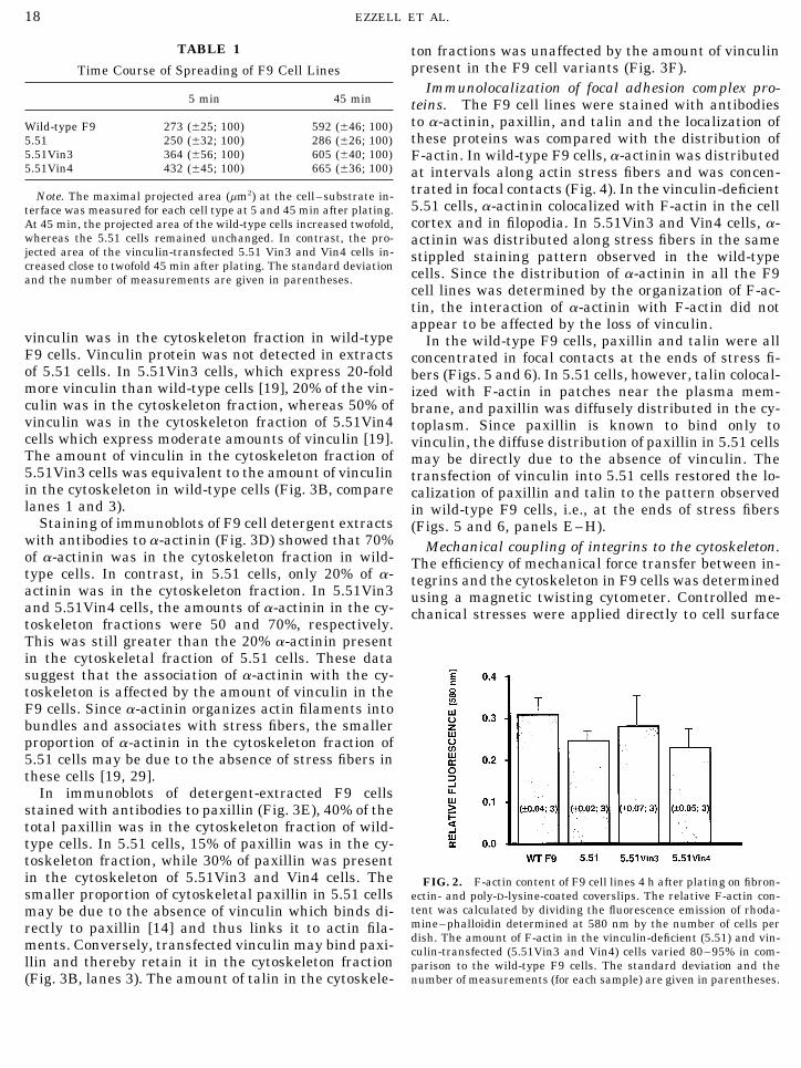
F-actin content. Rhodamine–phalloidin bindingRemovawells, Immunolon, IL) and cultured for 12 h before additionof 6.0-mm spherical ferromagnetic beads that were precoated with a assays were carried out to determine the amount ofsynthetic RGD-containing peptide (Peptite 2000, Telios Pharmaceu- polymerized (F-) and cross-linked actin in the differentticals, La Jolla, CA), which is a specific ligand for integrin receptors. F9 cell lines. The 5.51 cells, 5.51Vin3, and 5.51Vin4 allAfter 10 min, unbound beads were washed away with DMEM con-
contained between 80 and 95% of the amount of F-actintaining 1% BSA and the wells were placed into the magnetic twistingdevice and maintained at 377C. Controlled mechanical stresses were present in wild-type cells (Fig. 2). Thus, there was noapplied directly to cell surface integrins by mechanically twisting correlation between the absence or presence of vinculinthese surface-bound RGD-coated microbeads, as previously described and the total amount of F-actin in 5.51 cells. To deter-[25]. Mechanical stiffness of the focal adhesion and internal cytoskel-
mine the amount of cross-linked actin in the F9 celleton (i.e., its ability to resist mechanical deformation) was measuredlines, detergent (Triton X-100)-soluble and -insolubleby quantitating angular strain after applying a twisting magnetic
field of 13.3 Gauss (i.e., a stress of 40 dyn/cm2) to the attached RGD fractions were electrophoretically separated in 5–15%magnetic beads 20 s after they were initially magnetized by a 10-ms SDS–polyacrylamide gels (Fig. 3A), and the amount of1000-Gauss magnetic pulse. actin present in the extracts was determined by densi-
Confocal microscopy. F9 cell lines were cultured on glass cov- tometry of Coomassie blue-stained gels and immu-erslips coated with fibronectin and poly-D-lysine as described above.noblots stained with anti-actin antibodies (see Materi-Twelve hours after plating, the cells were fixed in 4% paraformalde-
hyde for 15 min followed by three washes in phosphate-buffered als and Methods). The insoluble fraction contained thesaline (PBS, pH 7.4) and extracted for 2 min in 0.2% Triton X-100. detergent-resistant cytoskeleton including cross-linkedAll procedures were done at room temperature (Ç227C). Following actin filaments. There was no detectable difference inextraction, cells were washed for 10 min in PBS and then stained
the distribution of actin in the detergent-soluble andwith rhodamine–phalloidin (diluted 1:100 in PBS). After staining,-insoluble fractions of wild-type F9, 5.51, 5.51Vin3, andthe coverslips were washed in PBS (three changes, 15 min each),
and a drop of 1% n-propyl gallate (used to reduce photobleaching of 5.51Vin4 cells (Fig. 3B). In all the cell lines, the deter-fluorescence, Sigma Chemical Co.) in a 8:2 mixture of glycerol and gent-soluble fraction contained 30% actin and the -in-PBS (pH 8.5) was added to each coverslip before sealing with nail soluble fraction contained 70% actin. This result sug-polish. The samples were examined using a confocal imaging system
gests that the amount of cross-linked actin in F9 cells[BioRad MRC 600 attached to a Zeiss Axiovert inverted microscope(Carl Zeiss Inc., Thornwood, NY)] with a 100X Zeiss Plan-Neofluar also is not affected by the lack of vinculin.objective. Distribution of focal adhesion complex proteins in F9
cell detergent extracts. To determine if the absence ofRESULTS vinculin affected the amount and distribution of focal
adhesion proteins in the detergent-soluble and -insolu-ble fractions (i.e., association with the detergent-resis-Cell spreading. Time-lapse video microscopy wastant cytoskeleton), immunoblots of F9 cell extractsused to examine and to compare the motile behavior ofwere stained with antibodies to focal adhesion proteinsthe wild-type and 5.51 cells (Fig. 1). Wild-type F9 cells
adhered to fibronectin-coated surfaces by first sending (Figs. 3C–3F). Based on densitometry, 40% of the total
AID ECR 3451 / 6i1a$$$$62 02-14-97 13:49:29 eca

17VINCULIN IN INTEGRIN-MEDIATED CELL ADHESION
FIG. 1. Spreading of a wild-type F9 cell (A, C, E) and a vinculin-deficient 5.51 cell (B, D, F). Trypsinized cells were added to fibronectin-coated coverslips and examined from below with an inverted microscope using video-enhanced Nomarski DIC optics, and images wererecorded onto a laser disk. Five minutes after addition to the coverslip (A), the wild-type cell was rounded and had filopodia that madecontact with the substrate (arrow in A). At 10 (C) and 15 (E) min, the cell was flattened and lamellipodia (arrowheads) extended outwardfrom the cell body. In contrast, the 5.51 cell remained rounded and only had extending and retracting filopodia (arrows in B, D, and F)when examined at the same time intervals as the wild-type cell. Bar in (F), 10 mm.
AID ECR 3451 / 6i1a$$3451 02-14-97 13:49:29 eca
18 EZZELL ET AL.
TABLE 1 ton fractions was unaffected by the amount of vinculinpresent in the F9 cell variants (Fig. 3F).Time Course of Spreading of F9 Cell Lines
Immunolocalization of focal adhesion complex pro-5 min 45 min teins. The F9 cell lines were stained with antibodies
to a-actinin, paxillin, and talin and the localization ofWild-type F9 273 ({25; 100) 592 ({46; 100)these proteins was compared with the distribution of5.51 250 ({32; 100) 286 ({26; 100)
5.51Vin3 364 ({56; 100) 605 ({40; 100) F-actin. In wild-type F9 cells, a-actinin was distributed5.51Vin4 432 ({45; 100) 665 ({36; 100) at intervals along actin stress fibers and was concen-
trated in focal contacts (Fig. 4). In the vinculin-deficientNote. The maximal projected area (mm2) at the cell–substrate in-5.51 cells, a-actinin colocalized with F-actin in the cellterface was measured for each cell type at 5 and 45 min after plating.cortex and in filopodia. In 5.51Vin3 and Vin4 cells, a-At 45 min, the projected area of the wild-type cells increased twofold,
whereas the 5.51 cells remained unchanged. In contrast, the pro- actinin was distributed along stress fibers in the samejected area of the vinculin-transfected 5.51 Vin3 and Vin4 cells in- stippled staining pattern observed in the wild-typecreased close to twofold 45 min after plating. The standard deviation cells. Since the distribution of a-actinin in all the F9and the number of measurements are given in parentheses.
cell lines was determined by the organization of F-ac-tin, the interaction of a-actinin with F-actin did notappear to be affected by the loss of vinculin.
vinculin was in the cytoskeleton fraction in wild-type In the wild-type F9 cells, paxillin and talin were allF9 cells. Vinculin protein was not detected in extracts concentrated in focal contacts at the ends of stress fi-of 5.51 cells. In 5.51Vin3 cells, which express 20-fold bers (Figs. 5 and 6). In 5.51 cells, however, talin colocal-more vinculin than wild-type cells [19], 20% of the vin- ized with F-actin in patches near the plasma mem-culin was in the cytoskeleton fraction, whereas 50% of brane, and paxillin was diffusely distributed in the cy-vinculin was in the cytoskeleton fraction of 5.51Vin4 toplasm. Since paxillin is known to bind only tocells which express moderate amounts of vinculin [19]. vinculin, the diffuse distribution of paxillin in 5.51 cellsThe amount of vinculin in the cytoskeleton fraction of may be directly due to the absence of vinculin. The5.51Vin3 cells was equivalent to the amount of vinculin transfection of vinculin into 5.51 cells restored the lo-in the cytoskeleton in wild-type cells (Fig. 3B, compare calization of paxillin and talin to the pattern observedlanes 1 and 3). in wild-type F9 cells, i.e., at the ends of stress fibers
Staining of immunoblots of F9 cell detergent extracts (Figs. 5 and 6, panels E–H).with antibodies to a-actinin (Fig. 3D) showed that 70% Mechanical coupling of integrins to the cytoskeleton.of a-actinin was in the cytoskeleton fraction in wild- The efficiency of mechanical force transfer between in-type cells. In contrast, in 5.51 cells, only 20% of a- tegrins and the cytoskeleton in F9 cells was determinedactinin was in the cytoskeleton fraction. In 5.51Vin3 using a magnetic twisting cytometer. Controlled me-and 5.51Vin4 cells, the amounts of a-actinin in the cy- chanical stresses were applied directly to cell surfacetoskeleton fractions were 50 and 70%, respectively.This was still greater than the 20% a-actinin presentin the cytoskeletal fraction of 5.51 cells. These datasuggest that the association of a-actinin with the cy-toskeleton is affected by the amount of vinculin in theF9 cells. Since a-actinin organizes actin filaments intobundles and associates with stress fibers, the smallerproportion of a-actinin in the cytoskeleton fraction of5.51 cells may be due to the absence of stress fibers inthese cells [19, 29].
In immunoblots of detergent-extracted F9 cellsstained with antibodies to paxillin (Fig. 3E), 40% of thetotal paxillin was in the cytoskeleton fraction of wild-type cells. In 5.51 cells, 15% of paxillin was in the cy-toskeleton fraction, while 30% of paxillin was presentin the cytoskeleton of 5.51Vin3 and Vin4 cells. The FIG. 2. F-actin content of F9 cell lines 4 h after plating on fibron-smaller proportion of cytoskeletal paxillin in 5.51 cells ectin- and poly-D-lysine-coated coverslips. The relative F-actin con-
tent was calculated by dividing the fluorescence emission of rhoda-may be due to the absence of vinculin which binds di-mine–phalloidin determined at 580 nm by the number of cells perrectly to paxillin [14] and thus links it to actin fila-dish. The amount of F-actin in the vinculin-deficient (5.51) and vin-ments. Conversely, transfected vinculin may bind paxi- culin-transfected (5.51Vin3 and Vin4) cells varied 80–95% in com-
llin and thereby retain it in the cytoskeleton fraction parison to the wild-type F9 cells. The standard deviation and thenumber of measurements (for each sample) are given in parentheses.(Fig. 3B, lanes 3). The amount of talin in the cytoskele-
AID ECR 3451 / 6i1a$$$$62 02-14-97 13:49:29 eca

19VINCULIN IN INTEGRIN-MEDIATED CELL ADHESION
FIG. 3. (A) SDS–polyacrylamide gel electrophoresis of the detergent-soluble and -insoluble (cytoskeleton) fractions of wild-type F9 (lane1), 5.51 (lane 2), 5.51Vin3 (lane 3), and 5.51Vin4 (lane 4) cells. Identical amounts of protein for each fraction and cell type were electrophoresedon 5–15% SDS–polyacrylamide gels and stained with Coomassie blue. (B–F) Distribution of actin and focal adhesion complex proteins inimmunoblots of detergent-extracted F9 cells. Detergent-soluble and -insoluble fractions of wild-type F9 (lanes 1), 5.51 (lanes 2), 5.51Vin3(lanes 3), and 5.51Vin4 (lanes 4) cells were electrophoresed on 5–15% SDS–polyacrylamide gels, transferred to nitrocellulose, and stainedwith antibodies to actin (B), vinculin (vinc, C), a-actinin (a-act, D), paxillin (pax, E), and talin (F).
integrins via bound RGD-coated magnetic microbeads toskeleton respond to stress application by gettingstiffer and stiffer whereas transmembrane metabolicand the cytoskeletal response was measured simulta-
neously using cell magnetometry [7]. Using this sys- receptors show little resistance to stress. As shown inFig. 7, the stiffness of the integrin-cytoskeletal linkagestem, transmembrane integrin receptors that form focal
adhesions and structurally couple to the internal cy- in 5.51 cells was reduced to ú50% of that measured in
AID ECR 3451 / 6i1a$$$$63 02-14-97 13:49:29 eca

FIG. 4. Colocalization of a-actinin (A, C, E, and G) and F-actin (B, D, F, H) in wild-type F9 (A, B), 5.51 (C, D), and vinculin-transfected5.51 [Vin3 (E, F) and Vin4 (G, H)] cells. Cells were stained with a mouse monoclonal antibody to human a-actinin and then with rhodamine–phalloidin. In the wild-type cells, a-actinin was associated with stress fibers in a stippled pattern (arrows in A and B). In the 5.51 cell, a-actinin was associated with F-actin in the cytoplasm and filopodia (arrows in C and D). In 5.51Vin3 and Vin4 cells, a-actinin was associatedwith the restored stress fibers (arrows in E–H). (G and H) Two 5.51Vin4 cells in contact. Bar (in H), 4 mm for wild-type F9, 5.51Vin3, andVin4 cells; 3 mm for 5.51 cells.
20
02-14-97 13:49:29 eca

FIG. 5. Colocalization of paxillin (A, C, E, and G) and F-actin (B, D, F, H) in wild-type F9 (A, B), 5.51 (C, D), and vinculin-transfected5.51 [Vin3 (E, F) and Vin4 (G, H)] cells. Cells were stained with a mouse monoclonal antibody to paxillin and then with rhodamine–phalloidin. In wild-type cells, paxillin was concentrated at the ends of stress fibers in focal contacts (arrows in A and B). In 5.51 cells,paxillin was diffusely distributed in the cytoplasm, including filopodia (arrows in C and D). In 5.51Vin3 and Vin4, paxillin was concentratedat the ends of stress fibers (arrows in E–H). Bar (in H), 4 mm for wild-type F9, 5.51Vin3 and Vin4 cells; 3 mm for 5.51 cells.
21
02-14-97 13:49:29 eca
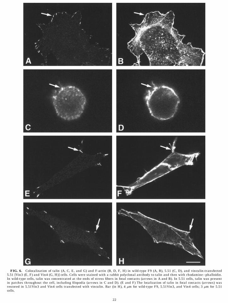
FIG. 6. Colocalization of talin (A, C, E, and G) and F-actin (B, D, F, H) in wild-type F9 (A, B), 5.51 (C, D), and vinculin-transfected5.51 [Vin3 (E, F) and Vin4 (G, H)] cells. Cells were stained with a rabbit polyclonal antibody to talin and then with rhodamine–phalloidin.In wild-type cells, talin was concentrated at the ends of stress fibers in focal contacts (arrows in A and B). In 5.51 cells, talin was presentin patches throughout the cell, including filopodia (arrows in C and D). (E and F) The localization of talin in focal contacts (arrows) wasrestored in 5.51Vin3 and Vin4 cells transfected with vinculin. Bar (in H), 4 mm for wild-type F9, 5.51Vin3, and Vin4 cells; 3 mm for 5.51cells.
22
02-14-97 13:49:29 eca

23VINCULIN IN INTEGRIN-MEDIATED CELL ADHESION
adhesion and spreading. When an anchorage-depen-dent cell adheres to a rigid ECM substrate, forces dueto local integrin binding begin to drive membrane flat-tening until balanced by the mechanical stiffness of thecortical membrane and associated cytoskeleton. Nor-mal cells overcome these forces by clustering multipleintegrins together with actin-associated moleculeswithin localized focal adhesion complexes that, in turn,physically couple to the ends of actin microfilamentsinside the cell. Formation of this molecular bridge re-sults in outward transfer of cytoskeletal tension to thecell’s ECM adhesions, resulting in what is known as a‘‘strengthening response’’ [30]. If the ECM can resistFIG. 7. Stiffness (calculated as the stress divided by the strain)
of RGD-coated magnetic microbeads bound to the surface of wild- these contractile forces (i.e., resist bending or compres-type F9, 5.51, and vinculin-transfected (5.51Vin3 and 5.51Vin4) cells. sion), then the cell can extend outward and progres-The standard deviation and the number of measurements (for each sively change from a spherical to a more flattened orsample) are given in parentheses.
spread morphology. Our results show that loss of vin-culin in 5.51 cells, disrupts focal adhesion complex for-mation, decreases the efficiency of transmembrane me-the wild-type cells (20 versus 37 dyn/cm2). Transfectionchanical coupling, and prevents cell adhesion andof vinculin into 5.51 cells restored the mechanical link-spreading. More importantly, all of these deficienciesage to the integrin-linked cytoskeleton (see Fig. 7).could be reversed by replacing vinculin through trans-These results indicate that the focal adhesions thatfection in two different F9 cell lines. Thus, at least inform in response to RGD-bead binding in 5.51 cells5.51 cells, vinculin appears to play a key role in shapewere less able to resist mechanical deformation andcontrol based on its ability to modulate focal adhesionless effective at transmitting mechanical stress to thestructure and function.internal cytoskeleton. Confocal microscopy confirmed
Most past work on cell shape control focused on thethat less F-actin was associated with the RGD-beadsmechanism by which motile cells extend small pro-in 5.51 cells than in wild-type cells (Fig. 8). The absencecesses, such as filopodia and lamellipodia. Much lessof an extensive F-actin network in this region may, inis known about how cells change from round to spreadpart, account for the decreased resistance to magneticfollowing adhesion or when they are nonmotile. How-twisting exhibited by 5.51 cells.ever, all cell shape changes are mediated through cy-toskeletal reorganization. For example, suspendedDISCUSSIONcells contain a continuous microfilament lattice that isloosely packed and isotropic (nonoriented) when roundTo understand the role of vinculin in cell shape con-
trol, it is necessary to consider the biophysical basis of and free of anchorage [27, 31, 32]. As cells attach and
FIG. 8. Organization of F-actin in wild-type F9 (A) and 5.51 (B) cells that have bound RGD-coated magnetic microbeads (arrows).Increased cortical actin and a dense cytoplasmic actin network was observed at the site of RGD-bead binding in the wild-type F9, but not5.51, cells. Bar (in B), 3 mm.
AID ECR 3451 / 6i1a$$$$63 02-14-97 13:49:29 eca
24 EZZELL ET AL.
spread on extracellular matrix, this loose network re- sions of lamellipodia while in its absence, only filopodiawould form. Another possibility is that vinculin pro-models locally between fixed focal adhesions and forms
microfilament bundles that are known as ‘‘stress fi- motes lamellipodia extension and cell spreading indi-rectly by stabilizing focal adhesion structure (e.g., stiff-bers.’’ Importantly, these changes in microfilament or-
ganization and cell shape proceed without altering to- ness), as demonstrated in the magnetic bead twistingstudies. This, in turn, would allow cells to exert cy-tal microfilament number [33] or the total amount of
F-actin [6, 34]. In fact, one possibility is that these toskeletal tension on the extracellular matrix, increaseinternal isometric tension within the whole cytoskele-changes in cytoskeletal organization result from ‘‘ten-
sion-molding’’ of the flexible microfilament networks ton and, hence, to drive local cytoskeletal remodelingevents in the apex of the cell as well as at the cell base.when cells apply tension to ECM and thereby increase
isometric tension in the cytoskeleton that stretches be- For example, structural modeling studies show thatincreased isometric tension in the cytoskeleton can ex-tween isolated focal adhesions [4, 35, 36]. Our finding
that transfection of vinculin into 5.51 cells promoted plain how triangulated nets of microfilaments form inapical regions of spreading cells as well as basal stressstress fiber formation and cell spreading by stabilizing
focal adhesions and supporting transfer of mechanical fibers, in the absence of net changes in actin polymer-ization or cross-linking [4, 5]. Importantly, vertices ofstresses across the cell surface, rather than by altering
the total amount of F-actin polymer or cross-linking, is triangulated actin nets (e.g., actin geodomes) appearto serve as preferred niduses for actin polymerizationconsistent with this hypothesis. In fact, ‘‘stress fiber’’
formation can be artificially induced by applying me- [40, 41]. Thus, failure to form well-developed focal ad-hesions in the vinculin-deficient cells could result inchanical tension to the cell surface both in vitro [37,
38] and in vivo [39]. both decreased stiffening and incomplete tension mold-ing of the actin cytoskeleton. This would suppress bothWork on the cytoskeletal basis of cell motility has
revealed that formation of both filopodia and lamelli- lateral membrane extension and cell spreading.Key to all of these processes is the clustering of inte-podia involves changes in microfilament assembly. The
stiff actin bundles that form the central cores of filo- grins and the formation of a stable focal adhesion com-plex that can bear mechanical loads. The presence ofpodia and extend from the central microfilament lattice
[40, 41] and push the cortical membrane outward are focal adhesions, as monitored by interference reflectionmicroscopy and staining for characteristic proteinsa result of actin polymerization, either alone or in com-
bination with osmotic forces [42–45]. Vinculin appar- (e.g., a-actinin, paxillin, and talin), coincided with theloss and reintroduction of vinculin in F9 cells [19, 29].ently is not required for this form of actin assembly
given that 5.51 cells continued to extend filopodia, even Furthermore, using atomic force microscopy and rheol-ogy, the 5.51 cells displayed marked differences in vis-when round. In contrast, lamellipodial extension was
only observed in cells that contained vinculin. Some coelasticity [52]. Although the sequence of events lead-ing to focal adhesion complex formation is not fullymodels of lamellipodial formation similarly depend on
actin polymerization, alone or in combination with os- understood there is reason to believe that adhesion canbe established by an alternative mechanism(s) involv-motic forces [42–45]. Others are based on myosin-de-
pendent movements of actin filaments at the leading ing a-actinin or talin, both of which can bind to actinand integrins [23, 24, 53, 54]. For example, talin andedge [46, 47]. The vinculin-deficient 5.51 cells con-
tained normal amounts of talin and a-actinin, two focal F-actin can accumulate before vinculin at the sitewhere focal adhesions will eventually form in spread-adhesion proteins capable of binding actin [48, 49], but
their presence alone was not sufficient to induce forma- ing fibroblasts [55, 56]. Talin also colocalizes with F-actin in patches near the plasma membrane in 5.51tion of lamellipodia or stress fibers. Actin polymeriza-
tion per se clearly was not inhibited since filopodia ex- cells. Our mechanical twisting data also show that F9cells exhibit some mechanical coupling between inte-tended normally in 5.51 cells and there was no signifi-
cant difference in the total amount of F-actin polymer grins and the cytoskeleton. However, the presence ofvinculin appears to be required for optimal mechanicalin the four different F9 cell lines. There also was no
difference in total actin cross-linking. stability of the focal adhesion complex, a structuralfeature that appears to be critical for cell adhesion andOne interpretation of these results is that talin sup-
ports linear membrane extension (i.e., filopodia) driven spreading as well as cytoskeletal reorganization.Recently, a vinculin knockout was created by homolo-by actin polymerization, in the absence of vinculin.
While talin self-associates to form dimers, vinculin gous recombination. These cells differ from the 5.51cells in that adhesion is only slightly inhibited [23, 24].forms multimers [16, 50]. Furthermore, talin dimers
may themselves be linked by vinculin [51]. In addition, It is possible that knocking out vinculin in these cellshas produced some compensatory mechanism that isvinculin binds to a-actinin, an actin cross-linking pro-
tein, while talin does not. In this manner, vinculin may not found in 5.51 cells or there is some other caveatthat remains to be determined. However, regardless ofsupport the broader, more stable cytoplasmic exten-
AID ECR 3451 / 6i1a$$$$63 02-14-97 13:49:29 eca

25VINCULIN IN INTEGRIN-MEDIATED CELL ADHESION
16. Johnson, R. P., and Craig, S. W. (1995) Nature 373, 261–264.the differences between these two models, the present17. Gilmore, A. P., and Burridge, K. (1996) Nature 381, 531–535.results show that loss of vinculin produces a particular18. Weekes, J., Barry, S. T., and Critchley, D. R. (1996) Biochem.phenotype in 5.51 cells and that most—if not all—of
J. 314, 827–832.this phenotype can be reversed by vinculin transfec-19. Samuels, M., Ezzell, R. M., Cardozo, T. J., Critchley, D. R., Coll,tion. In summary, many models have been proposed to
J. L., and Adamson, E. D. (1993) J. Cell Biol. 121, 909–921.explain how cells change shape and move. However,20. Varnum-Finney, B., and Reichardt, L. F. (1994) J. Cell Biol.all of these models require that the cell must adhere 127, 1071–1084.
to an extracellular matrix substrate that can resist cell-21. Rodriquez Fernandez, J. L., Geiger, B., Salomon, D., and Ben-
generated forces in order to promote cytoskeletal rear- Ze’ev, A. (1992) Cell Motil. Cytoskeleton 22, 127–134.rangements necessary for global cell shape changes 22. Rodriquez Fernandez, J. L., Geiger, B., Salomon, D., Sabanay,and to propel the cell forward. Many molecules are I., Zoller, M., and Ben-Ze’ev, A. (1992) J. Cell Biol. 119, 427–
438.known to localize to the site of integrin clustering23. Volberg, T., Geiger, B., Kam, Z., Pankov, R., Simcha, I., Saba-within the focal adhesion complex; however, their
nay, H., Coll, L. J., Adamson, E. D., and Ben-Ze’ev, A. (1995) J.structural functions remain unknown. The results ofCell Sci. 108, 2253–2260.the present study show that vinculin controls adhesion,
24. Coll, J. L., Ben-Ze’ev, A., Ezzell, R. M., Rodriguez Fernandez,cytoskeletal remodeling, and cell spreading by mechan-J. L., Baribault, H., Oshima, R. G., and Adamson, E. D. (1995)ically stabilizing the molecular bridge between actin Proc. Natl. Acad. Sci. USA 92, 9161–9165.
and integrins that forms the core of the focal adhesion 25. Wang, N., and Ingber, D. E. (1994) Biophys. J. 66, 2181–2189.and thereby, enhancing its ability to both transmit and 26. Condeelis, J., and Hall, A. L. (1991) Methods Enzymol. 196,resist cytoskeletal tension. 486–496.
27. Schliwa, M., van Blerkom, J., and Porter, K. (1981) Proc. Natl.We thank Dr. Eileen Adamson for providing the F9 cell lines and Acad. Sci. USA 80, 5417–5420.
helpful comments. This work was supported by the American Cancer 28. Matsudaira, P. T., and Burgess, D. R. (1978) Anal. Biochem. 87,Society, NASA, Deutsche Forschungsgemeinschaft, and NATO. Dr. 386–396.Ingber is a recipient of a Faculty Research Award from American
29. Goldmann, W. H., Schindl, M., Cardozo, T. J., and Ezzell, R. M.Cancer Society.(1995) Exp. Cell Res. 221, 311–319.
30. Lotz, M. M., Burdsal, C. A., Erickson, H. P., and McClay, D. R.REFERENCES (1989) J. Cell Biol. 109, 1795–1805.
31. Ben-Ze’ev, A., Duerr, A., Salomon, F., and Penman, S. (1979)1. Luna, E., and Hitt, A. L. (1992) Science 258, 955–963. Cell 17, 859–865.2. Burridge, K., Fath, K., Kelly, T., Nuckolls, G., and Turner, C. 32. Heuser, J., and Kirschner, M. (1980) J. Cell Biol. 86, 212–234.
(1988) Annu. Rev. Cell Biol. 4, 487–525.33. Revel, J. P., Hoch, P., and Ho, D. (1974) Exp. Cell Res. 84, 207–
3. Jockusch, B. M., Bubeck, P., Giehl, K., Kroemker, M., 218.Moschner, J., Rotkegel, M., Ruediger, M., Schiller, K., Stanke,
34. Bereiter-Hahn, J., Luck, M., Miebach, T., Stelzer, H. K., andG., and Winkler, J. (1995) Annu. Rev. Cell Dev. Biol. 11, 379–Voth, M. (1990) J. Cell Sci. 96, 171–188.416.
35. Bereiter-Hahn, J. (1987) in Cytomechanics: The Mechanical Ba-4. Ingber, D. E. (1993) J. Cell Sci. 104, 613–627.sis of Cell Form and Structure (Bereiter-Hahn, J., Anderson,
5. Ingber, D. E., Dike, L., Hansen, L., Karp, S., Liley, H., Maniotis, O. R., and Reif, W. E., Eds.), pp. 3–30, Springer-Verlag, Berlin.A., McNamee, H., Mooney, D., Plopper, G., Sims, J. et al. (1994)
36. Opas, M. (1987) in Cytomechanics: The Mechanical Basis ofInt. Rev. Cytol. 150, 173–224.Cell Form and Structure (Bereiter-Hahn, J., Anderson, O. R.,
6. Mooney, D. J., Langer, R., and Ingber, D. E. (1995) J. Cell Sci. and Reif, W. E., Eds.), pp. 273–286, Springer-Verlag, Berlin.108, 2311–2320.
37. Franke, R. P., Grafe, M., Schnittler, H., Seiffge, D., Mitter-7. Wang, N., Butler, J. P., and Ingber, D. E. (1993) Science 260, mayer, C., and Drenckhahn, D. (1984) Nature 307, 648–649.
1124–1127.38. Kolega, J. (1986) J. Cell Biol. 102, 1400–1411.
8. Crowley, E., and Horwitz, A. F. (1995) J. Cell Biol. 131, 525–39. Wong, A. J., Pollard, T. D., and Herman, I. M. (1983) Science537.
219, 867–869.9. Grover, A., Rosentraus, M. J., Sterman, B., Snook, M. E., and
40. Lazarides, E. (1976) J. Cell Biol. 68, 202–219.Adamson, E. D. (1987) Dev. Biol. 120, 1–11.41. Osborn, M., Born, T., Koitsch, H. J., and Weber, K. (1978) Cell10. Calogero, A., Samuels, M., Darland, T., Edwards, S. A., Kemler,
14, 477–488.R., and Adamson, E. D. (1991) Dev. Biol. 146, 499–508.42. Wang, J. L. (1986) J. Cell Biol. 101, 597–602.11. Jockusch, B. M., and Isenberg, G. (1981) Proc. Natl. Acad. Sci.43. Forscher, P., and Smith, S. (1988) J. Cell Biol. 107, 1505–1516.USA 78, 3005–3009.44. Oster, G. F., and Perelson, A. A. (1987) J. Cell Sci. Suppl. 8,12. Burridge, K., and Mangeat, P. (1984) Nature 308, 744–746.
35–54.13. Wachsstock, D. H., Wilkins, J. A., and Lin, S. (1987) Biochem.45. Small, J. (1989) Curr. Opin. Cell Biol. 1, 75–79.Biophys. Res. Commun. 146, 554–560.46. Sheetz, M. P., Wayne, D. B., and Pearlman, A. L. (1992) Cell14. Turner, C. E., Glenney, J. R., Jr., and Burridge, K. (1990) J.
Motil. Cytoskeleton 22, 160–169.Cell Biol. 111, 1059–1068.15. Johnson, R. P., and Craig, S. W. (1994) J. Biol. Chem. 269, 47. Heath, J. P., and Holifield, B. F. (1991) Cell Motil. Cytoskeleton
18, 245–257.12611–12619.
AID ECR 3451 / 6i1a$$$$63 02-14-97 13:49:29 eca

26 EZZELL ET AL.
48. Goldmann, W. H., and Isenberg, G. (1991) Biochem. Biophys. 52. Goldmann, W. H., and Ezzell, R. M. (1996) Exp. Cell Res. 226,234–237.Res. Commun. 178, 718–723.
53. Goldmann, W. H., Ezzell, R. M., Adamson, E. D., Niggli, V., and49. Muguruma, M., Matsumura, S., and Fukazawa, T. (1992) J.Isenberg, G. (1996) J. Muscle Res. Cell Motil. 17, 1–5.Biol. Chem. 267, 5621–5624.
54. Jockusch, B. M., and Ruediger, M. (1996) Trends Cell Biol. 6,50. Goldmann, W. H., Bremer, A., Haener, M., Aebi, U., and Isen- 311–315.
berg, G. (1994) J. Struct. Biol. 112, 3–10. 55. DePasquale, J. A., and Izzard, C. S. (1987) J. Cell Biol. 105,2803–2809.51. Gilmore, A. P., Wood, C., Ohanian, V., Jackson, P., Patel, B.,
Rees, D. J. G., Hynes, R. O., and Critchley, D. R. (1993) J. Cell 56. DePasquale, J. A., and Izzard, C. S. (1991) J. Cell Biol. 113,1351–1359.Biol. 122, 337–347.
Received August 21, 1996Revised version received November 15, 1996
AID ECR 3451 / 6i1a$$$$63 02-14-97 13:49:29 eca
Copyright © 2022 FDOKUMEN