Development of Mechanically Tailored Gelatin-Chondroitin Sulphate Hydrogel Films
9
Development of Mechanically Tailored Gelatin-Chondroitin Sulphate Hydrogel Films Sandra Van Vlierberghe, Sangram Keshari Samal, Peter Dubruel * Summary: The aim of the present work was to develop and characterize biohybrid hydrogels containing combinations of gelatin and chondroitin sulphate. Two approaches have been compared. First, physisorption of chondroitin sulphate to gelatin hydrogels was investigated using surface plasmon resonance measurements. Due to the limited interaction between both biopolymers, we developed in a second approach methacrylate-modified chondroitin sulphate hydrogels as such or in combination with methacrylamide-modified gelatin. Rheology measurements indi- cated that following this approach, mechanically stable hydrogel films could be obtained after UV irradiation in the presence of Irgacure 2959 as UV photo-initiator. When keeping the gelatin concentration and the modification degree constant, an increase in derivatization of the chondroitin sulphate component resulted in a high increase of the storage modulus. Keywords: characterization; chondroitin sulphate; gelatin; hydrogel; modification Introduction Extracellular matrix (ECM) constituents including collagen (i.e. starting product for gelatin synthesis) and glycosaminoglycans (GAGs) are among the most abundant in the body and play key roles in a number of biological processes. They are widely utilized to fabricate scaffolds, serving as an active analogue of native ECM. [1] Hydrogels are three-dimensional inso- luble polymeric networks from which biochemical factors can be delivered and through which diffusion of vital cell nutri- ents and waste products can occur. [2] Previous research already studied the application of photopolymerized hydrogels for tissue engineering applications. [3–5] Photopolymerization provides a fast and efficient method to crosslink polymers forming a hydrogel with significant tem- poral and spatial control. These hydrogels have already demonstrated their potential as three dimensional structures suitable for tissue engineering applications. [6,7] Very frequently, photopolymerised hydrogels are composed of synthetic polymers. In order to enhance their bioactivity and biocompat- ibility, biopolymers can be included. As an example, collagen-GAG scaffolds have been used extensively for in vitro studies of cell-ECM interactions and as a platform for tissue biosynthesis including in vivo studies of tissue or organ regenera- tion. [8,9] Favourable characteristics of scaf- folds from these natural materials stimulate host cells to repopulate and form new tissues that closely simulate the native organiza- tion. [10,11] In addition, they enhance biolo- gical interactions with cells and speed up tissue regeneration by introducing cell- specific ligands or extracellular signalling molecules, such as peptides and oligosac- charides. [12] In the present work, gelatin type B (i.e. processed from collagen via an alkali treatment) and chondroitin sulphate (CS) were selected as starting materials for the development of ECM mimicking materials. Gelatin has previously been successfully applied for a wide application range Macromol. Symp. 2011, 309/310, 173–181 DOI: 10.1002/masy.201100030 173 Polymer Chemistry & Biomaterials Research Group, Ghent University, Krijgslaan 281, S4-Bis, B-9000 Ghent, Belgium E-mail: [email protected] Copyright ß 2011 WILEY-VCH Verlag GmbH & Co. KGaA, Weinheim wileyonlinelibrary.com FOR PERSONAL USE
Transcript of Development of Mechanically Tailored Gelatin-Chondroitin Sulphate Hydrogel Films

Macromol. Symp. 2011, 309/310, 173–181 DOI: 10.1002/masy.201100030 173
Poly
Ghe
Ghe
E-m
Cop
Development of Mechanically Tailored
Gelatin-Chondroitin Sulphate Hydrogel Films
Sandra Van Vlierberghe, Sangram Keshari Samal, Peter Dubruel*
Summary: The aim of the present work was to develop and characterize biohybrid
hydrogels containing combinations of gelatin and chondroitin sulphate. Two
approaches have been compared. First, physisorption of chondroitin sulphate to
gelatin hydrogels was investigated using surface plasmon resonance measurements.
Due to the limited interaction between both biopolymers, we developed in a second
approach methacrylate-modified chondroitin sulphate hydrogels as such or in
combination with methacrylamide-modified gelatin. Rheology measurements indi-
cated that following this approach, mechanically stable hydrogel films could be
obtained after UV irradiation in the presence of Irgacure 2959 as UV photo-initiator.
When keeping the gelatin concentration and the modification degree constant, an
increase in derivatization of the chondroitin sulphate component resulted in a high
increase of the storage modulus.
Keywords: characterization; chondroitin sulphate; gelatin; hydrogel; modificationE
U
S IntroductionExtracellular matrix (ECM) constituents
including collagen (i.e. starting product for
gelatin synthesis) and glycosaminoglycans
(GAGs) are among the most abundant in
the body and play key roles in a number
of biological processes. They are widely
utilized to fabricate scaffolds, serving as an
active analogue of native ECM.[1]
Hydrogels are three-dimensional inso-
luble polymeric networks from which
biochemical factors can be delivered and
through which diffusion of vital cell nutri-
ents and waste products can occur.[2]
Previous research already studied the
application of photopolymerized hydrogels
for tissue engineering applications.[3–5]
Photopolymerization provides a fast and
efficient method to crosslink polymers
forming a hydrogel with significant tem-
poral and spatial control. These hydrogels
have already demonstrated their potential
FOR PERS
mer Chemistry & Biomaterials Research Group,
nt University, Krijgslaan 281, S4-Bis, B-9000
nt, Belgium
ail: [email protected]
yright � 2011 WILEY-VCH Verlag GmbH & Co. KGaA
as three dimensional structures suitable for
tissue engineering applications.[6,7] Very
frequently, photopolymerised hydrogels are
composed of synthetic polymers. In order to
enhance their bioactivity and biocompat-
ibility, biopolymers can be included.
As an example, collagen-GAG scaffolds
have been used extensively for in vitro
studies of cell-ECM interactions and as a
platform for tissue biosynthesis including
in vivo studies of tissue or organ regenera-
tion.[8,9] Favourable characteristics of scaf-
folds from these natural materials stimulate
host cells to repopulate and form new tissues
that closely simulate the native organiza-
tion.[10,11] In addition, they enhance biolo-
gical interactions with cells and speed up
tissue regeneration by introducing cell-
specific ligands or extracellular signalling
molecules, such as peptides and oligosac-
charides.[12]
In the present work, gelatin type B (i.e.
processed from collagen via an alkali
treatment) and chondroitin sulphate (CS)
were selected as starting materials for the
development of ECM mimicking materials.
Gelatin has previously been successfully
applied for a wide application range
ONAL
, Weinheim wileyonlinelibrary.com
Macromol. Symp. 2011, 309/310, 173–181174
including burn dressings, cardiovascular
surgery and 3D scaffolds for tissue engi-
neering of skin, bone, cartilage and other
tissues.[13] The combination of gelatin and
GAGs is often used in skin regeneration
since gelatin or GAGs as such cannot heal
full thickness wounds.[14] GAGs have also
been reported to significantly affect cellular
response, morphology and stiffness of
the biohybrid scaffolds.[15,16] In literature,
the interaction between proteoglycans
and extracellular matrix molecules was
reported earlier.[17] Most of the interactions
A
B
Gel NH2 + H2C C
CH3
C
O
O C
O
C
CH
C
OCOOH
OH
OH
O OOH
O
CH2OSO3Na
NHCOCH3
n
+
OO
COOH
OR
O C
O
CH2C
CH3
O OORCH2OSO3Na
NHCOCHn
R
Irgacure 295UV irradiati
10
R
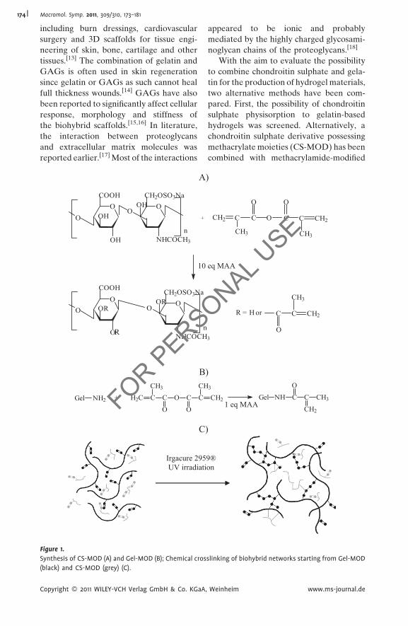
Figure 1.
Synthesis of CS-MOD (A) and Gel-MOD (B); Chemical cros
(black) and CS-MOD (grey) (C).
Copyright � 2011 WILEY-VCH Verlag GmbH & Co. KGaA
FOR PERS
appeared to be ionic and probably
mediated by the highly charged glycosami-
noglycan chains of the proteoglycans.[18]
With the aim to evaluate the possibility
to combine chondroitin sulphate and gela-
tin for the production of hydrogel materials,
two alternative methods have been com-
pared. First, the possibility of chondroitin
sulphate physisorption to gelatin-based
hydrogels was screened. Alternatively, a
chondroitin sulphate derivative possessing
methacrylate moieties (CS-MOD) has been
combined with methacrylamide-modified
)
) 3
CH2 Gel NH C
O
C
CH2
CH3
)
CH2 C
CH3
C
O
O C
O
C CH2
CH3
3
R = H or C
O
C CH2
CH3
9® on
1 eq MAA
eq MAA
slinking of biohybrid networks starting from Gel-MOD
, Weinheim www.ms-journal.de
ONAL USE

Macromol. Symp. 2011, 309/310, 173–181 175
gelatin (Gel-MOD) to develop biohybrid
chemically crosslinked hydrogels (see
Figure 1). The effect of the applied polymer
concentration and the hydrogel precursor
modification degree on the final properties
of the hydrogel films has been evaluated
using rheology.
Materials and Methods
Materials
Gelatin (type B), isolated from bovine skin
by an alkaline process, was kindly supplied
by Rousselot, Ghent, Belgium. Gelatin
samples with an approximate iso-electric
point of 5, a Bloom strength of 257 and a
viscosity (6.67%, 60 8C) of 4.88 mPa.s were
used. Methacrylic anhydride (MAA), chon-
droitin sulphate C, (sodium salt, from shark
cartilage), chondroitin sulphate A (sodium
salt, from bovine trachea) and monoclonal
anti-chondroitin sulphate (clone CS-56,
from mouse ascites fluid), were acquired
from Sigma-Aldrich (Bornem, Belgium).
1-[4-(2-Hydroxyethoxy)-phenyl]-2-hydroxy-
2-methyl-1-propane-1-one (Irgacure1 2959)
was a kind gift from Ciba Speciality Chemi-
cals N.V. (Groot-Bijgaarden, Belgium).
Surface Plasmon Resonance Measurements
The interaction between gelatin and CS was
measured using a Biacore-X (GE Health-
care Europe, Diegem, Belgium) equipped
with an internal 500ml Hamilton syringe.
All SPR measurements were performed at
25 8C using a phosphate buffer (0.05 M,
pH¼ 7.4). The flow rate was set to 50ml/min.
The sensor surface was spincoated using
90ml of an aqueous 5 w/v% Gel-MOD
(degree of substitution 60%) solution at a
speed of 6000 rpm during 90 seconds. After
spincoating, the gelatin coated sensor was
inserted into the SPR apparatus. After
stabilisation of the baseline, 50ml of various
concentrations of CS solutions was injected.
In a final step, after stabilisation of the
signal, 50ml of an chondroitin sulphate
antibody solution (200� dilution of the
stock) was injected. All values reported are
relative to a reference flow channel.
FOR PERS
Copyright � 2011 WILEY-VCH Verlag GmbH & Co. KGaA
Synthesis and Characterization of Hydrogel
Precursors
Chondroitin sulphate methacrylate (CS-
MOD) and gelatin-methacrylamide (Gel-
MOD) were synthesized as described
earlier.[7,19]
In brief, 1 g chondroitin sulphate was
dissolved in 50 ml double distilled water
at room temperature. Next, an excess
methacrylic anhydride (0.06 mol, 8.94 ml)
was added dropwise. Simultaneously,
the pH of the reaction mixture was adjusted
to 8, by adding NaOH (5 N). The ratio
between the added amounts of methacrylic
anhydride and NaOH was 1 to 1.12. Next,
the mixture was stirred at room tempera-
ture for 2 hours. Finally, the solution was
diluted with 50 ml double distilled water
and transferred to dialysis membranes
(Spectra/Por1 3, MWCO 3,500 Da, 3 days),
followed by lyophilization. Chondroitin
sulphate methacrylates with lower modifi-
cation degrees were obtained by adding
lower amounts of methacrylic anhydride.1H NMR-spectra of modified chondroi-
tin sulphate were recorded at room tem-
perature in deuterated water. The degree of
substitution could be obtained after com-
parison of the integrations of the charac-
teristic peaks of the methacrylate-substitu-
ent (1.95 ppm, 5.76 ppm and 6.19 ppm) and
the integration of the characteristic peak
corresponding to the methyl groups in
native CS (2.04 ppm). Consequently, the
degree of substitution could be calculated,
as indicated by the following equation:
DS ð%Þ ¼ 100 � ðI5:7 ppmÞ=ððI1:95 ppm
þ I2:04 ppm � 3 � ðI5:7 ppmÞÞ=3Þ
Hydrogel Preparation
CS-MOD (0.1–1 g) with various modifica-
tion degrees (degree of substitution, DS)
(5–40%) was dissolved in 20 ml double
distilled water at room temperature.
For the biohybrid hydrogels, Gel-MOD
(1.4–3 g, DS 65%) was added and the mixture
was stirred at 40 8C. Next, the photoinitiator
Irgacure1 2959 (2 mol% relative to the
methacrylates and methacrylamides of
ONAL USE
, Weinheim www.ms-journal.de

Macromol. Symp. 2011, 309/310, 173–181176
respectively CS-MOD and Gel-MOD) was
added, followed by injection of the mixture
between silanized glass plates, separated by
a 1 mm thick silicone spacer. Silanization of
the glass plates occurred by incubating the
glass plates overnight into an aqueous
solution of 2 v/v% H2SO4/HNO3, followed
by an overnight incubation in toluene,
containing 10 v/v% trimethylsilylchloride.
Finally, the hydrogel formed was UV
irradiated (276 nm, 10 mW/cm2) for 20 min-
utes on both sides or crosslinked in situ
during rheology. To enable the crosslinking
prior to the rheological evaluation, an
LWUV-lamp model VL-400L (Vilber
Lourmat, Marne La Vallee, France) with
an intensity of 10 mW/cm2 and a wave-
length range of 250–450 nm, was applied for
sample curing. The crosslinked hydrogels
were stored at 5 8C until further evaluation.
Hydrogel Characterization
The visco-elastic properties of the hydro-
gels were evaluated using a rheometer
type Physica MCR-301 (Anton Paar, Sint-
Martens-Latem, Belgium). First, the linear
visco-elastic range was determined as a
function of the deformation (0.01–1%) and
at constant frequency (1 Hz) (data not
shown). Next, the effect of the UV irradia-
tion applied, on the final mechanical
properties was monitored by performing
a time scan using the following parameters:
1 Hz, 0.5% strain, FN¼ 0.01 N and 21 8C.R PERS
-50
0
50
100
150
200
250
500450400350300250200150100Time (s)
Res
pons
e (R
U)
CS
100 µg/ml
500 µg/ml
1 mg/ml
anti-CS
Figure 2.
SPR sensorgram showing the effect of the CS con-
centration (100mg/l – 1 mg/ml) on the interaction
between gelatin and chondroitin sulphate A.
O
Results and DiscussionEvaluation of the Gelatin Chondroitin
Sulphate Affinity
Literature data previously indicated that
chondroitin sulphate E possesses specific
affinity for type V collagen.[18] The binding
requires a sequence of repeating units,
consisting of one glucuronic acid and
one N-acetyl-galactosamine, sulphated at
carbon-4 and carbon-6. Alternative oligo-
saccharides, consisting of other sequences,
however possessing the same charge, do not
interact with type V collagen.[20] The latter
demonstrates that the interaction between
F
Copyright � 2011 WILEY-VCH Verlag GmbH & Co. KGaA
chondroitin sulphate and gelatin might also
depend on various parameters. Therefore,
the interaction between gelatin type B and
two types of chondroitin sulphate (i.e. type
A and C) was studied in the present work
using SPR.
In a first part of the SPR experiment,
chondroitin sulphate A (CSA) solutions of
different concentrations were rinsed over
the sensor chip (100–1000mg/ml), pre-
viously spincoated with methacrylamide
modified gelatin type B (gel-MOD). In a
second part, an antibody specific for CSA
was injected in order to verify that the
response was related to the deposition of
the GAG onto the gelatin-coated SPR chip.
The response signal plotted as a function of
incubation time, giving an idea on the
interaction between gel-MOD and CSA, is
given in Figure 2. As a reference, the
antibody was rinsed over a gel-MOD coated
chip. As anticipated, the SPR sensorgram did
not show any response (data not shown).
From the figure, it can be derived that
the affinity between chondroitin sulphate A
and gel-MOD is relatively low. The amount
of adsorbed polysaccharide increases
slightly with increasing CS concentration.
The limited response can be explained by
the iso-electric point of the applied gelatin
(i.e. 5). Consequently, both gelatin B and
chondroitin sulphate A are negatively
charged, excluding the possibility for ionic
interactions. Literature data already
revealed a weak interaction between col-
lagen and chondroitin 4-sulphate.[21]
ONAL USE
, Weinheim www.ms-journal.de

-50
50
150
250
350
450
550
98009750970096509600955095009450Time (s)
Res
pons
e (R
U)
100 µg/ml
500 µg/ml 1 mg/ml CS
anti-CS
Figure 3.
SPR sensorgram showing the effect of the CS con-
centration (100mg/l – 1 mg/ml) on the interaction
between gelatin and chondroitin sulphate C.
0
510
15
20
25
30
35
40
45
121086420# equivalents methacrylic anhydride
DS
(%)
Figure 4.
Master curve of CS showing the degree of substitution
as a function of the amount of methacrylic anhydride
added.
Macromol. Symp. 2011, 309/310, 173–181 177
Interestingly, the adsorbed amount of
GAGs still enabled subsequent interaction
with CS antibodies, which was reflected by
the increased response signal after the
antibody injection (i.e. second injection)
(Figure 2).
In a following part of the SPR experi-
ments, we also investigated the interaction
between chondroitin sulphate type C and
gelatin (Figure 3).
When comparing the affinity between
both types of chondroitin sulphate and
gelatin type B, no significant difference in
response signal was observed. The SPR
data obtained clearly show that a chemical
modification of and a subsequent co-cross-
linking of chondroitin sulphate with cross-
linkable gelatin is essential to realize an
efficient incorporation of CS into hydrogel
films.
Synthesis and Characterization of Hydrogel
Precursors
The synthesis and characterization of
the methacrylate-modified chondroitin sul-
phate (CS-MOD) precursor has been
described earlier.[7] In brief, part of the
hydroxyl groups of CS were converted into
methacrylate groups. As methacrylic acid
(MA) is generated during the esterification,
NaOH was added as neutralizing agent,
avoiding possible acid catalysed degradation
of the polysaccharide. The glycosaminogly-
can containing crosslinkable methacrylate
groups was purified by membrane dialysis
against double distilled water for several
days, followed by isolation via lyophilization.
FOR PERS
Copyright � 2011 WILEY-VCH Verlag GmbH & Co. KGaA
The methacrylate substitution on CS was
quantified using 1H-NMR spectroscopy.
The two distinctive peaks at 5.76 and
6.19 ppm can be attributed to the two
protons on the double bond (C¼CH2),
while the peak at 1.95 ppm can be ascribed
to the methyl groups adjacent to the double
bonds (CH3-C¼CH2). The 1H-NMR region
from 1.6 to 2.5 ppm was expanded and the
peaks corresponding to the two methyl
groups were deconvoluted and integrated.
The ratio of the peak intensities at 1.95 ppm
to that at 2.04 ppm, corresponding to the
methyl groups on native CS, was used to
calculate the degree of substitution. In what
follows, the degree of substitution will be
expressed as the amount of modified
repeating disaccharide units. The degree
of substitution was calculated using the
following equation:
DS ð%Þ ¼ 100 � ðI5:7 ppmÞ=ððI1:95 ppm
þ I2:04 ppm � 3 � ðI5:7 ppmÞÞ=3Þ
We were able to show that the degree of
substitution can be easily varied by adjust-
ing the amount of added methacrylic
anhydride. Figure 4 indeed shows the
master curve for the CS modification in
which the degree of substitution is plotted
against the equivalents methacrylic anhy-
dride added.
The synthesis and characterization of
Gel-MOD as second hydrogel building
block was reported earlier by Van Den
Bulcke et al.[19]
ONAL USE
, Weinheim www.ms-journal.de

Macromol. Symp. 2011, 309/310, 173–181178
Hydrogel Preparation and Characterization
Both the biohybrid hydrogels and the
hydrogels composed of CS-MOD only were
prepared and evaluated as thin films. The
hydrogel films were prepared by mixing an
aqueous solution of the hydrogel precursors
(at 40 8C) in the presence of the photo-
initiator, followed by injection between two
silanized glass plates, separated by a 1 mm
silicone spacer. Chemical crosslinking
occurred via UV-irradiation (lex¼ 279 nm).
In the present work, Irgacure 2959 was
selected as a photo-initiator since previous
research already indicated its biocompat-
ibility.[6,7] Dubruel et al have already shown
that applying 2 mol% Irgacure 2959 to
the methacrylamides present in Gel-MOD
resulted in scaffolds suitable to support the
attachment and proliferation of a large panel
of human cells including endothelial cells,
glial cells, osteoblasts, fibroblasts and epithe-
lial cells.[6] We therefore do not anticipate
any problems regarding the material toxicity
when applying Irgacure 2959 as a photo-
initiator. The major advantage of combining
CS-MOD and Gel-MOD in stead of apply-
ing carbodiimide chemistry to couple amines
and carboxylic acids is the possibility to
introduce additional growth factors, if
needed, without affecting their biological
activity. When applying carbodiimide chem-
istry, the growth factors present would alsoPERS
0,01
0,1
1
10
100
1.000
10.000
100.000
Pa
G'
0 100 200 300 400 500 60Time t
Figure 5.
Influence of the modification degree of CS-MOD on the m
strain, 1 Hz, FN¼ 0.01 N, T¼ 21 8C).
Copyright � 2011 WILEY-VCH Verlag GmbH & Co. KGaA
FOR
react, while using the proposed approach,
only compounds containing double bonds
are coupled.
First, the linear visco-elastic range of the
hydrogels developed was determined using
an amplitude scan (data not shown). Next,
mechanical spectra were recorded, from
which it could be concluded that well-
structured networks were obtained follow-
ing the applied procedure (data not shown).
CS-MOD with varying modification
degrees were crosslinked in situ during
rheological evaluation. In contrast to gela-
tin-based hydrogels,[22,23] where the total
hydrogel network strength is the sum of
both the physical and the chemical cross-
linking, the strength of chondroitin
sulphate hydrogels only depends on the
chemical contribution, since CS has no
gelling properties. However, by derivatiza-
tion and subsequent irradiation, hydrogels
with storage moduli up to 20,000 could be
obtained (Figure 5).
The mechanical strength of the biohy-
brid hydrogels (containing both gelatin and
chondroitin sulphate) was also studied
using rheology. In this case, the total
hydrogel network strength was the sum of
different contributions: (1) the physical
gelation of gelatin as a consequence of
triple helix formation, (2) the chemical
network strength caused by Gel-MOD and
ONAL USE
DS 5%
0 700 800 900 1.000 1.100 1.300s
DS 10%
DS 30%
DS 40%
echanical properties of the hydrogels developed (0.5%
, Weinheim www.ms-journal.de

Table 1.Composition of the gelatin-chondroitin sulphate hydrogels and their mechanical strength, obtained by meansof rheology (0.5% strain, 1 Hz, FN¼ 0.01 N, T¼ 21 8C).
Composition G0 (20 8C) Pa
Gel-MOD CS-MOD
10w/v%, DS 65% 0.5 w/v%, DS 40% 1750010 w/v%, DS 65% 2w/v%, DS 40% 4230010 w/v%, DS 65% 5w/v%, DS 40% 9810010 w/v%, DS 65% 0.5w/v%, DS 5% 974010 w/v%, DS 65% 2w/v%, DS 5% 1580010 w/v%, DS 65% 5w/v%, DS 5% 446007 w/v%, DS 65% 5w/v%, DS 40% 8940010 w/v%, DS 65% 5w/v%, DS 40% 10150015 w/v%, DS 65% 5w/v%, DS 40% 1900007 w/v%, DS 65% 5w/v%, DS 5% 2790010 w/v%, DS 65% 5w/v%, DS 5% 3940015 w/v%, DS 65% 5w/v%, DS 5% 61700
Macromol. Symp. 2011, 309/310, 173–181 179
CS-MOD. The latter factor is referred to as
the total chemical network strength since
no distinction can be made between double
bonds in protein or glycosaminoglycan
side chains during the crosslinking process.
This part of the network is thermo-stable.
In a following part of the work, a large
variety of gelatin and chondroitin sulphate
derivatives with different modification
degrees were synthesized, enabling the
production of a broad selection of hydrogel
materials with varying mechanical proper-
ties. An overview of the polymer films
developed and their resulting storage moduli
are presented in Table 1. When keeping the
gelatin concentration and the modification PERS
0,001
0,01
0,1
1
10
100
1.000
100.000
Pa
G'
G''
0 100 200 300 400 500 6Time
CS-MOD10eq,10wt% CS-MOD10e
Figure 6.
Influence of the glycosaminoglycan concentration on
T¼ 21 8C).
Copyright � 2011 WILEY-VCH Verlag GmbH & Co. KGaA
FOR
degrees constant, an increase in derivatiza-
tion of the chondroitin sulphate component
(DS 5% versus 40%) results in a high
increase of the storage modulus (G0 � 2). All
hydrogels developed were thus crosslinked
very efficiently. The moduli remained
unaltered at elevated temperatures (40 8C)
(data not shown), implying that the physical
contribution to the hydrogel network
strength was minimal. Literature data already
indicated that the presence of covalent
bonds hinders the physical structuring of
gelatin.[19,24]
Figure 6 shows the in situ crosslinking of
CS-MOD hydrogels possessing varying
polymer concentrations using rheology.
ONAL USE
00 700 800 900 1.000 1.100 1.300st
q,3wt% CS-MOD10eq,5wt%
the storage modulus (0.5% strain, 1 Hz, FN¼ 0.01 N,
, Weinheim www.ms-journal.de

Macromol. Symp. 2011, 309/310, 173–181180
The results indicate that both the cross-
linking degree as well as the crosslinking
rate increased for higher polymer concen-
trations. Interestingly, the increase in
mechanical strength was higher than antici-
pated. The latter phenomenon was also
observed for the biohybrid hydrogels
composed of gelatin and chondroitin
sulphate (Table 1).
Conclusion
In the present work, we have compared two
approaches to develop biohybrid hydrogels
containing both chondroitin sulphate (CS)
and gelatin with the final aim to develop
ECM mimicking hydrogels. Due to the
limited interaction between CS and gelatin,
as obtained by SPR, the physisorption of CS
to gelatin hydrogels was not successful. An
alternative method, in which cross-linkable
CS and gelatin were copolymerized into
one single biohybrid hydrogel proved to be
successful. Finally, we showed that the
mechanical properties of the hydrogels
developed, depended on various para-
meters including the storage time, the
polymer concentration and the hydrogel
precursor modification degree. The final
network strength of the biohybrid hydro-
gels is determined by both a physical
gelation and a chemical crosslinking con-
tribution. In follow-up research, the mate-
rials developed will be screened for their
potential to act as cell scaffolds.
Acknowledgements: The authors would like toacknowledge Ghent University and the IWT forfinancial support in the frameworks of theUGent-BOF project 2009-2013 (Production ofporous polymer structures via Bioplotting forcardiovascular applications), the UGent-GOAproject 2010-2015 (BOF10/GOA/005, Biomedi-cal Engineering for Improved Diagnosis andPatient-Tailored Treatment of Aortic Aneur-ysms and Dissection), the UGent Multidisciplin-ary Research Partnership Nano- andbiophotonics (2010-2014) and the SBO HEP-STEM project IWT990066 respectively. Theauthors would also like to thank the PolExGeneconsortium. PolExGene is a STREP project(contract number 019114) funded under the EU
FOR PERS
Copyright � 2011 WILEY-VCH Verlag GmbH & Co. KGaA
6th framework programme. Sandra Van Vlier-berghe is post-doctoral fellow of the ResearchFoundation-Flanders (FWO, Belgium).
[1] T. W. Wang, J. S. Sun, Y. C. Huang, H. C. Wu, L. T.
Chen, F. H. Lin, Biomaterials 2006, 27, 5059.
[2] S. Van Vlierberghe, P. Dubruel, E. Schacht, Bioma-
cromolecules 2011, 12, 1387.
[3] S. Julien, T. Peters, F. Ziemssen, B. Arango-Gonza-
lez, S. Beck, H. Thielecke, H. Buth, S. Van Vlierberghe,
M. Sirova, P. Rossmann, B. Rihova, E. Schacht,
P. Dubruel, E. Zrenner, U. Schraermeyer, Biomaterials
2011, 32, 3890.
[4] S. H. Emami, A. M. A. Abad, S. Bonakdar, M. R.
Tahriri, A. Samadikuchaksaraei, M. A. Bahar, Inter-
national Journal of Materials Research 2010, 101, 1281.
[5] P. W. Madden, J. N. X. Lai, K. A. George, T. Giovenco,
D. G. Harkin, T. V. Chirila, Biomaterials 2011, 32, 4076.
[6] P. Dubruel, R. Unger, S. Van Vlierberghe, V. Cnudde,
P. J. S. Jacobs, E. Schacht, C. J. Kirkpatrick, Biomacro-
molecules 2007, 8, 338.
[7] S. Van Vlierberghe, P. Dubruel, E. Lippens,
B. Masschaele, L. Van Hoorebeke, M. Cornelissen,
R. Unger, C. J. Kirkpatrick, E. Schacht, Journal of
Materials Science-Materials in Medicine 2008, 19,
1459.
[8] Y. S. Pek, M. Spector, I. V. Yannas, L. J. Gibson,
Biomaterials 2004, 25, 473.
[9] C. R. Lee, A. J. Grodzinsky, M. Spector, Biomaterials
2001, 22, 3145.
[10] M. H. Spilker, K. Asano, I. V. Yannas, M. Spector,
Biomaterials 2001, 22, 1085.
[11] D. S. Torres, T. M. Freyman, I. V. Yannas,
M. Spector, Biomaterials 2000, 21, 1607.
[12] M. J. Moghaddam, T. Matsuda, Journal of Polymer
Science Part a-Polymer Chemistry 1993, 31, 1589.
[13] Y. C. Liu, X. Z. Shu, S. D. Gray, G. D. Prestwich,
Journal of Biomedical Materials Research Part A 2004,
68A, 142.
[14] Y. S. Choi, S. R. Hong, Y. M. Lee, K. W. Song, M. H.
Park, Y. S. Nam, Biomaterials 1999, 20, 409.
[15] J. S. Pieper, T. Hafmans, J. H. Veerkamp, T. H. van
Kuppevelt, Biomaterials 2000, 21, 581.
[16] C. S. Osborne, W. H. Reid, M. H. Grant, Biomaterials
1999, 20, 283.
[17] W. B. Stallcup, K. Dahlin, P. Healy, Journal of Cell
Biology 1990, 111, 3177.
[18] H. Munakata, K. Takagaki, M. Majima, M. Endo,
Glycobiology 1999, 9, 1023.
[19] A. I. Van den Bulcke, B. Bogdanov, N. De Rooze,
E. H. Schacht, M. Cornelissen, H. Berghmans, Bioma-
cromolecules 2000, 1, 31.
[20] K. Takagaki, H. Munakata, I. Kakizaki, M. Iwafune,
T. Itabashi, M. Endo, Journal of Biological Chemistry
2002, 277, 8882.
ONAL USE
, Weinheim www.ms-journal.de
Macromol. Symp. 2011, 309/310, 173–181 181
[21] B. Obrink, T. C. Laurent, B. Carlsson, Febs Letters
1975, 56, 166.
[22] S. Van Vlierberghe, P. Dubruel, E. Lippens,
M. Cornelissen, E. Schacht, Journal of Biomaterials
Science-Polymer Edition 2009, 20, 1417.
Copyright � 2011 WILEY-VCH Verlag GmbH & Co. KGaA
FOR PERS
[23] S. Van Vlierberghe, P. Dubruel, E. Schacht, Journal
of Bioactive and Compatible Polymers 2010, 25, 498.
[24] S. Van Vlierberghe, P. Dubruel, E. Schacht, Euro-
pean Polymer Journal 2011, DOI: 10.1016/j.eurpolymj.
2011.02.015.
, Weinheim www.ms-journal.de
ONAL USE