
Assessing the Implication of Organizational Leadership on ...

IKAP/Elp1 involvement in cytoskeleton regulationand implication for familial dysautonomia
David Cheishvili1,3, Channa Maayan3, Rachel Cohen-Kupiec2, Sharon Lefler2, Miguel Weil2,
Gil Ast4 and Aharon Razin1,∗
1Department of Developmental Biology and Cancer Research, Institute of Medical Research Israel-Canada,
The Hebrew University Hadassah Medical School, Jerusalem, Israel, 2Department of Cell Research and Immunology,
Tel-Aviv University, Ramat-Aviv, Israel, 3Department of Pediatrics, Israeli Familial Dysautonomia Center, Hadassah
University Hospital Mount Scopus, Jerusalem, Israel and 4Department of Human Molecular Genetics, Tel-Aviv
University Medical School, Ramat Aviv, Israel
Received November 29, 2010; Revised January 12, 2011; Accepted January 24, 2011
Deficiency in the IKAP/Elp1 protein leads to the recessive sensory autosomal congenital neuropathy which iscalled familial dysautonomia (FD). This protein was originally identified as a role player in transcriptionalelongation being a subunit of the RNAPII transcriptional Elongator multi-protein complex. Subsequently,IKAP/Elp1 was shown to play various functions in the cytoplasm. Here, we describe experiments performedwith IKAP/Elp1 downregulated cell lines and FD-derived cells and tissues. Immunostaining of the cytoskele-ton component a-tubulin in IKAP/Elp1 downregulated cells revealed disorganization of the microtubules(MTs) that was reflected in aberrant cell shape and process formation. In contrast to a recent report on thedecrease in a-tubulin acetylation in IKAP/Elp1 downregulated cells, we were unable to observe any effectof IKAP/Elp1 deficiency on a-tubulin acetylation in the FD cerebrum and in a variety of IKAP/Elp1 downregu-lated cell lines. To explore possible candidates involved in the observed aberrations in MTs, we focused onsuperior cervical ganglion-10 protein (SCG10), also called STMN2, which is known to be an MT destabilizingprotein. We have found that SCG10 is upregulated in the IKAP/Elp1-deficient FD cerebrum, FD fibroblasts andin IKAP/Elp1 downregulated neuroblastoma cell line. To better understand the effect of IKAP/Elp1 deficiencyon SCG10 expression, we investigated the possible involvement of RE-1-silencing transcription factor(REST), a known repressor of the SCG10 gene. Indeed, REST was downregulated in the IKAP/Elp1-deficientFD cerebrum and IKAP/Elp1 downregulated neuroblastoma cell line. These results could shed light on apossible link between IKAP/Elp1 deficiency and cytoskeleton destabilization.
INTRODUCTION
IKBKAP is the gene mutated in the neurodevelopmental,autosomal-recessive genetic disorder—familial dysautonomia(FD). The major mutation in the IKBKAP gene (99.5% occur-rence) is a single nucleotide (T�C) transition in the donorsplice site of intron 20. This mutation leads to the skippingof exon 20 by alternative splicing, producing both wild-typeand short (exon 20 skipped) transcripts in a tissue-specificmanner with full exon 20 skipping and consequently elimin-ation of the protein product IKAP/Elp1 in the nervoustissues (1).
While the biological consequences of the IKBKAPmutations have been described previously (1–4), the linkbetween IKAP/Elp1 deficiency, caused by these mutationsand the FD phenotype, is still not fully understood. IKAP/Elp1 is predominantly a cytoplasmic protein (5). Several poss-ible roles played by IKAP/Elp1 in the cytoplasm are currentlyunder investigation. It was suggested that IKAP/Elp1 plays arole in cytosolic stress signaling (5), in the regulation of exo-cytosis (6) and in tRNA modification (7,8). A suspected majortarget of the IKAP/Elp1 in the cytoplasm is the cytoskeleton.The indirect effect of IKAP/Elp1 on the cytoskeleton was firstproposed by experiments in IKAP/Elp1-deficient HeLa and
∗To whom correspondence should be addressed. Tel: +972 26758172; Fax: +972 26415848; Email: [email protected]
# The Author 2011. Published by Oxford University Press. All rights reserved.For Permissions, please email: [email protected]
Human Molecular Genetics, 2011, Vol. 20, No. 8 1585–1594doi:10.1093/hmg/ddr036Advance Access published on January 27, 2011
by guest on Decem
ber 2, 2015http://hm
g.oxfordjournals.org/D
ownloaded from
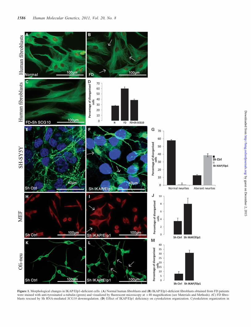
Figure 1. Morphological changes in IKAP/Elp1-deficient cells. (A) Normal human fibroblasts and (B) IKAP/Elp1-deficient fibroblasts obtained from FD patientswere stained with anti-tyrosinated a-tubulin (green) and visualized by fluorescent microscopy at ×40 magnification (see Materials and Methods). (C) FD fibro-blasts rescued by Sh RNA-mediated SCG10 downregulation. (D) Effect of IKAP/Elp1 deficiency on cytoskeleton organization. Cytoskeleton organization in
1586 Human Molecular Genetics, 2011, Vol. 20, No. 8
by guest on Decem
ber 2, 2015http://hm
g.oxfordjournals.org/D
ownloaded from

FD fibroblast cells where a subgroup of genes associated withcell migration and actin cytoskeleton regulation was shown tobe downregulated in a microarray assay (9). It was suggestedthat the Elongator complex plays a role in transcription ofthese genes presumably by its component Elp3 that displayshistone acetylation activity (9–11). In accordance, cellmigration defects were observed in IKAP/Elp1-depletedHeLa, SK-N-BE neuroblastoma, glioblastoma-derived U373and FD fibroblast cells (9). However, in recent studies, down-regulation of these genes was not observed in the cerebrum ofFD patients (12) and in other cells (13,14), leaving open thequestion of the observed effect on migration. It was alsosuggested that the IKAP/Elp1 protein is involved in celladhesion and migration through cytosolic interactions. Thiswas based on experiments in which the depletion of IKAP/Elp1 resulted in decreased staining of filamin A in membraneruffles and in disorganized actin cytoskeleton (13). IKAP/Elp1was also demonstrated to be colocalized with keratin but notwith microtubules (MTs), actin and vimentin in the cytoplasmof HeLa cells (15).
It has recently been shown that MTs, the major dynamicstructural component of the cytoskeleton, could be thetarget of IKAP/Elp1. Contrary to Gardiner et al.’s (15)observation in HeLa cells, Creppe et al. (14) demonstratedthat IKAP/Elp1 and Elp3, in fact, do interact with MTs incortical neurons. Moreover, Creppe et al. (14) provided evi-dence suggesting that the Elongator regulates radialmigration and branching of cortical projection neuronsthrough acetylation of the MT monomer a-tubulin. Whilea-tubulin deacetylases are well characterized (16–18), theenzyme that acetylates a-tubulin was proposed to be Elp3(14). Elp3 was shown to acetylate MT in vitro and IKAP/Elp1 and/or Elp3-deficient HT29 cells, and cortical neuronsshowed a significant decrease in acetylation of a-tubulin.Similar results were observed in C. elegans where the Elon-gator was shown to be critical for MT acetylation (19). Fur-thermore, the amount of Elp3 was found to be at asignificantly decreased level in cells where IKAP/Elp1 wasat lowered levels either by RNAi downregulation in HT29cells or in FD-derived fibroblasts (9,14).
MTs are composed of a/b-tubulin dimers as buildingblocks and involved in several vital processes occurringwithin the cell: development and maintenance of cellshape, cell reproduction, cell signaling, intracellular trans-port, neurite extension and cell migration (20). MTs are par-ticularly abundant in neurons (21) where they accomplishneurite outgrowth by reorganization. MTs show a highlydynamic instability, in which they are either growing(rescue) or shrinking (catastrophe) (22). The polymerizationof MTs is modulated by MT-associated proteins on the
one hand and destabilizing proteins (stathmins) on theother hand. Among the stathmin proteins, superior cervicalganglia neural-specific membrane-associated protein[superior cervical ganglion-10 protein (SCG10) also knownas STMN2) (23) attracted our attention since we found itto be upregulated in IKAP/Elp1-deficient cells. SCG10 is aneural-specific phosphoprotein that plays a critical role inneuronal development through its MT destabilizing activity.SCG10 is found in the Golgi area, along neurites, andenriched in growth cones (23) and plays a role in neuriteoutgrowth (24).
SCG10 is known to increase the rate of MT catastrophe,either through tubulin sequestration by forming a ternarycomplex or by direct stimulation at the plus end of MTs(reviewed in 24,25). Overexpression of SCG10 in COS-7cells, for example, had been shown to cause MT destabiliza-tion and disruption of MT networks (26). The activity ofSCG10 is known to be affected by phosphorylation (27).The MT destabilization function of SCG10 is inhibitedupon its phosphorylation (28). Interestingly, SCG10 wasdemonstrated to be phosphorylated by cJun N-terminalkinase (JNK1), which was shown earlier to be activated byIKAP/Elp1 (5). In addition, phosphorylation of SCG10 atserine residues in positions S62 and S73 is reduced in thebrain of mice lacking JNK1. It was shown that theexpression of SCG10 that could not be phosphorylated atthese two positions by JNK1 significantly inhibited theMTs’ turnover (27). It is noteworthy that the expression ofSCG10 is restricted to neural cells by the RE-1-silencingtranscription factor (REST) protein that is known to inhibitneuronal genes in non-neuronal cells (29).
In our current work, we demonstrate the function of IKAP/Elp1 in MT organization mediated by SCG10. We show thatIKAP/Elp1 deficiency upregulates the MT destabilizingprotein SCG10 and, in parallel, disorganizes the cytoskeletonin SH-SY5Y human neuroblastoma and FD fibroblast cells.The upregulation of SCG10 was also observed in the IKAP/Elp1-deficient cerebrum. The SCG10 elevation in cerebrumand in neuroblastoma IKAP/Elp1-deficient cells appears tobe associated with downregulation of REST that binds to theSCG10 promoter and acts as a specific repressor (29). In fibro-blasts of FD patients and in IKAP/Elp1-deficient mouseembryonic fibroblast (MEF) cells, the SCG10 gene is activatedas well, but in a REST-independent mechanism.
These data provide the first evidence that IKAP/Elp1 isinvolved in regulating the expression of the MT destabilizingprotein SCG10, lending support to the concept that SCG10elevation observed in IKAP/Elp1-deficient cells can alter theMT organization and dynamics which could play a pivotalrole in FD.
normal (N), IKAP/Elp1 deficient (FD) and rescued FD fibroblasts (FD+Sh SCG10). Results represent percentage of cells with disorganized cytoskeleton out oftotal counted cells. (E) SH-SY5Y human neuroblastoma cells transfected with Sh empty vector (Sh Ctrl) and (F) transfected with Sh IKAP/Elp1 plasmid (seeMaterials and Methods) were stained with anti-tyrosinated a-tubulin (green) and visualized by confocal microscopy. (G) Effect of IKAP/Elp1 deficiency onneurites production. Result represent the percent of cells with abnormal neurites out of total counted cells. (H) MEF cells transfected with Sh empty vector(Sh Ctrl), and (I) MEF cells transfected with Sh IKAP/Elp1 plasmid were stained with anti-tyrosinated a-tubulin (red) and visualized by fluorescent microscopyat ×40 magnification. (J) Effect of IKAP/Elp1 deficiency on production of processes in MEF cells. Results represent the percentage of multipolar cells out oftotal counted cells. (K) Oli-neu cells transfected with empty vector (Sh Ctrl). (L) Oli-neu cells transfected with Sh IKAP/Elp1 stained with anti-tyrosinateda-tubulin (green) and visualized by fluorescent microscopy at ×40 magnification. (M) Effect of IKAP/Elp1 deficiency on production of processes inOli-neu cells. Results represent the percent of multipolar cells out of total counted cells.
Human Molecular Genetics, 2011, Vol. 20, No. 8 1587
by guest on Decem
ber 2, 2015http://hm
g.oxfordjournals.org/D
ownloaded from

RESULTS
Recent reports suggested that IKAP/Elp1 is involved in cytos-keleton regulation. Depletion of IKAP/Elp1 resulted indecreased staining of filamin A in membrane ruffles and in dis-organized actin cytoskeleton in MEF cells. IKAP/Elp1 wasalso suggested to be involved in cell adhesion and migrationvia cytosolic interactions (13). Another report suggested thatthe six-subunit RNA polymerase II Elongator complexcontrols the migration of cortical neurons via acetylationof a-tubulin by the Elp3 acetylase component of theElongator (14).
We show here that IKAP/Elp1-deficient fibroblasts of FDpatients stained with a-tubulin antibody revealed morpho-logical aberrations reflected in MT disorganization when
compared with normal fibroblasts (compare Fig. 1A and B,see arrows). These results were validated by quantitativeanalysis that revealed a significant increase (.2-fold) inthe number of cells with disorganized cytoskeleton in FDfibroblasts when compared with normal fibroblasts (N andFD in Fig. 1D). Cytoskeleton disorganization was alsoobserved in IKAP/Elp1 downregulated neuroblastoma cells(Fig. 1F). In the control cells (Fig. 1E), tubulin is restrictedmainly to long-extended neurites, while in a-tubulin-stainedIKAP/Elp1 downregulated cells the tubulin network is con-centrated mostly in one pole of the cells (Fig. 1F, seeupper arrow), providing them with a comet-like structureand occasionally stretches to some of the neurites (Fig. 1F,see lower arrow). In addition, inter-tissue between the cellsof the IKAP/Elp1 downregulated cells is much wider,
Figure 2. SCG10 upregulation in IKAP/Elp1-deficient cells. (A) Expression of SCG10 in the cerebrum of normal individual (N) and FD patient (FD) assayed byreal-time PCR (see Materials and Methods). Results are presented as percent of relative expression, average of three experiments. (B) Western blot analysis ofSCG10 in the cerebrum of the normal (N) and of the FD patient (FD). b-Actin was used as control. (C) Expression of SCG10 assayed by real-time PCR in theSH-SY5Y neuroblastoma cell line transfected with empty vector (Sh Ctrl) and with IKAP/Elp1 Sh RNA (Sh IKAP/Elp1). Values are average of two independentanalyses (see Materials and Methods). (D) Western blot analysis of SCG10 in Sh Ctrl control and IKAP/Elp1 downregulated SH-SY5Y cells (Sh IKAP/Elp1).b-Actin was used as control. (E) Expression of SCG10 assayed by real-time PCR in human normal (N), FD fibroblasts (FD) and SCG10 Sh RNA-treated FDfibroblasts (FD + Sh SCG10). (F) Western blot analysis of SCG10 in normal (N) and (FD) fibroblasts b-actin used as control. (G) Expression of REST in cere-brum of FD patient (FD) and cerebrum of normal individual (N) assayed by real-time PCR and presented as percent of relative expression, average of threeexperiments. (H) Western blot analysis of REST in the cerebrum of the FD patient (FD) and normal individual (N). b-Actin used as control. (I) Expressionof REST assayed by real-time PCR in Sh Ctrl cells and IKAP/Elp1 downregulated SH-SY5Y cells (Sh IKAP/Elp1). (J) Western blot analysis of REST inSh Ctrl and IKAP/Elp1 downregulated SH-SY5Y cells (Sh IKAP/Elp1).
1588 Human Molecular Genetics, 2011, Vol. 20, No. 8
by guest on Decem
ber 2, 2015http://hm
g.oxfordjournals.org/D
ownloaded from
causing enhanced cell-to-cell adhesion (30). This is incontrast to what is seen in the control cells. In conclusion,the IKAP/Elp1 deficiency in SH-SY5Y cells caused asignificant elevation of the number of cells with aberrantneurites in comparison with normal neuroblastoma cells(Fig. 1G).
IKAP/Elp1 deficiency causes disorganization of cytoskele-ton in IKAP/Elp1-deficient MEF cells, as well (compareFigs. 1H and I). This is manifested in more condensedtubulin (Fig. 1I, see arrows) and in abnormal production ofprocesses (Fig. 1J).
IKAP/Elp1-deficient Oli-neu cells (a mouse oligodendro-cyte precursor cell line) showed abnormal staining witha-tubulin antibody. The IKAP/Elp1-depleted Oli-neu cellsdisplayed abnormal rounded shapes and aberrant processes(Fig. 1L, see arrows). This is in contrast to what isobserved in Oli-neu controls (Fig. 1K). In addition, a signifi-cant increase in the number of processes wasobserved (Fig. 1M) similar to what we observed in MEFs(Fig. 1J).
In conclusion, the results presented in Figure 1 show a clearcorrelation between IKAP/Elp1 deficiency and aberrantcytoskeleton organization which is in accord with a previousreport (13).
IKAP/Elp1 deficiency enhances the expression of the MTdestabilizing gene SCG10
We have previously performed a microarray expressionanalysis using RNA extracted from cerebrum of an11-year-old FD male patient and a 47-year-old female FDpatient and RNA extracted from the cerebrum of sex- andage-matched unaffected individuals and identified genesthat changed their expression level (12). One of the upregu-lated genes in the above microarray analysis that attractedour attention was SCG10, also known as STMN2. SCG10is a neuronal growth-associated protein and also known tobe a MT destabilizing protein. More specifically, SCG10 isknown to increase the rate of MT ‘catastrophe’, which rep-resents the conversion from a polymerization (growth) to adepolymerization (shrinkage) state of MTs (reviewed in23,31). Expression of SCG10 is known to be restricted toneurons (32,33). The upregulation of SCG10 was verifiedin the IKAP/Elp1-deficient FD cerebrum on the mRNAlevel by real-time polymerase chain reaction (PCR)(Fig. 2A) and on the protein level by western blot analysis(Fig. 2B). The upregulation of SCG10 was also observedin IKAP/Elp1 Sh RNA-depleted SH-SY5Y human neuroblas-toma cells on the mRNA level (Fig. 2C) and protein level(Fig. 2D). Interestingly, IKAP/Elp1 deficiency in FD fibro-blasts was accompanied by activation of the SCG10 genewhich is practically inactive in the normal fibroblast cells(Fig. 2E). We are intrigued by the fact that the protein ispractically not observed even in the IKAP/Elp1-deficientfibroblasts where SCG10 mRNA is high (Fig. 2F). It is poss-ible that the relative low level of SCG10 mRNA, which wasat least 10-fold lower in fibroblasts than the cerebrum, wastoo low to produce enough protein to be detected bywestern blotting.
Rescue of FD fibroblast phenotype by RNAi-mediatedSCG10 downregulation
To further support the contribution of SCG10 upregulation(Fig. 2E) in the above-presented cytoskeletal disorganization(Fig. 1B), we performed SCG10 Sh RNA downregulation ofSCG10 in FD fibroblast cells (FD+Sh SCG10 in Fig. 2E).Indeed, this treatment caused the significant downregulationto approximately the basal SCG10 level observed in normalfibroblasts (see N in Fig. 2E). The SCG10 downregulationresulted in a rescue of the observed defect in cytoskeletonorganization (Fig. 1C and D). These results indicate thatSCG10 upregulation is, in fact, involved in MT disorganiz-ation in IKAP/Elp1-deficient FD fibroblasts and possibly inother IKAP/Elp1-deficient cells.
Testing a possible mechanism for SCG10 upregulationin IKAP/Elp1-deficient cells
The zinc finger protein REST is a transcriptional repressor thatis known to repress neuronal genes in non-neuronal tissues.One of the REST target genes is SCG10 (29). Since the upre-gulation of the SCG10 gene in the IKAP/Elp1-deficient cere-brum appears to be on the transcriptional level (Fig. 2A) andreflected on the protein level (Fig. 2B), we asked whether adecrease in REST may be the reason for the upregulation ofSCG10 in IKAP/Elp1-deficient cells. To address this question,we performed real-time PCR and western blot analysis ofREST expression in the IKAP/Elp1-deficient cerebrum.Indeed, REST was found to be significantly downregulatedon both the RNA (Fig. 2G) and protein levels (Fig. 2H) inthe cerebrum of FD patient. This result is consistent with theupregulation of SCG10 observed in the IKAP/Elp1 cerebrumsuggesting that the SCG10 gene may be regulated by RESTand that SCG10 may be upregulated in IKAP/Elp1-deficientcells as a result of REST downregulation. Similar resultswere obtained with IKAP/Elp1 downregulated neuroblastomacells. SCG10 was upregulated on the RNA (Fig. 2C) andprotein levels (Fig. 2D), and we were able to demonstrate asignificant downregulation of the REST gene on the RNAlevel (Fig. 2I) and on the protein level (Fig. 2J). In humanFD fibroblasts, although the SCG10 gene was activated(Fig. 2E), no significant changes were observed in the RESTlevel between normal and FD fibroblasts (data not shown),suggesting that the activation of SCG10 in this case is inde-pendent of the REST level.
IKAP/Elp1 and Elp3 are not involved in acetylationof a-tubulin
It was recently reported (14) that in IKAP/Elp1-deficientHT29 and mouse cortical neuronal cells, a-tubulin acetylationis diminished. The decrease in acetylation was in accord with adecrease in the expression of Elp3, the acetyltransferasesubunit of the Elongator complex. It should be mentionedthat depolarization of microtubuli is associated with deacetyla-tion of a/b-tubulin dimers (reviewed in 34). Since the deace-tylation of a-tubulin could have been the reason for thedisorganization of MTs that we observed in IKAP/Elp1-deficient FD fibroblast cells, SH-SY5Y human neuroblastoma,
Human Molecular Genetics, 2011, Vol. 20, No. 8 1589
by guest on Decem
ber 2, 2015http://hm
g.oxfordjournals.org/D
ownloaded from

MEF and Oli-neu cells, stained with a-tubulin antibody(Fig. 1), we performed western blot analyses of acetylateda-tubulin in the IKAP/Elp1-deficient cells (Fig. 3). Surpris-ingly, in contrast to the reported findings in HT29 and inmouse cortical neuronal cells (14), we failed to detect anyreduction in a-tubulin acetylation in FD fibroblasts(Fig. 3A), FD human cerebrum (Fig. 3B), human neuroblas-toma SH-SY5Y cells (Fig. 3C), MEF cells (Fig. 3D) andmouse Oli-neu cells (Fig. 3E). In spite of a substantialdecrease in the acetyltransferase (Elp3) level, in MEF andOli-neu cells, no deacetylation of a-tubulin was observed(Fig. 3D and E). These results demonstrate that the Elp3level does not affect the level of a-tubulin acetylation. Itshould be noted that even in HT29 cells that were used byCreppe et al. (14), no decrease in acetylated tubulin wasdetected (data not shown).
IKAP/Elp1 is involved in adhesion capacityof Oli-neu cells and FD fibroblasts
As mentioned above, IKAP/Elp1-depleted Oli-neu cells showeda disorganized cytoskeleton and abnormal rounded shape(Fig. 1L). Such disorganization can affect the adhesion capacityof the cells. To test this possibility, we carried out a cell
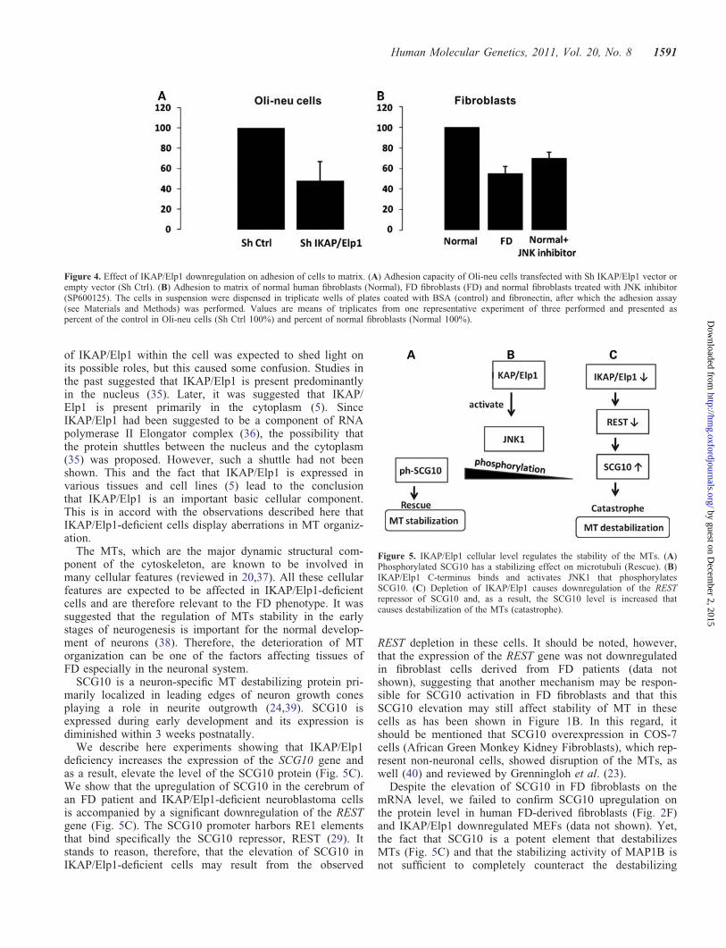
adhesion-to matrix assay. This assay revealed that in IKAP/Elp1-deficient Oli-neu cells, the adhesion capacity to fibronec-tin is reduced by 50% (Fig. 4A). The observed reduced adhesioncapacity of Oli-neu cells can affect the recognition process ofneurons by glial cells, which represents one of the main stepsduring myelination. Next, we asked whether the capacity ofadhesion is also affected in other IKAP/Elp1-deficient cells.For this purpose, we tested the adhesion of FD fibroblasts. A sig-nificant decrease (�50%) in adhesion capacity of these cells hadbeen observed as well (Fig. 4B).
These results are in accord with previous publications,where IKAP/Elp1 was shown to be involved in adhesion ofMEF cells (13) and where IKAP/Elp1-deficient SH-SY5Ycells showed reduced adhesion to lamina (30). We suggestthat the deterioration of the cytoskeleton shown above(Fig. 1) and of actin cytoskeleton that was shown by Johansenet al. (13) can explain the effect of IKAP/Elp1 deficiency oncell adhesion to matrix.
DISCUSSION
Although IKAP/Elp1 was found to be a multifunctionalprotein, the functions that are carried out by IKAP/Elp1 inthe cell are not completely understood. At first, localization
Figure 3. Effect of IKAP/Elp1 deficiency on the level of Elp3 and a-tubulin acetylation. Western blot analysis of IKAP/Elp1, Elp3 and acetylated tubulin(ac-tubulin) was performed in (A). Fibroblasts of normal individuals (N1, N2) and FD patients (FD1, FD2) in (B). Cerebrum of normal male (NF), normalfemale (NC), FD male (FDF) and FD female (FDC) in (C). SH-SY5Y neuroblastoma control (Sh Ctrl) and IKAP/Elp1-deficient (Sh IKAP/Elp1) cellsin (D). MEF control-Sh Ctrl and IKAP/Elp1-deficient (Sh IKAP/Elp1) cells in (E). Mouse oligodendrocyte progenitor (Oli-neu) control (Sh Ctrl) andIKAP/Elp1-deficient (Sh IKAP/Elp1) cells; GAPDH served as control.
1590 Human Molecular Genetics, 2011, Vol. 20, No. 8
by guest on Decem
ber 2, 2015http://hm
g.oxfordjournals.org/D
ownloaded from
of IKAP/Elp1 within the cell was expected to shed light onits possible roles, but this caused some confusion. Studies inthe past suggested that IKAP/Elp1 is present predominantlyin the nucleus (35). Later, it was suggested that IKAP/Elp1 is present primarily in the cytoplasm (5). SinceIKAP/Elp1 had been suggested to be a component of RNApolymerase II Elongator complex (36), the possibility thatthe protein shuttles between the nucleus and the cytoplasm(35) was proposed. However, such a shuttle had not beenshown. This and the fact that IKAP/Elp1 is expressed invarious tissues and cell lines (5) lead to the conclusionthat IKAP/Elp1 is an important basic cellular component.This is in accord with the observations described here thatIKAP/Elp1-deficient cells display aberrations in MT organiz-ation.
The MTs, which are the major dynamic structural com-ponent of the cytoskeleton, are known to be involved inmany cellular features (reviewed in 20,37). All these cellularfeatures are expected to be affected in IKAP/Elp1-deficientcells and are therefore relevant to the FD phenotype. It wassuggested that the regulation of MTs stability in the earlystages of neurogenesis is important for the normal develop-ment of neurons (38). Therefore, the deterioration of MTorganization can be one of the factors affecting tissues ofFD especially in the neuronal system.
SCG10 is a neuron-specific MT destabilizing protein pri-marily localized in leading edges of neuron growth conesplaying a role in neurite outgrowth (24,39). SCG10 isexpressed during early development and its expression isdiminished within 3 weeks postnatally.
We describe here experiments showing that IKAP/Elp1deficiency increases the expression of the SCG10 gene andas a result, elevate the level of the SCG10 protein (Fig. 5C).We show that the upregulation of SCG10 in the cerebrum ofan FD patient and IKAP/Elp1-deficient neuroblastoma cellsis accompanied by a significant downregulation of the RESTgene (Fig. 5C). The SCG10 promoter harbors RE1 elementsthat bind specifically the SCG10 repressor, REST (29). Itstands to reason, therefore, that the elevation of SCG10 inIKAP/Elp1-deficient cells may result from the observed
REST depletion in these cells. It should be noted, however,that the expression of the REST gene was not downregulatedin fibroblast cells derived from FD patients (data notshown), suggesting that another mechanism may be respon-sible for SCG10 activation in FD fibroblasts and that thisSCG10 elevation may still affect stability of MT in thesecells as has been shown in Figure 1B. In this regard, itshould be mentioned that SCG10 overexpression in COS-7cells (African Green Monkey Kidney Fibroblasts), which rep-resent non-neuronal cells, showed disruption of the MTs, aswell (40) and reviewed by Grenningloh et al. (23).
Despite the elevation of SCG10 in FD fibroblasts on themRNA level, we failed to confirm SCG10 upregulation onthe protein level in human FD-derived fibroblasts (Fig. 2F)and IKAP/Elp1 downregulated MEFs (data not shown). Yet,the fact that SCG10 is a potent element that destabilizesMTs (Fig. 5C) and that the stabilizing activity of MAP1B isnot sufficient to completely counteract the destabilizing
Figure 5. IKAP/Elp1 cellular level regulates the stability of the MTs. (A)Phosphorylated SCG10 has a stabilizing effect on microtubuli (Rescue). (B)IKAP/Elp1 C-terminus binds and activates JNK1 that phosphorylatesSCG10. (C) Depletion of IKAP/Elp1 causes downregulation of the RESTrepressor of SCG10 and, as a result, the SCG10 level is increased thatcauses destabilization of the MTs (catastrophe).
Figure 4. Effect of IKAP/Elp1 downregulation on adhesion of cells to matrix. (A) Adhesion capacity of Oli-neu cells transfected with Sh IKAP/Elp1 vector orempty vector (Sh Ctrl). (B) Adhesion to matrix of normal human fibroblasts (Normal), FD fibroblasts (FD) and normal fibroblasts treated with JNK inhibitor(SP600125). The cells in suspension were dispensed in triplicate wells of plates coated with BSA (control) and fibronectin, after which the adhesion assay(see Materials and Methods) was performed. Values are means of triplicates from one representative experiment of three performed and presented aspercent of the control in Oli-neu cells (Sh Ctrl 100%) and percent of normal fibroblasts (Normal 100%).
Human Molecular Genetics, 2011, Vol. 20, No. 8 1591
by guest on Decem
ber 2, 2015http://hm
g.oxfordjournals.org/D
ownloaded from

effect of SCG10 (40) suggest that a relatively low level ofSCG10 may be sufficient to destabilize MTs.
MT destabilizing activity displayed by SCG10 is regulatedby phosphorylation (reviewed in 23) (Fig. 5B). Interestingly,the serine residues S62 and S73 that are phosphorylated byJNK1 (27) had previously been suggested to be potentiallyactivated by IKAP/Elp1 (5). It has also been proposed thatIKAP/Elp1 may be important as a physiological regulator ofthe stress-associated JNK signaling pathway (5). Interestingly,inhibition of JNK by the specific JNK inhibitor SP600125(A.G. Scientific, Inc.) in normal fibroblast cells also causedsignificant adhesion impairment (Fig. 4B). These data indicatethat JNK inhibition results in a decrease of adhesion similar tothe effect of IKAP/Elp1 deficiency, suggesting that IKAP/Elp1affects adhesion through JNK. It should be noted that the poss-ible interaction between IKAP/Elp1, JNK and SCG10 hadbeen proposed by Gardiner et al. (15).
One of the genes that was found to be downregulated in amicroarray assay performed with cerebrum RNA of FD patientswas the axonal guidance protein Fez1 (12). Fez 1 together withthe JNK-interacting protein 1 bind to the Kinesin-1 motors andactivate MT-based motility (reviewed in 41). Thus, IKAP/Elp1deficiency may affect not only the MT stability via the MTdestabilizing protein SCG10 and JNK, but also theMT-dependent cell motility by the downregulation of Fez1.
Since the level of SCG10 is elevated in IKAP/Elp1-deficientcells, it can shift the equilibrium of MTs stability toward cat-astrophe and MT disruption. This in turn can reduce brain-derived neurotrophic factor (BDNF) transport through MTs.BDNF is a neurotrophic factor which is required in neuronaldevelopmental processes such as the development of tastepapillae in the fetus. This is interesting since FD patients areknown to lack taste papillae (42).
Intriguingly, SCG10 was recently demonstrated to be on thelist of downregulated genes during differentiation of FD iPScells toward neural crest lineages (43). These results, whichare contradictory to ours, may be attributed to the differentexperimental systems used by Lee et al. (43) and by us. Inany event, it demonstrates the importance of this protein inthe FD phenotype.
SCG10 is known to be upregulated during regeneration ofinjured postganglionic sympathetic nerve (44). Thus, the upre-gulation of SCG10 which we observed in IKAP/Elp1-deficientcells could reflect compensatory mechanisms that try to acti-vate restoration of the nervous system which may be relevantto the FD etiology.
The reduced adhesion abilities that we observed in IKAP/Elp1-deficient cells (Fig. 4) may be a result of our observed MTs desta-bilization mediated by SCG10 upregulation. Obviously, thisrequires further investigation. Interestingly, the adhesion defectwas shown to be rescued by reintroduction of the IKAP/Elp1expression vector to IKAP/Elp1 downregulated MEF cells (13).The migration capacity of IKAP/Elp1-deficient cells was alsoreported to be reduced (9,13,14). We propose that both effectson adhesion and migration are a result of the cytoskeleton aberra-tions affected by IKAP/Elp1 deficiency.
SCG10 regulates the cytoskeleton by destabilizing the MT,through altering dendrites as well as cell shape, size and polarity.Alterations in cellular architecture could explain some of themorphological changes reported by us here and by others (13,14).
MATERIALS AND METHODS
Tissues and cells
Oli-neu cells (gift from J. Trotter, University of Mainz) (45)were grown on poly-L-lysine-coated culture dishes in Satomedium consisting of Dulbecco modified Eagle medium(DMEM) supplemented with 2 mg/ml NaHCO3, 2 mM
L-glutamine, 10 mg/ml apotransferine, 10 mg/ml insulin,100 mM putrescine, 200 nm progesterone, 500 nm triiodo-L-thyronine, 220 nm sodium selenite, 520 mM L-thyroxin, 1%horse serum, gentamicin. Fibroblast cell lines (either clonedby us or obtained from Coriell Cell Repositories), 293Thuman embryo kidney cells and MEF cells were grown inDMEM in the presence of 10% fetal bovine serum, 5%glutamine and 1% penicillin–streptomycin.
SH-SY5Y human neuroblastoma cells were grown inmedium containing DMEM with 5% glutamine and 10%heat inactivated FCS, 2 mM sodium pyruvate (Invitrogen)and antibiotics (50 U/ml of penicillin, streptomycin andnystatin).
All reagents were purchased from Biological Industries,Israel.
All cells were maintained at 378C with 5% CO2 in a humidatmosphere.
Brain tissues were obtained from the NICHD Brain andTissue Bank for Developmental Disorders at the Universityof Maryland, Baltimore, MA, USA. Tissues used for thisstudy were the cerebrum of an FD 11-year-old patient(UMB# M3697M) and a 47-year-old FD female (UMB#M3783M) and age- and sex-matched normal brain (UMB#616 and UMB# 1910, respectively).
RNA interference treatment
To suppress IKBKAP gene expression lentiviral (pLKO) vectorwas used. Five different double-stranded IKBKAP shRNA,accession number NM_026079 (for mouse cells) andNM_003640 (for human cells) were purchased fromSigma-Aldrich (Israel). Plasmids were transformed byFuGene6 (Roche) into 293T cells according to the manufac-turer’s protocol. Lentiviruses were assembled using the follow-ing three vectors: pLKO (transfer vector)—includes the insert(Sigma); PHR—lentivirus polymerase; VSVG—virus envel-ope. The day before transfection, 1.2 × 10⁶ HEK293T cellswere plated in a 10 cm dish (20–30% confluence). Next day,three vectors were transfected at the following ratioPHR:VSVG:LVTR1 ¼ 9:1:10. Cells were incubated for 48 hfollowed by collection of the medium containing the virus fil-tered and used to infect the target cells. Selection with the1 mg/ml puromycin (Sigma) was started after 48 h post-infection. SCG10 Sh RNA was kindly provided by Elena Ain-binder (Prof. Michel Revel’s laboratory).
Adhesion assay
7The adhesion assay was carried out as described earlier (46).Cells were detached with minimal trypsinization, washed andresuspended in serum-free DMEM medium supplementedwith 1 mg/ml bovine serum albumin (BSA). Cell suspension(100 ml containing 1 × 10⁵ cells) was added to the wells of
1592 Human Molecular Genetics, 2011, Vol. 20, No. 8
by guest on Decem
ber 2, 2015http://hm
g.oxfordjournals.org/D
ownloaded from

96-well titer plates precoated with fibronectin and BSA and wasallowed to adhere for 1 h at 378C. Non-adherent cells wererinsed off with PBS containing 2 mM CaCl2, 1 mM MgCl2(warmed to 378C), and adherent cells were fixed and stainedin 2% toluidine blue/4% paraformaldehyde (PFA) (30 min at378C). Wells were then rinsed in tap water. Stained cells weresolubilized in 100 ml 1% sodium dodecyl sulfate (SDS), andabsorbance of the solution was measured at 595 nm in a micro-plate reader. Experiments were performed in triplicates andrepeated at least three times.
Antibodies for western blotting and immunostaining
Antibodies for western blotting and immunostaining were pur-chased from the following companies:
Anti IKAP Rabbit polyclonal IgG (Anaspec) Cat no. 54494;Anti glyceraldehyde-3-phosphate dehydrogenase (GAPDH)
monoclonal peroxidase (Sigma) Cat no. G9295;Ant b-actin monoclonal (Sigma) Cat no. A5411;Anti a-tubulin clone YL1/2 (Tyr-tubulin) Rat monoclonal
(Serotec) Cat no. MCA77G;Anti acetylated Mouse monoclonal (Clone [6–11B-1]a-tubulin (Santa Cruz) Cat no. sc–23950;
Anti REST Rabbit polyclonal (Milipore) Cat no. 07-579;Anti SCG10 and anti Elp3 were kindly obtained from Dr
Andre Sobel and Dr Jesper Svejstrup, respectively.
All secondary antibodies including Donkey anti Rat Cy2conjugated and Donkey anti-Rabbit Cy3 conjugated were pur-chased at Jackson IR.
Western immunoblotting
Cells were lysed in radioimmunoprecipitation assay buffer(150 mM NaCl, 0.1% SDS, 0.5% sodium deoxycholate,50 mM Tris–HCl pH 7.5, 1% NP-40, 1 mM phenylmethylsul-fonyl fluoride). The protein concentration of the soluble frac-tion was measured by the Bradford assay. Total cell lysateswere resolved by 8–12% SDS–polyacrylamide gel electro-phoresis and transferred to a nitrocellulose membrane. Afterstaining the membranes with reversible Ponceau red solutionto confirm equal protein loading, the membranes wereblocked with 5% Skim Milk in PBS (0.1% Tween-20) for1 h at room temperature. The membranes were subsequentlyincubated in PBS with 0.1% Tween-20 at room temperaturefor 1 h with the relevant primary antibodies. After threewashes in PBS 0.1% Tween-20, the blots were incubatedwith appropriate secondary antibodies (final antibody concen-tration was according to the manufacturer’s recommendation).
Real-time PCR
Total RNA was prepared from cells in culture using theTri-reagent (Sigma) according to the manufacturer’s protocol.Approximately 1 mg total RNA of each sample was reversetranscribed using the M-MLV Reverse Transcriptase(Promega) and random primers in a 20 ml reaction mixture.The reverse transcription reactions were carried out at 428Cfor 30 min. For each gene, the real-time PCR analysis was per-formed in triplicates with 2 ml of cDNA, primers (sequences
available upon request) at a concentration of 0.3 mM. TheSYBR Green (Applied Biosystems) was added to the 20 mlreaction mixture. An ABI PRISM 5700 Sequence DetectionSystem (Applied Biosystems) was programmed as follows:one cycle of 10 min at 958C, followed by 40 cycles of 15 sat 958C and 1 min at 608C. Data were analyzed using the7500 SDS system software. To control for the amount ofRNA, amplification of TATA box binding protein in mouseand 18S in human were performed and corrected for eachcDNA sample.
PCR analysis of IKBKAP expression
PCR analysis was carried out as described earlier (12).
Immunostaining
For staining, the cells were fixed in 4% PFA at room tempera-ture for 10 min, washed once in PBS. After permeabilizationin 1% Triton X-100 and 1% BSA in PBS for 5 min at roomtemperature, sections were washed twice for 15 min in PBSand blocked in 1% BSA in PBS for 20 min at room tempera-ture and incubated with primary antibody in blocking solutionat 48C overnight. The sections were then each washed twicewith PBS for 5 min and incubated with appropriate secondaryantibody for 45 min in blocking solution, washed twice withPBS for 5 min and mounted in Galvenol.
ACKNOWLEDGEMENTS
We thank Dr Mark Tarshish for his help with the microscopy,Dr Simcha Urieli-Shoval for her help with the adhesion assayand Ms Yevgenia Cheishvili for technical assistance, Dr AndreSobel for providing us with the SCG10 antibody and DrSvejstrup with the Elp3 antibody.
Conflict of Interest statement. None declared.
FUNDING
This work was partially supported by a donation fromMr Joseph Mizrahi, by the Dysautonomia Foundation, Inc.and by the Israeli Familial Dysautonomia Organization.
REFERENCES
1. Cuajungco, M.P., Leyne, M., Mull, J., Gill, S.P., Lu, W., Zagzag, D.,Axelrod, F.B., Maayan, C., Gusella, J.F. and Slaugenhaupt, S.A. (2003)Tissue-specific reduction in splicing efficiency of IKBKAP due to themajor mutation associated with familial dysautonomia. Am. J. Hum.Genet., 72, 749–758.
2. Anderson, S.L., Coli, R., Daly, I.W., Kichula, E.A., Rork, M.J., Volpi,S.A., Ekstein, J. and Rubin, B.Y. (2001) Familial dysautonomia is causedby mutations of the IKAP gene. Am. J. Hum. Genet., 68, 753–758.
3. Slaugenhaupt, S.A., Blumenfeld, A., Gill, S.P., Leyne, M., Mull, J.,Cuajungco, M.P., Liebert, C.B., Chadwick, B., Idelson, M., Reznik, L.et al. (2001) Tissue-specific expression of a splicing mutation in theIKBKAP gene causes familial dysautonomia. Am. J. Hum. Genet., 68,598–605.
4. Leyne, M., Mull, J., Gill, S.P., Cuajungco, M.P., Oddoux, C., Blumenfeld,A., Maayan, C., Gusella, J.F., Axelrod, F.B. and Slaugenhaupt, S.A.
Human Molecular Genetics, 2011, Vol. 20, No. 8 1593
by guest on Decem
ber 2, 2015http://hm
g.oxfordjournals.org/D
ownloaded from

(2003) Identification of the first non-Jewish mutation in familialDysautonomia. Am. J. Med. Genet., 118, 305–308.
5. Holmberg, C., Katz, S., Lerdrup, M., Herdegen, T., Jaattela, M.,Aronheim, A. and Kallunki, T. (2002) A novel specific role for I kappa Bkinase complex-associated protein in cytosolic stress signaling. J. Biol.Chem., 277, 31918–31928.
6. Rahl, P.B., Chen, C.Z. and Collins, R.N. (2005) Elp1p, the yeast homologof the FD disease syndrome protein, negatively regulates exocytosisindependently of transcriptional elongation. Mol. Cell, 17, 841–853.
7. Esberg, A., Huang, B., Johansson, M.J. and Bystrom, A.S. (2006)Elevated levels of two tRNA species bypass the requirement for elongatorcomplex in transcription and exocytosis. Mol. Cell, 24, 139–148.
8. Mehlgarten, C., Jablonowski, D., Wrackmeyer, U., Tschitschmann, S.,Sondermann, D., Jager, G., Gong, Z., Bystrom, A.S., Schaffrath, R. andBreunig, K.D. (2010) Elongator function in tRNA wobble uridinemodification is conserved between yeast and plants. Mol. Microbiol., 76,1082–1094.
9. Close, P., Hawkes, N., Cornez, I., Creppe, C., Lambert, C.A., Rogister, B.,Siebenlist, U., Merville, M.P., Slaugenhaupt, S.A., Bours, V. et al. (2006)Transcription impairment and cell migration defects in elongator-depletedcells: implication for familial dysautonomia. Mol. Cell, 22, 521–531.
10. Otero, G., Fellows, J., Li, Y., de Bizemont, T., Dirac, A.M., Gustafsson,C.M., Erdjument-Bromage, H., Tempst, P. and Svejstrup, J.Q. (1999)Elongator, a multisubunit component of a novel RNA polymerase IIholoenzyme for transcriptional elongation. Mol. Cell, 3, 109–118.
11. Wittschieben, B.O., Otero, G., de Bizemont, T., Fellows, J.,Erdjument-Bromage, H., Ohba, R., Li, Y., Allis, C.D., Tempst, P. andSvejstrup, J.Q. (1999) A novel histone acetyltransferase is anintegral subunit of elongating RNA polymerase II holoenzyme. Mol. Cell,4, 123–128.
12. Cheishvili, D., Maayan, C., Smith, Y., Ast, G. and Razin, A. (2007) IKAP/hELP1 deficiency in cerebrum of Familial Dysautonomia patients resultsin down regulation of genes involved in oligodendrocyte differentiationand in myelination. Hum. Mol. Genet., 16, 2097–2104.
13. Johansen, L.D., Naumanen, T., Knudsen, A., Westerlund, N., Gromova, I.,Junttila, M., Nielsen, C., Bottzauw, T., Tolkovsky, A., Westermarck, J.et al. (2008) IKAP localizes to membrane ruffles with filamin A andregulates actin cytoskeleton organization and cell migration. J. Cell Sci.,121, 854–864.
14. Creppe, C., Malinouskaya, L., Volvert, M.L., Gillard, M., Close, P.,Malaise, O., Laguesse, S., Cornez, I., Rahmouni, S., Ormenese, S. et al.(2009) Elongator controls the migration and differentiation of corticalneurons through acetylation of alpha-tubulin. Cell, 136, 551–564.
15. Gardiner, J., Barton, D., Marc, J. and Overall, R. (2007) Potential role oftubulin acetylation and microtubule-based protein trafficking in familialdysautonomia. Traffic, 8, 1145–1149.
16. Hubbert, C., Guardiola, A., Shao, R., Kawaguchi, Y., Ito, A., Nixon, A.,Yoshida, M., Wang, X.F. and Yao, T.P. (2002) HDAC6 is amicrotubule-associated deacetylase. Nature, 417, 455–458.
17. North, B.J., Marshall, B.L., Borra, M.T., Denu, J.M. and Verdin, E. (2003)The human Sir2 ortholog, SIRT2, is an NAD+-dependent tubulindeacetylase. Mol. Cell, 11, 437–444.
18. Zhang, Y., Li, N., Caron, C., Matthias, G., Hess, D., Khochbin, S. andMatthias, P. (2003) HDAC-6 interacts with and deacetylates tubulin andmicrotubules in vivo. EMBO J., 22, 1168–1179.
19. Solinger, J.A., Paolinelli, R., Kloss, H., Scorza, F.B., Marchesi, S.,Sauder, U., Mitsushima, D., Capuani, F., Sturzenbaum, S.R. and Cassata,G. (2010) The Caenorhabditis elegans Elongator complex regulatesneuronal alpha-tubulin acetylation. PLoS Genet., 6, e1000820.
20. Gelfand, V.I. and Bershadsky, A.D. (1991) Microtubule dynamics:mechanism, regulation, and function. Annu. Rev. Cell. Biol., 7, 93–116.
21. Ellisman, M.H. and Porter, K.R. (1980) Microtrabecular structure of theaxoplasmic matrix: visualization of cross-linking structures and theirdistribution. J. Cell. Biol., 87, 464–479.
22. Desai, A. and Mitchison, T.J. (1997) Microtubule polymerizationdynamics. Annu. Rev. Cell Dev. Biol., 13, 83–117.
23. Grenningloh, G., Soehrman, S., Bondallaz, P., Ruchti, E. and Cadas, H.(2004) Role of the microtubule destabilizing proteins SCG10 and stathminin neuronal growth. J. Neurobiol., 58, 60–69.
24. Riederer, B.M., Pellier, V., Antonsson, B., Di Paolo, G., Stimpson, S.A.,Lutjens, R., Catsicas, S. and Grenningloh, G. (1997) Regulation ofmicrotubule dynamics by the neuronal growth-associated protein SCG10.Proc. Natl Acad. Sci. USA, 94, 741–745.
25. Manna, T., Grenningloh, G., Miller, H.P. and Wilson, L. (2007) Stathminfamily protein SCG10 differentially regulates the plus and minus enddynamics of microtubules at steady state in vitro: implications for its rolein neurite outgrowth. Biochemistry, 46, 3543–3452.
26. Antonsson, B., Kassel, D.B., Di Paolo, G., Lutjens, R., Riederer, B.M. andGrenningloh, G. (1998) Identification of in vitro phosphorylation sites inthe growth cone protein SCG10. Effect of phosphorylation site mutants onmicrotubule-destabilizing activity. J. Biol. Chem., 273, 8439–8446.
27. Tararuk, T., Ostman, N., Li, W., Bjorkblom, B., Padzik, A., Zdrojewska,J., Hongisto, V., Herdegen, T., Konopka, W., Courtney, M.J. et al. (2006)JNK1 phosphorylation of SCG10 determines microtubule dynamics andaxodendritic length. J. Cell Biol., 173, 265–277.
28. Mori, N. and Morii, H. (2002) SCG10-related neuronal growth-associatedproteins in neural development, plasticity, degeneration, and aging.J. Neurosci. Res., 70, 264–273.
29. Mori, N., Schoenherr, C., Vandenbergh, D.J. and Anderson, D.J. (1992) Acommon silencer element in the SCG10 and type II Na+ channel genesbinds a factor present in nonneuronal cells but not in neuronal cells.Neuron, 9, 45–54.
30. Cohen-Kupiec, R., Weinstein, S., Kantor, G., Peer, D. and Weil, M.(2010) IKAP/hELP1 downregulation in neuroblastoma cells causesenhanced cell adhesion mediated by contactin overexpression. Cell. Adh.Migr., 4, 1–10.
31. Conde, C. and Caceres, A. (2009) Microtubule assembly, organization anddynamics in axons and dendrites. Nat. Rev. Neurosci., 10, 319–332.
32. Mori, N., Stein, R., Sigmund, O. and Anderson, D.J. (1990) A celltype-preferred silencer element that controls the neural-specific expressionof the SCG10 gene. Neuron, 4, 583–594.
33. Wuenschell, C.W., Mori, N. and Anderson, D.J. (1990) Analysis ofSCG10 gene expression in transgenic mice reveals that neural specificityis achieved through selective derepression. Neuron, 4, 595–602.
34. Janke, C. and Kneussel, M. (2010) Tubulin post-translationalmodifications: encoding functions on the neuronal microtubulecytoskeleton. Trends Neurosci., 33, 362–372.
35. Hawkes, N.A., Otero, G., Winkler, G.S., Marshall, N., Dahmus, M.E.,Krappmann, D., Scheidereit, C., Thomas, C.L., Schiavo, G.,Erdjument-Bromage, H. et al. (2002) Purification and characterization ofthe human elongator complex. J. Biol. Chem., 277, 3047–3052.
36. Svejstrup, J.Q. (2007) Elongator complex: how many roles does it play?Curr. Opin. Cell Biol., 19, 331–336.
37. Nogales, E. (2001) Structural insight into microtubule function. Annu.Rev. Biophys. Biomol. Struct., 30, 397–420.
38. Yamada, K., Matsuzaki, S., Hattori, T., Kuwahara, R., Taniguchi, M.,Hashimoto, H., Shintani, N., Baba, A., Kumamoto, N., Yoshikawa, T.et al. (2010) Increased stathmin1 expression in the dentate gyrus of micecauses abnormal axonal arborizations. PLoS ONE, 5, e8596.
39. Stein, R., Mori, N., Matthews, K., Lo, L.C. and Anderson, D.J. (1988) TheNGF-inducible SCG10 mRNA encodes a novel membrane-bound proteinpresent in growth cones and abundant in developing neurons. Neuron, 1,463–476.
40. Bondallaz, P., Barbier, A., Soehrman, S., Grenningloh, G. and Riederer,B.M. (2006) The control of microtubule stability in vitro and in transfectedcells by MAP1B and SCG10. Cell Motil. Cytoskeleton, 63, 681–695.
41. Hammond, J.W., Cai, D. and Verhey, K.J. (2008) Tubulin modificationsand their cellular functions. Curr. Opin. Cell Biol., 20, 71–76.
42. Smith, A., Farbman, A. and Dancis, J. (1965) Absence of taste-budpapillae in familial dysautonomia. Science, 147, 1040–1041.
43. Lee, G., Papapetrou, E.P., Kim, H., Chambers, S.M., Tomishima, M.J.,Fasano, C.A., Ganat, Y.M., Menon, J., Shimizu, F., Viale, A. et al. (2009)Modelling pathogenesis and treatment of familial dysautonomia usingpatient-specific iPSCs. Nature, 461, 402–406.
44. Anderson, D.J. (1989) Development and plasticity of a neural crestderived neuroendocrine sublineage. Alan R. Liss, Inc. The Assembly ofthe Nervous System. pp. 17–36.
45. Jung, M., Kramer, E., Grzenkowski, M., Tang, K., Blakemore, W.,Aguzzi, A., Khazaie, K., Chlichlia, K., von Blankenfeld, G., Kettenmann,H. et al. (1995) Lines of murine oligodendroglial precursor cellsimmortalized by an activated neu tyrosine kinase show distinct degrees ofinteraction with axons in vitro and in vivo. Eur. J. Neurosci., 7, 1245–1265.
46. Urieli-Shoval, S., Shubinsky, G., Linke, R.P., Fridkin, M., Tabi, I. andMatzner, Y. (2002) Adhesion of human platelets to serum amyloid A.Blood, 99, 1224–1229.
1594 Human Molecular Genetics, 2011, Vol. 20, No. 8
by guest on Decem
ber 2, 2015http://hm
g.oxfordjournals.org/D
ownloaded from
Copyright © 2022 FDOKUMEN