Genetic Basis of Familial Valvular Heart Disease
12
569 V alvular heart disease (VHD) is a major cause of morbid- ity and premature death from cardiovascular diseases, making it an important clinical entity. Despite a dramatic decline in the incidence of rheumatic heart disease in indus- trialized countries, VHD remains highly prevalent. Although many VHDs are acquired during adult life, familial cluster- ing and heritability have been noted for common heart valve defects, such as bicuspid aortic valve and myxomatous mitral valve prolapse, denoting an underlying genetic basis. Over the past decade, advances in our understanding of the genetic basis of familial VHD have been made through the unravel- ing of gene network and molecular mechanisms regulating normal valve development. Important progress has also arisen from a series of elegant studies that have focused on link- age analyses of large families with VHD, transgenic animal models, in vitro studies, and, more recently, microRNA and transcriptomic assessment of diseased tissues. Identification of the genes and molecular pathways responsible for the development of VHD has important implications in terms of improving current therapeutic strategies, as well as guiding the management of at-risk family members, with the ulti- mate aim to reduce the health burden of VHD. This article will summarize the current state of knowledge regarding the genetic basis of 2 common familial VHDs, namely mitral valve prolapse and bicuspid aortic valve, and highlight some of the recent findings that shed light on the pathogenesis of these diseases. Valvular heart disease (VHD) is a major cause of disabil- ity, diminished quality of life, and premature death from car- diovascular disease, 1 making it an important clinical entity. Despite a dramatic decline in the incidence of rheumatic heart disease in industrialized countries, VHD remains highly prevalent. 2 Although many VHDs are acquired during adult life, congenital forms present with abnormal valve struc- tures at birth, yet may not manifest as valvular dysfunction and disease until later in life. 3,4 Furthermore, familial cluster- ing and heritability have been noted for common heart valve defects, such as bicuspid aortic valve (BAV) and myxomatous mitral valve prolapse (MVP), implying an underlying genetic basis. 3,5,6 These familial VHDs can occur in isolation (nonsyn- dromic) or present as part of a clinical genetic syndrome, such as Marfan, Turner, and Noonan syndromes. 7 Traditionally, well-characterized multigeneration families have been invaluable for determining the genetic basis of disease, because they are amenable to marker-based genome- wide linkage analysis. However, the identification of causal genes in VHD has been hampered by complex genetic and phenotypic heterogeneity, incomplete penetrance, and the likely contribution of genetic modifier loci. Although in vitro studies and transgenic animal models that recapitulate the human phenotype have provided insights into the genetic basis of VHD, these findings do not always translate to humans. Therefore, concurrent studies on the molecular pathways reg- ulating valvulogenesis have sought to highlight gene networks relevant to VHD. This review article will focus primarily on the 2 most com- mon isolated VHDs, ie, MVP and BAV. The review will sum- marize the current knowledge regarding the genetic basis of MVP and BAV and highlight some of the recent findings that shed light on the pathogenesis of these diseases. Although there is much more to discover about the genetic causes of VHD, it is timely to review the current state of knowledge in these important cardiovascular genetic disorders. Overview of Development of Cardiac Valves A key aspect of cardiac development is the formation and function of the cardiac valves. Cardiac valve development begins immediately after cardiac looping, at embryonic days 31 to 35 in humans, with the formation of endocardial cushions in the atrioventricular canal and outflow tract. 8,9 Normal uridine diphosphate glucose dehydrogenase gene activity to synthesize glycosaminoglycan is critical for the early cell signaling events that establish the boundaries of these cushion-forming regions. 10 Cardiac cushion formation involves the transformation of a subset of endothelial cells into mesenchymal cells in the cushion-forming area, which is induced by bone morphogenetic protein-2 (BMP-2) signals derived from adjacent myocardial cells. 8 The mesenchymal progenitor cells migrate into the intervening cardiac jelly and proliferate to form swellings of valve primordial, which eventually give rise to valvuloseptal structures and adult valvular interstitial cells. 8,11–13 The atrioventricular cushions contribute to atrioventricular (mitral and tricuspid) valve leaflets, whereas the outflow tract cushions contribute to (Circ Cardiovasc Genet. 2012;5:569-580.) © 2012 American Heart Association, Inc. Circ Cardiovasc Genet is available at http://circgenetics.ahajournals.org DOI: 10.1161/CIRCGENETICS.112.962894 From the Agnes Ginges Centre for Molecular Cardiology, Centenary Institute, Sydney, Australia (R.P., R.D.B., C.S.); Sydney Medical School, University of Sydney, Sydney, Australia (R.P., C.S.); and Department of Cardiology, Royal Prince Alfred Hospital, Sydney, Australia (R.P., C.S.). Correspondence to Christopher Semsarian, MBBS, PhD, FRACP, Agnes Ginges Centre of Molecular Cardiology, Centenary Institute, Camperdown, NSW 2050, Australia. E-mail [email protected] Genetic Basis of Familial Valvular Heart Disease Ratnasari Padang, MBBS, FRACP; Richard D. Bagnall, PhD; Christopher Semsarian, MBBS, FRACP, PhD Advances in Genetics
Transcript of Genetic Basis of Familial Valvular Heart Disease

569
Valvular heart disease (VHD) is a major cause of morbid-ity and premature death from cardiovascular diseases,
making it an important clinical entity. Despite a dramatic decline in the incidence of rheumatic heart disease in indus-trialized countries, VHD remains highly prevalent. Although many VHDs are acquired during adult life, familial cluster-ing and heritability have been noted for common heart valve defects, such as bicuspid aortic valve and myxomatous mitral valve prolapse, denoting an underlying genetic basis. Over the past decade, advances in our understanding of the genetic basis of familial VHD have been made through the unravel-ing of gene network and molecular mechanisms regulating normal valve development. Important progress has also arisen from a series of elegant studies that have focused on link-age analyses of large families with VHD, transgenic animal models, in vitro studies, and, more recently, microRNA and transcriptomic assessment of diseased tissues. Identification of the genes and molecular pathways responsible for the development of VHD has important implications in terms of improving current therapeutic strategies, as well as guiding the management of at-risk family members, with the ulti-mate aim to reduce the health burden of VHD. This article will summarize the current state of knowledge regarding the genetic basis of 2 common familial VHDs, namely mitral valve prolapse and bicuspid aortic valve, and highlight some of the recent findings that shed light on the pathogenesis of these diseases.
Valvular heart disease (VHD) is a major cause of disabil-ity, diminished quality of life, and premature death from car-diovascular disease,1 making it an important clinical entity. Despite a dramatic decline in the incidence of rheumatic heart disease in industrialized countries, VHD remains highly prevalent.2 Although many VHDs are acquired during adult life, congenital forms present with abnormal valve struc-tures at birth, yet may not manifest as valvular dysfunction and disease until later in life.3,4 Furthermore, familial cluster-ing and heritability have been noted for common heart valve defects, such as bicuspid aortic valve (BAV) and myxomatous mitral valve prolapse (MVP), implying an underlying genetic basis.3,5,6 These familial VHDs can occur in isolation (nonsyn-dromic) or present as part of a clinical genetic syndrome, such as Marfan, Turner, and Noonan syndromes.7
Traditionally, well-characterized multigeneration families have been invaluable for determining the genetic basis of disease, because they are amenable to marker-based genome-wide linkage analysis. However, the identification of causal genes in VHD has been hampered by complex genetic and phenotypic heterogeneity, incomplete penetrance, and the likely contribution of genetic modifier loci. Although in vitro studies and transgenic animal models that recapitulate the human phenotype have provided insights into the genetic basis of VHD, these findings do not always translate to humans. Therefore, concurrent studies on the molecular pathways reg-ulating valvulogenesis have sought to highlight gene networks relevant to VHD.
This review article will focus primarily on the 2 most com-mon isolated VHDs, ie, MVP and BAV. The review will sum-marize the current knowledge regarding the genetic basis of MVP and BAV and highlight some of the recent findings that shed light on the pathogenesis of these diseases. Although there is much more to discover about the genetic causes of VHD, it is timely to review the current state of knowledge in these important cardiovascular genetic disorders.
Overview of Development of Cardiac ValvesA key aspect of cardiac development is the formation and function of the cardiac valves. Cardiac valve development begins immediately after cardiac looping, at embryonic days 31 to 35 in humans, with the formation of endocardial cushions in the atrioventricular canal and outflow tract.8,9 Normal uridine diphosphate glucose dehydrogenase gene activity to synthesize glycosaminoglycan is critical for the early cell signaling events that establish the boundaries of these cushion-forming regions.10 Cardiac cushion formation involves the transformation of a subset of endothelial cells into mesenchymal cells in the cushion-forming area, which is induced by bone morphogenetic protein-2 (BMP-2) signals derived from adjacent myocardial cells.8 The mesenchymal progenitor cells migrate into the intervening cardiac jelly and proliferate to form swellings of valve primordial, which eventually give rise to valvuloseptal structures and adult valvular interstitial cells.8,11–13 The atrioventricular cushions contribute to atrioventricular (mitral and tricuspid) valve leaflets, whereas the outflow tract cushions contribute to
(Circ Cardiovasc Genet. 2012;5:569-580.)© 2012 American Heart Association, Inc.
Circ Cardiovasc Genet is available at http://circgenetics.ahajournals.org DOI: 10.1161/CIRCGENETICS.112.962894
From the Agnes Ginges Centre for Molecular Cardiology, Centenary Institute, Sydney, Australia (R.P., R.D.B., C.S.); Sydney Medical School, University of Sydney, Sydney, Australia (R.P., C.S.); and Department of Cardiology, Royal Prince Alfred Hospital, Sydney, Australia (R.P., C.S.).
Correspondence to Christopher Semsarian, MBBS, PhD, FRACP, Agnes Ginges Centre of Molecular Cardiology, Centenary Institute, Camperdown, NSW 2050, Australia. E-mail [email protected]
Genetic Basis of Familial Valvular Heart DiseaseRatnasari Padang, MBBS, FRACP; Richard D. Bagnall, PhD;
Christopher Semsarian, MBBS, FRACP, PhD
Advances in Genetics

570 Circ Cardiovasc Genet October 2012
semilunar (aortic and pulmonary) valve leaflets.3,8 The development of the outflow tract and semilunar valve is complex and involves coordinated interactions between the endocardial-derived mesenchyme, cardiac, and smooth muscle progenitors from the anterior or secondary heart field and the cardiac neural crest.14 Whereas the secondary heart field progenitor cells contribute to the outflow tract myocardium, a population of neural crest-derived cells migrate into the distal (truncal) part of the outflow tract cushions, which subsequently divide the outflow tract into aortic and pulmonary trunks, and differentiate into the vascular smooth muscle layer of the aortic arch.9,14, 15 A number of signaling pathways including Wnt/β-catenin, NOTCH, transforming growth factor β receptor (TGF-β), BMP, vascular endothelial growth factor, NFATc1 and MAPK, and transcription factors, including Twist1, Tbx20, Msx1/2, and Sox9, are active during this early stage of valvulogenesis. These pathways are collectively important in the regulation of cell migration, proliferation, and extracellular matrix expression in the developing valves.8,13,16,17
Later in embryonic valve development, cell prolifera-tion decreases and the fused endocardial cushions remodel and thin out to form mature valve leaflets, which are char-acterized by increasing complexity and organization of the extracellular matrix, and compartmentalization of valvular interstitial cells.8,13 The molecular pathway that regulates this later stage of valvulogenesis is less well understood, but it involves periostin, a component of the extracellular matrix protein.18 The periostin gene has been identified as a molecular switch that promotes the differentiation of endo-cardial cushion cells into a fibroblastic cell lineage, rather than into myocardial or osteochondral cell lines, and regu-lates cushion remodeling, separation of the developing atrio-ventricular valve from the underlying myocardium, and its transformation into mature valve leaflet and the supporting apparatus.9,18 The mature valve leaflets are stratified into 3 layers with distinct mechanical properties arranged in orien-tation to the blood flow: a flexible, elastin-rich atrialis/ven-tricularis layer, a central shock-absorbing proteoglycan-rich spongiosa layer, and a tensile, collagen-rich fibrosa layer.19 The process of valve compartmentalization and remodeling occurs at late gestation and continues into postnatal life, suggesting that mechanisms of prenatal valve development persist during postnatal valve growth and maintenance.6,11 Furthermore, evidence suggests that developmental signal-ing pathways are shared between heart valves and the pre-cursors of cartilage, tendon, and bone, playing a critical role at this later stage of valve maturation/remodeling.19 These pathways include BMP-2–mediated induction of Sox9 and aggrecan, receptor activator of NFκB ligand–mediated induction of NFATc1, and FGF4-mediated induction of scleraxis and tenascin.17,19,20
Cardiac valve development during embryogenesis is there-fore complex, with an integrated mechanism of genetic fac-tors, activation of signaling pathways, as well as external environmental factors, such as the physical forces of blood flow. Because many of the signaling pathways and transcrip-tion factors responsible for valve development are reactivated in pediatric and adult valve diseases,3,21 the insights gained from understanding normal valvulogenesis have provided
a platform to identify potential candidate genes involved in familial VHDs.
Mitral Valve Prolapse
Clinical SignificanceMVP is a common disorder, which affects 2.4% of the gen-eral population and exhibits strong heritability.22-24 Because MVP is defined by the abnormal relationship of mitral leaf-lets to their surrounding structures, echocardiography has become a diagnostic standard to confirm its presence. In a pivotal 3D echocardiographic study in the late 1980s, Levine et al25 demonstrated that the mitral annulus was in fact saddle shaped, with the most superior aspects positioned anteri-orly and posteriorly and the most inferior aspects positioned medially and laterally. This improved understanding of nor-mal mitral valve anatomy has greatly enhanced the diagnos-tic specificity for MVP without loss of sensitivity.22 Because successful genetic studies rely on accurate phenotypic defini-tion, the increased diagnostic specificity for MVP has been central in enabling the advances to be made in MVP genetics over the past few years.
MVP is characterized by fibromyxomatous degeneration of the mitral valve, leading to progressive thickening and expan-sion of the leaflet(s) and lengthening of the chordae tendin-eae, which results in superior displacement of the leaflet(s) into the left atrium during systole.23,24,26 The clinical pheno-type of MVP is widely heterogeneous,24,27,28 ranging from a benign clinical course with normal life expectancy to adverse outcomes with significant morbidity and mortality resulting from the development of valvular insufficiency. Although most patients are asymptomatic, up to 13% of affected sub-jects develop serious MVP-related complications,28 attributed to significant mitral regurgitation, congestive heart failure, infective endocarditis, arrhythmias, and, in worst cases, sud-den cardiac death.22,23,27 MVP is the leading cause of isolated mitral regurgitation, requiring surgical intervention in devel-oping nations.12,22,27
Pathogenesis of MVPHistological examination of myxomatous MVP leaflets charac-teristically demonstrates activated interstitial myofibroblast-like cells, disorganized fragmentation of collagen and elastin fibers, and expansion of the spongiosa layer that have resulted from the accumulation of proteoglycans and glycosaminoglycans, which extend into the load-bearing fibrosa.22,29 This alteration of extracellular matrix synthesis and maladaptive remodeling by activated valvular interstitial cells result in a disruption in the mechanical integrity of the leaflet(s) which, together with normal wear and tear, leads to leaflet(s) stretching and expansion.12
Although the pathological features of MVP are well known, the precise cellular and molecular mechanisms that contribute to the development of MVP are less clear. Isolated MVP is absent in newborn babies,30 suggesting that it may develop from a combination of genetic variation with age-dependent penetrance, postnatal disruption of cel-lular signaling, and environmental factors, including repeti-tive mechanical stress from normal physiological wear and tear (Figure 1).27,31 It is postulated that hemodynamic shear

Padang et al Genetics of Familial Valve Disease 571
stress and impact-induced damage to the superficial lin-ing layer of valvular endothelial cells leads to the release of proinflammatory cytokines, such as TGF-β and vaso-active substance; eg, endothelin-1 and prostanoids.11,32–35 This then activates the residing valvular interstitial cells, which transform into myofibroblast-like cells and secrete excessive levels of catabolic enzymes, including the col-lagenases MMP-1 and MMP-13, the gelatinases MMP-2 and MMP-9, cysteine proteases cathepsin C and M, and interleukin-1β, a cytokine that induces the secretion of proteolytic enzymes.34 Moreover, a proportion of valvular interstitial cells transform toward a hyperplastic CD34+ fibrocyte phenotype, which synthesizes MMP-9 and colla-gen-III, and seem to take part in leaflet remodeling (Figure 2).31,36 Together, these changes alter the metabolism and composition of the extracellular matrix and, although most pronounced in the spongiosa layer, also affect the fibrosa backbone layer, hence compromising the leaflet structural integrity and biomechanics. Ultimately, remodeling of the extracellular matrix and excessive glycosaminoglycan and water accumulation, give the diseased leaflets their classical myxomatous appearance.22
Genetic Basis of MVPMVP can be sporadic, familial, or occur in the context of a syndrome, the latter occurring as part of heritable connective tissue disorders, which are often attributed to specific mutations in extracellular matrix genes. A summary of the genetic factors in MVP is provided in Table 1.
Mutations in fibrillin-1 (FBN-1) and, less commonly, in TGF-β receptor-2 (TGFBR2), cause Marfan syndrome Type I and Type II, respectively, in which MVP is common. Advances in understanding MVP pathogenesis have been made using an FBN1-deficient mouse model that recapitu-lates human Marfan syndrome in a landmark study by Dietz and colleagues.32 Fibrillin-1 is a major structural component of the extracellular matrix microfibrils and a key regulator of TGF-β availability.37 TGF-β regulates extracellular matrix content and remodeling, as well as valvular interstitial cell differentiation and activity, via autocrine signaling.11,34,37,38 TGF-β isoforms are synthesized in the intracellular compart-ment as large precursors that are cleaved into mature TGF-β and its propeptide, called latency-associated peptide, which are covalently bound to form inactive small latent com-plexes.38 This complex remains intracellular until it is bound
Figure 1. Factors contributing to MVP development and progression. MVP indi-cates mitral valve prolapse; BP, blood pressure; Fbn, fibrillin; and MMP, matrix metalloproteinases.

572 Circ Cardiovasc Genet October 2012
to latent TGF-β–binding protein, forming a large latent complex. Fibrillin-1 has been shown to bind to latent TGF-β–binding protein and the large latent complex, thus allow-ing fine control of TGF-β levels and activity by sequestering biologically active TGF-β into the extracellular matrix.37,38 Therefore, deficiency of fibrillin-1 results in increased TGF-β signaling within the extracellular matrix, as demonstrated by the increased levels of phosphorylated Smad2 and the expression of TGF-β–responsive extracellular matrix genes, such as Tgfbi, endothelin-1 (Edn1), and tissue inhibitor of metalloproteinase1 (Timp1).32 It is remarkable that TGF-β antagonism in vivo using neutralizing antibodies was able to rescue the valve phenotype of FBN1-deficient mice.32 Furthermore, Losartan, an angiotensin II type 1 receptor antagonist, prevents and possibly reverses the aortic root dil-atation and MVP in mice with Marfan syndrome, probably by decreasing TGF-β–mediated ERK1/2 activation, a prin-cipal effector of disease in FBN1-deficient mice.37,39 These insightful findings suggest that myxomatous MVP may result from dysregulation of conserved signaling pathways and be amenable to therapeutic intervention. Although link-age between FBN1 and several of the collagen genes have not been demonstrated in autosomal dominant MVP,40,41 the established role of TGF-β in the pathogenesis of MVP in Marfan syndrome suggests that it may be relevant in nonsyn-dromic forms of MVP.12
The contribution of genetic factors in nonsyndromic MVP is supported by family studies, which indicate genetic het-erogeneity with autosomal dominant and X-linked modes of inheritance.22 Incomplete penetrance with age- and sex-dependent expressivity further contributes to the striking clinical heterogeneity even within the same family.22,24,27,42 X-linked myxomatous valvular dystrophy is a rare form of familial multivalvular myxomatous degeneration caused by
mutations in the filamin A (FLNA) gene located on chromo-some Xq28, in which MVP is a frequent manifestation.43,44 FLNA encodes a large cytoskeletal actin-binding protein that directly coordinates the localization and activation of Smad proteins, especially Smad2, and serves as a positive regula-tor of TGF-β signaling.43 Although defective Smad signaling caused by FLNA mutation has not been shown, understanding the role of FLNA in modulating TGF-β signaling has provided another clue toward an improved understanding of myxoma-tous MVP pathogenesis. Furthermore, genome-wide linkage analyses in large families have mapped MVP loci to chro-mosomes 16p11.2-p12.1 (MMVP1),45 11p15.4 (MMVP2),46 and 13q31.3–31.2 (MMVP3),42 although the causative genes remain to be identified.
In sporadic MVP, where family studies are not informative, associations with single-nucleotide polymorphisms in genes implicated in extracellular matrix remodeling, collagen metabolism, and the renin–angiotensin–aldosterone system (RAAS) have been examined (Table 1). The association of sporadic MVP with the G/G genotype of a polymorphism in exon 31 of the collagen IIIα1 gene (COL3A1)47 and 3 polymorphisms in the FBN1 gene have been reported.38,39 However, the generalizability of these results is restricted by the small number of patients, and is in contrast with the previously mentioned linkage analyses on several pedigrees that showed no linkage of these genes to autosomal dominant MVP. A polymorphism in the 3’ untranslated region of the urokinase–plasminogen activator gene, which plays a role in the pathogenesis of elastin and collagen degradation in arterial aneurysms, was also reportedly associated with MVP.48 It is interesting that in another study of MVP patients, an MMP-3 promoter haplotype-1612 5A/5A polymorphism was associated with severe mitral regurgitation and more pronounced left ventricular remodeling.49 It is speculated
Figure 2. Schematic represen-tation of postulated pathogen-esis of mitral valve prolapse (MVP). MVP is believed to be the consequence of a maladaptive response of a genetically predisposed mitral valve to repetitive mechani-cal stress. As well as having a possible direct effect of shear stress on the residing valvular interstitial cells (VICs), this impact-induced damage to the superficial lining layer of valvular endothelial cells (VEC) leads to the release of proinflammatory cytokines and vasoactive substances. This, in turn, stimulates trans-formation of VICs to an active myofibroblast-like cells. The activated VICs then secrete excessive levels of catabolic enzymes, which result in col-lagen degradation, deposition of proteoglycans, extracellular matrix (ECM) disarray and remodeling, ultimately leading
to disruption in the mechanical integrity of the leaflet and leaflet prolapse. MMP indicates matrix metalloproteinases; BMP, bone mor-phogenetic protein; TGF-β, transforming growth factor type β.

Padang et al Genetics of Familial Valve Disease 573
that the MMP-3 promoter 5A/5A polymorphism may result in increased MMP-3 expression in the myocardium, which then accentuates the adaptive response of the myocardium to the volume overload of mitral regurgitation and hence more adverse disease course.49 This suggests that genetic variation influences the disease course in MVP.
A subset of MVP patients present with a variety of symptoms that seem to be independent from their underlying valve disease, including chest pain, dyspnea, dysrhythmia, anxiety, and syncope, and this is collectively termed MVP syndrome. The misperception that these symptoms frequently occur concomitantly with MVP has led to the practice of obtaining screening echocardiograms on patients with atypical or nonspecific cardiovascular symptoms.27 Thus, MVP syndrome
may be overdiagnosed as a result of 2 common things occurring together, without necessarily bearing any patho-physiological relationship. It is interesting that perturbations in autonomic, neuroendocrine, and renin–angiotensin–aldosterone system regulation have been reported among symptomatic patients with MVP.31 A number of studies have subsequently demonstrated genetic association between MVP syndrome and the components of the renin–angiotensin–aldosterone system, namely an angiotensin I–converting enzyme insertion/deletion polymorphism in intron 16,50 the TT genotype of the angiotensinogen Met235Thr polymorphism,51 and the C allele of the angiotensin II type 1 receptor A1166C polymorphism,52 although these studies are limited by the relatively small cohort of patients with MVP syndrome.
Table 1. Summary of Gene Anomalies and Polymorphisms Associated With the Development of Mitral Valve Prolapse
Study CohortsGenetic Anomalies and Polymorphisms Associated With
MVP Development Reference
Animal study
Mice FBN1-deficient mouse model 32
Cardiac-specific MMP-2 transgenic mouse model 54
Adamts9 haploinsufficient mice 53
Cavalier King Charles Spaniels Locus on CFA13 and CFA14 92
Human study
Familial MVP
Families with idiopathic autosomal dominant pattern of inheritance for MVP
Locus on Chr 16p11.2-p12.1 (MMVP1) 45
Locus on Chr 11p15.4 (MMVP 2) 46
Locus on Chr 13q31.3-q31.2 (MMVP3) 42
Families with X-linked form of MVP (myxomatous valvular dystrophy)
Filamin A gene mutation (Chr Xq28) 43, 44
Sporadic MVP
Chinese Han population cohort living in Taiwan
PLAU gene T4065C polymorphisms 48
Collagen IIIα1 exon 31 G2209A polymorphisms, particularly with GG genotype
47, 93
FBN1 exon 15 TT and exon 27 GG polymorphisms 51
Angiotensinogen gene M235T polymorphisms, particularly with TT genotype and T allele
ACE-I gene (Chr 17q23) insertion /deletion polymorphisms affecting intron 16
50
White population Angiotensin II type 1 receptor gene A1166C polymorphism, particularly with C allele
52
Pediatric cohort in Turkey FBN1 56GC intronic polymorphism 94
Syndromic MVP
Marfan syndrome FBN1 (Chr 15q21.1) and TGFβR2 (Chr 3p24.2-p25) mutations
3, 26
Loeys–Dietz syndrome TGFβR1 and TGFβR2 mutations
Ehler–Danlos syndrome Collagen type I, III, V, XI ,and tenascin mutations
Stickler syndrome Collagen type II and XI mutations
Williams–Beuren syndrome Elastin gene mutation
Osteogenesis imperfecta, Collagen type I mutation
Pseudoxanthoma elasticum ATP-binding cassette protein ABCC6 mutation
MVP indicates mitral valve prolapse; FBN, fibrillin; MMP, matrix metalloproteinases; CFA, canine chromosome; MMVP, myxomatous mitral valve prolapse; Chr, chromosome; PLAU, urokinase–plasminogen activator; ACE-I, angiotensin-converting enzyme inhibitor; and TGFβR, transforming growth factor β receptor.

574 Circ Cardiovasc Genet October 2012
However, further studies of asymptomatic patients with MVP have failed to show evidence of abnormal autonomic or neuroendocrine function, either at rest or during tilt testing.27 Therefore, although abnormal autonomic function might be responsible for symptoms in some patients with MVP, it remains unclear whether their MVP is directly related or incidental.27
Furthermore, studies in animal models have also high-lighted genetic factors involved in MVP pathogenesis. Proteoglycan accumulation is a hallmark of MVP,34 and mice haploinsufficient for the versican-specific protease, Adamts9, display abnormal valve morphogenesis and subsequently develop myxomatous mitral valve leaflets during adulthood as a result of versican accumulation.53 In addition, enzymes that degrade the extracellular matrix, such as the gelatinase MMP-2, may mediate myxomatous degeneration of the mitral valve leaflet, and the cardiac-specific transgenic expression of MMP-2 in mice reproduces many of the pathological features of MVP.54
Recently, gene expression microarray technology was used to gain a more unbiased, global view of the genetic signature of prolapsed mitral valve tissue from patients with idiopathic MVP, compared with control valve tissue from transplant donors. The study identified decreased expression of the metallothioneins 1 and 2 (MT1/2), which protect against oxi-dative stress, and ADAMTS-1, an abundant aggrecanase in the mitral valve leaflets that is implicated in proteoglycan deg-radation.55 Subsequent in vitro silencing of the expression of metallothioneins 1 and 2 in valvular interstitial cells culture resulted in the upregulation of TGF-β2 activity and TGF-β2 secretion, with consequent downregulation of ADAMTS-1, leading to versican accumulation and remodeling of the extra-cellular matrix, recapitulating the features of human MVP.55
Collectively, these findings provide an insight into the genetic basis of MVP, which is complex, highly heteroge-neous, and most likely involves causative gene mutations with genetic modifier loci that influence the disease course.
Bicuspid Aortic Valve
Clinical SignificanceBAV occurs when the aortic valve has 2 cusps, rather than 3, and represents the most common form of congenital cardiac malformation, affecting ≈1.4% of the general population.56,57 BAV has a male predominance of ≈3:1,58 and at least 35% of those affected develop serious complications including aortic stenosis and regurgitation requiring valve replacement, endo-carditis, ascending aortic aneurysm, and dissection.59 BAV underlies 70% to 85% of stenotic aortic valve in children and at least 50% of aortic stenosis in adults.12,60 Furthermore, BAV carries an 8-fold increased risk of aortic dissection, and over a 25-year period, the risk of valve replacement is 53%, aneu-rysm formation is 26%, and aortic surgery is 25%.56 As such, BAV represents a greater burden of disease than all other con-genital heart diseases combined.58,61
Disorders of aortic valvulogenesis tend to be regarded in the context of a phenotypic continuum, ranging from aortic valves with a single leaflet to those with 4 leaflets.59 Within this continuum, BAV exists and demonstrates a wide morpho-logical variation, as seen in human cases of BAV (Figure 3) and observed in BAV of Syrian hamsters.62 BAV morphologi-cal phenotypes range from the typical form with 2 unequal-sized leaflets and the larger leaflet having a central raphe that has resulted from commissural fusion (ie, functionally bicuspid), to the less common form with 2 approximately symmetrical leaflets without a raphe (ie, anatomically bicus-pid).58,63 Fusion of the right and left coronary cusps is the most common pattern of BAV and is associated with the coarctation of the aorta, whereas the fusion of the right and noncoronary cusps, the second commonest form, is associated with more cuspal pathology.58
Like MVP, BAV can occur sporadically as an isolated birth defect, can be familial, and can occur as part of a syndrome with more global clinical manifestations, such as Turner, Williams-Beuren, and Andersen syndromes. Furthermore, BAV is also recognized to coexist with other congenital cardiovascular malformations, most commonly
Figure 3. Different phenotypic spectrum of BAV morphology. A, Trileaflet aortic valve with complete fusion of the right and left (R-L) coronary cusps; B, Trileaflet aor-tic valve with complete fusion of the R-L coronary cusps and partial fusion of the right and noncoronary cusps; (C) and (D) Bileaflet aortic valve with extensive raphe in the fused leaflet; E, Bileaflet aortic valve with a vestigial raphe; F, True bicuspid aortic valve without raphe. Figures A and B are classified as functionally bicuspid and the remaining are classified as ana-tomically true bicuspid.

Padang et al Genetics of Familial Valve Disease 575
with coarctation of the aorta, and with cardiac septal defects or the genetically related hypoplastic left heart syndrome.58,64 This suggests that BAV is not only a disor-der of valvulogenesis, but it also represents a more com-plex coexistent genetic disease of the aorta and cardiac development.58,60
Pathogenesis of BAVDespite its high prevalence, the pathogenesis of BAV is largely undetermined, although gene mutations leading to alterations in cell migration and signal transduction, in conjunction with nongenetic factors such as blood flow during valvulogenesis, may contribute to its formation prenatally.3 Because the rela-tionship of the individual valve cusps to specific endocardial cushion progenitors is not known,8 BAV could result from a failure of separation of primordial cusps.3 Meanwhile, the pathogenesis of aortic valve dysfunction in BAV is thought to be the result of dysregulation in the complex molecular hierarchies controlling late valve development, which con-tinues into the postnatal life and results in the chronic pro-cess of valve tissue remodeling, leading to abnormal leaflet architecture, valvular thickening, and abnormal biomechanics (Figure 4).6,65,66 With increasing age, BAV leaflets are prone to develop premature fibrosis and calcification in response to hemodynamic shear stress,67,68 an active process that involves inflammation, endothelial dysfunction, and lipoprotein and calcium deposition, and results in the ossification of the leaf-lets and valvular stenosis.58 Meanwhile, the pathogenesis of aortic regurgitation in BAV patients is more complex. It is often a consequence of an intrinsic leaflet(s) redundancy or prolapse of the larger of the unequal-sized cusps; however, it can also occur secondary to other external factors, such as endocarditis after balloon valvuloplasty, or concomitant dila-tation of the aortic root.58,65,67
Histological examination of BAV tissue from pediatric patients revealed excessive extracellular matrix production and trilaminar matrix disorganization, with fragmentation and
reduction in elastin content, and accumulation of collagen and proteoglycan.6 BAV leaflets also show valvular interstitial cell disarray and varying degrees of inflammatory cell infiltrates, which secrete proteolytic enzymes implicated in collagen and elastin degradation in abdominal aortic aneurysm, such as MMP-2 and MMP-9.65 Supporting this, tissue microarray and immunohistochemical analysis of BAV leaflets demonstrated higher levels of MMP-2 and MMP-9, and lower level of the MMP inhibitor, TIMP1.65 The increased MMP activity extends to the aortic annulus and ascending aorta and may contribute to the related aortopathy.69
Over 50% of individuals with BAV also present with dilatation of the proximal aorta.59,67 Some controversies exist in the literature regarding the underlying pathogenesis of BAV-related aortopathy.70 These are of major clinical relevance because they will have an influence on the surgical management of the valve and the ascending aorta. One proposition is that aortic dilatation in BAV is a hemodynamic phenomenon, because the structurally defective valve alters blood flow within the aortic root, producing abnormal biomechanics inside the proximal aorta.71 The resulting shear stress and friction on the aortic endothelium subsequently lead to increased expression of MMPs and growth factors that regulate matrix degradation and vascular smooth muscle apoptosis. This proposition is supported by the increased expression of MMP-2 and a higher ratio of MMP-2 to TIMP1 activity in the tissue samples from aortic aneurysms in BAV patients.59,69 An alternate proposition is based on the observation that aortic dilatation can occur in patients with normally functioning BAV or after valve replacement.69,71 Moreover, an inherent structural alteration has also been described in the ascending aorta of BAV patients.69,71 The dilated aortas of BAV patients demonstrate degeneration of the aortic media with focal matrix disruption, fragmented elastic lamellae, and higher rates of vascular smooth muscle cell loss, which is also present in the no dilated aortas of BAV patients.56,58 This cell loss is similar to that observed in the aortas of Marfan syndrome patients, which may involve
Figure 4. Proposed pathogenetic mechanism of bicuspid aortic valve (BAV). Although BAV is largely genetically deter-mined, both environmental and stochastic factors play a contributory role in the determination of BAV morphogenesis and later manifestation of valve dysfunction and complication. It is likely that different BAV morphology have different etiologic entities. R-N indicates right and noncoro-nary; R-L, right and left coronary; L-N, left and noncoronary; A-P, anterior to poste-rior; and miRNA, microRNA.
576 Circ Cardiovasc Genet October 2012
the Bcl-2 mediator of apoptosis.67,69 Deficiency of fibrillin-1 has also been reported in BAV aortic tissue, and this may result in increased TGF-β signaling, as seen in Marfan syndrome.69 This suggests an underlying genetic defect in the pathogenesis of aortic dilatation in BAV, although both genetic and hemodynamic factors likely contribute to the development of osteopathy and subsequent disease progression.
Genetic Basis of BAVFamilial clustering demonstrates that BAV is heritable, with 9% prevalence in first-degree relatives of patients with BAV, and up to 24% in families with >1 person affected.58,61 Table 2 summarizes the key genetic findings in BAV identified to date. Genetic heterogeneity in BAV is demonstrated by the involvement of mutations in diverse genes encoding transcrip-tion factors, extracellular matrix proteins, and signaling path-ways that regulate cell proliferation, differentiation, adhesion, or apoptosis.61,66,72
To date, only the transcriptional regulator NOTCH1 gene at chromosome 9q34.3 has been linked to the development and calcify progression of nonsyndromic BAV in humans, in a limited number of familial cases and ≈4% of sporadic cases.5,73 The Notch signaling pathway is highly conserved and plays a critical role in cell fate determination and dif-ferentiation during organogenesis.72 NOTCH1 transcripts are abundant in the mesenchyme of the outflow tract and the developing aortic valve leaflets, which probably underlie the role of notch signaling in aortic valve development.5 There are 4 notch proteins in mammals (NOTCH1–4), which are large single-pass Tran membrane receptors that are activated upon binding with legends expressed on neighboring cells, egg, Delta-like proteins (DLL1, DLL3, and DLL4) and jag-ged proteins (JAG1 and JAG2). The ligand–receptor inter-action results in 2 independent proteolytic cleavages of the notch receptor, first, by a metalloprotease followed by a pre-senilin, which releases the notch intracellular domain.74 The notch intracellular domain then translocates to the nucleus where it, together with its cofactors, activates transcription of downstream target genes, including members of the hairy/enhancer of split (Hes/Hey) family of transcription factors.74 NOTCH1 signaling also represses BMP-2 and the down-stream osteogenic gene, RUNX2, a central transcriptional
regulator of osteoblast cell fate.5,75 Recently, NOTCH1 signaling has been shown to regulate the expression of Sox9, a key chondrogenic transcription factor and regula-tor of extracellular matrix genes that is required for normal valve development and maintenance.76 Although inhibition of NOTCH1 signaling downregulates Sox9 expression and promotes valvular calcification in vitro, overexpression of Sox9 markedly attenuates the calcific process that occurs with NOTCH1 inhibition.76 NOTCH1 mutations are there-fore associated with both defective development of the aortic valve and, later, derepression of an osteoblast gene program and dysregulation of valvular extracellular matrix genes, via a Sox9-dependent mechanism, leading to accelerated aortic valve calcification.
Further evidence of specific gene mutations as a cause of BAV has come from genome-wide marker-based linkage analyses. Families showing autosomal dominant inheritance of BAV and associated cardiovascular malformations have shown linkage to chromosomes 18q, 5q, and 13q, whereas families with BAV and ascending aortic aneurysms have shown linkage to chromosome 15q25-26.59,66 The causal gene(s), however, are yet to be identified. Furthermore, mutations in ACTA2 gene on chromosome 10q, which encode smooth muscle α-actin, cause thoracic aortic aneurysm and, in some instances, BAV.58 It has been suggested that the different phenotypes of BAV-associated aortopathy may be caused by unique pathogenetic mechanisms.77 A missense mutation in the TGFBR2 gene, c.1159G>A, which destabilizes the protein structure, was segregated in a family with nonsyndromic BAV with proximal aortic involvement,77 although the true incidence of TGFBR mutation in the overall BAV population is probably very low.78 Associations have also been reported between aneurysm risks in BAV patients with polymorphisms in eNOS, angiotensin I-converting enzyme, and MMP-9 and MMP-2 genes.79,80 Furthermore, downregulation of the ubiquitin fusion degradation 1–like gene, which is highly expressed in the cardiac outflow tract during embryogenesis, has also been noted in BAV, and may represent a candidate gene.81
In addition to the above studies in humans, animal studies have shed light on the genetic basis of BAV. In mice, homo-zygous deletion of the endothelial NO (eNOS) synthase gene
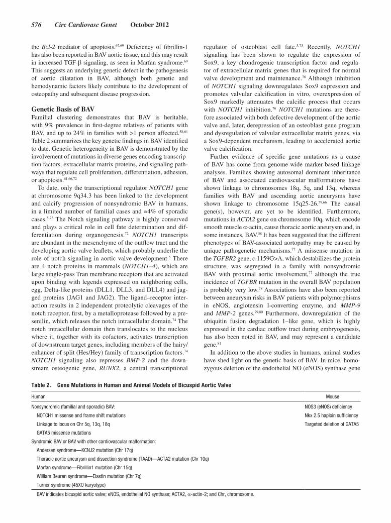
Table 2. Gene Mutations in Human and Animal Models of Bicuspid Aortic Valve
Human Mouse
Nonsyndromic (familial and sporadic) BAV: NOS3 (eNOS) deficiency
NOTCH1 missense and frame shift mutations Nkx 2.5 haploin sufficiency
Linkage to locus on Chr 5q, 13q, 18q Targeted deletion of GATA5
GATA5 missense mutations
Syndromic BAV or BAV with other cardiovascular malformation:
Andersen syndrome—KCNJ2 mutation (Chr 17q)
Thoracic aortic aneurysm and dissection syndrome (TAAD)—ACTA2 mutation (Chr 10q)
Marfan syndrome—Fibrillin1 mutation (Chr 15q)
William Beuren syndrome—Elastin mutation (Chr 7q)
Turner syndrome (45XO karyotype)
BAV indicates bicuspid aortic valve; eNOS, endothelial NO synthase; ACTA2, α-actin-2; and Chr, chromosome.

Padang et al Genetics of Familial Valve Disease 577
and haploinsufficiency in the cardiac homeobox gene Nkx2-5, are associated with a higher incidence of BAV.82,83 However, neither of these genes have been associated with BAV in humans.84,85 Furthermore, targeted deletion of the transcrip-tion factor Gata5 in mice leads to partially penetrant BAV with fusion of the right and noncoronary cusps.84 An essen-tial role of GATA5 in cardiac morphogenesis and aortic valve development has only recently been reported, whereby it regulates several pathways involved in endocardial cell dif-ferentiation, including those directed by Bmp4, Tbx20, and, notably, eNOS and NOTCH1.84 Downregulation of eNOS expression and attenuation of the notch pathway are observed in Gata5!/!mice,84 which may underlie the development of BAV as mutations in eNOS, and NOTCH1 are associated with BAV in mice and humans, respectively. It is interesting that a number of rare nonsynonymous variations within the highly conserved and functionally important GATA5 transcriptional activation domains have recently been reported in up to 4% of sporadic cases of human BAV.86 This provides support for the role of GATA5 as a potential candidate gene in modulating the pathogenesis of BAV disease in humans.
Animal studies are also at the forefront of research into the hypothesis that the different BAV morphologies have a differ-ent etiologic basis. Gata5!/! mice, eNOS knockout mice, and inbred Syrian hamsters each display a high incidence of BAV, but with different valve morphologies. BAV with fusion of the right coronary and noncoronary leaflets may result from morphogenetic defects that happen before outflow tract septa-tion, which probably relies on an exacerbated NO-dependent endothelial-to-mesenchymal transformation. BAV with fusion of right and left coronary leaflets may result from the anomalous septation of the proximal portion of the outflow tract, because of the distorted behavior of neural crest cells (Figure 4).62 This anomalous behavior of the cardiac neural crest is believed to explain the association of the right and left coronary leaflet fusion BAV morphology with coarctation of the aorta and the more pronounced aortic wall degeneration, compared with BAV with fusion of the right and noncoronary leaflets.62
It is interesting that Sans-Coma et al87 recently demon-strated that BAV in Syrian hamsters express a quantitative trait, subject to polygenic inheritance, with reduced pene-trance and variable expressivity. Furthermore, examination of aortic valve morphology in virtually isogenic Syrian hamsters produced by systematic inbreeding through full-sib mating, with the probability of homozygosity being 0.999 or higher, demonstrated the presence of BAV in up to five sixths of the hamsters studied, whereas the remaining one sixth had a trileaflet aortic valve. No significant asso-ciation was noted between the valvular phenotype in the parents and the offspring produced by crossing genetically alike hamsters, with the probability of homozygosity of at least 0.989. This suggests that a single underlying genotype may predispose to BAV and account for the whole range of valve morphology, and implies that nongenetic factors are acting during embryonic life, creating “developmental noise,” to influence the definitive anatomic configuration of the valve.87 This finding supports a complex inheritance
pattern, as seen in human BAV, and may explain the rela-tively low recurrence rate of BAV in first-degree relatives, despite its common prevalence within the general popu-lation.87 The findings also provide a potential explanation regarding aortic valve anatomy in monozygotic twins where BAV was present in 1 twin, but tricuspid aortic valve in the other.88
Although specific gene mutations can cause BAV in mice and humans, the variability in BAV morphology and its disease manifestations cannot be entirely explained. MicroRNAs (miRNAs) are a class of evolutionarily con-served small, noncoding RNAs that regulate gene expression in development and disease. Reduced expression of miR-26a, miR-30b, and miR-195 in stenotic BAV associated with cal-cification, compared with a regurgitant phenotype, has been reported.89 In vitro transfection of each of these miRNAs into human aortic valvular interstitial cells indirectly modulated the expression of several calcification-related genes, includ-ing RUNX2, BMP-2, alkaline phosphatase (ALPL), SMAD1, and SMAD3.89 Patients with BAV-associated aortic stenosis have also been shown to have attenuated the expression of miRNA-141, a repressor of BMP-2.90 Together, these stud-ies suggest that miRNA dysregulation contributes to aortic stenosis in BAV, and that miRNAs represent a potential new therapeutic target to limit progressive aortic valve calcifica-tion and dysfunction.
Most recently, global gene expression levels have been compared in aortic tissue from patients with a bicuspid or tricuspid aortic valve, with and without thoracic aortic aneu-rysm.91 Only 7 genes were differentially expressed between the nondilated BAV and nondilated tricuspid groups; the 4 upregulated genes were: left–right determination factor 2, a TGF-β family member; Fraser syndrome 1, a member of the extracellular matrix family of protein; Src homology 2 domain containing family, member 4;and death-associated protein kinase 3, a proapoptotic gene; and the 3 down-regulated genes were: VEGFC (a member of the vascular endothelial growth factor family); NFASC (neurofascin homolog, a member of the L1 family of cell adhesion mol-ecule); and LSP1 (lymphocyte-specific protein 1). Given the possible shared genetic underpinnings of BAV and related aortopathy, an investigation of these genes in BAV is war-ranted. The study also showed that immune responsive genes were upregulated in patients with tricuspid valve and with a dilated aorta, compared with those with a dilated aorta and BAV, suggesting that immune response genes are not directly involved in BAV-related aneurysms. Rather, BAV patients with aortic dilation showed an almost exclu-sive expression of the TGF-β–binding proteins LTBP3/4, ADAMTSL1, and an alternatively spliced isoform of fibro-nectin-1 (FN1).91
Collectively, these findings support a genetic basis for BAV, the pathogenesis of which involves a complex inter-play between specific gene mutations, environmental influences, and stochastic factors. The balance of this inter-action is likely to have major influence on disease progres-sion and severity, including valve dysfunction and aortic complications.

578 Circ Cardiovasc Genet October 2012
Conclusions and Future DirectionsCurrent evidence points to heritable and familial VHD being a final common pathway for a wide variety of altered molecular and genetic defects. Determination of the genetic basis underlying these disorders is complex. Not only that there are large number of genes involved in valvulogenesis (genetic heterogeneity) but epigenetic, environmental, and stochastic factors are also important in modulating phenotype expression and thus contributing to the wide spectrum of disease manifestations (clinical heterogeneity). Recent advances in the fields of valve development and familial VHD, combined with major developments in genetic analysis technologies, place the field at the precipice of accelerated discovery into the causative and pathogenetic mechanisms responsible for common heart valve defects.
Furthermore, the present limitation of pharmacological therapy in delaying or stopping disease progression has left surgical therapies as the most effective cure for many cases of familial VHD. Improved understanding of the genetic basis, molecular pathways, and cellular mediators involved in the pathogenesis of VHD will create great opportunities for the development of novel pharmacological treatment and pre-vention strategies in VHD. This knowledge has the potential to also be used to stratify treatment modalities, time surgi-cal intervention, and guide management of at-risk family members, in terms of diagnostic, therapeutic, and prevention strategies, with the overall aim of reducing the health burden of VHD. With the advancement of genetic technologies, the increasing availability of next-generation sequencing, coupled with well-characterized families with VHD, we are hopeful that many of the key genes and elusive pathways in VHD will be identified in coming years.
AcknowledgmentsDr Padang is supported by a cofunded Postgraduate Scholarship (PC 10S 5530) from the National Health and Medical Research Council (NHMRC) and the National Heart Foundation of Australia (NHFA), Australia. Dr Semsarian is the recipient of a National Health and Medical Research Council (NHMRC) of Australia Practitioner Fellowship. This work was supported by a research project grant from NHMRC (No. 1023318). We thank our surgical colleagues from the Cardiothoracic Department of Royal Prince Alfred Hospital, Sydney, Australia, for their support.
DisclosuresNone.
References 1. Mensah GA. The burden of valvular heart disease. In: Otto CM, Bonow
RO, eds. Valvular Heart Disease: A Companion to Braunwald’s Heart Disease. Philadelphia, PA: Elsevier Inc; 2009:1–18.
2. Nkomo VT, Gardin JM, Skelton TN, Gottdiener JS, Scott CG, Enriquez-Sarano M. Burden of valvular heart diseases: a population-based study. Lancet. 2006;368:1005–1011.
3. Lincoln J, Yutzey KE. Molecular and developmental mechanisms of con-genital heart valve disease. Birth Defects Res Part A Clin Mol Teratol. 2011;91:526–534.
4. Markwald RR, Norris RA, Moreno-Rodriguez R, Levine RA. Develop-mental basis of adult cardiovascular diseases: valvular heart diseases. Ann N Y Acad Sci. 2010;1188:177–183.
5. Garg V, Muth AN, Ransom JF, Schluterman MK, Barnes R, King IN, et al. Mutations in NOTCH1 cause aortic valve disease. Nature. 2005;437:270–274.
6. Hinton RB Jr, Lincoln J, Deutsch GH, Osinska H, Manning PB, Benson DW, et al. Extracellular matrix remodeling and organization in developing and diseased aortic valves. Circ Res. 2006;98:1431–1438.
7. Pierpont ME, Basson CT, Benson DW Jr, Gelb BD, Giglia TM, Gold-muntz E, et al; American Heart Association Congenital Cardiac Defects Committee, Council on Cardiovascular Disease in the Young. Genetic ba-sis for congenital heart defects: current knowledge: a scientific statement from the American Heart Association Congenital Cardiac Defects Com-mittee, Council on Cardiovascular Disease in the Young: endorsed by the American Academy of Pediatrics. Circulation. 2007;115:3015–3038.
8. Combs MD, Yutzey KE. Heart valve development: regulatory networks in development and disease. Circ Res. 2009;105:408–421.
9. Butcher JT, Markwald RR. Valvulogenesis: the moving target. Philos Trans R Soc Lond, B, Biol Sci. 2007;362:1489–1503.
10. Walsh EC, Stainier DY. UDP-glucose dehydrogenase required for cardiac valve formation in zebrafish. Science. 2001;293:1670–1673.
11. Liu AC, Joag VR, Gotlieb AI. The emerging role of valve interstitial cell phenotypes in regulating heart valve pathobiology. Am J Pathol. 2007;171:1407–1418.
12. Schoen FJ. Evolving concepts of cardiac valve dynamics: the continuum of development, functional structure, pathobiology, and tissue engineer-ing. Circulation. 2008;118:1864–1880.
13. Hinton RB, Yutzey KE. Heart valve structure and function in development and disease. Annu Rev Physiol. 2011;73:29–46.
14. de la Pompa JL, Epstein JA. Coordinating tissue interactions: Notch sig-naling in cardiac development and disease. Dev Cell. 2012;22:244–254.
15. Niessen K, Karsan A. Notch signaling in cardiac development. Circ Res. 2008;102:1169–1181.
16. Benson DW. Thar’s tendons in them thar valves! Circ Res. 2008;103:914–915.
17. Chakraborty S, Combs MD, Yutzey KE. Transcriptional regulation of heart valve progenitor cells. Pediatr Cardiol. 2010;31:414–421.
18. Norris RA, Moreno-Rodriguez RA, Sugi Y, Hoffman S, Amos J, Hart MM, et al. Periostin regulates atrioventricular valve maturation. Dev Biol. 2008;316:200–213.
19. Lincoln J, Lange AW, Yutzey KE. Hearts and bones: shared regulatory mechanisms in heart valve, cartilage, tendon, and bone development. Dev Biol. 2006;294:292–302.
20. Lin CY, Lin CJ, Chen CH, Chen RM, Zhou B, Chang CP. The secondary heart field is a new site of calcineurin/Nfatc1 signaling for semilunar valve development. J Mol Cell Cardiol. 2012;52:1096–1102.
21. Wirrig EE, Yutzey KE. Transcriptional regulation of heart valve develop-ment and disease. Cardiovasc Pathol. 2011;20:162–167.
22. Levine RA, Slaugenhaupt SA. Molecular genetics of mitral valve pro-lapse. Curr Opin Cardiol. 2007;22:171–175.
23. Freed LA, Levy D, Levine RA, Larson MG, Evans JC, Fuller DL, et al. Prevalence and clinical outcome of mitral-valve prolapse. N Engl J Med. 1999;341:1–7.
24. Grau JB, Pirelli L, Yu PJ, Galloway AC, Ostrer H. The genetics of mitral valve prolapse. Clin Genet. 2007;72:288–295.
25. Levine RA, Handschumacher MD, Sanfilippo AJ, Hagege AA, Harrigan P, Marshall JE, et al. Three-dimensional echocardiographic reconstruction of the mitral valve, with implications for the diagnosis of mitral valve prolapse. Circulation. 1989;80:589–598.
26. Guy TS, Hill AC. Mitral valve prolapse. Annu Rev Med. 2012;63:277–292. 27. Hayek E, Gring CN, Griffin BP. Mitral valve prolapse. Lancet.
2005;365:507–518. 28. Avierinos JF, Gersh BJ, Melton LJ 3rd, Bailey KR, Shub C, Nishimura
RA, et al. Natural history of asymptomatic mitral valve prolapse in the community. Circulation. 2002;106:1355–1361.
29. Rabkin E, Aikawa M, Stone JR, Fukumoto Y, Libby P, Schoen FJ. Ac-tivated interstitial myofibroblasts express catabolic enzymes and me-diate matrix remodeling in myxomatous heart valves. Circulation. 2001;104:2525–2532.
30. Nascimento R, Freitas A, Teixeira F, Pereira D, Cardoso A, Dinis M, et al. Is mitral valve prolapse a congenital or acquired disease? Am J Cardiol. 1997;79:226–227.
31. Loardi C, Alamanni F, Trezzi M, Kassem S, Cavallotti L, Tremoli E, et al. Biology of mitral valve prolapse: the harvest is big, but the workers are few. Int J Cardiol. 2011;151:129–135.
32. Ng CM, Cheng A, Myers LA, Martinez-Murillo F, Jie C, Bedja D, et al. TGF-beta-dependent pathogenesis of mitral valve prolapse in a mouse model of Marfan syndrome. J Clin Invest. 2004;114:1586–1592.
33. Leask RL, Jain N, Butany J. Endothelium and valvular diseases of the heart. Microsc Res Tech. 2003;60:129–137.

Padang et al Genetics of Familial Valve Disease 579
34. Prunotto M, Caimmi PP, Bongiovanni M. Cellular pathology of mitral valve prolapse. Cardiovasc Pathol. 2010;19:e113–e117.
35. Stein PD, Wang CH, Riddle JM, Sabbah HN, Magilligan DJ Jr, Hawkins ET. Scanning electron microscopy of operatively excised severely regurgitant floppy mitral valves. Am J Cardiol. 1989;64:392–394.
36. Barth PJ, Köster H, Moosdorf R. CD34+ fibrocytes in normal mitral valves and myxomatous mitral valve degeneration. Pathol Res Pract. 2005;201:301–304.
37. Judge DP, Rouf R, Habashi J, Dietz HC. Mitral valve disease in Marfan syndrome and related disorders. J Cardiovasc Transl Res. 2011;4:741–747.
38. Kaartinen V, Warburton D. Fibrillin controls TGF-beta activation. Nat Genet. 2003;33:331–332.
39. Habashi JP, Judge DP, Holm TM, Cohn RD, Loeys BL, Cooper TK, et al. Losartan, an AT1 antagonist, prevents aortic aneurysm in a mouse model of Marfan syndrome. Science. 2006;312:117–121.
40. Henney AM, Tsipouras P, Schwartz RC, Child AH, Devereux RB, Leech GJ. Genetic evidence that mutations in the COL1A1, COL1A2, COL3A1, or COL5A2 collagen genes are not responsible for mitral valve prolapse. Br Heart J. 1989;61:292–299.
41. Wordsworth P, Ogilvie D, Akhras F, Jackson G, Sykes B. Genetic segrega-tion analysis of familial mitral valve prolapse shows no linkage to fibrillar collagen genes. Br Heart J. 1989;61:300–306.
42. Nesta F, Leyne M, Yosefy C, Simpson C, Dai D, Marshall JE, et al. New locus for autosomal dominant mitral valve prolapse on chromosome 13: clinical insights from genetic studies. Circulation. 2005;112:2022–2030.
43. Kyndt F, Gueffet JP, Probst V, Jaafar P, Legendre A, Le Bouffant F, et al. Mutations in the gene encoding filamin A as a cause for familial cardiac valvular dystrophy. Circulation. 2007;115:40–49.
44. Trochu JN, Kyndt F, Schott JJ, Gueffet JP, Probst V, Bénichou B, et al. Clinical characteristics of a familial inherited myxomatous valvular dys-trophy mapped to Xq28. J Am Coll Cardiol. 2000;35:1890–1897.
45. Disse S, Abergel E, Berrebi A, Houot AM, Le Heuzey JY, Diebold B, et al. Mapping of a first locus for autosomal dominant myxomatous mitral-valve prolapse to chromosome 16p11.2-p12.1. Am J Hum Genet. 1999;65:1242–1251.
46. Freed LA, Acierno JS Jr, Dai D, Leyne M, Marshall JE, Nesta F, et al. A locus for autosomal dominant mitral valve prolapse on chromosome 11p15.4. Am J Hum Genet. 2003;72:1551–1559.
47. Chou HT, Hung JS, Chen YT, Wu JY, Tsai FJ. Association between COL3A1 collagen gene exon 31 polymorphism and risk of floppy mitral valve/mitral valve prolapse. Int J Cardiol. 2004;95:299–305.
48. Chou HT, Chen YT, Wu JY, Tsai FJ. Association between urokinase-plas-minogen activator gene T4065C polymorphism and risk of mitral valve prolapse. Int J Cardiol. 2004;96:165–170.
49. Oceandy D, Yusoff R, Baudoin FM, Neyses L, Ray SG. Promoter poly-morphism of the matrix metalloproteinase 3 gene is associated with regur-gitation and left ventricular remodelling in mitral valve prolapse patients. Eur J Heart Fail. 2007;9:1010–1017.
50. Chou HT, Chen YT, Shi YR, Tsai FJ. Association between angiotensin I-converting enzyme gene insertion/deletion polymorphism and mitral valve prolapse syndrome. Am Heart J. 2003;145:169–173.
51. Chou HT, Hung JS, Chen YT, Shi YR, Tsai FJ. Association between an-giotensinogen gene M235T polymorphism and mitral valve prolapse syn-drome in Taiwan Chinese. J Heart Valve Dis. 2002;11:830–836.
52. Szombathy T, Jánoskúti L, Szalai C, Császár A, Miklósi M, Mészáros Z, et al. Angiotensin II type 1 receptor gene polymorphism and mitral valve prolapse syndrome. Am Heart J. 2000;139(1 Pt 1):101–105.
53. Kern CB, Wessels A, McGarity J, Dixon LJ, Alston E, Argraves WS, et al. Reduced versican cleavage due to Adamts9 haploinsufficiency is associ-ated with cardiac and aortic anomalies. Matrix Biol. 2010;29:304–316.
54. Mahimkar R, Nguyen A, Mann M, Yeh CC, Zhu BQ, Karliner JS, et al. Cardiac transgenic matrix metalloproteinase-2 expression induces myxo-matous valve degeneration: a potential model of mitral valve prolapse dis-ease. Cardiovasc Pathol. 2009;18:253–261.
55. Hulin A, Deroanne CF, Lambert CA, Dumont B, Castronovo V, Defraigne JO, et al. Metallothionein-dependent up-regulation of TGF-ß2 partici-pates in the remodelling of the myxomatous mitral valve. Cardiovasc Res. 2012;93:480–489.
56. Michelena HI, Khanna AD, Mahoney D, Margaryan E, Topilsky Y, Suri RM, et al. Incidence of aortic complications in patients with bicuspid aor-tic valves. JAMA. 2011;306:1104–1112.
57. Roger VL, Go AS, Lloyd-Jones DM, Benjamin EJ, Berry JD, Bor-den WB, et al; American Heart Association Statistics Committee and Stroke Statistics Subcommittee. Heart disease and stroke statistics–2012 update: a report from the American Heart Association. Circulation. 2012;125:e2–e220.
58. Siu SC, Silversides CK. Bicuspid aortic valve disease. J Am Coll Cardiol. 2010;55:2789–2800.
59. Vallely MP, Semsarian C, Bannon PG. Management of the ascending aorta in patients with bicuspid aortic valve disease. Heart Lung Circ. 2008;17:357–363.
60. Cripe L, Andelfinger G, Martin LJ, Shooner K, Benson DW. Bicuspid aor-tic valve is heritable. J Am Coll Cardiol. 2004;44:138–143.
61. Ward C. Clinical significance of the bicuspid aortic valve. Heart. 2000;83:81–85.
62. Fernández B, Durán AC, Fernández-Gallego T, Fernández MC, Such M, Arqué JM, et al. Bicuspid aortic valves with different spatial orienta-tions of the leaflets are distinct etiological entities. J Am Coll Cardiol. 2009;54:2312–2318.
63. Sievers HH, Schmidtke C. A classification system for the bicuspid aor-tic valve from 304 surgical specimens. J Thorac Cardiovasc Surg. 2007;133:1226–1233.
64. Hinton RB, Martin LJ, Rame-Gowda S, Tabangin ME, Cripe LH, Ben-son DW. Hypoplastic left heart syndrome links to chromosomes 10q and 6q and is genetically related to bicuspid aortic valve. J Am Coll Cardiol. 2009;53:1065–1071.
65. Koullias GJ, Korkolis DP, Ravichandran P, Psyrri A, Hatzaras I, Eleft-eriades JA. Tissue microarray detection of matrix metalloproteinases, in diseased tricuspid and bicuspid aortic valves with or without pathology of the ascending aorta. Eur J Cardiothorac Surg. 2004;26:1098–1103.
66. Martin LJ, Ramachandran V, Cripe LH, Hinton RB, Andelfinger G, Taban-gin M, et al. Evidence in favor of linkage to human chromosomal regions 18q, 5q and 13q for bicuspid aortic valve and associated cardiovascular malformations. Hum Genet. 2007;121:275–284.
67. Fedak PW, Verma S, David TE, Leask RL, Weisel RD, Butany J. Clinical and pathophysiological implications of a bicuspid aortic valve. Circula-tion. 2002;106:900–904.
68. Rajamannan NM. Bicuspid aortic valve disease: the role of oxidative stress in Lrp5 bone formation. Cardiovasc Pathol. 2011;20:168–176.
69. Tadros TM, Klein MD, Shapira OM. Ascending aortic dilatation associ-ated with bicuspid aortic valve: pathophysiology, molecular biology, and clinical implications. Circulation. 2009;119:880–890.
70. Girdauskas E, Borger MA, Secknus MA, Girdauskas G, Kuntze T. Is aor-topathy in bicuspid aortic valve disease a congenital defect or a result of abnormal hemodynamics? A critical reappraisal of a one-sided argument. Eur J Cardiothorac Surg. 2011;39:809–814.
71. Aicher D, Urbich C, Zeiher A, Dimmeler S, Schäfers HJ. Endothelial nitric oxide synthase in bicuspid aortic valve disease. Ann Thorac Surg. 2007;83:1290–1294.
72. Garg V. Molecular genetics of aortic valve disease. Curr Opin Cardiol. 2006;21:180–184.
73. Mohamed SA, Aherrahrou Z, Liptau H, Erasmi AW, Hagemann C, Wrobel S, et al. Novel missense mutations (p.T596M and p.P1797H) in NOTCH1 in patients with bicuspid aortic valve. Biochem Biophys Res Commun. 2006;345:1460–1465.
74. Gridley T. Notch signaling in vascular development and physiology. De-velopment. 2007;134:2709–2718.
75. Nigam V, Srivastava D. Notch1 represses osteogenic pathways in aortic valve cells. J Mol Cell Cardiol. 2009;47:828–834.
76. Acharya A, Hans CP, Koenig SN, Nichols HA, Galindo CL, Garner HR, et al. Inhibitory role of Notch1 in calcific aortic valve disease. PLoS ONE. 2011;6:e27743.
77. Girdauskas E, Schulz S, Borger MA, Mierzwa M, Kuntze T. Transform-ing growth factor-beta receptor type II mutation in a patient with bicuspid aortic valve disease and intraoperative aortic dissection. Ann Thorac Surg. 2011;91:e70–e71.
78. Arrington CB, Sower CT, Chuckwuk N, Stevens J, Leppert MF, Yet-man AT, et al. Absence of TGFBR1 and TGFBR2 mutations in patients with bicuspid aortic valve and aortic dilation. Am J Cardiol. 2008;102: 629–631.
79. Pisano C, Maresi E, Balistreri CR, Candore G, Merlo D, Fattouch K, et al. Histological and genetic studies in patients with bicuspid aortic valve and ascending aorta complications. Interact Cardiovasc Thorac Surg. 2012;14:300–306.
80. Foffa I, Murzi M, Mariani M, Mazzone AM, Glauber M, Ait Ali L, et al. Angiotensin-converting enzyme insertion/deletion polymorphism is a risk factor for thoracic aortic aneurysm in patients with bicuspid or tricuspid aortic valves. J Thorac Cardiovasc Surg. 2012;144:390–395.
81. Mohamed SA, Hanke T, Schlueter C, Bullerdiek J, Sievers HH. Ubiquitin fusion degradation 1-like gene dysregulation in bicuspid aortic valve. J Thorac Cardiovasc Surg. 2005;130:1531–1536.

580 Circ Cardiovasc Genet October 2012
82. Biben C, Weber R, Kesteven S, Stanley E, McDonald L, Elliott DA, et al. Cardiac septal and valvular dysmorphogenesis in mice heterozygous for mutations in the homeobox gene Nkx2-5. Circ Res. 2000;87:888–895.
83. Lee TC, Zhao YD, Courtman DW, Stewart DJ. Abnormal aortic valve de-velopment in mice lacking endothelial nitric oxide synthase. Circulation. 2000;101:2345–2348.
84. Laforest B, Andelfinger G, Nemer M. Loss of Gata5 in mice leads to bi-cuspid aortic valve. J Clin Invest. 2011;121:2876–2887.
85. Majumdar R, Yagubyan M, Sarkar G, Bolander ME, Sundt TM 3rd. Bi-cuspid aortic valve and ascending aortic aneurysm are not associated with germline or somatic homeobox NKX2-5 gene polymorphism in 19 pa-tients. J Thorac Cardiovasc Surg. 2006;131:1301–1305.
86. Padang R, Bagnall RD, Richmond DR, Bannon PG, Semsarian C. Rare non-synonymous variations in the transcriptional activation do-mains of GATA5 in bicuspid aortic valve disease. J Mol Cell Cardiol. 2012;53:277–281.
87. Sans-Coma V, Carmen Fernández M, Fernández B, Durán AC, Anderson RH, Arqué JM. Genetically alike Syrian hamsters display both bifoliate and trifoliate aortic valves. J Anat. 2012;220:92–101.
88. Lewis NP, Henderson AH. Calcific aortic stenosis in twins: a clue to its pathogenesis? Eur Heart J. 1990;11:90–91.
89. Nigam V, Sievers HH, Jensen BC, Sier HA, Simpson PC, Srivastava D, et al. Altered microRNAs in bicuspid aortic valve: a comparison between stenotic and insufficient valves. J Heart Valve Dis. 2010;19:459–465.
90. Yanagawa B, Lovren F, Pan Y, Garg V, Quan A, Tang G, et al. miRNA-141 is a novel regulator of BMP-2-mediated calcification in aortic stenosis. J Thorac Cardiovasc Surg. 2012;144:256–262.
91. Folkersen L, Wågsäter D, Paloschi V, Jackson V, Petrini J, Kurtovic S, et al. Unraveling divergent gene expression profiles in bicuspid and tricuspid aortic valve patients with thoracic aortic dilatation: the ASAP study. Mol Med. 2011;17:1365–1373.
92. Madsen MB, Olsen LH, Häggström J, Höglund K, Ljungvall I, Falk T, et al. Identification of 2 loci associated with development of myxomatous mitral valve disease in Cavalier King Charles Spaniels. J Hered. 2011;102 (suppl 1):S62–S67.
93. Chou HT, Shi YR, Hsu Y, Tsai FJ. Association between fibrillin-1 gene exon 15 and 27 polymorphisms and risk of mitral valve prolapse. J Heart Valve Dis. 2003;12:475–481.
94. Ozdemir O, Olgunturk R, Karaer K, Ergun MA, Tunaoglu FS, Kula S, et al. Fibrillin-1 gene intron 56 polymorphism in Turkish children with mitral valve prolapse. Cardiol Young. 2010;20:173–177.
KEY WORDS: aortic valve ◼ genes ◼ genetic heart disease ◼ mitral valve ◼ valves