High performance cDNA sequence analysis using grid technology
Upload
independentCategory
view
3download
0
Eur. J. Biochem. 230, 977-986 (1995) 0 FEBS 1995
Binding of thyroxine to pig transthyretin, its cDNA structure, and other properties Wei DUAN’, Samantha J. RICHARDSON’, Josef KOHRLE’, Linus CHANG’, Bridget R. SOUTHWELL’, Paul J. HARMS’, Charlotte M. BRACK’, Tom M. PETTERSSON3 and Gerhard SCHREIBER’
’ Russell Grimwade School of Biochemistry, University of Melbourne, Australia Medizinische Poliklinik der Universitat Wiirzburg, Germany Department of Clinical Chemistry, Danderyd Hospital, Danderyd, Sweden
(Received 1 March 1995) - EJB 95 0325/3
Thyroxine binding to proteins in pig plasma during electrophoresis was observed in the albumin, but not in the prealbumin and post-albumin regions.
Transthyretin could be identified in medium from in vitro pig choroid plexus incubations by size and number of subunits and a very high rate of synthesis and secretion. Its electrophoretic mobility was intermediate between that of thyroxine-binding globulin and albumin. It bound thyroxine, retinol-binding protein, anti-(rat transthyretin) antibodies and behaved similarly to transthyretins from other vertebrate species when plasma was extracted with phenol. Inhibition experiments with the synthetic flavonoid F 21388, analysing the binding of thyroxine, suggested that transthyretin is not a major thyroxine carrier in the bloodstream of pigs.
Cloning and sequencing of transthyretin cDNA from both choroid plexus and liver showed that the same transthyretin mRNA is expressed in pig choroid plexus and liver. The amino acid sequence derived from the nucleotide sequence revealed that pig transthyretin differs from the transthyretins of all other studied vertebrate species by an unusual C-terminal extension consisting of the amino acids glycine, alanine and leucine. This extension results from the mutation of a stop codon into a codon for glycine. The unusual C-terminal extensions do not seem to interfere with the access of thyroxine to its binding site in the central channel of transthyretin.
Keywords. Pig transthyretin ; thyroxine binding ; pig transthyretin cDNA ; protein evolution ; thyroxine transport.
Thyroxine, the major thyroid hormone in the blood, has a strong tendency to partition from the blood into lipid membranes (Hillier, 1970; Mendel et al., 1987; Dickson et al., 1987). Plasma proteins markedly reduce the thyroxine disappearance from the blood (Mendel et al., 1987). Without thyroxine-binding proteins in the bloodstream, an even distribution of thyroid hor- mones in perfused tissues is not possible, because thyroid hor- mones quickly disappear into the cells first encountered by the perfusion medium (Mendel et al., 1987, 1988; Mendel and Wei- siger, 1990). More that 99.9 % of the intravascular pool of circu- lating thyroxine is protein-bound and the free fraction of thyrox- ine is below 0.1 % of total plasma thyroxine in most higher ver- tebrate species. One of the thyroxine binding plasma proteins is transthyretin. Thyroxine is bound to one of two specific sites of the transthyretin molecule (Blake et al., 1974). The two thyrox- ine-binding sites are located in a central channel formed by the four identical subunits of transthyretin. A binding site for reti-
Corresporzdence to G. Schreiber, Russell Grimwade School of Bio- chemistry, University of Melbourne, Parkville, 3052, Victoria, Australia
Fax: +61 3 9341 7730. Abbreviations. F 21388, 3’,5’-dibromo-6,4’-dihydroxy-3-methyl-fla-
Enzyme. Restriction endonucleases (EC 3.1.21.4). Note. The novel nucleotide sequence data published here have been
deposited with the Genbank sequence data bank, accession number U16131 and the EMBL nucleotide sequence database, accession number X82258.
vone; F 49209, 3’,5’-diiodo-6,4’-dihydroxy-3-methyl-flavone.
nol-binding protein, which is different from that for thyroxine (Nilsson and Peterson, 1971), exists on the outer surface of transthyretin (Wallace et al., 1988; Berni et al., 1994).
Transthyretin mRNA has been found in the liver of many eutherians, including pigs (Harms et al., 1991). No clear identifi- cation of transthyretin was achieved in electrophoresis of pig serum proteins (Larsson et al., 1985). However, transthyretin is abundantly synthesised and secreted by choroid plexus (Dickson et al., 1985a,b, 1986) where it has been proposed to be involved in transport and distribution of thyroxine (Schreiber et al., 1990; Southwell et al., 1993). This abundant synthesis by choroid plexus was used in this study to prepare radioactive pig trans- thyretin which was then used as a marker to identify transthyre- tin in pig blood. Transthyretin in pig blood plasma bound anti- bodies against rat transthyretin raised in rabbits. Binding of thy- roxine to purified pig transthyretin was investigated using a spe- cific inhibitor of binding of thyroxine, namely the synthetic fla- vonoid F 21388.
Complementary cDNA libraries were prepared from both choroid plexus and liver, transthyretin cDNA clones were iso- lated and their nucleotide sequences were analyzed. The derived amino acid sequence was used to compare pig transthyretin with the transthyretins from other vertebrate species.
MATERIALS AND METHODS Animals and materials. Choroid plexus from Large White
pigs (S. scrofa) were dissected from brains obtained from a local

97 8 Duan et al. (Eur J. Biochem. 230)
abattoir. Blood and liver were a gift from Dr John Walker (School of Veterinary Science, University of Melbourne). Only plasma from pigs which were not in an acute-phase response was used, since an acute-phase response leads to lower trans- thyretin levels in plasma (Dickson et al., 1982). Choroid plexus were dissected from brains within 30 rnin after death and were quickly frozen in liquid nitrogen. They were kept at -70°C until use for the isolation of RNA. A pig liver cDNA library in bacte- riophage /@l1 was a gift from Dr G. Baldwin (Ludwig Institute, Melbourne). Rats (Rattus nowegicus, Buffalo strain) were from a colony kept in the Russell Grimwade School of Biochemistry. They were fed a 20 % protein diet.
The sources of chemicals and enzymes were as described previously (Duan et al., 1991 ; Richardson et al., 1994). Amplify and a chemiluminescence kit were from Amersham. ~-[3’,5’- ‘251]Thyroxine (> 1.2 Ci/mg) was either from Amersham or from Du Pont de Nemours; XAR-5 film from Kodak. RPMI 1640 cell culture medium without leucine was purchased from Cytosystems (Castle Hill, NSW, Australia). Centricon-I0 con- centrators were from Amicon Corp. The two synthetic flavo- noids, specific competitors for thyroxine binding to transthyretin (Kohrle, 1992), F 21388 (3’,5’-dibromo-6,4’-dihydroxy-3-meth- yl-flavone) and F 49209 (3’,5’-diiodo-6,4’-dihydroxy-3-methyl- flavone) were obtained from Dr Priicher (Merck, Darmstadt). The preparation of transthyretin from rat serum and the raising of antibodies in rabbits were as previously described by Dickson et al. (1982) and Schreiber et al. (1968), respectively.
[14C]Leucine labelling of transthyretin from pig choroid plexus. Choroid plexus, freshly removed from the brain within 10 min of death, were initially placed in RPMI 1640 cell culture medium for 30 rnin at 37 “C. Choroid plexus was then incubated at 37°C in leucine-free RPMI 1640 cell culture medium with L- [U-14C]leucine (300 Ci/mol, 4 pCi/ml incubation medium) for a period of 5 h. Other conditions for incubations were as described previously (Edwards et al., 1976). After incubation, the medium was separated from tissue by centrifugation in a microfuge at 12000Xg for 5 rnin and the supernatant stored at -20°C. The incorporation of radioactive amino acid into secreted proteins was determined as described by Mans and Novelli (1961).
Purification of transthyretin by affinity chromatography. Human retinol-binding protein was purified and coupled to Sepharose-4B, as previously described (Larsson et al., 1985). A sample of 18 ml pig serum was dialysed against 40 mM Tris/ HCl pH 7.4, containing 0.5 M NaCl, and loaded onto a glass column (20X 1.5 cm) containing 15 ml retinol-binding-protein- Sepharose which had been equilibrated in the same buffer at 4°C. After washing with more buffer, pig transthyretin was eluted in water. The purity of eluted transthyretin was assessed by SDSPAGE. The yield was 2.3 mg transthyretin.
Radioactively labelled transthyretin from pig choroid plexus incubations and transthyretin from rat plasma were purified sim- ilarly to the purification of transthyretin from pig plasma.
Estimation of the molecular mass of proteins in choroid plexus incubation medium. The molecular masses of native proteins were estimated by fast protein liquid chromatography/ gel-permeation chromatography using a Superose-12 (HR 10/ 30) column equilibrated in 0.15 M ammonium bicarbonate pH 8.0. The column was calibrated with chymotrypsin (25 m a ) , hen ovalbumin (43 m a ) , and bovine serum albumin (67 kDa and dimer of 134 m a ) . The log of the relative molecular mass was plotted versus K,, where Kav = (V,-Vo)(Vt-K)-’. V, is the elution volume of the protein, V, the exclusion volume of the column, and V, the total gel bed volume.
Western analysis of pig transthyretin with anti-(rat transthyretin). Samples of rat serum transthyretin and pig se- rum were analysed by SDS/PAGE and transferred electrophoret-
ically (30 V, 18 h) to a nitrocellulose membrane. After the transfer, remaining protein binding sites on the membrane were blocked by incubation with 5% skimmed milk and the mem- brane was incubated with rabbit anti-(rat transthyretin) antise- rum (1 : lOOOO), then with horseradish-peroxidase-conjugated donkey anti-(rabbit immunoglobulin) (dilution of antiserum 1:lOOOO). The second antibody was detected by enhanced chemiluminescence, as described by the manufacturer (Amer- sham). Approximately 20-fold more pig serum than rat serum was required to give equivalent immunostaining.
Electrophoretic analysis of thyroxine binding to plasma proteins. ~-[3’,5’-’~~I]Thyroxine was isolated as described by Mendel et al. (1989) and analysed for purity according to Par- dridge and Mietus (1980). Combinations of aliquots of plasma, choroid plexus incubation medium, purified transthyretins and 2.1 nCi ~-[3‘,5’-”’II]thyroxine were incubated at room temper- ature for 1 h before being analysed by non-denaturing polyacryl- amide gel (10%) electrophoresis in 0.05 M Tris/glycine pH 8.6, 4°C. Proteins were stained with Coomassie brilliant blue R-250. For visualization of ~-[3‘,5’-’~~I]thyroxine, gels were autoradio- graphed on Kodak XAR-5 film at -70°C. For visualization of [‘4C]leucine-labelled protein, lanes were soaked in Amplify, dried, then fluorographed on Kodak XAR-5 film at -70°C. Vol- umes of plasma and choroid plexus incubation medium loaded onto each lane are indicated in the figure legends.
Thyroid hormone binding of pig serum and competition by flavonoids. Binding of ~-[3’,5‘-’~~I]thyroxine to serum and competition by flavonoids was analysed in vitro, as previously described (Kohrle et al., 1986). In brief, 10 pl serum or 19 pmol human transthyretin were incubated without or with binding competitors with 71 000 dpm ~-[3’,5’-’~~I]thyroxine (correspond- ing to approximately 45 pmol thyroxineil) in 500 pl buffer (0.1 M Tris/HCl, 1 mM EDTA, 0.1 M NaCI, pH 8.0) for 1 h at 25°C. The reaction was stopped by addition of 0.5 ml ice-cold dextran-coated charcoal. Bound and free ~-[3‘,5‘-’~~I]thyroxine were separated after a 10-min incubation at 4°C by a 10-min centrifugation at 3000Xg. Radioactivity in supernatant (bound thyroxine) and pellet (free thyroxine fraction) was measured in a LKB 1270 Rackgamma I1 gamma counter. Nonspecific binding (2.3 % of total), i.e. tracer displacement by a saturating concen- tration of 10 pM L-thyroxine, was subtracted for calculation of specific binding. L-Thyroxine and the flavonoids F 21 388 and F 49209 were dissolved in 0.01 M NaOH and diluted in H,O to final concentrations immediately before the assay.
Determination of Kd for thyroxine and transthyretin. The equilibrium binding of thyroxine to transthyretin purified from plasma was analysed by separation of free and transthyretin- bound thyroxine by adsorption of free thyroxine to charcoal, as described by Munro et al. (1989), and by equilibrium dialysis. All binding assays were performed in 0.04 M Tris/HCl pH 7.4, containing 0.04 M HC1, 0.06 M NaC1, 4 mM mM KC1, 3.3 mM CaCI, and 0.6 mM MgC1, (buffer A; Irvine, 1974). The concen- tration of transthyretin was 0.10 pM, that of ~-[3’,5’-’~~I]thyrox- ine (1.5 Ci/mg) was 0.01-0.03 nM (14000 dpdtube). Non-ra- dioactive thyroxine was added at concentrations from 0.01 nM to 1 pM. All incubations were prepared, in duplicate, at 4°C with a total volume of 0.5 ml for 14-18 h. The methyl-cellu- losekharcoal suspension (10 g/l activated charcoal and 1 g/l methyl-cellulose; 0.25 ml at 0°C) was mixed with the incuba- tion mixture and immediately centrifuged (lOOOXg, 30 min, 4°C). The supernatant was aspirated and radioactivity bound to the charcoal was determined. The affinity of the purified trans- thyretin for thyroxine was analysed using non-linear regression analysis of the binding data followed by Scatchard analysis.
For equilibrium dialysis, pig transthyretin, at concentrations between 5-500 nM was incubated for 15 min at room temper-

Duan et al. (Eul: J. Biochern. 230) 979
Fig. 1. Analysis of the binding of thyroxine to proteins in pig plasma by non-denaturing polyacrylamide gel electrophoresis. Autoradio- graphy following electrophoresis of 8 p1 plasma incubated with 2.1 nCi ~-[3‘,5’-’~~I]thyroxine in the presence of 0, 0.5, 2 or 8 p M unlabelled L- thyroxine (from left to right). 10% polyacrylamide gel, pH 8.6, 4°C. TBG indicates the thyroxine-binding globulin.
ature with ~-[3’,5’-’*~I]thyroxine at 1 nM to give approximately 1.71 X 10⁶ dpdml . An aliquot of 250 p1 of this mixture was dia- lysed against 20 ml buffer A at 37°C for 16-18 h in 10-mm flat-width dialysis tubing (Union Carbide). Radioactivity was determined for both dialysate and dialysand after MgCl, precipi- tation in the presence of unlabelled thyroxine to remove contam- inating free 9 o d i d e (Stockigt et al., 1981).
RNA isolation, cDNA synthesis, library construction, and sequencing. A pig choroid plexus cDNA library was con- structed in bacteriophage &lo, as described previously (Duan et al., 1991). Approximately 1000 plaques from an unamplified cDNA library were screened by hybridisation (Sambrook et al., 1989) with a sheep transthyretin cDNA probe (Tu et al., 1989), labelled with [a-32P]dATP as described previously (Duan et al., 1991). A pig choroid plexus transthyretin cDNA of 0.66 kb was subcloned into plasmid pGEM-7Zf( +).
Three pig transthyretin cDNA clones were isolated from a pig liver cDNA library in bacteriophage Agtl 1 by hybridisation, using pig choroid plexus transthyretin cDNA as a probe. Recom- binant plasmids containing pig transthyretin cDNA were pre- pared by a method involving boiling with lysozyme (Sambrook et al., 1989). The DNA sequence was determined for both strands (Sanger et al., 1977).
RESULTS
Analysis of thyroxine-binding to pig plasma proteins. In order to determine the relative thyroxine-binding capacity of thyrox- ine-binding proteins in pig plasma, a 10-pl sample of plasma was incubated for 1 h with 2.1 nCi ~-[3’,5’-’~~I]thyroxine and either 0, 0.5, 2 or 8 pM unlabelled L-thyroxine. Proteins were then separated by electrophoresis in polyacrylamide gel under non-denaturing conditions, followed by autoradiography (see Materials and Methods). The results are illustrated in Fig. 1. Ra- dioactive thyroxine bound to proteins in pig plasma which were found in two bands. The protein with the greater electrophoretic mobility was identified as albumin, as it comigrated with the
Fig. 2. Analysis of thyroxine-binding proteins in choroid plexus incu- bation medium. Non-denaturing electrophoresis in polyacrylamide gel was followed by autoradiography (lanes 2, 5-7, numbering from left to right), fluorography (lanes 3 and 4) or staining with Coomassie brilliant blue R-250 (lanes 1 and 8). Lane 1, 2 p1 pig plasma, the position of albumin is marked by an open triangle; lane 2, 5 p1 pig plasma which had been incubated with 0.72 nCi ~-[3’,5’-’~~I]thyroxine; lane 3, 40 p1 pig choroid plexus incubation medium; lane 4, 40 p1 pig choroid plexus incubation medium, incubated with 5 p1 diluted (1 :9) pig plasma; lane 5, 40 p1 choroid plexus incubation medium with 5 pl diluted (1 :9) pig plasma and 0.72 nCi ~-[3’,5’-’~’I]thyroxine; lane 6, 40 p1 pig choroid plexus incubation medium which had been incubated with 0.72 nCi L- [3‘,5’-‘251]thyroxine, the position of the thyroxine-binding protein in cho- roid plexus incubation medium is marked by a black triangle; lane 7, 5 pl human plasma which had been incubated with 0.72 nCi ~-[3‘,5’- ‘251]thyroxine, the position of albumin is marked by an open triangle and that of transthyretin by a black triangle; lane 8, 2 pl human plasma.
major Coomassie brilliant blue R-250-staining protein in plasma. Increasing the amount of non-radioactive thyroxine added lead to an increasing displacement of ~-[3’,5’-’~~I]thyrox- ine from the more cathodal thyroxine-binding plasma protein, suggesting that this protein had a lower thyroxine-binding capa- city than the more anodal thyroxine-binding protein. A thyrox- ine-binding protein which migrated faster than albumin was not observed in pig plasma.
Binding of thyroxine to proteins secreted by pig choroid plexus. Transthyretin is the major protein synthesised and secreted by the choroid plexus in the rat (Dickson et al., 1986) and in pigs (Harms et al., 1991). Thus, choroid plexus tissue incubated in vitro with [14C]leucine can be conveniently used for the synthesis of radioactive pig transthyretin as a marker. The analysis of radioactive proteins synthesised and secreted by pig choroid plexus, incubated in vitro with [ ‘‘C]leucine, by electro- phoresis and autoradiography is shown in Fig. 2. Proteins were also exposed to binding of ~-[3’,5’-’*~I]thyroxine for 60 min at 25OC before electrophoresis. Thus, a I4C signal indicated pro- teins synthesised by choroid plexus and a ‘’‘1 signal indicated proteins binding thyroxine. Proteins were stained with Coomas- sie blue R-250. Lane 1 (numbering from left to right) contained

980 Duan et al. (EM J. Biochem. 230)
.- * $ 40M)-
g 20m-
0 U .-
0
1 40
0 0 0 0 0 0
U V U U L n l n q y p - o N
Log (MasdDa)
. , . , . , . , . , . , . I .
competitor (pmollL)
Fig. 3. Competition of binding of ~-[3’,5’-’~~I]thyroxine to porcine se- rum by thyroxine and synthetic flavonoids. Porcine serum was incu- bated with ~-[3‘,5’-’~~I]thyroxine and increasing concentrations of non- radioactive L-thyroxine, the synthetic brominated flavonoid F 21388, or the iodinated flavonoid F 49209 for 1 h at 25°C. Bound and free L- [3’,5’-’**I]thyroxine were separated by dextran-coated charcoal and cen- trifugation. Nonspecific binding (2.3 % of total radioactivity) in excess of 10 pM L-thyroxine was subtracted for determination of specific bind- ing. Data give the mean+-SEM of two (L-thyroxine) or three (flavo- noids) experiments performed independently in duplicates. (0) Thyrox- ine; (U) F 21388, (A) F 49209.
Volume (ml)
Fig. 4. Gel filtration chromatography of radioactive proteins in pig choroid plexus incubation medium. Proteins (107 000 dpm of radio- active protein) from medium of pig choroid plexus incubation were sepa- rated by gel filtration on a Superose-12 column. Fractions of 0.25 ml were collected and 50-pl aliquots were analysed according to Mans and Novelli (1961) for protein-associated radioactivity by liquid scintillation spectrometry. Proteins with known molecular masses were also chro- matographed to calibrate the column. The major radioactive protein (marked with an arrow) that eluted from the column had a calculated molecular mass of 57 ma. See Materials and Methods for further de- tails.
pig plasma; the band corresponding to albumin is indicated by an open triangle. Lane 2 shows pig plasma which had been incu- bated with ~-[3‘,5’-’~~I]thyroxine; this lane shows the position of thyroxine-binding globulin and albumin. Lane 3 shows the analysis of pig choroid plexus incubation medium ; [‘4C]leucine- labelled transthyretin can be clearly seen. Lane 4 shows the analysis of a plasma sample mixed (‘spiked’) with proteins secreted by the choroid plexus ; the migration of I4C-labelled transthyretin added to plasma is similar to that of 14C-labelled transthyretin in choroid plexus incubation medium and, in both
Fig. 5. Immunoblot (Western) analysis of transthyretin in rat and pig serum. Samples of rat and pig serum were analysed by SDSFAGE. Proteins were then electrophoretically transferred to nitrocellulose filters. The filter was incubated with rabbit anti-rat transthyretin antiserum (1 : 10000) followed by horseradish-peroxidase-conjugated anti-(rabbit immunoglobulin) (1 : 10000). Detection was by enhanced chemilumines- cence. Lanes are numbered below the blot. Positions of protein size markers are indicated on the right of the blot in kDa. The position of the transthyretin subunit is indicated (b) on the left of the blot. Rat serum was diluted 1 : 100. A sample of 5 p1 of this dilution and further samples obtained by serial 2-fold dilutions were loaded onto the gel (lanes 1 to 5) . Pig serum was diluted 1 :2. A sample of 5 pl, of the dilution and subsequent 2-fold dilutions were loaded in lanes 6-10. Sample dilutions are indicated above the blot.
cases, between that of thyroxine-binding globulin and albumin. Lane 5 shows the result for a plasma sample which had been incubated with choroid plexus incubation medium and ~-[3‘,5‘- ‘251]thyroxine ; the electrophoretic migration of albumin and thy- roxine-binding globulin was similar in plasma ‘spiked‘ with choroid plexus incubation medium and in plasma alone, both proteins binding ~-[3’,5’-’~~I]thyroxine. Lane 6 shows the analy- sis of choroid plexus incubation medium incubated with ~-[3’,5‘- ‘251]thyroxine, showing that transthyretin from choroid plexus incubation medium bound thyroxine ; the position of the signal, i.e. bound ~-[3’,5’-’~~I]thyroxine, indicating transthyretin (black triangle in Fig. 2), was intermediate between that of thyroxine- binding globulin and albumin. For lane 7 human plasma had been incubated with ~-[3’,5’-~~~I]thyroxine; the position of albu- min is indicated by an open triangle and that of transthyretin by a black triangle. Lane 8 shows Coomassie blue staining of pro- teins in human plasma.
Competition between thyroxine and flavonoids for the bind- ing to pig serum transthyretin. In human, dog and rat serum, synthetic flavonoids are potent and selective competitors of thy- roxine binding to transthyretin (Kohrle et al., 1989; Kohrle, 1992; Rajatanavin et al., 1989). Therefore, in vitro competition experiments were performed with pig serum in order to identify the presence of transthyretin. Fig. 3 shows that both the bromi- nated (F 21388) and the iodinated (F 49209) flavonoid displaced thyroxine from binding to pig sera with relative potencies simi- lar to those previously reported for human serum (Kohrle et al., 1991). As already shown for competition with purified human transthyretin (Kohrle et al., 1988), the iodinated flavonoid F 49209 was more potent than the brominated derivative F 21388, compatible with the preferential binding of iodinated ligands to the transthyretin-binding site. A concentration of 0.5 pM F 21388 almost completely displaced ~-[3’,5’-’~~I]thyroxine from purified human transthyretin, but not from human albumin or human thyroxine-binding globulin (data not shown). Thyroxine

Duan et al. ( E m J. Biochem. 230) 981
025 7
020 - .O a n
Fig. 6. Characterisation of thyroxine binding to pig transthyretin purified by affinity chromatography. Transthyretin, albumin and whole serum were incubated with ~-[3’,5’-‘~’I]thyroxine (2.5 nCi/track) for 30 min at room temperature in the absence or presence of flavonoid F 21388. Incubated proteins were then separated on a 10% polyacryl- amide gel under non-denaturing conditions. Half the gel was stained with Coomassie blue to visualise the separated proteins (A), the other half of the gel was dried and exposed to a XAR-5 film for analysis of ~-[3’,5’-’~~I]thyroxine binding (B). The order of proteins in A and B was identical. Lane 1, 5 p1 diluted (1 :9) human serum, lane 2, 5 p1 diluted (1 :9) pig serum; lane 3, lO pg pig albumin; lane 4, 10 pg pig transthyre- tin; lane 5,6 pg human transthyretin; lane 6 , 5 pl diluted (1 :9) pig serum and 5 p1 of flavonoid F 21388 (1 pM); lane 7, 10 pg pig transthyretin and 5 pl of flavonoid F 21388 (1 pM); lane 8, 5 p1 diluted (1:9) human serum. The strong radioactive signals at the lower ends of lanes 3, 6, and 7 are produced by free thyroxine.
tracer displacement by non-radioactive L-thyroxine in pig serum was also comparable to that for human serum, suggesting the presence of a similar mixture of thyroid hormone-binding pro- teins in pig and human sera.
J
0.00 0.50 1.00 1.50 2.00
Free T4 pM
Fig. 7. Equilibrium binding of thyroxine to human (0) and pig (0) transthyretin (0.1 pM). Protein-bound and free thyroxine were sepa- rated by binding free thyroxine to methyl-cellulose-charcoal. Maximum binding was 60 % of total radioactivity. Each point is the mean of dupli- cate determinations. The affinity (KJ of the isolated transthyretin prepa- rations for thyroxine was between 0.04-0.08 pM.
Identification of pig transthyretin from choroid plexus by binding to retinol-binding protein and determination of qua- ternary structure. Medium from choroid plexus incubated in vitro with [14C]leucine was chromatographed on a column with a matrix of retinol-binding protein coupled to Sepharose-4B. Protein was eluted with distilled water. The molecular mass of the major radioactive protein was determined by gel filtration on Sepharose-12 column. The elution profile is shown in Fig. 4. A molecular mass of 57 kDa was obtained by comparison with the elution position of calibration proteins (insert in Fig. 4.). This, combined with the previously determined mass of the pig transthyretin subunit of about 15 kDa (Harms et al., 1991), indi- cated a tetrameric structure for the major radioactive protein synthesised and secreted by pig choroid plexus.
Western analysis of transthyretin in serum. In preliminary experiments it was found that an antiserum raised in rabbits against rat transthyretin also reacted with pig transthyretin from choroid plexus incubation medium in a Western analysis (al- though about 20-fold less strongly). Therefore, this antiserum could be used for the demonstration of transthyretin in pig se- rum. Immunostaining of transthyretin in rat and pig serum (Fig. 5) indicated that 5 p1 of 1 : 100 diluted rat serum (lane 1) gave equivalent staining to 5 p1 pig serum, diluted 1:16 (lane 9).
Isolation of transthyretin from pig serum by affinity chroma- tography. Binding to retinol-binding protein of transthyretin secreted into the incubation medium by pig choroid plexus had been used for the purification of the transthyretin analyzed in Fig. 4, suggesting that it could be feasible to purify larger amounts of transthyretin from pig serum by affinity chromatog- raphy. A total of 2.3 mg transthyretin was obtained from 18 ml serum from non-stressed pigs by chromatography on a retinol- binding-protein- Sepharose column, using the procedure de- scribed in Materials and Methods. Assuming 100 % recovery of pig transthyretin, the concentration of transthyretin in pig serum would be about 3 pM. This is similar to the concentration of transthyretin in rat serum.
This transthyretin was characterised and compared to human transthyretin (Fig. 6). In contrast to the large difference between the electrophoretic mobilities of human albumin (Fig. 6A, lane 1) and human transthyretin (Fig. 6A, lane 5) the electrophoretic mobilities of pig transthyretin (Fig. 6A, lane 4) and pig albumin (Fig. 6A, lane 3) were very similar. The flavonoid F 21388 completely inhibited the binding of ~-[3’,5‘-*~~1]thyroxine to pig transthyretin (Fig. 6B, lane 7). Pig albumin did not bind ~-[3’,5’- 1251]thyroxine (Fig. 6B, lane 3). For comparison, whole human
982 Duan et al. (Eur: J. Biochern. 230)
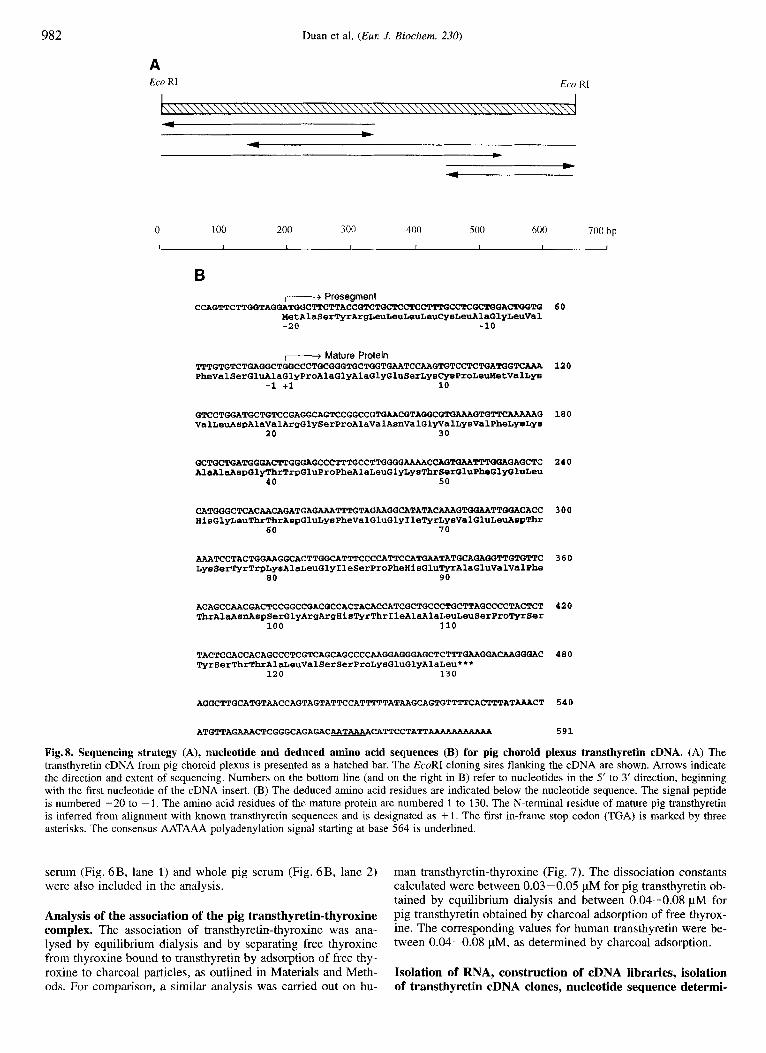
A Eco RI Eco RI
0 100 200 300 400 500 600 700 bp
L 1 I I I I I I
B I--+ Presegment
MetAlaSerTyrArgLeuLeuLeuLeuCysLeuAlaG1yLeuVa1 -20 -10
CCAGTTCTTOOTAGGATGGCTTCTTACCOTCTOCTCCGTCTGCTCCTCC~~CTCGC~GAC~TG
,-I Mature Protein T T T G T G T C T G A G G C T G G C C C T G C O O G T G C T G G T G M T C C A PheVa1SerGluAlaGlyProAlaGlyAlaGlyGluSerLysCysProLeuddetValLys
-1 +1 10
GTCCTGGATGCTGTCCGAGGCAGTCCGGCCGTGMCGTAGGCGTGAAAGTGTTCAAAAAG ValLeuAspAlaValArgGlySerProAlaValAsnValGlyValLysValPheLysLys
20 30
GCTGCTGATGGGACTTGGGAGCCCTTTGCCTTGGGGAAAACCAGWM~G~GAGCTC AlaAlaAspGlyThrTrpGluProPheA1aLeuGlyLysThrSerGluPheGlyGluLeu
40 50
CATGGGCTCACAACAGATGAGAAATTTGTAGAAGGCATATACAAAGTGGMTWGACACC HisGlyLeuThrThrAspGluLysPheValGluGlyIle~rLysValGluLe~s~Thr
60 7 0
AAATCCTACTGGAAGGCACTTGGCATTTCCCCATTCCATGMTATGCAGAGGTTGTGTTC LysSer'PyrTrpLysAlaLeuGlyIleSerProPheHisGlu~rAlaGluValValPhe
80 90
ACAGCCAACGACTCCGGCCGACGCCACTACACCATCGCTGCCCTGCTTAGCC~CT~CTCT ThrAlaAsnAspSerGlyArgArgHis~rThrIleAl~laLeuLeuSerPro~rSer
100 110
TACTCCACCACAGCCCTCGTCAGCAGCCCCMGGAGGGAGCTCTTT~GGA~GGGAC TyrSerThrThrAlaLeuValSerSerProLysGluGlyAlaLeu***
120 130
ATGTTAGAAACTCGGGCAGAGACAATAAAACATTCCTATT-
60
120
180
240
300
360
420
480
540
591
Fig. 8. Sequencing strategy (A), nucleotide and deduced amino acid sequences (B) for pig choroid plexus transthyretin cDNA. (A) The transthyretin cDNA from pig choroid plexus is presented as a hatched bar. The EcoRI cloning sites flanking the cDNA are shown. Arrows indicate the direction and extent of sequencing. Numbers on the bottom line (and on the right in B) refer to nucleotides in the 5' to 3' direction, beginning with the first nucleotide of the cDNA insert. (B) The deduced amino acid residues are indicated below the nucleotide sequence. The signal peptide is numbered - 20 to - 1. The amino acid residues of the mature protein are numbered 1 to 130. The N-terminal residue of mature pig transthyretin is inferred from alignment with known transthyretin sequences and is designated as + 1 . The first in-frame stop codon (TGA) is marked by three asterisks. The consensus AATAAA polyadenylation signal starting at base 564 is underlined.
serum (Fig. 6B, lane 1) and whole pig serum (Fig. 6B, lane 2 ) were also included in the analysis.
Analysis of the association of the pig transthyretin-thyroxine complex, The association of transthyretin-thyroxine was ana- l ysed by equilibrium dialysis and by separating free thyroxine from thyroxine bound to transthyretin by adsorption of free thy- roxine to charcoal particles, as outlined in Materials and Meth- ods. For comparison, a similar analysis was carried out on hu-
man transthyretin-thyroxine (Fig. 7). The dissociation constants calculated were between 0.03-0.05 pM for pig transthyretin ob- tained by equilibrium dialysis and between 0.04-0.08 pM for pig transthyretin obtained by charcoal adsorption of free thyrox- ine. The corresponding values for human transthyretin were be- tween 0.04-0.08 pM, as determined by charcoal adsorption.
Isolation of RNA, construction of cDNA libraries, isolation of transthyretin cDNA clones, nucleotide sequence determi-

Duan et al. (Eur J . Biochem. 230) 983
Human Sheep Rabbit Rat Mouse Pig S.Glider Wa 1 1 aby Monodel . Dunnart Chicken Lizard
---a- helix-l Gstrand FI I-&strand G--1 I-&strand HI Human ALGISPEEH AEYVFT&IDS GPRRYZISL @PY-TA VJTNPKE Sheep S*f******y * + * * * * * * * * *L*H*f**** * * * * i t * * * * L*SS*** Rabbit *********y A * * * * * * * * * *HIIS****** ***F****** **S**Q*
*********y * t t f * f * * * * *H*H*'**** * * * * * * * * * * *"S**QN Mouse T'*******F *D******** *H*H*+**** * * * * f t t t * * **S*'QN Rat
Pig t********y * t t + * * * * * * *R*H****** * * * * * * * * * * L*SS***GAL S,Glider +t+V*****y *D*+*****A *H+H*****Q *++++F**t* I*S'*T* Wallaby ***v*****y *D*t*****A *H*H*****Q *****F**** I*S**T* Monodel, ***V*****y *D***K***A *H*H**t*** f * * t t * * * t * * * S * * * * Dunnart TF*******Y *D******IA *H*H***ffQ **fF*F**** **S***D Chicken G**L*****y *D*******i *H*H***t** ***F****** **SD*Q* Lizard t*+V*+**'y *Dt*fSft+f tH*H****** **fF****** ++SD**t
I I I I I 90 100 110 120 1 2 7
E x o n 3 4- E x o n 4 Fig. 9. Alignment of amino acid sequences of 12 transthyretins. The deduced amino acid sequence of pig transthyretin (this paper) is aligned with the amino acid sequence of rabbit transthyretin (Sundelin et al., 1985) and those deduced from cDNA sequences for human (Mita et al., 1984), sheep (Tu et al., 1989), rat (Duan et al., 1989), mouse (Wakasugi et al., 1985), wallaby (Brack et al., 1995), sugar glider, South American short- tailed grey possum (designated as 'Monodel') and striped-faced dunnart (Duan et al., 1995), chicken (Duan et al., 1991) and lizard (Achen et al., 1993). Numbers below the sequences refer to the amino acids of human transthyretin. The N-terminal residue of mature transthyretin is designated as + I . F'resegments contain the residues numbered from -20 to -1. Those residues in sheep, rabbit, rat, mouse, pig, sugar glider, South American short-tailed grey possum, striped-faced dunnart, chicken and lizard transthyretins which are identical to those in human transthyretin are represented by asterisks. Gaps (in position 3-6) were introduced to optimise the alignment. Features of secondary structure in human transthyretin (Blake et al., 1974, 1978) are indicated above the sequence for human transthyretin. Residues located in the core of human transthyretin subunit are singly underlined, those located in the central channel of the tetramer are doubly underlined. The arrows below the amino acid sequences indicate the intron-exon boundaries in the human (Tsuzuki et al., 1985), rat (Fung et a]., 1988) and mouse (Wakasugi et al., 1986) transthyretin genes. The a/Iy below the amino acids 4-6 denotes the additional amino acid residues present in the marsupial, bird and reptile transthyretins, but absent in rodent and large mammal transthyretins.
nation and derivation of the primary structure for pig trans- thyretin. A 0.66-kb pig transthyretin cDNA was isolated from a pig choroid plexus cDNA library and the complete nucleotide sequence was determined as described in Materials and Meth- ods. The strategy for DNA sequencing analysis, the complete nucleotide and the deduced amino acid sequence are shown in Fig. 8. The pig transthyretin subunit consisted of 130 amino acid residues beginning with the first methionine at nucleotides 16- 18 and ending with a stop codon at nucleotides 465-467. The cDNA contained a 5'-untranslated region of 15 nucleotides and also a 3'-untranslated region consisting of 11 5 nucleotides with a polyadenylated segment of 11 bases, following a consensus AATAAA polyadenylation signal at residues 564. The pig trans-
thyretin cDNA sequence displayed 8 5 % , 87%, 76%, 78%, 72%, 72%, 71%, 73% and 68% identity to the cDNA se- quences of human (Mita et al., 1984), sheep (Tu et al., 1989), rat (Duan et al., 1989), mouse (Wakasugi et al., 1985), wallaby (Brack et al., 1994), sugar glider, stripe-faced dunnart, South American short-tailed grey opossum (Duan et al., 1995), chicken (Duan et al., 1991) and lizard transthyretin (Achen et al., 1993), respectively.
The calculated mass of the subunit of pig transthyretin de- duced from the cDNA is 13 930 Da, in good agreement with the electrophoretic mobility in SDSPAGE and suggesting the absence of post-translational modifications. No potential glyco- sylation site, Asn-Xaa-SerJThr, was found in the derived amino

984 Duan et al. (EUK J. Biochern. 230)
acid sequence. Compared with the known amino acid sequences of other transthyretins, pig transthyretin has three extra amino acid residues at the carboxyl-terminus of each subunit. To verify this unique feature of pig transthyretin, another two transthyretin cDNA clones were isolated from a pig liver cDNA library con- structed in bacteriophage jlgtll. The nucleotide sequences of segments from nucleotides 420- 591 determined from these clones were identical to that of the transthyretin cDNA from pig choroid plexus cDNA library. Thus, a single nucleotide substitu- tion from T to G at nucleotide 456 of pig transthyretin cDNA resulted in the presence of three extra amino acid residues (com- pared to human transthyretin) at the carboxyl-terminal of each pig transthyretin subunit.
Comparison of transthyretin amino acid sequences. The de- duced amino acid sequence of pig transthyretin was compared with derived amino acid sequences of the transthyretins pub- lished earlier for rat (Dickson et al., 1985b; Duan et al., 1989), sheep (Tu et al., 1989), marsupials (Duan et al., 1995), chicken (Duan et al., 1991) and lizard (Achen et al., 1993), and the se- quences published by others for human (Mita et al., 1984), mouse (Wakasugi et al., 1985) and rabbit transthyretins (Sunde- lin et al., 1985) in Fig. 9. The presence of three extra amino acids at its carboxyl-terminal resulted in a total length of 130 amino acids for the pig transthyretin subunit, compared with 127 amino acids for the subunit of other eutherian transthyretins. Similarly to other eutherian transthyretins (Duan et al., 1995), pig transthyretin also lacked the Val-Ser-His sequence which is found for chicken and lizard transthyretins in positions 4-6 from the N-terminus. The overall identity of the amino acid se- quences of pig and those of eutherian transthyretin was between 85% and 91%. The amino acid sequence identities were be- tween 73 % and 74 % for pig and marsupial, 75 % for pig and chicken, and 70 % for pig and lizard transthyretins.
DISCUSSION
Transthyretin is the only one of the three thyroxine-binding plasma proteins in placental mammals for which natural null mutants have never been observed. Genetic deficiencies of the other two thyroid hormone-transporting plasma proteins, albu- min and thyroxine-binding globulin, are tolerated without major effects on thyroid status and health (for reviews see Refetoff, 1989; Kallee and Ott, 1992; Wood and Ramaker, 1992). Trans- thyretin is also the only one of the thyroxine-transporting plasma proteins which is strongly expressed in the choroid plexus of the brain. Perhaps, a particular functional importance of transthyre- tin in the brain (Dickson et al., 1987; Schreiber et al., 1990; Southwell et al., 1993) could explain why transthyretin null mu- tations are not compatible with survival. This interpretation is reinforced by studies of the evolution of the thyroid hormone transporting system in vertebrates, which suggest the appearance of transthyretin synthesis in the brain at the stage of the stem reptiles (Achen et al., 1992, 1993), whereas the liver only begins to provide transthyretin to the blood in placental mammals, di- protodont marsupials and birds (Richardson et al., 1993, 1994; Schreiber et al., 1993).
The preliminary identification of thyroxine-binding proteins in electrophoresis under non-denaturing conditions is possible by the analysis of the binding of radioactive thyroxine. In this way, transthyretin has been identified as a thyroxine-binding plasma protein migrating faster than albumin in polyacrylamide gel at pH 8.6, present in the plasma of all eutherian, avian and diprotodont marsupial species studied previously by this method (Richardson et al., 1993, 1994). However, no thyroxine-binding
Table 1. Ratio of negatively charged amino acid residues (Glu, Asp) over positively charged amino acid residues (Lys, Arg) in transthyre- tins.
Species Residues/molecule (Glu + Asp)/ transthyretin of (LY s + Arg)
Glu + Asp Lys + Arg
Human 17 12 1.42 Sheep 16 13 1.23 Rabbit 15 12 1.25 Rat 15 12 1.25 Mouse 15 12 1.25 Pig 15 14 1.07 Sugar glider 17 13 1.31 Wallaby 19 12 1.58 Monodel 18 12 1.50 Dunnart 18 11 1.64 Chicken 17 10 1.70 Lizard 18 11 1 .a
plasma protein migrating faster than albumin could be detected in pig plasma (Fig. 1).
Transthyretin can be easily identified in the medium from choroid plexus incubated in vitro since it accounts for about half of all proteins newly synthesised and secreted by choroid plexus (Dickson et al., 1986). In the work reported here, transthyretin synthesised by pig choroid plexus incubated in vitro was iden- tified by the intensity of the incorporation of radioactive leucine (Fig. 2), the binding of thyroxine (Fig. 2), the binding of retinol- binding protein and the analysis of the molecular mass of the native protein (Fig. 4). Transthyretin from pig plasma was iden- tified here by binding of thyroxine (Figs 2 and 6), retinol-bind- ing protein (Materials and Methods), specific antibodies (Fig. 5 ) , and subunit mass (Fig. 5) .
Transthyretin from both pig choroid plexus and plasma was found to have an electrophoretic mobility midway between that of pig albumin and pig thyroxine-binding globulin (Figs 2 and 6). An electrophoretic mobility greater than that of albumin at pH 8.6 is one of the criteria often used for the definition of a plasma protein as transthyretin (formerly called prealbumin or thyroxine-binding prealbumin). The electrophoretic mobility of pig transthyretin at pH 8.6 was less than that of all other verte- brate transthyretins hitherto studied. The ratio of the number of negatively charged amino acid residues Glu and Asp over that of the positively charged amino acid residues Lys and Arg was found to be smaller in pig transthyretin than in the other studied transthyretins. The smaller overall negative charge of pig trans- thyretin (Table 1) is probably the explanation for its migration towards the anode being slowest amongst the investigated trans- thyretins.
In vitro and in vivo binding and displacement studies in the rat and in vitro competition experiments using whole human se- rum or purified human thyroid hormone-binding serum proteins such as albumin, thyroxine-binding globulin and transthyretin have shown that the flavonoids F 21388 and F 49209 compete for thyroxine binding to transthyretin, but not to thyroxine-bind- ing globulin or albumin (Kohrle et al., 1989; Kohrle, 1992; Ra- jatanavin et al., 1989). Selective and specific flavonoid competi- tion was demonstrated for transthyretin from various species and also for the deiodinase isozymes, which are effectively inhibited by natural and synthetic flavonoids in vitro and in vivo (Kohrle et al., 1989, 1990; Kohrle, 1992; Rajatanavin et al., 1989; Schroder-van der Elst et al., 1993). Similar to the observations with rat, dog and human serum (Kohrle et al., 1989; Kohrle,

Duan et al. (Eul: J. Biochem. 230) 985
1992; Rajatanavin et al., 1989), the displacement of ~-[3’,5’- ‘z51]thyroxine binding to pig serum proteins by the flavonoids F 21388 and F 49209 suggested the presence of transthyretin in pig serum. Thyroxine binding to transthyretin isolated from pig serum (by affinity chromatography) was displaced by the flavo- noid F 21388 (Fig. 6). The affinities of transthyretins from hu- man and pig plasma for L-thyroxine were similar.
The construction of cDNA libraries from RNA from choroid plexus and liver from pigs, the isolation of transthyretin cDNA clones, and the analysis of their nucleotide sequences, allowed the deduction of the primary structure of pig transthyretin. Transthyretin cDNAs from pig choroid plexus and pig liver had identical nucleotide sequences. This is consistent with the as- sumption that only one transthyretin gene copy exists per hap- loid pig genome and is expressed in both choroid plexus and liver. Comparison of Southern analysis of rat genomic DNA and rat transthyretin genomic DNA clones had earlier suggested that only one copy exists in the rat genome (Fung et al., 1988). In humans, the transthyretin gene is located at a single site on chro- mosome 18 (Wallace et al., 1985).
The sequence analysis of pig transthyretin showed an un- usual feature (Fig. 8). A mutation in the stop codon lead to read- ing through the mutated ‘stop’ signal and to the production of a transthyretin with a C-terminal extension of three amino acids (Gly-Ala-Leu) to each subunit, compared with transthyretins from other species (Fig. 9). The C-termini of transthyretin are located near the entrance of the central channel of transthyretin which contains the binding site for thyroxine (Blake et al., 1978). However, the change to the C-terminus of transthyretin does not seem to interfere with the binding of thyroxine.
We thank Mrs Elsbeth Fekete for technical assistance and Mrs Min- nie Cai for her assistance in the nucleotide sequencing of transthyretin cDNA and computer manipulations of the data. The help of Mrs Judy Guest in the word processing of the manuscript is also much appreciated. This work was supported by the National Health and Medical Research Council of Australia, the Australian Research Council, the Deutsche ~orschungsgemeinscha~ (J. K.), and the European Community Science Programme (J. K.)
REFERENCES Achen, M. G., Harms, P. J., Thomas, T., Richardson, S. J., Wettenhall,
R. E. H. & Schreiber, G. (1992) Protein synthesis at the blood-brain barrier: the major protein secreted by amphibian choroid plexus is a lipocalin, J. Biol. Chem. 267, 23 170-23 174.
Achen, M. G., Duan, W., Pettersson, T. M., Harms, P. J., Richardson, S. J., Lawrence, M. C., Wettenhall, R. E. H., Aldred, A. R. & Schreiber, G. (1993) Transthyretin gene expression in choroid plexus first evolved in reptiles, Am. J. Physiol. 265, R982-R989.
Berni, R., Malpeli, G., Folli, C., Murrell, J. R., Liepnieks, J. J. & Benson, M. D. (1994) The Ile84+Ser amino acid substitution in transthyretin interferes with the interaction with plasma retinol-binding protein, J. Biol. Ckem. 269, 23 395 -23 398.
Blake, C . C . F., Geisow, M. J., Swan, I. D. A., Rerat, $. & Rerat, B. (1974) Structure of human plasma prealbumin at 2.5 A resolution. A preliminary report on the polypeptide chain conformation, quaternary structure and thyroxine binding, J. Mol. Biol. 88, 1 - 12.
Blake, C . C. F., Geisow, M. J., Oatley, S. J., Rkrat, B. & RCrat, C. (1978) Structure of prealbumin: Secondary, tertiary y d quaternary interactions determined by Fourier refinement at 1.8 A. J. Mol. Biol. 121, 339-356.
Brack, C. M., Duan, W., Hulbert, A. J. & Schreiber, G. (1995) Wallaby transthyretin, Comp. Biochem. Physiol. IIOB, 523 -529.
Dickson, P. W., Howlett, G. J. & Schreiber, G. (1982) Metabolism of prealbumin in rats and changes induced by acute inflammation, Eul: J. Biochem. 129, 289-293.
Dickson, P. W., Aldred, A. R., Marley, P. D., Tu, G.-F., Howlett, G. J. & Schreiber, G. (1985a) High prealhumin and transferrin mRNA levels
in the choroid plexus of rat brain, Biochem. Biophys. Res. Comrnun. 127, 890-895.
Dickson, P. W., Howlett, G. J. & Schreiber, G. (1985b) Rat transthyretin (prealbumin) : Molecular cloning, nucleotide sequence, and gene ex- pression in liver and brain, J. Biol. Chem. 260, 8214-8219.
Dickson, P. W., Aldred, A. R., Marley, D., Bannister, D. & Schreiber, G. (1986) Rat choroid plexus specializes in the synthesis and the secretion of transthyretin (prealbumin) - Regulation of transthyretin synthesis in choroid plexus is independent from that in liver, J. Biol. Chem. 261, 3475-3478.
Dickson, P. W., Aldred, A. R., Menting, J. G. T., Marley, P. D., Sawyer, W. H. & Schreiber, G. (1987) Thyroxine transport in choroid plexus, J. Biol. Chem. 262, 13907-13915.
Duan, W., Cole, T. & Schreiber, G. (1989) Cloning and nucleotide se- quencing of transthyretin (prealbumin) cDNA from rat choroid plexus and liver, Nucleic Acids Res. 17, 3978-3980.
Duan, W., Achen, M. G., Richardson, S. J., Lawrence, M. C., Wettenhall, R. E. H., Jaworowski, A. & Schreiber, G. (1991) Isolation, character- ization, cDNA cloning and gene expression of an avian transthyretin: implications for the evolution of structure and function of transthyre- tin in vertebrates, Eul: J. Biochem. 200, 679-687.
Duan, W., Richardson, S. J., Babon, J. J., Heyes, R. J., Southwell, B. R., Harms, P. J., Wettenhall, R. E H., Dziegielewska, K. M., Selwood, L., Bradley, A. J., Brack, C. M. & Schreiber, G. (1995) Evolution of transthyretin in marsupials, Eul: J. Biochem. 227, 396-406.
Edwards, K., Schreiber, G., Dryburgh, H., Urban, J. & Inglis, A. S. (1976) Synthesis of albumin via a precursor protein in cell suspen- sions from rat liver, Eul: J. Biochem. 63, 303-311.
Fung, W.-P., Thomas, T., Dickson, P. W., Aldred, A. R., Milland, J., Dziadek, M., Power, B., Hudson, P. & Schreiber, G. (1988) Structure and expression of the rat transthyretin (prealbumin) gene, J. Bid. Chem. 263,480-488.
Harms, P. J., Tu, G.-F., Richardson, S. J., Aldred, A. R., Jaworowski, A. & Schreiber, G. (1991) Transthyretin (prealbumin) gene expres- sion in choroid plexus is strongly conserved during evolution of ver- tebrates, Comp. Biochem. Physiol. 99B, 239 - 349.
Hillier, A. P. (1970) The binding of thyroid hormones to phospholipid membranes, J. Physiol. (Lond.) 211, 585-597.
Irvine, C. H. (1974) Measurement of free thyroxine in human serum by a Sephadex binding method, J. Clin. Endocrinol. Metab. 38, 655- 662.
Kallee, E. & Ott, H. (1992) Albuminpolymorphien: Analbuminamie, Bisalbuminamie und dysalbuminamische Hyperthyroxinamie, in In- nere Medizin in Pruxis und Klinik (Hornbostel, H., Kaufmann, W. & Siegenthaler, W., eds) 4th edn, pp. 108-119, Georg Thieme Verlag,
Kohrle, J., Auf’mkolk, M., Rokos, H., Hesch, R. D., Auf dem Brinke, D. & Cody, V. (1986) Rat liver iodothyronine monodeiodinase: eval- uation of the iodothyronine binding site, J. Biol. Chem. 261,11613- 11 622.
Kohrle, J., Spanka, M. & Hesch, R. D. (1988) Flavenoid effects on trans- port, metabolism and action of thyroid hormones, in Pluntflavenoids in biology and medicine II: biochemical, cellular and medicinal properties (Cody, V., Middleton, E., Harbome, J. B. & Beretz, A,, eds) pp. 323-340, Liss, New York.
Kohrle, J., Fang, S. L., Yang, Y., Irmscher, K., Hesch, R. D., Pino, S., Alex, S. & Braverman, L. E. (1989) Rapid effects of the flavonoid EMD 21388 on serum thyroid hormone binding and thyrotropin reg- ulation in the rat, Endocrinology 125, 532-537.
Kohrle, J., Rasmussen, U. B., Ekenbarger, D. M., Alex, S., Rokos, H., Hesch, R. D. & Leonard, J. L. (1990) Affinity labeling of rat liver and kidney type I 5’-deiodinase: identification of the 27-kDa sub- strate binding subunit, J. Biol. Chem. 265, 6155-6163.
Kohrle, J., Fang, S.-L., Lueprasitsakul, W., Irmscher, K., Spanka, M., Hesch, R.-D. & Braverman, L. E. (1991) The flavonoid EMD 21388, derived from naturally occurring phenolic secondary metabolites of plants, rapidly affects rat serum thyroid hormone binding, rnetabo- lism and TSH regulation, in The thyroid gland, environment and autoimmunity (Drexhage, H. A., de Vijlder, J. J. M. & Wiersinga, W. M., eds) pp. 121-124, Elsevier, Amsterdam.
Kohrle, J. (1992) The trace components - selenium and flavonoids - affect iodothyronine deiodinases, thyroid hormone transport and TSH regulation, Acta Med. Austriaca 19, 13-17.
Stuttgart.

986 Duan et al. (Eur J. Biochem. 230)
Larsson, M., Pettersson, T. & Carlstrom, A. (1985) Thyroid hormone binding in serum of 15 vertebrate species: isolation of thyroxine- binding globulin and prealbumin analogs, Gen. Comp. Endocrinol.
Mans, R. J. & Novelli, G. D. (1961) Measurement of the incorporation of radioactive amino acids into protein by a filter-paper disk method, Arch. Biochem Biophys. 94, 48-53.
Mendel, C. M., Weisiger, R. A,, Jones, A. L. & Cavalieri, R. R. (1987) Thyroid hormone-binding proteins in plasma facilitate uniform dis- tribution of thyroxine within tissues: a perfused rat liver study, En- docrinology 120, 1742-1749.
Mendel, C. M., Cavalieri, R. R. & Weisiger, R. A. (1988) Uptake of thyroxine by the perfused rat liver: implications for the free hormone hypothesis, Am. J. Physiol. 255, E110-E119.
Mendel, C . M., Cavalieri, R. R., Gavin, L. A,, Pettersson, T. & Inoue, M. (1989) Thyroxine transport and distribution in Nagase analbu- minemic rats. J. Clin. Invest. 83, 143-148.
Mendel, C. M. & Weisiger, R. A. (1990) Thyroxine uptake by the per- fused rat liver, J. Clin. Invest. 86, 1840-1847.
Mita, S., Maeda, S., Shimada, K. & Araki, S. (1984) Cloning and se- quence analysis of cDNA for human prealbumin, Biochem. Biophys. Res. Commun. 124,558-564.
Munro, S. L., Lim, C.-F., Hall, J. G., Barlow, J. W., Craik, D. J., Topliss, D. J. & Stockigt, J. R. (1989) Drug competition for thyroxine binding to transthyretin (prealbumin) : comparison with effects on thyroxine- binding globulin, J , Clin. Endocrinol. Metab. 68, 1141 - 1147.
Nilsson, S. F. & Peterson, P. A. (1971) Evidence for multiple thyroxine- binding sites in human prealbumin. J. Bid. Chem. 246, 6098-6105.
Pardridge, W. M. & Mietus, L. J. (1980) Influx of thyroid hormones into rat liver in vivo: differential availability of thyroxine and triiodothy- ronine bound by plasma proteins. J . Clin. Invest. 66, 367-374.
Rajatanavin, R., Fang, S. L., Pino, S., Laurberg, P., Braverman, L. E., Smith, M. & Bullock, L. P. (1989) Thyroid hormone antibodies and Hashimoto’s thyroiditis in mongrel dogs, Endocrinology 124, 2535- 2540.
Refetoff, S. (1989) Inherited thyroxine-binding globulin abnormalities in man, Endocrine Rev. 10, 275-293.
Richardson, S. J., Bradley, A. J., Dnan, W., Southwell, B. R., Selwood, L. & Schreiber, G. (1993) The expression of the transthyretin gene in liver evolved during the radiation of diprotodont marsupials in Australia, Gen. Comp. Endocrinol. 90, 177- 182.
Richardson, S. J., Bradley, A. J., Duan, W., Wettenhall, R. E. H., Harms, P. J., Babon, J. J., Southwell, B. R., Nicol, S., Donnellan, S. C. & Schreiber, G. (1994) Evolution of marsupial and other vertebrate thy- roxine-binding plasma proteins, Am. J. Physiol. 266, R1359-R1370.
Sambrook, J., Fritsch, E. F. & Maniatis, T. (1989) Molecular cloning: a laboratory manual, 2nd edn, Cold Spring Harbor Laboratory Press, Cold Spring Harbor NY.
Sanger, F., Nicklen, S. & Coulson, A. R. (1977) DNA sequencing with chain-terminating inhibitors, Proc. Natl Acad. Sci. USA 74, 5463- 5467.
58, 360-375.
Schreiber, G., Rotermund, H.-M., Dimigen, E. & Maeno, H. (1968) Im- munoprecipitation on chromatography paper disks for the determina- tion of mono- and double-labelled specific proteins, Z. Anal. Chem.
Schreiber, G., Aldred, A. R., Jaworowski, A,, Nilsson, C., Achen, M. G. & Segal, M. B. (1990) Thyroxine transport from blood to brain via transthyretin synthesis in choroid plexus, Am. J. Physiol. 258,
Schreiber, G., Pettersson, T. M., Southwell, B. R., Aldred, A. R., Harms, P. J., Richardson, S . J., Wettenhall, R. E. H., Duan, W. & Nicol, S. C. (1993) Transthyretin expression evolved more recently in liver than in brain, Comp. Biochem. Physiol. 105B, 317-325.
Schroder-van der Elst, J. P., Van der Heide, D. & Kohrle, J. (1991) In vivo effects of flavonoid EMD 21388 on thyroid hormone secretion and metabolism in rats, Am. J. Physiol. 261, E227-E232.
Southwell, B. R., Duan, W., Alcom, D., Brack, C., Richardson, S . J., Kohrle, J. & Schreiber, G. (1993) Thyroxine transport to the brain: role of protein synthesis by the choroiod plexus, Endocrinology 133,
Stockigt, J. R., Topliss, D. J., Barlow, J. W., White, E. L., Hurley, D. M. & Taft, P. (1981) Familial euthyroid thyroxine excess: an appro- priate response to abnormal thyroxine binding associated with albu- min, J. Clin. Endocrin. Metab. 53, 353-359.
Sundelin, J., Melhus, H., Das, S., Eriksson, U., Lind, P., Tragirdh, L., Peterson, P. A. & Rask, L. (1985) The primary structure of rabbit and rat prealbumin and a comparison with the tertiary structure of human prealbumin, J. B i d Chem. 260, 6481-6487.
Tu, G.-F., Cole, T., Duan, W. & Schreiber, G. (1989) The nucleotide sequence of transthyretin cDNA isolated from a sheep choroid plexus cDNA library, Nucleic Acids Res. 17, 6384.
Tsuzuki, T., Mita, S., Maeda, S., Araki, S. & Shimada, K. (1985) Struc- ture of the human prealbumin gene, J. Biol. Chem. 260, 12224- 12227.
Wakasugi, S., Maeda, S., Shimada, K., Nakashima, H. & Migita, S. (1985) Structural comparisons between mouse and human prealbu- min, J. Biochem. (Tokyo) 98, 1707-1714.
Wakasugi, S., Maeda, S. & Shimada, K. (1986) Structure and expression of the mouse prelabumin gene, J. Biochem. (Tokyo) 100, 49-58.
Wallace, M. R., Naylor, S. L., Kluve-Beckerman, B., Long, G. L., Mc- Donald, L., Shows, T. B. & Benson, M. D. (1985) Localization of the human prealbumin gene to chromosome 18, Biochem. Biophys. Res. Commun. 129, 753-758.
Wallace, M. R., Conneally, P. M. & Benson, M. D. (1988) A DNA test for IndiandSwiss hereditary amyloidosis (FAP 11), Am. J . Hum. Genet. 43, 182-187.
Wood, W. G. & Ramaker, J. (1992) What value has the actual serum triiodothyroxine (T,) level and what role does transthyretin (TTR) play in illness and recovery? A physio-logical answer! Klin. Lab.
243, 173-183.
R338-R345.
2116-2126.
38, 60-62.
Copyright © 2022 FDOKUMEN