Treatment of murine lupus with cDNA encoding IFN-γR/Fc
9
Introduction Lupus, the prototypic systemic autoimmune disease, is characterized by a high female predominance, mul- tiorgan pathology, and a broad spectrum of autoan- tibodies, of which those against nuclear antigens typ- ically predominate (1). The etiology of this disease is still unknown, but a strong genetic predilection appears to be a dominant factor (reviewed in ref. 2). Despite considerable advances in the management of this disease, morbidity and mortality remain high, and intense efforts are ongoing to develop less toxic and more efficacious treatments. Among the many avenues pursued in experimental models, those relat- ed to immunointervention with blocking peptides (3), antibodies, and other agents that inhibit coreceptor or costimulatory molecules (4–6), and those using agonist and antagonists of cytokines (reviewed in ref. 7 and 8), appear to be promising. Many cytokine abnormalities have been identified in lupus-predisposed mice and humans (reviewed in ref. 7 and 8), the most prominent of which is increased levels of IFN-γ in serum, lymphoid organs, and afflicted tissues. The importance of this Th1 type inflammatory cytokine in lupus pathogenesis was suggested by the initial demonstration of Jacob et al. (9) that (NZBxW)F 1 lupus mice treated with IFN-γ or its inducers showed accelerated disease, whereas those treated with anti–IFN-γ Ab beginning at the early stage showed significant delay in disease onset. The most compelling evidence for the deleterious effects of IFN-γ in lupus was obtained recently by us (10) and others (11–14) with several lupus-predisposed mouse strains congenic for deletions of either the IFNγ or IFN-γR genes, wherein significant decreases in the humoral and histologic characteristics of the disease were uniformly reported. Of particular interest was our observation that even MRL-Fas lpr mice heterozy- gous for the IFN- γ deletion (IFN-γ levels half that of the nondeleted mice) were protected from kidney dis- ease and early mortality (10). Here, we describe the application of intramuscular injections of plasmids (VICAL VR1255) with a cDNA insert encoding an IFN-γR/IgG1Fc fusion protein as a means to block the disease-promoting effects of IFN-γ in MRL-Fas lpr mice. These mice, due to a defect in the Fas apoptosis-promoting gene, develop an early and severe lupus-like disease, together with massive lym- phoaccumulation (reviewed in ref. 2). We applied this regimen at both the predisease and, more relevantly, the advanced disease stages. In addition, we examined the potentiating effects of electroporation at the site of plasmid injection in the systemic expression of the IFN-γR/Fc fusion protein. We observed significant reduction in all disease manifestations when treatment was initiated early and, notably, such treatment was efficacious even when initiated at the advanced disease stage. This is one of the few examples in which an experimental treatment had a clear-cut effect in halting and, more importantly, ameliorating established sys- temic autoimmune disease in an appropriate sponta- neous mouse model, indicating considerable promise in the treatment of the human disorder. The Journal of Clinical Investigation | July 2000 | Volume 106 | Number 2 207 Treatment of murine lupus with cDNA encoding IFN- γR/Fc Brian R. Lawson, 1 Gerald J. Prud’homme, 2 Yigang Chang, 2 Humphrey A. Gardner, 1 Jason Kuan, 1 Dwight H. Kono, 1 and Argyrios N. Theofilopoulos 1 1 Department of Immunology, The Scripps Research Institute, La Jolla, California, USA 2 Department of Pathology, McGill University, Montreal, Canada Address correspondence to: Argyrios N. Theofilopoulos, Department of Immunology, The Scripps Research Institute, 10550 North Torrey Pines Road/IMM3, La Jolla, California 92037, USA. Phone: (858) 784-8135; Fax: (858) 784-8361; E-mail: [email protected]. Received for publication April 25, 2000, and accepted in revised form June 13, 2000. IFN-γ, a pleiotropic cytokine, is a key effector molecule in the pathogenesis of several autoimmune diseases, including lupus. Importantly, deletion of IFN-γ or IFN-γR in several lupus-predisposed mouse strains resulted in significant disease reduction, suggesting the potential for therapeutic intervention. We evaluated whether intramuscular injections of plasmids with cDNA encoding IFN-γR/Fc can retard lupus development and progression in MRL-Fas lpr mice. Therapy signifi- cantly reduced serum levels of IFN-γ, as well as disease manifestations (autoantibodies, lymphoid hyperplasia, glomerulonephritis, mortality), when treatment was initiated at the predisease stage, particularly when IFN-γR/Fc expression was enhanced by electroporation at the injection site. Remarkably, disease was arrested and even ameliorated when this treatment was initiated at an advanced stage. This therapy represents a rare example of disease reversal and makes application of this nonviral gene therapy in humans with lupus (and perhaps other autoimmune/inflamma- tory conditions) highly promising. J. Clin. Invest. 106:207–215 (2000).
-
Upload
independent -
Category
Documents
-
view
4 -
download
0
Transcript of Treatment of murine lupus with cDNA encoding IFN-γR/Fc

IntroductionLupus, the prototypic systemic autoimmune disease,is characterized by a high female predominance, mul-tiorgan pathology, and a broad spectrum of autoan-tibodies, of which those against nuclear antigens typ-ically predominate (1). The etiology of this disease isstill unknown, but a strong genetic predilectionappears to be a dominant factor (reviewed in ref. 2).Despite considerable advances in the management ofthis disease, morbidity and mortality remain high,and intense efforts are ongoing to develop less toxicand more efficacious treatments. Among the manyavenues pursued in experimental models, those relat-ed to immunointervention with blocking peptides (3),antibodies, and other agents that inhibit coreceptoror costimulatory molecules (4–6), and those usingagonist and antagonists of cytokines (reviewed in ref.7 and 8), appear to be promising.
Many cytokine abnormalities have been identifiedin lupus-predisposed mice and humans (reviewed inref. 7 and 8), the most prominent of which isincreased levels of IFN-γ in serum, lymphoid organs,and afflicted tissues. The importance of this Th1 typeinflammatory cytokine in lupus pathogenesis wassuggested by the initial demonstration of Jacob et al.(9) that (NZBxW)F1 lupus mice treated with IFN-γ orits inducers showed accelerated disease, whereas thosetreated with anti–IFN-γ Ab beginning at the earlystage showed significant delay in disease onset. Themost compelling evidence for the deleterious effectsof IFN-γ in lupus was obtained recently by us (10) and
others (11–14) with several lupus-predisposed mousestrains congenic for deletions of either the IFNγ orIFN-γR genes, wherein significant decreases in thehumoral and histologic characteristics of the diseasewere uniformly reported. Of particular interest wasour observation that even MRL-Faslpr mice heterozy-gous for the IFN-γ deletion (IFN-γ levels half that ofthe nondeleted mice) were protected from kidney dis-ease and early mortality (10).
Here, we describe the application of intramuscularinjections of plasmids (VICAL VR1255) with a cDNAinsert encoding an IFN-γR/IgG1Fc fusion protein as ameans to block the disease-promoting effects of IFN-γin MRL-Faslpr mice. These mice, due to a defect in theFas apoptosis-promoting gene, develop an early andsevere lupus-like disease, together with massive lym-phoaccumulation (reviewed in ref. 2). We applied thisregimen at both the predisease and, more relevantly,the advanced disease stages. In addition, we examinedthe potentiating effects of electroporation at the site ofplasmid injection in the systemic expression of theIFN-γR/Fc fusion protein. We observed significantreduction in all disease manifestations when treatmentwas initiated early and, notably, such treatment wasefficacious even when initiated at the advanced diseasestage. This is one of the few examples in which anexperimental treatment had a clear-cut effect in haltingand, more importantly, ameliorating established sys-temic autoimmune disease in an appropriate sponta-neous mouse model, indicating considerable promisein the treatment of the human disorder.
The Journal of Clinical Investigation | July 2000 | Volume 106 | Number 2 207
Treatment of murine lupus with cDNA encoding IFN-γR/Fc
Brian R. Lawson,1 Gerald J. Prud’homme,2 Yigang Chang,2 Humphrey A. Gardner,1
Jason Kuan,1 Dwight H. Kono,1 and Argyrios N. Theofilopoulos1
1Department of Immunology, The Scripps Research Institute, La Jolla, California, USA2Department of Pathology, McGill University, Montreal, Canada
Address correspondence to: Argyrios N. Theofilopoulos, Department of Immunology, The Scripps Research Institute, 10550 North Torrey Pines Road/IMM3, La Jolla, California 92037, USA. Phone: (858) 784-8135; Fax: (858) 784-8361; E-mail: [email protected].
Received for publication April 25, 2000, and accepted in revised form June 13, 2000.
IFN-γ, a pleiotropic cytokine, is a key effector molecule in the pathogenesis of several autoimmunediseases, including lupus. Importantly, deletion of IFN-γ or IFN-γR in several lupus-predisposedmouse strains resulted in significant disease reduction, suggesting the potential for therapeuticintervention. We evaluated whether intramuscular injections of plasmids with cDNA encodingIFN-γR/Fc can retard lupus development and progression in MRL-Faslpr mice. Therapy signifi-cantly reduced serum levels of IFN-γ, as well as disease manifestations (autoantibodies, lymphoidhyperplasia, glomerulonephritis, mortality), when treatment was initiated at the predisease stage,particularly when IFN-γR/Fc expression was enhanced by electroporation at the injection site.Remarkably, disease was arrested and even ameliorated when this treatment was initiated at anadvanced stage. This therapy represents a rare example of disease reversal and makes applicationof this nonviral gene therapy in humans with lupus (and perhaps other autoimmune/inflamma-tory conditions) highly promising.
J. Clin. Invest. 106:207–215 (2000).

MethodsMice. MRL-Faslpr mice were obtained from the Scrippsrodent breeding colony. Mice were given food andwater ad libitum and housed in a specific pathogen-freefacility. All procedures were approved by the institu-tional animal research committee.
IFN-γR/Fc plasmids. The extracellular portion ofmouse IFN-γR alpha-chain and mouse IgG1 constantheavy-chain cDNA were produced by RT-PCR, as wehave described previously (15). RNA extraction, reverse-transcription, and PCR amplification were performedusing Pfu DNA polymerase (Stratagene, La Jolla, Cali-fornia, USA), as described (16). These cDNA fragmentswere designed for overlap to generate a full-length IFN-γR/IgG1Fc cDNA segment by PCR. This fragment wasthen inserted into the EcoRV and EcoRI restriction sitesof the VICAL VR1255 vector (17) from which the orig-inal luciferase cDNA sequence had been deleted. Thefinal active plasmid was designated VR1255-IFN-γR/Fc. VR1255 directs eukaryotic gene expression froma cassette containing the human cytomegalovirus(CMV) immediate-early enhancer/promoter, the CMVintron A sequence, a cloning polylinker for insertion ofprotein-coding sequences, and a transcriptional termi-nator region derived from the rabbit β-globin gene.Plasmid DNA was prepared by the alkaline lysismethod using an endotoxin-free extraction kit (QiagenInc, Santa Clarina, California, USA), diluted to 2 µg/µLin sterile saline and stored at –20°C. The supernatantsof COS-7 cells transfected with the VR1255-IFN-γ/Fcvector contained the 130-kD fusion protein, whichexhibited more than 90% inhibition of NO release froma macrophage cell line cultured with IFN-γ andlipopolysaccharide (15, and data not shown).
Treatments. Treatments were initiated at the predis-ease (1 month of age) or advanced disease stages (4months of age). Predisease mice received no treatment,intramuscular injections of blank VR1255 plasmid, orVR1255-IFN-γR/Fc plasmid. In parallel, another set ofpredisease mice received the above treatments coupledwith electroporation at the injection site (electropora-tion alone; electroporation plus blank VR1255; elec-troporation plus VR1255-IFN-γR/Fc). A separate set ofmice with advanced disease received either VR1255 orVR1255-IFN-γR/Fc with electroporation at the injec-tion site. Each of the above groups consisted of 8–14mice in equal distributions of both sexes. All groupsinitially received two intramuscular injections at week-ly intervals; thereafter, injections were at monthly inter-vals for up to 6 months, and subsequently bimonthlyuntil termination of the experiment.
Electroporation in vivo. Electroporation was performedessentially as described (18). Briefly, anesthetized micewere injected in the bilateral tibialis anterior muscleswith either VR1255 or VR1255-IFN-γR/Fc plasmids(100 µg/50 µL saline) using a 27-gauge needle fittedwith a plastic collar limiting muscle penetration toapproximately 5 mm. Immediately thereafter, a pair ofelectrode needles (BTX Corp., San Diego, California,
USA) spaced 5 mm apart were inserted into the musclebed on either side of the injection sites, and three 100-V square wave pulses (spaced 1 second apart) wereapplied, followed by three pulses in the opposite polar-ity for 50 milliseconds in duration using an ElectroSquare Porator T820M (BTX). No inflammation wasobserved at the injection sites.
Serologic analysis. IgG subclasses (IgG1, 2a, 2b, 3) werecaptured on ELISA plates coated with Fc-specificF(ab)′2 fragment of goat anti-mouse IgG (5 µg/mL)(Jackson ImmunoResearch Laboratories, West Grove,Pennsylvania, USA), or with mouse chromatin (3.5µg/mL; a gift from R. Rubin). Bound IgG subclasseswere measured using alkaline phosphatase–conjugat-ed goat anti-mouse IgG subclass-specific antibodies(Caltag, San Francisco, California, USA) (10). SerumIFN-γ levels were measured with a commercial ELISAkit per the manufacturer’s instructions (PharMingen,La Jolla, California, USA). Serum IFN-γR/Fc fusionprotein levels were determined using ELISA platescoated with rat anti-mouse IFN-γR Ab (clone GR-20;American Type Culture Collection, Rockville, Mary-land, USA). After blocking with 5% BSA in saline, serawere added to plates, followed by biotinylated rat anti-mouse IgG1 (PharMingen) and alkaline phosphatase-streptavidin (AP-streptavidin) conjugate (Bio-RadLaboratories, Hercules, California, USA). Blood ureanitrogen (BUN) levels were ascertained from freshblood using AZ0STIX strips (Bayer, Elkhart, Indiana,USA). BUN grades were recorded on a scale of 1–4 cor-responding to levels from a low of 5–15 mg/dL to amaximum of 50–90 mg/dL.
Flow cytometric analysis and bromodeoxyuridine label-ing. Splenocyte comparisons were determined bytriple staining using antibodies against CD3, CD4,CD8, CD19, and CD44 (all from PharMingen) andanalyses on a FACScan flow cytometer (Becton Dick-inson Immunocytometry Systems, San Jose, Califor-nia, USA). For intracellular IFN-γ determination,splenocytes were stimulated with PMA and iono-mycin, surface stained for CD4, fixed, permeabilizedwith saponin, and stained with anti–IFN-γ-phycoery-thrin (anti–IFN-γ-PE) (19). For bromodeoxyuridine(BrdU) labeling, mice received drinking water con-taining 0.8 mg/mL BrdU (Sigma Chemical Co., St.Louis, Missouri, USA) for 9 days, prepared fresh everyother day, as described (10). BrdU-labeled cells werestained with one or more of the above antibodies tosurface markers, fixed, permeabilized, and stainedwith FITC-conjugated anti-BrdU Ab (Becton Dickin-son) as described (10).
Cytokine levels. Splenic mRNA expression levels for IFN-γ, IL-2, IL-4, IL-10, IL-12, IL-18, and TNF-α were meas-ured using a multiprobe RNase protection assay accord-ing to manufacturer’s instructions (PharMingen).
Histology and immunohistochemistry. Histologic exami-nation of kidneys was done in a blind manner, andseverity of glomerulonephritis (GN) was defined on ascale of 0 to 4+ (10). For immunohistochemistry, sec-
208 The Journal of Clinical Investigation | July 2000 | Volume 106 | Number 2

tions of snap-frozen OCT-embedded kidneys were airdried, fixed in acetone, and blocked with anavidin/biotin blocking kit (Vector Labs, Burlingame,California, USA), as well as 10% goat serum or 5% BSAin 0.1 M Tris/0.01% Triton X-100 buffer. Subsequent-ly, sections were incubated with various primary anti-bodies, including anti–IgG-FITC (Vector Labs), anti-CD3-biotin (PharMingen), anti–F4/80-biotin (Caltag),anti–ICAM-1 (PharMingen), anti–MHC class II(PharMingen), and anti–MCP-1 (Santa Cruz Biotech-nology, Santa Cruz, California, USA). When required,sections were sequentially incubated with biotinylatedsecondary antibodies (Jackson ImmunoResearch). Inthese cases, sections were incubated with streptavidinhorseradish peroxidase (Vector Labs), developed with aperoxidase substrate AEC kit (Vector Labs), and coun-terstained with Mayer’s hematoxylin.
Statistics. For group mean comparisons, statistical sig-nificance was determined by the Student’s t test, where-as significance for survival curves was determined bythe Kaplan-Meier method. P values of less than 0.05were considered significant.
ResultsSerum IFN-γR/Fc levels and IFN-γ–neutralizing activity invivo. An ELISA detecting IFN-γR/Fc fusion protein,but not native IFN-γR, was performed to determineserum levels of this protein in MRL-Faslpr mice inject-ed with the cDNA plasmid with or without local elec-troporation. Mice (n = 16) injected 1.5 months earlierwith two intramuscular doses of this plasmid (200 µgeach, 2 weeks apart) had IFN-γR/Fc levels below thesensitivity level of our assay (< 10 ng/mL). In contrast,mice (n = 16) in which plasmid injections were cou-pled with local electroporation had dramaticallyincreased values, ranging from 10 to 360 ng/mL(mean ± SEM = 144.4 ± 18.3). Among individual mice,however, there was considerable variability: four micehad values between 10–30 ng/mL, three between40–55 ng/mL, and nine between 100–360 ng/mL.Reassessment one month later showed fusion proteinlevels had declined in half these animals, whereas theremainder had steady or even moderately increasedlevels. Based on these results, the treatment schedulewas initially set at monthly intervals (up to 6 monthsof age) and bimonthly thereafter until sacrifice.
The in vivo blocking activity of the expressed recep-tor was ascertained by measuring serum IFN-γ levels atthe time of sacrifice. Surprisingly, MRL-Faslpr miceinjected with the VR1255-IFN-γR/Fc plasmid (activeplasmid) without electroporation, whose serum recep-tor levels were undetectable in our assay, had half theIFN-γ levels of either nontreated mice or mice injectedwith control VR1255 plasmid (blank plasmid) (Figure1). Declines in IFN-γ values were more pronounced inboth young and older mice in which injections werecoupled with electroporation (approximately 10–25%of those receiving the blank plasmid), in accordancewith the high levels of expressed IFN-γR/Fc.
Survival rates. Treatment of MRL-Faslpr mice initiatedat the predisease stage (1 month old) with VR1255-IFN-γR/Fc injections without electroporation was asso-ciated with increased survival compared with controls(Figure 2a). At 8 months of age, the point at which thestudy of these groups was terminated, 62% of the miceinjected with the active plasmid were alive, whereasunmanipulated mice or those injected with the blankplasmid had all died. Survival increased to 90% at 11months of age when injections with the VR1255-IFN-γR/Fc plasmid initiated at an early age were coupledwith local electroporation, whereas control mice (localelectroporation only or together with the blank plas-mid) died by 7.5 months (Figure 2b).
To determine whether the VR1255-IFN-γR/Fc plas-mid, in addition to prevention, could control estab-lished systemic autoimmune responses, we tested itsefficacy in a cohort of mice with advanced disease (4months old). Remarkably, mice treated with the activeplasmid and local electroporation were all alive at 14months of age, the latest point of observation, where-
The Journal of Clinical Investigation | July 2000 | Volume 106 | Number 2 209
Figure 1Serum IFN-γ levels for treatment and control groups. Levels were deter-mined by solid-phase ELISA. (a) Treatments initiated at 1 month of agewithout local electroporation. (b) Treatments initiated at 1 month ofage with electroporation. (c) Treatments initiated at 4 months of agewith electroporation. Circles, no treatment; squares, blank plasmid(VR1255); triangles, active plasmid (VR1255-IFN-γR/Fc). The meanand SEM are shown. In a, P < 0.001, and in b and c, P < 10–6 betweenactive plasmid– and blank plasmid–treated groups (n = 3–8).
Figure 2Cumulative survival rates for treatment and control groups. (a) Treat-ments initiated at 1 month of age without local electroporation. (b)Treatments initiated at 1 month of age with electroporation. (c)Treatments initiated at 4 months of age with electroporation. Cir-cles, no treatment; squares, blank plasmid (VR1255); triangles,active plasmid (VR1255-IFN-γR/Fc). Indicated P values are derivedfrom comparisons of IFN-γR/Fc cDNA plasmid–treated vs. blankplasmid–treated mice (n = 8–14 mice per group).

as control mice receiving blank plasmid injections withelectroporation died by 7.5 months (Figure 2c).
Polyclonal and anti-chromatin IgG subclass levels. At thetime of sacrifice, or the latest observation point, mice inwhich treatment with VR1255-IFN-γR/Fc plus electro-poration was initiated at an early age had significantlyreduced levels of polyclonal IgG2a and, conversely, sig-nificant increases in polyclonal IgG1 compared withcontrols receiving electroporation alone or with blankplasmid (Figure 3, upper panel). A significant reductionin polyclonal IgG2a and a minimal increase in IgG1 weredetected when this treatment was initiated at the late dis-ease stage. Mice injected with the active plasmid at anearly age without electroporation alsoshowed a decline in polyclonal IgG2aand an increase in IgG1, but thesechanges did not reach statistical sig-nificance.
The anti-chromatin autoantibodyresponse in all control groups wasdominated by the IgG2a subclass(Figure 3, lower panel), as reportedpreviously (10, 20). Mice receivingthe active plasmid either early or latein the disease process showed signif-icant reductions in this autoantibodysubclass, particularly when injectionswere coupled with electroporation.Significantly, this reduction wasdetectable not only when comparingthe active plasmid-treated and con-trol groups, but also between pre-
and posttreatment levels in late-disease mice. It is alsonoteworthy that the drop in IgG2a anti-chromatin lev-els caused by IFN-γ blockade was not associated witha compensatory increase in anti-chromatin autoanti-body of the Th2-dependent IgG1 subclass.
Lymphoid hyperplasia and cellular composition. The Faslpr
mutation is associated with massive lymphoid hyper-plasia, primarily due to the expansion of the apoptosis-defective double-negative (DN; CD4–8–) cells (reviewedin ref. 2). Thus, we assessed the effects of the varioustreatments on lymphoid hyperplasia as well as pheno-typic and cycling characteristics of splenic T cells (Table1). VR1255-IFN-γR/Fc treatment initiated at predisease
210 The Journal of Clinical Investigation | July 2000 | Volume 106 | Number 2
Figure 3Serum polyclonal IgG subclasses (upper panels)and anti-chromatin IgG subclasses (lower pan-els). (a and d) Treatments initiated at 1 monthof age without electroporation. (b and e) Treat-ments initiated at 1 month of age with electro-poration. (c and f) Treatments initiated at 4months of age with electroporation. AP < 0.001,n = 8–14 mice per group.
Table 1Reduction of lymphoid hyperplasia and T-cell phenotype changes of IFN-γR/Fc–treated mice
Disease stage and Lymphoid HyperplasiaB DN subsetC CD8+ SubsetC CD4+ subsetC
treatment LN Spleen Total DN CD44hi DN CD8+ CD8+ CD4+ CD4+ CD4+
BrdUhi CD44hi BrdUhi CD44hi BrdUhi IFN-γ+
PrediseasedA
No Rx 3.9 ± 0.4 0.74 ± 0.1 38.6 ± 2.2 13.8 ± 1.5Blank plasmid 3.6 ± 0.3 0.71 ± 0.1 42.2 ± 2.1 16.3 ± 5.8IFN-γR/Fc plasmid 2.3 ± 0.3D 0.54 ± 0.1 21.2 ± 1.8D 35.2 ± 3PrediseasedElectroporation alone 3.2 ± 0.2 0.63 ± 0.2 42.5 ± 3.5 43.3 ± 0.5 46.9 ± 3.2 20.4 ± 0.5 15.6 ± 1.2 36.1 ± 0.9 20.4 ± 0.9 17.1 ± 2.9Blank plasmid + electroporation 3.7 ± 0.6 0.88 ± 0.1 42.1 ± 2.1 48.6 ± 3.5 52.2 ±4.3 19.9 ± 2.1 16.1 ± 1.1 39.5 ± 1.6 20.7 ± 2.1 14.8 ± 0.8IFN-γR/Fc plasmid + electroporation 0.7 ± 0.4D 0.41 ± 0.1D 15.4 ± 2.2D 21.1 ± 1.7D 17.4 ± 0.7D 19.3 ± 1.2 23.3 ± 1.1 59.0 ± 2.3D 31.3 ± 2.1D 42.1 ± 6.2D
DiseasedA
Blank plasmid + electroporation 3.1 ± 0.5 0.66 ± 0.1 41.2 ± 5.1 40.9 ± 4.1 42.2 ± 2 15.9 ± 2.0 17.6 ± 1 42.7 ± 2.3 24.1 ± 3.1 19.3 ± 2.2IFN-γR/Fc plasmid + electroporation 1.6 ± 0.3D 0.32 ± 0.1D 16 ± 2.1D 5.9 ± 1.5D 8.2 ± 0.7D 17.7 ± 1.6 13.1 ± 0.6 75.7 ± 1.0D 46.7 ± 2.4D 31.8 ± 2.4D
APrediseased, treatment started at 1 month of age; diseased, treatment started at 4 months of age. BLymph node (axillary, inguinal, cervical, and mesenteric) andspleen weights in grams. CTotal DN cells are given as percentages of CD3+ splenic cells, whereas all others denote percentages of the indicated subsets (mean ± SEM).DP < 0.05 between active plasmid– and blank plasmid–treated groups (n = 8–14 mice per group, except for the BrdUhi and CD4+IFN-γ+ groups where n = 4).

or advanced disease stages resulted in drastic reductionsof both lymph node and spleen weights, as well as theDN cell population. These reductions were even morepronounced when treatment was coupled with localelectroporation. Furthermore, the frequency of activat-ed DN CD44hi cells was reduced from 48.6% in theblank plasmid–treated mice to 21.1% in the groupreceiving the active plasmid plus electroporation begin-ning at an early age, and an even more impressive reduc-tion in the frequency of these cells was observed in micewhen this treatment was initiated at the advanced dis-ease stage (40.9% in the blank plasmid–treated vs. 5.9%in the active plasmid–treated mice). The proportions ofboth CD19+ and CD8+ cells (not shown), includingthose with the activated phenotype (CD44hi) (Table 1),was unaltered in the various treatment groups.Although the proportion of CD4+ cells was similar in allgroups (not shown), there was a significant increase inCD4+ cells expressing the activation CD44hi marker,unlike DN cells, as well as intracellular IFN-γ stainingin mice treated with the active plasmid (Table 1). Thephenotypic results on activation markers coincided wellwith the frequency of cycling cells, as assessed by in vivoBrdU uptake, i.e., reduction in DN BrdUhi cells, unal-tered frequency in CD8+ BrdUhi cells, and increased fre-quency in CD4+ BrdUhi cells (Table 1 and Figure 4).
As defined by multiprobe RNase protection assay,with the exception of a twofold increase in IL-18, therewere no changes in expression levels for IFN-γ, IL-2, IL-4, IL-10, IL-12, and TNF-α in splenic RNA derived fromthe various groups of mice at sacrifice.
Kidney disease and immunocytochemistry. The grade ofGN in control mice (nontreated, blank plasmid–treat-ed, with or without electroporation at predisease oradvanced disease stages) was 3.62 ± 0.05 (mean ± SEM),whereas that of the prediseased or diseased mice treat-ed with VR1255-IFN-γR/Fc plus electroporation was2.36 ± 0.03 (P < 10–5). BUN grades were similarlyreduced in active plasmid–treated mice compared withcontrols (3.95 ± 0.03 vs. 1.22 ± 0.11, P < 10–7) (Table 2).
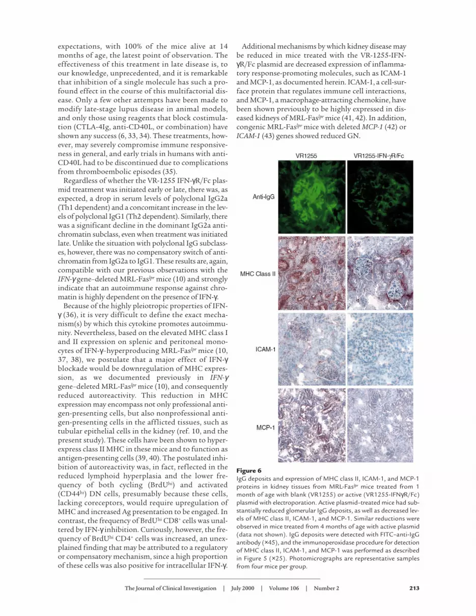
Kidney-infiltrating T cells (CD3+) and macrophages(F4/80+) were diminished in mice injected with VR1255-IFN-γR/Fc at both the predisease and advanced diseasestages regardless of electroporation (Figure 5). Decreasedkidney IgG deposits as well as protein expression ofMHC class II, ICAM-1, and MCP-1 were also observed inthe active plasmid–treated mice (Figure 6).
DiscussionWe demonstrated that: (a) intramuscular injections ofplasmids with cDNA encoding IFN-γR/Fc protectedMRL-Faslpr mice from development of lymphoid hyper-plasia and lupus-like disease, (b) electroporation at theinjection site significantly enhanced the expression ofthe fusion protein and its protective effects, and (c) thismode of treatment was highly effective in prolongingsurvival and ameliorating the serologic and histologicparameters of the disease, even when initiated at theadvanced stage, thereby making it a good candidate forhuman application.
Previous studies have attempted to neutralize IFN-γin mouse lupus models using polyclonal Ab’s (9) ormAb’s (21), as well as soluble recombinant IFN-γR (22).These approaches, however, have limitations: for exam-ple, with regard to Ab’s, large quantities may berequired, the Ab may not attain sufficient concentra-tions in secondary lymphoid organs or sites of inflam-mation, and it may be neutralized by host immune
The Journal of Clinical Investigation | July 2000 | Volume 106 | Number 2 211
Figure 4Long-term in vivo BrdU incorporation (9 days in drinking water) inT-cell subsets of mice treated late in the disease with either the blankplasmid (left panels) or the IFN-R/Fc cDNA plasmid (right panels)coupled with electroporation. Assessments were made 7 monthsafter initiation of treatment. Cells were first stained with the indi-cated surface markers, fixed, stained with anti-BrdU, and analyzedby FACS. Profiles are representative of one mouse, and mean ± SEMof BrdUhi (cycling) cells are derived from four mice per group.
Table 2Reduction of kidney disease in IFN-γR/Fc plasmid–treated mice
Disease stage Treatment Kidney disease
GNA BUNA
Prediseased No Rx 3.5 ± 0.1 3.9 ± 0.1Blank plasmid 3.5 ± 0.1 4.0 ± 0.0IFN-γR/Fc plasmid 2.8 ± 0.1 2.1 ± 0.3B
Prediseased Electroporation alone 3.7 ± 0.2 4.0 ± 0.0Blank plasmid + electroporation 3.7 ± 0.2 4.0 ± 0.0IFN-γR/Fc plasmid + electroporation 2.4 ± 0.1B 1.3 ± 0.3B
Diseased Blank plasmid + electroporation 3.8 ± 0.1 3.9 ± 0.1IFN-γR/Fc plasmid + electroporation 2.3 ± 0.2B 1.1 ± 0.4B
AGN and BUN grades (mean ± SEM) scored from 0–4. BP < 0.001 betweenactive plasmid– and blank plasmid–treated groups (n = 8–14 mice per group).

responses. With regard to soluble recombinant recep-tors, rapid turnover may affect efficacy and necessitatefrequent administration. These constraints mightexplain the negative result with anti-IFNγ mAb treat-ment of MRL-Faslpr mice reported previously (21) andthe finding that treatment with recombinant solubleIFN-γR in (NZB × W)F1 lupus mice was effective onlywhen initiated early, but not late, when IFN-γ levels aresignificantly higher (22).
Several of these problems may be overcome by intra-muscular injection of nonviral vectors, which caninduce sustained production of cytokine agonists orantagonists. This approach was originally implement-ed in MRL-Faslpr mice by Raz et al. (23, 24), who showedthat injections of a cDNA vector encoding TGF-β com-mencing early in life significantly reduced diseaseparameters. We have now used a nonviral vector encod-ing a secreted IFN-γR/Fc (IFN-γ inhibitory fusion pro-tein) as a means to assess its prophylactic and thera-peutic effects in this lupus model. We used IFN-γRfused to IgG1 Fc as the inhibitor instead of the trun-cated receptor alone, since fusion molecules secreted ashomodimers have been reported to have much longerhalf-lives than truncated IFN-γR (40 vs. 1–3 hours,respectively) (25, 26), and dimeric IFN-γR/Fc fusionproteins exhibit higher ligand avidity than single-chainreceptors (27). Finally, IgG1 Fc, commonly used inthese cases, was chosen as the partner to create theenhanced half-life–displaying biomolecule because thisIgG subclass does not activate complement. In addi-tion, to enhance systemic fusion protein expression, wecoupled the intramuscular injections with local elec-troporation. Among the many methods used to pro-mote naked plasmid DNA cellular transfer and geneexpression, application of low field strength, square-wave electric pulses through external or invasive elec-trodes appears to produce markedly higher transferefficiency (18, 28–31). This has been attributed toincreased numbers of muscle fibers that take up plas-mid DNA and probably increased copy number of plas-
mids introduced into each muscle cell. Indeed, when weapplied this procedure, levels of IFN-γR/Fc in themajority of treated animals exceeded 100 ng/mL, andthe ligand levels were consequently reduced to approx-imately 10–25% of those in controls.
Prophylactic treatment initiated at the prediseasestage with injections of the VR-1255-IFN-γR/Fc plas-mid without local electroporation effectively protect-ed mice from early lethality and reduced serologic andhistologic disease markers. These benefits wereobserved despite low serum levels of the fusion pro-tein, which may, to some extent, be due to the seques-tration of the receptor in IFN-γ–producing lymphoidorgans and inflammatory sites. This possibility is sup-ported by the fact that decreases in IFN-γ serum lev-els were still substantial. The degree of prophylaxisafforded by this regimen is not unlike the protectionwe reported previously in MRL-Faslpr mice heterozy-gous for the IFN-γ gene deletion (10) and that report-ed by others (32) in a subline of long-lived MRL-Fasl-
pr mice in which IFN-γ levels were one-third that ofconventional MRL-Faslpr mice. In the heterozygousIFN-γ gene–deleted mouse, however, despite reducedGN and prolonged survival, autoantibody levels andIgG kidney deposits were unaffected (10). It appears,therefore, that the active plasmid treatment is morepotent than the drop in serum IFN-γ levels suggests,presumably due to the postulated effects of thesequestered receptor in inflammatory sites. Overall,the data indicate that even a fractional inhibition ofIFN-γ is sufficient to reduce disease progression andseverity. Nevertheless, injections of the active plasmidcombined with electroporation significantlyimproved protection commensurate with several-foldincreases in serum levels of the IFN-γ inhibitoryfusion protein.
The positive results with early-life treatment pro-vided the impetus to apply this mode of therapy tomice with established disease. Despite the severity ofthe underlying illness, survival was extended beyond
212 The Journal of Clinical Investigation | July 2000 | Volume 106 | Number 2
Figure 5CD3+ and F4/80+ cells in kidney tissue from MRL-Faslpr mice treated with blank (VR1255) or active(VR1255-IFN-γR/Fc) plasmid with electropora-tion from 1 month of age. The numbers of T cells(CD3+), and especially macrophages (F4/80+),are markedly reduced in the active plasmid–treat-ed groups. Similar reductions were observed inmice treated from 4 months of age with activeplasmid (data not shown). Tissue sections werestained with either biotinylated anti-CD3 or anti-F4/80, incubated with streptavidin-horseradishperoxidase, and developed with AEC (see Meth-ods). Photomicrographs are representative sec-tions from four mice in each group. ×25.
expectations, with 100% of the mice alive at 14months of age, the latest point of observation. Theeffectiveness of this treatment in late disease is, toour knowledge, unprecedented, and it is remarkablethat inhibition of a single molecule has such a pro-found effect in the course of this multifactorial dis-ease. Only a few other attempts have been made tomodify late-stage lupus disease in animal models,and only those using reagents that block costimula-tion (CTLA-4Ig, anti-CD40L, or combination) haveshown any success (6, 33, 34). These treatments, how-ever, may severely compromise immune responsive-ness in general, and early trials in humans with anti-CD40L had to be discontinued due to complicationsfrom thromboembolic episodes (35).
Regardless of whether the VR-1255 IFN-γR/Fc plas-mid treatment was initiated early or late, there was, asexpected, a drop in serum levels of polyclonal IgG2a(Th1 dependent) and a concomitant increase in the lev-els of polyclonal IgG1 (Th2 dependent). Similarly, therewas a significant decline in the dominant IgG2a anti-chromatin subclass, even when treatment was initiatedlate. Unlike the situation with polyclonal IgG subclass-es, however, there was no compensatory switch of anti-chromatin from IgG2a to IgG1. These results are, again,compatible with our previous observations with theIFN-γ gene–deleted MRL-Faslpr mice (10) and stronglyindicate that an autoimmune response against chro-matin is highly dependent on the presence of IFN-γ.
Because of the highly pleiotropic properties of IFN-γ (36), it is very difficult to define the exact mecha-nism(s) by which this cytokine promotes autoimmu-nity. Nevertheless, based on the elevated MHC class Iand II expression on splenic and peritoneal mono-cytes of IFN-γ–hyperproducing MRL-Faslpr mice (10,37, 38), we postulate that a major effect of IFN-γblockade would be downregulation of MHC expres-sion, as we documented previously in IFN-γgene–deleted MRL-Faslpr mice (10), and consequentlyreduced autoreactivity. This reduction in MHCexpression may encompass not only professional anti-gen-presenting cells, but also nonprofessional anti-gen-presenting cells in the afflicted tissues, such astubular epithelial cells in the kidney (ref. 10, and thepresent study). These cells have been shown to hyper-express class II MHC in these mice and to function asantigen-presenting cells (39, 40). The postulated inhi-bition of autoreactivity was, in fact, reflected in thereduced lymphoid hyperplasia and the lower fre-quency of both cycling (BrdUhi) and activated(CD44hi) DN cells, presumably because these cells,lacking coreceptors, would require upregulation ofMHC and increased Ag presentation to be engaged. Incontrast, the frequency of BrdUhi CD8+ cells was unal-tered by IFN-γ inhibition. Curiously, however, the fre-quency of BrdUhi CD4+ cells was increased, an unex-plained finding that may be attributed to a regulatoryor compensatory mechanism, since a high proportionof these cells was also positive for intracellular IFN-γ.
Additional mechanisms by which kidney disease maybe reduced in mice treated with the VR-1255-IFN-γR/Fc plasmid are decreased expression of inflamma-tory response-promoting molecules, such as ICAM-1and MCP-1, as documented herein. ICAM-1, a cell-sur-face protein that regulates immune cell interactions,and MCP-1, a macrophage-attracting chemokine, havebeen shown previously to be highly expressed in dis-eased kidneys of MRL-Faslpr mice (41, 42). In addition,congenic MRL-Faslpr mice with deleted MCP-1 (42) orICAM-1 (43) genes showed reduced GN.
The Journal of Clinical Investigation | July 2000 | Volume 106 | Number 2 213
Figure 6IgG deposits and expression of MHC class II, ICAM-1, and MCP-1proteins in kidney tissues from MRL-Faslpr mice treated from 1month of age with blank (VR1255) or active (VR1255-IFNγR/Fc)plasmid with electroporation. Active plasmid–treated mice had sub-stantially reduced glomerular IgG deposits, as well as decreased lev-els of MHC class II, ICAM-1, and MCP-1. Similar reductions wereobserved in mice treated from 4 months of age with active plasmid(data not shown). IgG deposits were detected with FITC–anti-IgGantibody (×45), and the immunoperoxidase procedure for detectionof MHC class II, ICAM-1, and MCP-1 was performed as describedin Figure 5 (×25). Photomicrographs are representative samplesfrom four mice per group.

Gene therapy for autoimmune diseases continues toreceive considerable attention (44, 45). The delivery ofIFN-γ inhibitory molecules by intramuscular injectionof plasmid vectors is simple and appears to be nontox-ic and safe. The use of this approach for gene therapyof autoimmune diseases circumvents several problemsencountered with viral vectors (46) because the plasmidwill not reactivate to a pathogenic state, is unlikely tobe incorporated in genomic DNA or to be neutralizedby the host’s immune response, and does not stimulatea local inflammatory response. This approach may alsobe superior to recombinant soluble molecules in that itprovides a depot of genetic material for long-termexpression of the active biomolecule. In addition, desir-able effects on affected microenvironments may bemore potent, since recent studies (47) have shown thatnaked DNA is transported from the injection site tonot only the regional lymph nodes, but also to distantorgans such as the spleen and very likely to the afflict-ed tissues. Finally, since inhibition of IFN-γ has beenshown to be of benefit in other autoimmune diseases,such as myasthenia gravis (48, 49) and insulin-depend-ent diabetes mellitus (50), the mode of therapy out-lined here may have broader application.
AcknowledgmentsThis is Publication No. 13208-IMM from the Depart-ment of Immunology, The Scripps Research Institute,10550 North Torrey Pines Road, IMM3, La Jolla, Cal-ifornia 92037. The results reported herein were sup-ported, in part, by NIH grants AR31203, AG15061,and AR39555. B.R. Lawson was supported by NIHTraining Grant AG-00080. We thank Ms. M. K.Occhipinti for editorial assistance.
1. Boumpas, D.T., et al. 1995. Systemic lupus erythematosus: emerging con-cepts. Part 2: dermatologic and joint disease, the antiphospholipid anti-body syndrome, pregnancy and hormonal therapy, morbidity and mor-tality, and pathogenesis. Ann. Int. Med. 123:42–53.
2. Theofilopoulos, A.N., and Kono, D.H. 1999. The genes of systemicautoimmunity. Proc. Assoc. Am. Physicians. 111:228–240.
3. Kaliyaperumal, A., Michaels, M.A., and Datta, S. 1999. Antigen-specifictherapy of murine lupus nephritis using nucleosomal peptides: toler-ance spreading impairs pathogenic function of autoimmune T and Bcells. J. Immunol. 162:5775–5783.
4. Wofsy, D. 1993. Treatment of murine lupus with anti-CD4 monoclonalantibodies. Immunol. Ser. 59:221–236.
5. Mohan, C., Shi, Y., Laman, J.D., and Datta, S.K. 1995. Interactionbetween CD40 and its ligand gp39 in the development of murine lupusnephritis. J. Immunol. 154:1470–1480.
6. Finck, B.K., Linsley, P.S., and Wofsy, D. 1994. Treatment of murine lupuswith CTLA4Ig. Science. 265:1225–1227.
7. Theofilopoulos, A.N., and Lawson, B.R. 1999. Tumour necrosis factorand other cytokines in murine lupus. Ann. Rheum. Dis. 58:149–155.
8. Kelley, V.R., and Wuthrich, R.P. 1999. Cytokines in the pathogenesis ofsystemic lupus erythematosus. Semin. Nephrol. 19:57–66.
9. Jacob, C.O., Van der Meide, P.H., and McDevitt, H.O. 1987. In vivo treat-ment of (NZB×NZW)F1 lupus-like nephritis with monoclonal antibodyto gamma interferon. J. Exp. Med. 166:798–803.
10. Balomenos, D., Rumold, R., and Theofilopoulos, A.N. 1998. Interferon-gamma is required for lupus-like disease and lymphoaccumulation inMRL-lpr mice. J. Clin. Invest. 101:364–371.
11. Peng, S.L., Moslehi, J., and Craft, J. 1997. Roles of interferon-gamma andinterleukin-4 in murine lupus. J. Clin. Invest. 99:1936–1946.
12. Haas, C., Ryffel, B., and LeHir, M. 1998. IFN-gamma receptor deletionprevents autoantibody production and glomerulonephritis in lupus-prone (NZB×NZW)F1 mice. J. Immunol. 160:3713–3718.
13. Haas, C., Ryffel, B., and LeHir, M. 1997. IFN-gamma is essential for the
development of autoimmune glomerulonephritis in MRL/lpr mice. J.Immunol. 158:5485–5491.
14. Schwarting, A., Wada, T., Kinoshita, K., Tesch, G., and Kelley, V.R. 1998.IFN-gamma receptor signaling is essential for the initiation, accelera-tion, and destruction of autoimmune kidney disease in MRL-Faslpr mice.J. Immunol. 161:494–503.
15. Piccirillo, C.A., and Prud’homme, G.J. 1999. Prevention of experimentalallergic encephalomyelitis by intramuscular gene transfer with cytokine-encoding plasmid vectors. Hum. Gene Ther. 10:1915–1922.
16. Prud’homme, G.J., Kono, D.H., and Theofilopoulos, A.N. 1995. Quanti-tative polymerase chain reaction analysis reveals marked overexpressionof interleukin-1β, interleukin-10, and interferon-gamma mRNA in thelymph nodes of lupus-prone mice. Mol. Immunol. 32:495–503.
17. Hartikka, J., et al. 1996. An improved plasmic DNA expression vector fordirect injection into skeletal muscle. Hum. Gene Ther. 20:1205–1207.
18. Aihara, H., and Miyazaki, J.-I. 1998. Gene transfer into muscle by elec-troporation in vivo. Nat. Biotechnol. 16:867–870.
19. Jung, T., Schauer, U., Heusser, C., Neumann, C., and Rieger, C. 1993.Detection of intracellular cytokines by flow cytometry. J. Immunol. Meth-ods. 159:197–207.
20. Theofilopoulos, A.N., and Dixon, F.J. 1985. Murine models of systemiclupus erythematosus. Adv. Immunol. 37:269–390.
21. Nicoletti, F., et al. 1992. In vivo treatment with a monoclonal antibody tointerferon-gamma neither affects the survival nor the incidence of lupus-nephritis in the MRL/lpr-lpr mouse. Immunopharmacology. 24:11–16.
22. Ozmen, L., et al. 1995. Experimental therapy of systemic lupus erythe-matosus: the treatment of NZB/W mice with mouse soluble interferon-gamma receptor inhibits the onset of glomerulonephritis. Eur. J.Immunol. 25:6–12.
23. Raz, E., et al. 1993. Systemic immunological effects of cytokine genesinjected into skeletal muscle. Proc. Natl. Acad. Sci. USA. 90:4523–4527.
24. Raz, E., et al. 1995. Modulation of disease activity in murine systemiclupus erythematosus by cytokine gene delivery. Lupus. 4:286–292.
25. Ozmen, L., et al. 1993. Mouse soluble IFN-gamma receptor as IFN-gamma inhibitor. J. Immunol. 150:2698–2705.
26. Kurschner, C., Ozmen, L., Garotta, G., and Dembic, Z. 1992. IFN-gammareceptor-Ig fusion proteins. Half-life, immunogenicity, and in vivo activ-ity. J. Immunol. 149:4096–4100.
27. Kurschner, C., Garotta, G., and Dembic, Z. 1992. Construction, purifi-cation, and characterization of new interferon gamma (IFN gamma)inhibitor proteins. Three IFN gamma receptor-immunoglobulin hybridmolecules. J. Biol. Chem. 267:9354–9360.
28. Muramatsu, T., Nakjamura, A., and Park, H.-M. 1998. In vivo electropo-ration: a powerful and convenient means of nonviral gene transfer to tis-sues of living animals. Int. J. Mol. Med. 1:55–62.
29. Rols, M.-P., et al. 1998. In vivo electrically mediated protein and genetransfer in murine melanoma. Nat. Biotechnol. 16:168–171.
30. Horton, H.M., et al. 1999. A gene therapy for cancer using intramuscu-lar injection of plasmid DNA encoding interferon-alpha. Proc. Natl. Acad.Sci. USA. 96:1553–1558.
31. Mir, L.M., et al. 1999. High-efficiency gene transfer into skeletal musclemediated by electric pulses. Proc. Natl. Acad. Sci. USA. 96:4262–4267.
32. Takahashi, S., et al. 1996. Imbalance towards Th1 predominance is asso-ciated with acceleration of lupus-like autoimmune syndrome in MRLmice. J. Clin. Invest. 97:1597–1604.
33. Kalled, S.L., Cutler, A.H., Datta, S.K., and Thomas, D.W. 1998. Anti-CD40 ligand antibody treatment of SNF1 mice with established nephri-tis: preservation of kidney function. J. Immunol. 160:2158–2165.
34. Daikh, D.I., Finck, B.K., Linsley, P.S., Hollenbaugh, D., and Wofsy, D.1997. Long-term inhibition of murine lupus by brief simultaneousblockade of the B7/CD28 and CD40/gp39 costimulation pathways. J.Immunol. 159:3104–3108.
35. Kawai, T., Andrews, D., Colvin, R.B., Sachs, D.H., and Cosimi, A.B. 2000.Thromboembolic complications after treatment with monoclonal anti-body against CD40 ligand. Nat. Med. 6:114.
36. Boehm, U., Klamp, T., Groot, M., and Howard, J.C. 1997. Cellularresponses to interferon-gamma. Annu. Rev. Immunol. 15:749–795.
37. Kofler, R., Schreiber, R.D., Dixon, F.J., and Theofilopoulos, A.N. 1984.Macrophage I-A/I-E expression and macrophage-stimulating lym-phokines in murine lupus. Cell Immunol. 87:92–100.
38. Kelley, V.E., and Roths, J.B. 1982. Increase in macrophage Ia expressionin autoimmune mice: role of the lpr gene. J. Immunol. 129:923–925.
39. Wuthrich, R.P., et al. 1997. Enhanced MHC class II expression in renalproximal tubules precedes loss of renal function in MRL/lpr mice withlupus nephritis. Am. J. Pathol. 134:45–51.
40. Wuthrich, R.P., et al. 1990. MHC class II, antigen presentation andtumor necrosis factor in renal tubular epithelial cells. Kidney Int.37:783–792.
41. Wuthrich, R.P., Jevnikar, A.M., Takei, F., Glimcher, L.H., and Kelley, V.E.1990. Intercellular adhesion molecule-1 (ICAM-1) expression is upregu-
214 The Journal of Clinical Investigation | July 2000 | Volume 106 | Number 2

lated in autoimmune murine lupus nephritis. Am. J. Pathol. 136:441–450.42. Tesch, G.H., Maifert, S., Schwarting, A., Rollins, B.J., and Kelley, V.R.
1999. Monocyte chemoattractant protein 1-dependent leukocytic infil-trates are responsible for autoimmune disease in MRL-Faslpr mice. J. Exp.Med. 190:1813–1824.
43. Bullard, D.C., et al. 1997. Intercellular adhesion molecule-1 deficiencyprotects MRL/MpJ-Fas (lpr) mice from early lethality. J. Immunol.159:2058–2067.
44. Mathisen, P.M., and Tuohy, V.K. 1998. Gene therapy in the treatment ofautoimmune disease. Immunol. Today. 19:103–105.
45. Evans, C.H., Whalen, J.D., Evans, C.H., Ghivizzani, S.C., and Robbins, P.D.1998. Gene therapy in autoimmune diseases. Ann. Rheum. Dis. 57:125–127.
46. Mulligan, R.C. 1993. The basic science of gene therapy. Science.
260:926–931.47. La Cava, A., et al. 2000. Cell-mediated DNA transport between distant
inflammatory sites following intradermal DNA immunization in the pres-ence of adjuvant. J. Immunol. 164:1340–1345.
48. Balasa, B., et al. 1997. Interferon gamma (IFN-gamma) is necessary for thegenesis of acetylcholine receptor-induced clinical experimental autoim-mune myasthenia gravis in mice. J. Exp. Med. 186:385–391.
49. Zhang, G.-X., et al. 1999. Mice with IFN-γreceptor deficiency are less sus-ceptible to experimental autoimmune myasthenia gravis. J. Immunol.162:3775–3781.
50. Nicoletti, F., et al. 1996. The effects of a nonimmunogenic form of murinesoluble interferon-γreceptor on the development of autoimmune diabetesin the NOD mouse. Endocrinology. 137:5567–5575.
The Journal of Clinical Investigation | July 2000 | Volume 106 | Number 2 215