Molecular cloning and nucleotide sequence of a human renin cDNA fragment
10
Volume 11 Number 20 1983 Nucleic Acids Research Molecular cloning and nadeotide sequence of a human renin cDNA fragment Florent Soubrier* + , Jean-Jacques Panthier + , Pierre Corvol* and Francois Rougeon + Unite de Genetique et Biochimie du Developpement, Institut Pasteur, ERA CNRS 851, 25 rue du Docteur Roux, 75724 Paris Cedex 15, and * Institut National de la Sante et de la Recherche Medicale, Unite 36, 17 rue du Fer a Moulin, 75005 Paris, France Received 1 August 1983; Revised and Accepted 22 September 1983 ABSTRACT We have studied human renin messenger RNA by hybridiza- tion with the mouse submaxillary gland (SMG) renin cDNA probe. The human kidney messenger RNA is about 1.6 kilobase (kb) long, similarly to the mouse SMG renin mRNA. A kidney renin cDNA clone of 1.1 kb length was obtained. A comparison of nucleotide se- quences of mouse and human cDNA clones reveals conservation of residues involved in catalytic mechanisms and a potential glyco- sylation site. The human renin molecular probe allowed us to study renin expression in human chorionic tissue. The chorionic and kidney renin messenger RNAs are similar in length. The Southern blot analysis reveals the presence of a single renin gene in human DNA. INTRODUCTION Interest in the renin angiotensin system is related to the facts that renin plays a major role in the regulation of blood pressure and water and salt metabolism, and that pharmaco- logical blockade of this system is extremely effective in the treatment of hypertension. The main source of human renin is the juxtaglomerular cells of the afferent arterioles in the kidney. Other sources of renin or renin-like enzymes are the chorion (1) and the central nervous system (2). In the kidney, renin is found mainly as an active enzyme, whereas chorionic cells con- tain an inactive form of the enzyme which is trypsin activable. Biosynthetic experiments with chorionic cell cultures suggest that inactive renin is the proform of the active enzyme (3). Similarly in renin-secreting tumors, an inactive 54 KD renin precursor is converted into an active 44 KD enzyme (4). Although human renal renin has been purified to homogeneity (5), there are no data concerning its primary structure, due to its low concentration in the kidney. To date none of the extra renal © IRL Press Limited, Oxford, England. 7181 by guest on July 27, 2016 http://nar.oxfordjournals.org/ Downloaded from
Transcript of Molecular cloning and nucleotide sequence of a human renin cDNA fragment

Volume 11 Number 20 1983 Nucleic Acids Research
Molecular cloning and nadeotide sequence of a human renin cDNA fragment
Florent Soubrier* + , Jean-Jacques Panthier+, Pierre Corvol* and Francois Rougeon
+ Unite de Genetique et Biochimie du Developpement, Institut Pasteur, ERA CNRS 851, 25 rue duDocteur Roux, 75724 Paris Cedex 15, and * Institut National de la Sante et de la RechercheMedicale, Unite 36, 17 rue du Fer a Moulin, 75005 Paris, France
Received 1 August 1983; Revised and Accepted 22 September 1983
ABSTRACTWe have studied human renin messenger RNA by hybridiza-
tion with the mouse submaxillary gland (SMG) renin cDNA probe.The human kidney messenger RNA is about 1.6 kilobase (kb) long,similarly to the mouse SMG renin mRNA. A kidney renin cDNA cloneof 1.1 kb length was obtained. A comparison of nucleotide se-quences of mouse and human cDNA clones reveals conservation ofresidues involved in catalytic mechanisms and a potential glyco-sylation site. The human renin molecular probe allowed us tostudy renin expression in human chorionic tissue. The chorionicand kidney renin messenger RNAs are similar in length. TheSouthern blot analysis reveals the presence of a single reningene in human DNA.
INTRODUCTION
Interest in the renin angiotensin system is related to
the facts that renin plays a major role in the regulation of
blood pressure and water and salt metabolism, and that pharmaco-
logical blockade of this system is extremely effective in the
treatment of hypertension. The main source of human renin is the
juxtaglomerular cells of the afferent arterioles in the kidney.
Other sources of renin or renin-like enzymes are the chorion (1)
and the central nervous system (2). In the kidney, renin is
found mainly as an active enzyme, whereas chorionic cells con-
tain an inactive form of the enzyme which is trypsin activable.
Biosynthetic experiments with chorionic cell cultures suggest
that inactive renin is the proform of the active enzyme (3).
Similarly in renin-secreting tumors, an inactive 54 KD renin
precursor is converted into an active 44 KD enzyme (4). Although
human renal renin has been purified to homogeneity (5), there
are no data concerning its primary structure, due to its low
concentration in the kidney. To date none of the extra renal
© IRL Press Limited, Oxford, England. 7181
by guest on July 27, 2016http://nar.oxfordjournals.org/
Dow
nloaded from

Nucleic Acids Research
renin-like enzymes in man have been extensively purified, and
their relation to the kidney enzyme remains speculative,
although iramunohistochemical studies have shown imraunological
similarities. The purpose of this communication is to report
the partial amino-acid sequence of human renal renin deduced
from the nucleotide sequence, which shows a striking homology
with both mouse submaxillary gland (SMG) renin and other aspar-
tyl proteases. In addition, we provide evidence that human
renal and chorionic renin mRNAs are the same length and that in
these different organs, they are transcribed from a single gene.
MATERIALS AND METHODS
Mouse SMG renin probe
Clone pRnl-4, 1427 nucleotide length, contains an essen-
tially complete renin mRNA transcript and is inserted in the
PstI site of pBR322 (6) . Double-stranded DNA was labeled by
nick-translation (7) . Purified insert was isolated from a 3,5 %
acrylamide gel for Southern blot hybridization and screening of
the cDNA library.
DNA preparation
Human female and mouse BALB/c male DNAs were extracted
respectively from frozen placenta and liver. High molecular
weight DNA was prepared by a modification of the methods of
Blin and Stafford (8). Restriction digest, gel electrophoresis
and Southern blot hybridization were performed as described by
Heldmann and Rougeon (9). Mouse SMG renin cDNA insert and a
EcoRI-SacI restriction fragment isolated from human cDNA insert,
ligated and nick-translated, were used as probes. Filters were
washed using high stringency conditions, as defined in this
paper.
mRNA Isolation and Northern blot technique
SMG, kidney, chorion and leukemic lymphoid total RNA was
prepared by the lithium chloride-urea method as described (10).
Fractionation of poly(A)-containing RNA on oligo(dT)cellulose
was performed as described (10) . Glyoxal-dimethylsulfoxide
treated RNA was electrophoresed, transferred to nitrocellulose
filter and hybridized with the mouse SMG renin probe according
to Thomas (11). Washing conditions were 1 x SSC (50°C) for inter-
7182
by guest on July 27, 2016http://nar.oxfordjournals.org/
Dow
nloaded from

Nucleic Acids Research
species hybridization and 0.1 x SSC (50°C) for homologous hybri-
dization (Fig. 1) .
Construction of a cDNA library from kidney mRNA
Poly(A) mRNAs were fractionated on a 5-20 % sucrose
gradient. Fractions were checked for renin mRNA by hybridization
of an aliquot with mouse SMG cDNA probe by the Northern blot
technique (11) . Fractions enriched for renin sequence were used
as a template for reverse transcription (12). The cDNA obtained
was converted into double-stranded SI nuclease resistant DNA (121
Double-stranded cDNA was fractionated on a 5-20 % sucrose gra-
dient and larger cDNA was inserted into the PstI site of pBR322
by the oligo-d(CG) tailing method (12). Host bacteria were
transformed and colonies were screened by hybridization with
the nick-translated cDNA insert of pRnl-4 as a probe. =< 10,000
clones were obtained and the recombinant clone pHRn22, contai-
ning the larger insert, 1,100 base pairs, was kept for further
analysis among five clones exhibiting common restriction sites.
DNA sequence analysis (Fig. 2)
DNA fragments were labeled at their 5' ends by the poly-
nucleotide kinase exchange reaction or at their 3' ends, either
by the terminal deoxynucleotidyl-transferase or the Klenow frag-
ment of DNA polymerase I. The chemical degradation reaction was
performed using the method of Maxam and Gilbert (13).
1743 •» ~ m1431 <—
1 2 3 4
Figure 1 : Characterization of human renin mRNA by Northern blothybridization with the mouse SMG renin cDNA probe.Lane 1, pBR322 digested by Avail. Lane 2, human kidneymRNA (15 yg). Lane 3, kidney mRNA from SWISS female(10 ug). Lane 4, SWISS SMG mRNA (0.1 yg).In another experiment, human mRNA from leukemic cellswas used as a negative control and no hybridizationwas detectable (data not shown).
7183
by guest on July 27, 2016http://nar.oxfordjournals.org/
Dow
nloaded from

Nucleic Acids Research
Pltl1 Avail
no
••
Figure
Hbtfl
^
2 :
Unfl
Sequencing
Hlnfl-Snfl
— •—•* • —
—•
strategy of
flnfl Wall
— • ' . — : ^• -»
•-• • <—
recombinant plasmid
>acl
'UT 1' •
ilOObp
pHRn22.
Pltl
1-•
DNA fragments were labeled at their 5' ends by thepolynucleotide kinase exchange reaction (# +•) or attheir 3' ends (• ••) either by the terminal deoxy-nucleotidyltransferase or the klenow fragment of DNApolymerase I.
RESULTS AND DISCUSSION
Molecular cloning of SWISS mouse submaxillary gland renin
has allowed the determination of the complete amino acid sequen-
ce of the precursor (44 KD). This precursor is converted into
active renin, which consists of two chains of 35 and 3 KD (6).
The general strategy was to use the mouse SMG cDNA as a molecu-
lar probe to study human renin mRNA in the kidney and the cho-
rion, as well as the human renin structural gene. Since infarc-
ted kidney synthesizes increased amounts of renin, messenger RNA
was prepared from a surgically removed infarcted kidney due to
arterial thrombosis and causing severe arterial hypertension.
Characterization of the renin mRNA was performed by the
Northern blot technique. The autoradiogram presented in Figure 1
shows that among kidney poly(A) containing RNA, a mRNA popula-
tion hybridizes specifically with the mouse SMG probe. The 1.6
kb length of this mRNA is similar to that of the mouse kidney
and SMG renin mRNA.
Because of its low abundance, kidney mRNA was enriched
for renin message by fractionation on a sucrose gradient.
Aliquots of each fraction were tested by Northern blot hydridi-
zation with the mouse SMG renin probe. Fractions containing
renin mRNA were pooled and used to construct a cDNA library of
approximately 10,000 clones. Colonies were screened by hybridi-
zation with the mouse SMG renin P cDNA as a probe. One clone,
pHRn22, containing a 1,100 base pairs insert, was chosen among
five hybridizing colonies for further experiments.
That pHRn22 is derived from the 1.6 kb renin mRNA was
7184
by guest on July 27, 2016http://nar.oxfordjournals.org/
Dow
nloaded from
Nucleic Acids Research
t .1 - i «
* LJ t— 5E-, «t CJ O
C CJ I M<B CD I «co <c i co
to O ICO I— I
P. r -
C Q C JXT CD
1t—
GG
-
CJ
CO
I r-il' U|
'S Si
Y l
i £CJ t.
Gin
i K
t-J CD 1
+} CD I
A C J IT-1 CD 1
o 3 •
2 CD CJ
5
Mat
3u
Lcn
j
ciSCD P.
K CD i E l
C? t— I «
t j CJ I ~̂J I
» I— CJ C0|
St i Si
o«( i SI
tth I COJ
CJ 3 CJ tj
a CJ i ol
3 CO I 3 |
CJ CJ CD 3i-3 CJ h- Ca•q; CD CJ O
,5 s : 51t, co CJ £J
£X*-J i
t (3 •
CO CJ I—
>~~t « : co ^
C CD I 3O CJ CD CJ
A»< CJ ^Jr-l CD I " - l |
gfe
S3
SO I t N B C . CO r-JCL, (_> I QJI C***C CD I CJ
S7c5 I r-» J3j«-q; < CD CJ E~< h
B t— C J t-» • CJ
1 * £ CD S». O, (—
C CD I C «(
CJ CJ I f ? l Qj h
c j cD t *q; N."^ (
r CD i clO O I Ol
e-s : el
3>-i CJ I t ,S O r— CJ Ci)N^- CO r- CO
CD CD I CD I
3>S i H
J CJ I 3
O I »J|C CJ I t ,(0 *C CD COH< I CO
CO CJ CD I--*
i-i I— I <J
fVr- I CI
CO CJ I C0|*ri «f • ~ri|a=5 i ft:!
SjCDCJ SJr-i CD I r-i]CJ CD I Cj]
c; 5c j cj
3:3 i 4
3H- I O. O I IJ-|
1 CD I i-i
E CD t
CJ 5 CD
Tili l li l l
»CD I 3J
i. <r i 3CJ CD ' CJ
i ,31• O |
T-» CO I r-l l
i)CD i aJ<—1 O I t- t lCJ CD I l j |
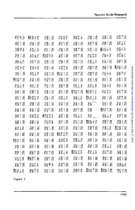
Figure 3
7185
by guest on July 27, 2016http://nar.oxfordjournals.org/
Dow
nloaded from

Nucleic Acids Research
au i ql»•-* CJ I 1-J
a>< i »
<J 3 I Ca|
J CD I 3 |
£3 ! 3\
3O< rCJ CD CJ CJ
•WCD I - U |« I— I C&
* * E ' %1
3 CJ • «S i— 1 ^»-5 CJ -a: k-i
l c j c3 <c ^
• <C CD 3M 1— 1 <a
t. CJ t *,[« CJ I tt>to I— 1 co|
F. CJ I M« CD 1 co
to - x 1 co|
*,<_> CD U
Ei< 1 t i l
» CJ 1 e
fO K~ CJ (X
CO I— 1 CO|
H o 1 «
N^Ot— I Co|
^ CD H- COt~-» CD I t ^ lO CD I C j |
fc. C J H - ^ |
£X<-J 1 DJQ)< I SI
<=c c 3 1 -*:
a CJ 1 «|
I t !g|Ot,h CJ t,|
o w n 1 u5|
II
: CJ t,
CJ CD CJ CJ
C CJ I I,
^ < I CO
O c o l -M SiCD
I * l
g-s?•q; CD I
U t -CDco CD Ito < 1 •
S f, O 1CD CJ I
f>V} t— CJ (
r31— 1 a
t,i-u tl
£>? : £|acj 1 gj
CO CD CJ Z,3 j < CD CokA«£ 1 Co
"CJ^^I 03c
i l l
B-S !
TilCD 3J
I Cj|
O I . I - I *Hr - j ; CJ 1 -sfn h <c 1 &H|
-1 CJ I
53 i-W CD
• < 1 5:
j CJ 1 q
2 CJ I— CD
= C O I EC
1 SI -*:h-
ISTIItoCD ^ OJ
i§ i n
His53 ! iH
i3<Ur-Jcj3 1 •*
5l?|CJ CD 1 CJ
s§is1Id ill•3^ : gS: CJ «X. ^
O ^ CJ« t J CJ«•«?: CD
3 CD« 1—
1 Ei ^
1 co CJ
1 ISCD CD
V.S
1 a
Thr
1 co l
.'I• •-SI
! §j• ^
CD CD
CO C J
gtBCJ
h CJ-C CJE-. <
a CJ»»» CJ
1
(_)
1—CJ«cCJ
3CD
—
CJCD
CAG
CJCJCDCD
I—CJ
GCC
CJ
1
111
C J2̂
CDt—
CJ
<c
«cCJ
11
<
111
<c1
1I
CD 1
I— CJ
h- ICJ 1CJ 1
CJ « tCD 1
3YCD 1h - CJCD <r
3 :CD »
^— C£
<c 1—
CJ rf
8Y<C I
x h- 1— x
CJ I—CJ ICJ CD
tiCJ I—
si
3 :t J
CD CJ
C D *
CD « t•a: CJCJ t—
CJ 1
3 i
3 t
7186
by guest on July 27, 2016http://nar.oxfordjournals.org/
Dow
nloaded from

Nucleic Acids Research
demonstrated by nucleotide sequence analysis of the clone
(Fig. 3). Alignment of the nucleotide sequence with the corres-
ponding sequence of mouse SMG renin cDNA reveals considerable
homology. The overall homology between human renal and mouse SMG
renin is 77 % at the nucleotide level in the coding region, as
expected from mouse-human divergence. The rate of silent subs-
titutions is 6.3 %. A deletion of 9 nucleotides occurred in SMG
renin sequence after residue 228 and a deletion of 3 nucleotides
in human renin is present at residue 310. At the amino acid
level, the homology in the sequenced region is 69.6 %. In the
protein sequence encoded by the clone, amino acid residues
known to be involved in the catalytic mechanisms are conserved.
Aspartate 286, corresponding to aspartate 215 of pepsin, is
present as well as the two following residues which are also
present in mouse SMG renin and other aspartyl proteases. Other
araino acids involved in the catalytic mechanisms, such as tryp-
tophan 108 and tyrosine 146, corresponding to tryptophan 39 and
tyrosine 75, respectively, of pepsin are present. Cysteines in-
volved in disulphide bonds 114-121 and 277-281 are conserved at
the same position.A potential N-glycosylation site is shown by
the presence of the sequence Asn-Gly-Thr located at residues
138-139-140. This results was expected since human renal reninis
stained by Schiff reaction (5) and binds to Con-A-sepharose (14).
Comparison with the mouse SMG renin sequence shows that the di-
basic Arg352 - Arg353 pair is replaced by a Ser - Ser dipeptide.
Figure 3 : Sequence of human renin cDNA and comparison with mouseSMG renin cDNA.The two upper lines show the nucleotide sequence(below) and predicted amino acid sequence (above) ofhuman renin. The two lower lines indicate nucleotidesequence of the homologous region (above) and thepredicted amino acid sequence (below) of mouse SMGrenin. Only nucleotides which differ from the humanrenin chain are shown. Homologous amino acids areunderlined. Deletions are indicated by parentheses. Themouse SMG renin nucleotide and amino acid sequencespreviously published (6) have been revised from residue195 to residue 198 in view of the amino acid sequencepublished by Misono et al. (17) . Since the pHRn22clone does not include the 5' region of the codingsequence, nucleotides and amino acids numberscorrespond to the mouse SMG renin sequence, beginningwith the first residue of renin precursor.
7187
by guest on July 27, 2016http://nar.oxfordjournals.org/
Dow
nloaded from

Nucleic Acids Research
17431431
1 2 3Figure 4 : Extra renal expression of renin.
The human renin cDNA clone pHRn22 was used as a probe.Lane 1, pBRn22 digested by Avail. Lane 2, kidney mRNA(10 pg). Lane 3, chorion mRNA (40 pg).
The cleavage site between chain A and B of submaxillary renin
occurs after this Arg - Arg pair and might be located after the
dibasic pair Lys35it- Lys355 in the case of human renin. However,
there is no evidence, at the present time, for such a processing
of the human enzyme. It can be noticed that there is a 87 % homo-
logy between the B chain of mouse SMG renin and its correspon-
ding region in the human enzyme (Leu35g Argmji) •
The human renin clone was also used to investigate the
extra renal expression of renin. Chorion was isolated from a
healthy woman undergoing full term cesarean section. Poly(A)
containing RNA was prepared and analyzed by the Northern blot
technique. As shown in Figure 4, a unique mRNA, that is the same
size as renal renin mRNA, hybridizes with human renin cDNA probe.
To determine if renin mRNA from both kidney and chorion
is transcribed from different genes, we made a human DNA blot
hybridization with mouse SMG cDNA as a probe, since this cDNA
contains an almost complete mRNA transcript. Human DNA was pre-
pared from a placenta of a female fetus after cesarean section
and digested with EcoRI restriction enzyme, which cuts in human
renin cDNA at one site. As shown in Figure 5, two fragments of
6.0 and 3.5 kb are identified by the mouse cDNA probe.
Furthermore, with the human cDNA EcoRI-Sacl restriction fragment,
a unique 6.0 kb fragment was detected showing that there is only
one renin gene per haploid genome in humans. This single gene
appears to be transcribed into mRNAs of similar size in at least
7188
by guest on July 27, 2016http://nar.oxfordjournals.org/
Dow
nloaded from

Nucleic Acids Research
Figure 5 : Genomic pattern of human renin gene.Human DNA is digested by EcoRi. Lane 1, humanDNA hybridized with the mouse SMG renin cDNAprobe. Lane 2, human DNA hybridized with theEcoRi-SacI fragment of the human renin cDNAprobe.
two organs. However, until chorionic renin mRNA structure is
known, we cannot exclude the possibility of differential spli-
cing leading to slightly different mRNAs, but similar in sizes.
Because human renin mRNA from kidney and chorion, and
mouse SHG mRNA are approximately the same size, the molecular
weight of the human renin precursor should be similar to that of
the mouse SMG renin precursor. Based on acellular translation
and calculation from the primary amico acid sequence deduced
from the nucleotide sequence, we have previously found that the
molecular weight of mouse SMG is 44 KD (6, 15). The discrepancy
between the expected 44 KD prorenin and the tentative 54 KD human
renin precursor could well be explained by the presence of glyco-
sylated chains interfering with the molecular weight determina-
tion by SDS gel electrophoresis. Furthermore, these results
suggest that higher molecular weight renins (16) are not present
in kidney as precursors of the active renin. Renal and chorionic
renins are transcribed from a single gene into mRNAs of similar
sizes. Differences in the level of activation of renin in these
two tissues could be related either to a differental splicing of
the renin mRNAs or to different post-translational processing of
the precursor.
7189
by guest on July 27, 2016http://nar.oxfordjournals.org/
Dow
nloaded from

Nucleic Acids Research
ACKNOWLEGMENTS
This work was supported by grants from C.N.R.S. (A.T.P.
n° Al-5054 and 960008), INSERM (PRC-Genie Genetique), and the
Fondation pour la Recherche M6dicale Francaise.
REFERENCES
1 - Skinner S.L., Lumbers E.R. and Symonds E.M. (1968)Am. J. Obstet. Gynecol. 101, 529-533.
2 - Daul C.B., Heath R.G. and Garey R.E. (1975) Neuropharraaco-logy 14, 75-80.
3 - Acker G.M., Galen F.X., Devaux C , Foote S., Papernick E.,Pesty A., Mfinard J. and Corvol P. (1982) J. Clin. Endocrinol.Metab. 55, 902-909.
4a- Leckie B.J., McGhee N.K., Lever A.F. and Robertson J.I.S.(1981) in : Sambhi M.P. (ed.) Heterogeneity of renln andrenin substrate. Elsevier North-Holland Inc. New York159-174.
4b- Corvol M.T. et al., submitted.5 - Galen F.X., Devaux C , Guyenne T., Mfinard J. and Corvol P.
(1979) J. Biol. Chem. 254, 4848-4854.6 - Panthier J.J., Foote S,, Chambraud B., Strosberg A.D.,
Corvol P. and Rougeon F. (1982) Nature 298, 90-92.7 - Rigby P.W.J., Dieckmann M., Rhodes C. and Berg P. (1977)
J. Mol. Biol. 113, 237-251.8 - Blin N. and Stafford W. (1976) Nucl. Acids Res. 3, 2303-2308.9 - Heidmann 0. and Rougeon F. (1982) Cell 28, 507-513.
10 - Auffray C. and Rougeon F. (1980) Eur. J. Biochem. 107,303-314.
11 - Thomas P.S. (1980) Proc. Natl. Acad. Sci. USA 77, 5201-5205.12 - Auffray C , Nageotte R., Chambraud B. and Rougeon F. (1980)
Nucl. Acids Res. 8, 1231-1241.13 - Maxam A.M. and Gilbert W. (1980) Methods Enzymol. 65, 499-
560.14 - Yokosawa A.H., Holladay L.A., Inagami T., Haas E. and
Murakami K. (1980) J. Biol. Chem. 255, 3498-3502.15 - Rougeon F., Chambraud B., Footes S., Panthier J.J., Nageotte
R. and Corvol P. (1981) Proc. Natl. Acad. Sci. USA 78,6367-6371.
16 - Barett J.D., Eggena P. and Sambhi M.P. (1977) Circ. Res.suppl. 2, 41, 7.
17 - Misono K.S., Chang J.J., Inagami T. (1982) Proc. Natl. Acad.Sci. USA 79, 4858-4862.
7190
by guest on July 27, 2016http://nar.oxfordjournals.org/
Dow
nloaded from