The intrarenal renin-angiotensin system in autosomal dominant polycystic kidney disease
15
doi:10.1152/ajprenal.00370.2003 287:775-788, 2004. First published Jun 8, 2004; Am J Physiol Renal Physiol Mahmoud Loghman-Adham, Carlos E. Soto, Tadashi Inagami and Lisa Cassis dominant polycystic kidney disease The intrarenal renin-angiotensin system in autosomal You might find this additional information useful... 64 articles, 28 of which you can access free at: This article cites http://ajprenal.physiology.org/cgi/content/full/287/4/F775#BIBL 4 other HighWire hosted articles: This article has been cited by [PDF] [Full Text] [Abstract] , March 1, 2005; 16 (3): 755-762. J. Am. Soc. Nephrol. St. George-Hyslop and Y. Pei A. D. Paterson, R. Magistroni, N. He, K. Wang, A. Johnson, P. R. Fain, E. Dicks, P. Parfrey, P. Autosomal Dominant Polycystic Kidney Disease Progressive Loss of Renal Function Is an Age-Dependent Heritable Trait in Type 1 [PDF] [Full Text] [Abstract] , April 15, 2005; 25 (8): 2938-2945. Mol. Cell. Biol. J. D. Engel, A. Fukamizu and K. Tanimoto T. Shimizu, T. Oishi, A. Omori, A. Sugiura, K. Hirota, H. Aoyama, T. Saito, T. Sugaya, Y. Kon, Transgene Coplacement and Site-Specific Recombination Identification of cis-Regulatory Sequences in the Human Angiotensinogen Gene by [PDF] [Full Text] [Abstract] , August 1, 2005; 53 (8): 979-988. J. Histochem. Cytochem. M. Loghman-Adham, C. E. Soto, T. Inagami and C. Sotelo-Avila Polycystic Kidney Disease Expression of Components of the Renin-angiotensin System in Autosomal Recessive [PDF] [Full Text] [Abstract] , January 1, 2006; 290 (1): R1-R10. Am J Physiol Regulatory Integrative Comp Physiol B. T. Alexander Fetal programming of hypertension including high-resolution figures, can be found at: Updated information and services http://ajprenal.physiology.org/cgi/content/full/287/4/F775 can be found at: AJP - Renal Physiology about Additional material and information http://www.the-aps.org/publications/ajprenal This information is current as of April 4, 2007 . http://www.the-aps.org/. American Physiological Society. ISSN: 0363-6127, ESSN: 1522-1466. Visit our website at (monthly) by the American Physiological Society, 9650 Rockville Pike, Bethesda MD 20814-3991. Copyright © 2005 by the respective cells and vasculature, as well as to the control of body fluid volume and composition. It is published 12 times a year publishes original manuscripts on a broad range of subjects relating to the kidney, urinary tract, and their AJP - Renal Physiology on April 4, 2007 ajprenal.physiology.org Downloaded from
-
Upload
independent -
Category
Documents
-
view
3 -
download
0
Transcript of The intrarenal renin-angiotensin system in autosomal dominant polycystic kidney disease

doi:10.1152/ajprenal.00370.2003 287:775-788, 2004. First published Jun 8, 2004;Am J Physiol Renal Physiol
Mahmoud Loghman-Adham, Carlos E. Soto, Tadashi Inagami and Lisa Cassis dominant polycystic kidney disease The intrarenal renin-angiotensin system in autosomal
You might find this additional information useful...
64 articles, 28 of which you can access free at: This article cites http://ajprenal.physiology.org/cgi/content/full/287/4/F775#BIBL
4 other HighWire hosted articles: This article has been cited by
[PDF] [Full Text] [Abstract]
, March 1, 2005; 16 (3): 755-762. J. Am. Soc. Nephrol.St. George-Hyslop and Y. Pei A. D. Paterson, R. Magistroni, N. He, K. Wang, A. Johnson, P. R. Fain, E. Dicks, P. Parfrey, P.
Autosomal Dominant Polycystic Kidney DiseaseProgressive Loss of Renal Function Is an Age-Dependent Heritable Trait in Type 1
[PDF] [Full Text] [Abstract], April 15, 2005; 25 (8): 2938-2945. Mol. Cell. Biol.
J. D. Engel, A. Fukamizu and K. Tanimoto T. Shimizu, T. Oishi, A. Omori, A. Sugiura, K. Hirota, H. Aoyama, T. Saito, T. Sugaya, Y. Kon,
Transgene Coplacement and Site-Specific RecombinationIdentification of cis-Regulatory Sequences in the Human Angiotensinogen Gene by
[PDF] [Full Text] [Abstract], August 1, 2005; 53 (8): 979-988. J. Histochem. Cytochem.
M. Loghman-Adham, C. E. Soto, T. Inagami and C. Sotelo-Avila Polycystic Kidney Disease
Expression of Components of the Renin-angiotensin System in Autosomal Recessive
[PDF] [Full Text] [Abstract], January 1, 2006; 290 (1): R1-R10. Am J Physiol Regulatory Integrative Comp Physiol
B. T. Alexander Fetal programming of hypertension
including high-resolution figures, can be found at: Updated information and services http://ajprenal.physiology.org/cgi/content/full/287/4/F775
can be found at: AJP - Renal Physiologyabout Additional material and information http://www.the-aps.org/publications/ajprenal
This information is current as of April 4, 2007 .
http://www.the-aps.org/.American Physiological Society. ISSN: 0363-6127, ESSN: 1522-1466. Visit our website at (monthly) by the American Physiological Society, 9650 Rockville Pike, Bethesda MD 20814-3991. Copyright © 2005 by therespective cells and vasculature, as well as to the control of body fluid volume and composition. It is published 12 times a year
publishes original manuscripts on a broad range of subjects relating to the kidney, urinary tract, and theirAJP - Renal Physiology
on April 4, 2007
ajprenal.physiology.orgD
ownloaded from

The intrarenal renin-angiotensin system in autosomaldominant polycystic kidney disease
Mahmoud Loghman-Adham, Carlos E. Soto, Tadashi Inagami, and Lisa CassisDepartment of Pediatrics and Pediatric Research Institute, Saint Louis University, St. Louis, Missouri 07920
Submitted 21 October 2003; accepted in final form 1 June 2004
Loghman-Adham, Mahmoud, Carlos E. Soto, Tadashi In-agami, and Lisa Cassis. The intrarenal renin-angiotensin system inautosomal dominant polycystic kidney disease. Am J Physiol RenalPhysiol 287: F775–F788, 2004. First published June 8, 2004; 10.1152/ajprenal.00370.2003.—Hypertension is a common complication ofautosomal dominant polycystic kidney disease (ADPKD), oftenpresent before the onset of renal failure. A role for the renin-angiotensin system (RAS) has been proposed, but studies of systemicRAS have failed to show a correlation between plasma renin activityand blood pressure in ADPKD. Ectopic renin expression by cystepithelium was first reported in 1992 (Torres VE, Donovan KA, SicliG, Holley KE, Thibodeau ST, Carretero OA, Inagami T, McAteer JA,and Johnson CM. Kidney Int 42: 364–373, 1992). It is not known,however, whether other RAS components are also expressed by cystsin ADPKD. We show that, in addition to renin, angiotensinogen(AGT) is produced by some cysts and dilated tubules. Angiotensin-converting enzyme, ANG II type 1 receptor, and ANG II peptide arealso present within cysts and in many tubules; and some cyst fluidscontain high ANG II concentrations. Additionally, cyst-derived cellsin culture continue to express the components of the RAS at both theprotein and mRNA levels. We further show that renin is expressedprimarily in cysts of distal tubule origin and in cyst-derived cells withdistal tubule characteristics, whereas AGT is expressed primarily incysts of proximal tubule origin and in cyst-derived cells with proximaltubule characteristics. Renin production by cyst-derived cells appearsto be regulated by extracellular Na� concentration. Based on theseobservations, we propose a model of an autocrine/paracrine RAS inpolycystic kidney disease, whereby overactivity of the intrarenalsystem results in sustained increases in intratubular ANG II concen-trations.
hypertension; cyst epithelium; polycystic kidney disease; pressure-natriuresis; renin
AUTOSOMAL DOMINANT POLYCYSTIC kidney disease (ADPKD) is acommon genetic disorder resulting in the formation of cysticdilatation of renal tubules, leading to a gradual destruction ofrenal parenchyma and renal failure in half of the patients byage 60 (59). Mutations in two genes, PKD1 and PKD2, thatencode membrane-associated proteins polycystin-1 and -2 ac-count for almost all cases of ADPKD, with PKD1 mutationsaccounting for �85% of the cases (23, 59). In ADPKD, cystsmay originate from any nephron segment, including proximaland distal tubules or the collecting ducts (1, 10).
Hypertension is observed in over half of the patients withADPKD, often present before the onset of renal insufficiency(3, 8, 35). It is a major factor in the progression towardend-stage renal disease (31). The mechanisms leading to hy-pertension in ADPKD are not well understood. Hypertensionappears to be associated with larger kidney size, which may
reflect a larger number of cysts (45). Involvement of therenin-angiotensin system (RAS) has been postulated, but noconsistent relationship has been found between blood pressureand plasma renin activity or plasma aldosterone concentrations(3, 7, 14, 45). Only indirect evidence is available to supportinvolvement of the RAS in blood pressure control in ADPKD(3, 7, 14). For example, the administration of captopril, aconverting-enzyme inhibitor, results in a significantly greaterrise in plasma renin activity in hypertensive compared withnormotensive ADPKD patients, suggesting overactivity of theRAS (3). Although it is difficult to correlate the increasedactivity of the systemic RAS with hypertension in ADPKD,there is some evidence to support the overactivity of theintrarenal RAS in this condition. Torres et al. (52) reportedstrong renin immunostaining in dilated tubules and cysts inADPKD kidneys. Furthermore, they showed that cyst-derivedepithelial cells in culture contain immunostainable renin andexpress renin mRNA, suggesting local renin synthesis (52).These studies showed that, in ADPKD, cyst epithelium couldproduce renin but did not explore whether other RAS compo-nents are also present in ADPKD cysts and whether increasedtubulocystic renin may lead to increased ANG II production.
In the present study, we have confirmed the observations ofTorres et al. (52) and discovered that, in addition to renin,angiotensinogen (AGT), angiotensin-converting enzyme(ACE), ANG II receptor, and ANG II peptide are also presentin cysts and in dilated tubules in ADPKD kidneys. Based onthese findings, we hypothesize that ectopic renin and AGTproduction by cyst epithelium could result in increased forma-tion of ANG I, followed by increased ANG II production. Highintratubular ANG II concentrations could cause increased so-dium and water reabsorption by the functioning tubules, whichover time could result in hypertension. We propose a possiblemodel of autocrine/paracrine intrarenal RAS to account for saltand water retention and hypertension observed in polycystickidney disease.
METHODS
Immunohistochemistry of tissue sections. Polycystic kidneys weresurgical specimens shipped on ice from many U.S. sites and processedwithin 24 h of nephrectomy. The main reason for the nephrectomywas preparation for a kidney transplant. Kidney sections were fixed in10% buffered formalin, pH 7.4, and paraffin embedded. Sections (4�m) were used for immunohistochemistry. The sections were depar-affinized in HemoDe (Fisher Scientific, Pittsburgh, PA) and thenrehydrated in graded alcohols. Antigen retrieval was performed with0.1 M citrate buffer, pH 6.0, at 60°C for 30 min. The slides wererinsed two times with PBS, followed by the addition of 0.6% H2O2 in
Address for reprint requests and other correspondence: M. Loghman-Adham, 26 Huntington Rd., Basking Ridge, NJ 07920 (E-mail: [email protected]).
The costs of publication of this article were defrayed in part by the paymentof page charges. The article must therefore be hereby marked “advertisement”in accordance with 18 U.S.C. Section 1734 solely to indicate this fact.
Am J Physiol Renal Physiol 287: F775–F788, 2004.First published June 8, 2004; 10.1152/ajprenal.00370.2003.
0363-6127/04 $5.00 Copyright © 2004 the American Physiological Societyhttp://www.ajprenal.org F775
on April 4, 2007
ajprenal.physiology.orgD
ownloaded from

20% methanol for 20 min at room temperature to block endogenousperoxidase. The slides were washed three times with PBS and thenblocked with normal horse serum for 20 min at room temperature. Theprimary antibodies were added at the dilutions indicated in Table 1.The slides were incubated either for 1 h at room temperature orovernight at 4°C and then washed three times with PBS-0.1% Tween20, followed by the addition of the second biotinylated antibody andincubated for 30 min at room temperature.
For lectin-binding studies, biotinylated Lotus tetragonolobus(LTA) and Arachis hypogaea (PNA) lectins were used directly at thisstage. LTA is a marker of proximal tubules, and PNA is a marker ofdistal and collecting tubules. Tissue sections were washed three timeswith PBS followed by the addition of one drop of the ABC reagent(Vectastain Elite kit; Vector Laboratories, Burlingame, CA) andincubated at room temperature for 30 min. The slides were washedthree times with PBS, followed by the addition of peroxidase substratefor 6–10 min. They were rinsed in distilled water, counterstained withhematoxylin (Gill No 3; Sigma Diagnostics) for 60–90 s, washedextensively in running water, and mounted. They were viewed with aZeiss Axioplan microscope and photographed with Kodak Ekta-chrome 64T film.
Culture of cyst-derived cells. We used a trypsin/EDTA digestionmethod similar to that described by McAteer et al. (32). Briefly, cysttops were excised, washed extensively in PBS, and incubated with 1�trypsin/EDTA at 37°C for 20 min. To obtain cells from individualcysts, each cyst was processed separately. In some experiments,several cysts were pooled and digested together. The tubes containingthe cyst fragments were vortexed vigorously every 5 min. Thereafter,ice-cold Hanks’ buffered salt solution (HBSS) containing 10% FBSwas added to inactivate trypsin. The cells released from the fibrouscyst wall were washed two times with HBSS, centrifuged, resus-pended in fresh culture medium, and seeded on Primaria cultureplates. We also used normal human renal cortical tubule cells grownin primary culture as controls for cyst-derived epithelial cells.
To allow attachment, the cells were grown for 24–48 h in completemedium containing 10% FBS. Thereafter, the medium was changed toa defined, hormone-supplemented medium containing 2% FBS con-sisting of a 1:1 mixture of DMEM/Ham’s F-12, supplemented with 5�g/ml insulin, 5 �g/ml transferrin, 5 �g/ml selenium, 36 ng/mlhydrocortisone, 10�8 M triiodothyronine, 10 ng/ml epidermal growthfactor, 50 ng/ml PGE2, 100 U/ml penicillin, and 100 �g/ml strepto-mycin (50). In some experiments, the culture medium was changedovernight to a special DMEM containing 30 mM NaCl (normalmedium contains 137 mM NaCl). Osmolality was maintained constantby replacing NaCl with sufficient N-methyl-D-glucamine. The cellswere grown at 37°C in a humidified incubator in 5% CO2-95% air.The culture medium was changed every 2–3 days, until confluencywas reached. The cells were propagated by releasing them with 0.05%trypsin-0.53 mM EDTA and seeded on collagen I-coated culturedishes. A total of 11 polycystic kidneys were used for the presentstudy. Because there was no difference in the characteristics of the
cells cultured by these two methods, the data are combined. The cellscould be propagated for three passages, after which they becamesenescent and stopped dividing. The majority of the experiments wereperformed at first or second passage on cyst-derived cells grown asprimary culture. The epithelial origin of the cells was confirmed bydemonstrating the presence of cytokeratin (data not shown). Two of30 cyst cell isolates from 11 kidneys were found to be of proximaltubule origin, as evidenced by binding of FITC-labeled LTA lectin orby positive staining with an antibody against aminopeptidase N(CD13), a protein expressed by proximal tubules. The other cells wereof distal tubule origin, as demonstrated by positive staining withpeanut lectin (PNA) or Dolichos biflorus (DBA) lectin. These cyst-derived cells have also been immortalized with a chimeric adeno-SV40 virus (30). In preliminary experiments, we have shown that theimmortalized ADPKD cells continue to express renin or AGT aftermultiple passages (30). The development and characterization ofimmortalized cyst-derived cells has been described (30).
Development of rabbit anti-human renin antibody. Anti-renin an-tibodies were produced against a 14-mer antigenic peptide based onthe human renin sequence. Two rabbits were injected with the con-jugated peptide, and antibody titers were determined 3 wk after eachinjection. A preimmune serum was obtained, followed by three morecollections. All collected rabbit sera showed high antibody titers(1:30,000 to 1:50,000), as measured with an ELISA. The antibodywas subsequently affinity purified to obtain the IgG fraction. Immu-nocytochemistry on renin-expressing Calu-6 cells was positive at 1:50to 1:200 dilutions. Immunohistochemistry on formalin-fixed paraffinsections of human kidneys detected renin in afferent arterioles at1:500 or higher dilutions. The antibody also detected renin by Westernblotting in homogenates of renin-expressing cyst-derived cell lines.This antibody was used in a limited number of experiments to confirmthe observations made, using a previously established antibody(6, 52).
Immunocytochemistry of cell monolayers. The cells were seeded oncollagen I-coated coverslips and grown at 37°C for 24–48 h beforestudy. They were washed three times with PBS and fixed for 15 minwith 4% paraformaldehyde in PBS followed by three washes withPBS. The cells were permeabilized for 5 min at room temperaturewith 0.5% Triton X-100 in PBS, washed two times with PBS, andblocked with 0.5% BSA in PBS for 20 min. After incubation withprimary antibodies for 1 h at room temperature, the cells were washedthree times with PBS, followed by incubation with the FITC-labeledsecondary antibodies (anti-mouse or anti-rabbit IgG, used at 1:100 to1:200 dilution) for 30 min in the dark. The dilutions for the primaryantibodies are indicated in Table 1 and in RESULTS. The cells werewashed with PBS and mounted on slides, using FluoroGuard antifademounting solution (Bio-Rad, Hercules, CA). The slides were viewedwith a Zeiss Axioplan microscope equipped with epifluorescence andphotographed using Kodak Elitechrome 400 film at either 8- or 15-sexposures.
Table 1. Information on antibodies used
Antigenic Protein Host or Origin Type Source
Useful Dilutions
Paraffin tissuesections
Paraformaldehyde-fixed cells
Purified human kidney renin Rabbit Polyclonal antiserum T. Inagami 1:2,000–1:8,000 1:100–1:500Human renin 14-mer peptide Rabbit Polyclonal affinity purified M. Loghman-Adham 1:500–1:1,000 1:50–1:200Human angiotensinogen Rabbit Polyclonal antiserum D. Tewksbury 1:4,000–1:32,000 1:500ACE Mouse Monoclonal Chemicon 1:100–1:500 1:100–1:200ANG II (AT1) receptor Rabbit Polyclonal affinity purified Santa Cruz BT 1:500–1:1,000 1:250ANG I/ANG II peptides Mouse Monoclonal Santa Cruz BT 1:500–1:1,000 1:250ANG II peptide Rabbit Polyclonal affinity purified Peninsula Labs 1:250–1:500 1:50–1:100Aminopeptidase N (CD-13) Mouse Monoclonal Santa Cruz BT ND 1:250
ACE, angiotensin-converting enzyme; AT1, ANG II type 1 receptor; ND, not done; BT, Biotechnology.
F776 RENIN-ANGIOTENSIN SYSTEM IN ADPKD
AJP-Renal Physiol • VOL 287 • OCTOBER 2004 • www.ajprenal.org
on April 4, 2007
ajprenal.physiology.orgD
ownloaded from
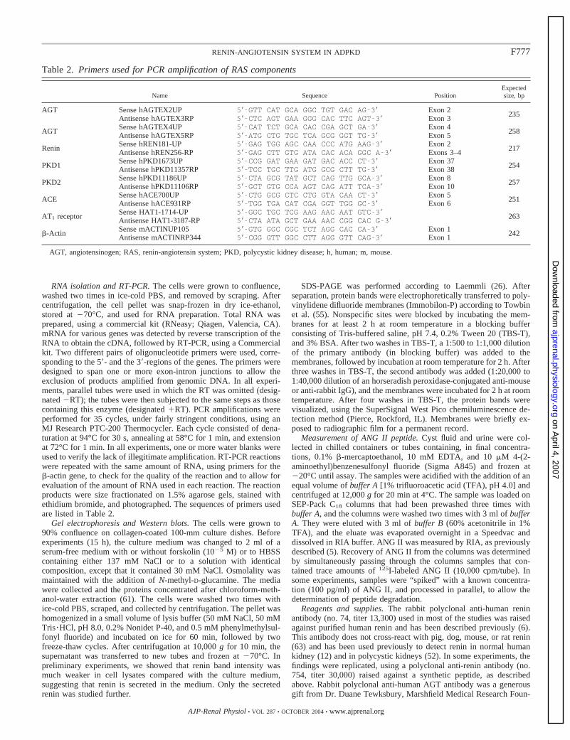
RNA isolation and RT-PCR. The cells were grown to confluence,washed two times in ice-cold PBS, and removed by scraping. Aftercentrifugation, the cell pellet was snap-frozen in dry ice-ethanol,stored at �70°C, and used for RNA preparation. Total RNA wasprepared, using a commercial kit (RNeasy; Qiagen, Valencia, CA).mRNA for various genes was detected by reverse transcription of theRNA to obtain the cDNA, followed by RT-PCR, using a Commercialkit. Two different pairs of oligonucleotide primers were used, corre-sponding to the 5�- and the 3�-regions of the genes. The primers weredesigned to span one or more exon-intron junctions to allow theexclusion of products amplified from genomic DNA. In all experi-ments, parallel tubes were used in which the RT was omitted (desig-nated �RT); the tubes were then subjected to the same steps as thosecontaining this enzyme (designated �RT). PCR amplifications wereperformed for 35 cycles, under fairly stringent conditions, using anMJ Research PTC-200 Thermocycler. Each cycle consisted of dena-turation at 94°C for 30 s, annealing at 58°C for 1 min, and extensionat 72°C for 1 min. In all experiments, one or more water blanks wereused to verify the lack of illegitimate amplification. RT-PCR reactionswere repeated with the same amount of RNA, using primers for the�-actin gene, to check for the quality of the reaction and to allow forevaluation of the amount of RNA used in each reaction. The reactionproducts were size fractionated on 1.5% agarose gels, stained withethidium bromide, and photographed. The sequences of primers usedare listed in Table 2.
Gel electrophoresis and Western blots. The cells were grown to90% confluence on collagen-coated 100-mm culture dishes. Beforeexperiments (15 h), the culture medium was changed to 2 ml of aserum-free medium with or without forskolin (10�5 M) or to HBSScontaining either 137 mM NaCl or to a solution with identicalcomposition, except that it contained 30 mM NaCl. Osmolality wasmaintained with the addition of N-methyl-D-glucamine. The mediawere collected and the proteins concentrated after chloroform-meth-anol-water extraction (61). The cells were washed two times withice-cold PBS, scraped, and collected by centrifugation. The pellet washomogenized in a small volume of lysis buffer (50 mM NaCl, 50 mMTris �HCl, pH 8.0, 0.2% Nonidet P-40, and 0.5 mM phenylmethylsul-fonyl fluoride) and incubated on ice for 60 min, followed by twofreeze-thaw cycles. After centrifugation at 10,000 g for 10 min, thesupernatant was transferred to new tubes and frozen at �70°C. Inpreliminary experiments, we showed that renin band intensity wasmuch weaker in cell lysates compared with the culture medium,suggesting that renin is secreted in the medium. Only the secretedrenin was studied further.
SDS-PAGE was performed according to Laemmli (26). Afterseparation, protein bands were electrophoretically transferred to poly-vinylidene difluoride membranes (Immobilon-P) according to Towbinet al. (55). Nonspecific sites were blocked by incubating the mem-branes for at least 2 h at room temperature in a blocking bufferconsisting of Tris-buffered saline, pH 7.4, 0.2% Tween 20 (TBS-T),and 3% BSA. After two washes in TBS-T, a 1:500 to 1:1,000 dilutionof the primary antibody (in blocking buffer) was added to themembranes, followed by incubation at room temperature for 2 h. Afterthree washes in TBS-T, the second antibody was added (1:20,000 to1:40,000 dilution of an horseradish peroxidase-conjugated anti-mouseor anti-rabbit IgG), and the membranes were incubated for 2 h at roomtemperature. After four washes in TBS-T, the protein bands werevisualized, using the SuperSignal West Pico chemiluminescence de-tection method (Pierce, Rockford, IL). Membranes were briefly ex-posed to radiographic film for a permanent record.
Measurement of ANG II peptide. Cyst fluid and urine were col-lected in chilled containers or tubes containing, in final concentra-tions, 0.1% �-mercaptoethanol, 10 mM EDTA, and 10 �M 4-(2-aminoethyl)benzenesulfonyl fluoride (Sigma A845) and frozen at�20°C until assay. The samples were acidified with the addition of anequal volume of buffer A [1% trifluoroacetic acid (TFA), pH 4.0] andcentrifuged at 12,000 g for 20 min at 4°C. The sample was loaded onSEP-Pack C18 columns that had been prewashed three times withbuffer A, and the columns were washed two times with 3 ml of bufferA. They were eluted with 3 ml of buffer B (60% acetonitrile in 1%TFA), and the eluate was evaporated overnight in a Speedvac anddissolved in RIA buffer. ANG II was measured by RIA, as previouslydescribed (5). Recovery of ANG II from the columns was determinedby simultaneously passing through the columns samples that con-tained trace amounts of 125I-labeled ANG II (10,000 cpm/tube). Insome experiments, samples were “spiked” with a known concentra-tion (100 pg/ml) of ANG II, and processed in parallel, to allow thedetermination of peptide degradation.
Reagents and supplies. The rabbit polyclonal anti-human reninantibody (no. 74, titer 13,300) used in most of the studies was raisedagainst purified human renin and has been described previously (6).This antibody does not cross-react with pig, dog, mouse, or rat renin(63) and has been used previously to detect renin in normal humankidney (12) and in polycystic kidneys (52). In some experiments, thefindings were replicated, using a polyclonal anti-renin antibody (no.754, titer 30,000) raised against a synthetic peptide, as describedabove. Rabbit polyclonal anti-human AGT antibody was a generousgift from Dr. Duane Tewksbury, Marshfield Medical Research Foun-
Table 2. Primers used for PCR amplification of RAS components
Name Sequence PositionExpectedsize, bp
AGT Sense hAGTEX2UP 5�-GTT CAT GCA GGC TGT GAC AG-3� Exon 2235
Antisense hAGTEX3RP 5�-CTC AGT GAA GGG CAC TTC AGT-3� Exon 3
AGTSense hAGTEX4UP 5�-CAT TCT GCA CAC CGA GCT GA-3� Exon 4
258Antisense hAGTEX5RP 5�-ATG CTG TGC TCA GCG GGT TG-3� Exon 5
ReninSense hREN181-UP 5�-GAG TGG AGC CAA CCC ATG AAG-3� Exon 2
217Antisense hREN256-RP 5�-GAG CTT GTG ATA CAC ACA GGC A-3� Exons 3–4
PKD1Sense hPKD1673UP 5�-CCG GAT GAA GAT GAC ACC CT-3� Exon 37
254Antisense hPKD11357RP 5�-TCC TGC TTG ATG GCG CTT TG-3� Exon 38
PKD2Sense hPKD11186UP 5�-CTA GCG TAT GCT CAG TTG GCA-3� Exon 8
257Antisense hPKD11106RP 5�-GCT GTG CCA AGT CAG ATT TCA-3� Exon 10
ACESense hACE700UP 5�-CTG GCG CTC CTG GTA CAA CT-3� Exon 5
251Antisense hACE931RP 5�-TGG TGA CAT CGA GGT TGG GC-3� Exon 6
AT1 receptorSense HAT1-1714-UP 5�-GGC TGC TCG AAG AAC AAT GTC-3�
263Antisense HAT1-3187-RP 5�-CTA ATA GCT GAA AAC CGG CAC G-3�
�-ActinSense mACTINUP105 5�-GTG GGC CGC TCT AGG CAC CA-3� Exon 1
242Antisense mACTINRP344 5�-CGG GTT GGC CTT AGG GTT CAG-3� Exon 1
AGT, angiotensinogen; RAS, renin-angiotensin system; PKD, polycystic kidney disease; h, human; m, mouse.
F777RENIN-ANGIOTENSIN SYSTEM IN ADPKD
AJP-Renal Physiol • VOL 287 • OCTOBER 2004 • www.ajprenal.org
on April 4, 2007
ajprenal.physiology.orgD
ownloaded from

dation (Marshfield, WI). This antibody was not affinity purified.Mouse monoclonal antibody against ACE was purchased fromChemicon International (Temecula, CA). Mouse monoclonal antibodyagainst ANG II type 1 receptor (AT1) and mouse monoclonal anti-body against ANG I and ANG II peptides were purchased from SantaCruz Biotechnology (Santa Cruz, CA). Rabbit polyclonal antibodyagainst ANG II peptide was purchased from Peninsula Laboratories(San Carlos, CA). Human renal cortical tubule epithelial cells werepurchased at passage 1 from Clonetics (Walkersville, MD). FITC-labeled secondary antibodies (anti-mouse, anti-rabbit, or anti-goatIgG) were purchased from Pierce. FITC-labeled LTA, PNA, and DBAlectins were purchased from Sigma (St. Louis, MO). Biotin-labeledlectins and the immunohistochemistry kit (Vectastain Elite) werepurchased from Vector Laboratories. RNA preparation kit (RNeasy)was purchased from Qiagen. RT-PCR kit (Access RT-PCR) waspurchased from Promega (Madison, WI). Oligonucleotide primerswere synthesized by GIBCO Invitrogen (Carlsbad, CA). Rat tailcollagen I (catalog no. 354236) was purchased from Beckton-Dick-inson (Bedford, MA). Tissue culture media and FBS were purchasedfrom GIBCO Invitrogen. Other reagents of highest-purity grades werepurchased from Sigma, Fisher, or other commercial suppliers.
Human subjects. Eleven polycystic kidneys were used for theseexperiments. The kidneys were obtained from many centers aroundthe United States. We also used tissue specimens from two normalnoncystic kidneys as controls. The nephrologists or surgeons wereasked to complete a brief questionnaire on each patient, includinginformation on hypertension and its duration, the latest availableblood pressure, and antihypertensive medications. When nephrectomywas planned, the patients contacted the Polycystic Kidney ResearchFoundation to express interest in donating their kidney(s) for research.The investigator’s laboratory was notified by the Polycystic KidneyResearch Foundation, and arrangements were made for the surgicalspecimen to be collected and shipped to the laboratory in sterilefashion. In addition to the standard surgical informed consent, eachpatient signed a second informed consent to allow the use of theirkidney tissue for research, including future use of archived kidneytissue. The procedures for the use of human kidneys and the consentform were approved by the Saint Louis University InstitutionalReview Board and was administered by the staff at the institutionwhere the nephrectomy was performed, then signed by the principalinvestigator. The information on patients who provided a kidneyspecimen for this research is summarized in Table 3. Clinical infor-mation could be ascertained on 8 of the 11 subjects who donated akidney. Incomplete information was provided for another patient. Allpatients were hypertensive at the time of nephrectomy. All except twowere on maintenance hemodialysis. The main reason for nephrectomyin all patients was preparation for a kidney transplant. Despite anti-hypertensive treatment, five patients had systolic blood pressure �140mmHg, and five patients had diastolic blood pressure �90 mmHg. Allbut one patient were treated with diuretics and antihypertensive agentsbefore nephrectomy. Angiotensin receptor blockers were used in four
patients, with two patients each receiving an ACE inhibitor and a�-blocker. Diuretics were administered in six patients.
RESULTS
Expression of renin by ADPKD kidneys. In normal humankidney sections, renin staining was confined to the afferentarterioles of the glomeruli, with no staining seen in the tubules(Fig. 1). In ADPKD kidneys, we observed renin staining incells lining the cysts and in dilated tubules. The intensity ofrenin staining was variable, with some cyst-lining cells show-ing intense staining and others showing mild staining or nostaining. Only about one-half of the cysts showed renin stain-ing (Fig. 1A). The available kidney sections had a few globallysclerosed glomeruli, none of which showed renin staining ofthe afferent arterioles. Renin staining was also not observed inafferent arterioles in areas where relatively normal glomeruliwere found. By contrast, in normal human kidneys, strongrenin staining was seen in the afferent arterioles of manyglomeruli (Fig. 1B). The findings suggest that juxtaglomerularapparatus (JGA) renin may be downregulated in ADPKDkidneys. No staining was seen when sections were incubatedwith rabbit preimmune IgG, confirming specificity (Fig. 1C).
Expression of AGT by ADPKD kidneys. In normal humankidney tissues, AGT was seen only in proximal tubules (Fig.2C). In ADPKD kidneys, we observed moderately strong AGTimmunostaining in cyst-lining cells and in many proximaltubules (Fig. 2, A and B). Only some cysts, presumably ofproximal origin (see below), showed AGT staining. Within agiven cyst, the intensity of AGT staining was variable, withsome cyst-lining cells showing only mild staining or no stain-ing. We presume this to be a result of chronic disease, delay intissue fixation, or antigen accessibility. The staining intensityof cysts decreased with antibody dilution, persisting down to1:32,000 dilution, whereas proximal tubule staining persisteddown to 1:64,000 dilution. Sections incubated with rabbitpreimmune IgG showed no staining, confirming specificity(data not shown).
Because only a portion of cysts in each ADPKD kidneyexpressed renin or AGT, and because AGT is known to beexpressed by proximal tubules (37), we performed additionalstudies to determine whether the cysts expressing renin or AGTmight originate from different tubule segments. To determinethe tubule origin of the cysts, adjacent sections of the sameADPKD kidney blocks were alternately stained for renin andfor AGT (Fig. 3). Other adjacent sections were stained withbiotinylated lectins to determine the tubule origin of the cysts
Table 3. Clinical information on subjects whose nephrectomy samples were used for study
Age, yrYears onDialysis
Duration ofHTN, yr
Blood Pressure, mmHg Antihypertensive Medications
Systolic Diastolic Diuretics ACEIs ARBs �-Blockers CCB
49 1.5 12 132 80 X X41 0 10 167 99 X X X51 0.5 5 152 82 X X51 5 5 140 90 X X X51 NK 20 140 90 X X62 10 15 150 90 X X X61 1.5 41 116 4946 0 5 136 100 X X
Information could not be ascertained for 3 of the subjects. HTN, hypertension; ACEI, angiotensin-converting enzyme inhibitor; ARB, angiotensin II receptorblocker; CCB, calcium channel blocker; NK, not known.
F778 RENIN-ANGIOTENSIN SYSTEM IN ADPKD
AJP-Renal Physiol • VOL 287 • OCTOBER 2004 • www.ajprenal.org
on April 4, 2007
ajprenal.physiology.orgD
ownloaded from

(13, 46). We used LTA as a marker of proximal tubules andPNA as a distal tubule marker. Based on studies performed infour different ADPKD kidneys, we showed that renin and AGTare localized on different cysts. Renin was expressed by cystsof distal tubule origin, as evidenced by positive staining with
PNA, whereas AGT was expressed by cysts of proximal tubuleorigin and by proximal tubules, as evidenced by positivestaining with LTA (Fig. 3). Because a limited number of cystswere available for examination, we cannot exclude the possi-bility that some cysts might express both renin and AGT.
Expression of other RAS components by ADPKD kidneys.Using immunohistochemistry with specific antibodies, wedemonstrated the presence of other components of the RAS in
Fig. 2. Angiotensinogen (AGT) expression by ADPKD kidney. Sections fromADPKD and normal kidneys stained for AGT. A: portion of a cyst with AGTstaining of cyst-lining cells (arrows). Inset: higher magnification of AGT-expressing cells. B: section from another ADPKD kidney showing strong AGTstaining of several tubules (arrows). C: section from a normal human kidneyshowing AGT staining of proximal tubules (arrows). Magnification �400.
Fig. 1. Renin expression by autosomal dominant polycystic kidney disease(ADPKD) kidney. Sections from an ADPKD kidney (A) and a normal humankidney (B) stained for renin. A: renin staining is seen in 3 cysts (arrowheads)while several other cysts and the glomerulus at the center are not stained. Notesevere interstitial fibrosis and tubular damage. B: in normal kidney, reninstaining is confined to the afferent arterioles of glomeruli (arrows). C: sectionof normal kidney stained with preimmune IgG shows no staining. Magnifica-tion �400.
F779RENIN-ANGIOTENSIN SYSTEM IN ADPKD
AJP-Renal Physiol • VOL 287 • OCTOBER 2004 • www.ajprenal.org
on April 4, 2007
ajprenal.physiology.orgD
ownloaded from

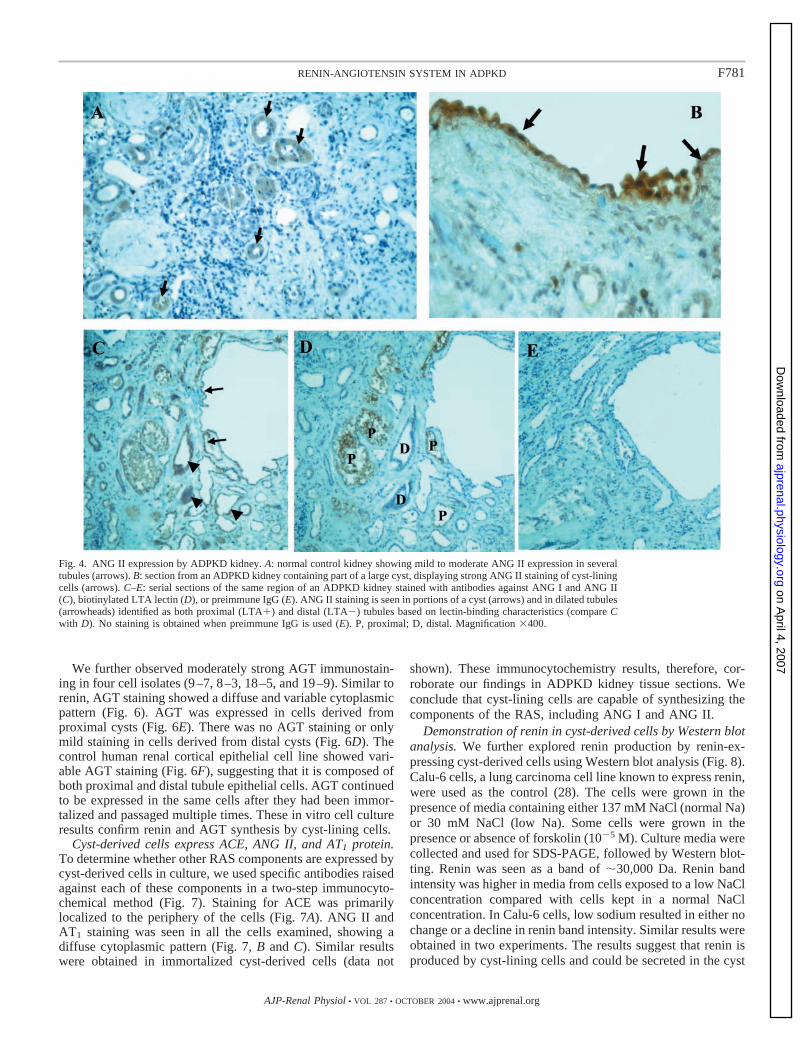
cyst-lining cells and in previously described locations withinthe kidney. We observed immunostaining for ACE in bothproximal and distal tubules and in many dilated tubules andcysts (Fig. 4, B and C). In control kidney sections, ACEstaining was confined to proximal tubules (Fig. 4A). We alsoobserved strong immunostaining for AT1 receptor in mostproximal tubules and in some small- and medium-sized cysts(Fig. 4, E and F).
The presence of immunostainable AGT, renin, ACE, andAT1 receptors in cysts and some dilated tubules suggests thatANG I and ANG II could be produced in situ within cysts anddilated tubules in ADPKD. To examine this possibility, westained ADPKD kidney sections with a monoclonal antibodythat recognizes both ANG I and ANG II and with a morespecific polyclonal antibody against ANG II with low cross-reactivity against ANG I. Using these two antibodies, wedetected ANG I and ANG II immunostaining in cyst-liningcells and within the lumen of some cysts (Fig. 5). ANG IIstaining was also present in tubule epithelial cells and withinthe lumen of proximal tubules. Some distal tubules also stainedpositive for ANG II (Fig. 4C). The results show that thecomponents of the RAS, including the effector peptide ANG II,are present within cysts and dilated tubules of polycystickidneys. The presence of ANG II-positive particles within thecyst lumen suggests that ANG II is either secreted in the lumenor formed within the cyst lumen from conversion of ANG I toANG II.
If ANG II is secreted in or formed within the cyst lumen, onewould expect relatively high concentrations of the peptide incyst fluid, provided that it is not degraded. ANG II presentwithin cysts could find its way to the lumen of the tubules towhich they are connected. We therefore measured ANG IIconcentrations in cyst fluid collected at �24 h after nephrec-tomy (Table 4). Fluids collected from 12 cysts were used forthese experiments. ANG II concentration was below the de-tection limit of the assay in five cysts. The (mean � SE) cystfluid ANG II concentration in the remaining seven cysts was
1,063 � 81 pg/ml (�1 pmol/ml). As a crude measure of ANGII in distal tubules (37), ANG II concentrations were measuredin the urine of three ADPKD subjects with normal renalfunction but were detectable in only one subject. ANG IIconcentration in the urine of this individual was 88.2 ng/ml(�84 pmol/ml). In comparison, ANG II concentrations in theurine of two control subjects were 61 and 101 ng/ml (�58 and96.6 pmol/ml).
Cyst-derived cells in culture express renin and AGT protein.Our findings suggest that, in ADPKD, the components of theRAS can be produced by cysts and dilated tubules, but wecannot rule out nonspecific uptake by cyst-lining cells. Plasmarenin (mol wt �30,000) is partially filtered at the glomerulusand degraded in the proximal tubule (25). The presence ofrenin within the cyst epithelium could therefore representendocytic uptake of filtered renin from the lumen. AGT (molwt �60,000) is not filtered but can be produced by proximaltubules. Therefore, it might reach the microcysts attached tothe tubules in a retrograde fashion and endocytosed by cyst-lining cells. To prove that cyst-lining cells synthesize renin andAGT, we isolated epithelial cells from multiple individualcysts of 11 polycystic kidneys. Both ADPKD cells in primaryculture and immortalized ADPKD cells were used for thestudies described below.
Using immunohistochemical techniques, we showed strongrenin immunostaining in the majority of cyst-derived cells inculture. Diffuse renin staining was observed within the cyto-plasm of the cells. When the tubule origin of the cells wasidentified, renin appeared to be expressed in cells derived fromdistal cysts (Fig. 6A). No renin staining was seen in cells fromproximal cysts (Fig. 6B). Calu-6 cells, a lung carcinoma cellline known to express renin (28), showed strong renin staining(Fig. 6C). No staining was seen when preimmune rabbit IgGwas used at the same dilution (data not shown). Renin contin-ued to be expressed in at least 11 cell isolates after they hadbeen immortalized and passaged multiple times.
Fig. 3. Distribution of renin and AGT be-tween cysts of ADPKD kidney. Serial sec-tions from an ADPKD kidney stained withantibodies against renin (A), AGT (B), orwith biotinylated Arachis hypogaea (PNA;C) or Lotus tetragonolobus (LTA; D) lectins.The sections show part of a large cyst andseveral smaller cysts and tubules. A: strongrenin staining is seen in cells lining the largecyst (arrowheads) with weak renin stainingin some of the dilated tubules. B: AGT stain-ing is confined to a few tubules (arrows) withno staining of the large cyst. C: same areastained with PNA lectin shows strong stain-ing of distal tubules and the large cyst (ar-rowheads). D: same area as in C at a slightlydifferent depth stained with LTA lectinshows staining of two proximal tubules (ar-rows). Distal tubules and the large cyst arenot stained. The ability of the large cyst tobind PNA lectin but not LTA lectin identifiesthe cyst to be of distal origin.
F780 RENIN-ANGIOTENSIN SYSTEM IN ADPKD
AJP-Renal Physiol • VOL 287 • OCTOBER 2004 • www.ajprenal.org
on April 4, 2007
ajprenal.physiology.orgD
ownloaded from
We further observed moderately strong AGT immunostain-ing in four cell isolates (9–7, 8–3, 18–5, and 19–9). Similar torenin, AGT staining showed a diffuse and variable cytoplasmicpattern (Fig. 6). AGT was expressed in cells derived fromproximal cysts (Fig. 6E). There was no AGT staining or onlymild staining in cells derived from distal cysts (Fig. 6D). Thecontrol human renal cortical epithelial cell line showed vari-able AGT staining (Fig. 6F), suggesting that it is composed ofboth proximal and distal tubule epithelial cells. AGT continuedto be expressed in the same cells after they had been immor-talized and passaged multiple times. These in vitro cell cultureresults confirm renin and AGT synthesis by cyst-lining cells.
Cyst-derived cells express ACE, ANG II, and AT1 protein.To determine whether other RAS components are expressed bycyst-derived cells in culture, we used specific antibodies raisedagainst each of these components in a two-step immunocyto-chemical method (Fig. 7). Staining for ACE was primarilylocalized to the periphery of the cells (Fig. 7A). ANG II andAT1 staining was seen in all the cells examined, showing adiffuse cytoplasmic pattern (Fig. 7, B and C). Similar resultswere obtained in immortalized cyst-derived cells (data not
shown). These immunocytochemistry results, therefore, cor-roborate our findings in ADPKD kidney tissue sections. Weconclude that cyst-lining cells are capable of synthesizing thecomponents of the RAS, including ANG I and ANG II.
Demonstration of renin in cyst-derived cells by Western blotanalysis. We further explored renin production by renin-ex-pressing cyst-derived cells using Western blot analysis (Fig. 8).Calu-6 cells, a lung carcinoma cell line known to express renin,were used as the control (28). The cells were grown in thepresence of media containing either 137 mM NaCl (normal Na)or 30 mM NaCl (low Na). Some cells were grown in thepresence or absence of forskolin (10�5 M). Culture media werecollected and used for SDS-PAGE, followed by Western blot-ting. Renin was seen as a band of �30,000 Da. Renin bandintensity was higher in media from cells exposed to a low NaClconcentration compared with cells kept in a normal NaClconcentration. In Calu-6 cells, low sodium resulted in either nochange or a decline in renin band intensity. Similar results wereobtained in two experiments. The results suggest that renin isproduced by cyst-lining cells and could be secreted in the cyst
Fig. 4. ANG II expression by ADPKD kidney. A: normal control kidney showing mild to moderate ANG II expression in severaltubules (arrows). B: section from an ADPKD kidney containing part of a large cyst, displaying strong ANG II staining of cyst-liningcells (arrows). C–E: serial sections of the same region of an ADPKD kidney stained with antibodies against ANG I and ANG II(C), biotinylated LTA lectin (D), or preimmune IgG (E). ANG II staining is seen in portions of a cyst (arrows) and in dilated tubules(arrowheads) identified as both proximal (LTA�) and distal (LTA�) tubules based on lectin-binding characteristics (compare Cwith D). No staining is obtained when preimmune IgG is used (E). P, proximal; D, distal. Magnification �400.
F781RENIN-ANGIOTENSIN SYSTEM IN ADPKD
AJP-Renal Physiol • VOL 287 • OCTOBER 2004 • www.ajprenal.org
on April 4, 2007
ajprenal.physiology.orgD
ownloaded from

lumen. Furthermore, renin production by cyst-derived cellscould be regulated by extracellular NaCl concentrations.
Cyst-derived cells express renin and AGT mRNA. If reninand AGT are synthesized by the cyst epithelium, one wouldexpect the presence of mRNA for these proteins in cystepithelium and in cyst-derived cells in culture. The presence ofrenin and AGT mRNA was evaluated by RT-PCR of total
RNA isolated from cultured cyst-derived cells. Renin mRNAwas expressed by the majority of cell isolates studied (Figs. 9and 10A). Furthermore, renin mRNA expression appeared to behigher in cells grown on collagen I-coated plates comparedwith cells grown on plastic (Fig. 9A). AGT mRNA wasexpressed by only a few cyst-derived cell isolates (Fig. 9B).
In addition to mRNA for renin and AGT, RT-PCR analysisof RNA isolated from cultured cyst-derived cells showed thepresence of mRNA for ACE and for AT1 in all cyst-derivedcells studied (Fig. 9, C and D), regardless of tubule origin.
Differential expression of renin and AGT by cysts. We foundthat, in general, renin-expressing cells did not express AGTand AGT-expressing cells did not express renin. Using tubule-specific lectins, we confirmed that AGT mRNA is expressed bycyst-derived cells of proximal tubule origin, whereas reninmRNA is expressed by cyst-derived cells of distal tubule origin(Fig. 10A). For those cells in which both immunocytochemistryand RT-PCR data were available, there was concordancebetween renin and AGT protein and mRNA expression. Forexample, cyst-derived cells of distal tubule origin (e.g., 11–6)express renin mRNA at the exclusion of AGT mRNA (Fig.10A). The same cells also show moderately strong reninstaining by immunocytochemistry but no staining for AGT(Fig. 10B).
Fig. 5. Expression of angiotensin-convertingenzyme (ACE; A–C) and ANG II type 1receptor (AT1; D–F) by ADPKD kidney. A:section from a normal kidney showing mild tomoderate ACE staining of several tubules(arrows). B: section from an ADPKD kidneyshowing ACE staining in several dilated tu-bules (arrows). The two cysts at the center arenot stained. C: section from another ADPKDkidney showing ACE staining of both tubulesand a large cyst (arrows). D: section from anormal kidney showing staining for AT1 re-ceptors in tubules (arrows) and in afferentarteriole (arrowhead). E: section from anADPKD kidney showing strong AT1 stainingin cystic dilated tubules (arrows). F: ADPKDkidney showing AT1 staining of a cyst inaddition to several tubules (arrows). Magnifi-cation �400.
Table 4. ANG II concentrations in cyst fluid and urine
Cyst FluidANG II Concentration,
pg/ml UrineANG II Concentration,
ng/ml
Cyst 6 BDL U1* BDLCyst 7 1,415 U2* BDLCyst 8 1,223 U3* 88.2Cyst 9 BDLCyst 10 739Cyst 11 BDLCyst 12 BDL U1† 61.1Cyst 13 BDL U2† 101.3Cyst 14 1,022Cyst 15 1,076Cyst 16 1,033Cyst 17 936
BDL, below detection limit of the assay. Urine values are not corrected forCr concentration. *ADPKD urine. †Control urine.
F782 RENIN-ANGIOTENSIN SYSTEM IN ADPKD
AJP-Renal Physiol • VOL 287 • OCTOBER 2004 • www.ajprenal.org
on April 4, 2007
ajprenal.physiology.orgD
ownloaded from

Taken together, the results indicate that, in ADPKD, cyst-lining epithelia can synthesize the components of the RAS.This is associated with production and secretion of ANG II bycysts and a relatively high intratubular ANG II concentration.
DISCUSSION
The mechanism of hypertension in ADPKD remains poorlyunderstood. Involvement of the RAS has been postulated, but
the data correlating plasma renin activity with blood pressurein ADPKD patients are inconclusive. Although some studiesshow increased plasma renin activity in hypertensive ADPKDpatients compared with control subjects, a strong associationhas not been established in all studies (7, 22), casting doubt on therole of systemic RAS in blood pressure regulation in ADPKD.
The role of intrarenal RAS in hypertensive disorders. It isnow well accepted that increased activity of the intrarenal
Fig. 6. Expression of renin and AGT by cyst-derived cells in culture. A: cells derived from a distal cyst (9–9) show strong reninstaining. B: cells from a proximal cyst (9–7) do not express renin. C: Calu-6 cells show strong renin staining. D: cells from a distalcyst (9–9) show weak or no AGT staining. E: cells from a proximal cyst (19–9) show strong AGT staining. F: control humancortical epithelial cell line (RCTEC) shows AGT staining in about one-half of the cells, presumably of proximal tubule origin. G:cyst-derived cells (9–7) shown in B express CD13, identifying them to be of proximal origin. H: proximal cyst cells (9–7) stainedwith preimmune IgG. Magnification �400.
Fig. 7. Expression of other renin-angiotensin system (RAS) components by cyst-derived cells in culture. A: cells derived from adistal cyst (9–9) show moderately strong staining for ACE, taking a peripheral membrane-associated pattern. B: cells from aproximal cyst (19–9) show strong ANG II staining. C: same cells (19–9) also express AT1 receptors. Magnification �400.
F783RENIN-ANGIOTENSIN SYSTEM IN ADPKD
AJP-Renal Physiol • VOL 287 • OCTOBER 2004 • www.ajprenal.org
on April 4, 2007
ajprenal.physiology.orgD
ownloaded from

rather than the systemic RAS is responsible for many forms ofhypertension (11, 37, 58). It has been suggested that persistentelevation of intrarenal ANG II production with an inability toreduce ANG II formation in response to a high sodium intakewill result in resetting of the pressure-natriuresis relationshiptoward higher blood pressures, thus leading to hypertension(18, 20). Several lines of evidence suggest that a similarparadigm may apply to polycystic kidney disease. In ADPKDpatients, the pressure-natriuresis curve is shifted to the rightalong with increased blood pressure sensitivity to salt loading(44, 54). Sodium retention and volume expansion have beenreported in hypertensive ADPKD patients, before the onset ofrenal failure (35, 55). These patients also have higher renalvascular resistance compared with normotensive controls andan exaggerated vascular response to converting-enzyme inhi-bition (7, 53, 60). Additionally, in normotensive ADPKDpatients, the renal vasculature is insensitive to ANG II infusion,with a lower relative rise in vascular resistance compared withcontrol subjects (2). Taken together, these observations suggestincreased intrarenal ANG II production in ADPKD. Becausethe majority of tubules continue to function, high ANG IIconcentrations within the tubule lumen could result in in-creased renal tubular sodium reabsorption and hypertension ifsalt intake is not reduced (19, 20).
Overexpression of RAS in polycystic kidneys. In the presentstudy, we show that, in ADPKD, the components of the RAScan be produced by cysts and dilated tubules. Renin expressionis confined to distal cysts and some dilated distal tubules,whereas AGT expression is confined to proximal cysts andproximal tubules. Because AGT is known to be present inproximal tubules, its expression by cysts of proximal tubuleorigin is not surprising. Similarly, renin expression by cysts of
distal tubule origin is in line with recent reports of reninexpression in mouse connecting and collecting tubules (43).We have recently made similar observations in kidneys of pcymice, considered a model of polycystic kidney disease (unpub-lished observations).
Several lines of evidence suggest that renin, AGT, and otherRAS proteins are synthesized by cyst epithelium in ADPKDkidneys: 1) the cultured cells used for the present studies werederived from large superficial cysts that are cut off from the
Fig. 9. Expression of renin, AGT, and ANG II receptor mRNA by cyst-derived cells in culture. Cells were isolated from individual cysts and usedeither as primary culture or after they had been immortalized. RT-PCR wasperformed with specific oligonucleotide primers for renin, AGT, ACE, or AT1
receptor. A: renin mRNA band shows a higher intensity in ADPKD cells grownon collagen compared with cells grown on plastic. Human kidney mRNA isused as a positive control for renin. B: AGT mRNA band is seen in cyst-derived cells, using two different primer sets. Human liver mRNA is used aspositive control for AGT. C: ACE mRNA is seen in a cyst-derived cell. D: AT1
mRNA band is seen in 3 different immortalized cyst-derived cells and in thecontrol human renal cortical epithelial cell line (wtRCTEC). �RT, controlRNA sample, not exposed to RT; �RT, samples treated with RT before PCR.
Fig. 8. Demonstration of renin in cyst-derived cells in culture by Westernblotting. Renin-expressing immortalized cyst-derived cells (wt 9–9) weregrown in the presence of culture media containing 137 mM NaCl [normal Na(NL Na)] until confluent and then either kept in normal-sodium medium orswitched to a medium containing 30 mM NaCl (low Na). The cells were alsogrown in the presence or absence of forskolin (10�5 M). Calu-6 cells, a lungcarcinoma cell line known to express renin, were used as control. Culturemedia were collected, concentrated, and used for SDS-PAGE, followed byWestern blotting. Protein (30 �g) was loaded in each lane. Renin is seen as aband of �30,000. Renin band intensity is increased in ADPKD cells exposedto low NaCl concentrations compared with cells exposed to normal NaClconcentrations. Renin band intensity is somewhat increased in cells exposed toforskolin to increase intracellular cAMP. Right: renin expression in Calu-6cells does not change with forskolin. Lower renin band intensity in low-sodiummedium is likely because of uneven protein loading.
F784 RENIN-ANGIOTENSIN SYSTEM IN ADPKD
AJP-Renal Physiol • VOL 287 • OCTOBER 2004 • www.ajprenal.org
on April 4, 2007
ajprenal.physiology.orgD
ownloaded from

rest of the nephron, eliminating the possibility of renin absorp-tion from the tubule lumen; 2) renin and AGT were stilldetected after the cells were grown in primary culture for atleast 1 wk; and 3) immortalized cyst-derived cells continued toexpress RAS components after multiple passages (30).
Torres et al. (52) described ectopic production of renin bytubulocystic epithelium in ADPKD. Renin appeared to besecreted in the cyst fluid, with a higher concentration ingradient cysts (52). There was a negative correlation betweencyst fluid sodium concentration and cyst fluid active renin (52).This observation suggests that cyst renin production may beregulated by changes in extracellular sodium concentration.Our Western blot analysis provides evidence for regulatedrelease of renin by the cyst epithelium. Renin secretion wasincreased in cyst-derived cells exposed to low NaCl concen-trations. Although Torres et al. (52) reported renin expression
in both proximal tubules and in cysts of distal tubule origin, ourresults suggest that renin is primarily, if not exclusively,produced by cysts of distal tubule origin. Rohrwasser et al. (43)showed low levels of immunoreactive renin in mouse connect-ing and cortical collecting tubules, which could be augmentedacutely by dietary sodium restriction. The finding that dilateddistal tubules and cysts of distal tubule origin both producerenin is in agreement with the above observation and mayrepresent derepression of renin release by distal tubules aftercystic transformation.
Graham and Lindop (17) have reported increased numbersof renin-secreting cells in ADPKD kidneys with immunoreac-tive renin extending beyond the afferent arterioles and in smallrenal arteries. Contrary to these earlier reports, we seldomobserved prominent JGA or arteriolar renin staining in ourADPKD kidney specimens, even though several patients were
Fig. 10. Differential expression of renin and AGT mRNA in cyst-derived cells. A: mRNA analysis. Three immortalizedcyst-derived cells, previously characterized to be of distal tubule origin (30), were used for RT-PCR analysis, using oligonucleotideprimers for renin and AGT. Renin mRNA is expressed in all 3 cyst-derived cells and in human kidney, used as positive control.By contrast, none of the 3 cyst-derived cells of distal origin express AGT mRNA. A strong AGT mRNA band is seen in humanliver. B: immunohistochemistry of renin and AGT in one of the cyst-derived cells (11–6) of distal tubule origin shown in A. Thedistal origin of the cells is confirmed by positive staining with PNA lectin (a) and negative staining with LTA lectin (b). The cellsshow moderately strong staining for renin (d) but only sparse staining for AGT (e). No staining is seen when preimmune IgG isused as primary antibody (c).
F785RENIN-ANGIOTENSIN SYSTEM IN ADPKD
AJP-Renal Physiol • VOL 287 • OCTOBER 2004 • www.ajprenal.org
on April 4, 2007
ajprenal.physiology.orgD
ownloaded from

treated with diuretics, angiotensin receptor blockers, or ACEinhibitors (Table 3). A possible explanation is that manyglomeruli were scarred or destroyed in these end-stage kid-neys, leading to involution of their arterioles. However, even inkidney sections where intact glomeruli were available, JGArenin was reduced or absent (Fig. 1A). Increased renin expres-sion by renal arterioles is also difficult to reconcile with theobservations that systemic renin activity is not generally in-creased in ADPKD patients.
Paracrine intrarenal RAS in ADPKD. In the early stages ofADPKD, only a minority of tubules undergo a “second hit”somatic mutation and develop into cysts over several decades(40). The majority of nephrons remain normal and presumablyfunctional before becoming damaged by compression fromadjacent growing cysts. Because a significant number of cystsare connected to tubules (49), AGT produced by cystic andnoncystic proximal tubules could gain access to distal tubules(43). Therefore, a paracrine system could exist whereby AGTproduced by proximal tubules and proximal cysts could reachthe distal tubules where it is cleaved by renin from distal cyststo form ANG I (see Fig. 11). ACE is present on the brushborder of proximal tubules and within distal tubules (4),allowing ANG I to be cleaved readily to ANG II. ACE-independent, chymase-mediated ANG II formation has alsobeen reported in the interstitium of ADPKD kidney tissues(33). ANG II could bind to apical AT1 receptors at these tubulesites and increase sodium and water reabsorption (15, 24, 51,57). Although 24-h delay in sample collection may haveresulted in significant degradation of ANG II, relatively highANG II concentrations were measured in fluids collected from7 of the 12 cysts studied, but ANG II remained undetectable in5 cysts (Table 4). For those cysts in which ANG II could bemeasured, the average ANG II concentration in cyst fluid was1 pmol/ml, well within the range of tubular ANG II reportedpreviously (36, 38). These concentrations are also higher thanthose measured in plasma (38), suggesting in situ productionby cyst-lining epithelium. If one considers that a significantfraction of tubular ANG II is derived from cysts draining in thetubules, the ANG II concentrations measured here are wellwithin the range that can stimulate tubular sodium reabsorp-tion.
Previous studies in cyst epithelium have demonstrated thepresence of apical chloride channels and reversed polarity ofNa�-K�-ATPase pumps with an apical instead of basolaterallocation (62). The net effect of these channels is sodium andchloride secretion in the cyst lumen and cyst growth (39, 62).The mechanism described here might appear incompatible withoverall sodium and chloride reabsorption by the renal tubules.Because 1% of tubules develop into cysts (40), the contri-bution of the secretory flux described above is negligiblecompared with the ANG II-stimulated absorptive flux. There-fore, the net effect of increased intrarenal RAS activity wouldbe sodium and water retention. Recently, it was shown thatADPKD cyst-lining epithelial cells absorb sodium by an amilo-ride-sensitive pathway (42). Because of similarity of ADPKDand ADPKD, it is likely that cysts in ADPKD can also absorbsodium.
Proposed mechanism of hypertension in ADPKD. Based onthe findings of this study and the classical work of Guyton andHall (19, 20), we propose the following mechanism for thedevelopment of hypertension in ADPKD (Fig. 11). Ectopic and
excessive production of AGT and renin by proximal and distalcysts could lead to increased intratubular ANG II concentra-tions and excessive renal tubular sodium reabsorption (37).Renin production may not become suppressed when dietarysodium is increased. This could lead to a shift of the pressure-natriuresis curve to the right and chronic hypertension, unlessthe dietary sodium intake is reduced. Increased ANG II pro-duction may also contribute to cellular proliferation and cystgrowth, and to interstitial fibrosis (39). The mechanism de-scribed above might contribute to hypertension in early stagesof ADPKD. Later in the course of the disease, hypertensioncould be sustained by a combination of factors that include saltand water retention and RAS overactivity resulting fromnephron damage.
Possible mechanisms of intrarenal RAS upregulation inADPKD. Changes in extracellular NaCl or intracellular cal-cium have been shown to regulate renin production in juxta-glomerular cells (43). Polycystin-2 may represent a componentof a nonselective sodium or calcium channel (16, 34), whichrequires assembly with polycystin-1 for adequate ion channelfunction (21). Therefore, it is possible that the polycystinscould be involved in the regulation of renin production bymodulating sodium or calcium fluxes across the cyst epithe-lium. Defective channel activity resulting from specific poly-cystin mutations (9) could reduce intracellular sodium andcalcium concentrations and stimulate renin production. In JGAcells, renin release is also controlled by changes in early distaltubular flow (29). Recently, Nauli et al. (36) showed thatpolycystins act as flow sensors in renal tubules, transducingmechanical fluid flow signals into calcium signals. In cultured
Fig. 11. Speculative model of autocrine/paracrine tubular RAS in polycystickidney disease. AGT is produced by proximal tubules, where it can be cleavedto ANG I by filtered renin (R). ANG I is converted to ANG II by ACE presenton the brush-border membrane. ANG II binds to apical AT1 receptors tostimulate sodium reabsorption via apical Na�/H� antiporter. In early stages ofdisease, many microcysts maintain continuity with the rest of the nephron.Accordingly, AGT produced by proximal tubules and proximal cysts can reachthe distal tubules where it can be cleaved by renin emerging from distal cystsand dilated distal tubules. Cleavage of AGT by renin results in the formationof ANG I, which is readily converted to ANG II by ACE, present in thisnephron segment. ANG II produced at distal tubule sites can lead to increasedsodium reabsorption via stimulation of the epithelial sodium channels. Exces-sive sodium intake may influence ANG II production/accumulation by 1)decreasing AGT production by proximal tubules and cysts and 2) reducingrenin production by distal tubules and cysts. Inability of polycystic kidneys todownregulate the proximal and distal limbs of this paracrine intratubular RAScould result in sustained increases in intratubular ANG II levels. One canspeculate that increased ANG II could lead to salt and water retention andhypertension.
F786 RENIN-ANGIOTENSIN SYSTEM IN ADPKD
AJP-Renal Physiol • VOL 287 • OCTOBER 2004 • www.ajprenal.org
on April 4, 2007
ajprenal.physiology.orgD
ownloaded from

cells with a homozygous polycystin mutation, the cilia fail tosense fluid flow (36). We also speculate that increased reninexpression by cyst-lining epithelia might be related to failure ofmechanosensation resulting from polycystin mutations.
The mechanism of ectopic AGT expression in ADPKDremains speculative. In cardiac myocytes, mechanical stretchactivates the AGT gene expression and the formation of ANGII, which is involved in stretch-induced myocardial hypertro-phy (48). Hypoxia can also stimulate AGT gene expression(27). It is possible that mechanical stretch or hypoxia couldexert similar effects on cyst-lining cells. The polycystin com-plex triggers the formation of a number of transcription factors.One could speculate that polycystic kidney disease mutationsmay also interfere with the formation of trans-acting factorsthat can regulate AGT gene expression. Recent isolation ofimmortalized cyst-derived cells from ADPKD kidneys (30)should provide the tools necessary to test these hypotheses.
ACKNOWLEDGMENTS
The shipping of nephrectomy specimens from various surgical programswas facilitated by the PKD Foundation and by patients who offered toparticipate in the study. The following surgical specialists provided kidneytissue for this study: Drs. Angel Alsina, Steven Bridge, Michael Brown,Thomas Cooper, James Eason, Albin Gritsch, Donald Jablonski, Mark Klein,George Lipkowitz, Deepak Mital, Ali Naji, Arnold Serota, Hans Solinger,Harvey Solomon, Frank Stuart, and Harold Yang. The work of Dr. Jean-MarcLalouel on intratubular RAS provided the initial framework for the hypothesesdeveloped in this paper. We thank Barbara Kariuki for technical assistance andDr. Farhad Sedarati for reviewing the manuscript.
GRANTS
This work was supported by a grant from the Polycystic Kidney ResearchFoundation and in part by a grant from the Fleur-de-Lis Foundation.
REFERENCES
1. Baert L. Hereditary polycystic kidney disease (adult form): a microdis-section study of two cases at an early stage of the disease. Kidney Int 13:519–525, 1978.
2. Barrett BJ, Foley R, Morgan J, Hefferton D, and Parfrey P. Differ-ences in hormonal and renal vascular responses between normotensivepatients with autosomal dominant polycystic kidney disease and unaf-fected family members. Kidney Int 46: 1118–1123, 1994.
3. Bell PE, Hossack KF, Gabow PA, Durr JA, Johnson AM, and SchrierRW. Hypertension in autosomal dominant polycystic kidney disease.Kidney Int 34: 683–690, 1988.
4. Casarini DE, Boim MA, Stella RC, Krieger-Azzolini MH, Krieger JE,and Schor N. Angiotensin I-converting enzyme activity in tubular fluidalong the rat nephron. Am J Physiol Renal Physiol 272: F405–F409, 1997.
5. Cassis L, Laughter A, Fettinger M, Akers S, Speth R, Burke G, KingV, and Dwoskin L. Cold exposure regulates the renin-angiotensin system.J Pharmacol Exp Ther 286: 718–726, 1998.
6. Celio MR and Inagami T. Angiotensin II immunoreactivity coexists withrenin in juxtaglomerular cells of the kidney. Proc Natl Acad Sci USA 78:3897–3900, 1981.
7. Chapman AB, Johnson A, Gabow PA, and Schrier WS. The renin-angiotensin-aldosterone system, and autosomal dominant polycystic kid-ney disease. N Engl J Med 323: 1091–1096, 1990.
8. Chapman AB and Schrier RW. Pathogenesis of hypertension in auto-somal dominant polycystic kidney disease. Semin Nephrol 11: 653–660,1991.
9. Chen X-Z. Segal Y, Basora N, Guo L, Peng J-B, Babakhanlou H,Vassilev PM, Brown EM, Hediger MA, and Zhou J. Transport functionof the naturally occurring pathogenic polycystin-2 mutant R742X. Bio-chem Biophys Res Commun 282: 1251–1256, 2001.
10. Cuppage FE, Huseman RA, Chapman A, and Grantham JJ. Ultra-structure and function of cysts from human adult polycystic kidneys.Kidney Int 17: 372–381, 1980.
11. Davisson RL, Ding Y, Stec DE, Catterall JF, and Sigmund CD. Novelmechanism of hypertension revealed by cell-specific targeting of humanangiotensinogen in transgenic mice. Physiol Genomics 1: 3–9, 1999.
12. Faraggiana T, Gresik E, Tanaka T, Inagami T, and Lupo A. Immu-nohistochemical localization of renin in the human kidney. J HistochemCytochem 30: 459–65, 1982.
13. Faraggiana T, Malchiodo F, Prado A, and Churg J. Lectin-peroxidaseconjugate in normal human kidney. J Histochem Cytochem 30: 451–458,1982.
14. Gabow PA, Chapman AB, Johnson AM, Tangel DJ, Duley IT, KaehnyWD, Manco-Johnson M, and Schrier RW. Renal structure and hyper-tension in autosomal dominant polycystic kidney disease. Kidney Int 38:1177–1180, 1990.
15. Geibel J, Giebisch G, and Boron WF. Angiotensin II stimulates bothNa/H exchange and Na/HCO3 cotransport in the rabbit proximal tubule.Proc Natl Acad Sci USA 87: 7917–7920, 1990.
16. Gonzalez-Perrett S, Kim K, Ibarra C, Damiano A, Zotta E, Batelli M,Harris PC, Reisin IL, Arnaout MA, and Cantiello HF. Polycystin 2, theprotein mutated in autosomal dominant polycystic kidney disease, is aCa2�-permeable nonselective channel. Proc Natl Acad Sci USA 98:1182–1187, 2001.
17. Graham PC and Lindop GB. The anatomy of the renin secreting cell inadult polycystic kidney disease. Kidney Int 33: 1084–1090, 1988.
18. Gross V, Roman RJ, and Cowley AW Jr. Abnormal pressure-natriuresisin transgenic renin gene rats. J Hypertens 12: 1029–1034, 1994.
19. Hall JE, Brands MW, and Henegar JR. Angiotensin II and long-termarterial pressure regulation. The overriding dominance of the kidney. J AmSoc Nephrol 10: 5258–5265, 1999.
20. Hall JE, Guyton AC, and Brands MW. Pressure-volume regulation inhypertension. Kidney Int 49: S35–S41, 1996.
21. Hanaoka K, Qian F, Boletta A, Bhunia A, Piontek K, Tsiokas L,Sukhatme VP, Guggino WB, and Germino GG. Co-assembly of poly-cystin-1 and -2 produces unique cation-permeable currents. Nature 408:990–994, 2000.
22. Harrap SB, Davies DL, Macnicol AM, Dominiczak AF, Fraser R,Wright AF, Watson ML, and Briggs JD. Renal, cardiovascular andhormonal characteristics of young adults with autosomal dominant poly-cystic kidney disease. Kidney Int 40: 501–508, 1991.
23. Harris PC. Autosomal dominant polycystic kidney disease: clues topathogenesis. Hum Mol Genet 8: 1861–1866, 1999.
24. Harrison-Bernard LM, Navar LG, Ho MM, Vinson GP, and El-DahrS. Immunohistochemical localization of ANG II AT1 receptor in adult ratkidney using a monoclonal antibody. Am J Physiol Renal Physiol 273:F170–F177, 1997.
25. Kim S, Iwao H, Nakamura N, Ikemoto F, Yamamoto K, Mizuhira V,and Yokofujita J. Metabolism of circulating renin by liver, and kidney ofrats. J Cardovasc Pharmacol 10, Suppl 7: S94–S95, 1987.
26. Laemmli UK. Cleavage of structural proteins during the assembly of thehead of bacteriophage T4. Nature 227: 680–685, 1970.
27. Lam SY and Leung PS. Chronic hypoxia activates a local angiotensin-generating system in rat carotid body. Mol Cell Endocrinol 203: 147–153,2003.
28. Lang JA, Yang G, Kern JA, and Sigmund CD. Endogenous humanrenin expression and promoter activity in Calu-6, a pulmonary carcinomacell line. Hypertension 25: 704–710, 1995.
29. Leyssac PP. Changes in single nephron renin release are mediated bytubular fluid flow rate. Kidney Int 30: 332–339, 1986.
30. Loghman-Adham M, Nauli SM, Soto CE, Kariuki B, and Zhou J.Immortalized epithelial cells from human autosomal dominant polycystickidney cysts. Am J Physiol Renal Physiol 285: F397–F412, 2003.
31. Marcelli D, Locatelli F, Alberti D, Graziani G, Buccianti G, RedaelliB, Giangrande A, and the Northern Italian Cooperative Study Group.Hypertension as a factor in chronic renal insufficiency progression inpolycystic kidney disease. Nephrol Dial Transplant 10, Suppl 6: 15–17,1995.
32. McAteer JA, Carone FA, Grantham JJ, Kempson SA, Gardner KDJr, and Evan A. Explant culture of human polycystic kidney. Lab Invest59: 126–136, 1988.
33. McPherson EA, Luo Z, Brown RA, Lebard LS, Corless CC, SpethRC, and Bagby SP. Chymase-like angiotensin II-generating activity inend-stage human autosomal dominant polycystic kidney disease. J Am SocNephrol 15: 493–500, 2004.
34. Mochizuki T, Wu G, Hayashi T, Xenophontos SL, Veldhuisen B, SarisJJ, Reynolds DM, Cai Y, Gabow PA, Pierides A, Kimberling WJ,
F787RENIN-ANGIOTENSIN SYSTEM IN ADPKD
AJP-Renal Physiol • VOL 287 • OCTOBER 2004 • www.ajprenal.org
on April 4, 2007
ajprenal.physiology.orgD
ownloaded from

Breuning MH, Deltas CC, Peters DJM, and Somlo S. PKD2, a gene forpolycystic kidney disease that encodes an integral membrane protein.Science 272: 1339–1342, 1996.
35. Nash DA Jr. Hypertension in polycystic kidney disease without renalfailure. Arch Intern Med 137: 1571–1575, 1977.
36. Nauli SM, Alenghat FJ, Luo Y, Williams E, Vassilev P, Li X, EliaAEH, Lu W, Brown EM, Quinn SJ, Ingber DE, and Zhou J. Polycystin1, and 2 mediate mechanosensation in the primary cilium of kidney cells.Nat Genet 33: 129–137, 2003.
37. Navar LG, Harrison-Bernard LM, Nishiyama A, and Kobori H.Regulation of intrarenal angiotensin II in hypertension. Hypertension 39:316–322, 2002.
38. Navar LG, Lewis L, Hymel A, Braam B, and Mitchell KD. Tubularfluid concentration, and kidney contents of angiotensin I and II in anes-thetized rats. J Am Soc Nephrol 5: 1153–1158, 1994.
39. Noble NA and Border WA. Angiotensin II in renal fibrosis: shouldTGF-beta rather than blood pressure be the therapeutic target? SeminNephrol 17: 455–466, 1997.
40. Reeders ST. Multilocus polycystic disease. Nat Genet 1: 235–237, 1992.41. Reilly AM, Harris PJ, and Williams DA. Biphasic effect of angiotensin
II on intracellular sodium concentration in rat proximal tubules. Am JPhysiol Renal Fluid Electrolyte Physiol 269: F374–F380, 1995.
42. Rohatgi R, Greenberg A, Burrow CR, Wilson PD, and Satlin LM. Natransport in autosomal recessive polycystic kidney disease (ARPKD) cystlining epithelial cells. J Am Soc Nephrol 14: 827–836, 2003.
43. Rohrwasser A, Morgan T, Dillon HF, Zhao L, Callaway CW, Hillas E,Zhang S, Cheng T, Inagami T, Ward K, Terreros DA, and LalouelJ-M. Elements of a paracrine tubular renin-angiotensin system along theentire nephron. Hypertension 34: 1265–1274, 1999.
44. Schmid M, Mann JF, Stein G, Herter M, Nussberger J, Klingbeil A,and Ritz E. Natriuresis-pressure relationship in polycystic kidney disease.J Hypertens 8: 277–283, 1990.
45. Seeman T, Sikut M, Konrad M, Vondrichova H, Janda J, and ScharerK. Blood pressure and renal function in autosomal dominant polycystickidney disease. Pediatr Nephrol 11: 592–596, 1997.
46. Silva FG, Nadasdy T, and Laszik Z. Immunohistochemical and lectindissection of the human nephron in health and disease. Arch Pathol LabMed 117: 1233–1239, 1993.
47. Sullivan LP, Wallace DP, and Grantham JJ. Epithelial transport inpolycystic kidney disease. Physiol Rev 78: 1165–1191, 1998.
48. Tamura K, Umemura S, Nyui N, Hibi K, Ishigami T, Kihara M, ToyaY, and Ishii M. Activation of angiotensinogen gene in cardiac myocytesby angiotensin II and mechanical stretch. Am J Physiol Regul Integr CompPhysiol 275: R1–R9, 1998.
49. Tanner GA, Gretz N, Connors BA, Evan AP, and Steinhausen M. Roleof obstruction in autosomal dominant polycystic kidney disease in rats.Kidney Int 50: 873–886, 1996.
50. Taub M, Chuman L, Saier MH, and Sato G. Growth of Madin-Darbycanine kidney epithelial cell (MDCK) line in hormone-supplemented,serum-free medium. Proc Natl Acad Sci USA 76: 3338–3342, 1979.
51. Thekkumkara TJ, Cookson R, and Linas SL. Angiotensin (AT1A)receptor-mediated increase in transcellular sodium transport in proximaltubule cells. Am J Physiol Renal Physiol 274: F897–F905, 1998.
52. Torres VE, Donovan KA, Sicli G, Holley KE, Thibodeau ST, Carret-ero OA, Inagami T, McAteer JA, and Johnson CM. Synthesis of reninby tubulocystic epithelium in autosomal dominant polycystic kidneydisease. Kidney Int 42: 364–373, 1992.
53. Torres VE, Wilson DM, Burnett JC Jr, Johnson CM, and Offord KP.Effect of inhibition of converting enzyme on renal hemodynamics andsodium management in polycystic kidney disease. Mayo Clin Proc 66:1010–1017, 1991.
54. Torres VE, Wilson DM, Offord KP, Burnette JC Jr, and Romero JC.Natriuretic response to volume expansion in polycystic kidney disease.Mayo Clin Proc 64: 509–515, 1989.
55. Towbin HT, Staehelin T, and Gordon J. Electrophoretic transfer ofproteins from polyacrylamide gels to nitrocellulose sheets: procedure, andsome applications. Proc Natl Acad Sci USA 76: 4350–4354, 1979.
56. Wang D and Strandgaard S. The pathogenesis of hypertension inautosomal dominant polycystic kidney disease. J Hypertens 15: 925–933,1997.
57. Wang T and Giebisch G. Effects of angiotensin II on electrolyte transportin the early and late distal tubule in rat kidney. Am J Physiol Renal FluidElectrolyte Physiol 271: F143–F149, 1996.
58. Warnock DG. Prevention, protection, and the intrarenal renin-angiotensinsystems. Semin Nephrol 21: 593–602, 2001.
59. Watnick T and Germino GG. Molecular basis of autosomal dominantpolycystic kidney disease. Semin Nephrol 19: 327–343, 1999.
60. Watson ML, Macnicol AM, Allan PL, and Wright AF. Effects ofangiotensin converting enzyme inhibition in adult polycystic kidney dis-ease. Kidney Int 41: 206–210, 1992.
61. Wessel D and Flugge UL. A method for the quantitative recovery ofprotein in dilute solution in the presence of detergents and lipids. AnalBiochem 138: 141–143, 1984.
62. Wilson PD, Sherwood AC, Palla K, Du J, Watson R, and Norman JT.Reversed polarity of Na�-K�-ATPase: mislocation to apical plasmamembranes in polycystic kidney epithelia. Am J Physiol Renal FluidElectrolyte Physiol 260: F420–F430, 1991.
63. Yokosawa H, Inagami T, and Haas E. Purification of human renin.Biochem Biophys Res Commun 83: 306–312, 1978.
F788 RENIN-ANGIOTENSIN SYSTEM IN ADPKD
AJP-Renal Physiol • VOL 287 • OCTOBER 2004 • www.ajprenal.org
on April 4, 2007
ajprenal.physiology.orgD
ownloaded from