Evaluation of Resuscitation Cardiopulmonary Training for the ...
Upload
independentCategory
view
0download
0
Tin the Postural Regulation of Renin
RAMIRO A. SANCHEZ, MD, ENRIQUE J. MARCO, MD, CARLOS OLIVERI, MD, FERNANDO J. OTERO, MD, OSVALDO DEGROSSI, MD, LUIS I. MOLEDO, MD,
and STEVO JULIUS, MD, ScD
To change the stretch on cardiopulmonary mecha- noreceptors, large shifts of blood in the capacity space were elicited by tilting and by exerting posi- tive lower body pressure in the tilted position. Twelve volunteers underwent invasive hemodynam- ic studies and in 10 other subjects cardiac size was determined by radionuclide cardiography. In all 22 subjects tilting caused the expected increase of re- nin, which was abolii by lower body compres-
sion. Decompression caused renin to increase again. Right atria1 pressure ln invasive studies and end-systolic and end-diastolic counts in noninvasive studies showed a signkant and strong negative correlation wlth renin and noreplnephrine levels. Thus, the degree of stretch of the cardiopulmonary mechanoreceptors is a major determlnant of reflex regulation of renln release in humans.
(Am J Cardlol 1987;59:881-888)
T here is good experimental evidence for the pres- ence of mechanoreceptors in atria, ventricles and pul- monary vessels of anima1s.Q Because these stretch receptors are placed in the highly compliant, low-pres- sure segment of the circulation, they are sensitive to changes in intravascular volume and are primarily in- volved in control of the extracellular fluid volume.
Expansion of the blood volume increases stretch on cardiopulmonary receptors, which, in turn, through the central nervous system, reduces efferent sympa- thetic tone, particularly to the kidneys.3 This leads to reduction of renal vascular resistance4 and suppres- sion of the secretion of renin.5,6 Similarly, atria1 stretch during volume expansion inhibits secretion of the anti- diuretic hormone’ and enhances release of the atria1 natriuretic factor (ANF).8 These factors, then, contrib- ute to the enhanced sodium excretion that accompa- nies volume expansion. There is, however, some doubt as to whether increased ANF has a physiologic rolea
From the Hospital Instituto de Cardiologia, Fundacion Hermen- egilda Pombo de Rodreiguez, Academia National de Medicina, Buenos Aires Hospital Aleman, Buenos Aires, Argentina, and the Division of Hypertension, Department of Internal Medicine, University of Michigan Medical Center, Ann Arbor, Michigan. Manuscript received June 26.1986; revised manuscript received October 23, 1986, accepted October 24, 1986.
Address for reprints: Stevo Julius, MD, ScD, Division of Hy- pertension, 3918 Taubman Center, University of Michigan Medical Center, Ann Arbor, Michigan 48109-0356.
Whereas a role for cardiopulmonary receptors has been well documented in animals,l-’ the functional significance of these receptors in humans is not clear because of the difficulty in devising methods to elicit isolated changes of cardiopulmonary blood volume in humans. However, negative pressure respiration* and total body water immersion9 cause a reasonably selec- tive shift of the blood from the peripheral to the central intrathoracic capacitance space, whereas lower body negative pressure10s** and tourniquets around the thighsi decrease cardiopulmonary blood volume. We investigated the influence of cardiopulmonary mecha- noreceptors on circulating renin in 22 normal volun- teers. We used a technique that permits selective change of the pressure and volume in the intrathoracic capacitance space.
Methods Twenty-two normal volunteers (5 women, 17 men),
average age 21 f 4 years (range 16 to 26), participated in the experiment. To affect changes in the distribution of the blood volume, we used a double-walled suit that resembles fisherman waders and can be filled with water. The suit, originally developed by Julius et a1,13 when filled with water in upright posture exerts a graded pressure on the lower extremities and lower abdomen and thereby causes translocation of the blood from extremities toward the cardiopulmonary space.
002 CARDIOPULMONARY RECEPTORS AND RENIN IN HUMANS
The whole study lasted 180 minutes and was sepa- rated into six 30-minute periods. The first set of mea- surements was taken after 30 minutes of recumbency (period 1). This was followed by 30 minutes of 45 head- up tilt (period 2). Then, the suit was filled with water at 32 to 34°C with the subjects remaining tilted for anoth- er hour [periods 3 and 4). At that point the suit was evacuated and the subject remained in the tilted posi- tion (without compression) for another hour (periods 5 and 6).
Invasive hemodynamic studies were completed in 12 volunteers [group A). The procedure was started at 0800 hours and was finished before noon. A No. 7Fr Swan-Ganz catheter with a flow-directed thermodilu- tion sensor was threaded through the cephalic vein
and appropriately positioned under x-ray monitoring. Cardiac output was determined by the 9520 Evans lab- oratory cardiac output computer. The measurements at each period represent an average of 3 separate ther- modilution injectiond4 into the right atrium at the be- ginning of each of the 3 consecutive minutes. Right atria1 pressure was measured through the thermodilu- tion catheter. The strain gauge (Statham PB50) was placed in recumbency at the midaxillary line of the subject. Upon tilting, the strain gauge was repositioned to be in the same relative position to the heart as in recumbency. The radial artery pressure was measured through a Teflon@ catheter. Both cardiac and arterial pressures were recorded continuously on a Hewlett Packard 8890B recorder.
FIGURE 1. Representative cardiac images. Each frame expresses the end-diastolic radioactivlty durlng each posltlon studled In the same subject. A = tilt; B and C = tilt wlth the suit fllled (30 and 60 minutes); D and E = tilt continued, suit emptled (30 and 60 mln- utes). BKD = background; EDC = end-diastolic counts; %E.F. = ejection fraction; ESC = end-systolic counts.

April 1.1987 THE AMERICAN JOURNAL OF CARDIOLOGY Volume 59 883
At the end of each period blood was collected to measure plasma renin activity by radioimmunoassay15 and to measure plasma norepinephrine by radioenzy- matic assay.“’ All measurements were done in tripli- cate and the results averaged.
Ten volunteers in group B underwent the same ba- sic protocol in regard to observation periods, the posi- tions and filling of the suit, but the blood pressure was measured by sphygmomanometry. In these volunteers we measured the size of cardiac chambers by radioiso- tope ventriculography [Fig. 1). Five minutes after the beginning of the procedures we injected a radionu- elide (30 mCi of technetium-99m). In vivo labeling of red blood cells was achieved with intravenous injec- tion of pyrophosphate (10 ng of TCK 14CO) 15 minutes before injection of the radionuclide.
The ventriculogram was recorded at the end of each observation period using a gamma camera (Dy- nacamera 41561 Picker) with a collimator of low ener- gy and high resolution placed in a modified left an- terior oblique position (under 40 to 60’ left rotation and caudal inclination of 15 to 267, which permitted a separation of cardiac chambers. Data were acquired by the multiple-gated electrocardiographically trig- gered system. Computer-enhanced imaging was achieved through a computer A2 of MDS using 64 X 64 byte matrix. Each cardiac cycle was separated into 20 images, accumulating 200,000 counts per image. Val- ues for 300 to 500 cardiac cycles occurring within 5 to 7 minutes were averaged.
Data processing allowed for measurement of global ejection fraction and regional ejection fractions, e.g., separate assessment of the volumes of ventricular chambers. Global ejection fraction was determined by integrating the time-activity curve, expressing changes in the volume as a function of time. The latter value was determined with the use of a locally developed program for the computer A2 of Medical Data Systems, which allowed delineation of borders of left and right ventricles in a semiautom.atic fashion. Regional ejec- tion fraction was analyzed by another standard pro- gram in the software of the computer. Because mea- suring changes of the activity over the ventricles could be affected by the counts from the atria, we carefully positioned the collimator to separate the atria1 from the ventricular values. However, some contribution of the left atrium to the total count cannot be ruled out. Con- sequently, only data on the end-systolic and end-dia- stolic counts of the right ventricle are reported (Fig. 1).
The following conditions had to be met in order to accept radioactive counts per minute as a valid index of cardiac chambers: correct marking of the pool of the blood with technetium99m, acquisition of at least 200,000 cumulative counts per image in all studies, and the relative position between the subject and the colli- mator had to be constant during the whole experiment. Conditions 1 and 2 have been met for all cases. With respect to condition 3, a constant relation could be maintained only for periods 2 to 6, as the relative posi- tions to the collimator changed when the previously recumbent subjects were tilted. Consequently, the val-
60
800
T
FIGURE 2. Results ot invasive hemodynamlc studies In group A (n = 12). BP = blood pressure; Cl = cardiac index; HR = heart rate; NE = norepinephrlne; PRA = plasma renln activity; RAP = right atria1 pressure.
ues at 60 minutes-the first period of tilt-are given as initial values for subjects in group B.
Values are expressed as mean and standard de- viation, using for statistical calculation an analy sis of variance with Tukey’s criterion for levels of significance.
The studies were approved by the institutional re- view board and the purpose of them was clearly ex- plained to all subjects.

CARDIOPULMONARY RECEPTORS AND RENIN IN HUMANS
Results Group A: Results are given in Figure 2 and Table I.
Plasma renin activity increased significantly during tilting (p <O.OOOl). Filling of the suit in periods 3 and 4 induced a significant decrease of plasma renin activity levels (p <O.OOOl). Draining of the suit in periods 5 and 6 again induced an increase of plasma renin activity to levels similar to the period 2. Right atria1 pressure decreased significantly on tilt (p <O.OOOl), increased during filling of the suit [periods 3 and 4, p <O.OOOl), and decreased again when the suit was drained in periods 5 and 6. Across all conditions a negative rela- tion existed between plasma renin activity and right atria1 pressure (Fig. 3). Plasma norepinephrine values mirrored changes in plasma renin activity: They sig nificantly increased during tilt (p <O.OOOl), were sup- pressed with filling of the suit in periods 3 and 4 (p <O.OOOl) and increased again after the suit was drained in periods 5 and 6 (p <O.OOOl).
Cardiac index fell slightly only during the first con- dition of tilt at 60 minutes (p <0.05).
Heart rate increased with tilt, was not affected by filling of the water suit and increased further after draining of the suit so that at period 6 it was significant- ly elevated over period 2 (p CO.0001).
Group B: Table II lists values for the experiments using radioactive ventriculography. Plasma renin ac- tivity on tilt was initially high and then were sup- pressed during filling of the water suit at 90 and 120 minutes, and returned to high levels when the suit was drained.
Compression with the suit caused a significant in- crease of end-diastolic and end-systolic counts of the right ventricle; upon decompression the counts de- creased to values comparable to those at the beginning of tilt [period 2). Similar trends could not be found in regard to left end-systolic or end-diastolic counts. Sys- tolic and diastolic blood pressures and heart rate did not change significantly throughout this procedure. The failure of heart rate to increase in periods 5 and 6 in this group although it increased in group A may be explained by the discomfort of the invasive proce- dures in group A.
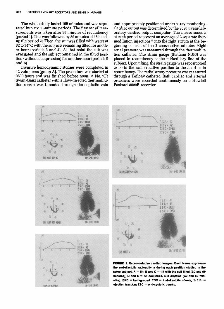
Since right ventricular end-systolic and end-dia- stolic counts showed wide interindividual variability, we analyzed the relation of the counts to plasma renin activity within persons across the 5 periods of observa- tion (Fig. 4). Negative slopes were achieved for all study participants and the values for the fit were strong in all but 2 instances.
Discussion The role of the cardiopulmonary mechanorecep-
tors in the regulation of renin in humans is controver- sial. Pooling of the blood in the peripheral veins through lower body negative pressure produces a de- crease of right atria1 pressure17 and cardiac output,18 but the reported effects on renin levels are discordant. Fasola and MartzIg reported elevation, whereas Mark et alzo failed to observe such a renin response to low degrees of lower body negative suction. With lower body negative suction, the negative pressure is trans-

April I, 1987 THE AMERICAN JOURNAL OF CARDIOLOGY Volume 59 805
3 4 5 ngimlih
PRA
FIGURE 3. Relation between right atriai pressure (RAP) and plasma renin (PRA) activity across 6 periods In study A. Bar represents standard deviation.
3 mitted throughout the lower abdominal cavity, as evi- 3 dented by a lower position of the diaphragm.21 This negative pressure probably elicits reflexes from the
5 ;
richly innervated abdominal cavity,22 and it is also 8 possible that the transmural renal pressure is altered. E
To induce a selective decrease of right atria1 pres- sure without changes in intraabdominal pressure, Kiowski and Julius12 used inflatable tourniquets placed over both thighs. Right atria1 pressure, cardio- pulmonary blood volume and cardiac output de- creased and plasma renin activity increased. This increase of plasma renin was believed to be reflex- -. __
11000
10000
9000
6000
7000
6000
5000
4000
3000
2000
1000
0
18000
17000
16000
15000
14000
13000
12000
11000
10000
9000
8000
7000
6000 -.-T----- I~.-l--~~-r-.--,~.-, 0 1 2 3 4 5 6
Plasma Renin Activity
mediated because (11 renin failed to increase in pa- FIGURE 4. Relation between plasma renin activity and end-systolic tients with transplanted, and therefore denervated, and end-diastolic counts. A linear regression was calculated for 5 kidneys: (2) /3 blockade abolished the renin response to periods for each sub)ect. Corresponding r values are given for each thigh cuff inflation: and (3) an increase of plasma nor- individual line.
TABLE ii Resuits of the Radionuciide imaaina Study
60 Minutes 90 Minutes 120 Minutes Tilt Suit Filled Suit Filled
150 Minutes 180 Minutes Tilt- Tilt-
Suit Empty Suit Empty
PRA 3.34 f 1.39 1.65 f 0.78 1.19 f 0.59 2.71 f 1.15 3.73 f 1.64 (rig/ml/hour) p <0.05 NS p <0.05 NS
EDC (RV) 9,564 l 1,115 11,930 f 1,637 11,642 f 1.419 10,038 f 1,501 -9,130 f 879 (cpm) p <O.Ol NS p <0.05 NS
ESC (RV) 5,841 f 994 7,315 f 1,040 7,761 f 1,264 6,135 f 1,395 5,607 f 1,074 (cpm) p <0.05 NS p <O.Ol NS
EDC (LV) 8,226 f 1,584 0.703 l 2,029 8,868 f 1,666 8,767 f 1,345 8,286 f 1.220 (cpm) NS NS NS NS
ESC (LV) 3,357 f 886 3,723 f 1,721 3,388 f 1,000 3,350 f 726 3,277 f 729 @pm) NS NS NS NS
SBP 108 f 9 113 f 7 114 f 6 113 f 6 114 i 8 (mm Ha) NS NS NS NS
DBP 69 f 10 71 f 10 73 f 7 74 f 7 75 f a (mm Hs) NS NS NS NS
HR 68 f 3 68f 11 69 f 12 69 f 12 69 i 13 (beatslmin) NS NS NS NS
The p values denote results of analysis of variance with the Tukey criterion for the level of significance from the preceding period. EDC = enddiastolic counts; ESC = end-systolic counts; RV = right ventricle; LV = left ventricle: other abbreviations as in Table I.

888 CARDIOPULMONARY RECEPTORS AND RENIN IN HUMANS
epinephrine levels accompanied the plasma renin in- References crease. Later, Egan et alI3 used numerous other ma- neuvers to separate the effects of the arterial and 1. Linden RJ. Function of cardiac receptors. Circulation 1968;46:463-480.
2. Mancia G, Donald DE. Demonstration that the atria, ventricles and lungs cardiopulmonary receptors and concluded that the each are respons&Ie for a tonic inhbition of the vasomotor center in the dog.
cardiopulmonary receptors have a predominant role Circ Res 1975;36:310-316.
in the control of renin release. 3. Clement DL, Pelletier CL, Shepherd JT. Role of vagal afferents in the control of renal sympathetic nerve activity in the rabbit. Circ Res 1972;31:624-
Our studies complement the investigations of Julius 830. and co-workers in 2 important ways: They have not 4. Oberg 8, Thoren P. Circulatory responses to stimulation of medullated and
carried out offset and onset studies of the effect of non-medullated afferents in the cardiac nerve of the cat. Acta Physiol Stand 1973;67:121-132.
increasing the stretch of cardiopulmonary receptors on 5. Mancia G, Romero JC. Shepherd JT. Continuous inhibition of renin release
renin levels and they have not independently assessed in dogs by vagally innervated receptors in the cardiopulmonary region. Circ Res 1975;36:529-535.
the cardiac size during their experiments. 8. Thames MD. Reflex suppression of renin release by ventricular receptors
In the present experiments, tilting induced the well with vaga1 afferents. Am J Physiol 1977;233:H181-H184.
known compensatory activation of the renin-angioten- 7. De Torrente A, Robertson GL, MC Donald KM, Schrier RW. Mechanism of diuretic response to increased left atria1 pressure in the anesthetized dog.
sin and sympathetic nervous system to upright posture, Kidney Int 1975:8:355-36X
which was fully abolished upon filling of the suit with 8. Siekar HO, Gauer OH, Henry JP. The effect of continuous negative pres-
water. Filling and draining of the suit caused signifi- sure breathing on water and electrolyte excretion by the human kidney. J Clin Invest 1954;33:572-577.
cant changes of right atria1 pressure (Table I, Fig. 2) 9. Epstein M. Renal effects of head-out water immersion in man: implications
and right-heart chamber volumes by ventriculography for on understanding of volume homeostasis. Physiol Rev 1978;58:529-581.
(Table II). Under all conditions right atria1 pressure 10. Brown E, Goei JS, Greenfield ADM, Plassaras GC. Circulatory responses to stimulated gravitational shifts of blood in men induced hy exposure of the
levels (Fig. 3) and radioactivity counts over the right body below the iliac crests to s&atmospheric pressure. J Physiol [London)
ventricle (Table II) showed a clear negative correla- 1g66;.63:607-627. 11. Wolthuis RA, Hoffler GW. Johnson RL. Lower body negative pressure as
tion to plasma renin activity. We therefore conclude an assay technique for orthostatic tolerance. III. A comparison of the individ-
that the major determinants of the renin response to ual response to incremental leg negative pressure vs increment lower nega-
upright posture are the pressures and volumes main- tive pressure. Aerospace Med l970;41:1354-1357. 12. Kiowski W. Julius S. Renin response to stimulation of cardiopulmonary
tained in the cardiopulmonary capacitance space. The mechanoreceptors in man. J C1in Invest 1978;82:856-683.
observed parallel changes of renin and norepineph- 13. Egan B, Julius S. Cottier C, Sanchez RA. Julius S. Role of cardiovascular
rine levels (Table I and II] support the assumption that receptors on neural regulation of renin release in normal man. Hypertension 1983;5:779-786.
this relation is due to reflex release of renin. That the 14. Forrester J, Ganz W. Diamond G, McHugh T, Chonette DW. Swan HJC.
relation is not secondary to hemodynamic changes can Thermodilution cardiac output determination with a single flow directed catheter. Am Heart J 1972;83:306-311.
be seen in Figure 2. 15. Haber E. Koerner T. Page LB, Kliman B. Furnode A. Application of
Filling of the water suit caused significant shifts in radioimmunoassay for angiotensin I to the physiologic measurements of pIas-
cardiopulmonary blood volume, without large effects ma renin activity in normal human subjects. J Clin Endocrino11969;29:1349- 1355.
on cardiac output (Fig. 2). In that regard, our experi- 18. Peuler JD. Johnson GA. Simultaneous single isotope radioenzymatic assay
ments offer a clearer picture than total body water $$Iasma norepinephrine. epinephrine and dopamine. Life Sci 1977;21:825-
immersion,g which also elicits renin suppression but 17. Zoller RP, Mark AL, Abboud FM, Schmid PG, Heistad DD. The role of 1ow
is associated with very large increases of central ve- pressure haroreceptors in reflex vasoconstrictor responses in man. J Clin
nous pressure23 and substantial elevation of cardiac Invest 1972;51:2967-2972. 18. Murray RH, Thompson LJ, Bowers JA, Albright CD. Hemodynamic ef-
output.z4 fects of graded hypovolemia and vasodepressor syncope induced by lower
The role of ANF should be discussed. Increases in body negative pressure. Am Heart J 1988;76:799-811.
right atria1 pressure elicit increases of ANFaz5 ANF can 19. Fasola AF, Martz BL. Peripheral venous renin activity during 70” tilt and lower body negative pressure. Aerospace Med 1972;43:713-715.
suppress plasma renin levels in animals.2e However, 20. Mark A, Abboud F, Fitz AE. Effects of low and high pressure baroreceg
experiments by Goetz et aP7 refute that changes in tors on plasma renin activity in humans. Am J Physiol 1978;235:H29-H33.
ANF have a physiologic role at least in regard to vol- 21. Millege RD. Musgrave FS, Zechman FW. Radiographic studies of the chest during changes in posture and lower body negative pressure. Radiology
ume diuresis. Furthermore, previous research strongly 1g88;go’674-676. indicates that the renin increase that followed the de-
22. Tuttle RS, McClealy M. Mesenteric baroreceptors. Am J Physiol 1975;229:1514-1519.
crease of right atria1 pressure is neurogenic because it 23. Arborelius M Jr. Balldin UI, Lilja B, Lundgren CEG. Hemodynamic
does not occur in patients with denervated kidneysI changes in mon during immersion wlth the head above water. Aerospace
It is difficult to visualize 2 mechanisms, one of a neuro- Med 1g72;43:5g2-5g8. 24. Begin R. Epstein M. Sackner MA, Levinson R, Dougherty R. Duncan D.
genie increase in renin when right atria1 pressure Effects of water immersion to the neck on pulmonary circulation and tissue
decreases and another humorally mediated renin sup volume in man. J Appl Physiol 1976;40:293-299. 25. Bates ER, Schenker Y, Grekin R. Relationship of hemodynamic parame-
pression when right atria1 pressure increases. Conse- ters to plasma level of immunoreactive atricd natriuretic hormone in man.
quently. we interpreted the renin decrease in our Circulation 1986;73:1155-1161.
experiments to reflect a direct suppression of sympa- 28. Ledsome JR, Wilson N, Courneya CA, Rankin AJ. Release of atria1 natri- uretic peptide by atria1 distension. Can J Physiol Pharmacol 1965;83:739-
thetic discharge to the juxtaglomerular cells in the kid- 742. ney. It remains to be seen whether further research 27. Goetz KL, Wang BC, Geer PG. Leadley RJ. Jr, Reinhardt HW. Atria1
may show also a role of ANF in these circumstances. stretch increases sodium excretion independently of release of atria1 pep- tides. Am J Physiol 1986:25O:R946-B950.
Copyright © 2022 FDOKUMEN